Doses from Medical X-Ray Procedures - Health Physics Society
Doses from Medical X-Ray Procedures - Health Physics Society
Doses from Medical X-Ray Procedures - Health Physics Society

You also want an ePaper? Increase the reach of your titles
YUMPU automatically turns print PDFs into web optimized ePapers that Google loves.
<strong>Doses</strong> <strong>from</strong> <strong>Medical</strong> X‐<strong>Ray</strong> <strong>Procedures</strong><br />
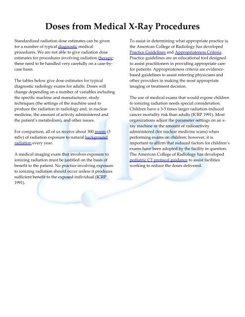
Standardized radiation dose estimates can be given<br />
for a number of typical diagnostic medical<br />
procedures. We are not able to give radiation dose<br />
estimates for procedures involving radiation therapy;<br />
these need to be handled very carefully on a case‐by‐<br />
case basis.<br />
The tables below give dose estimates for typical<br />
diagnostic radiology exams for adults. <strong>Doses</strong> will<br />
change depending on a number of variables including<br />
the specific machine and manufacturer, study<br />
techniques (the settings of the machine used to<br />
produce the radiation in radiology and, in nuclear<br />
medicine, the amount of activity administered and<br />
the patient’s metabolism), and other issues.<br />
For comparison, all of us receive about 300 mrem (3<br />
mSv) of radiation exposure to natural background<br />
radiation every year.<br />
A medical imaging exam that involves exposure to<br />
ionizing radiation must be justified on the basis of<br />
benefit to the patient. No practice involving exposure<br />
to ionizing radiation should occur unless it produces<br />
sufficient benefit to the exposed individual (ICRP<br />
1991).<br />
To assist in determining what appropriate practice is,<br />
the American College of Radiology has developed<br />
Practice Guidelines and Appropriateness Criteria.<br />
Practice guidelines are an educational tool designed<br />
to assist practitioners in providing appropriate care<br />
for patients. Appropriateness criteria are evidence‐<br />
based guidelines to assist referring physicians and<br />
other providers in making the most appropriate<br />
imaging or treatment decision.<br />
The use of medical exams that would expose children<br />
to ionizing radiation needs special consideration.<br />
Children have a 3‐5 times larger radiation‐induced<br />
cancer mortality risk than adults (ICRP 1991). Most<br />
organizations adjust the parameter settings on an x‐<br />
ray machine or the amount of radioactivity<br />
administered (for nuclear medicine scans) when<br />
performing exams on children; however, it is<br />
important to affirm that reduced factors for children’s<br />
exams have been adopted by the facility in question.<br />
The American College of Radiology has developed<br />
pediatric CT protocol guidance to assist facilities<br />
working to reduce the doses delivered.
Estimates of the dose an individual might receive <strong>from</strong> one x ray.<br />
Single Radiograph Effective Dose, mrem (mSv)<br />
Skull (PA or AP) 1 3 (0.03)<br />
Skull (lateral) 1 1 (0.01)<br />
Chest (PA) 1 2 (0.02)<br />
Chest (lateral) 1 4 (0.04)<br />
Chest (PA and lateral) 2 6 (0.06)<br />
Thoracic spine (AP) 1 40 (0.4)<br />
Thoracic spine (lateral) 1 30 (0.3)<br />
Lumbar spine (AP) 1 70 (0.7)<br />
Lumbar spine (lateral) 1 30 (0.3)<br />
Abdomen (AP) 1 70 (0.7)<br />
Abdomen 3 53 (0.53)<br />
Pelvis (AP) 1 70 (0.7)<br />
Pelvis or hips 3 83 (0.83)<br />
Bitewing dental film 3 0.4 (0.004)<br />
Limbs and joints 3 6 (0.06)<br />
Estimates of the dose an individual might receive if undergoing an entire procedure (e.g., a lumbar<br />
spine series typically consists of five films).<br />
Complete Exams Effective Dose, mrem (mSv)<br />
Intravenous pyelogram (kidneys, 6 films) 1 250 (2.5)<br />
Barium swallow (24 images, 106 sec fluoroscopy) 1 150 (1.5)<br />
Barium meal (11 images, 121 sec fluoroscopy) 1 300 (3.0)<br />
Barium follow‐up (4 images, 78 sec fluoroscopy) 1 300 (3.0)<br />
Barium enema (10 images, 137 sec fluoroscopy) 1 700 (7.0)<br />
CT head 1 200 (2.0)<br />
CT chest 1 800 (8.0)<br />
CT abdomen 1 1,000 (10)<br />
CT pelvis 1 1,000 (10)<br />
CT (head or chest) 2 1,110 (11.1)<br />
PTCA (heart study) 3 750–5,700 (7.5–57)<br />
Coronary angiogram 3 460–1,580 (4.6–15.8)<br />
Mammogram 3 13 (0.13)<br />
Lumbar spine series 3 180 (1.8)<br />
Thoracic spine series 3 140 (1.4)<br />
Cervical spine series 3 27 (0.27)
References for the two tables<br />
1. Wall BF, Hart D. Revised radiation doses for typical x‐ray examinations. The British Journal of Radiology<br />
70:437‐439; 1997 (5,000 patient dose measurements <strong>from</strong> 375 hospitals).<br />
2. National Council on Radiation Protection and Measurements. Sources and magnitude of occupational and<br />
public exposures <strong>from</strong> nuclear medicine procedures. Bethesda, MD: National Council on Radiation<br />
Protection and Measurements; NCRP Report 124; 1996.<br />
3. United Nations Scientific Committee on the Effects of Atomic Radiation. Sources and effects of ionizing<br />
radiation, Vol. 1: Sources. New York, NY: United Nations Publishing; 2000.<br />
Other references and resources<br />
American College of Radiology white paper on radiation dose in medicine:<br />
http://www.acr.org/SecondaryMainMenuCategories/quality_safety/white_paper_dose.aspx<br />
American College of Radiology practice guideline for diagnostic reference levels in medical x‐ray imaging:<br />
http://www.acr.org/SecondaryMainMenuCategories/quality_safety/RadSafety/RadiationSafety/guideline‐<br />
diagnostic‐reference.aspx<br />
International Commission on Radiological Protection. 1990 recommendations of the International Commission on<br />
Radiological Protection. Oxford: Pergamon Press; ICRP Publication 60; Ann ICRP 21(1‐3); 1991.<br />
Glossary<br />
background radiation: Background radiation includes radiation <strong>from</strong> cosmic sources, naturally occurring radioactive<br />
materials (including radon), and global fallout (<strong>from</strong> the testing of nuclear explosive devices). The typically quoted<br />
average individual exposure <strong>from</strong> background radiation is 0.30 rem per year.<br />
becquerel: The becquerel (Bq) is the unit in the International System of Units to replace the curie (see curie).<br />
curie: The curie (Ci) is the original term used to describe the amount of radioactive material present or strength of<br />
the source. It is based upon the radioactive decay rate of the radionuclide. One curie is equal to 3.7 x 10 10<br />
disintegrations (37 trillion decays) per second (dps); one becquerel is equal to 1 dps. The most common activity<br />
levels used in laboratories are the millicurie (mCi) and microcurie (μCi). A millicurie (mCi) is 1/1,000 th of a curie and<br />
a microcurie (μCi) is 1/1,000,000 th of a curie. In the International System of Units, the becquerel (Bq) describes the<br />
amount of radioactive material present. One curie is equal to 3.7 x 10 9 Bq.
diagnostic: In medicine, diagnosis or diagnostics is the process of identifying a medical condition or disease by its<br />
signs and symptoms and <strong>from</strong> the results of various procedures. As used when referring to medical exams<br />
involving radiation, it is the use of x rays or radioactive materials to identify the medical condition.<br />
dose: Dose is a general term used to express (quantify) how much radiation exposure something (a person or other<br />
material) has received. The exposure can subsequently be expressed in terms of the absorbed, equivalent,<br />
committed, and/or effective dose based on the amount of energy absorbed and in what tissues.<br />
effective dose: Radiation exposures to the human body, whether <strong>from</strong> external or internal sources, can involve all or a<br />
portion of the body. The health effects of one unit of dose to the entire body are more harmful than the same dose to<br />
only a portion of the body, e.g., the hand or the foot. To enable radiation protection specialists to express partial‐<br />
body exposures (and the accompanying doses) to portions of the body in terms of an equal dose to the whole body,<br />
the concept of effective dose was developed. Effective dose, then, is the dose to the whole body that carries with it<br />
the same risk as a higher dose to a portion of the body. As an example, 8 rem (80 mSv) to the lungs is roughly the<br />
same potential detriment as 1 rem (10 mSv) to the whole body based on this idea.<br />
exposure: Exposure is commonly used to refer to being around a radiation source; e.g., if you have a chest x ray, you<br />
are exposed to radiation. By definition, exposure is a measure of the amount of ionizations produced in air by<br />
photon radiation.<br />
exposure rate: Exposure rate is the amount of exposure or dose you are receiving per unit time (e.g., 1 mrem/hour).<br />
gamma rays: Gamma rays are high‐energy electromagnetic radiation (photons) emitted in an attempt by the<br />
radionuclide to become stable, i.e., radioactive decay. Gamma rays have moderate‐to‐high penetrating power, are<br />
often able to penetrate deep into the body, and generally require some form of shielding, such as lead or concrete.<br />
Visible light is also in the form of photons. Gamma photons behave similarly to light, but they are invisible.<br />
high‐level radiation: High‐level radiation refers to radiation doses greater than 10 rem to a human body.<br />
low‐level radiation: Low‐level radiation refers to radiation doses less than 10 rem to a human body.
observable health effect: An observable health effect is a change in physical health that can be detected medically.<br />
Observable health effects may include changes in blood cell counts, skin reddening, cataracts, etc. Whether or not it<br />
is an observable harmful health effect depends on whether damage to the body has occurred and whether that<br />
damage impairs how the body is able to function.<br />
radiation: Radiation is a term commonly used to describe ionizing radiation (i.e., x and gamma rays, alpha and beta<br />
particles, neutrons). Ionizing radiation is radiation that is capable of producing ions by passing through matter.<br />
rem: Rem is the term used to describe equivalent or effective radiation dose. In the International System of Units, the<br />
sievert (Sv) describes equivalent or effective radiation dose. One sievert is equal to 100 rem. One mrem is one<br />
thousandth of a rem.<br />
risk: Risk is defined in most health‐related fields as the probability or odds of incurring injury, disease, or death.<br />
roentgen: The roentgen (R) is the term used to describe radiation exposure. This term for exposure only describes the<br />
amount of ionization in air. In the International System of Units, the coulomb/kilogram (C/kg) describes radiation<br />
exposure. One roentgen is equal to 2.58 x 10 ‐4 C/kg.<br />
safe: Safe, as it is being used in the information on this Web site, is defined as an activity that is generally considered<br />
acceptable to us. This is not to say there is absolutely no risk with an activity that is considered safe; there may be a<br />
risk <strong>from</strong> the activity or the exposure to radiation, but it is the same or lower than the risks <strong>from</strong> everyday actions.<br />
At a level of radiation that is considered safe, an effect is either nonexistent or too small to observe.<br />
sievert: Sievert (Sv) is the unit in the International System of Units to replace the rem (see rem).<br />
therapy: Therapy is the medical treatment of disease or disorders. With respect to radiation therapy, therapeutic<br />
doses (e.g., external beam treatments for tumors, radioiodine treatment for thyroid disorders) are significantly<br />
greater than those received <strong>from</strong> diagnostic procedures (e.g., chest x‐rays, CT scans, nuclear medicine procedures,<br />
etc.).<br />
x rays: X rays are electromagnetic radiation (photons) that can be emitted <strong>from</strong> radionuclides or <strong>from</strong> certain types<br />
of devices. Generally, x rays have lower energies than gamma rays, but like gamma rays, x rays can penetrate into<br />
the body. Sometimes lead or concrete may be used as shielding for x rays.