Plant Physiology - Stony Brook University
Plant Physiology - Stony Brook University
Plant Physiology - Stony Brook University

You also want an ePaper? Increase the reach of your titles
YUMPU automatically turns print PDFs into web optimized ePapers that Google loves.
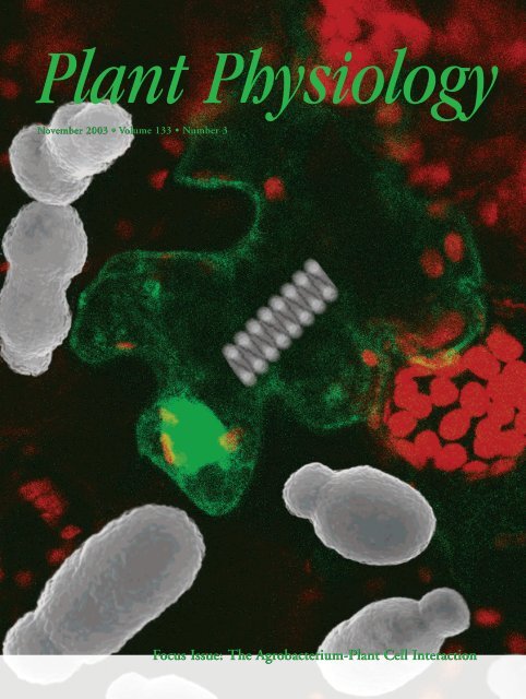
<strong>Plant</strong> <strong>Physiology</strong><br />
November 2003 • Volume 133 • Number 3<br />
Focus Issue: The Agrobacterium-<strong>Plant</strong> Cell Interaction
Site-Specific Integration of Agrobacterium tumefaciens<br />
T-DNA via Double-Stranded Intermediates 1<br />
Tzvi Tzfira*, Leah Renée Frankman, Manjusha Vaidya, and Vitaly Citovsky<br />
Department of Biochemistry and Cell Biology, State <strong>University</strong> of New York, <strong>Stony</strong> <strong>Brook</strong>, New York<br />
11794 (T.T., M.V., V.C.); and Westhampton Beach High School, 340 Mill Road, Westhampton Beach,<br />
New York 11978 (L.R.F.)<br />
Agrobacterium tumefaciens-mediated genetic transformation involves transfer of a single-stranded T-DNA molecule (T strand)<br />
into the host cell, followed by its integration into the plant genome. The molecular mechanism of T-DNA integration, the<br />
culmination point of the entire transformation process, remains largely obscure. Here, we studied the roles of doublestranded<br />
breaks (DSBs) and double-stranded T-DNA intermediates in the integration process. We produced transgenic<br />
tobacco (Nicotiana tabacum) plants carrying an I-SceI endonuclease recognition site that, upon cleavage with I-SceI, generates<br />
DSB. Then, we retransformed these plants with two A. tumefaciens strains: one that allows transient expression of I-SceI to<br />
induce DSB and the other that carries a T-DNA with the I-SceI site and an integration selection marker. Integration of this<br />
latter T-DNA as full-length and I-SceI-digested molecules into the DSB site was analyzed in the resulting plants. Of 620<br />
transgenic plants, 16 plants integrated T-DNA into DSB at their I-SceI sites; because DSB induces DNA repair, these results<br />
suggest that the invading T-DNA molecules target to the DNA repair sites for integration. Furthermore, of these 16 plants,<br />
seven plants incorporated T-DNA digested with I-SceI, which cleaves only double-stranded DNA. Thus, T-strand molecules<br />
can be converted into double-stranded intermediates before their integration into the DSB sites within the host cell genome.<br />
Agrobacterium tumefaciens is the only known organism<br />
capable of inter-kingdom DNA transfer (Stachel<br />
and Zambryski, 1989). In nature, this bacterium<br />
causes crown gall disease in many dicotyledonous<br />
plant species by transferring a specific DNA fragment,<br />
the transferred DNA (T-DNA), from its tumorinducing<br />
(Ti) plasmid into the host cell (for review,<br />
see Gelvin, 2000; Tzfira et al., 2000; Zupan et al., 2000;<br />
Gelvin, 2003). The wild-type T-DNA carries two<br />
types of genes: oncogenes and genes for biosynthesis<br />
of amino acid derivatives, opines. Expression of the<br />
integrated T-DNA, therefore, results in uncontrolled<br />
cell division and formation of tumors (Gaudin et al.,<br />
1994; Das, 1998) that produce and secrete opines that<br />
are utilized by A. tumefaciens and several other microorganisms<br />
as the source of carbon and nitrogen<br />
(for review, see Savka et al., 2002). The T-DNA itself<br />
does not encode genes required for its transfer and<br />
integration and is defined only by two 25-bp direct<br />
repeats, termed T-DNA left and right borders (Zambryski<br />
et al., 1982). Thus, the entire wild-type T-DNA<br />
sequence between the borders can be replaced with a<br />
gene of interest that will be transferred to plants and<br />
1 This work was supported by the U.S.-Israel Binational Research<br />
and Development Fund (grant to T.T.), by the National<br />
Institutes of Health (grant to V.C.), by the National Science Foundation<br />
(grant to V.C.), by the U.S. Department of Agriculture<br />
(grant to V.C.), and by the U.S.-Israel Binational Science Foundation<br />
(grant to V.C.).<br />
* Corresponding author; e-mail ttzfira@notes.cc.sunysb.edu; fax<br />
631–632–8575.<br />
Article, publication date, and citation information can be found<br />
at http://www.plantphysiol.org/cgi/doi/10.1104/pp.103.032128.<br />
integrated in the plant genome, representing the molecular<br />
basis for plant genetic engineering (for review,<br />
see Gelvin, 1998b, 2000, 2003; Potrykus et al.,<br />
1998).<br />
Several A. tumefaciens chv (chromosomal virulence)<br />
genes and a set of vir (virulence) genes were contained<br />
on the Ti plasmid code for the protein machinery<br />
of the T-DNA transport (for review, see Sheng<br />
and Citovsky, 1996; Gelvin, 2000,2003; Tzfira and<br />
Citovsky, 2000). The VirA/VirG two-component signal<br />
transduction system senses secretion of phenolics<br />
and specific sugar compounds from the wounded<br />
susceptible plant tissues and induces expression of<br />
other vir genes (Winans et al., 1994). The VirD2/<br />
VirD1 complex nicks the bottom strand of the Ti<br />
plasmid at the T-DNA borders and, with the assistance<br />
of the bacterial DNA synthesis and repair machinery,<br />
a single-stranded copy of the T-DNA, the T<br />
strand, is liberated from the Ti plasmid (Wang et al.,<br />
1987; Filichkin and Gelvin, 1993; Scheiffele et al.,<br />
1995). VirD1 is then released, whereas VirD2 remains<br />
covalently attached to the 5� end of the T strand<br />
(Ward and Barnes, 1988; Young and Nester, 1988;<br />
Howard et al., 1989). The resulting protein-DNA<br />
complex is exported to the host cell through the type<br />
IV secretion system encoded by the virD4 gene and<br />
virB operon, with 11 open reading frames (for review,<br />
see Christie, 1997; Christie and Vogel, 2000). The<br />
VirD4/VirB channel also mediates export of VirE2<br />
(Vergunst et al., 2000), a single-stranded DNAbinding<br />
protein (Gietl et al., 1987; Christie et al., 1988;<br />
Citovsky et al., 1988, 1989; Das, 1988; Sen et al., 1989)<br />
<strong>Plant</strong> <strong>Physiology</strong>, November 2003, Vol. 133, pp. 1011–1023, www.plantphysiol.org © 2003 American Society of <strong>Plant</strong> Biologists 1011
Tzfira et al.<br />
thought to associate cooperatively with the T-strand<br />
in the plant cell cytoplasm (Citovsky et al., 1992;<br />
Gelvin, 1998a), producing T-complex (Howard et al.,<br />
1990; Zupan and Zambryski, 1997), a semirigid, hollow,<br />
cylindrical filament composed of a T-strand<br />
molecule attached to one molecule of VirD2 and<br />
coated by multiple VirE2 monomers (Citovsky et al.,<br />
1997). Both VirD2 and VirE2 are thought to facilitate<br />
import of the T-complex into the host cell nucleus<br />
(Howard et al., 1992; Rossi et al., 1993; Citovsky et al.,<br />
1994; Ziemienowicz et al., 2001), after which the<br />
T-DNA is integrated into the plant genome by illegitimate<br />
recombination (Gheysen et al., 1991; Mayerhofer<br />
et al., 1991; Bundock and Hooykaas, 1996; Tinland,<br />
1996).<br />
Initially, two different models have been suggested<br />
for T-DNA integration, double-stranded break (DSB)<br />
repair and single-stranded gap repair (Mayerhofer et<br />
al., 1991). The first model predicts that unwound<br />
ends of a double-stranded T-DNA molecule anneal<br />
with single-stranded overhangs of a DSB in the plant<br />
DNA, the residual 5� and 3� overhangs are removed,<br />
and the inserted T-DNA is ligated. The second model<br />
suggests that integration initiates with a nick, which<br />
leads to a gap, in the plant DNA, both ends of a<br />
single-stranded T-DNA molecule anneal to this gap,<br />
the residual 5� and 3� overhangs are removed, and the<br />
integration is completed by repair synthesis of<br />
the second T-DNA stand (Mayerhofer et al., 1991).<br />
Both models also suggest a role for VirD2 in recognition<br />
of nicks or gaps by interaction with plant<br />
factors (Mayerhofer et al., 1991).<br />
The single-stranded gap repair model was extensively<br />
revised (Tinland and Hohn, 1995; Tinland et<br />
al., 1995) to suggest that the 3� end of the T strand<br />
acts as a driving force in the selection of the integration<br />
site. According to this current model, the integration<br />
process is initiated by microhomologies between<br />
the 3� end of the T strand and the upper strand<br />
of the plant DNA. The lower plant DNA strand is<br />
then nicked, and VirD2 ligates the 5� end of the T<br />
strand into this nick. The 3� overhang of the T strand<br />
is removed, and the second strand of the integrated<br />
T-DNA is filled in by the DNA repair machinery of<br />
the host cell (Tinland and Hohn, 1995; Tinland et al.,<br />
1995). This model is based on four lines of circumstantial<br />
evidence: T-DNA is transported into the<br />
plant cell as a single-stranded molecule (Tinland et<br />
al., 1994; Yusibov et al., 1994), nucleotide sequence<br />
comparison between the 3� ends of the T-DNA and<br />
the integration sites identifies regions of microhomology<br />
(Tinland et al., 1995; Brunaud et al., 2002),<br />
VirD2 associates with the 5� end of the T-DNA (Ward<br />
and Barnes, 1988; Young and Nester, 1988; Howard<br />
et al., 1989) and cleaves the T-DNA left border sequence<br />
and ligates the adjoining DNA strands in<br />
vitro (Pansegrau et al., 1993; Jasper et al., 1994), and<br />
mutations in VirD2 result in imprecise integration of<br />
the 5� end of the T-DNA (Tinland et al., 1995). Con-<br />
versely, this model cannot account for the formation<br />
of several complex T-DNA integration patterns (e.g.<br />
Krizkova and Hrouda, 1998; McCormac et al., 2001),<br />
it relies on the ligase activity of VirD2 that may be<br />
dispensable for integration in vitro (Ziemienowicz et<br />
al., 2000), and it does not explain the fast kinetics of<br />
transient T-DNA expression (Janssen and Gardner,<br />
1990; Tinland et al., 1994; Narasimhulu et al., 1996;<br />
De Neve et al., 1997) or rapid expression of T strands<br />
corresponding to the coding strand of the T-DNA<br />
(Narasimhulu et al., 1996), all of which imply that T<br />
strands can be converted to double-stranded molecules<br />
already early in the infection process.<br />
Recent studies further supported the notion that<br />
the T-strand molecule becomes double stranded before<br />
its integration (De Neve et al., 1997; Salomon and<br />
Puchta, 1998; De Buck et al., 1999). For example,<br />
because 5� ends of single-stranded DNA molecules<br />
cannot be ligated to each other, two T-DNA right<br />
borders integrated “head to head” suggested that the<br />
T strands may have been converted to a doublestranded<br />
form before integration; these observations,<br />
however, did not rule out the possibility of this conversion<br />
occurring during the first integration event<br />
followed by immediate integration of the second<br />
T-strand molecule (De Neve et al., 1997; De Buck et<br />
al., 1999). A more direct indication of the involvement<br />
of double-stranded integration intermediates<br />
came from detection of T-DNA inserts within the<br />
sites of DSB repair in the host DNA that suggested<br />
ligation of double-stranded T-DNA molecules to<br />
both sides of the break (Salomon and Puchta, 1998).<br />
Here, we present direct evidence for the role of<br />
DSBs and double-stranded T-DNA intermediates in<br />
T-DNA integration. Specifically, we used transient<br />
expression of an intron-encoded endonuclease I-SceI<br />
(Monteilhet et al., 1990) to create DSBs in the genomes<br />
of plants transgenic for the I-SceI recognition<br />
site. Our data indicate that T-DNA is preferentially<br />
targeted and integrated into these DSB sites. Furthermore,<br />
the invading T-DNA that itself contained the<br />
I-SceI recognition site was often integrated after being<br />
digested by I-SceI, indicating its conversion to a<br />
double-stranded form before integration.<br />
RESULTS<br />
Experimental System<br />
To investigate a role that DSB may play in T-DNA<br />
integration and to determine whether T-DNA integrates<br />
as a single- or double-stranded molecule, we<br />
utilized a genetic system for in vivo induction of<br />
DSBs (Salomon and Puchta, 1998). In this approach,<br />
DSB is induced by transient expression of the I-SceI<br />
endonuclease in plants carrying a 35S-I-SceI-codA<br />
transgene composed of the 35S promoter, the I-SceI<br />
recognition site, and the cytosine deaminase gene<br />
codA (Stougaard, 1993; Salomon and Puchta, 1998)<br />
encoded by the pBNE3I binary vector (Fig. 1A). Im-<br />
1012 <strong>Plant</strong> Physiol. Vol. 133, 2003
Figure 1. Maps of the T-DNA regions. A, pBNE3I. B, pBIG-HYGterGUS/I-SceI.<br />
C, Nucleotide sequence of the I-SceI site between the<br />
T-DNA left border and the uidA gene in pBIG-HYG-terGUS/I-SceI.<br />
LB, T-DNA left border; RB, T-DNA right border; 35S, cauliflower<br />
mosaic virus 35S RNA promoter; nptII, kanamycin resistance gene;<br />
codA, cytosine deaminase gene, uidA; �-glucoronidase (GUS) gene;<br />
hpt, hygromycin resistance gene;NOS5�, nopaline synthase promoter;NOS3�,<br />
nopaline synthase terminator; pAg7, agropine synthase<br />
terminator.<br />
proper repair of such DSBs, which may result from<br />
deletions or insertions of endogenous DNA segments<br />
and from capture of integrating T-DNA molecules, is<br />
detected by selection for loss of function of the cytosine<br />
deaminase transgene (Salomon and Puchta,<br />
1998). In our experimental system, however, we did<br />
not select for the absence of the cytosine deaminase<br />
activity; instead, we selected for hygromycin resistance,<br />
which is encoded by the integrating I-SceIuidA-hpt<br />
T-DNA derived from the pBIG-HYGterGUS/I-SceI<br />
binary vector (Fig. 1B). This approach<br />
allowed us to detect all transformation events rather<br />
than only those that occurred within the DSB sites.<br />
The initial selection was followed by screening of the<br />
resulting population of hygromycin-resistant plants<br />
for the cytosine deaminase activity to determine the<br />
frequency of specific insertions into DSB within the<br />
35S-I-SceI-codA transgene.<br />
T-DNA Integration via Double-Stranded Intermediates<br />
To determine the structure of the integrating<br />
T-DNA molecules, we designed the T-DNA to carry<br />
an I-SceI recognition sequence between the promoterless<br />
uidA gene and the T-DNA left border (Fig. 1B).<br />
If this T-DNA becomes double stranded to allow its<br />
cleavage with I-SceI before integration, and if the<br />
resulting fragment carrying the promoterless uidA<br />
integrates into the DSB created by I-SceI cleavage<br />
between the 35S promoter and the I-SceI site in the<br />
35S-I-SceI-codA transgene, we expect to detect the<br />
uidA gene expression, i.e. GUS activity, in the transgenic<br />
plants. Thus, our experimental design aims to<br />
examine concurrently two important characteristics<br />
of the integration process: frequency of targeted integration<br />
into the specific DSB sites out of the total<br />
number of transformation events and frequency of<br />
formation of double-stranded T-DNA intermediates<br />
before integration.<br />
Using this experimental design, we generated a<br />
total of 24 independent transgenic lines transformed<br />
with pBNE3I containing the 35S-I-SceI-codA T-DNA.<br />
Fifteen lines, showing a 3:1 kanamycin resistance<br />
segregation ratio, were analyzed by PCR to identify<br />
plants that do not contain tandem insertions of the<br />
35S-I-SceI-codA transgene (Table I). In addition, sequence<br />
analysis of the amplified PCR fragments revealed<br />
that, in all 15 lines, the sequence of the integrated<br />
35S-I-SceI-codA transgene was identical to the<br />
original sequence of this transgene within pBNE3I<br />
(data not shown). <strong>Plant</strong>s were then tested for their<br />
cytosine deaminase activity, which indicates the degree<br />
of expression of the 35S-I-SceI-codA transgene,<br />
using leaf disc regeneration assay in the presence of<br />
5-fluorocytosine (5-FC); 5-FC is known to select<br />
against cells that express cytosine deaminase (Stougaard,<br />
1993). Five heterozygous lines (Sc1, Sc4, Sc33,<br />
Sc59, and Sc70) showing the highest sensitivity to<br />
5-FC, i.e. very low or abolished ability to regenerate<br />
on a medium containing 5-FC, were selected for further<br />
studies.<br />
Next, these 35S-I-SceI-codA transgenic plants were<br />
transformed with a mixture of A. tumefaciens strains<br />
harboring the binary vectors pCISceI, with the<br />
I-SceIORF T-DNA, and pBIG-HYG-terGUS/I-SceI,<br />
Table I. Primer combinations used for amplification of T-DNA/DSB junctions a<br />
Primer Set Amplified Junction Expected Size (bp)<br />
35S-F2, codA-R 35S-I-SceI-codA transgene 509<br />
35S-F2 codA-R Disrupted 35S-I-SceI-codA (no T-DNA integration) Unpredicted<br />
35S-F2, T-DNA-RB 35S/T-DNA right border Unpredicted<br />
codA-R, T-DNA-RB T-DNA right border/codA Unpredicted<br />
35S-F2, T-DNA-LB 35S/T-DNA left border Unpredicted<br />
codA-R, T-DNA-LB T-DNA left border/codA Unpredicted<br />
35S-F2, GUS-R 35S / I-SceI-digested T-DNA left border 350<br />
codA-R, GUS-R I-SceI-digested T-DNA left border/codA Approximately 672<br />
a<br />
Primer sequences: 35S-F2, 5�CGTTCCAACCACGTCTTCAAAGC3�; codA-R, 5�GCCTTCAAACAG<br />
CGTGCCGG; T-DNA-RB, 5�CCCAGTCATAGCCGAATAGCC3�; T-DNA-LB, 5�GGCGTAATAGCGAA-<br />
GAGGCCC3�; GUS-R, 5�CCTGATTATTGACC CACACTTTGCCG3�.<br />
<strong>Plant</strong> Physiol. Vol. 133, 2003 1013
Tzfira et al.<br />
with the I-SceI-uidA-hpt T-DNA; these vectors encode<br />
the transiently expressed I-SceI endonuclease and<br />
provide the integrating I-SceI-uidA-hpt T-DNA, respectively.<br />
A total of 620 hygromycin-resistant transgenic<br />
lines were recovered and analyzed for their<br />
sensitivity to 5-FC; the 5-FC-resistant plants were<br />
then tested for GUS activity and the nucleotide sequence<br />
of their integration junctions was determined.<br />
Efficiency of T-DNA Integration during DSB Repair<br />
From 620 hygromycin-resistant plants, 82 plants<br />
exhibited significant 5-FC resistance. We then PCR<br />
amplified the putative integration junctions around<br />
the DSB site in 68 of these plants and examined them<br />
for small deletions or insertions in the DSB site. To<br />
this end, the 35S-I-SceI-codA transgene was amplified<br />
using the 35S-F2 and codA-R primers (Table I) and a<br />
short extension time (1 min). All 68 plants exhibited<br />
sequence alterations within the codA gene, indicating<br />
approximately 11% frequency of DSB induction and<br />
improper repair and demonstrating that the 5-FC<br />
resistance of these lines was not simply and solely<br />
due to silencing of this transgene. Forty-five of 620<br />
plants (7.25%) yielded short PCR fragments that<br />
ranged from 300 to 600 bp and, thus, could not represent<br />
the true T-DNA integration (Table II). Sequence<br />
analysis of five randomly chosen PCR fragments<br />
revealed deletions and/or insertions of short<br />
filler DNA that were most likely responsible for loss<br />
of function of the cytosine deaminase gene (data not<br />
shown).<br />
Twenty-three of 620 plants (3.71%) did not show<br />
any visible PCR bands after short amplification, but<br />
four of them yielded longer PCR fragments, 2.2 kb,<br />
2.8 kb, and two bands of 2.9 kb each (Table II), when<br />
longer PCR extension time (3 min) was used. Sequence<br />
analysis of these fragments revealed the integration<br />
of the full-length or truncated I-SceIORF<br />
T-DNA into DSB sites. The remaining 19 plants were<br />
subjected to a series of PCR analyses using different<br />
primer combinations shown in Table I. Table II<br />
shows that five plants contained the full-length or<br />
truncated I-SceI-uidA-hpt T-DNA integrated in their<br />
Table II. Frequency of site-specific T-DNA integration<br />
Experimental Step No. of <strong>Plant</strong>s<br />
DSBs, whereas in seven plants, the integrated I-SceIuidA-hpt<br />
T-DNA was found to have been digested by<br />
I-SceI before integration. The last seven plants did not<br />
yield discrete PCR products, potentially due to large<br />
deletions or insertions (Table II). Thus, out of 620<br />
transgenic lines examined in this study, we observed<br />
16 site-specific T-DNA integration events, including<br />
those of the I-SceIORF and I-SceI-uidA-hpt transgenes,<br />
corresponding to 2.58% frequency of targeted integration.<br />
Furthermore, this is the minimal estimate of<br />
the frequency of insertions into DSB sites because it<br />
assumes that transient expression of I-SceI created<br />
DSBs in all 620 transgenic lines.<br />
Orientation of Integrated T-DNA within DSB Sites<br />
Determination of the nucleotide sequence of T-<br />
DNA/DSB site integration junctions allowed their<br />
classification based on the orientation of the inserted<br />
T-DNA within the DSB site (Fig. 2). For the junctions<br />
in which the T-DNA left border was found ligated to<br />
the left side of DSB (DSB-L/LB, Fig. 2A) and the<br />
T-DNA right border was found ligated to the right<br />
side of DSB (RB/DSB-R, Fig. 2A), the sequences of<br />
the DSB site and the T-DNA were presented in sense<br />
orientation in Figures 3 to 5. When the T-DNA integrated<br />
in reverse orientation, we classified the junctions<br />
as DSB-L/RB and LB/DSB-R (Fig. 2B); in this<br />
case, to show the sense strand of the junction, we<br />
reversed and complemented the T-DNA sequence in<br />
Figures 3 to 5. Finally, when the I-SceI-digested<br />
T-DNA integrated into DSB, recreating the I-SceI site,<br />
we classified two additional types of junctions, DSB-<br />
L/I-SceI (Fig. 2C) and I-SceI/DSB-R (Fig. 2D). The<br />
T-DNA sequences of these junctions were presented<br />
in Figures 3 to 5 as sense (for DSB-L/I-SceI) or<br />
reversed and complemented sequences (for<br />
I-SceI/DSB-R).<br />
Integration of I-SceIORF T-DNA<br />
In four plant lines, the full-length or truncated<br />
I-SceIORF T-DNA integrated into the DSB site (Table<br />
II). In three of these lines (Sc4-2, Sc59-5, and Sc59-9),<br />
Frequency of<br />
Transformation Events<br />
% of Total<br />
Hygromycin 620 100<br />
5-FC-resistant plants 82 13.22<br />
<strong>Plant</strong>s analyzed by PCR 68 10.96<br />
PCR fragment � 600 bp or no PCR fragment 23 3.71<br />
All cases of T-DNA integration 16 2.58<br />
Integration of I-SceIORF T-DNA 4 0.64<br />
Integration of full-length or slightly truncated T-DNA 5 0.81<br />
Integration of I-SceI-digested uidA-hpt T-DNA 7 1.13<br />
Large deletion/insertion 7 1.13<br />
PCR fragment � 600 bp 45 7.25<br />
1014 <strong>Plant</strong> Physiol. Vol. 133, 2003
Figure 2. Orientation of the integrated T-DNA within DSB sites. A<br />
and B, Two possible orientations for integration of the I-SceI-uidAhpt<br />
T-DNA. C and D, Two possible orientations for integration of the<br />
I-SceI-digested I-SceI-uidA-hpt T-DNA. I-SceI sites are shown within<br />
the integrating T-DNA and in the DSB site. Arrowheads indicate the<br />
left-to-right border direction of T-DNA. Designations of each T-DNA<br />
border/DSB site integration junction are indicated.<br />
almost the entire I-SceIORF T-DNA integrated into<br />
DSB, whereas in the fourth line (Sc70-3), a deletion of<br />
797 bp was observed (Table III; Fig. 3). Sequence<br />
analysis of the DSB-L/LB junctions revealed that<br />
small deletions of 13 to 62 bp had occurred at the<br />
T-DNA left border. These deletions in the integrated<br />
T-DNA were accompanied by deletions, insertions,<br />
or no changes in the DSB site sequence (Sc4-2, Sc59-5,<br />
Sc70-3, and Sc59-9, Table III). The RB/DSB-R junctions<br />
were more conserved, showing no T-DNA right<br />
border alterations in three out of four cases (Sc4-2,<br />
Sc59-5, and Sc59-9) and were accompanied by small<br />
deletions or insertions in the DSB site (Table III). In<br />
the Sc59-5 line, insertion of 4 bp led to restoration of<br />
the TTAT fragment, originally located within the<br />
cleaved I-SceI sequence of DSB (Fig. 3). In the Sc59-9<br />
line, insertion of TA nucleotides was found at the<br />
LB/DSB-R junction (Fig. 3). In the Sc70-3 line, a large<br />
T-DNA right border deletion of 797 bp occurred together<br />
with a 10-bp deletion in the flanking DSB site<br />
(Table III; Fig. 3). Sequence homologies between the<br />
integrated T-DNA and the DSB site—analyzed<br />
within 10 bp from the integration point—were small,<br />
ranging between 3 and 10 bp at the left border integration<br />
point to 0 and 2 bp at the right border integration<br />
point (Table III; Fig. 3).<br />
Integration of Full-Length I-SceI-uidA-hpt T-DNA<br />
In five lines (Sc1-10, Sc33-4, Sc33-7, Sc4-6, and Sc1-<br />
5), the full-length or only slightly (�29 bp) truncated<br />
I-SceI-uidA-hpt T-DNA integrated into the DSB site<br />
(Table II). Similar to integration of the I-SceIORF<br />
T-DNA Integration via Double-Stranded Intermediates<br />
T-DNA, the RB/DSB-R integration junctions of the<br />
I-SceI-uidA-hpt T-DNA were conserved and exhibited<br />
no alterations in the sequence of the T-DNA right<br />
border in four lines (Sc1-10, Sc33-4, and Sc1-5) and a<br />
small 18-bp deletion in one line (Sc4-6; Table III; Fig.<br />
4). These deletions coincided with 0- to 24-bp deletions<br />
in the flanking DSB sequences (Table III). The<br />
DSB-L/LB junctions were less conserved, exhibiting<br />
deletions of 4 to 29 bp in the T-DNA left border and<br />
1 to 3 bp in the DSB site in four lines (Sc1-10, Sc33-4,<br />
Sc4-6, and Sc1-5; Table III). In the fifth line, Sc33-4,<br />
GT nucleotides were inserted between the T-DNA<br />
left border and the DSB site (Fig. 4); however, unlike<br />
an insertion in the Sc59-5 line (Fig. 3) that recreated<br />
part of the I-SceI recognition sequence, the junction of<br />
the Sc33-4 line did not restore this restriction site.<br />
Finally, in the Sc33-7 line, the T-DNA integrated in its<br />
entirety and without alterations, but the right and left<br />
sides of the DSB site underwent deletions of 22 and<br />
26 bp, respectively (Fig. 4; Table III). Homologies<br />
between the integrated T-DNA and the DSB site<br />
ranged from 0 to 6 bp at the left side of DSB to 0 to 5<br />
bp at the right side of DSB (Table III; Fig. 4).<br />
Integration of I-SceI-Digested I-SceI-uidA-hpt T-DNA<br />
Seven transgenic lines (Sc4-5, Sc4-8, Sc1-3, Sc4-12,<br />
Sc59-2, Sc33-9, and Sc70-9) contained only a part of<br />
the I-SceI-uidA-hpt T-DNA integrated into the DSB<br />
site. In all seven lines, the integrated portion of the<br />
T-DNA lacked the sequences from the T-DNA left<br />
border to the I-SceI site, probably due to cleavage of<br />
the T-DNA itself with I-SceI (Table III; Fig. 5). Also, in<br />
six of these lines, a portion of the I-SceI recognition<br />
sequence in the I-SceI-uidA-hpt T-DNA was identified<br />
within the integration junction (lines Sc4-5, Sc4-8,<br />
Sc1-3, Sc59-2, Sc33-9, and Sc70-9 in Fig. 5), whereas in<br />
the remaining Sc4-12 line, the digested I-SceI sequence<br />
was most likely deleted (see below).<br />
In lines Sc4-5, Sc4-8, and Sc1-3, the T-DNA was<br />
inserted in sense orientation to the I-SceI restriction<br />
site, producing the DSB-L/I-SceI type of the integration<br />
junction (see Fig. 2C). This resulted in a precise<br />
match between the digested I-SceI sequence of DSB<br />
and the digested I-SceI sequence of the I-SceI-uidA-hpt<br />
T-DNA, recreating the intact restriction site (Fig. 5).<br />
PCR fragments obtained from amplification of DSB-<br />
L/I-SceI junctions of all three lines were cleavable by<br />
a recombinant I-SceI enzyme (New England Biolabs,<br />
Beverly, MA) in vitro (data not shown). No homologies<br />
were observed between the I-SceI-digested end<br />
of the integrating T-DNA and the DSB site sequences<br />
in these lines (Table III). In lines Sc59-2 and Sc33-9, in<br />
which the T-DNA also integrated in sense orientation<br />
to the I-SceI site, the integration event did not recreate<br />
the restriction site due to deletions of 1 to 2 bp<br />
and 2 to 10 bp in the T-DNA and in the DSB site,<br />
respectively (Table III; Fig. 5); the amplified junction<br />
fragments from these lines failed to be digested with<br />
<strong>Plant</strong> Physiol. Vol. 133, 2003 1015
Tzfira et al.<br />
Figure 3. Nucleotide sequence of integration junctions between the I-SceIORF T-DNA and DSB. Upper strands of the<br />
T-DNA and the DSB site are shown. The I-SceI site is indicated in capital letters, and homologous base pairs are highlighted<br />
in gray. Numbers within the nucleotide sequences indicate the size of large deletions (see also Table III).<br />
I-SceI in vitro (data not shown). No homology between<br />
DSB and the I-SceI-digested end of the T-DNA<br />
was observed in line Sc33-9, whereas a 5-bp homology<br />
was found in line Sc59-2 (Table III; Fig. 5).<br />
Integration of the I-SceI-digested I-SceI-uidA-hpt<br />
T-DNA in sense orientation to the I-SceI site of DSB<br />
should place the promoterless uidA gene in the<br />
T-DNA directly downstream of the 35S promoter in<br />
the DSB site (Fig. 2C). All five lines exhibiting such<br />
integration (Sc4-5, Sc4-8, Sc1-3, Sc59-2, and Sc33-9)<br />
expressed GUS activity in their tissues as detected by<br />
histochemical staining (data not shown). GUS expression<br />
was not detected in plants in which the<br />
full-length I-SceI-uidA-hpt integrated downstream of<br />
the 35S promoter (see above) because the distance<br />
between the T-DNA left border and the uidA gene<br />
(see Fig. 1) was too large to allow promoter activity.<br />
In lines Sc70-9 and Sc4-12, the T-DNA integrated in<br />
reverse orientation to the I-SceI site of DSB, producing<br />
the I-SceI/DSB-R type of the integration junction<br />
(see Fig. 2D). In Sc70-9, the integration junction contained<br />
only 2 bp of the I-SceI site located in the<br />
T-DNA and 3 bp of the I-SceI site derived from DSB.<br />
Sequence analysis of the integration junction from<br />
the Sc4-12 line showed a 17-bp deletion in the<br />
T-DNA, resulting in elimination of its entire I-SceI<br />
site and a 4-bp deletion in DSB. DSB and the I-SceIdigested<br />
end of the T-DNA showed no homologies to<br />
each other in line Sc70-9, whereas a 3-bp homology<br />
was found in line Sc4-12 (Table III; Fig. 5).<br />
The T-DNA right border of the I-SceI-digested<br />
I-SceI-uidA-hpt, which had no I-SceI sequences (see<br />
Fig. 2, C and D), integrated in a relatively conserved<br />
fashion in all lines, exhibiting deletions of 0 to 4 bp<br />
in the T-DNA (with the exception of line Sc33-9,<br />
which had a 52-bp deletion) and 7- to 36-bp deletions<br />
or 1- to 12-bp insertions in the DSB site (Table<br />
III; Fig. 5). In Sc70-9, the 30-bp deletion in DSB was<br />
also accompanied by an insertion of a 12-bp DNA<br />
fragment that did not originate from the T-DNA<br />
sequences. Homologies between the integrated<br />
T-DNA right border and the DSB site ranged from 0<br />
to 5 bp (Table III; Fig. 5).<br />
Our sequence analysis of the RB/DSB-R integration<br />
junctions shown in Figure 5 also helped to address<br />
the likelihood of T-DNA integration in the<br />
vicinity of, rather than within, the I-SceI site followed<br />
by subsequent digestion and religation of the resulting<br />
two I-SceI sites, one within the plant DNA and<br />
the other near the T-DNA left border. In this scenario,<br />
which would give an appearance of I-SceI digestion<br />
before integration, the right border-host DNA junction<br />
is not expected to coincide with the original DSB.<br />
Our sequence data, however, did not detect such<br />
integration junctions, thus arguing against this<br />
possibility.<br />
1016 <strong>Plant</strong> Physiol. Vol. 133, 2003
DISCUSSION<br />
T-DNA Molecules Preferentially Integrate into DSBs<br />
Our findings indicate that DSBs in the host cell<br />
genome play an important role in T-DNA integration,<br />
representing the preferred sites for integration.<br />
This idea is based on the observations that, from a<br />
total number of 620 transgenic lines analyzed in this<br />
study, 16 lines contained T-DNA within the induced<br />
DSB site, corresponding to the 2.58% frequency of<br />
site-specific T-DNA integration. In reality, this number<br />
could be even higher because a sizable proportion<br />
of the 620 transgenic plants may not have contained<br />
DSBs, for example, due to insufficient levels of<br />
transient I-SceI expression and/or activity. For comparison,<br />
the probability of random T-DNA integration<br />
into a specific single DSB within a plant genome<br />
is infinitesimally small. Based on the 4.5-Gb genome<br />
size of tobacco, the plant species used in our study,<br />
the calculated frequency of random T-DNA integration<br />
into a 18-bp-long site, corresponding to the I-SceI<br />
recognition sequence, is 4 � 10 �9 . Importantly, the<br />
2.58% frequency was observed for integration into<br />
the specific DSB within the 35S-I-SceI-codA transgene.<br />
Potentially, a much higher percentage of the 620<br />
T-DNA Integration via Double-Stranded Intermediates<br />
Figure 4. Nucleotide sequence of integration junctions between the I-SceI-uidA-hpt T-DNA and DSB. Upper strands of the<br />
T-DNA and the DSB site are shown. The I-SceI site is indicated in capital letters, and homologous base pairs are highlighted<br />
in gray.<br />
transgenic plants had derived from T-DNA integration<br />
into other DSBs, naturally occurring in the plant<br />
genome.<br />
We also calculated the frequency of a DSB accepting<br />
a T-DNA insert instead of undergoing improper<br />
repair. From 63 lines identified to have a disrupted<br />
DSB, 16 lines verifiably contained T-DNA within<br />
their DSB site, corresponding to a 25% frequency.<br />
Previously, direct selection for a transgene insertion<br />
into DSBs was reported to result in 14% frequency of<br />
T-DNA integration in plants (Salomon and Puchta,<br />
1998) and 18% frequency of insertion of a plasmid<br />
DNA in mammals (Pipiras et al., 1998). That x-ray<br />
irradiation, known to cause DSBs (Leskov et al.,<br />
2001), enhances transgene integration in plants<br />
(Kohler et al., 1989) lends additional support to the<br />
role of DSBs in T-DNA integration. We suggest that<br />
I-SceI-induced DSBs mimic the naturally occurring<br />
DSBs. Such DSBs and/or reactions of DNA repair<br />
associated with DSBs may act as molecular “baits,”<br />
attracting incoming molecules of foreign DNA,<br />
which then integrate into the break using the cellular<br />
DNA repair machinery. This notion helps to explain<br />
the well-known phenomenon of integration of multiple<br />
T-DNA molecules, even those originating from<br />
<strong>Plant</strong> Physiol. Vol. 133, 2003 1017
Tzfira et al.<br />
Figure 5. Nucleotide sequence of integration junctions between the I-SceI-digested I-SceI-uidA-hpt T-DNA and DSB. Upper<br />
strands of the T-DNA and the DSB site are shown. The I-SceI site is indicated in capital letters, and homologous base pairs<br />
are highlighted in gray.<br />
different A. tumefaciens strains, into a single site in the<br />
host genome (De Block and Debrouwer, 1991; De<br />
Neve et al., 1997; Krizkova and Hrouda, 1998).<br />
It remains unknown how T strands are targeted to<br />
their integration sites. One obvious candidate for this<br />
“pilot” function is VirD2, which is attached to the 5�<br />
end of the T strand (Ward and Barnes, 1988; Young<br />
and Nester, 1988; Howard et al., 1989) and has been<br />
proposed to recognize nicks and gaps in the plant<br />
DNA (Mayerhofer et al., 1991). This function of<br />
VirD2 in the integration process is supported by the<br />
recent observations that VirD2 interacts with the<br />
plant transcription machinery in vivo (Bako et al.,<br />
2003). Another protein proposed to participate in<br />
targeting of the T strands to the host genome is VIP1,<br />
a plant protein that interacts with VirE2 and VirE2single-stranded<br />
DNA complexes and facilitates nuclear<br />
import of VirE2 in the host cells (Tzfira et al.,<br />
2001). Because VIP1 contains a basic zipper motif, a<br />
hallmark of many transcription factors (van der Krol<br />
and Chua, 1991), it may also target VirE2 and its<br />
cognate T strand to chromosomal regions of transcrip-<br />
1018 <strong>Plant</strong> Physiol. Vol. 133, 2003
Table III. Sequence alterations and homology at the T-DNA integration sites<br />
<strong>Plant</strong><br />
Line<br />
Junction<br />
Type a<br />
DSB Site Deletion/<br />
Insertion (bp)<br />
tional regulation, where the host DNA is more exposed<br />
and, thus, better suitable for T-DNA integration<br />
(Tzfira et al., 2001; Tzfira and Citovsky, 2002). In fact,<br />
a recent analysis of more than 80,000 T-DNA insertion<br />
site sequences indicated that the density of T-DNA<br />
insertion events closely parallels gene density along<br />
the Arabidopsis chromosomes, further supporting the<br />
idea that T-DNA molecules are targeted to “hot spots”<br />
of transcription (Alonso et al., 2003).<br />
If the invading T strands are targeted to DSBs for<br />
conversion to double strands and integration, it is the<br />
frequency of DSBs in the host cell genome and the<br />
genome stability, therefore, that may represent a limiting<br />
factor for the efficiency of plant genetic transformation<br />
by A. tumefaciens. A statistical analysis of<br />
T-DNA integration sites in Arabidopsis revealed that<br />
T-DNA molecules mainly integrate in the vicinity of<br />
T-rich genomic regions (Brunaud et al., 2002), and<br />
several reports demonstrated that, in various plant<br />
T-DNA Integration via Double-Stranded Intermediates<br />
T-DNA Deletion/<br />
Insertion (bp)<br />
Homology b<br />
(bp)<br />
Sc4-2 DSB-L/LB �22 �13 12<br />
Sc59-5 DSB-L/LB 0 �21 4<br />
Sc70-3 DSB-L/LB �30 �72 5<br />
Sc4-2 RB/DSB-R �7 0 2<br />
Sc59-5 RB/DSB-R �2 0 0<br />
Sc70-3 RB/DSB-R �10 �797 2<br />
Sc59-9 DSB-L/RB 0 0 0<br />
Sc59-9 LB/DSB-R �2 �62 3<br />
Sc1-10 DSB-L/RB 0 0 0<br />
Sc33-4 DSB-L/RB �4 0 0<br />
Sc33-7 DSB-L/RB �22 0 3<br />
Sc1-10 LB/DSB-Rt �2 �9 0<br />
Sc33-4 LB/DSB-R �2 �12 2<br />
Sc33-7 LB/DSB-R �26 0 6<br />
Sc4-6 DSB-L/LB �2 �29 1<br />
Sc1-5 DSB-L/LB �3 �4 2<br />
Sc4-6 RB/DSB-R �13 �18 5<br />
Sc1-5 RB/DSB-R �24 0 1<br />
Sc4-5 DSB-L/I-SceI 0 0 0<br />
Sc4-8 DSB-L/I-SceI 0 0 0<br />
Sc1-3 DSB-L/I-SceI 0 0 0<br />
Sc59-2 DSB-L/I-SceI �10 �2 5<br />
Sc33-9 DSB-L/I-SceI �2 �1 0<br />
Sc4-5 RB/DSB-R �2 0 0<br />
Sc4-8 RB/DSB-R �11 �1 0<br />
Sc1-3 RB/DSB-R �7 0 2<br />
Sc59-2 RB/DSB-R �36 0 5<br />
Sc33-9 RB/DSB-R �1 �52 3<br />
Sc70-9 DSB-L/RB �30 0 1<br />
Sc4-12 DSB-L/RB �17 �4 2<br />
Sc70-9 I-SceI/DSB-R �3 �7 0<br />
Sc4-12 I-SceI/DSB-R �4 �17 3<br />
a<br />
Classification of junction types is explained in text.<br />
the integration point.<br />
b<br />
Homology was determined within 10 bp from<br />
species, genomic sequences flanking T-DNA insertions<br />
are AT rich (Gheysen et al., 1987; Takano et al.,<br />
1997; Kumar and Fladung, 2001). Such AT-rich regions,<br />
characterized by low DNA duplex stability<br />
(Breslauer et al., 1986) and strong bending (Bolshoy<br />
et al., 1991; Muller et al., 1999), are prefect candidates<br />
for naturally occurring DSBs. Also, DSBs are likely to<br />
occur during increased DNA metabolism in regenerating<br />
cells within plant wounds, contributing to the<br />
well-known susceptibility of wounded tissues to A.<br />
tumefaciens infection (Kahl, 1982).<br />
Microhomology between the T strand and the plant<br />
DNA plays a critical role in the single-stranded<br />
T-DNA integration model (Tinland and Hohn, 1995;<br />
Tinland et al., 1995). However, our sequence analysis<br />
of both T-DNA right and left border integration junctions<br />
did not detect any consistent patterns of such<br />
homology; moreover, in nearly one-half of all analyzed<br />
junctions, no homology was observed at all. On<br />
<strong>Plant</strong> Physiol. Vol. 133, 2003 1019
Tzfira et al.<br />
the other hand, previous studies indicated that even<br />
an extensive 500-bp homology between T-DNA and<br />
the host genome did not elicit homologous<br />
recombination-like events during T-DNA integration<br />
into DSBs (Puchta, 1998). Thus, microhomology may<br />
play only a minor role, if any, during T-DNA integration<br />
into DSBs; instead, this process most likely<br />
resembles a ligation reaction between two doublestranded<br />
DNA molecules, probably by a nonhomologous<br />
end joining (NHEJ) mechanism. This<br />
notion is consistent with the recently reported involvement<br />
of the NHEJ machinery in T-DNA integration<br />
in yeast cells (van Attikum et al., 2001; van<br />
Attikum and Hooykaas, 2003). It remains unclear,<br />
however, which components of the NHEJ machinery<br />
may be involved in the T-DNA integration in plants;<br />
for example, whereas one study suggested that the<br />
DNA ligase IV of Arabidopsis is not required for this<br />
process (van Attikum et al., 2003), another study<br />
indicated a role for both Arabidopsis ligase IV and<br />
Ku80 in the T-DNA integration (Friesner and Britt,<br />
2003).<br />
T Strands Are Converted to a Double-Stranded<br />
Form before Integration into DSB<br />
T strands corresponding to the coding strand of a<br />
reporter gene are rapidly expressed, which requires<br />
their conversion to a double-stranded form already<br />
early in the infection process (Narasimhulu et al.,<br />
1996). Here, we present the first direct evidence, to<br />
our knowledge, that such double-stranded forms of<br />
T-DNA, previously suggested to represent a potential<br />
dead end in the integration process (Gelvin,<br />
1998b, 2000), are in fact the essential intermediates of<br />
T-DNA integration into DSBs. We showed that<br />
nearly one-half of all integration events within DSBs<br />
involved insertion of the I-SceI-digested T-DNA. Because<br />
this restriction endonuclease cleaves only<br />
double-stranded DNA (Monteilhet et al., 1990; Jasin,<br />
1996), the T strands must have become double<br />
stranded before their cleavage. Moreover, that the<br />
I-SceI-cleaved T-DNA molecules integrated precisely<br />
into the I-SceI-cleaved DSB indicates that they could<br />
not have been digested after integration because, in<br />
such a case, the I-SceI site within DSB would no<br />
longer have been present. Also, the possibility that<br />
the T-DNA first integrated in the vicinity of DSB and<br />
only later became double-stranded, digested, and religated<br />
to the I-SceI-cleaved DSB was ruled out because<br />
the sequences of the right border-host DNA<br />
junctions indicated that the integration occurred specifically<br />
within DSB itself. Thus, conversion of the T<br />
strand to a double-stranded molecule may be a prerequisite<br />
for its integration into DSBs.<br />
Formation of double-stranded intermediates before<br />
integration may also explain the well-known conservation<br />
of the 5� end sequences of the integrated<br />
T-DNA and frequent deletions of its 3� end sequences<br />
(Matsumoto et al., 1990; Gheysen et al., 1991; Kumar<br />
and Fladung, 2002). The second strand synthesis<br />
must begin from the 3� end of the T-strand template,<br />
most probably by random priming. Such priming<br />
may not occur precisely at the 3� end, allowing completion<br />
of the synthesis to faithfully replicate the 5�<br />
end of the T strand, but leaving the 3� end uncomplemented<br />
and, thus, susceptible to removal by<br />
exonucleases. In an alternative pathway, the 3� end<br />
of the T strand may be ligated to the 5� end of one<br />
side of DSB, but the 5� end of the T strand may still<br />
remain free. This partially integrated T strand may<br />
then be converted to a double-stranded form using<br />
the free 3� end of the same DSB side as primer.<br />
Finally, the resulting the double-stranded T-DNA<br />
molecule is digested by I-SceI and ligated to the<br />
second side of DSB.<br />
It is important to emphasize that our data indicate<br />
the importance of conversion of T strands into<br />
double-stranded molecules before site-specific integration<br />
into DSBs. Although integration via doublestranded<br />
intermediates into DSBs most likely represents<br />
one of the major mechanisms of T-DNA<br />
integration, an alternative integration pathway via<br />
single-stranded intermediates into microhomology<br />
regions (Brunaud et al., 2002) is certainly possible.<br />
MATERIALS AND METHODS<br />
DNA Constructs<br />
Binary vectors pBNE3I and pCISceI, carrying within their T-DNA regions<br />
the cytosine deaminase (codA) and I-SceI genes, respectively, each<br />
driven by the cauliflower mosaic virus 35S promoter, were kindly provided<br />
by Dr. Holger Puchta (Puchta, 1998). The T-DNA region of pBNE3I<br />
also contained an I-SceI recognition site between the 35S promoter and<br />
codA (Fig. 1A) and carried the kanamycin resistance gene as selectable<br />
marker in transgenic plants. The T-DNA region of pCISceI, on the other<br />
hand, contained no I-SceI sites and no plant selection marker. To construct<br />
pBIG-HYG-terGUS/I-SceI, the 35S-uidA cassette of pBIG-HYG-GUS (Tzfira<br />
et al., 1997), was removed by EcoRI-HindIII digestion and replaced with<br />
PCR-amplified promoterless uidA-NOS-terminator fragment from pBI121<br />
(Jefferson, 1987; Jefferson et al., 1987; Fig. 1B). To place this fragment in<br />
sense orientation to the T-DNA left border, the orientation of the EcoRI and<br />
HindIII sites flanking the uidA-NOS-terminator cassette were reversed<br />
using the forward 5�CGGAATTCATTACCCTGTTATCCCTAAT-<br />
GTTACGTCCTGTAGAAACCCA3� and reverse 5�CCCAAGCTTCT-<br />
TCTAGTAACATAGATGACACC3� primers. In addition, a new I-SceI site<br />
was incorporated between uidA and the EcoRI site in the forward primer<br />
(Fig. 1B). The pBIG-HYG-terGUS/I-SceI T-DNA also contained the hpt<br />
(hygromycin resistance) gene as a selectable marker in transgenic plants.<br />
All binary plasmids were introduced into Agrobacterium tumefaciens strain<br />
EHA105 using the calcium chloride transformation method (Tzfira et<br />
al., 1997).<br />
Transgenic <strong>Plant</strong>s<br />
Transgenic tobacco (Nicotiana tabacum cv Turk) plants carrying the I-SceI<br />
site were generated using pBNE3I and the leaf disc transformation protocol<br />
as previously described (Horsch et al., 1985), selected on 100 �g mL �1<br />
kanamycin, and maintained in tissue culture on basal Murashige and Skoog<br />
medium (Murashige and Skoog, 1962) with no exogenous growth regulators.<br />
<strong>Plant</strong>s were then transferred to soil in a greenhouse, allowed to set<br />
seed, and the transgenic progeny was selected by germinating the seeds on<br />
Murashige and Skoog agar in the presence of kanamycin. Transgenic lines<br />
showing 3:1 segregation ratio of the kanamycin resistance gene were ana-<br />
1020 <strong>Plant</strong> Physiol. Vol. 133, 2003
lyzed by PCR for the presence of 35S-I-SceI-codA as described below to<br />
identify plants without tandem T-DNA repeats. The resulting plant lines<br />
were propagated vegetatively to maintain their heterozygosity for the single<br />
35S-I-SceI-codA cassette; having only one copy of this DSB site helped to<br />
reduce the complexity of subsequent integration events and facilitate interpretation<br />
of the resulting data.<br />
To identify transgenic lines with high cytosine deaminase activity, leaf<br />
explants from the 35S-I-SceI-codA plants were placed on regeneration medium<br />
(Horsch et al., 1985) with or without 250 �g mL �1 5-FC, which is toxic<br />
to cells expressing cytosine deaminase (Stougaard, 1993). Transgenic lines<br />
with high sensitivity to 5-FC and, by implication, high levels of cytosine<br />
deaminase activity (lines Sc1, Sc4, Sc33, Sc59 and Sc70) were transformed,<br />
using the leaf disc transformation protocol (Horsch et al., 1985), with a<br />
mixture of A. tumefaciens strains carrying the pCISceI and pBIG-HYGterGUS/I-SceI<br />
vectors. For efficient DSB induction, a ratio of 3:1 between<br />
pCISceI and pBIG-HYG-terGUS/I-SceI was used in this second transformation<br />
cycle. To select for pBIG-HYG-terGUS/I-SceI integration, transgenic<br />
plants were grown in the presence of 25 �gmL �1 hygromycin. A total of 620<br />
hygromycin-resistant transgenic plants were obtained and maintained in<br />
tissue culture on basal Murashige and Skoog medium (Murashige and<br />
Skoog, 1962) without exogenous growth regulators.<br />
Hygromycin-resistant transgenic lines with disrupted cytosine deaminase<br />
activity were identified by culturing their leaf explants in the presence<br />
or absence of 250 �g mL �1 5-FC as described above. To identify transgenic<br />
plants with targeted integration of the functional I-SceI-uidA-hpt T-DNA,<br />
leaf explants from lines resistant both to hygromycin and 5-FC were assayed<br />
for GUS activity by histochemical staining with chromogenic substrate<br />
5-bromo-4-chloro-3-indolyl-�-glucuronic acid as described by Jefferson<br />
(1987).<br />
PCR and DNA Sequence Analyses of Integration Sites<br />
Genomic DNA was extracted from transgenic leaf tissues using the<br />
DNeasy <strong>Plant</strong> Mini DNA extraction kit (Qiagen USA, Valencia, CA) and<br />
analyzed by PCR using primer sets shown in Table I. To amplify the<br />
35S-I-SceI-codA insertion for identification of transgenic plants from the first<br />
transformation cycle and to analyze hygromycin-resistant transgenic plants<br />
with disrupted cytosine deaminase activity from the second transformation<br />
cycle, we used the 35S-F2 and codA-R primers (Table I); amplification of<br />
undisrupted 35S-I-SceI-codA was predicted to produce a 509-kb DNA fragment<br />
(Table I). To amplify the right border integration junction of pBIG-<br />
HYG-terGUS/I-SceI T-DNA in the I-SceI cleavage site, the primer<br />
T-DNA-RB located 431 bp downstream of the T-DNA right border of<br />
pBIG-HYG-terGUS/I-SceI was used in combination with either the 35S-F2 or<br />
codA-R primers specific for the 35S promoter or the codA gene, respectively<br />
(Table I). To amplify the left border integration junction of pBIG-HYGterGUS/I-SceI<br />
T-DNA in the I-SceI cleavage site, the primer T-DNA-LB,<br />
located 568 bp upstream of the T-DNA left border of pBIG-HYG-terGUS/<br />
I-SceI, was used in combination with either 35S-F2 or codA-R (Table I). To<br />
amplify the integration junction of I-SceI-digested T-DNA of pBIG-HYGterGUS/I-SceI<br />
in the I-SceI cleavage site, the primer GUS-R located 310 bp<br />
upstream of the first codon of uidA in the T-DNA of pBIG-HYG-terGUS/I-<br />
SceI was used in combination of either 35S-F2 or codA-R; these combinations<br />
of primers were predicted to produce 350-bp and approximately 672-bp<br />
PCR fragments, respectively (Table I).<br />
All PCR reactions were carried out in a 50-�L volume containing 20 ng of<br />
DNA, 0.1 nm dNTP, 2.5 mm of each primer, and 2 units of TKARA EX-Taq<br />
polymerase (Pan Vera Corp., Madison, WI) with 5 �L of EX-Taq 10�<br />
reaction buffer. After 5 min of denaturation at 94°C, the junction fragment<br />
was amplified for 36 cycles of 60 s each at 94°C,60sat55°C, and 1 or 3 min<br />
at 72°C. The resulting amplified junction fragments were either directly<br />
sequenced using the corresponding primers or first subcloned into pSL301<br />
and then sequenced.<br />
Distribution of Materials<br />
Upon request, all novel materials described in this publication will be<br />
made available in a timely manner for noncommercial research purposes,<br />
subject to the requisite permission from any third party owners of all or<br />
parts of the material. Obtaining any permissions will be the responsibility of<br />
the requestor.<br />
T-DNA Integration via Double-Stranded Intermediates<br />
ACKNOWLEDGMENT<br />
We thank Dr. Holger Puchta for the generous gift of the pBNE3I and<br />
pCISceI plasmids.<br />
Received August 21, 2003; returned for revision August 28, 2003; accepted<br />
August 28, 2003.<br />
LITERATURE CITED<br />
Alonso JM, Stepanova AN, Leisse TJ, Kim CJ, Chen H, Shinn P, Stevenson<br />
DK, Zimmerman J, Barajas P, Cheuk R et al. (2003) Genome-wide<br />
insertional mutagenesis of Arabidopsis thaliana. Science 301: 653–657<br />
Bako L, Umeda M, Tiburcio AF, Schell J, Koncz C (2003) The VirD2 pilot<br />
protein of Agrobacterium-transferred DNA interacts with the TATA boxbinding<br />
protein and a nuclear protein kinase in plants. Proc Natl Acad Sci<br />
USA 100: 10108–10113<br />
Bolshoy A, McNamara P, Harrington RE, Trifonov EN (1991) Curved DNA<br />
without A-A: experimental estimation of all 16 DNA wedge angles. Proc<br />
Natl Acad Sci USA 88: 2312–2316<br />
Breslauer KJ, Frank R, Blocker H, Marky LA (1986) Predicting DNA duplex<br />
stability from the base sequence. Proc Natl Acad Sci USA 83: 3746–3750<br />
Brunaud V, Balzergue S, Dubreucq B, Aubourg S, Samson F, Chauvin S,<br />
Bechtold N, Cruaud C, DeRose R, Pelletier G et al. (2002) T-DNA<br />
integration into the Arabidopsis genome depends on sequences of preinsertion<br />
sites. EMBO Rep 3: 1152–1157<br />
Bundock P, Hooykaas PJJ (1996) Integration of Agrobacterium tumefaciens<br />
T-DNA in the Saccharomyces cerevisiae genome by illegitimate recombination.<br />
Proc Natl Acad Sci USA 93: 15272–15275<br />
Christie PJ (1997) Agrobacterium tumefaciens T-complex transport apparatus:<br />
a paradigm for a new family of multifunctional transporters in eubacteria.<br />
J Bacteriol 179: 3085–3094<br />
Christie PJ, Vogel JP (2000) Bacterial type IV secretion: conjugation systems<br />
adapted to deliver effector molecules to host cells. Trends Microbiol 8:<br />
354–360<br />
Christie PJ, Ward JE, Winans SC, Nester EW (1988) The Agrobacterium<br />
tumefaciens virE2 gene product is a single-stranded-DNA-binding protein<br />
that associates with T-DNA. J Bacteriol 170: 2659–2667<br />
Citovsky V, De Vos G, Zambryski PC (1988) Single-stranded DNA binding<br />
protein encoded by the virE locus of Agrobacterium tumefaciens. Science<br />
240: 501–504<br />
Citovsky V, Guralnick B, Simon MN, Wall JS (1997) The molecular structure<br />
of Agrobacterium VirE2-single stranded DNA complexes involved in<br />
nuclear import. J Mol Biol 271: 718–727<br />
Citovsky V, Warnick D, Zambryski PC (1994) Nuclear import of Agrobacterium<br />
VirD2 and VirE2 proteins in maize and tobacco. Proc Natl Acad Sci<br />
USA 91: 3210–3214<br />
Citovsky V, Wong ML, Zambryski PC (1989) Cooperative interaction of<br />
Agrobacterium VirE2 protein with single stranded DNA: implications for<br />
the T-DNA transfer process. Proc Natl Acad Sci USA 86: 1193–1197<br />
Citovsky V, Zupan J, Warnick D, Zambryski PC (1992) Nuclear localization<br />
of Agrobacterium VirE2 protein in plant cells. Science 256: 1802–1805<br />
Das A (1988) Agrobacterium tumefaciens virE operon encodes a singlestranded<br />
DNA-binding protein. Proc Natl Acad Sci USA 85: 2909–2913<br />
Das A (1998) DNA transfer from Agrobacterium to plant cells in crown gall<br />
tumor disease. Subcell Biochem 29: 343–363<br />
De Block M, Debrouwer D (1991) Two T-DNAs co-transformed into Brassica<br />
napus by a double Agrobacterium tumefaciens infection are mainly<br />
integrated at the same locus. Theor Appl Genet 82: 257–263<br />
De Buck S, Jacobs A, Van Montagu M, Depicker A (1999) The DNA<br />
sequences of T-DNA junctions suggest that complex T-DNA loci are<br />
formed by a recombination process resembling T-DNA integration. <strong>Plant</strong><br />
J 20: 295–304<br />
De Neve M, De Buck S, Jacobs A, Van Montagu M, Depicker A (1997)<br />
T-DNA integration patterns in co-transformed plant cells suggest that T-<br />
DNA repeats originate from co-integration of separate T-DNAs. <strong>Plant</strong> J<br />
11: 15–29<br />
Filichkin SA, Gelvin SB (1993) Formation of a putative relaxation intermediate<br />
during T-DNA processing directed by the Agrobacterium tumefaciens<br />
VirD1, D2 endonuclease. Mol Microbiol 8: 915–926<br />
Friesner J, Britt AB (2003) Ku80- and DNA ligase IV-deficient plants are<br />
sensitive to ionizing radiation and defective in T-DNA integration. <strong>Plant</strong><br />
J 34: 427–440<br />
<strong>Plant</strong> Physiol. Vol. 133, 2003 1021
Tzfira et al.<br />
Gaudin V, Vrain T, Jouanin L (1994) Bacterial genes modifying hormonal<br />
balances in plants. <strong>Plant</strong> Physiol Biochem 32: 11–29<br />
Gelvin SB (1998a) Agrobacterium VirE2 proteins can form a complex with T<br />
strands in the plant cytoplasm. J Bacteriol 180: 4300–4302<br />
Gelvin SB (1998b) The introduction and expression of transgenes in plants.<br />
Curr Opin Biotechnol 9: 227–232<br />
Gelvin SB (2000) Agrobacterium and plant genes involved in T-DNA transfer<br />
and integration. Annu Rev <strong>Plant</strong> Physiol <strong>Plant</strong> Mol Biol 51: 223–256<br />
Gelvin SB (2003) Agrobacterium-mediated plant transformation: the biology<br />
behind the “gene-jockeying” tool. Microbiol Mol Biol Rev 67: 16–37<br />
Gheysen G, Van Montagu M, Zambryski P (1987) Integration of Agrobacterium<br />
tumefaciens transfer DNA (T-DNA) involves rearrangements of<br />
target plant DNA. Proc Natl Acad Sci USA 84: 6169–6173<br />
Gheysen G, Villarroel R, Van Montagu M (1991) Illegitimate recombination<br />
in plants: a model for T-DNA integration. Genes Dev 5: 287–297<br />
Gietl C, Koukolikova-Nicola Z, Hohn B (1987) Mobilization of T-DNA<br />
from Agrobacterium to plant cells involves a protein that binds singlestranded<br />
DNA. Proc Natl Acad Sci USA 84: 9006–9010<br />
Horsch RB, Fry JE, Hoffman NL, Eichholtz D, Rogers SG, Fraley RT (1985)<br />
A simple and general method for transferring genes into plants. Science<br />
227: 1229–1231<br />
Howard E, Zupan J, Citovsky V, Zambryski PC (1992) The VirD2 protein of<br />
A. tumefaciens contains a C-terminal bipartite nuclear localization signal:<br />
implications for nuclear uptake of DNA in plant cells. Cell 68: 109–118<br />
Howard EA, Citovsky V, Zambryski PC (1990) The T-complex of Agrobacterium<br />
tumefaciens. UCLA Symp Mol Cell Biol 129: 1–11<br />
Howard EA, Winsor BA, De Vos G, Zambryski PC (1989) Activation of the<br />
T-DNA transfer process in Agrobacterium results in the generation of a<br />
T-strand protein complex: tight association of VirD2 with the 5� ends of<br />
T-strands. Proc Natl Acad Sci USA 86: 4017–4021<br />
Janssen BJ, Gardner RC (1990) Localized transient expression of GUS in leaf<br />
discs following cocultivation with Agrobacterium. <strong>Plant</strong> Mol Biol 14: 61–72<br />
Jasin M (1996) Genetic manipulation of genomes with rare-cutting endonucleases.<br />
Trends Genet 12: 224–228<br />
Jasper F, Koncz C, Schell J, Steinbiss H-H (1994) Agrobacterium T-strand<br />
production in vitro: sequence-specific cleavage and 5� protection of<br />
single-stranded DNA templates by purified VirD2 protein. Proc Natl<br />
Acad Sci USA 91: 694–698<br />
Jefferson RA (1987) Assaying chimeric genes in plants: the GUS gene fusion<br />
system. <strong>Plant</strong> Mol Biol Rep 5: 387–405<br />
Jefferson RA, Kavanagh TA, Bevan MW (1987) GUS fusions: betaglucuronidase<br />
as a sensitive and versatile gene fusion marker in higher<br />
plants. EMBO J 6: 3901–3907<br />
Kahl G (1982) Molecular biology of wound healing: the conditioning phenomenon.<br />
In G Kahl, J Schell, eds, Molecular Biology of <strong>Plant</strong> Tumors.<br />
Academic Press, New York, pp 211–268<br />
Kohler F, Cardon G, Pohlman M, Gill R, Schider O (1989) Enhancement of<br />
transformation rates in higher plants by low-does irradiation: are DNA<br />
repair systems involved in incorporation of exogenous DNA into the<br />
plant genome? <strong>Plant</strong> Mol. Biol 12: 189–199<br />
Krizkova L, Hrouda M (1998) Direct repeats of T-DNA integrated in tobacco<br />
chromosome: characterization of junction regions. <strong>Plant</strong> J 16: 673–680<br />
Kumar S, Fladung M (2001) Gene stability in transgenic aspen (Populus): II.<br />
Molecular characterization of variable expression of transgene in wild<br />
and hybrid aspen. <strong>Plant</strong>a 213: 731–740<br />
Kumar S, Fladung M (2002) Transgene integratin in aspen: structures of<br />
integration sites and mechanism of T-DNA integration. <strong>Plant</strong> J 31:<br />
543–551<br />
Leskov KS, Criswell T, Antonio S, Li J, Yang CR, Kinsella TJ, Boothman<br />
DA (2001) When X-ray-inducible proteins meet DNA double strand<br />
break repair. Semin Radiat Oncol 11: 352–372<br />
Matsumoto M, Ito Y, Hosoi T, Takahashi Y, Machida Y (1990) Integration<br />
of Agrobacterium T-DNA into a tobacco chromosome: possible involvement<br />
of DNA homology between T-DNA and plant DNA. Mol Gen Genet<br />
224: 309–316<br />
Mayerhofer R, Koncz-Kalman Z, Nawrath C, Bakkeren G, Crameri A,<br />
Angelis K, Redei GP, Schell J, Hohn B, Koncz C (1991) T-DNA integration:<br />
a mode of illegitimate recombination in plants. EMBO J 10: 697–704<br />
McCormac AC, Fowler MR, Chen DF, Elliott MC (2001) Efficient cotransformation<br />
of Nicotiana tabacum by two independent T-DNAs, the<br />
effect of T-DNA size and implications for genetic separation. Transgenic<br />
Res 10: 143–155<br />
Monteilhet C, Perrin A, Thierry A, Colleaux L, Dujon B (1990) Purification<br />
and characterization of the in vitro activity of I-Sce I, a novel and highly<br />
specific endonuclease encoded by a group I intron. Nucleic Acids Res 18:<br />
1407–1413<br />
Muller AE, Kamisugi Y, Gruneberg R, Niedenhof I, Horold RJ, Meyer P<br />
(1999) Palindromic sequences and A�T-rich DNA elements promote<br />
illegitimate recombination in Nicotiana tabacum. J Mol Biol 291: 29–46<br />
Murashige T, Skoog F (1962) A revised medium for rapid growth and bio<br />
assays with tobacco tissue cultures. Physiol <strong>Plant</strong> 15: 473–497<br />
Narasimhulu SB, Deng X-B, Sarria R, Gelvin SB (1996) Early transcription<br />
of Agrobacterium T-DNA genes in tobacco and maize. <strong>Plant</strong> Cell 8:<br />
873–886<br />
Pansegrau W, Schoumacher F, Hohn B, Lanka E (1993) Site-specific cleavage<br />
and joining of single-stranded DNA by VirD2 protein of Agrobacterium<br />
tumefaciens Ti plasmids: analogy to bacterial conjugation. Proc Natl<br />
Acad Sci USA 90: 11538–11542<br />
Pipiras E, Coquelle A, Bieth A, Debatisse M (1998) Interstitial deletions<br />
and intrachromosomal amplification initiated from a double-strand<br />
break targeted to a mammalian chromosome. EMBO J 17: 325–333<br />
Potrykus I, Bilang R, Futterer J, Sautter C, Schrott M, Spangenberg G<br />
(1998) Genetic Engineering of Crop <strong>Plant</strong>s, Ed 1. Marcel Dekker, New<br />
York<br />
Puchta H (1998) Repair of genomic double-strand breaks in somatic plant<br />
cells by one-sided invasion of homologous sequences. <strong>Plant</strong> J 13: 77–78<br />
Rossi L, Hohn B, Tinland B (1993) The VirD2 protein of Agrobacterium<br />
tumefaciens carries nuclear localization signals important for transfer of<br />
T-DNA to plant. Mol Gen Genet 239: 345–353<br />
Salomon S, Puchta H (1998) Capture of genomic and T-DNA sequences<br />
during double-strand break repair in somatic plant cells. EMBO J 17:<br />
6086–6095<br />
Savka MA, Dessaux Y, Oger P, Rossbach S (2002) Engineering bacterial<br />
competitiveness and persistence in the phytosphere. Mol <strong>Plant</strong>-Microbe<br />
Interact 15: 866–874<br />
Scheiffele P, Pansegrau W, Lanka E (1995) Initiation of Agrobacterium<br />
tumefaciens T-DNA processing: purified proteins VirD1 and VirD2 catalyze<br />
site- and strand-specific cleavage of superhelical T-border DNA in<br />
vitro. J Biol Chem 270: 1269–1276<br />
Sen P, Pazour GJ, Anderson D, Das A (1989) Cooperative binding of<br />
Agrobacterium tumefaciens VirE2 protein to single-stranded DNA. J Bacteriol<br />
171: 2573–2580<br />
Sheng J, Citovsky V (1996) Agrobacterium-plant cell interaction: have virulence<br />
proteins, will travel. <strong>Plant</strong> Cell 8: 1699–1710<br />
Stachel SE, Zambryski PC (1989) Generic trans-kingdom sex? Nature 340:<br />
190–191<br />
Stougaard J (1993) Substrate-dependent negative selection in plants using a<br />
bacterial cytosine deaminase gene. <strong>Plant</strong> J 3: 755–761<br />
Takano M, Egawa H, Ikeda JE, Wakasa K (1997) The structures of integration<br />
sites in transgenic rice. <strong>Plant</strong> J 11: 353–361<br />
Tinland B (1996) The integration of T-DNA into plant genomes. Trends<br />
<strong>Plant</strong> Sci 1: 178–184<br />
Tinland B, Hohn B (1995) Recombination between prokaryotic and eukaryotic<br />
DNA: integration of Agrobacterium tumefaciens T-DNA into the plant<br />
genome. Genet Eng 17: 209–229<br />
Tinland B, Hohn B, Puchta H (1994) Agrobacterium tumefaciens transfers<br />
single-stranded transferred DNA (T-DNA) into the plant cell nucleus.<br />
Proc Natl Acad Sci USA 91: 8000–8004<br />
Tinland B, Schoumacher F, Gloeckler V, Bravo-Angel AM, Hohn B (1995)<br />
The Agrobacterium tumefaciens virulence D2 protein is responsible for<br />
precise integration of T-DNA into the plant genome. EMBO J 14:<br />
3585–3595<br />
Tzfira T, Citovsky V (2000) From host recognition to T-DNA integration:<br />
the function of bacterial and plant genes in the Agrobacterium-plant cell<br />
interaction. Mol <strong>Plant</strong> Pathol 1: 201–212<br />
Tzfira T, Citovsky V (2002) Partners-in-infection: host proteins involved in<br />
the transformation of plant cells by Agrobacterium. Trends Cell Biol 12:<br />
121–129<br />
Tzfira T, Jensen CS, Wangxia W, Zuker A, Altman A, Vainstein A (1997)<br />
Transgenic Populus: a step-by-step protocol for its Agrobacteriummediated<br />
transformation. <strong>Plant</strong> Mol Biol Rep 15: 219–235<br />
Tzfira T, Rhee Y, Chen M-H, Citovsky V (2000) Nucleic acid transport in<br />
plant-microbe interactions: the molecules that walk through the walls.<br />
Annu Rev Microbiol 54: 187–219<br />
1022 <strong>Plant</strong> Physiol. Vol. 133, 2003
Tzfira T, Vaidya M, Citovsky V (2001) VIP1, an Arabidopsis protein that<br />
interacts with Agrobacterium VirE2, is involved in VirE2 nuclear import<br />
and Agrobacterium infectivity. EMBO J 20: 3596–3607<br />
van Attikum H, Bundock P, Hooykaas PJJ (2001) Non-homologous endjoining<br />
proteins are required for Agrobacterium T-DNA integration.<br />
EMBO J 20: 6550–6558<br />
van Attikum H, Bundock P, Overmeer RM, Lee LY, Gelvin SB, Hooykaas<br />
PJ (2003) The Arabidopsis AtLIG4 gene is required for the repair of DNA<br />
damage, but not for the integration of Agrobacterium T-DNA. Nucleic<br />
Acids Res 31: 4247–4255<br />
van Attikum H, Hooykaas PJJ (2003) Genetic requirements for the targeted<br />
integration of Agrobacterium T-DNA in Saccharomyces cerevisiae. Nucleic<br />
Acids Res 31: 826–832<br />
van der Krol AR, Chua N-H (1991) The basic domain of plant B-ZIP proteins<br />
facilitates import of a reporter protein into plant nuclei. <strong>Plant</strong> Cell 3:<br />
667–675<br />
Vergunst AC, Schrammeijer B, den Dulk-Ras A, de Vlaam CMT,<br />
Regensburg-Tuink TJ, Hooykaas PJJ (2000) VirB/D4-dependent protein<br />
translocation from Agrobacterium into plant cells. Science 290: 979–982<br />
Wang K, Stachel SE, Timmerman B, Van Montagu M, Zambryski PC<br />
(1987) Site-specific nick occurs within the 25 bp transfer promoting<br />
border sequence following induction of vir gene expression in Agrobacterium<br />
tumefaciens. Science 235: 587–591<br />
T-DNA Integration via Double-Stranded Intermediates<br />
Ward E, Barnes W (1988) VirD2 protein of Agrobacterium tumefaciens very<br />
tightly linked to the 5� end of T-strand DNA. Science 242: 927–930<br />
Winans SC, Mantis NJ, Chen CY, Chang CH, Han DC (1994) Host recognition<br />
by the VirA, VirG two-component regulatory proteins of Agrobacterium<br />
tumefaciens. Res Microbiol 145: 461–473<br />
Young C, Nester EW (1988) Association of the VirD2 protein with the 5� end<br />
of T-strands in Agrobacterium tumefaciens. J Bacteriol 170: 3367–3374<br />
Yusibov VM, Steck TR, Gupta V, Gelvin SB (1994) Association of singlestranded<br />
transferred DNA from Agrobacterium tumefaciens with tobacco<br />
cells. Proc Natl Acad Sci USA 91: 2994–2998<br />
Zambryski PC, Depicker A, Kruger K, Goodman HM (1982) Tumor induction<br />
by Agrobacterium tumefaciens: analysis of the boundaries of T-DNA. J<br />
Mol Appl Genet 1: 361–370<br />
Ziemienowicz A, Merkle T, Schoumacher F, Hohn B, Rossi L (2001) Import<br />
of Agrobacterium T-DNA into plant nuclei: Two distinct functions of<br />
VirD2 and VirE2 proteins. <strong>Plant</strong> Cell 13: 369–384<br />
Ziemienowicz A, Tinland B, Bryant J, Gloeckler V, Hohn B (2000) <strong>Plant</strong><br />
enzymes but not Agrobacterium VirD2 mediate T-DNA ligation in vitro.<br />
Mol Cell Biol 20: 6317–6322<br />
Zupan J, Muth TR, Draper O, Zambryski PC (2000) The transfer of DNA<br />
from Agrobacterium tumefaciens into plants: a feast of fundamental insights.<br />
<strong>Plant</strong> J 23: 11–28<br />
Zupan J, Zambryski PC (1997) The Agrobacterium DNA transfer complex.<br />
Crit Rev <strong>Plant</strong> Sci 16: 279–295<br />
<strong>Plant</strong> Physiol. Vol. 133, 2003 1023