PDF version - Children's Hospital Boston
PDF version - Children's Hospital Boston
PDF version - Children's Hospital Boston

Create successful ePaper yourself
Turn your PDF publications into a flip-book with our unique Google optimized e-Paper software.
© 2008 Nature Publishing Group http://www.nature.com/naturebiotechnology<br />
ARTICLES<br />
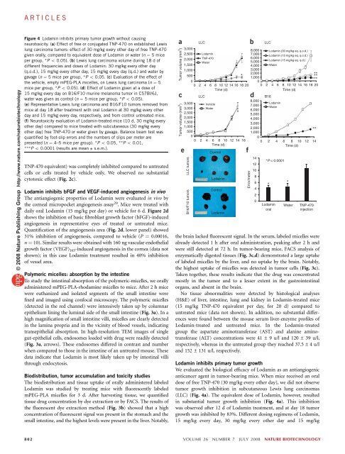
Figure 4 Lodamin inhibits primary tumor growth without causing<br />
neurotoxicity. (a) Effect of free or conjugated TNP-470 on established Lewis<br />
lung carcinoma tumors: effect of 30 mg/kg every other day of free TNP-470<br />
given orally, compared to equivalent dose of Lodamin or water (n ¼ 5 mice<br />
per group, *P o 0.05). (b) Lewis lung carcinoma volume during 18 d of<br />
different frequencies and doses of Lodamin: 30 mg/kg every other day<br />
(q.o.d.), 15 mg/kg every other day, 15 mg/kg every day (q.d.) and water by<br />
gavage (n ¼ 5 mice per group, *P o 0.05. (c) Evaluation of the effect of<br />
the vehicle, empty mPEG-PLA micelles, on Lewis lung carcinoma (n ¼ 5<br />
mice per group, *P o 0.05). (d) Effect of Lodamin given at a dose of<br />
15 mg/kg every day on B16/F10 murine melanoma tumor in C57Bl/6J,<br />
water was given as control (n ¼ 5 mice per group, *P o 0.05).<br />
(e) Representative Lewis lung carcinoma and B16/F10 tumors removed from<br />
mice at day 18 after treatment with oral Lodamin at 30 mg/kg every other<br />
day and 15 mg/kg every day, respectively, and from control untreated mice.<br />
(f) Neurotoxicity evaluation of Lodamin-treated mice (10 d, 30 mg/kg every<br />
other day) compared to mice treated with subcutaneous (30 mg/kg every<br />
other day) free TNP-470 or water given by gavage. Balance beam test was<br />
quantified by foot-slip errors and the numbers of slips per meter are<br />
presented (n ¼ 4–5 mice per group). *P o 0.05, **P o 0.01,<br />
***P o 0.0001 (results are mean ± s.e.m.).<br />
TNP-470 equivalent) was completely inhibited compared to untreated<br />
cells or cells treated by vehicle only. We observed no substantial<br />
cytotoxic effect (Fig. 2c).<br />
Lodamin inhibits bFGF and VEGF-induced angiogenesis in vivo<br />
The antiangiogenic properties of Lodamin were evaluated in vivo by<br />
the corneal micropocket angiogenesis assay 29 . Mice were treated with<br />
daily oral Lodamin (15 mg/kg per day) or vehicle for 6 d. Figure 2d<br />
shows the inhibition of basic fibroblast growth factor (bFGF)-induced<br />
angiogenesis in representative eyes of treated or untreated mice.<br />
Quantification of the angiogenesis area (Fig. 2d, lower panel) showed<br />
31% inhibition of angiogenesis, compared to vehicle (P ¼ 0.00016,<br />
n ¼ 10). Similar results were obtained with 160 ng vascular endothelial<br />
growth factor (VEGF) 165-induced angiogenesis in the cornea (data not<br />
shown); in this case Lodamin treatment resulted in 40% inhibition<br />
of vessel area.<br />
Polymeric micelles: absorption by the intestine<br />
To study the intestinal absorption of the polymeric-micelles, we orally<br />
administered mPEG-PLA-rhodamine micelles to mice. After 2 h mice<br />
were euthanized and isolated segments of the small intestine were<br />
fixed and imaged using confocal microscopy. The polymeric micelles<br />
(detected in the red channel) were intensively taken up by columnar<br />
epithelium lining the luminal side of the small intestine (Fig. 3a). In a<br />
high magnification of small intestine villi, micelles are clearly detected<br />
in the lamina propria and in the vicinity of blood vessels, indicating<br />
transepithelial absorption. In high-resolution TEM images of single<br />
gut-epithelial cells, endosomes loaded with drug were readily detected<br />
(Fig. 3a, arrows). These endosomes differed in contrast and number<br />
when compared to those in the intestine of an untreated mouse. These<br />
data indicate that Lodamin is most likely taken up by intestinal villi<br />
through endocytosis.<br />
Biodistribution, tumor accumulation and toxicity studies<br />
The biodistribution and tissue uptake of orally administered labeled<br />
Lodamin was studied by treating mice with fluorescently labeled<br />
mPEG-PLA micelles for 3 d. After harvesting tissue, we quantified<br />
tissue drug concentration by dye extraction or by FACS. The results of<br />
the fluorescent dye extraction method (Fig. 3b) showed that a high<br />
concentration of fluorescent signal was present in the stomach and the<br />
small intestine, and the highest levels were present in the liver. Notably,<br />
a LLC<br />
b<br />
Tumor volume (mm 3 )<br />
c d<br />
Tumor volume (mm 3 )<br />
3,000<br />
2,500<br />
2,000<br />
1,500<br />
Lodamin<br />
TNP-470<br />
Water<br />
8,000<br />
7,000<br />
6,000<br />
5,000<br />
4,000<br />
Lodamin (30 mg/kg eq. q.o.d.)<br />
Lodamin (15 mg/kg eq. q.o.d.)<br />
Lodamin (15 mg/kg eq. q.d.)<br />
Water<br />
1,000<br />
3,000<br />
500<br />
0<br />
*<br />
2,000<br />
1,000<br />
0<br />
**<br />
**<br />
***<br />
0 2 4 6 8 10 12 14 16 18 20 0 2 4 6 8 10 12 14 16 18 20<br />
Time (d) Time (d)<br />
LLC B16<br />
3,500<br />
3,000<br />
2,500<br />
2,000<br />
1,500<br />
1,000<br />
500<br />
Vehicle<br />
Water<br />
8,000<br />
7,000<br />
6,000<br />
5,000<br />
4,000<br />
3,000<br />
2,000<br />
1,000<br />
Lodamin<br />
Water<br />
**<br />
0<br />
0 2 4 6 8 10 12 14 16<br />
0<br />
0 2 4 6 8 10 12 14<br />
Time (d)<br />
Control<br />
Time (d)<br />
e f<br />
LLC tumors<br />
B16/F10 tumors<br />
Lodamin<br />
Control<br />
Lodamin<br />
Error/meter<br />
the brain lacked fluorescent signal. In the serum, labeled micelles were<br />
already detected 1 h after oral administration, peaking after 2 h and<br />
were still detected at 72 h. In tumor-bearing mice, FACS analysis of<br />
enzymatically digested tissues (Fig. 3c,d) demonstrated a large uptake<br />
of labeled micelles by the liver, and no uptake by the brain. Notably,<br />
the highest uptake of micelles was detected in tumor cells (Fig. 3c).<br />
Taken together, these results indicate that the drug was concentrated<br />
mostly in the tumor and to a lesser extent in the gastrointestinal<br />
organs, and absent in the brain.<br />
No tissue abnormalities were detected by histological analyses<br />
(H&E) of liver, intestine, lung and kidney in Lodamin-treated mice<br />
(15 mg/kg TNP-470 equivalent per day, for 20 d) compared to<br />
untreated mice (data not shown). In addition, no substantial differences<br />
were found between the mouse serum liver-enzyme profiles of<br />
Lodamin-treated and untreated mice. In the Lodamin-treated<br />
group the aspartate aminotransferase (AST) and alanine aminotransferase<br />
(ALT) concentrations were 41 ± 9 u/l and 120 ± 39 u/l,<br />
respectively, whereas in the untreated group they reached 37.5 ± 4 u/l<br />
and 152 ± 131 u/l, respectively.<br />
Lodamin inhibits primary tumor growth<br />
We evaluated the biological efficacy of Lodamin as an antiangiogenic<br />
anticancer agent in tumor-bearing mice. When mice received an oral<br />
dose of free TNP-470 (30 mg/kg every other day), we did not observe<br />
tumor growth inhibition in subcutaneous Lewis lung carcinomas<br />
(LLC) (Fig. 4a). The equivalent dose of Lodamin, however, resulted<br />
in substantial tumor growth inhibition (Fig. 4a). This inhibition<br />
was observed after 12 d of Lodamin treatment, and at day 18 tumor<br />
growth was inhibited by 83%. Different dosing regimens of Lodamin,<br />
15 mg/kg every day, 30 mg/kg every other day and 15 mg/kg<br />
14<br />
12<br />
10<br />
8<br />
6<br />
4<br />
2<br />
0<br />
LLC<br />
*P < 0.0001<br />
*<br />
*<br />
Lodamin Water<br />
oral<br />
TNP-470<br />
injection<br />
802 VOLUME 26 NUMBER 7 JULY 2008 NATURE BIOTECHNOLOGY