PRRS Compendium Producer Edition - National Pork Board
PRRS Compendium Producer Edition - National Pork Board
PRRS Compendium Producer Edition - National Pork Board

You also want an ePaper? Increase the reach of your titles
YUMPU automatically turns print PDFs into web optimized ePapers that Google loves.
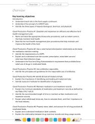
2.<br />
3.<br />
4.<br />
The pigs were recently infected with virus but have not yet had time to seroconvert.<br />
The pigs were infected with virus some time ago, but have since become seronegative.<br />
The result was falsely negative due to poor sensitivity of the test or a laboratory error.<br />
Therefore, if using single point-in-time samples, <strong>PRRS</strong> serology must be used in conjunction with valid<br />
population sampling methods and knowledge of a herd’s history to determine if the herd has been exposed<br />
to <strong>PRRS</strong> virus. Current serologic tests are better suited for determining the status of a population,<br />
not necessarily the status of individual animals.<br />
Diagnosis of <strong>PRRS</strong> virus infection as the cause of reproductive failure or respiratory disease can be<br />
achieved by showing a change in antibody titer (i.e., rising antibody titer) in paired serum samples. However,<br />
a definitive diagnostic evaluation of <strong>PRRS</strong> with respect to clinical disease requires that serological<br />
information be interpreted in combination with results from virus isolation and/or detection of antigenic or<br />
genomic material (Christianson and Joo, 1994; Goyal, 1993). It is important to bear in mind that the current<br />
serologic assays used in the diagnostic setting cannot routinely differentiate vaccine-derived antibodies<br />
from field isolate-derived antibodies.<br />
The occurrence of false positive serologic results on the commercial ELISA has been a concern for diagnosticians<br />
and practitioners, particularly in expected-negative herds or groups (Keay et al., 2002; O’Connor<br />
et al., 2002; Torremorell et al., 2002). Field observations in “expected-negative” herds have suggested that<br />
false positive animals (“singleton reactors”) occur at a rate of 0.5 to 2%. Most suspected false positive animals<br />
have ELISA S/P values around the 0.4 cut off value, but S/P ratios can occasionally exceed 1.0 in these<br />
animals. At present, no information regarding the occurrence of false positives is available on the recently<br />
released 2XR version of the HerdChek ® ELISA. Current recommendations for evaluating suspected false<br />
positive animals include repeating the test, testing by other serological methods in conjunction with PCR,<br />
re-sampling the suspected false positive animal and re-testing, or occasionally, sacrificing the animal and<br />
conducting a complete diagnostic work-up. A few diagnostic laboratories have developed in-house ELISAs<br />
for more specific detection of positive animals (Ferrin et al., 2002; Zhou et al 2001). Such tests are available<br />
on an experimental basis at present.<br />
Differential Testing<br />
Serologic assays cannot routinely differentiate antibodies to field isolates from vaccine-derived antibodies.<br />
However, characterization of virus isolates is possible by several methods. Panels of monoclonal antibodies<br />
can easily differentiate European isolates from North American isolates and vice versa (Dea et al.,<br />
1996; Drew et al., 1995; Nelson et al., 1993, 1996; Yang et al., 1999, 2000). Using this technique, Nelson et<br />
al. (1996) could find no evidence of the Lelystad virus (LV) or LV-like <strong>PRRS</strong> viruses in Midwestern US swine<br />
herds after evaluating 306 field isolates collected before 1995. However, recent reports of “Euro<strong>PRRS</strong>”<br />
virus, i.e., Lelystad-like virus or European <strong>PRRS</strong> virus in North America (Dewey et al., 2000) suggest that<br />
monoclonal antibody analysis of isolates may be useful for differentiation of isolates. Monoclonal antibody<br />
analysis can also be used to differentiate commercial modified-live vaccine virus from field isolates<br />
(Yang et al 2000).<br />
Molecular biology has also made it possible to characterize <strong>PRRS</strong> virus isolates using PCR, a restriction<br />
fragment length polymorphism (RFLP) assay (Wesley et al., 1998a; Umthun et al. 1999), and direct DNA sequencing<br />
(Kapur et al., 1996; Yoon et al 2001). A PCR-based technique has been developed to differentiate<br />
North American from European isolates (Christopher-Hennings et al., 1995a; Egli et al., 2001; Gilbert et al.,<br />
1997; Mardassi et al., 1994). Although the investigators demonstrated its usefulness, PCR was not routinely<br />
used for that purpose in North American until the recent report of the presence of European-like <strong>PRRS</strong><br />
virus in North America (Dewey et al., 2000). Currently, several diagnostic laboratories conduct differential<br />
PCR on suspect cases as per request or at the diagnostician’s discretion. More recently, a heteroduplex<br />
mobility assay (HMA) has been developed for a rapid identification and differentiation of vaccine-like virus<br />
from field viruses (Key et al., 2002a) but has only limited availability.<br />
The RFLP assay is a crude technique for differentiating one <strong>PRRS</strong> virus isolate from another. It was developed<br />
early on in the genesis of <strong>PRRS</strong> virus diagnostic tools but has recently fallen out of favor. The RFLP<br />
technique involves virus isolation followed by restriction endonuclease digestion. Restriction endonucleases<br />
are enzymes that make cuts at specific places in a genomic sequence. Different <strong>PRRS</strong> viruses differ in<br />
their genomic sequences, so fragments of different sizes are produced. The lengths of these fragments are<br />
PAGE 4 PIG 04-01-09