PRRS Compendium Producer Edition - National Pork Board
PRRS Compendium Producer Edition - National Pork Board
PRRS Compendium Producer Edition - National Pork Board

You also want an ePaper? Increase the reach of your titles
YUMPU automatically turns print PDFs into web optimized ePapers that Google loves.
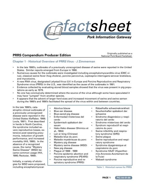
<strong>PRRS</strong> <strong>Compendium</strong> <strong>Producer</strong> <strong>Edition</strong><br />
Chapter 1 - Historical Overview of <strong>PRRS</strong> Virus - J Zimmerman<br />
•<br />
•<br />
•<br />
•<br />
•<br />
•<br />
In the late 1980’s, outbreaks of a previously unrecognized disease of swine were reported in the United<br />
States. Similar reports emerged from Europe in 1990.<br />
Numerous causes for the outbreaks were investigated including encephalomyocarditis virus (EMC virus),<br />
classical swine fever (hog cholera), porcine parvovirus, Leptospira interrogans serovar bratislava,<br />
and mycotoxins.<br />
A new RNA virus, designated Lelystad Virus (LV) in Europe and Porcine Reproductive and Respiratory<br />
Syndrome virus (<strong>PRRS</strong>) in the U.S., was identified as the cause of the outbreaks in 1991.<br />
Evidence collected by evaluating stored blood samples showed that the virus was present in pig populations<br />
as early as 1979.<br />
No one has conclusively determined where the source of the virus although some have speculated it<br />
may have “jumped” from another species.<br />
It appears that the advent of larger herd sizes and increased movement of swine and swine semen<br />
during the 1980’s and 1990’s facilitated the spread of the virus within and between countries.<br />
In the late 1980’s, catastrophic<br />
clinical outbreaks of<br />
a previously unrecognized<br />
disease were reported in the<br />
United States (Keffaber, 1989;<br />
Loula, 1991). First described<br />
in herds in North Carolina,<br />
the syndrome included severe<br />
reproductive losses, extensive<br />
post-weaning pneumonia,<br />
reduction of growth<br />
performance, and increased<br />
mortality (Hill, 1990). In the<br />
absence of a recognized<br />
cause, the name “Mystery<br />
Swine Disease” (MSD) became<br />
common usage (Hill,<br />
1990; Reotutar, 1989).<br />
Initially, a variety of etiologies<br />
for MSD were proposed,<br />
including encephalomyocar-<br />
•<br />
•<br />
•<br />
•<br />
•<br />
•<br />
•<br />
•<br />
•<br />
•<br />
•<br />
•<br />
•<br />
•<br />
•<br />
Abortus blauw<br />
Blue ear disease<br />
Blue-eared pig disease<br />
Enfermedad misteriosa del<br />
cerdo<br />
Epidemisch spätabort der<br />
sauen<br />
Heko-Heko disease (Shimizu et<br />
al., 1994)<br />
Lan er bing (Chinese)<br />
Maladie blue du porc<br />
Maladie mystérieuse du porc<br />
Mystery pig disease<br />
Mystery swine disease (MSD)<br />
New pig disease<br />
Plague of 1988 - 1989<br />
Porcine epidemic abortion and<br />
respiratory syndrome (PEARS)<br />
Porcine reproductive and respiratory<br />
syndrome (<strong>PRRS</strong>)<br />
•<br />
•<br />
•<br />
•<br />
•<br />
•<br />
•<br />
•<br />
•<br />
•<br />
•<br />
Originally published as a<br />
<strong>National</strong> <strong>Pork</strong> <strong>Board</strong> Factsheet.<br />
Rätselhafte schweinekrankheit<br />
Seuchenhafter spätabort der<br />
schweine<br />
Síndrome disgenésico y respiratorio<br />
del cerdo<br />
Síndrome misterioso del cerdo<br />
Syndrom reproductif et respiratoire<br />
du porc<br />
Swine infertility and respiratory<br />
syndrome (SIRS)<br />
Swine plague<br />
Swine reproductive and respiratory<br />
syndrome<br />
Syndrome dysgénésique et<br />
respiratoire du porc<br />
Syndrome HAAT (Hyperthermie-Anorexie-Avortement<br />
de<br />
la Truie)<br />
Wabash syndrome<br />
Table 1: Historical names for <strong>PRRS</strong><br />
PAGE 1<br />
PIG 04-01-09
ditis virus, classical swine fever (hog cholera) virus, porcine enterovirus, porcine parvovirus, pseudorabies<br />
virus (Aujeszky’s disease), Leptospira interrogans serovar bratislava, Chlamydia psittaci, and contamination<br />
of feed with mycotoxins (Bane et al., 1990; Daniels, 1990; Hoeffling, 1990; Joo, 1988; Joo et al., 1990;<br />
Quaife, 1989; Reotutar, 1989). In Canada, a new subtype of Influenza A virus was isolated from piglets suffering<br />
severe respiratory disease and added to the list as a possible agent of MSD (Dea et al., 1992c; Elazhary<br />
et al., 1991). Identifying the etiology was complicated by the fact that one or more of the suspected<br />
pathogens, as well as other infectious agents, were commonly isolated from cases of MSD.<br />
In Europe, clinical outbreaks notably similar to MSD were reported in November 1990 near Münster, Germany<br />
(OIE, 1992). The disease spread rapidly and over 3,000 outbreaks were documented in Germany in<br />
May 1991. No link was found between outbreaks in Germany and MSD in the U.S. (Anon, 1991). The disease<br />
was reported in the Netherlands in January 1991 and in Belgium in March 1991 (OIE, 1992). The first<br />
clinical case of <strong>PRRS</strong> in Spain was detected in January 1991 associated with the importation of live pigs<br />
(Plana Duran et al., 1992). Three outbreaks were reported in Spain—two in the province of Huesca and one<br />
in the province of Lerida—and all animals were quickly slaughtered (OIE, 1992). In Great Britain, “blueeared”<br />
pig disease appeared in May 1991 (Edwards et al., 1992). In spite of the application of control measures,<br />
the disease spread. By the end of October 1991, 58 outbreaks had been confirmed. In the case of<br />
the United Kingdom, it was noted that no imports of live pigs, semen, or embryos had been received from<br />
countries known to have had MSD during the preceding 12 months; thus, there was no apparent explanation<br />
for its source (Robertson, 1992). In France, the first outbreaks appeared in Brittany in November 1991<br />
(Baron et al., 1992; OIE, 1992), followed by outbreaks in Denmark in March 1992 (Bøtner et al., 1994). The<br />
disease was confirmed to be present in Poland in 1992 (Pejsak and Markowska-Daniel, 1996), and the Czeck<br />
Republic in 1995 (Valíček et al., 1997).<br />
In Asia, outbreaks occurred in Japan in 1988 (Hirose et al., 1995) and in Taiwan in 1991 (Chang et al., 1993).<br />
Thus, the pandemic had spread to most of the major swine producing centers of the world in the space of<br />
a few years.<br />
Until 1991, the lack of a specific etiologic agent led to a rapid proliferation of colorful and descriptive terms<br />
for the disease based on clinical signs, none of which, in the absence of a defined cause, could be considered<br />
either inappropriate or incorrect. A partial list of names can be found in Table 1.<br />
<strong>PRRS</strong> Virus<br />
The cause of MSD was resolved in 1991 when Koch’s postulates were fulfilled with a previously unrecognized,<br />
enveloped RNA virus (Terpstra et al., 1991a; Wensvoort et al., 1991). Shortly thereafter, the virus was<br />
isolated in the U.S. (Collins, 1991; Collins et al., 1992) and Canada (Dea et al., 1992a, 1992b). The first virus<br />
isolates in the Netherlands and U.S. were designated Lelystad virus and Swine Infertility and Respiratory<br />
Syndrome (SIRS) virus (BIAH-001), respectively. Both virus isolates were shown to induce reproductive<br />
failure and respiratory signs under experimental conditions (Collins et al., 1992; Terpstra et al., 1991a). The<br />
virus is now commonly referred to as Porcine Reproductive and Respiratory Syndrome (<strong>PRRS</strong>) virus in<br />
much of the world.<br />
Early Evidence of <strong>PRRS</strong> Virus<br />
The earliest direct evidence of <strong>PRRS</strong> virus infection in domestic swine comes from a retrospective serologic<br />
study of herds in Ontario, Canada. Carman et al. (1995) found that none of 50 herds sampled in 1978<br />
were serologically positive for antibodies against <strong>PRRS</strong> virus by enzyme linked immunosorbent assay<br />
(ELISA) or indirect fluorescent antibody (IFA), but antibodies were detected in serum samples from 2 of 51<br />
(3.9%) herds collected in 1979 and 8 of 51 (15.7%) herds sampled in 1980.<br />
In the U.S., a retrospective survey found no evidence of infection in 1,425 serum samples collected from<br />
118 Iowa swine herds in 1980 (Zimmerman et al., 1997). One of 26 herds (3.8%) sampled in 1985 was <strong>PRRS</strong><br />
virus-infected and each successive year showed an increase in prevalence. The data from samples collected<br />
in 1985 suggests that the virus entered Iowa during or shortly prior to 1985. By 1988, 17 of 27 herds<br />
(63.0%) and 313 of 658 (47.6%) animals were seropositive. Similar to the Iowa data, the earliest evidence of<br />
infection in the state of Minnesota was found in banked serum samples originally collected in 1986 (Yoon<br />
et al., 1992).<br />
PAGE<br />
PIG 04-01-09
Most of Europe followed the pattern seen in Canada and the U.S., prevalence increasing rapidly in 1988<br />
and 1989. About 48 percent of 1,480 serum samples collected in eastern Germany prior to the outbreaks in<br />
northwestern Germany in 1990 were positive for <strong>PRRS</strong> virus antibodies.<br />
In Asia, antibodies against <strong>PRRS</strong> virus were retrospectively documented in serum from pigs imported into<br />
the Republic of Korea (South Korea) in October 1985 (Shin et al., 1993), in serum samples collected in 1987<br />
in Taiwan (see Section 6.17), and in samples collected in June 1988 in Japan (Hirose et al., 1995). Again,<br />
well before the recognition of clinical disease.<br />
Molecular studies of <strong>PRRS</strong> virus isolates provide indirect evidence of the early presence of the virus.<br />
Based on a study by Forsberg et al. (2001), <strong>PRRS</strong> virus isolates from Denmark, Italy, the United Kingdom,<br />
and the Lelystad virus were linked to a common ancestor that existed about 1979, i.e., more than 10 years<br />
prior to the outbreaks in Europe.<br />
Thus, the serological and virological evidence indicate that <strong>PRRS</strong> virus was circulating in the domestic<br />
swine population by 1979 in North American and perhaps Europe, as well, i.e., several years prior to the<br />
actual recognition of clinical disease: This raises several questions, but two in particular: Where was <strong>PRRS</strong><br />
virus before 1979? and Why were clinical signs not reported prior to 1987?<br />
Regarding the first question, Forsberg et al. (2000) suggest two possibilities:<br />
1.<br />
2.<br />
The virus came from another species.<br />
The virus circulated in an isolated pig population for a “long time” prior to the pandemic, during<br />
which time it acquired a high degree of genetic diversity.<br />
There is no direct evidence to fully support or repudiate either of these hypotheses, but further discussion<br />
is warranted because of the consequences <strong>PRRS</strong> virus has had on swine health and because there is no<br />
reason to believe something similar could not happen in the future.<br />
Forsberg et al. (2000) reasoned that, if <strong>PRRS</strong> had been the result of a single interspecies transmission<br />
event followed by explosive transmission in the swine population, i.e., a “point-source epidemic,” then<br />
current virus isolates should be genetically linked to a common viral ancestor that emerged around the<br />
time of the outbreaks—which they are not. Instead, extensive genetic diversity already existed within<br />
Europe, within North America, and between Europe and North America at the time <strong>PRRS</strong> was first recognized.<br />
In support of the hypothesis that <strong>PRRS</strong> virus circulated undetected in isolated swine populations is the observation<br />
that smaller herds tend to have fewer episodes of clinical <strong>PRRS</strong>. For example, the USDA (2002)<br />
reported that 15% of “smaller breeding herds,” defined as sites fewer than 250 sows and gilts, reported<br />
clinical <strong>PRRS</strong>, versus 40% of “medium herds” (250 to 499 females) and 58% of “large herds” (500 or more<br />
females). These data are important because they link to the past. From the perspective of current production<br />
systems, the average breeding herd size for most of the 20 th century was extremely small. According<br />
to the U.S. Census of Agriculture, the average total inventory among U.S. swine operations in 1959 was<br />
37 animals (USDA, 1997). This increased to a mean of 81 per operation in 1969, 130 in 1978, 215 in 1987,<br />
and 301 in 1992 (USDA, 1997). Thus, if <strong>PRRS</strong> virus produces few clinical signs in small herds, then the virus<br />
could conceivably have circulated in the small herds that were the standard in the past without attracting<br />
excessive attention.<br />
Of course, this still leaves the problem of identifying the original source of introduction. At present, we<br />
simply have insufficient data with which to resolve the question.<br />
Changes in Swine Production<br />
As suggested by Nelsen et al. (1999) and elsewhere, the dramatic changes in swine production and management<br />
that occurred in the latter half of the 20 th century may have created an environment well-suited<br />
to the dissemination and perpetuation of the virus in the domestic swine population. Changes favorable<br />
to the virus included extensive horizontal integration resulting in fewer but larger herds, reliance on fewer<br />
and larger companies for replacement breeding stock, increased transport of live animals both within and<br />
between countries, and greater use of artificial insemination in breeding programs. It should be recog-<br />
PAGE<br />
PIG 04-01-09
nized that the changes that occurred in swine production and management in the last two decades have<br />
imposed an entirely new epidemiology on the infectious diseases of swine. In the case of <strong>PRRS</strong> virus,<br />
larger herds and increased movement of pigs and semen facilitated the spread of the virus within and between<br />
countries (Dewey et al., 2000; Milián Suazo et al., 1994; Plana Duran et al., 1992; OIE, 1994; Shin et<br />
al., 1993).<br />
Summary<br />
In the late 1980’s, catastrophic clinical outbreaks of a previously unrecognized disease were reported in<br />
the United States, followed by outbreaks in Europe and Asia in the early 1990’s. In 1991, European workers<br />
reported the cause to be a previously unrecognized Arterivirus and introduced the term “porcine reproductive<br />
and respiratory syndrome.”<br />
The earliest direct evidence of <strong>PRRS</strong> virus infection in domestic swine is the presence of anti-<strong>PRRS</strong> virus<br />
antibodies in serum samples collected in 1979 in Canada. Retrospective studies also found antibodies in<br />
samples collected in the U.S. in 1985, the Republic of Korea in 1985, in Japan in 1988, and in the former<br />
East Germany in 1987. Molecular studies of <strong>PRRS</strong> virus isolates suggest that the virus may have been present<br />
in Europe as early as 1979. The original source of the virus is unknown, but once introduced into domestic<br />
swine, the larger herds and increased movement of pigs and semen that became increasingly common<br />
in the 1980’s and 1990’s facilitated the spread of the virus both within and between countries.<br />
References<br />
Anon. 1991. The new pig disease: conclusions reached at the seminar held. In: The new pig disease. Porcine reproductive and respiratory syndrome.<br />
A report on the seminar/workshop held in Brussels on 29-30 April and organized by the European Commission (Directorate General for Agriculture), pp.<br />
82-86.<br />
Bane DP, Hall WF. 1990. Fumonisin as a predisposing factor for “Mystery Swine Disease.” Proc Mystery Swine Disease Committee Meeting, Livestock<br />
Conservation Institute, Denver, Colorado, pp. 77-79.<br />
Baron T, Albina E, Leforban Y, et al. 1992. Report on the first outbreaks of the porcine reproductive and respiratory syndrome (<strong>PRRS</strong>) in France: Diagnosis<br />
and viral isolation. Ann Rech Vet 23:161-166.<br />
Bøtner A, Nielsen J, Bille-Hansen V. 1994. Isolation of porcine reproductive and respiratory syndrome (<strong>PRRS</strong>) virus in a Danish swine herd and experimental<br />
infection of pregnant gilts with the virus. Vet Microbiol 40:351-360.<br />
Carman S, Sanford SE, Dea S. 1995. Assessment of seropositivity to porcine reproductive and respiratory syndrome (<strong>PRRS</strong>) virus in swine herds in<br />
Ontario--1978 to 1982. Can Vet J 36:776-777.<br />
Chang CC, Chung WB, Lin MW, et al.1993. Porcine reproductive and respiratory syndrome (<strong>PRRS</strong>) in Taiwan. I. viral isolation. J Chin Soc Vet Sci19:268-276.<br />
Collins J, Benfield DA, Christianson WT, et al. 1992. Isolation of swine infertility and respiratory syndrome virus (isolate ATCC VR-2332) in North America<br />
and experimental reproduction of the disease in gnotobiotic pigs. J Vet Diagn Invest 4:117-126.<br />
Collins JE. 1991. Diagnostic note: newly recognized respiratory syndromes in North American swine herds. American Association of Swine Practitioners<br />
Newsletter 3:7-11.<br />
Daniels ED. 1990. Chlamydia proteins found in swine reproductive failure cases. Proc Mystery Swine Disease Committee Meeting, Livestock Conservation<br />
Institute, Denver, Colorado, pp. 80-81.<br />
Dea S, Bilodeau R, Athanaseous R, et al. 1992a. <strong>PRRS</strong> syndrome in Quebec: isolation of a virus serologically related to Lelystad virus [letter]. Vet Rec<br />
130:167.<br />
Dea S, Bilodeau R, Athanassious R, et al. 1992b. Swine reproductive and respiratory syndrome in Quebec: isolation of an enveloped virus serologicallyrelated<br />
to Lelystad virus. Can Vet J 33:801-808.<br />
Dea SA, Bilodeau R, Athanassious R, et al. 1992c. <strong>PRRS</strong> in Quebec: virology and pathologic aspects. American Association of Swine Practitioners<br />
Newsletter 4:2-7.<br />
Dewey C, Charbonneau G, Carman S, et al. 2000. Lelystad-like strain of porcine reproductive and respiratory syndrome virus (<strong>PRRS</strong>V) identified in Canadian<br />
swine. Can Vet J 41: 493-494.<br />
Edwards S, Robertson IB, Wilesmith JW, et al. 1992. <strong>PRRS</strong> (“blue-eared pig disease”) in Great Britain. American Association of Swine Practitioners<br />
Newsletter 4:32-36.<br />
Elazhary Y, Weber J, Bikour H, et al. 1991. “Mystery swine disease” in Canada. Vet Rec 129:495-496.<br />
Forsberg R, Oleksiewicz MB, Petersen AM, et al. 2001. A molecular clock dates the common ancestor of European-type porcine reproductive and respiratory<br />
syndrome virus at more than 10 years before the emergence of disease. Virology 289:174-179.<br />
Hill H. 1990. Overview and history of Mystery Swine Disease (Swine infertility/respiratory syndrome). Proc Mystery Swine Disease Committee Meeting,<br />
Livestock Conservation Institute, Denver, Colorado, pp. 29-31.<br />
PAGE 4<br />
PIG 04-01-09
Hirose O, Kudo H, Yoshizawa S, et al. 1995. Prevalence of porcine reproductive and respiratory syndrome virus in Chiba prefecture. J Jpn Vet Med Assoc<br />
48:650-653.<br />
Hoeffling DC. 1990. Mystery swine disease. Proc US Anim Health Assoc 94:501-504.<br />
Joo HS, Christianson B, Kim H. 1990. Encephalomyocarditis virus as a potential cause for “Mystery swine disease.” Proc Mystery Swine Disease Committee<br />
Meeting, Livestock Conservation Institute, Denver, Colorado, pp. 62-66.<br />
Joo HS. 1988. EMC virus causes poor reproduction. Int Pigletter 8:3-4.<br />
Keffaber KK. 1989. Reproductive failure of unknown etiology. American Association of Swine Practitioners Newsletter 1:1-10.<br />
Loula T. 1991. Mystery pig disease. Agri Prac 12:23-34.<br />
Milián Suazo F, Canto Alarcon GJ, Weimersheimer Rubi J, et al. 1994. (Seroprevalence of antibodies against porcine reproductive and respiratory syndrome<br />
virus in Mexico.) Estudio seroepidemiológico para determinar la presencia de anticuerpos contra el virus del síndrome disgenésico del cerdo en<br />
México. Técnica Pecuaria en México 32: 139-144.<br />
Nelsen CJ, Murtaugh MP, Faaberg KS. 1999. Porcine reproductive and respiratory syndrome virus comparison: Divergent evolution on two continents.<br />
J Virol 73:270-280.<br />
OIE (Office International des Épizooties). 1992. World Animal Health 1991. Volume VII. Number 2. Animal Health Status and Disease Control Methods<br />
(Part One: Reports), p. 126.<br />
OIE (Office International des Épizooties). 1994. World Animal Health in 1993. Part 1. Reports on the Animal Health Status and Disease Control Methods<br />
and List A Disease Outbreaks - Statistics, p. 17.<br />
Pejsak Z, Markowska Daniel I. 1996. Viruses as a reason for reproductive failure in pig herds in Poland. Reprod Dom Anim 31:445-447.<br />
Plana Duran J, Vayreda M, Vilarrasa J, et al. 1992. PEARS (“Mystery Swine Disease”) - summary of the work conducted by our group. American Association<br />
of Swine Practitioners Newsletter 4:16-18.<br />
Quaife T. 1989. Scramble is on to solve mystery disease. Swine Pract, July 5-10.<br />
Reotutar R. 1989. Swine reproductive failure syndrome mystifies scientists. J Am Vet Med Assoc 195:425-428.<br />
Robertson IB. 1992. Porcine reproductive and respiratory syndrome (Blue eared pig disease): Some aspects of its epidemiology. Proc Soc Vet Epidemiol<br />
Prev Med, pp. 24-38.<br />
Shimizu M, Yamada S, Murakami Y, et al. 1994. Isolation of porcine reproductive and respiratory syndrome (<strong>PRRS</strong>) virus from Heko-Heko disease of<br />
pigs. J Vet Med Sci 56:389-391.<br />
Shin J-H, Kang Y-B, Kim Y-J, et al. 1993. Sero-epidemiological studies on porcine reproductive and respiratory syndrome in Korea. I. Detection of indirect<br />
fluorescent antibodies. RDA J Agri Sci 35:572-576.<br />
Terpstra C, Wensvoort G, Pol JMA. 1991a. Experimental reproduction of porcine epidemic abortion and respiratory syndrome (mystery swine disease)<br />
by infection with Lelystad virus: Koch’s postulates fulfilled. Vet Q 13:131-136.<br />
USDA (United States Department of Agriculture). 1997. Part III. Changes in the U.S. <strong>Pork</strong> Industry 1990-1995. USDA:APHIS:VS, CEAH, <strong>National</strong> Animal<br />
Health Monitoring System, Fort Collins, CO.<br />
USDA (United States Department of Agriculture). 2002. Part II. Reference of Swine Health and Health Management in the United States, 2000. USDA:<br />
APHIS:VS, CEAH, <strong>National</strong> Animal Health Monitoring System, Fort Collins, CO #N355.0202.<br />
Valíček L, Pšikal I, čmid B, et al.<br />
Wensvoort G, Terpstra C, Pol JMA, et al. 1991. Mystery swine disease in the Netherlands: the isolation of Lelystad virus. Vet Q 13:121-130.<br />
Yoon IJ, Joo HS, Christianson WT, et al. 1992. An indirect fluorescent antibody test for the detection of antibody to swine infertility and respiratory syndrome<br />
virus in swine sera. J Vet Diagn Invest 4:144-147.<br />
Zimmerman J, Yoon K-J, Wills RW, Swenson SL. 1997. General overview of <strong>PRRS</strong>V: A perspective from the United States. Vet Microbiol 55:187-196.<br />
Chapter - <strong>PRRS</strong> Virus Infection and Disease - B Thacker<br />
•<br />
•<br />
•<br />
The severity of <strong>PRRS</strong> virus infection can vary widely and range from a near complete lack of clinical<br />
signs to devastating outbreaks of reproductive and respiratory disease.<br />
<strong>PRRS</strong> virus infection occurs mainly in a cell of the immune system called a macrophage. Macrophages<br />
are found throughout many tissues in the body.<br />
Clinical signs in the breeding herd may include anorexia (off-feed), fever, lethargy, nervous signs, purplish<br />
discoloration of the ears and vulva, and abortion. Litters born to recently infected dams may have<br />
increased rates of mummification, still births, and weak-born piglets. Piglets in these litters may carry<br />
PAGE<br />
PIG 04-01-09
•<br />
•<br />
•<br />
the virus for an extended period.<br />
Boars infected with <strong>PRRS</strong> virus can show similar signs as sows. Infection does not usually impact the<br />
fertility of the semen but virus can be found in the ejaculate of boars for several months after being<br />
infected.<br />
Newborn piglets infected with the virus can demonstrate severe respiratory disease. Nervous signs as<br />
well as anorexia and lethargy have also been reported. Other common diseases of young pigs may become<br />
more prevalent and severe during a <strong>PRRS</strong> outbreak.<br />
In an experimental setting, piglets infected with <strong>PRRS</strong> virus often show minimal clinical signs. However,<br />
in a field setting, <strong>PRRS</strong> virus infection frequently has a more pronounced effect due to its interaction<br />
with the pig’s environment, immune status, and concurrent diseases.<br />
Introduction<br />
The severity of disease and clinical manifestations of <strong>PRRS</strong> virus infection vary widely. Subclinical infections<br />
can occur in a herd over a long period of time based on field observations of herds that remain clinically<br />
normal despite ample serological evidence that <strong>PRRS</strong> virus (<strong>PRRS</strong>V) circulation occurs continuously<br />
(B Thacker, personal observations). On the other hand, the upper limits of severity can be extreme. For example,<br />
some outbreaks that occurred in 1996 were initially considered so severe that other agents beside<br />
<strong>PRRS</strong> virus, or “super virulent” strains of <strong>PRRS</strong> virus, were suspected to be the cause (Hurd et al., 2001).<br />
This marked variation in the severity of <strong>PRRS</strong> in a herd can be explained by several factors. These factors<br />
include <strong>PRRS</strong> virus strain variation, the presence of other pathogens, age of the pig at the time of infection,<br />
stage of reproduction at the time of infection, level of immunity in the herd, herd size, housing, and<br />
environment. This paper will describe the clinical manifestations of <strong>PRRS</strong> virus induced disease by animal<br />
category and/or phase of production.<br />
<strong>PRRS</strong> virus infection occurs mainly in a cell called a macrophage. Macrophages are widespread throughout<br />
all tissues. Accordingly, systemic disease resulting in fever and reduced appetite is obvious after infection<br />
with virulent strains. Additionally, the infection of macrophages found in the lungs results in respiratory<br />
disease signs manifested primarily as elevated respiration rates and a pronounced breathing pattern.<br />
Cough is not a prominent feature of uncomplicated <strong>PRRS</strong> virus infection. Infection of developing embryos<br />
and fetuses results in a full range of reproductive problems in pregnant females (unexpected returns to<br />
estrus, abortion) along with the expected symptoms of a generalized, systemic disease (fever, anorexia,<br />
lethargy). Infection in boars can result in abnormal semen and shedding of virus in semen.<br />
Characterization of Infection and Disease<br />
Unfortunately for the clinician, none of the clinical signs attributed to <strong>PRRS</strong> are specific only to infection<br />
with <strong>PRRS</strong> virus. Understanding the clinical severity of <strong>PRRS</strong> within an infected herd is confusing at best,<br />
and a nightmare at worst. The challenge for the clinician is determining the contribution of <strong>PRRS</strong>V infection<br />
to poor herd performance when numerous other stressors are known to already exist on the farm. In<br />
most infected herds, <strong>PRRS</strong> virus will continue to circulate until a concerted effort is made to eliminate the<br />
virus from the herd (Joo and Dee, 1993).<br />
The severity of an outbreak on farms previously infected (or vaccinated) with <strong>PRRS</strong>V can be similar to<br />
herds that were previously naïve. This was clearly illustrated by the severe disease observed in previously<br />
infected herds involved in the “atypical” or “acute <strong>PRRS</strong>” outbreaks in late 1996 (to be discussed in more<br />
detail later in this chapter). To the contrary, evidence suggests that asymptomatic infections occur in breeding<br />
herds. This is based on the absence of obvious clinical disease and reproductive failure in adult animals,<br />
or infection of the offspring prior to weaning, when serum antibody levels suggest that the herd was<br />
recently infected.<br />
In a laboratory setting, nursery pigs infected with a moderately virulent <strong>PRRS</strong> virus causes respiratory disease<br />
that resolves within 28 days—if no other pathogens are present and under ideal environmental and<br />
animal care conditions. With regard to the persistence of <strong>PRRS</strong>V infections, determining a fixed endpoint<br />
where infected animals no longer shed virus has been elusive. Viral shedding following experimental inoculation<br />
of young pigs has been observed for at least 157 days after challenge (Wills et al., 1997). Latency,<br />
the potential for an infectious virus to be carried by a pig not showing any signs of the disease (as seen<br />
PAGE<br />
PIG 04-01-09
with pseudorabies), has not been demonstrated with <strong>PRRS</strong> virus. However, fetuses that survive being infected<br />
in late term gestation (80-90 days of gestation) have been reported to be persistently infected. Piglets<br />
born under these conditions can carry the virus in their bloodstream for up to 11 weeks and intermittently<br />
shed virus for up to 30 weeks.<br />
Clinical Disease in the Breeding Herd - Sows and Gilts<br />
Clinical signs most frequently observed in adult animals include anorexia, fever, and lethargy (Keffaber,<br />
1989; Loula, 1991). Occasionally, subcutaneous and hind limb edema, nervous signs, and skin lesions,<br />
such as purplish discoloration of the ears and vulva, are observed (Hopper et al., 1992, Rossow et al, 1998).<br />
Diseases with similar presentations include pseudorabies (PRV) and swine influenza virus (SIV) infections.<br />
Unlike viral diseases such as PRV, SIV, and transmissible gastroenteritis (TGE) virus, which typically spread<br />
rapidly through the herd so that the onset of clinical disease is relatively synchronous in the population,<br />
<strong>PRRS</strong> virus can move slowly through the herd. This slow spread can result in a “rolling” or periodic anorexia<br />
that persists for several days to several weeks on a herd basis (B Thacker, 1992). <strong>PRRS</strong> virus infection<br />
in adults can be fatal, especially in late-term gestating females.<br />
The slow progression of the disease on some farms has created some confusion regarding the effects of<br />
<strong>PRRS</strong> virus at various stages of gestation. Many of the clinical signs of reproductive failure are not specific<br />
to one stage of gestation. Initially, the main effect of <strong>PRRS</strong> virus-induced reproductive failure was<br />
believed to be late-term abortions (Cromwijk, 1991; Hill, 1990). Early experimental studies confirmed that<br />
reproductive failure was relatively easy to induce in late pregnancy while early pregnancies were relatively<br />
resistant. However, in many severe field outbreaks, reproductive failure occurred regardless of the stage of<br />
gestation and later studies were able to demonstrate reproductive failure in early gestation. Accordingly,<br />
<strong>PRRS</strong> virus-induced reproductive failure can present clinically as increased regular and delayed returns to<br />
estrus, not-in-pig sows, abortions, mummified fetuses, stillbirths, and weak-born pigs. The increased rate<br />
of regular returns to estrus may be attributable to reduced fertility in boars following <strong>PRRS</strong> virus infection.<br />
Insemination of naïve gilts with <strong>PRRS</strong> virus-contaminated, but otherwise fertile semen, had little impact<br />
on fertility although the gilts did become infected (Lager et al., 1997). Wean-to-estrus intervals can be prolonged<br />
and the intensity of estrus can be reduced when clinical disease occurs in sows during and after<br />
lactation. Cycling of gilts can be delayed or disrupted, as well. Reduction in feed intake due to generalized<br />
illness is most likely responsible for these outcomes.<br />
Clinical Disease in the Breeding Herd - Boars<br />
Boars can show generalized clinical disease similar to sows, although the severity and the percentage of<br />
boars exhibiting clinical disease may be lower than sows. The impact of infection in boars on semen quality<br />
and mating ability is highly variable. Several studies have demonstrated increased sperm abnormalities,<br />
while several other studies found no influence on semen quality. Of particular importance is the finding<br />
that experimental infection of boars can induce sperm abnormalities even though the boars exhibit<br />
minimal clinical signs.<br />
Fietsma et al. (1992) reported their observations on field infection in five artificial insemination centers.<br />
Out of 230 boars, approximately 25% exhibited reduced appetite, fever, and in some cases, diminished<br />
libido, with recovery in one week. Sperm counts were not affected in any of the boars, but motility was<br />
reduced in boars that exhibited clinical disease. After experimental challenge of 4 boars, Swenson et al.<br />
(1995) observed mild respiratory signs (sneezing and coughing) for one day, but appetite, behavior, libido,<br />
and semen quality remained normal.<br />
The impact of <strong>PRRS</strong> virus on semen quality is highly variable and may or may not play a significant role<br />
in <strong>PRRS</strong> virus-induced reproductive failure. The influence of genetics on the severity of <strong>PRRS</strong> virus infection<br />
in boars has been studied but the low number of boars evaluated precluded any meaningful statistical<br />
analysis of the data. In that study, Landrace, Yorkshire, and Hampshire boars were evaluated and the data<br />
suggested that Yorkshire boars were more resistant to shedding <strong>PRRS</strong> virus in semen compared to Landrace<br />
boars (Christopher-Hennings et al., 2001). Contamination of semen with <strong>PRRS</strong> virus following infection<br />
or immunization with modified live vaccine is a common event (Swenson et al., 1994). The duration<br />
of shedding in semen however, can be quite variable. The maximum length of virus shedding has been<br />
reported to be 92 days (Christopher-Hennings et al., 1995).<br />
PAGE<br />
PIG 04-01-09
In the female, the reproductive consequences of receiving virus-contaminated semen are minimal, providing<br />
semen quality is acceptable. Several studies have been unable to demonstrate reduced fertilization<br />
or conception rates following insemination of virus contaminated semen (Lager et al., 1997; Prieto et al.,<br />
1997). Of more importance is the likelihood of transmitting <strong>PRRS</strong> virus to the recipient female resulting in<br />
clinical disease in that animal and subsequent spread to other animals in the herd (Prieto et al., 1997; Yaeger<br />
et al., 1993). Introduction of <strong>PRRS</strong> virus-contaminated semen is a constant threat to herd biosecurity.<br />
Clinical Disease in Lactating Sows and Neonates<br />
Clinical signs of <strong>PRRS</strong> virus infection observed in neonates during acute outbreaks can be quite remarkable<br />
(Keffaber, 1989; Loula, 1991). The first sign is often severe respiratory disease in pigs less than two<br />
weeks of age. The respiratory rate becomes markedly elevated and the depth of respiration increases to<br />
the point where each breath is evident by the pronounced movement of the chest and abdomen. Severely<br />
affected pigs will exhibit open-mouth breathing and the respiration will become so rapid as to make it<br />
impossible to accurately determine the respiratory rate. Central nervous signs, including drowsiness and<br />
anorexia, along with corresponding microscopic lesions in the brain were reported by Rossow et al. (1999).<br />
Other clinical signs reported in neonates include edema around the eyes, conjunctivitis, blue discoloration<br />
of ears, bruising of the skin, diarrhea, shaking, rough hair coats and profuse bleeding post-injection (Rossow,<br />
1998). Whether some of these clinical signs, such as diarrhea, are directly attributable to <strong>PRRS</strong> virus<br />
infection is debatable and may be due other secondary infections.<br />
Simultaneously, or within a few days of the appearance of disease in neonates, weak-born pigs and stillbirths<br />
become more frequent. Weak-born pigs fail to move beyond the rear of the sow and often die within<br />
a few hours. The rate of stillborn pigs can reach 75%. Often as an outbreak progresses and dead fetuses<br />
have more time to undergo the process of mummification, the rate of stillbirths declines and the rate of<br />
mummified fetuses increases. Finally, small litters may be observed due to embryonic death during early<br />
pregnancy.<br />
The impact on the health status of lactating sows is similar to gestating sows. Loss of appetite and fever<br />
leads to agalactia, which results in starvation of the piglets or development of diseases that are controlled<br />
by maternal immunity such as colibacillosis. In total, preweaning mortality rates can exceed 80% in severe<br />
cases (Christianson et al., 1991).<br />
Clinical Disease from Weaning to Market<br />
As with the breeding herd, the impact of <strong>PRRS</strong> virus infections in pigs after weaning can be highly variable.<br />
Pig age influences the severity of disease (Rossow et al., 1994). In the experience of this author, it<br />
appears that 8 weeks of age is an important break point with regard to the severity of disease induced by<br />
experimental challenge. In the field, most clinicians would agree that younger pigs tend to develop more<br />
severe disease, although in an individual herd, the timing of infections with other pathogens will influence<br />
disease severity as well. <strong>PRRS</strong> virus infections of weaned pigs can persist within a herd essentially<br />
forever because each new group of young pigs is susceptible to infection following the decay of colostral<br />
immunity (Dee et al., 1997). Infection during pregnancy can sometimes occur without any obvious harm<br />
to the pig. Infection with <strong>PRRS</strong> virus alone can markedly reduce growth rates, although mortality rates are<br />
usually only mildly elevated unless other pathogens are present. In field cases, reduced growth rates are<br />
frequently observed and increased rates of cull or light pigs occur (Keffaber, 1989). Nursery daily gains can<br />
be reduced by 50-75% and mortality rates can rise to 10-25% in field situations (Keffaber, 1989). In a recent<br />
study, Regula et al. (2000) reported that finishing pigs that seroconverted to <strong>PRRS</strong> virus gained 40 grams<br />
per day less than pigs that did not seroconvert.<br />
Experimentally, infection of high health status pigs results in no, or slight, death loss and a 25-40% reduction<br />
in daily gain in the first 28 days following experimental challenge (Thacker et al., 1998). Clinical signs<br />
include fever (>40° C), anorexia, and an increased respiration rate that is especially pronounced after handling<br />
the pigs for rectal temperature assessment or blood collection.<br />
As stated above, the earlier in life that a pig becomes infected with <strong>PRRS</strong> virus, the more severe the clinical<br />
outcome will be. This finding is corroborated by field experiences that suggest avoiding circulation of<br />
<strong>PRRS</strong> virus in nurseries appears to be very important with respect to the overall performance of the finishing<br />
herd. Under field conditions, pigs are typically infected with other pathogens prior to, during, and/or<br />
PAGE PIG 04-01-09
after infection with <strong>PRRS</strong> virus. Depending on the relative timing of infection and duration of disease following<br />
infection, the severity of clinical signs will vary widely. Agents commonly associated with PPRS<br />
infection in the field, or vice versa, include porcine circovirus type 2, Mycoplasma hyopneumoniae, Streptococcus<br />
suis, Haemophilus parasuis, Salmonella choleraesuis, swine influenza virus, porcine respiratory<br />
coronavirus, Pasteurella multocida and Actinobacillus pleuropneumoniae.<br />
<strong>PRRS</strong> virus infection also appears to reduce the benefits of medications and vaccinations (Keffaber, 1989;<br />
Loula, 1991). In the case of medications, lower feed intake or water consumption can reduce the effectiveness<br />
of mass medication in the diet or drinking water. With regard to vaccination, Thacker et al. (2000)<br />
showed that infection with <strong>PRRS</strong> reduced the effectiveness of vaccinating against Mycoplasma hyopneumoniae.<br />
Risk Factors and Disease Severity<br />
Risk factors associated with the severity of <strong>PRRS</strong> outbreaks are a concern and a frequent topic of discussion<br />
with clinicians. But with <strong>PRRS</strong>, it is usually difficult to sort out variations in disease severity resulting<br />
from strain variation versus those resulting from associated risk factors. Even over time, it is difficult<br />
to determine if a reduction in severity is due to an improved control over risk factors, a change in strain<br />
virulence, or perhaps a higher level of population immunity. Herd size appears to be an important risk<br />
factor and introduction of large numbers of susceptible or recently infected gilts appears to be especially<br />
problematic. Goldberg and co-workers (2000) reported that large herd size increased the rate of sow<br />
deaths and the severity of respiratory disease in nursery pigs. All-in/all-out management of nursery pigs<br />
was associated with reduced reproductive disease in the sows and all-in/all-out management of finishing<br />
pigs was associated with increased reproductive disease. The influence of environment on the outcome of<br />
<strong>PRRS</strong>V infection has not been rigorously studied.<br />
Acute <strong>PRRS</strong><br />
In late 1996, severe outbreaks of reproductive failure were described in endemically infected herds (vaccinated<br />
and unvaccinated herds) in the U.S. (OIE, 1997; Rossow et al., 1997). Initially termed “sow abortion<br />
and mortality syndrome” (SAMS) or “atypical <strong>PRRS</strong>,” the terminology ultimately accepted to describe<br />
these outbreaks was “acute <strong>PRRS</strong>.” This terminology was considered appropriate because there were no<br />
features, characteristics, or clinical signs setting acute <strong>PRRS</strong> apart from previous severe <strong>PRRS</strong> outbreaks<br />
except perhaps the magnitude of losses on individual farms.<br />
Acute <strong>PRRS</strong> outbreaks were characterized by mortality greater than 5% in sows and boars, and abortion<br />
rates greater than 10% and as high as 60% (Rossow et al., 1997). The reproductive outbreaks were of short<br />
duration (2-4 weeks) and, at least in the early stages of the disease, did not involve other stages of production<br />
(nursery or grower-finisher). Later on, death loss was severe in some nurseries that received pigs following<br />
the initial outbreak in the breeding herd.<br />
Because of the sudden onset of these severe outbreaks and the heightened awareness of emerging diseases,<br />
the USDA responded to producer’s request for help by sending an Emergency Response Team to<br />
investigate outbreaks on ten farms in southeastern Iowa in December 1996. The primary question was<br />
whether acute <strong>PRRS</strong> was caused by a more virulent strain of <strong>PRRS</strong> virus, a pathogen other than <strong>PRRS</strong> virus,<br />
a combination of <strong>PRRS</strong> virus plus another pathogen, or whether the outbreaks were compatible with<br />
previous reports of <strong>PRRS</strong>. Although the outbreaks were severe, the investigators found no indication that<br />
any agent besides <strong>PRRS</strong> virus was involved.<br />
A large number of risk factors were evaluated in this study but only three were found to be significant:<br />
isolation of <strong>PRRS</strong> virus, swine influenza reported, and females purchased from <strong>PRRS</strong> virus-positive or unknown<br />
status sources (Bush et al., 1997).<br />
Subsequent to the USDA investigation, <strong>PRRS</strong> virus isolates from the acute <strong>PRRS</strong> outbreaks were studied<br />
experimentally. These isolates did appear to be more virulent than previous isolates (Halbur et al., 1998).<br />
Pregnant gilts inoculated at 85-89 days of gestation developed severe clinical disease following inoculation,<br />
including death (1 of 8 gilts), reproductive failure (2 of 8 sows aborted), and increased stillborn and<br />
mummified pigs (Lager et al., 1998). In this study, the authors implied that <strong>PRRS</strong> virus was solely responsible<br />
for the acute outbreaks.<br />
PAGE 9 PIG 04-01-09
References<br />
Bush EJ, Corso B, Zimmerman JJ, et al. 1997. Update on the acute <strong>PRRS</strong> investigative study. Swine Health and Production 7(4):179-180.<br />
Christianson WT, Collins JE, Pijoan C, et al. 1991. Swine infertility and respiratory syndrome. Pig Vet J 27:9-12.<br />
Christopher-Hennings J, Holler LD, Benfield DA, Nelson EA. 2001. Detection and duration of porcine reproductive and respiratory syndrome virus in<br />
semen, serum, peripheral blood mononuclear cells, and tissues from Yorkshire, Hampshire and Landrace boars. J Vet Diagn Invest 13:133-142.<br />
Christopher-Hennings J, Nelson EA, et al. 1995. Persistence of porcine reproductive and respiratory syndrome virus in serum and semen of adult boars.<br />
J Vet Diagn Invest 7:456-464.<br />
Cromwijk W. 1991. The ‘new’ pig disease - further observations in Dutch herds. In: The new pig disease. Porcine reproductive and respiratory syndrome.<br />
A report on the seminar/workshop held in Brussels on 29-30 April and organized by the European Commission (Directorate General for Agriculture),<br />
pp. 20-27.<br />
Dee SA, Joo HS, Polson DD, Marsh WT. 1997. Evaluation of the effects of nursery depopulation on the profitability of 34 pig farms. Vet Rec 140:498-500.<br />
Goldberg TL, Weigel RM, Hahn EC, Scherba G. 2000. Associations between genetics, farm characteristics and clinical disease in field outbreaks of porcine<br />
reproductive and respiratory syndrome virus. Prev Vet Med 43:293-302.<br />
Halbur PG, Sorden SD, Lager KM. 1998. Evaluation of the pathogenecity of “atypical <strong>PRRS</strong>” virus isolates in caesarean-derived-colostrum-deprived<br />
(CDCD) pigs. Proceedings of the International Pig Veterinary Society Congress, p. 136.<br />
Hill H. 1990. Overview and history of Mystery Swine Disease (Swine infertility/respiratory syndrome). Proceedings of the Mystery Swine Disease Committee<br />
Meeting, Livestock Conservation Institute, Denver, Colorado, pp. 29-31.<br />
Hopper SA, White ME, Twiddy N. 1992. An outbreak of blue-eared pig disease (porcine reproductive and respiratory syndrome) in four pig herds in<br />
Great Britain. Vet Rec 131:140-144.<br />
Hurd HS, Bush EJ, Losinger W, et al. 2001. Outbreaks of porcine reproductive failure: Report on a collaborative field investigation. J Swine Health Prod<br />
9:103-108.<br />
Joo HS, Dee SA. 1993. Recurrent <strong>PRRS</strong> problems in nursery pigs. Proceedings of the Allen D. Leman Swine Conference, pp. 85-86.<br />
Keffaber KK. 1989. Reproductive failure of unknown etiology. American Association of Swine Practitioners Newsletter 1:1-10.<br />
Lager KM, Halbur PG, Mengeling WL. 1998. Evaluation of the pathogenic effects of an ‘atypical <strong>PRRS</strong>’ virus isolate in pregnant gilts. Proceedings of the<br />
International Pig Veterinary Society Congress, p. 137.<br />
Lager KM, Mengeling WL, Brockmeier SL. 1997. Homologous challenge of porcine reproductive and respiratory syndrome virus immunity in pregnant<br />
swine. Vet Microbiol 58:113-125.<br />
Loula T. 1991. Mystery pig disease. Agri-Practice 12:23-34.<br />
Office International des Épizooties (OIE). 1997. World Animal Health in 1996. Part 1. Reports on the Animal Health Status and Disease Control Methods<br />
and List A Disease Outbreaks – Statistics, p. 322.<br />
Prieto C, Suarez P, Simarro I, et al. 1997. Insemination of susceptible and preimmunized gilts with boar semen containing porcine reproductive and respiratory<br />
syndrome virus. Theriogenology 47:647-654.<br />
Regula G, Lichtensteiger CA, Mateus-Pinilla NE, et al. 2000. Comparison of serologic testing and slaughter evaluation for assessing the effects of subclinical<br />
infection on growth in pigs. J Am Vet Med Assoc 217:888-895.<br />
Rossow K, Vaughn E, Nelson E, et al. 1997. Investigation of an abortion storm in a Resp<strong>PRRS</strong>® vaccinated swine herd from SE Iowa. Proceedings of the<br />
Allen D. Leman Swine Conference (Research Abstracts), p. 1.<br />
Rossow KD, Bautista EM, Goyal SM, et al. 1994. Experimental porcine reproductive and respiratory syndrome virus infection in one-, four-, and 10week-old<br />
pigs. J Vet Diagn Invest 6:3-12.<br />
Rossow KD, Shivers JL, Yeske PE, et al. 1999. Porcine reproductive and respiratory syndrome virus infection in neonatal pigs characterised by marked<br />
neurovirulence. Vet Rec 144:444-448.<br />
Rossow KD. 1998. Porcine reproductive and respiratory syndrome. Vet Pathol 35:1-20.<br />
Swenson SL, Hill HT, Zimmerman J, et al. 1995. <strong>PRRS</strong> virus infection in boars: isolation from semen and effect on semen quality. 1995 Research Investment<br />
Report. <strong>National</strong> <strong>Pork</strong> <strong>Producer</strong>s Council, PO Box 10383, Des Moines, Iowa 50308, pp. 93-96.<br />
Swenson SL, Hill HT, Zimmerman JJ, et al. 1994. Excretion of porcine reproductive and respiratory syndrome virus in semen after experimentally induced<br />
infection in boars. J Am Vet Med Assoc 204:1943-1948.<br />
Thacker BJ. 1992. Serological surveys in a herd before, during, and after an outbreak of SIRS. American Association of Swine Practitioners Newsletter<br />
4:40.<br />
Thacker EL, Thacker BJ, Young TF, Halbur PG. 2000. Effect of vaccination on the potentiation of porcine reproductive and respiratory syndrome virus<br />
(<strong>PRRS</strong>V)-induced pneumonia by Mycoplasma hyopneumoniae. Vaccine 18:1244-1252.<br />
Thacker EL, Thacker BJ, Young TF, Halbur. 1998. Unpublished data.<br />
PAGE 10<br />
PIG 04-01-09
Wills RW, Zimmerman JJ, Yoon K-J, et al. 1997. Porcine reproductive and respiratory syndrome virus: A persistent infection. Vet Microbiol 55:231-240.<br />
Yaeger MJ, Prieve T, Collins J, et al. 1993. Evidence for the transmission of porcine reproductive and respiratory syndrome (<strong>PRRS</strong>) virus in boar semen.<br />
Swine Health and Production 1(5):7-9.<br />
Chapter - “<strong>PRRS</strong> Plus” – <strong>PRRS</strong> Virus Infection in Combination with Other Agents - PG<br />
Halbur<br />
•<br />
•<br />
•<br />
•<br />
•<br />
•<br />
The severity of <strong>PRRS</strong> virus infection is often related to its interactions with other concurrent diseases.<br />
<strong>PRRS</strong> virus isolates are known to differ in their ability to cause clinical signs and lesions. The molecular<br />
basis for these inherent differences has not been identified and cannot be predicted with our current<br />
diagnostic tests.<br />
It has been suggested, but not proven, that <strong>PRRS</strong> virus has a direct immune suppressive effect in the<br />
pig.<br />
Experimentally, pigs have been subjected to co-infections with <strong>PRRS</strong> virus and a number of different<br />
infectious agents including Streptococcus suis, Haemophilus parasuis, Mycoplasma hyopneumoniae,<br />
Pasteurella multocida, Actinobacillus pleuropneumoniae, Salmonella cholerasuis, porcine Circovirus<br />
type 2, and swine influenza virus. Generally, co-infections result in much more significant disease than<br />
either agent acting alone does.<br />
Immunity that develops after infection with a single strain of <strong>PRRS</strong> virus will frequently not result in<br />
protection against other strains of <strong>PRRS</strong> virus.<br />
Laboratory techniques (including genomic sequencing) have not been developed that can accurately<br />
predict cross-protection between different strains of <strong>PRRS</strong> virus. Additionally, the ability of commercial<br />
vaccines to protect against a specific field strain of the virus cannot be predicted through laboratory<br />
testing.<br />
Introduction<br />
The clinical manifestations of <strong>PRRS</strong> virus infection vary from subclinical to severe reproductive failure<br />
and/or respiratory disease. Severity of <strong>PRRS</strong> virus-associated disease may result from interactions among<br />
factors involving differences in virulence among <strong>PRRS</strong> virus isolates, differences in concurrent infections<br />
(other viruses and bacteria), genetically-based differences in pig susceptibility, environmental differences<br />
from farm to farm, varying management factors among farms (i.e., weaning age, pig flow, gilt acclimatization<br />
strategies), level of herd immunity to <strong>PRRS</strong> virus, and other factors or circumstances. This review will<br />
primarily focus on differences in virulence among <strong>PRRS</strong> virus isolates, the effect of co-infections on the<br />
severity of <strong>PRRS</strong> virus-associated disease, and host genetic susceptibility differences. Although equally<br />
important, less documentation is available in the literature on the influence of environment and production<br />
style on the manifestation of <strong>PRRS</strong> virus-associated disease.<br />
Differences among <strong>PRRS</strong> Viruses<br />
It is now widely accepted that <strong>PRRS</strong> virus isolates differ in virulence. In neonatal and growing pigs, <strong>PRRS</strong><br />
virus isolates vary markedly in the severity of experimentally induced pneumonia (Halbur et al. 1995,<br />
1996a, 1996b; Thanawongnuwech et al., 1998a). Some isolates produce subclinical disease and minimal lesions<br />
while others cause severe disease and severe interstitial pneumonia lesions. Differences in virulence<br />
among <strong>PRRS</strong> virus isolates have also been demonstrated in the pregnant female (Mengeling et al., 1994,<br />
1996, 1998; Park et al., 1996). It is widely accepted that <strong>PRRS</strong> virus isolates differ in genetic and antigenic<br />
make-up (Meng et al., 1994, 1995b, 1996a; Meng, 2000). However, a link between specific molecular sequences<br />
and the degree of clinical severity among <strong>PRRS</strong> virus isolates has not yet been identified. At this<br />
time, molecular and antigenic characterization of isolates is used as an epidemiological tool rather than a<br />
virulence determinant or vaccine selection tool.<br />
Viral Pathogenesis<br />
The fact that <strong>PRRS</strong> virus replication occurs in cells of the macrophage lineage is consistent with the idea<br />
that <strong>PRRS</strong> virus leads to immunosuppression (Done and Paton, 1995; Done et al., 1996; Meier et al., 1999;<br />
Molitor et al., 1997). However, experimental data has not always supported the perception of a systemic<br />
immunosuppressive effect of <strong>PRRS</strong> virus. It has been proposed that what is perceived as recurrent <strong>PRRS</strong><br />
infections may be better explained as successive waves of acute infections in susceptible pigs along with<br />
infection by opportunistic bacteria (Albina et al., 1998).<br />
PAGE 11 PIG 04-01-09
<strong>PRRS</strong> virus primarily infects cells of the macrophage cell lineage. These cells are extremely important in a<br />
number of immunological responses, including the destruction of bacteria. Several groups have demonstrated<br />
that <strong>PRRS</strong> virus replicates in and damages pulmonary alveolar macrophages (PAMs) (Molitor et<br />
al., 1997; Thanawongnuwech et al., 1997, 1998a, 1998b) and pulmonary intravascular macrophages (PIMs)<br />
(Thanawongnuwech et al., 1997, 1998a, 1998b, 2000). One proposed hypothesis is that <strong>PRRS</strong> virus destroys<br />
the PAMs and PIMs, which are then replaced by immature cells that are less effective in containing bacterial<br />
infections, thereby resulting in pneumonia and septicemia (Pijoan et al., 1994). The fact that <strong>PRRS</strong> virus<br />
can persist in the bloodstream for several weeks despite the presence of anti-<strong>PRRS</strong>V antibodies certainly<br />
suggests that the immune system is not efficient in clearing <strong>PRRS</strong> virus infection. Complete clearance<br />
of <strong>PRRS</strong> virus often takes as long as 5 months in immunologically competent pigs (Allende et al., 1999).<br />
These remarkable findings emphasize the phenomenal challenges we face in preventing and/or controlling<br />
<strong>PRRS</strong> virus-associated disease outbreaks.<br />
Bacterial Co-infection Models<br />
Several groups have attempted<br />
to develop models<br />
to study <strong>PRRS</strong> virus<br />
co-infections with various<br />
bacteria. There is strong<br />
experimental evidence<br />
to support <strong>PRRS</strong> virusinduced<br />
predisposition<br />
to Streptococcus suis in<br />
nursery age pigs (Galina<br />
et al., 1994; Halbur et<br />
al., 2000; Thanawongnuwech<br />
et al., 2000) and in<br />
neonatal pigs from sows<br />
that were experimentally<br />
infected with <strong>PRRS</strong> virus<br />
in late gestation (Feng et<br />
al., 2001). There is also<br />
experimental evidence<br />
to support <strong>PRRS</strong> virusinduced<br />
increased susceptibility<br />
to Salmonella<br />
choleraesuis (Wills et al.,<br />
2000), Bordetella bronchiseptica<br />
(Brockmeier et al.,<br />
2000), and Mycoplasma<br />
hyopneumoniae (Thacker<br />
et al., 1999) infection and<br />
disease. Experimental efforts<br />
have been generally<br />
unsuccessful in demonstrating<br />
predisposition to<br />
disease induced by Haemophilus<br />
parasuis (Cooper<br />
et al., 1995; Segales<br />
et al., 1999; Solano et<br />
al., 1997), Actinobacillus<br />
pleuropneumoniae (Pol et<br />
al., 1997), or Pasteurella<br />
multocida (Carvalho et<br />
al., 1997). Table 1 summarizes<br />
much of the information<br />
in the literature<br />
Reference Coinfection Conclusions<br />
Brockmeier<br />
et al., 2000<br />
Carvalho et<br />
al., 1997<br />
Cooper et<br />
al., 1995<br />
Cooper et<br />
al., 1995<br />
Cooper et<br />
al., 1995<br />
Cooper et<br />
al., 1995<br />
Feng et al.,<br />
2001<br />
Galina et al.,<br />
1994<br />
Pol et al.,<br />
1997<br />
Segales et<br />
al., 1999<br />
Solano et<br />
al., 1997<br />
Thacker et<br />
al., 1999<br />
Thanawongnuwech<br />
et<br />
al., 2000<br />
Van Alstine<br />
et al., 1996<br />
Wills et al.,<br />
2000<br />
Bordetella bronchiseptica<br />
Pasteurella multocida<br />
Clinical disease was more severe in coinfected<br />
pigs.<br />
No clear interaction between <strong>PRRS</strong><br />
virus and Pasteurella multocida.<br />
Streptococcus suis No predisposition to S. suis.<br />
Salmonella choleraesuis<br />
Haemophilus parasuis<br />
Pasteurella multocida<br />
No predisposition to Salmonella choleraesuis.<br />
No predisposition to Haemophilus<br />
parasuis.<br />
No predisposition to Pasteurella multocida.<br />
Streptococcus suis In utero <strong>PRRS</strong> virus infection increases<br />
the susceptibility of neonates to S.<br />
suis.<br />
Streptococcus suis <strong>PRRS</strong> virus predisposed pigs to S. suis.<br />
Actinobacillus pleuropneumoniae<br />
Haemophilus parasuis<br />
Haemophilus parasuis<br />
Mycoplasma hyopneumoniae<br />
Minimal predisposition to Actinobacillus<br />
pleuropneumoniae.<br />
No predisposition to Haemophilus<br />
parasuis.<br />
No predisposition to Haemophilus<br />
parasuis.<br />
M. hyopneumonia infection potentiated<br />
the severity and duration of <strong>PRRS</strong><br />
virus-induced pneumonia.<br />
Streptococcus suis <strong>PRRS</strong> virus predisposed pigs to S. suis.<br />
<strong>PRRS</strong> virus strains differed in degree<br />
of predisposition to S. suis. <strong>PRRS</strong> virus<br />
MLV vaccines predisposed pigs to S.<br />
suis.<br />
Mycoplasma hyopneumoniae<br />
Salmonella choleraesuis<br />
No predisposition to Mycoplasma hyopneumoniae.<br />
Synergism of <strong>PRRS</strong> virus and Salmonella<br />
choleraesuis.<br />
Table 1: <strong>PRRS</strong> virus and bacterial coinfection models<br />
PAGE 1<br />
PIG 04-01-09
on experimental models using <strong>PRRS</strong> virus and bacterial co-infections.<br />
That <strong>PRRS</strong> virus predisposes pigs to S. suis infection is clear from the numbers of field cases seen in the<br />
diagnostic laboratory and from the research. Practitioners commonly report poor success in treating or<br />
preventing this problematic co-infection in nursery pigs. The efficacy of several <strong>PRRS</strong> virus and S. suis<br />
vaccines and antimicrobial treatment regimens were tested in a well-established <strong>PRRS</strong> virus/S. suis co-infection<br />
model (Halbur et al., 2000; Schmitt et al., 2001). Ceftiofur injections and an experimental live autogenous<br />
S. suis vaccine were the only treatments that significantly reduced mortality associated with <strong>PRRS</strong><br />
virus/S. suis co-infection. This suggests that if <strong>PRRS</strong> virus/S. suis co-infection cannot be prevented, it is<br />
most cost effective to focus treatment strategies on controlling S. suis infections.<br />
Porcine respiratory disease complex (PRDC) associated with <strong>PRRS</strong> virus/M. hyopneumoniae co-infection is<br />
the most common respiratory problem in grow-finish pigs today (Clark, 1998; Dee et al., 1996, 1997; Desrosiers,<br />
1998; Halbur, 1998). Frequently, there are one or more additional opportunistic pathogens, such<br />
as Pasteurella multocida, complicating the <strong>PRRS</strong> virus/M. hyopneumoniae PRDC cases. A M. hyopneumoniae/<strong>PRRS</strong><br />
virus co-infection model that closely mimicked the field situation was developed and studied<br />
in detail (Thacker et al., 1999). Unexpectedly, the model showed that M. hyopneumoniae actually made<br />
the <strong>PRRS</strong> virus-induced pneumonia more severe and for a longer duration (Thacker et al., 1999). The <strong>PRRS</strong><br />
virus/M. hyopneumoniae model has recently been used to test the efficacy of M. hyopneumoniae vaccination,<br />
<strong>PRRS</strong> virus vaccination, and dual vaccination with <strong>PRRS</strong> virus and M. hyopneumoniae (Thacker et<br />
al., 2000). Vaccination with M. hyopneumoniae bacterin significantly decreased the severity of the <strong>PRRS</strong><br />
virus-induced pneumonia in dually infected pigs. Vaccination with MLV <strong>PRRS</strong> virus vaccine alone did not<br />
decrease the severity of the pneumonia in the dually infected pigs. Vaccination with both MLV <strong>PRRS</strong> virus<br />
vaccine and M. hyopneumoniae bacterin eliminated the benefit of the M. hyopneumoniae vaccination.<br />
Viral Co-infection Models<br />
<strong>PRRS</strong> virus and other viral co-infections are also common. At the Iowa State University Veterinary Diagnostic<br />
Laboratory, <strong>PRRS</strong> virus/porcine circovirus type 2 (PCV2) and <strong>PRRS</strong> virus/swine influenza virus (SIV)<br />
co-infections are commonly diagnosed. Researchers in Belgium concluded that dual infection with <strong>PRRS</strong><br />
virus and SIV, or <strong>PRRS</strong> virus and porcine respiratory coronavirus (PRCV), resulted in more severe disease<br />
and growth retardation than single <strong>PRRS</strong> virus infection (van Reeth et al., 1996, 1999). Others have also dually<br />
infected pigs with <strong>PRRS</strong> virus and porcine respiratory coronavirus (PRCV) and similarly concluded that<br />
this resulted in enhanced disease and lesions (Halbur et al., unpublished data). <strong>PRRS</strong> virus is found along<br />
with PCV2 in the majority of postweaning multisystemic wasting syndrome (PMWS) field cases and many<br />
of the PRDC cases submitted to the Iowa State University Veterinary Diagnostic Laboratory (Harms et al.,<br />
2001; Sorden, 2000). Perhaps the most convincing experimental evidence of virus-virus interaction to date<br />
is that demonstrated by <strong>PRRS</strong> virus/PCV2 co-infection models. These models clearly established an additive<br />
or synergistic effect of <strong>PRRS</strong> virus and PCV2. Harms et al. (2001) demonstrated more severe clinical<br />
signs, lesions, and mortality in pigs dually inoculated with <strong>PRRS</strong> virus and PCV2, as compared to singular<br />
infections. Evidence from diagnostic submissions, together with the above-described models, strongly<br />
supports an important role for PCV2 in PMWS and PRDC and enhanced disease with <strong>PRRS</strong> virus-PCV2 coinfection.<br />
Host Susceptibility Differences<br />
Besides differences in virulence among virus isolates, genetic differences among pigs affecting their susceptibility<br />
to <strong>PRRS</strong> virus should also be considered (Halbur et al., 1998). It is common to observe marked<br />
differences in the severity of clinical disease and resulting financial losses due to <strong>PRRS</strong> in herds with similar<br />
production and management styles, but with different pig genetics. <strong>Producer</strong>s with multiple sources<br />
and types of genetics on the same farm frequently report that certain sources or breeds of pigs are more<br />
severely affected by <strong>PRRS</strong> virus-induced disease (P. Halbur, personal observation). Pigs or sows within a<br />
herd also vary in their ability to clear <strong>PRRS</strong> virus infection over time (Bierk et al., 2001). Christopher-Hennings<br />
et al. (2001) also reported differences in the duration of <strong>PRRS</strong> virus shedding in semen of boars of<br />
different breeds. A pig that is resistant to <strong>PRRS</strong> virus infection has not yet been discovered. The ability to<br />
select resistant or less susceptible pigs would be a tremendous benefit to the swine industry. The genetic<br />
basis for the apparent differences in pig genetic susceptibility has yet to be defined.<br />
Protective Immunity against Different <strong>PRRS</strong> Virus Strains<br />
PAGE 1<br />
PIG 04-01-09
Protective immunity against re-challenge with the same <strong>PRRS</strong> virus appears to be of long duration. However,<br />
protection against challenge with different strains of <strong>PRRS</strong> virus may be variable and incomplete (Lager<br />
et al., 1999), as supported by field observations.<br />
For example, in the late summer of 1996, the number of cases of severe abortion storms in U.S. swine<br />
herds reported to diagnostic laboratories increased sharply (Halbur et al., 1997). The syndrome came to be<br />
described as “sow abortion and mortality syndrome” (SAMS) or “atypical <strong>PRRS</strong>” or “acute <strong>PRRS</strong>” by clinicians<br />
and diagnosticians. Clinical outbreaks were characterized by mid- or late-term abortions with 10-50%<br />
of the herd affected in a 1-5 week period. Sows typically were anorexic and had fevers of 104° F to 106° F<br />
for 2-4 days. Sow mortality increased with reports of losses of 5-10% of the inventory in a 1-5 week period.<br />
Increased preweaning mortality and decreased nursery pig performance, primarily due to respiratory disease,<br />
was common in these “acute <strong>PRRS</strong>” herds. Although the losses were severe, epidemiological investigations<br />
found that clinical losses associated with these outbreaks were not extraordinarily different than<br />
those observed in the late 1980’s when <strong>PRRS</strong> virus was first recognized (Bush et al., 1997). Although most<br />
of the herds had used a modified live virus (MLV) <strong>PRRS</strong> vaccine, in the majority of the cases, diagnosticians<br />
observed microscopic lesions typical of <strong>PRRS</strong> virus (interstitial pneumonia, encephalitis, myometritis)<br />
and were able to demonstrate the presence of <strong>PRRS</strong> virus antigen associated with the lesions. <strong>PRRS</strong><br />
virus was frequently isolated from the tissue or serum of affected pigs. Most <strong>PRRS</strong> virus isolates from<br />
these outbreaks were determined to be field isolates (wild type), but in some cases, the virus recovered<br />
was found to be highly similar to the MLV <strong>PRRS</strong> virus vaccine utilized in the affected herds.<br />
Isolation of vaccine-like strains of <strong>PRRS</strong> virus from cases of reproductive failure in breeding herds and/or<br />
respiratory disease in growing pigs occurs in the U.S. Experimental evidence to support vaccine-induced<br />
disease has been demonstrated in growing pigs in the U.S. (Thanawongnuwech et al., 1998a; 2000; Halbur<br />
et al., 2000). Because of these findings, there continues to be debate and discussion among researchers,<br />
diagnosticians, practitioners, and producers over the safe and efficacious use of vaccines (Key et al., 2001;<br />
Meng et al., 2000; Nielsen et al., 2001). Ultimately, practitioners and producers must weigh the risks and<br />
benefits for themselves.<br />
Summary<br />
It is clear that <strong>PRRS</strong> virus isolates vary in virulence and this may account for much of the variability in the<br />
clinical signs of <strong>PRRS</strong> in the field. The molecular tools are not yet available to predict virulence, determine<br />
the level of cross protection between <strong>PRRS</strong> virus isolates, or select the most appropriate vaccines for use<br />
in specific herds or geographic locations. Concurrent viral and bacterial co-infections can certainly influence<br />
the severity and duration of disease and mortality associated with <strong>PRRS</strong> virus infection and account<br />
for a great deal of the differences in losses attributed to <strong>PRRS</strong> from herd to herd.<br />
References<br />
Albina E, Piriou L, Hutet E, et al. 1998. Immune responses in pigs infected with porcine reproductive and respiratory syndrome virus (<strong>PRRS</strong>V). Vet Immunol<br />
Immunopath 61:49-66.<br />
Allende R, Zuckerman F, Wills RW, et al. 1999. <strong>PRRS</strong>V: Viral persistence and immune response after infection of immune competent pigs. Proceedings<br />
of the 3rd International Symposium on Porcine Reproductive and Respiratory Syndrome (<strong>PRRS</strong>), pp. 87-88.<br />
Bierk MD, Dee SA, Rossow KD, et al. 2001. Diagnostic investigation of chronic porcine reproductive and respiratory syndrome virus in a breeding herd<br />
of pigs. Vet Rec 148:687-690.<br />
Brockmeier SL, Palmer MV, Bolin SR. 2000. Effects of intranasal inoculation of porcine reproductive and respiratory syndrome virus, Bordetella bronchiseptica,<br />
or a combination of both organisms in pigs. Am J Vet Res 61:892-899.<br />
Bush EJ, Corso B, Zimmerman JJ, et al. 1997. Update on the acute <strong>PRRS</strong> investigative study. Swine Health and Production 7(4):179-180.<br />
Carvalho LFOS, Segales J, Pijoan C. 1997. Effect of porcine reproductive and respiratory syndrome virus on subsequent Pasteurella multocida challenge<br />
in pigs. Vet Microbiol 55:241-246.<br />
Christopher-Hennings J, Holler LD, Benfield DA, Nelson EA. 2001. Detection and duration of porcine reproductive and respiratory syndrome virus in<br />
semen, serum, peripheral blood mononuclear cells, and tissues from Yorkshire, Hampshire and Landrace boars. J Vet Diagn Invest 13:133-142.<br />
Clark LK. 1998. New rearing technologies: Influence on health, growth, and production economics of swine. Proceedings of the International Pig Veterinary<br />
Society Congress, Vol 1, pp. 281-288.<br />
Cooper VL, Doster AR, Hesse RA, Harris NB. 1995. Porcine reproductive and respiratory syndrome: NEB-1 <strong>PRRS</strong>V infection did not potentiate bacterial<br />
pathogens. J Vet Diagn Invest 7:313-320.<br />
PAGE 14<br />
PIG 04-01-09
Dee SA. 1996. The porcine respiratory disease complex: Are subpopulations important? Swine Health and Production 4(3):147-149.<br />
Dee SA. 1997. Porcine respiratory disease complex: “The 18 week wall”. Proceedings of the American Association of Swine Practitioners, pp. 465-466.<br />
Desrosiers R. 1998. Control of bacterial respiratory diseases. Proceedings of the International Pig Veterinary Society Congress, Vol 1, pp. 21-25.<br />
Done SH, Paton DJ, White MEC. 1996. Porcine reproductive and respiratory syndrome (<strong>PRRS</strong>): A review, with emphasis on pathological, virological,<br />
and diagnostic aspects. Br Vet J 152:153-174.<br />
Done SH, Paton DJ. 1995. Porcine reproductive and respiratory syndrome: Clinical disease, pathology, and immunosuppression. Vet Rec 136:32-35.<br />
Feng W-H, Laster SM, Tompkins M, et al. 2001. In utero infection by porcine reproductive and respiratory syndrome virus is sufficient to increase susceptibility<br />
of piglets to challenge with Streptococcus suis type II. J Virol 75:4889-4895.<br />
Galina L, Pijoan C, Sitjar M, et al. 1994. Interaction between Streptococcus suis serotype 2 and porcine reproductive and respiratory syndrome virus in<br />
specific pathogen-free piglets. Vet Rec 134:60-64.<br />
Halbur PG, Bush E. 1997. Update on abortion storms and sow mortality. Swine Health and Production 5(2):73.<br />
Halbur PG, Paul PS, Frey ML, et al. 1995. Comparison of the pathogenicity of two U.S. porcine reproductive and respiratory syndrome virus isolates<br />
with that of the Lelystad virus. Vet Pathol 32:648-660.<br />
Halbur PG, Rothschild MF, Thacker BJ, et al. 1998. Differences in susceptibility of Duroc, Hampshire, and Meishan pigs to infection with a high virulence<br />
strain (VR2385) of porcine reproductive and respiratory syndrome virus (<strong>PRRS</strong>V). J Anim Breed Genet 115:181-189.<br />
Halbur PG, Thanawongnuwech R, Brown G, et al. 2000. Efficacy of treatment and vaccination regimens for control of porcine reproductive and respiratory<br />
syndrome virus and Streptococcus suis coinfection of nursery pigs. J Clin Microbiol 38:1156-1160.<br />
Halbur PG. 1998. Porcine viral respiratory diseases. Proceedings of the International Pig Veterinary Society Congress, Vol 1, pp. 1-10.<br />
Harms PA, Sorden SD, Halbur PG, et al. 2001. Experimental reproduction of severe disease in CD/CD pigs concurrently infected with type 2 porcine<br />
circovirus and porcine reproductive and respiratory syndrome virus. Vet Pathol 38:528-539.<br />
Key KF, G. Haqshenas, Guenette DK, et al. 2001. Genetic variation and phylogenetic analyses of the ORF5 gene of acute porcine reproductive and respiratory<br />
syndrome virus isolates. Vet Microb 83:249-263.<br />
Lager KM, Mengeling WL, Brockmeier SL. 1999. Evaluation of the protective immunity in gilts inoculated with the NADC-8 isolate of porcine reproductive<br />
and respiratory syndrome virus (<strong>PRRS</strong>V) and challenge-exposed with an antigenically distinct <strong>PRRS</strong>V isolate. Am J Vet Res 60:1022-1027.<br />
Meier W, Wheeler J, Husmann, et al. 1999. Characteristics of the immune response of pigs to <strong>PRRS</strong> virus. Proceedings of the 3rd International Symposium<br />
on Porcine Reproductive and Respiratory Syndrome (<strong>PRRS</strong>), pp. 71-74.<br />
Meng X-J, Paul PS, Halbur PG, Lum MA. 1996a. Characterization of a high-virulence US isolate of porcine reproductive and respiratory syndrome virus<br />
in a continuous cell line, ATCC CRL11171. J Vet Diagn Invest 8:374-381.<br />
Meng X-J, Paul PS, Halbur PG, Morozov I. 1995b. Sequence comparison of open reading frames 2 to 5 of low and high virulence United States isolates<br />
of porcine reproductive and respiratory syndrome virus. J Gen Virol 76:3181-3188.<br />
Meng X-J, Paul PS, Halbur PG. 1994. Molecular cloning and nucleotide sequencing of the 3’terminal genomic RNA of porcine reproductive and respiratory<br />
syndrome virus. J Gen Virol 75:1795-1801.<br />
Meng X-J. 2000. Heterogeneity of porcine reproductive and respiratory syndrome virus: implications for current vaccine efficacy and future vaccine<br />
development. Vet Microbiol 74:309-329.<br />
Mengeling WL, Lager KM, Vorwald AC. 1994. Temporal characterization of transplacental infection of porcine fetuses with porcine reproductive and<br />
respiratory syndrome virus. Am J Vet Res 55:1391-1398.<br />
Mengeling WL, Vorwald AC, Lager KM, et al. 1996. Comparison among strains of porcine reproductive and respiratory syndrome virus for their ability to<br />
cause reproductive failure. Am J Vet Res 57:834-839.<br />
Mengeling, WL, Lager KM, Vorwald AC. 1998. Clinical consequences of exposing pregnant gilts to strains of porcine reproductive and respiratory syndrome<br />
(<strong>PRRS</strong>) virus isolated from field cases of “atypical” <strong>PRRS</strong>. Am J Vet Res 59:1540-1544.<br />
Molitor TW, Bautista EM, Choi CS. 1997. Immunity to <strong>PRRS</strong>V: Double-edged sword. Vet Microbiol 55:265-276.<br />
Nielsen HS, Oleksiewicz MB, Forsberg, et al. 2001. Reversion of a live porcine reproductive and respiratory syndrome virus vaccine investigated by<br />
parallel mutations. J Gen Virol 82:1263-1272.<br />
Park BK, Yoon IJ, Joo HS. 1996. Pathogenesis of plaque variants of porcine reproductive and respiratory syndrome virus in pregnant sows. Am J Vet<br />
Res 57:320-323.<br />
Pijoan C, Solano G, Segales J. 1994. <strong>PRRS</strong> virus and secondary disease. Proceedings of the Allen D. Leman Swine Conference, pp. 225-226.<br />
Pol JMA, van Leengoed LAMG, Stockhofe N, et al. 1997. Dual infections of <strong>PRRS</strong>V/influenza or <strong>PRRS</strong>V/Actinobacillus pleuropneumoniae in the respiratory<br />
tract. Vet Microbiol 55:259-264.<br />
Schmitt C, Halbur P, Roth J, et al. 2001. Influence of ampicillin, ceftiofur, attenuated live <strong>PRRS</strong>V vaccine, and reduced dose Streptococcus suis exposure<br />
PAGE 1<br />
PIG 04-01-09
on disease associated with <strong>PRRS</strong>V and S. suis coinfection. Vet Microbiol 78:29-37.<br />
Segales J, Domingo M, Solano GI, Pijoan C. 1999. Porcine reproductive and respiratory virus and Haemophilus parasuis antigen distribution in dually<br />
infected pigs. Vet Microbiol 64:287-297.<br />
Solano GI, Segales J, Collins J, et al. 1997. Porcine reproductive and respiratory syndrome virus (<strong>PRRS</strong>V) interaction with Haemophilus parasuis. Vet<br />
Microbiol 55:247-257.<br />
Sorden S. 2000. Diagnostic Notes: Update on porcine circovirus and postweaning multisystemic wasting syndrome. Swine Health and Production<br />
8(3):133-136.<br />
Thacker E, Halbur P, Ross RF, et al. 1999. Mycoplasma hyopneumoniae potentiation of porcine reproductive and respiratory syndrome virus-induced<br />
pneumonia. J Clin Microbiol 37:620-627.<br />
Thacker EL, Thacker BJ, Young P, Halbur PG. 2000. Effect of vaccination on the potentiation of porcine reproductive and respiratory syndrome virus<br />
(<strong>PRRS</strong>V)-induced pneumonia by Mycoplasma hyopneumoniae. Vaccine 18:1244-1252.<br />
Thanawongnuwech R, Brown GB, Halbur PG, et al. 2000. Pathogenesis of porcine reproductive and respiratory syndrome virus-induced increase in<br />
susceptibility in Streptococcus suis infection. Vet Pathol 37:143-152.<br />
Thanawongnuwech R, Halbur PG, Ackermann MR, et al. 1998a, Effects of low (modified-live virus vaccine) and high (VR-2385)- virulence strains of porcine<br />
reproductive and respiratory syndrome virus (<strong>PRRS</strong>V) on pulmonary clearance of copper particles in pigs. Vet Pathol 35:398-406.<br />
Thanawongnuwech R, Thacker EL, Halbur PG. 1997. Effect of porcine reproductive and respiratory syndrome virus (<strong>PRRS</strong>V) (isolate VR-2385) infection<br />
on bactericidal activity of porcine pulmonary intravascular macrophages (PIMs): In vitro comparisons with pulmonary alveolar macrophages (PAMs).<br />
Vet Immunol Immunopathol 59:323-335.<br />
Thanawongnuwech R, Thacker EL, Halbur PG. 1998b. Influence of pig age on virus titer and bactericidal activity of porcine reproductive and respiratory<br />
syndrome virus (<strong>PRRS</strong>V)-infected porcine intravascular macrophages (PIMs). Vet Microbiol 63:177-187.<br />
Van Alstine WG, Stevenson GW, Kanitz CL. 1996. Porcine reproductive and respiratory syndrome virus does not exacerbate Mycoplasma hyopneumoniae<br />
infection in young pigs. Vet Microbiol 49:297-303.<br />
van Reeth K, Labarque G, Nauwynck H, et al. 1999. Differential production of proinflammatory cytokines in the pig lung during different respiratory tract<br />
infections; correlations with pathogenicity. Res Vet Sci 67:47-52.<br />
van Reeth K, Nauwynek H, Pensaert M. 1996. Dual infections of feeder pigs with porcine reproductive and respiratory syndrome virus followed by porcine<br />
respiratory coronavirus or swine influenza virus. a clinical and virological study. Vet Microbiol 48:325-335.<br />
Wills RW, Gray JT, Fedorka-Cray PJ, et al. 2000. Synergism between porcine reproductive and respiratory syndrome virus (<strong>PRRS</strong>V) and Salmonella<br />
choleraesuis in swine. Vet Micribiol 71:177-192.<br />
Chapter 4 - Epidemiology and Ecology - J Zimmerman<br />
•<br />
•<br />
•<br />
•<br />
•<br />
•<br />
•<br />
<strong>PRRS</strong> virus is highly infectious (a pig can become infected by exposure to just a few viral particles) but<br />
not highly contagious (is not transmitted from one pig or contaminated surface to another pig very<br />
easily).<br />
<strong>PRRS</strong> virus can be transmitted vertically (from a mother to her offspring) during gestation. This is<br />
known as in utero or “transplacental” infection. The virus can be transmitted through milk but the significance<br />
of this is not known.<br />
Horizontal infection (from an infected pig to an uninfected pig) is also possible. This may result from<br />
exposure to body fluids (semen, blood, oral and nasal secretions), feces, contaminated surfaces or<br />
vectors, and possibly through the air.<br />
Vectors that may contribute to transmission of <strong>PRRS</strong> virus include, needles and syringes, insects,<br />
clothing and outerwear, and birds although their significance is unknown.<br />
Feral pigs can become infected with <strong>PRRS</strong> virus but their importance in transmission or maintenance<br />
of the infection in an area is unknown.<br />
<strong>PRRS</strong> virus can be found under specific conditions in pork meat. However, the ingestion of pork meat<br />
is not thought to be important in transmission of the virus.<br />
<strong>PRRS</strong> virus is found in nearly all areas of the world where pigs are located.<br />
Introduction<br />
The section summarizes current knowledge regarding the distribution of <strong>PRRS</strong> virus in populations and<br />
describes what we know about how <strong>PRRS</strong> virus is maintained in populations and how it interacts with the<br />
environment. This information is the key to <strong>PRRS</strong> prevention and/or control. Under the best circumstances,<br />
the basic sciences are able to develop effective tools to use against infectious diseases, especially efficacious<br />
vaccines and good diagnostic assays. Ultimately, however, the effective application of even the best<br />
tools science can develop must be grounded in population medicine.<br />
PAGE 1<br />
PIG 04-01-09
Transmission<br />
Transmission requires that the pathogen successfully exits an infected host, escape potential threats to its<br />
existence in the environment, avoid the host defense system, and reach the site of replication in a new,<br />
susceptible host. Transmission is a process with an associated probability of success. Intuitively, it can be<br />
recognized that this probability is not fixed, but varies depending on the stage of infection and the specific<br />
circumstances. For example, the probability of transmitting <strong>PRRS</strong> virus from a pig is greater during the<br />
acute phase of the infection, when virus is being shed in great amounts, than during the chronic stage of<br />
infection. Specific circumstances are important, as well. For example, infected sows in gestation crates are<br />
less likely to transmit virus than pen-housed sows. We do not currently have good estimates of the probability<br />
of transmission for various circumstances and stages of disease, but some probability of transmission<br />
exists as long as infectious <strong>PRRS</strong> virus is present in the pig.<br />
<strong>PRRS</strong> virus is highly infectious, but not highly contagious. This may sound like a contradiction, but it is<br />
not. To be highly “infectious” means that exposure of the animal to relatively few virus particles results in<br />
transmission. Thus, Yoon et al. (1999) estimated that intranasal or intramuscular exposure to 10 or fewer<br />
virus particles were sufficient to produce infection. By comparison, many other diseases of swine considered<br />
to be highly infectious (such as pseudorabies) require several thousand virus particles to produce<br />
infection. “Contagious” means that transmission occurs by contact with infected animals or virus-contaminated<br />
surfaces. A highly contagious pathogen transmits readily from infected to susceptible individuals. In<br />
contrast, producers often comment that it is difficult to intentionally transmit <strong>PRRS</strong> virus by housing negative<br />
animals with infected animals and that it is common to find negative animals within infected groups.<br />
In 1992, Potter (1994) investigated nine herds that had received infected breeding stock from a single <strong>PRRS</strong><br />
virus-positive herd and found no serological evidence of transmission. For <strong>PRRS</strong> virus, the “highly infectious,<br />
but not highly contagious” viral strategy has been effective because infectious virus is present in<br />
carrier animals for months and because the population density in swine herds essentially guarantees that<br />
a circumstance will eventually occur that brings a carrier together with a susceptible animal.<br />
Vertical Transmission<br />
Vertical transmission is defined as transmission from one generation to the next by infection of the embryo<br />
or fetus in utero (within the uterus). Some experts include transmission of infectious agents to offspring<br />
in milk as vertical transmission, as well.<br />
Transplacental transmission (transmission of fetus occurring through passage of the virus across the<br />
placenta) was first reported by Christianson et al. (1992). To date, the cumulative evidence indicates that<br />
transplacental transmission occurs most readily during the third trimester of pregnancy (Benfield et al.,<br />
1997, Christianson et al., 1993, Kranker et al., 1998, Lager et al., 1997a, Mengeling et al., 1994), but Molitor<br />
et al. (2001) discovered that some isolates are capable of transplacental transmission as early as 30 days<br />
of gestation. Further, Prieto et al. (1996b, 1997a, 1997b) found that inoculation or insemination of gilts with<br />
semen contaminated with Spanish <strong>PRRS</strong> virus strain 5710 did not interfere with conception and fertilization,<br />
but virus was recovered from 20 day-old embryos in 3 of 5 litters, indicating early infection and death<br />
of embryos. Once infected, immunity against transplacental transmission upon subsequent challenge with<br />
the same strain of the virus appears to be long term. However, for reasons unknown at this time, immunity<br />
is incomplete against different strains of the virus. That is, re-challenge of previously infected sows with<br />
viruses other than the original virus isolate may result in transplacental transmission (Benson et al., 2000,<br />
Lager et al., 1999).<br />
Vertical transmission has been shown to be a potential means of biosecurity breech. Dewey et al. (2000)<br />
reported the case of 350 pigs imported into Canada from the Netherlands in May 1999. The pigs were caesarian-derived,<br />
raised in isolation until they were 2-3 weeks of age, then shipped to Canada. Upon arrival,<br />
they were quarantined in a federally controlled facility for 3 months, then moved to a farm isolation facility.<br />
In August, tonsil biopsies were collected from 26 animals, of which 13 were positive for <strong>PRRS</strong> virus.<br />
Eleven of the samples showed greater than 99% homology to Lelystad virus (the European strain of the<br />
virus, not found in Canada at that time). The only possible conclusion is that these animals were infected<br />
in utero.<br />
It is suspected that neonatal pigs may become infected after birth by exposure to infected dams, but the<br />
mechanism(s) of transmission has not been demonstrated. <strong>PRRS</strong> virus has been detected in milk under<br />
PAGE 1<br />
PIG 04-01-09
experimental conditions (Wagstrom et al., 2000) and transmission to neonates is assumed possible, but<br />
unproven. Transmission by contact of neonates with virus-contaminated oronasal secretions from the dam<br />
may occur, but is also unproven.<br />
Direct transmission<br />
Direct transmission involves the immediate transfer of an infectious agent by physical contact with the<br />
infected individual or by contact with virus-contaminated material from an infected individual. <strong>PRRS</strong> virus<br />
transmission most commonly occurs by direct transmission, i.e., close contact between animals (nose-tonose)<br />
or by exposure to contaminated body fluids (semen, virus-tainted blood, or perhaps mammary secretions).<br />
Social behavior and the character of pig-to-pig interactions are important in direct transmission,<br />
particularly the aggressive behavior associated with establishing a social order within a group. Typically,<br />
fighting involves slashes or bites in the shoulders, neck, and head and results in the exchange of blood<br />
and saliva. Bierk et al. (2001) associated transmission with aggressive behavior between carrier sows and<br />
naive contacts. Non-aggressive behavior that results in exchange of blood and saliva, i.e., tail-biting and<br />
ear-biting, may also function in transmission. The frequency of these behaviors is related to facilities, management<br />
system, group sizes, group stability, mixing, and other population factors (Hafez and Signoret,<br />
1969, Whittemore, 1998).<br />
Indirect transmission (fomites, arthropods, aerosols)<br />
By definition, indirect transmission means transmission by an intermediate vehicle, such as inanimate objects<br />
or substances (e.g., water, food), living carriers (insect, bird, wildlife vectors), or aerosols.<br />
Fomites - As previously discussed, <strong>PRRS</strong> virus is shed at low levels in a variety of secretions and excretions<br />
and is inactivated in the environment at a rate that depends on the ambient temperature and moisture<br />
conditions. Under warm dry conditions, inactivation is relatively rapid (Pirtle and Beran, 1996). Otake<br />
et al. (2002a) showed that <strong>PRRS</strong> virus was present on workers’ coveralls, boots, and hands following 60<br />
minutes of contact with acutely infected pigs. However, elementary sanitation procedures, such as changing<br />
coveralls, changing boots, and washing hands, was sufficient to stop transmission. Under experimental<br />
conditions, Dee et al. (2002a, 2002b) showed that <strong>PRRS</strong> virus could be moved easily on fomites in the<br />
field under winter conditions, but to a much lesser degree during warm weather.<br />
Contamination of instruments and medications with body fluids from <strong>PRRS</strong> virus-infected animals can result<br />
in transmission. This includes instruments used for ear notching, tail docking, teeth clipping, or tattooing,<br />
as well as and needles, syringes, medications, and biologics. Recently, Otake et al. 2002b confirmed<br />
needle-borne transmission of <strong>PRRS</strong> virus under experimental conditions. Preventing the transmission of<br />
<strong>PRRS</strong> virus via commonly used instruments will require awareness of the risk and strict adherence to procedures<br />
that prevent transmission.<br />
Insects - Insects are well recognized for their role in the transmission of a variety of infectious agents. Insects<br />
may serve as mechanical vectors, in which the infectious agent is carried either internally or externally<br />
(body surface) to the next susceptible individual, or biological vectors, meaning that the pathogen<br />
replicates in the insect prior to transmission to the host. Research in insect-borne infections is challenging<br />
because the interactions between vertebrate host, insect, pathogen, and environment are often highly<br />
complex.<br />
The role of insects in the transmission of <strong>PRRS</strong> virus has recently become an active area of investigation.<br />
Otake et al. (2002c) reported detection of <strong>PRRS</strong> virus in mosquitoes captured on a farm undergoing a<br />
<strong>PRRS</strong> outbreak. Subsequently, they demonstrated mechanical transmission under experimental conditions<br />
in 2 of 4 attempts by allowing mosquitoes to feed on viremic pigs (pigs that had <strong>PRRS</strong> virus circulating in<br />
their blood). Otake et al. (2002d) also examined the transmission of <strong>PRRS</strong> virus by houseflies. By abrading<br />
the skin of infected and recipient pigs so as to provide access to blood, houseflies that fed on infected pigs<br />
were able to mechanically transmit virus to recipient pigs. In a separate study, house flies were allowed to<br />
feed on a viremic pig, then were held at 27°C (81°F) and tested for the presence of <strong>PRRS</strong> virus over time.<br />
<strong>PRRS</strong> virus could be detected in the flies zero and 6 hours after feeding but later samples (12, 24, 48, 72,<br />
and 96 hours) were negative.<br />
Overall, the current data indicate that flies and mosquitoes are mechanical vectors of <strong>PRRS</strong> virus under<br />
PAGE 1 PIG 04-01-09
experimental conditions. Whether <strong>PRRS</strong> virus is actually an insect-borne disease will require additional<br />
experimental and field studies. If it is proven that certain insect species participate in the transmission of<br />
<strong>PRRS</strong> virus in any one region, it will need to be established that the appropriate vector/host/environment<br />
relationships are in place elsewhere in the world where pigs are raised.<br />
Aerosol transmission - Airborne virus was once considered the primary route of <strong>PRRS</strong> virus transmission,<br />
with aerosol transmission suspected to occur over distances of up to 20km (Anon, 1991). Since <strong>PRRS</strong><br />
virus is present in the upper respiratory tract and oropharyngeal area of infected pigs for an extended<br />
period, this was not an unreasonable hypothesis. Wills et al. (1997b), however, found that transmission<br />
by direct contact occurred much more readily than transmission across a space of up to one meter. Other<br />
workers reported similar results. Torremorell et al. (1997) demonstrated transmission over a distance of<br />
one meter in one of two attempts. Likewise, Lager and Mengeling (2000) successfully transmitted <strong>PRRS</strong><br />
virus in one of two attempts from infected pigs to susceptible pigs via a tube 8cm in diameter and 50cm<br />
in length. Otake et al. (2002e) reported transmission over a distance of 2.5m from infected animals to susceptible<br />
pigs sharing the same air space, but aerosols emitted from exhaust fans over distances of 1-30m<br />
did not transmit <strong>PRRS</strong> virus to sentinel pigs. The one exception to this pattern of poor transmissibility via<br />
aerosols is a report by Kristensen et al. (2002). In three trials, approximately 50 acutely infected pigs transmitted<br />
<strong>PRRS</strong> virus over a distance of one meter to approximately 50 negative pigs when 1, 10 or 70% of air<br />
was exchanged.<br />
Available data suggest that the minimum infectious dose via aerosol exposure is probably low (Yoon et<br />
al., 1999). If the threshold exposure dose is low, however, then the experiments described above suggest<br />
either that infected pigs aerosolized very little virus or that aerosolized virus was rapidly inactivated under<br />
the environmental conditions under which most of the experiments were conducted.<br />
Factors of undetermined significance in transmission<br />
Differences among virus isolates, age of pig at time of infection, stress, bacterial or viral co-infections, diet,<br />
and host genetic factors, should be included on the list of factors of undetermined significance in transmission.<br />
In addition, the following are factors with a possible, but uncertain, role in <strong>PRRS</strong> virus transmission.<br />
Alternate hosts – It is possible that a wildlife species was the original source of the virus, therefore, one or<br />
more alternate host species may exist. The identification of alternate hosts is important because of their<br />
potential role in transmitting <strong>PRRS</strong> virus between herds (area spread) and the possibility that an unrecognized<br />
species might serve as a source of new strains of <strong>PRRS</strong> virus.<br />
Feral swine are susceptible to <strong>PRRS</strong> virus infection, but the occurrence of infection in free-ranging animals<br />
is relatively rare. Overall, the data suggest that <strong>PRRS</strong> virus did not originate in feral swine, but moved<br />
from domestic swine into the feral population. Feral swine generally live in small groups of fewer than 20<br />
individuals (Hafez and Signoret, 1969), a population probably not sufficiently large to maintain the infection<br />
indefinitely. Nevertheless, in areas where feral swine interact with domestic swine, feral swine might<br />
serve as a source of <strong>PRRS</strong> virus.<br />
A number of species have been examined and found not to be susceptible to <strong>PRRS</strong> virus, including mice,<br />
rats (Hooper et al., 1994), and guinea pigs (J. Zimmerman, unpublished data). Likewise, Wills et al. (2000b)<br />
found no evidence of <strong>PRRS</strong> virus replication in cats, dogs, mice, opossums, raccoons, rats, skunks, house<br />
sparrows, or starlings. In addition, 30 dogs and 5 deer captured in suburban areas in Kanagawa Prefecture<br />
(Japan) were negative for <strong>PRRS</strong> virus antibodies (Neagari et al., 1998). Zimmerman et al. (1997) reported<br />
that some avian species, mallard ducks in particular, were susceptible to <strong>PRRS</strong> virus. Mallards exposed<br />
to <strong>PRRS</strong> virus in drinking water shed virus in feces and virus was recovered from fecal samples collected<br />
from 8 of 20 ducks 39 days after exposure. In a second experiment, mallard-to-mallard transmission was<br />
demonstrated by infecting ducks with feces from ducks shedding <strong>PRRS</strong> virus. Swine were shown to be<br />
susceptible to the mallard-derived virus. However, subsequent workers have been unable to replicate<br />
these experiments (F. Osorio, personal communication). At this juncture, the issue of non-swine host species<br />
is unresolved.<br />
<strong>PRRS</strong> virus in pig meat (cannibalism) - Questions regarding the ability of infectious <strong>PRRS</strong> virus to persist<br />
in pork or pork products quickly surfaced among international trading partners in the early 1990s. Under-<br />
PAGE 19 PIG 04-01-09
standably, <strong>PRRS</strong> virus-free countries had no wish to introduce the virus through the importation of viruscontaminated<br />
pork products if it was determined that ingestion of pork meat was an important route of<br />
transmission.<br />
Several studies have evaluated the presence of <strong>PRRS</strong> virus in meat. Bloemraad et al. (1994) reported that<br />
virus was present, although at low titer, in muscle tissue collected from viremic pigs and that the quantity<br />
of virus was only slightly affected by storage for up to 48h at 4°C (39°F). In a research setting, Magar et al.<br />
(1995b), inoculated 6-month-old pigs with <strong>PRRS</strong> virus and muscle tissue samples were collected at 7 and<br />
14 days post inoculation. Virus was isolated from samples collected 7 days post inoculation but samples<br />
collected 14 days after inoculations were negative. In a slaughterhouse study, muscle tissue samples were<br />
collected from 44 carcasses from herds known to be <strong>PRRS</strong> virus positive but no virus could be isolated.<br />
The investigators concluded that meat does not retain detectable amounts of <strong>PRRS</strong> virus and that transmission<br />
of virus through pork is unlikely. Overall, the data suggest that virus is present at low levels, or<br />
not at all, in the meat of market-aged swine and that the risk of transmission to swine by the consumption<br />
of pig meat is low.<br />
Transmission Within Herds<br />
Once infected, <strong>PRRS</strong> virus tends to circulate within a herd indefinitely. Investigators have reported isolation<br />
of virus from nursery pigs up to 2.5 years after the initial <strong>PRRS</strong> outbreak (Joo and Dee, 1993, Stevenson<br />
et al., 1993). However, spontaneous elimination of <strong>PRRS</strong> virus from commercial herds has been<br />
reported, but the circumstances under which this occurs are not well defined (Freese et al., 1993). The key<br />
components appear to be persistent <strong>PRRS</strong> virus infection in clinically normal carrier animals and the continual<br />
introduction of susceptible animals either through birth or purchase (Wills et al., 1997c). In a typical<br />
scenario, the virus is perpetuated by a cycle of transmission from dams to pigs either in utero or post<br />
partum, or by commingling susceptible animals with infected animals in later stages of production. In<br />
neonatal pigs, maternal antibodies may provide some protection from infection. However, the degree of<br />
protection is not very well characterized and appears to be of short duration. Under conditions in which<br />
susceptible and infectious pigs are mixed, e.g. at weaning, a large proportion of the population may quickly<br />
become infected. Dee and Joo (1994) reported 80-100% of pigs in three swine herds were infected by 8-9<br />
weeks of age and Maes (1997) found 96% of market hogs sampled from 50 herds to be positive. However,<br />
the pattern of infection in <strong>PRRS</strong> virus-endemic herds often deviates from this description of rapid, uniform<br />
spread. Within infected herds, marked differences in infection rates between groups, pens, or rooms of<br />
animals are often observed. Houben et al. (1995) found transmission to vary even within litters, with some<br />
littermates becoming positive as early as 6-8 weeks and other individuals as late as 10 to 12 weeks of age.<br />
In some cases, litters of pigs reached 12 weeks of age, the end of the monitoring period, and still remained<br />
free of <strong>PRRS</strong> virus infection. Thus, it is possible for animals in endemically infected herds to escape infection<br />
for an extended period of time. Dee et al. (1996) concluded that the presence of susceptible animals<br />
in breeding herds provided a mechanism to maintain persistent viral transmission in chronically infected<br />
farms.<br />
Albina et al. (1994) described the mechanisms that allow <strong>PRRS</strong> virus to persist in infected farms as,<br />
1.<br />
2.<br />
3.<br />
4.<br />
5.<br />
Incomplete infection of the susceptible population during the acute phase.<br />
Introduction of new susceptible pigs in the form of replacement breeders.<br />
A persistent viral infection in individual pigs with the potential to excrete virus under certain<br />
conditions, such as animal grouping, farrowing or weaning.<br />
A rapid decrease in maternal immunity, with young pigs becoming susceptible to infection or<br />
re-infection several months later.<br />
Lack of protective immunity, or variable periods of active immunity, in infected pigs.<br />
Transmission Between Herds<br />
Frequently, elimination of <strong>PRRS</strong> virus from herds in swine dense areas is commonly followed by re-introduction<br />
of the virus weeks or months later. The introduction of virus into a herd in the absence of any<br />
apparent animal or human contact is termed “area spread.” Under most circumstances, the source of the<br />
virus is unproven. Possible sources for consideration include the introduction of infected animals or contaminated<br />
fomites onto the premises, or spread via insects, aerosols, water, or non-porcine hosts.<br />
Dee (1992) was the first to recognize the primary role of infected animals in herd-to-herd transmission.<br />
PAGE 0<br />
PIG 04-01-09
Following outbreaks of <strong>PRRS</strong> in late 1990, Dee (1992) reported that, of 10 farms surveyed, 8 had purchased<br />
breeding stock from the same source. The interval from arrival of the stock to appearance of clinical signs<br />
ranged from 3-24 days. In a regional <strong>PRRS</strong> virus control program in France, Le Potier et al. (1997) estimated<br />
that 56% (66 of 118) of herds acquired the infection through infected pigs, 20% (23 of 118) through<br />
infected semen, 21% (25 of 118) through fomites/slurry, and 3% (4 of 118) through unidentified sources. In<br />
Illinois, Weigel et al. (2000) noted that the purchase of semen or boars was associated with increased risk,<br />
whereas isolation of gilts after purchase was associated with decreased risk of infection. Other investigators<br />
have also noted that proximity to infected herds increased the risk of acquiring <strong>PRRS</strong> virus. For<br />
example, in Denmark, it was observed that the risk of a herd becoming <strong>PRRS</strong> virus-positive increased with<br />
the density of <strong>PRRS</strong> virus-positive neighboring herds, but decreased with distance from them (Zhuang<br />
et al., 2002). In France, Le Potier et al. (1997) found that 45% of herds suspected to have become infected<br />
through area spread were located within 500m (0.3 miles) of the suspected source herd and only 2% were<br />
more than one kilometer from the initial outbreak.<br />
Using a molecular approach to the problem of area spread, Goldberg et al. (2000) evaluated the gene<br />
sequences from 55 field isolates collected in Illinois and eastern Iowa with the objective of determining<br />
whether the genetic similarity of <strong>PRRS</strong> virus isolates reflected their geographical proximity. Somewhat<br />
surprisingly, they found that the genetic similarity of isolates did not correlate with their geographical distance<br />
and, on that basis, concluded that <strong>PRRS</strong> virus was most commonly introduced into herds through<br />
animals or semen, as opposed to mechanisms associated with spread from neighboring herds.<br />
Recent publications by Dee et al. (2002a, 2002b) have demonstrated the ease with which <strong>PRRS</strong> virus can<br />
be moved between farms on commonplace equipment and objects common to swine farms, e.g., styrofoam<br />
semen coolers, metal toolboxes, plastic lunch pails, and cardboard boxes, especially when wet and<br />
cold. It is highly unlikely that clean-appearing and ordinary objects would be recognized as the source of<br />
the introduction days or weeks later, when the infection became apparent.<br />
Overall, the cumulative information suggests that the risk of introducing <strong>PRRS</strong> virus can be reduced by<br />
carefully monitoring the <strong>PRRS</strong> virus status of replacement livestock and boar studs that supply semen for<br />
artificial insemination and by implementing procedures to avoid the introduction of <strong>PRRS</strong> virus on contaminated<br />
fomites.<br />
Summary<br />
<strong>PRRS</strong> virus is found in most areas of the world. Within infected countries, 60-80% of herds are typically infected,<br />
with in-herd prevalence highly variable. Estimates of prevalence are complicated by the use of MLV<br />
vaccines in most parts of the world. MLV vaccines have been available since 1994 and antibodies against<br />
vaccine virus are not easily differentiated from antibodies against <strong>PRRS</strong> virus field strains. Population<br />
density has a marked effect on the prevalence of <strong>PRRS</strong> within herds and regions. Even within the same<br />
area, larger herds tend to have higher in-herd prevalence than smaller herds.<br />
Swine are susceptible to <strong>PRRS</strong> virus by several routes of exposure, including intranasal, intramuscular, intraperitoneal,<br />
oral, and vaginal. Exposure to 10 or fewer <strong>PRRS</strong> virus particles by intranasal, intramuscular,<br />
and probably intraperitoneal routes results in infection (Yoon et al., 1999).<br />
Infection of susceptible animals results in the shedding of virus in saliva, nasal secretions, urine, semen,<br />
and perhaps feces, with shedding occurring simultaneously from many sites at low levels or perhaps intermittently.<br />
Pregnant susceptible females inoculated in late gestation have been shown to shed virus in<br />
mammary secretions (Wagstrom et al. 2001). The infection is a chronic, persistent infection whereby virus<br />
replicates in susceptible cells of infected individuals for several months. Shedding of <strong>PRRS</strong> virus in secretions<br />
and excretions results in environmental contamination and creates the potential for transmission<br />
via fomites. The virus is generally short-lived in the environment and is quickly inactivated by drying, but<br />
it can remain infectious for an extended time under specific conditions of temperature, moisture, and pH.<br />
Dee et al. (2002a, 2002b) illustrated that <strong>PRRS</strong> virus could be moved easily on fomites in the field under<br />
winter conditions but to a much lesser degree during warm weather. Standard disinfection and sanitation<br />
procedures are effective against the virus, but they must be studiously applied.<br />
<strong>PRRS</strong> virus transmission most commonly occurs by direct transmission, i.e., close contact between<br />
animals or by exposure to contaminated body fluids (semen, virus-tainted blood, or perhaps mammary<br />
PAGE 1 PIG 04-01-09
secretions). The aggressive behavior associated with establishing a social order within a group that<br />
involves slashes or bites in the shoulders, neck, and head and results in the exchange of blood and<br />
saliva and transmission of <strong>PRRS</strong> virus. Indirect transmission by fomites, vectors, or aerosols may also<br />
occur. Of these, transmission via instruments and medications contaminated with body fluids from<br />
<strong>PRRS</strong> virus-infected animals is the most important. This includes instruments used for ear notching, tail<br />
docking, teeth clipping, or tattooing, as well as and needles, syringes, medications, and biologics. Recent<br />
research has shown that flies and mosquitoes are capable of mechanical transmission of <strong>PRRS</strong> virus<br />
under experimental conditions (Otake et al., 2002c, 2002d). Establishing whether <strong>PRRS</strong> virus is an insectborne<br />
disease will require additional experimental and field studies. Aerosol transmission is also still an<br />
open question. Results of pig-to-pig aerosol transmission experiments have been mixed and essential<br />
information (the quantity of virus excreted by pigs and the rate of inactivation of aerosolized virus) is<br />
missing.<br />
The ability of <strong>PRRS</strong> virus to establish persistent infections in animals is the primary challenge to successful<br />
prevention and control programs. Establishing and maintaining herd immunity in the face of persistent<br />
infection is problematic because vaccines that induce long-term protective immunity against heterologous<br />
isolates and eliminate or reduce virus shedding are not yet available. Finally, if elimination is achieved,<br />
herds are vulnerable to re-infection with <strong>PRRS</strong> virus through the introduction of subclinically-infected<br />
animals or by area spread. This scenario is reminiscent of other infectious agents, i.e., classical swine<br />
fever virus (hog cholera) or African swine fever, which have been successfully controlled and/or eliminated<br />
in the past through coordinated regional efforts.<br />
References<br />
Albina E, Madec F, Cariolet R, Torrison J. 1994. Immune response and persistence of the porcine reproductive and respiratory syndrome virus in<br />
infected pigs and farm units. Vet Rec 134:567-573.<br />
Anon. 1991. The new pig disease: Conclusions reached at the seminar. In: The new pig disease. Porcine reproductive and respiratory syndrome. A<br />
report on the seminar/workshop held in Brussels on 29-30 April 1991 and organized by the European Commission Directorate General for Agriculture,<br />
pp. 82-86.<br />
Benfield DA, Christopher-Hennings J, Nelson EA, et al. 1997. Persistent fetal infection of porcine reproductive and respiratory syndrome (<strong>PRRS</strong>) virus.<br />
Proceedings of the American Association of Swine Veterinarians, pp. 455-458.<br />
Benson JE, Yaeger MJ, Lager KM. 2000. Effect of porcine reproductive and respiratory syndrome virus (<strong>PRRS</strong>V) exposure dose on fetal infection in<br />
vaccinated and nonvaccinated swine. Swine Health and Production 8(4):155-160.<br />
Bierk MD, Dee SA, Rossow KD, et al. 2001. Transmission of porcine reproductive and respiratory syndrome virus from persistently infected sows to<br />
contact controls. Can J Vet Res 65:261-266.<br />
Bloemraad M, de Kluijver EP, Petersen A, et al. 1994. Porcine reproductive and respiratory syndrome: temperature and pH stability of Lelystad virus and<br />
its survival in tissue specimens from viraemic pigs. Vet Microbiol 42:361-371.<br />
Christianson WT, Choi CS, Collins JE, et al. 1993. Pathogenesis of porcine reproductive and respiratory syndrome virus infection in mid-gestation sows<br />
and fetuses. Can J Vet Res 57:262-268.<br />
Christianson WT, Collins JE, Benfield DA, et al. 1992. Experimental reproduction of swine infertility and respiratory syndrome in pregnant sows. Am J<br />
Vet Res 53:485-488.<br />
Dee S, Deen J, Rossow K, et al. 2002a. Mechanical transmission of porcine reproductive and respiratory syndrome virus throughout a coordinated<br />
sequence of events during cold weather. Can J Vet Res 66:232-239.<br />
Dee S, Deen J, Rossow K, et al. 2002b. Mechanical transmission of porcine reproductive and respiratory syndrome virus throughout a coordinated<br />
sequence of events during warm weather. Can J Vet Res (in press).<br />
Dee SA, Joo HS, Henry S, et al. 1996. Detecting subpopulations after <strong>PRRS</strong> virus infection in large breeding herds using multiple serologic tests. Swine<br />
Health and Production 4(4):181-184.<br />
Dee SA, Joo HS. 1994. Prevention of the spread of porcine reproductive and respiratory syndrome virus in endemically infected pig herds by nursery<br />
depopulation. Vet Rec 135:6-9.<br />
Dee SA. 1992. Investigation of a nationwide outbreak of SIRS using a telephone survey. American Association of Swine Practitioners Newsletter 4:41-<br />
44.<br />
Dewey C, Charbonneau G, Carman S, et al. 2000. Lelystad-like strain of porcine reproductive and respiratory syndrome virus (<strong>PRRS</strong>V) identified in<br />
Canadian swine. Can Vet J 41:493-494.<br />
Freese WR, Joo HS, Simonson RR. 1993. A potential spontaneous elimination of porcine endemic abortion respiratory syndrome (PEARS) virus spread<br />
in a commercial swine herd. Proceedings of the International Pig Veterinary Society Congress, p. 66.<br />
Goldberg TL, Hahn EC, Weigel RM, Scherba G. 2000. Genetic, geographical and temporal variation of porcine reproductive and respiratory syndrome<br />
PAGE<br />
PIG 04-01-09
virus in Illinois. J Gen Virol 81:171-179.<br />
Hafez ESE, Signoret JP. 1969. The behavior of swine. In: The Behavior of Domestic Animals. Hafez ESE (ed). The Williams and Wilkins Company,<br />
Baltimore, Maryland, pp. 349-390.<br />
Hooper CC, Van Alstine WG, Stevenson GW, Kanitz CL. 1994. Mice and rats (laboratory and feral) are not a reservoir for <strong>PRRS</strong> virus. J Vet Diagn Invest<br />
6:13-15.<br />
Houben S, van Reeth K, Pensaert MB. 1995. Pattern of infection with the porcine reproductive and respiratory syndrome virus on swine farms in<br />
Belgium. J Vet Med [B] 42:209-215.<br />
Joo HS, Dee SA. 1993. Recurrent <strong>PRRS</strong> problems in nursery pigs. Proceedings of the Allen D. Leman Swine Conference, pp. 85-86.<br />
Kranker S, Nielsen J, Bille-Hansen V, Bøtner A. 1998. Experimental inoculation of swine at various stages of gestation with a Danish isolate of porcine<br />
reproductive and respiratory syndrome virus (<strong>PRRS</strong>V). Vet Microbiol 61:21-31.<br />
Kristensen CS, Bøtner A, Angen Ø, et al. 2002. Airborne transmission of A. pleuropneumoniae and <strong>PRRS</strong> virus between pig units. Proceedings of the<br />
International Pig Veterinary Society Congress, 1:272.<br />
Lager KM, Mengeling WL, Brockmeier SL. 1997a. Homologous challenge of porcine reproductive and respiratory syndrome virus immunity in pregnant<br />
swine. Vet Microbiol 58:113-125.<br />
Lager KM, Mengeling WL, Brockmeier SL. 1999. Evaluation of protective immunity in gilts inoculated with the NADC-8 isolate of porcine reproductive<br />
and respiratory syndrome virus (<strong>PRRS</strong>V) and challenge-exposed with an antigenically distinct <strong>PRRS</strong>V isolate. Am J Vet Res 60:1022-1027.<br />
Lager KM, Mengeling WL. 2000. Experimental aerosol transmission of pseudorabies virus and porcine reproductive and respiratory syndrome virus.<br />
Proceedings of the American Association of Swine Practitioners, pp. 409-410.<br />
Le Potier M-F, Blanquefort P, Morvan E, Albina E. 1997. Results of a control programme for the porcine reproductive and respiratory syndrome in the<br />
French “Pays de la Loire” region. Vet Microbiol 55:355-360.<br />
Maes D. 1997. Descriptive epidemiological aspects of the seroprevalence of five respiratory disease agents in slaughter pigs from fattening herds.<br />
Epidémiol Santé Anim 31-32:05.B.19.<br />
Magar R, Robinson Y, Dubuc C, Larochelle R. 1995b. Evaluation of the persistence of porcine reproductive and respiratory syndrome virus in pig<br />
carcasses. Vet Rec 137:559-561.<br />
Mengeling WL Lager KM, Vorwald AC. 1994. Temporal characterization of transplacental infection of porcine fetuses with porcine reproductive and<br />
respiratory syndrome virus. Am J Vet Res 55:1391-1398.<br />
Molitor T, Papenfuss T, Joo HS. 2001. Hormonal profiles in sows infected with atypical porcine reproductive and respiratory syndrome virus (<strong>PRRS</strong>V), A<br />
mechanism of virus-induced abortion. NPPC Project #98-020. 2000 Research Investment Report. <strong>National</strong> <strong>Pork</strong> <strong>Producer</strong>s Council, PO Box 10383, Des<br />
Moines, Iowa 50308.<br />
Neagari Y, Sakai T, Nogami S, et al. 1998. [Incidence of antibodies in raccoon dogs and deer inhabiting suburban areas]. Kansenshogaku Zasshi 72:331-<br />
334.<br />
Otake S, Dee SA, Jacobson L, et al. 2002e. Evaluation of aerosol transmission of porcine reproductive and respiratory syndrome virus under controlled<br />
field conditions. Vet Rec 150:804-808.<br />
Otake S, Dee SA, Rossow KD, et al. 2002a. Transmission of porcine reproductive and respiratory syndrome virus by fomites (boots and coveralls). J<br />
Swine Health Prod 10(2):59-65.<br />
Otake S, Dee SA, Rossow KD, et al. 2002b. Transmission of porcine reproductive and respiratory syndrome virus by needles. Vet Rec 150:114-115.<br />
Otake S, Dee SA, Rossow KD, et al. 2002c. Mechanical transmission of porcine reproductive and respiratory syndrome virus by mosquitoes, Aedes<br />
vexans (Meigen). Can J Vet Res 66:191-195.<br />
Otake S, Dee SA, Rossow KD, et al. 2002d. Evaluation of mechanical transmission of porcine reproductive and respiratory syndrome virus by<br />
houseflies, Musca domestica (Linnaeus). Vet Rec (in press).<br />
Pirtle EC, Beran GW. 1996. Stability of porcine reproductive and respiratory syndrome virus in the presence of fomites commonly found on farms. J<br />
Am Vet Med Assoc 208:390-392.<br />
Potter RA. 1994. Non-transmission of porcine reproductive and respiratory syndrome virus by seropositive pigs from an infected herd. Vet Rec<br />
134:304-305.<br />
Prieto C, Sanchez R, Martin Rillo S, et al. 1996b. Exposure of gilts in early gestation to porcine reproductive and respiratory syndrome virus. Vet Rec<br />
138:536-539.<br />
Prieto C, Suárez P, Simarro I, et al. 1997b. Transplacental infection following exposure of gilts to porcine reproductive and respiratory syndrome virus at<br />
the onset of gestation. Vet Microbiol 57:301-311.<br />
Prieto C, Suarez P, Simarro I, et al., 1997a. Insemination of susceptible and preimmunized gilts with boar semen containing porcine reproductive and<br />
respiratory syndrome virus. Theriogenology 47:647-654.<br />
Rowson KEK, Mahy BWJ. 1975. Lactic dehydrogenase virus. Virology Monographs, p. 100.<br />
PAGE<br />
PIG 04-01-09
Stevenson GW, Van Alstine WG, Kanitz CL, Keffaber KK. 1993. Endemic porcine reproductive and respiratory syndrome virus infection of nursery pigs in<br />
two swine herds without current reproductive failure. J Vet Diagn Invest 5:432-434.<br />
Torremorell M, Pijoan C, Janni K, et al. 1997. Airborne transmission of Actinobacillus pleuropneumoniae and porcine reproductive and respiratory<br />
syndrome virus in nursery pigs. Am J Vet Res 58:828-832.<br />
Wagstrom EA, Chang C-C, Yoon K-J, Zimmerman JJ. 2001. Shedding of porcine reproductive and respiratory syndrome (<strong>PRRS</strong>) virus in mammary<br />
secretions of sows. Am J Vet Res 62:1876-1880.<br />
Weigel RM, Firkins LD, Scherba G. 2000. Prevalence and risk factors for infection with porcine reproductive and respiratory syndrome virus (<strong>PRRS</strong>V) in<br />
swine herds in Illinois (USA). Vet Res 31:87-88.<br />
Whittemore C. 1998. The Science and Practice of Pig Production (2nd edition). Blackwell Science, Ltd., Oxford, pp. 146, 153, 260.<br />
Wills RW, Osorio FA, Doster AR. 2000b. Susceptibility of selected non-swine species to infection with <strong>PRRS</strong> virus. Proceedings of the American<br />
Association of Swine Practitioners, pp. 411-413.<br />
Wills RW, Zimmerman JJ, Swenson SL, et al. 1997b. Transmission of porcine reproductive and respiratory syndrome virus by direct close or indirect<br />
contact. Swine Health and Production 5(6):213-218.<br />
Wills RW, Zimmerman JJ, Yoon K-J, et al. 1997c. Porcine reproductive and respiratory syndrome virus: A persistent infection. Vet Microbiol 55:231-240.<br />
Yoon K-J, Zimmerman JJ, Chang C-C, et al. 1999. Effect of challenge dose and route on porcine reproductive and respiratory syndrome virus (<strong>PRRS</strong>V)<br />
infection in young swine. Vet Res 30:629-638.<br />
Zhuang Q, Barfod K, Wachmann H, et al. 2002. Serological surveillance for <strong>PRRS</strong> in danish genetic pig herds and risk factors for <strong>PRRS</strong> infection.<br />
Proceedings of the International Pig Veterinary Society Congress 2:231.<br />
Zimmerman JJ, Yoon K-J, Pirtle EC, et al. 1997. Studies of porcine reproductive and respiratory syndrome (<strong>PRRS</strong>) virus infection in avian species. Vet<br />
Microbiol 55:329-336.<br />
Chapter - <strong>PRRS</strong> Virus – What Happens After a Pig Becomes Infected with <strong>PRRS</strong> Virus?<br />
- J Zimmerman<br />
•<br />
•<br />
•<br />
•<br />
•<br />
•<br />
•<br />
Swine can become infected with <strong>PRRS</strong> virus through intranasal, intramuscular, oral, and vaginal routes<br />
of transmission.<br />
After infection, pigs can shed the virus for as little as a few weeks to as long as several months. While<br />
<strong>PRRS</strong> virus infection can persist in pigs for many months in some situations, the existence of a permanent<br />
carrier status in pigs has not been confirmed.<br />
Pigs that become persistently infected with <strong>PRRS</strong> virus are the most important reason for failure in<br />
control and eradication efforts.<br />
In persistently infected pigs, the virus is most likely found in lymph tissues (tonsils, lymph nodes).<br />
The degree to which an infection persists is dependent on numerous factors including the age of the<br />
pig at the time of infection, innate immunity of the pig, and characteristics of the specific virus strain.<br />
<strong>PRRS</strong> virus does not appear to remain viable in the normal environment for more than a few days.<br />
Temperature, moisture, the presence of organic matter, and pH all impact the length of time it can remain<br />
infective.<br />
Standard cleaning and disinfection protocols should be effective in controlling <strong>PRRS</strong> virus in the envi-<br />
ronment.<br />
Introduction<br />
Much research has been done exploring what happens to a pig after it becomes infected with <strong>PRRS</strong> virus.<br />
This chapter aims to describes the potential means by which a pig can become infected as well as a discussion<br />
about how one pig can pass the virus to another pig. These topics are critical to the development<br />
of <strong>PRRS</strong> control and eradication programs, and new vaccine development.<br />
Routes of Exposure<br />
Swine are susceptible to <strong>PRRS</strong> virus by several routes of exposure, including intranasal, intramuscular,<br />
oral, and vaginal. By either intranasal or intramuscular routes, the minimum infectious dose is low and<br />
young swine are readily infected by exposure to 10 or fewer <strong>PRRS</strong> virus particles (Yoon et al., 1999).<br />
Infection by oral exposure has been demonstrated experimentally. Hypothetically, infection in the field<br />
could occur through oral exposure to virus-contaminated feed or water, but it has not been documented.<br />
Dispensing vaccine via drinking water offers significant labor saving advantages over vaccination of indi-<br />
PAGE 4<br />
PIG 04-01-09
vidual animals, but anecdotal reports<br />
indicate that attempts to date have<br />
given negative results. This suggests<br />
that the minimum infectious dose by<br />
oral exposure is much higher than by<br />
intranasal or injection routes. Even<br />
so, the potential for infection by oral<br />
exposure to <strong>PRRS</strong> virus-contaminated<br />
imported pig meat has become<br />
a trade issue.<br />
Outbreaks apparently associated<br />
with the use of artificial insemination<br />
led investigators to consider the<br />
transmission of virus in semen (Robertson,<br />
1992). Shortly thereafter, infection<br />
was demonstrated in females<br />
following artificial insemination with<br />
undiluted semen from <strong>PRRS</strong> virusinfected<br />
boars (Yaeger et al., 1993),<br />
extended semen from infected boars<br />
(Gradil et al., 1996, Swenson et al.,<br />
1995a), and semen to which <strong>PRRS</strong> virus was added (Prieto et al., 1997a, Swenson et al. 1995a). In one field<br />
study, transmission via semen was reported as second in importance only to the introduction of infected<br />
pigs as a source of virus (Le Potier et al., 1997).<br />
Routes of shedding<br />
pH 4° C ( 9° F) 1° C ( 0° F) ° C (99° F) ° C (1 ° F)<br />
5.00 18.8 hours — 0.7 hours —<br />
5.25 — — 0.6 hours —<br />
5.50 — — 3.1 hours —<br />
5.75 — — 5.7 hours —<br />
6.00 — — 6.5 hours —<br />
6.25 50.0 hours — 4.1 hours —<br />
6.50 — — 2.9 hours —<br />
7.00 — — 2.4 hours —<br />
7.50 139.0 hours 20 hours 1.4/3.0 hours 2 6 minutes<br />
7.75 — — 1.4 hours —<br />
8.00 — — 1.4 hours —<br />
8.25 — — 1.3 hours —<br />
8.50 33.3 hours — 1.3 hours —<br />
Table 1: Estimated half-life of <strong>PRRS</strong> virus under various conditions of pH<br />
and temperature. Table adapted from Bloemraad et al., 1994. Both half-life<br />
estimates reported in Bloemraad et al., 1994<br />
Infection of susceptible animals results in the shedding of virus in saliva, nasal secretions, urine, semen,<br />
and perhaps feces, with shedding occurring simultaneously from many sites at low levels or perhaps<br />
intermittently. Pregnant susceptible females inoculated in late gestation have been shown to shed virus in<br />
mammary secretions (Wagstrom et al. 2001).<br />
Shedding of <strong>PRRS</strong> virus in semen was proven early on (Swenson and Zimmerman, 1993, Swenson<br />
et al., 1994a). The period of shedding varies widely among boars (Christopher-Hennings et al., 1996).<br />
Swenson et al. (1994a) found infectious virus in the semen of experimentally infected boars for up to 43<br />
days following exposure. Using a nested reverse transcription-polymerase chain reaction, Christopher-<br />
Hennings et al. (1995a) detected viral RNA in the semen of experimentally infected boars for up to 92<br />
days post exposure and isolated <strong>PRRS</strong> virus from the bulbourethral gland of a boar euthanized 101 days<br />
after inoculation. Frequently, clinical signs in acutely infected boars are mild and transient (Christopher-<br />
Hennings, 2001). Feitsma et al. (1992) observed <strong>PRRS</strong> virus infection in approximately 230 boars, of which<br />
about 25 percent showed clinical signs, including poor appetite, fever, and, in some cases, loss of libido.<br />
Most boars recovered within one week. Therefore, clinical signs are not an accurate diagnostic measure of<br />
<strong>PRRS</strong> virus infection in boars. Intermittent shedding of virus in semen can occur. Thus, neither negative<br />
polymerase chain reaction (PCR) and/or negative virus isolation (VI) results on serum samples nor specific<br />
serum antibody levels (S/P values) are reliable indicators of the absence of semen shedding (Christopher-<br />
Hennings et al., 1995a, Christopher-Hennings et al., 1996, Christopher-Hennings, 2000).<br />
Voicu et al. (1994) were the first to suggest that <strong>PRRS</strong> virus might be shed in milk and colostrum, thereby<br />
serving as a means of transmission in endemically infected herds. Hypothetically, shedding of virus in milk<br />
and colostrum was a possible explanation for the failure of early weaning protocols to predictably eliminate<br />
<strong>PRRS</strong> virus (Clark et al., 1994, Fangman et al., 1996, Senn et al., 1998). Wagstrom et al. (2001) showed<br />
that exposure of susceptible gilts to MLV vaccine or field virus between days 85-97 of gestation resulted in<br />
the shedding of virus in mammary secretions in the subsequent lactation. Overall, the data suggested that<br />
susceptible dams exposed to virus during late gestation shed virus in mammary secretions, but prior immunity<br />
inhibited the likelihood of shedding.<br />
The characteristics of fecal shedding of <strong>PRRS</strong> virus remain unresolved. Yoon et al. (1993) reported extensive<br />
fecal shedding by young pigs over a 35-day observation period. In contrast, Rossow et al. (1994)<br />
PAGE PIG 04-01-09
found only 2 positives among 120 samples collected over a period of 28 days after inoculation. Christianson<br />
et al. (1993) reported recovery of virus from fecal swabs through day 9 (12 positives among 56<br />
samples) from sows experimentally inoculated with isolate VR-2332, but the investigators raised the possibility<br />
that blood contamination of fecal materials may have resulted in the presence of virus in feces.<br />
Wills et al. (1997b) did not isolate virus from 36 fecal samples collected from 6 pigs over a 42 day period<br />
after challenge. Traditionally, producers expose animals to feces from infected animals in order to infect<br />
animals with pathogens known to exist in the herd in order to improve herd immunity and prevent clinical<br />
outbreaks. In the case of <strong>PRRS</strong> virus, the data suggest that fecal feedback will not consistently accomplish<br />
this goal. Separate from the question of the presence or absence of <strong>PRRS</strong> virus in feces, but relevant to the<br />
issue, Pirtle and Beran (1996) reported rapid inactivation of virus in fecal slurry.<br />
Persistent <strong>PRRS</strong> virus infection<br />
<strong>PRRS</strong> virus produces a chronic, persistent infection in pigs. Virus replicates in susceptible cells of infected<br />
individuals for several months, thereby resulting in clinically inapparent carrier animals. This is the single<br />
most significant epidemiological feature of <strong>PRRS</strong> virus infection. It profoundly affects all efforts at prevention<br />
and control of the disease.<br />
Persistent <strong>PRRS</strong> virus infection has been extensively documented through transmission experiments and<br />
by detection of virus in persistently infected animals. Within a year of the first published report of the identification<br />
of the virus, Zimmerman et al. (1992) had reported transmission of <strong>PRRS</strong> virus from a sow infected<br />
99 days earlier to susceptible sentinels. Following in utero exposure at day 90 of gestation, Benfield et<br />
al. (2000b) isolated virus from tonsil and lymph nodes from pigs for up to 132 days after farrowing. Wills et<br />
al. (1997c) reported isolation of virus from one of four pigs at 157 days post-inoculation. Many additional<br />
studies have been completed to further characterize the persistence of <strong>PRRS</strong> virus in different ages and<br />
classifications of pigs.<br />
Allende et al. (2000) aptly described <strong>PRRS</strong> virus persistence as a “smoldering” infection in which the virus<br />
is present at lower levels in a continuously decreasing percentage of recovering animals over time. Overall,<br />
the data show that persistent infection is a reflection of the ability of the virus to evade the immune<br />
system and not a function of pig age at the time of infection. The mechanism(s) by which the virus is able<br />
to persevere in the face of an active immune response is not known.<br />
Detection of carriers is problematic. Under experimental conditions in which animals were followed for<br />
up to 105 days post inoculation, Horter et al. (2002) reported no significant difference in the antibody response<br />
of carrier versus non-carrier animals. That is, it was not possible to predict carrier status based on<br />
the enzyme-linked immunosorbent assay (ELISA) serological test response. In the field, Kleiboeker et al.<br />
(2002) reported that oral scraping samples from 54 of 191 sows in one herd were PCR-positive. All serum<br />
samples from the 54 PCR-positive animals were both PCR and VI negative. Disturbingly, 9 of the 54 were<br />
serum antibody (ELISA) negative both at the time of sampling and 4 weeks earlier. In a second herd, 11 of<br />
56 oral scraping samples from sows were PCR positive and 4 of the 11 were VI positive, as well. Again, all<br />
serum samples from the 11 PCR-positive animals were both PCR and VI negative. Two of the 11 animals<br />
were also serum antibody (ELISA) negative. Although the virus is known to persist in lymphoid tissue, particularly<br />
the tonsil, after it is no longer detectable by PCR or VI in serum, the tonsil is not a convenient ante<br />
mortem diagnostic sample to collect from adult animals. Thus, practical, accurate, and cost-effective diagnostic<br />
techniques for the identification of persistently infected pigs are lacking.<br />
Ultimately, control of <strong>PRRS</strong> must be implemented at the population level. Precise estimates of the percentage<br />
of animals in a population that remain persistently infected over time, and the virus loads they carry,<br />
are needed. In addition, we need estimates of the probability of transmission between carrier and susceptible<br />
animals and the circumstances under which transmission occurs.<br />
Factors of undetermined significance in virus shedding and persistence<br />
Hypothetically, several factors could alter virus shedding and persistence patterns and, thereby, affect the<br />
epidemiology of <strong>PRRS</strong> virus by changing transmission parameters. The most obvious of these is immunity<br />
from prior exposure to the virus and is discussed in another chapter. With the exception of prior immunity,<br />
none of these factors discussed below has actually been proven to affect either the rate or duration of<br />
shedding or persistence.<br />
PAGE PIG 04-01-09
Differences among virus isolates - Differences in virulence among virus isolates is associated with higher<br />
levels of virus circulating within the pig. Halbur et al. (1996) reported that significantly more virus was<br />
present in the lungs, lymph nodes, and tonsils of pigs infected with higher virulence isolates as compared<br />
to lower virulence isolates. Haynes et al. (1997) found that more tissues were positive in pigs infected with<br />
a high-virulence isolate (VR-2385) versus a low-virulence isolate (VR-2431) at 10 and 21 DPI. These data<br />
suggest the possibility that isolates that are more virulent might be shed at higher levels for a longer period<br />
but other research seems to contradict these findings.<br />
Age of pig at time of infection - Direct comparisons of the effect of age on virus replication in the pig are<br />
nearly non-existent in the literature. As discussed above, age has no apparent effect on virus persistence.<br />
In one of the few studies examining the age effect, Rossow et al. (1994) found no differences in the duration<br />
of detectable virus in the bloodstream (viremia) or virus shedding among one-, 4-, and 10-week old<br />
pigs. However, the general perception is that viremia resolves more quickly in adult versus young animals<br />
and other studies confirm this.<br />
Stress - The effect of stress on shedding and transmission by persistently infected animals is unclear but<br />
probably of minor importance. Some research has been done in this area and has reinforced this notion.<br />
Bacterial or viral co-infections - Although data is sparse, the available information does not support the<br />
hypothesis that co-infections, through direct or indirect effects on macrophages, affect either the level or<br />
duration of <strong>PRRS</strong> virus replication in pigs.<br />
Diet - The impact of a few specific dietary factors on <strong>PRRS</strong> virus have been studied under controlled experimental<br />
conditions, but none have been tested on a broad scale in the field. If present, effects have been<br />
limited to the acute phase of the infection. Early in the <strong>PRRS</strong> pandemic, Bane and Hall (1990) hypothesized<br />
a link between to dietary exposure of swine to fumonisin, an immunosuppressive mycotoxin, and “mystery<br />
swine disease.” A case-control study conducted in mid-1990 found a statistically significant association<br />
(p=0.017) between fumonisin contamination of feed and the risk of “mystery swine disease “ (Bane<br />
et al., 1992). Farms with >20 parts per million (ppm) of fumonisin contamination in the feed were at a significantly<br />
higher risk (OR=11.2, p=0.037) and, the risk of “mystery swine disease” increased as the level of<br />
fumonisin in the feed increased. Information corroborating an interaction between fumonisin and <strong>PRRS</strong><br />
virus infection has not been forthcoming.<br />
Host genetic factors - Innate host resistance to disease is an area of strong interest because of the possibility<br />
of breeding disease-resistant livestock. To date, this potential has been exploited extensively by poultry<br />
breeders and to a much lesser extent by swine breeders. The data on innate host resistance to <strong>PRRS</strong> virus,<br />
as measured by replication of virus within the pig, is sparse. In a small study, Christopher-Hennings et al.<br />
(2001) compared the presence of virus in serum, semen, or peripheral blood mononuclear cells over time<br />
in adult Hampshire (n=3), Yorkshire (n=3), and Landrace (n=2) boars inoculated with a <strong>PRRS</strong> virus field<br />
isolate (SD-23983). The small sample size and the variation in response among boars precluded the possibility<br />
of detecting statistically significant differences among breeds. Halbur et al. (1998) infected Duroc,<br />
Hampshire, and Meishan pigs with <strong>PRRS</strong> virus (VR-2385) at 22-38 days of age and compared the lesions 10<br />
DPI. Hampshire pigs had significantly more severe lung lesions than Duroc or Meishan pigs. Meishan pigs<br />
had significantly less <strong>PRRS</strong> virus detected in the lungs, but significantly more Meishan pigs had heart and<br />
brain lesions. Durocs had significantly lower serum antibody titers against <strong>PRRS</strong> virus. The investigators<br />
concluded that the differences observed could, in part, be influenced by breed genetics.<br />
Virus Stability in the Environment<br />
Shedding of virus in saliva, urine, and perhaps feces, results in environmental contamination and creates<br />
the potential for transmission via fomites. (“Fomites” are defined as inanimate objects that convey infection<br />
because they have become contaminated with the infectious agent.) <strong>PRRS</strong> virus is a fragile virus that<br />
is quickly inactivated by drying, however, it can remain infectious for an extended time under specific<br />
conditions of temperature, moisture, and pH. Benfield et al. (1992) examined the effect of temperature on<br />
the inactivation of virus isolate VR-2332 suspended in laboratory medium (minimum essential medium)<br />
and found that virus infectivity was reduced by 50% after incubation for 12h at 37°C (99°F). The virus was<br />
completely inactivated after 48h at 37°C or 45 minutes at 56°C (133°F). Infectivity was unchanged after one<br />
month at 4°C (39°F) or 4 months at -70°C (-94°F). Bloemraad et al. (1994) reported the inactivation of <strong>PRRS</strong><br />
PAGE PIG 04-01-09
virus under various conditions of temperature and pH as measured by virus half-life. A half-life is the time<br />
required for the virus population to decline by one-half. Measuring inactivation as half-lifes, rather than<br />
absolute numbers, makes comparisons of different treatments and experiments easy. The calculation of<br />
half-lives and half-life confidence intervals is described elsewhere (Bryan et al., 1990). As shown in Table 1,<br />
under the conditions of the study, inactivation of virus was highly dependent upon both temperature and<br />
pH.<br />
Pirtle and Beran (1996) studied the stability of <strong>PRRS</strong> virus in or on 16 fomites, including plastic, stainless<br />
steel, rubber, alfalfa, wood shavings, straw, corn, swine starter feed, denim cloth, phosphate buffered saline,<br />
well water, city water, swine saliva, urine, and fecal slurry. At 25° to 27°C (77° to 81°F), infectious virus<br />
was not detected on fomites beyond day zero. However, infectious virus was detected in phosphate buffered<br />
saline through day 3, well water through day 8, and city water through day 11.<br />
Disinfection<br />
Effective disinfection first requires removal of all organic material. Thereafter, infectious agents are inactivated<br />
in a temperature- and contact time-dependent fashion specific to the agent and the disinfectant. At<br />
“room temperature,” Shirai et al. (2000) reported complete inactivation of <strong>PRRS</strong> virus with chlorine (0.03%)<br />
in 10 minutes, iodine (0.0075%) in one minute, and a quaternary ammonium compound (0.0063%) in one<br />
minute. The effects of temperature or pH were not explored. Given that <strong>PRRS</strong> virus is relatively fragile in<br />
the environment (Pirtle and Beran, 1996), standard protocols for cleaning and disinfecting facilities should<br />
be effective in the control of <strong>PRRS</strong> virus.<br />
Summary<br />
<strong>PRRS</strong> virus is found in most areas of the world. Within infected countries, 60-80% of herds (prevalence)<br />
are typically infected. Estimates of prevalence are confounded by the use of MLV vaccines in most parts of<br />
the world. MLV vaccines have been available since 1994 and antibodies against vaccine virus are not easily<br />
differentiated from antibodies against <strong>PRRS</strong> virus field strains. Population density has a marked effect<br />
on the prevalence of <strong>PRRS</strong> within herds and regions. Even within the same area, larger herds tend to have<br />
higher in-herd prevalence than smaller herds.<br />
Swine are susceptible to <strong>PRRS</strong> virus by several routes of exposure, including intranasal, intramuscular,<br />
oral, and vaginal. Exposure to 10 or fewer <strong>PRRS</strong> virus particles by intranasal and intramuscular routes<br />
results in infection (Yoon et al., 1999). Benfield et al. (2000a) determined that a <strong>PRRS</strong> virus concentration<br />
of 2x10 3 TCID 50 per 50ml of semen was sufficient to infect females through artificial insemination. The minimum<br />
infectious dose by oral exposure has not been established.<br />
Infection of susceptible animals results in the shedding of virus in saliva, nasal secretions, urine, semen,<br />
and perhaps feces, with shedding occurring simultaneously from many sites at low levels or perhaps intermittently.<br />
Pregnant susceptible females inoculated in late gestation have been shown to shed virus in<br />
mammary secretions (Wagstrom et al. 2001). The infection is a chronic, persistent infection whereby virus<br />
replicates in susceptible cells of infected individuals for several months. Shedding of <strong>PRRS</strong> virus in secretions<br />
and excretions results in environmental contamination and creates the potential for transmission<br />
via fomites. The virus is relatively labile in the environment and is quickly inactivated by drying, but it can<br />
remain infectious for an extended time under specific conditions of temperature, moisture, and pH. ee<br />
et al. (2002a, 2002b) illustrated that <strong>PRRS</strong> virus could be moved extensively on fomites in the field under<br />
winter conditions, i.e., below 0°C (32°F), but to a much lesser degree during warm weather, i.e., 10°-16°C<br />
(50°-61°F). Standard disinfection and sanitation procedures are effective against the virus, but they must<br />
be correctly applied.<br />
<strong>PRRS</strong> virus transmission most commonly occurs by direct transmission, i.e., close contact between animals<br />
or by exposure to contaminated body fluids (semen, virus-tainted blood, or perhaps mammary secretions).<br />
The behavior associated with establishing a social order within a group that involves slashes or<br />
bites in the shoulders, neck, and head and results in the exchange of blood and saliva and transmission of<br />
<strong>PRRS</strong> virus. Indirect transmission by fomites, vectors, or aerosols may also occur. Of these, transmission<br />
via instruments and medications contaminated with body fluids from <strong>PRRS</strong> virus-infected animals is the<br />
most important. This includes instruments used for ear notching, tail docking, teeth clipping, or tattooing,<br />
as well as needles, syringes, medications, and vaccines. Recent research has shown that flies and mosqui-<br />
PAGE PIG 04-01-09
toes are capable of mechanical transmission of <strong>PRRS</strong> virus under experimental conditions (Otake et al.,<br />
2002c, 2002d). Aerosol transmission is still an open question. Results of pig-to-pig aerosol transmission<br />
experiments are mixed and essential information, e.g., the quantity of virus excreted by pigs and the rate<br />
of inactivation of aerosolized virus, is missing.<br />
The ability of <strong>PRRS</strong> virus to establish carrier animals is the primary challenge to prevention and control.<br />
Establishing and maintaining herd immunity in the face of persistent infection is problematic because vaccines<br />
that induce long-term protective immunity against different <strong>PRRS</strong>V isolates and eliminate or reduce<br />
virus shedding are not yet available. Finally, if elimination is achieved, herds are vulnerable to re-infection<br />
with <strong>PRRS</strong> virus through the introduction of carrier animals or by area spread. This scenario is reminiscent<br />
of other infectious agents, i.e., classical swine fever virus (hog cholera) or African swine fever, which have<br />
been successfully controlled and/or eliminated in the past through coordinated regional efforts.<br />
References<br />
Allende R, Laegreid WW, Kutish GF, et al. 2000. Porcine reproductive and respiratory syndrome virus: description of persistence in individual pigs upon<br />
experimental infection. J Virol 74:10834-10837.<br />
Bane DP, Neumann EJ, Hall WF, et al. 1992. Relationship between fumonisin contamination of feed and Mystery Swine Disease. Mycopathol 117:121-<br />
124.<br />
Benfield D, Nelson J, Rossow K, et al. 2000b. Diagnosis of persistent or prolonged porcine reproductive and respiratory syndrome virus infections. Vet<br />
Res 31:71.<br />
Benfield DA, Nelson C, Steffen M, Rowland RRR. 2000a. Transmission of <strong>PRRS</strong>V by artificial insemination using extended semen seeded with different<br />
concentrations of <strong>PRRS</strong>V. Proceeding of the American Association of Swine Practitioners, pp. 405-408.<br />
Benfield DA, Nelson E, Collins JE, et al. 1992. Characterization of swine infertility and respiratory syndrome (SIRS) virus (isolate ATCC VR-2332). J Vet<br />
Diagn Invest 4:127-133.<br />
Bloemraad M, de Kluijver EP, Petersen A, et al. 1994. Porcine reproductive and respiratory syndrome: temperature and pH stability of Lelystad virus and<br />
its survival in tissue specimens from viraemic pigs. Vet Microbiol 42:361-371.<br />
Bryan M, Zimmerman JJ, Berry WJ. 1990. The use of half-lives and associated confidence intervals in biological research. Vet Res Comm 14:235-240.<br />
Christianson WT, Choi CS, Collins JE, et al. 1993. Pathogenesis of porcine reproductive and respiratory syndrome virus infection in mid-gestation sows<br />
and fetuses. Can J Vet Res 57:262-268.<br />
Christopher-Hennings J, Holler LD, Benfield DA, Nelson EA. 2001. Detection and duration of porcine reproductive and respiratory syndrome virus in<br />
semen, serum, peripheral blood mononuclear cells, and tissues from Yorkshire, Hampshire, and Landrace boars. J Vet Diagn Invest 13:133-142.<br />
Christopher-Hennings J, Nelson EA, Benfield DA. 1996. Detecting <strong>PRRS</strong>V in boar semen. Swine Health and Production 4(1):37-39.<br />
Christopher-Hennings J, Nelson EA, Hines RJ. 1995a. Persistence of porcine reproductive and respiratory syndrome virus in serum and semen of adult<br />
boars. J Vet Diagn Invest 7:456-464.<br />
Christopher-Hennings J, Nelson EA, Nelson JK, Benfield DA. 1997. Effects of a modified-live virus vaccine against porcine reproductive and respiratory<br />
syndrome in boars. Am J Vet Res 58:40-45.<br />
Christopher-Hennings J, Nelson EA, Nelson JK, et al. 1995b. Detection of porcine reproductive and respiratory syndrome virus in boar semen by PCR.<br />
J Clin Microbiol 33:1730-1734.<br />
Christopher-Hennings J. 2000. The pathogenesis of porcine reproductive and respiratory syndrome virus (<strong>PRRS</strong>V) in the boar. Vet Res 31:57-58.<br />
Christopher-Hennings J. 2001. Monitoring for porcine reproductive and respiratory syndrome virus (<strong>PRRS</strong>V) in the boar stud. J Swine Health Prod<br />
9(4):186-188.<br />
Clark LK, Hill MA, Kniffen TS, et al. 1994. An evaluation of the components of medicated early weaning. Swine Health and Production 2(3):5-11.<br />
Dee S, Deen J, Rossow K, et al. 2002a. Mechanical transmission of porcine reproductive and respiratory syndrome virus throughout a coordinated<br />
sequence of events during cold weather. Can J Vet Res 66:232-239.<br />
Dee S, Deen J, Rossow K, et al. 2002b. Mechanical transmission of porcine reproductive and respiratory syndrome virus throughout a coordinated<br />
sequence of events during warm weather. Can J Vet Res (in press).<br />
Fangman TJ, Tubbs RC, Henningsen Dyer K. 1996. Influence of weaning site, weaning age, and viral exposure on production performance in earlyweaned<br />
nursery pigs. Swine Health and Production 4(5):223-229.<br />
Feitsma H, Grooten HJ, Schie FWV, Colenbrander B. 1992. The effect of porcine epidemic abortion and respiratory syndrome (PEARS) on sperm<br />
production. Proceedings of the 12th International Congress on Animal Reproduction, pp. 1710-1712.<br />
Gradil C, Dubuc C, Eaglesome MD. 1996. Porcine reproductive and respiratory syndrome virus: Seminal transmission. Vet Rec 138:521-522.<br />
Halbur PG, Paul PS, Frey ML, et al. 1996. Comparison of the antigen distribution of two US porcine reproductive and respiratory syndrome virus<br />
PAGE 9<br />
PIG 04-01-09
isolates with that of the Lelystad virus. Vet Pathol 33:159-170.<br />
Halbur PG, Rothschild MF, Thacker BJ, et al. 1998. Differences in susceptibility of Duroc, Hampshire, and Meishan pigs to infection with a high virulence<br />
strain (VR2385) of porcine reproductive and respiratory syndrome virus (<strong>PRRS</strong>V). J Anim Breed Genet 115:181-189.<br />
Haynes JS, Halbur PG, Sirinarumitr T, et al. 1997. Temporal and morphologic characterization of the distribution of porcine reproductive and respiratory<br />
syndrome virus (<strong>PRRS</strong>V) by in situ hybridization in pigs infected with isolates of <strong>PRRS</strong>V that differ in virulence. Vet Pathol 34:39-43.<br />
Horter DC, Pogranichniy RM, Chang CC, et al. 2002. Characterization of the carrier state in porcine reproductive and respiratory syndrome virus<br />
infection. Vet Microbiol 86:213-228.<br />
Kleiboeker SB, Lehman JR, Fangman TJ. 2002. Concurrent use of reverse transcription polymerase chain reaction testing of oropharyngeal scrapings<br />
and paired serological testing for detection of porcine reproductive and respiratory syndrome virus infection in sows. J Swine Health Prod 10(6):251-<br />
258.<br />
Le Potier M-F, Blanquefort P, Morvan E, Albina E. 1997. Results of a control programme for the porcine reproductive and respiratory syndrome in the<br />
French “Pays de la Loire” region. Vet Microbiol 55:355-360.<br />
Otake S, Dee SA, Rossow KD, et al. 2002c. Mechanical transmission of porcine reproductive and respiratory syndrome virus by mosquitoes, Aedes<br />
vexans (Meigen). Can J Vet Res 66:191-195.<br />
Otake S, Dee SA, Rossow KD, et al. 2002d. Evaluation of mechanical transmission of porcine reproductive and respiratory syndrome virus by<br />
houseflies, Musca domestica (Linnaeus). Vet Rec (in press).<br />
Pirtle EC, Beran GW. 1996. Stability of porcine reproductive and respiratory syndrome virus in the presence of fomites commonly found on farms. J<br />
Am Vet Med Assoc 208:390-392.<br />
Prieto C, Suarez P, Simarro I, et al., 1997a. Insemination of susceptible and preimmunized gilts with boar semen containing porcine reproductive and<br />
respiratory syndrome virus. Theriogenology 47:647-654.<br />
Robertson IB. 1992. Transmission of blue-eared pig disease. Vet Rec 130:478-479.<br />
Rossow KD, Bautista EM, Goyal SM, et al. 1994. Experimental porcine reproductive and respiratory syndrome virus infection in one- four- and 10-weekold<br />
pigs. J Vet Diagn Invest 6:3-12.<br />
Rossow KD, Collins JE, Goyal SM, et al. 1995. Pathogenesis of <strong>PRRS</strong> virus infection in gnotobiotic pigs. Vet Pathol 32:361-373.<br />
Senn MK, Yoon K-J, Zimmerman JJ, et al. 1998. Characterization of porcine reproductive and respiratory syndrome virus antibody levels in neonatal<br />
swine nursing immune dams. Proceedings of the International Pig Veterinary Society Congress, 2:130.<br />
Shirai J, Kanno T, Tsuchiya Y, et al. 2000. Effects of chlorine, iodine, and quaternary ammonium compound disinfectants on several exotic disease<br />
viruses. J Vet Med Sci 62:85-92.<br />
Swenson S, Zimmerman J, Evans L, et al. 1995a. Exposure of gilts to <strong>PRRS</strong> virus by artificial insemination. Proceedings of the 3rd International<br />
Symposium on Porcine Reproductive and Respiratory Syndrome (<strong>PRRS</strong>), p. 42.<br />
Swenson SL, Hill HT, Zimmerman JJ, et al. 1994a. Excretion of porcine reproductive and respiratory syndrome virus in semen after experimentally<br />
induced infection in boars. J Am Vet Med Assoc 204:1943-1948.<br />
Swenson SL, Zimmerman J. 1993. Porcine reproductive and respiratory syndrome virus in experimentally infected boars: isolation from semen.<br />
Proceedings of the American Association of Swine Practitioners, pp. 719-720.<br />
Voicu IL, Silim A, Morin M, Elazhary MASY. 1994. Interaction of porcine reproductive and respiratory syndrome virus with swine monocytes. Vet Rec<br />
134:422-423.<br />
Wagstrom EA, Chang C-C, Yoon K-J, Zimmerman JJ. 2001. Shedding of porcine reproductive and respiratory syndrome (<strong>PRRS</strong>) virus in mammary<br />
secretions of sows. Am J Vet Res 62:1876-1880.<br />
Wills RW, Zimmerman JJ, Swenson SL, et al. 1997b. Transmission of porcine reproductive and respiratory syndrome virus by direct close or indirect<br />
contact. Swine Health and Production 5(6):213-218.<br />
Wills RW, Zimmerman JJ, Yoon K-J, et al. 1997c. Porcine reproductive and respiratory syndrome virus: A persistent infection. Vet Microbiol 55:231-240.<br />
Yaeger MJ, Prieve T, Collins J, et al. 1993. Evidence for the transmission of porcine reproductive and respiratory syndrome (<strong>PRRS</strong>) virus in boar semen.<br />
Swine Health and Production 1(5):7-9.<br />
Yoon IJ, Joo HS, Christianson WT, et al. 1993. Persistent and contact infection in nursery pigs experimentally infected with porcine reproductive and<br />
respiratory syndrome (<strong>PRRS</strong>) virus. Swine Health and Production 1(4):5-8.<br />
Yoon K-J, Zimmerman JJ, Chang C-C, et al. 1999. Effect of challenge dose and route on porcine reproductive and respiratory syndrome virus (<strong>PRRS</strong>V)<br />
infection in young swine. Vet Res 30:629-638.<br />
Zimmerman J, Sanderson T, Eernisse K, et al. 1992. Transmission of SIRS virus from convalescent animals to commingled penmates under<br />
experimental conditions. Am Assoc Swine Practitioners Newsletter 4(4):27.<br />
Chapter - The Financial Impact of <strong>PRRS</strong> Virus - JT Holck and DD Polson<br />
•<br />
<strong>PRRS</strong> virus infection can result in financial losses due to increased death loss, poor reproductive per-<br />
PAGE 0 PIG 04-01-09
•<br />
•<br />
•<br />
•<br />
formance, an increased significance of other diseases, increased use of vaccines and medications, and<br />
increased diagnostic costs.<br />
Acute outbreaks in breeding herds have been estimated to be around $250 per sow with some estimates<br />
as high as $302 per sow.<br />
Costs associated with persistent infections in breeding herds or growing pig herds are much more<br />
difficult to quantify. Some authors have estimated the costs to range from $6.25-$15.25 per pig. An increase<br />
in the significance of other swine pathogens on a farm after a <strong>PRRS</strong> outbreak can represent an<br />
enormous cost to the producer.<br />
It is typical for producers to spend more money on diagnostic testing and herd monitoring after their<br />
herd becomes infected with <strong>PRRS</strong> virus. This diagnostic work is often necessary in order to develop<br />
comprehensive control or eradication strategies.<br />
Partial budget analysis can provide a simple method for estimating the cost of the disease on a farm<br />
and help to justify the amount of money that can be spent to develop control and eradication strategies.<br />
Introduction<br />
Since it was first described in the U.S. in 1987 as “mystery swine disease” (Dial and Parsons, 1989), the<br />
virus now recognized as causing <strong>PRRS</strong> has had a huge impact on the global pig industry. Much of our<br />
combined efforts in swine health over the last decade have focused on understanding, preventing, or controlling<br />
<strong>PRRS</strong>. As its name implies, <strong>PRRS</strong> virus affects both reproductive performance in the breeding herd<br />
and, in its respiratory form, growth performance in their offspring. Anyone who has experienced <strong>PRRS</strong><br />
firsthand is well aware of its often-devastating impact on breeding herd performance (Polson et al., 1990)<br />
and its chronic effect on health and growth rates in post weaning pigs (Keffaber et al., 1992). These visible<br />
effects on pig health and performance have financial consequences, as well (Polson et al., 1992a, 1992b;<br />
Polson et al., 1994). However, although the impact of an acute outbreak of <strong>PRRS</strong> on herd performance is<br />
easily measured, the financial impact is not. For that reason, while the impact of <strong>PRRS</strong> on reproduction,<br />
morbidity, and mortality has been extensively documented, there is little information in the literature describing<br />
the financial ramifications of <strong>PRRS</strong>. In this section we will attempt to provide a brief overview of<br />
the potential financial impact of <strong>PRRS</strong> on the breeding and growing pig herd, some “rules of thumb” formulas<br />
to estimate the potential impact on a herd, and a partial budget approach for estimating the cost of<br />
current intervention strategies. Examples and illustrations give values in U.S. dollars ($).<br />
The Financial Impact of <strong>PRRS</strong> in the Breeding Herd<br />
When a herd is first infected with <strong>PRRS</strong>, the initial clinical presentation can be dramatic and costly.<br />
Hoefling (1992) estimated the “cost” of an initial breeding herd outbreak in four Illinois herds to be $100,<br />
$170, $428, and $510 per breeding female, respectively. In this case, the term “breeding females” included<br />
all females in the breeding herd, i.e., unmated gilts, primiparous gilts, and sows mated one or more times<br />
in their lifetime. These estimates were based upon the decreased production of weaned pigs and increased<br />
costs associated with animal health. Polson et al. (1992a) described a four-month outbreak in a 250-sow<br />
herd in Minnesota, estimating the cost at $236 per breeding female for the year of outbreak. This loss was<br />
composed primarily of lost opportunity for revenues on 966 pigs that would have otherwise been produced<br />
had the herd performed at its baseline productivity levels (derived from the same period for the<br />
previous three years). This opportunity loss represented a reduction of 3.8 pigs weaned per female per<br />
year and translated into a decrease in profits of $59,781 for the year of the outbreak, or a decrease in profit<br />
per hundredweight of $9.42. Dee et al. (1997) documented losses averaging $228 per sow over a 12-month<br />
period due to elevated mortality rates, reduced growth rates, and increased medication and vaccination<br />
costs.<br />
Using these documentaries of initial outbreaks of <strong>PRRS</strong> in the breeding herd, the estimated average cost<br />
of an acute outbreak would be $255/sow:<br />
Hoefling, 1992 $302/sow<br />
Polson et al., 1992a $236/sow<br />
Dee et al., 1997 $228/sow<br />
Average cost $255/sow<br />
Unfortunately, the economic impact of <strong>PRRS</strong> in the breeding herd is not confined to the initial or acute<br />
phase of an outbreak. Relative to pre-infection performance, diminished reproductive performance can<br />
PAGE 1<br />
PIG 04-01-09
continue into the post-acute or<br />
chronic phase. While the literature<br />
documenting the financial impact<br />
of chronic <strong>PRRS</strong> infection in the<br />
breeding herd is scarce, formulas<br />
are available to estimate the impact<br />
in specific herds. The financial<br />
impact of changes in farrowing rate<br />
(FR), liveborn (LB), and prewean<br />
mortality (PWM) can be estimated<br />
using the margin-over-variable-cost<br />
(MOVC) method. Baseline assumptions<br />
for these estimates include<br />
80% FR, 10 pigs per litter LB, 12%<br />
PWM, 5% postwean mortality, 7day<br />
weaning-to-service interval,<br />
115-day gestation length, 21-day<br />
lactation length, and 65-day service-to-nonfarrowing<br />
interval. A<br />
range of $20-$50 per pig MOVC,<br />
with a mean of $35/pig MOVC, is<br />
used to provide a range in outcome<br />
representing potential market conditions.<br />
Using these baseline assumptions<br />
and ranges in MOVC, a 1% decrease<br />
in FR equates to a potential<br />
loss of $3.20-$8.00 per mated female<br />
per year, with a mean (using<br />
$35 MOVC) of $5.60/mated female<br />
per year. A 1.0 pig per litter decrease<br />
in LB equates to a potential<br />
loss of $37.00-$92.00 per mated<br />
female per year, with a mean of<br />
$64.00/mated female per year. And a 1% increase in percent PWM equates to a potential loss of $4.20-<br />
$10.40 per mated female per year, with a mean of $7.30/mated female per year. These rules-of-thumb can<br />
be used to estimate losses where sufficient production information is available and a reduction in performance<br />
can be proven to be the result of <strong>PRRS</strong> virus infection. You can insert your own herd size (i.e.,<br />
mated female inventory) and estimates in Table 1 to estimate the annual financial impact of <strong>PRRS</strong> in your<br />
breeding herd.<br />
In addition to lost production, the impact of a <strong>PRRS</strong> outbreak on animal health costs can be dramatic.<br />
Expenses associated with prevention and treatment of secondary infections increased 60% during the 12<br />
months following an outbreak of <strong>PRRS</strong> in a 2700 sow operation in Poland (Pejsak et al., 1997). During the<br />
peak of the outbreak, the animal health costs were four times higher than prior to the <strong>PRRS</strong> infection and a<br />
year after the outbreak this parameter had not returned to pre-<strong>PRRS</strong> levels.<br />
The Financial Impact of <strong>PRRS</strong> in the Growing Pig Herd<br />
Financial impact of reduced farrowing rate (FR): (% reduction in farrowing<br />
rate)x(MF herd size)x($5.60 per MF per year)<br />
Example herd<br />
5% farrowing rate in a 1000<br />
sow (MF) herd = 5x1000x$5.60<br />
=$28,000 per year<br />
Your herd<br />
__ % farrowing rate in a ___ sow<br />
(MF) herd = __ x ___ x $5.60<br />
= $ per year<br />
Financial impact of reduced liveborn/litter (LB): (Reduction in<br />
liveborn)x(MF herd size)x($64.00 per MF per year)<br />
Example herd<br />
0.3 liveborn in a 1000 sow (MF)<br />
herd = 0.3x1000x$64.00<br />
= $19,200 per year<br />
Your herd<br />
__ liveborn in a ___ sow (MF)<br />
herd = __ x ___ x$64.00<br />
= $ per year<br />
Financial impact of increased % pre-wean mortality (% PWM):<br />
(Increase in % PWM)x(MF herd size)x($7.30 per MF per year)<br />
Example herd<br />
3.0% PWM in a 1000 sow (MF)<br />
herd = 3.0 x 1000 x $7.30 =<br />
$21,900 per year<br />
Your herd<br />
__ % PWM ___ sow (MF) herd =<br />
__ x ___ x $7.30<br />
= $ per year<br />
Total estimated financial impact using MOVC method ($35 MOVC<br />
estimate). These estimates are approximately additive and, therefore,<br />
the financial impact of <strong>PRRS</strong> in your breeding herd would be<br />
the obtained by taking the sum of the estimates for FR, LB, & PWM:<br />
Example Herd<br />
Farrowing Rate $28,000<br />
Liveborn/litter $19,200<br />
Prewean Mortality $21,900<br />
Cumulative Total $69,100 / year<br />
Your Herd<br />
$<br />
$<br />
$<br />
$<br />
Table 1: Estimating the financial impact of chronic <strong>PRRS</strong> in the breeding<br />
herd using mated female (MF) inventory<br />
While <strong>PRRS</strong> virus by itself can have devastating affect in the breeding herd, in the growing pig it is usually<br />
most harmful in combination with other pathogens, such as Mycoplasma hyopneumoniae, swine influenza<br />
virus, Salmonella choleraesuis, or Streptococcus suis. Therefore, it is difficult to separate the impact<br />
of <strong>PRRS</strong> virus on growth performance and the subsequent financial implications from those imposed by<br />
concurrent infections.<br />
Based upon productivity levels in herds that had successfully eliminated <strong>PRRS</strong> virus by nursery depopulation,<br />
Dee and Joo (1993) estimated an increase of 14-30 days in the finishing stage attributable to <strong>PRRS</strong><br />
virus in combination with secondary bacterial agents. They estimated an additional cost to finish such pigs<br />
PAGE PIG 04-01-09
at $7.50-$15.00 per pig<br />
marketed due to reduction<br />
in growth rates,<br />
increased mortality, and<br />
increased numbers of<br />
non-marketable pigs<br />
associated with <strong>PRRS</strong><br />
virus infection. Kerkaert<br />
et al. (1994) reported a<br />
70% loss in profits in<br />
a feeder pig operation<br />
due to endemic <strong>PRRS</strong><br />
virus infection in the<br />
Mortality<br />
No<br />
Change<br />
in ADG 1<br />
scenario<br />
Dee and Joo (1993) $7.50 to $15.00 per pig<br />
Kerkaert et al. (1994) $5.17 per feeder pig<br />
Dee and Joo (1994) $10.50 to $12.50 per pig<br />
Polson et al. (1994) $0.73 to $18.21 per pig<br />
Average cost $6.25 to 15.25 per pig<br />
Episode duration<br />
in 0.1 ADG<br />
scenario<br />
Episode duration<br />
in 0.3 ADG<br />
scenario<br />
Episode duration<br />
in 0.5 ADG<br />
scenario<br />
2 weeks 6 weeks 2 weeks 6 weeks 2 weeks 6 weeks<br />
——<br />
nursery. A reduction in over $5.00 per pig was attributed in the<br />
nursery stage alone due to decreased growth rates, increased<br />
feed conversion, and increased mortality. Dee and Joo (1994)<br />
estimated the cost of an endemic <strong>PRRS</strong> nursery problem in a<br />
600-sow herd at $225 per sow per year ($10.50-12.50 per pig<br />
marketed). This cost estimate was based on a 10% nursery<br />
mortality, a 50 percent reduction in average daily gain, and a<br />
33% rate of non-marketable pigs.<br />
Using a financial model, Polson et al. (1994) estimated that<br />
the difference between affected and non-affected nursery<br />
pigs ranged from $0.73 per head placed on feed to $18.21 per<br />
head placed. This model utilized baseline assumptions (below)<br />
while varying the impact on ADG and mortality:<br />
1. Average daily gain (ADG) = 0.8 pounds/head/day<br />
2. Feed-to-gain (F/G) = 1.75 pounds<br />
3. Weigh of pigs entering the nursery = 12 pounds<br />
4. Days on feed = 42 days<br />
5.<br />
6.<br />
7.<br />
Non-feed variable costs per pig = $1.00 per pig<br />
Feed costs = $200 per ton<br />
Feeder pig price = $0.95 per pound<br />
As shown in Table 2, as either ADG worsened or mortality increased,<br />
the observed difference in financial impact between<br />
affected and non-affected pigs increased. These estimates<br />
also provided for the length of an acute outbreak or episode<br />
as it affected ADG. As expected, the longer the episode, the<br />
greater the increase in financial impact.<br />
Based on the information in the published literature, the average<br />
cost of <strong>PRRS</strong> in the growing pig would be between estimated<br />
at between $6.25 and $15.25 per pig.<br />
2 $ 0.003 $ 0.95 $ 3.16 $ 3.16 $ 9.47 $ 5.37 $ 15.79<br />
+ 2% $ 0.73 $ 1.66 $ 3.82 $ 3.82 $ 10.01 $ 5.99 $ 16.19<br />
+ 7% $ 2.55 $ 3.43 $ 5.48 $ 5.48 $ 11.34 $ 7.53 $ 17.20<br />
+ 12% $ 4.37 $ 5.20 $ 7.14 $ 7.14 $ 12.67 $ 9.08 $ 18.21<br />
Table : Financial impact of <strong>PRRS</strong> in nursery pigs for a farrow-to-feeder pig farm. 1Aver age daily gain; No change from baseline; Values reported on a “per head placed” basis<br />
Low impact scenario: 0.05 decreased<br />
ADG, 0.05 increased F/G, and a 3% decrease<br />
in percent marketable pigs<br />
Feed cost / lb<br />
$0.060<br />
Live hog price<br />
$0.42 $0.45 $0.48<br />
$ 4.59 $ 4.99 $ 5.40<br />
$0.075 $ 4.33 $ 4.73 $ 5.13<br />
$0.090 $ 4.06 $ 4.47 $ 4.87<br />
Moderate impact scenario: 0.1 decreased<br />
ADG, 0.1 increased F/G, and a<br />
5% decrease in percent marketable pigs<br />
Feed cost / lb<br />
$0.060<br />
Live hog price<br />
$0.42 $0.45 $0.48<br />
$8.42 $9.16 $9.90<br />
$0.075 $7.95 $8.69 $9.42<br />
$0.090 $7.47 $8.21 $8.95<br />
High impact scenario: 0.15 decreased<br />
in ADG, 0.15 increased F/G, and a 10%<br />
decrease in percent marketable pigs<br />
Feed cost / lb<br />
$0.060<br />
Live hog price<br />
$0.42 $0.45 $0.48<br />
$14.92 $16.22 $17.52<br />
$0.075 $14.11 $15.41 $16.71<br />
$0.090 $13.29 $14.59 $15.89<br />
Table : Sensitivity analyses for low, moderate,<br />
and high impact scenarios of <strong>PRRS</strong> in<br />
finishing stage with varying live hog prices<br />
and feed costs.<br />
While the estimates in the literature are accurate for the herds that were evaluated, the impact of <strong>PRRS</strong><br />
virus on performance (and subsequently, on profits) will vary for specific herds. In addition, market conditions,<br />
such as feed costs and live hog prices, will affect the magnitude of the financial impact. For those<br />
reasons, sensitivity analyses are useful to estimate the impact of <strong>PRRS</strong> in specific herds.<br />
Using the key parameters affected, namely, average daily gain (ADG), feed efficiency (F/G), and percent<br />
marketable pigs (excludes culls and mortality), the results of a simple sensitivity analysis is provided in<br />
PAGE PIG 04-01-09
Table 3 for low, moderate,<br />
and high impact<br />
scenarios. The baseline<br />
assumptions for these<br />
analyses included an<br />
incoming pig weight<br />
of 50 pounds, 130 days<br />
on feed, 1.6 pounds of<br />
gain per day, and a feed<br />
efficiency of 3.0. The<br />
low impact scenario<br />
assumes a 3% improvement<br />
in marketability (an<br />
increase from 85-88%<br />
associated with a reduction<br />
in mortality from 5<br />
to 4% and a reduction in<br />
culls from 10 to 8%) and a<br />
0.05 improvement in ADG<br />
and F/G. The moderate<br />
impact scenario assumes<br />
an improvement of 5%<br />
marketability (an increase<br />
from 85 to 90% associated<br />
with a reduction in mortality<br />
from 5 to 3% and a reduction<br />
in culls from 10 to<br />
7%), 0.1 ADG, and 0.1 F/G.<br />
And the high impact scenario<br />
and assumes a 10%<br />
Breeding herd Growing pig herd Transmission Intervention objective<br />
Positive 1 , Unstable 2 Positive, Unstable<br />
Vertical and<br />
horizontal<br />
Positive, Stable 3 Positive, Unstable Horizontal<br />
Positive, Stable Negative None<br />
Stabilize breeding<br />
herd<br />
Stabilize growing pig<br />
herd<br />
Eliminate virus in<br />
breeding herd<br />
Negative 4 Negative None Prevention<br />
Table 4: Classification of herds based upon <strong>PRRS</strong> virus transmission. 1 Are now and/or<br />
have been previously infected with <strong>PRRS</strong> virus; Unstable denotes virus is circulating<br />
within and/or among production stages (e.g., among sows, and/or between sows and<br />
pigs); Stable denotes virus is not circulating within and/or among production stages;<br />
4 Not infected with <strong>PRRS</strong> virus<br />
Sow<br />
inventory<br />
Pig<br />
inventory<br />
# pig<br />
flows<br />
# samples<br />
Cost of<br />
serology<br />
Cost / sow Cost / pig<br />
250 2500 1 720 $ 3600 $14.40 $ 0.720<br />
500 5000 1 720 $ 3600 $ 7.20 $ 0.360<br />
1000 10000 1 720 $ 3600 $ 3.60 $ 0.180<br />
2500 25000 1 720 $ 3600 $ 1.44 $ 0.072<br />
2500 25000 2 1080 $ 5400 $ 2.16 $ 0.108<br />
5000 50000 1 720 $ 3600 $ 0.72 $ 0.036<br />
5000 50000 2 1080 $ 5400 $ 1.08 $ 0.054<br />
5000 50000 4 1800 $ 9000 $ 1.80 $ 0.090<br />
Table : Annual cost of transmission profiling based upon herd size<br />
improvement in marketability (an increase from 80 to 90% associated with a reduction in mortality from 7<br />
to 4% and a reduction in culls from 13 to 6%) and a 0.15 improvement in ADG and F/G.<br />
The Financial Impact of <strong>PRRS</strong> Intervention: The importance and cost of the proper diagnosis<br />
<strong>PRRS</strong> is often blamed for performance problems that either are not caused by <strong>PRRS</strong> virus or are not due<br />
to <strong>PRRS</strong> virus alone. In breeding herds, when farrowing rates or litter size fall below expectations, <strong>PRRS</strong><br />
is frequently held accountable. In the growing pig, <strong>PRRS</strong> virus is often considered responsible for respiratory<br />
disease and poor performance. Sometimes the blame is justified; all too often, it is not. Many times,<br />
there are infectious and/or management factors which, in addition to <strong>PRRS</strong> (or even instead of <strong>PRRS</strong>), are<br />
deserving of as much or more attention. Although a complete and thorough diagnostic workup may cost<br />
hundreds of dollars, the question must be asked, “What is the cost of not having the proper diagnosis?”<br />
It is not sufficient to determine that a herd is <strong>PRRS</strong> virus positive or negative; nor is it enough to simply<br />
associate clinical problem(s) with <strong>PRRS</strong> virus isolation or antibody seroconversion. Instead, a thorough<br />
diagnostic effort must make the effort to identify other disease-causing agents that may be contributing to<br />
<strong>PRRS</strong>, or perhaps have even been mistaken for it. Due consideration must also be given to the non infectious<br />
factors that often precipitate infectious diseases and even cause the problems that are blamed on<br />
infectious agents. To not do so leads to waste as financial and time resources are invested in useless intervention/prevention<br />
efforts and lost production opportunity, while problems continue to go unresolved.<br />
Once a definitive <strong>PRRS</strong> diagnosis has been made, the first consideration for intervention is to determine<br />
where the circulation of the <strong>PRRS</strong> virus is occurring within a herd. The primary transmission of <strong>PRRS</strong> virus<br />
is pig-to-pig, which can be either vertical (sow-to-piglet) or horizontal (pig-to-pig, sow-to-sow). It is critical<br />
to understand the cycle of transmission within a herd before beginning intervention, especially intervention<br />
in the growing pig herd. If the sow herd is unstable and there is vertical transmission to the growing<br />
pigs, an intervention strategy to eliminate <strong>PRRS</strong> virus in the growing pigs will fail both biologically and<br />
fiscally by not providing a return on investment. On the other hand, if the sow herd is stable (not transmit-<br />
PAGE 4 PIG 04-01-09
ting <strong>PRRS</strong> virus to offspring),<br />
then an elimination strategy such<br />
as nursery depopulation or mass<br />
vaccination and unidirectional<br />
flow can be beneficial.<br />
The specific <strong>PRRS</strong> virus transmission<br />
pattern within a herd can be<br />
determined by serologic monitoring,<br />
beginning with a crosssectional<br />
profile of the breeding<br />
herd and growing stages (sites).<br />
The transmission patterns or<br />
classifications are described in<br />
Table 4. The cost of transmission<br />
profiling will vary depending<br />
upon the size of the herd and<br />
frequency of testing. Frequency<br />
of testing is related to the confidence<br />
of findings, especially for<br />
elimination or detection strategies.<br />
Table 5 describes the annual<br />
Intervention<br />
objective<br />
Stabilization of<br />
breeding herd<br />
Stabilization of the<br />
growing pig<br />
Elimination in the<br />
breeding herd<br />
Elimination in the<br />
growing pig<br />
Intervention strategy Reference<br />
cost of testing a herd on a per sow and per pig basis (assuming 20 pigs per sow per year) for a variety of<br />
herd size and pig flow combinations. Assumptions in estimates presented in Table 5 include:<br />
1. $5.00 per sample cost at the laboratory. This does not include the cost of collection, which may<br />
increase costs an additional 20 percent above that described in Table 5.<br />
2. 30 randomly selected animals from the breeding herd are tested monthly.<br />
3. 15 pigs are sampled at exit from each nursery and finisher from each pig flow.<br />
4. The number of pigs tested is the same regardless of group size and, therefore, less on a per pig<br />
basis in larger groups.<br />
While this sampling protocol may be adequate for transmission profiling, more samples (and thus higher<br />
costs) would be required to provide adequate power for detection in negative herds. Finally, a single pointin-time<br />
(cross-sectional) testing could be considered as a first step. This would reduce the initial testing<br />
costs, but would need to be interpreted carefully in terms of herd classification and providing the basis for<br />
intervention strategies.<br />
A partial budget approach to selecting an intervention strategy<br />
Herd closure Torremorell et al., 2000<br />
Mass vaccination Sanford, 2000<br />
Management<br />
(McRebel®)<br />
McCaw and Henry, 1995<br />
(also see Section 4.2)<br />
Vaccination Gorcyca et al., 1995<br />
Herd closure<br />
Torremorell and Christianson,<br />
2001 (also see<br />
Section 4.7)<br />
Test and removal Dee et al., 2001<br />
Depopulation/<br />
Repopulation<br />
Vaccination and unidirectional<br />
flow<br />
Leman, 1991<br />
Dee and Philips, 1998<br />
(also see Section 4.5)<br />
Nursery depopulation Dee et al., 1998<br />
Table : Intervention options<br />
<strong>PRRS</strong> intervention should be viewed as a step-wise progression of achieving stabilization (moving from<br />
unstable to stable) that may or may not be followed by elimination (stable to negative). Potential intervention<br />
options to achieve either stabilization or elimination objectives are listed in Table 6.<br />
The selection of a specific <strong>PRRS</strong> intervention strategy to achieve either stabilization or elimination should<br />
be based upon estimated costs of the intervention and the benefit or return on that investment. Partial<br />
budgeting, as described by Boehlje and Eidman (1984), can be a valuable tool in the intervention process.<br />
Partial budgets are used to estimate the change in profit or loss associated with some change in the operations<br />
procedures (i.e., intervention to stabilize or eliminate <strong>PRRS</strong> virus). The general format of a partial<br />
budget consists of four categories that should be interpreted as changes from the base plan (i.e., no <strong>PRRS</strong><br />
intervention).<br />
In a partial budget, the first category is any additional income (AI) that the intervention could be expected<br />
to provide (i.e., improved performance resulting in more pounds of pork sold). The second category is any<br />
reduced expense (RE) that could be associated with the implementation of the intervention strategy (i.e.,<br />
reduction in individual animal treatment costs). The third category is any reduced income (RI) (i.e., herd<br />
closure resulting in decreased throughput and thus less pounds of pork sold) anticipated from the intervention<br />
strategy. The fourth category accounts for any additional expenses (AE) from the intervention itself<br />
(i.e., costs of vaccine and labor to vaccinate) as well as any other costs that could be associated with the<br />
intervention (a net increase in feed consumed as a result of an increase in average daily gain that is not<br />
PAGE PIG 04-01-09
offset by any decrease in feed conversion). The<br />
net partial budget equation yields a value for net<br />
difference (ND). ND = [(AI + RE) – (RI + AE)]<br />
An example of the partial budget format is<br />
shown in Table 7.<br />
To use the partial budget assessment, estimate a<br />
value for each of the appropriate categories comparing<br />
the impact of the intervention strategy to<br />
your baseline (current performance with no intervention).<br />
Once all potential changes associated<br />
with the intervention are accounted for, a net<br />
difference to the operations can be calculated.<br />
The subtotal of (additional income + reduced expenses)<br />
minus the subtotal of (reduced income +<br />
additional expenses) is an estimate of the impact<br />
the intervention will have on the operation measured<br />
as a “net difference.”<br />
The net difference (or net return) for the interven- Additional facilities X<br />
tion can then be used to calculate the benefit: Table : Partial budget format with potential intervention<br />
cost ratio. The “net difference” from the partial<br />
budget includes the influence of the direct cost<br />
budget items<br />
of the intervention. In contrast, the “benefit:cost ratio” (BCR) is calculated by dividing the net return, excluding<br />
the direct cost of the intervention (DCI) (i.e., “benefit”), by the direct cost of the intervention (i.e.,<br />
“cost”), resulting in a “benefit:cost ratio.” That is, BCR = AI + RERI + AE - DCIDCI<br />
An example of the use of partial budgeting and the subsequent benefit:cost ratio is described in the literature<br />
as a case study in which the impact of vaccination with a modified-live <strong>PRRS</strong> vaccine at weaning in<br />
a system with a <strong>PRRS</strong> stabilized breeding herd and unstable growing pig herd was evaluated (Polson et<br />
al., 2000). Weighing the “benefit” (as measured by a reduction in mortality from 5 to 3% and an improvement<br />
in ADG from 0.59 to 0.79 pounds per head per day) against the “cost” (additional expense of vaccine<br />
and labor to administer) resulted in a benefit:cost ratio range of 2.4:1 to 22:1, depending upon the value of<br />
feeder pigs ($0.30 to 0.90 per pound).<br />
A BCR of 2.0 means that there is a $2.00 benefit for every $1.00 of direct intervention cost. In contrast, a<br />
BCR of 0.5 means that there is only a $0.50 benefit for every $1.00 of direct intervention cost. Where the<br />
BCR is 1.0, there would be a $1.00 benefit for every $1.00 of direct intervention cost.<br />
When doing partial budgets, it is also of value to do a “sensitivity analysis” (Boehlje and Eidman, 1984).<br />
As previously demonstrated in Table 3, a sensitivity analysis involves varying some of the input measures<br />
(impact on performance, feed cost, market price, intervention cost) to see how they change the output<br />
measure (net difference or benefit:cost ratio). In this way, you can assess the potential impact of an intervention<br />
if the effect was smaller and/or greater than what you expected.<br />
References<br />
Boehlje MD, Eidman VR. 1984. Farm Management, pp. 237-242.<br />
Potential intervention<br />
budget items<br />
Dee SA, Bierk MD, Deen J, Molitor TW. 2001. An evaluation of test and removal for the elimination of porcine reproductive and respiratory syndrome<br />
virus from five swine farms. Can J Vet Res 65:22-27.<br />
Dee SA, Joo HS, Park BK, et al. 1998. Attempted elimination of porcine reproductive and respiratory syndrome virus from a seedstock farm by<br />
vaccination of the breeding herd and nursery depopulation. Vet Rec 142:569-572.<br />
Dee SA, Joo HS, Polson DD, Marsh WE. 1997. Evaluation of the effects of nursery depopulation on the profitability of 34 pig farms. Vet Rec 140:498-500.<br />
Dee SA, Joo HS. 1993. <strong>PRRS</strong> clinical management and control: Eradication from herds. Proceedings of the Allen D. Leman Swine Conference, pp. 93-97<br />
Dee SA, Joo HS. 1994. Factors involved in successful eradication of <strong>PRRS</strong> virus using nursery depopulation. Proceedings of the American Association<br />
of Swine Practitioners, pp. 239-243<br />
Dee SA, Philips RC. 1998. Using vaccination and unidirectional pigflow to control <strong>PRRS</strong> virus transmission. Swine Health and Production 6(1):21-25.<br />
Additional<br />
income<br />
Potential outcomes<br />
Increased pigs weaned X<br />
Increased pounds sold X<br />
PAGE<br />
Reduced<br />
expenses<br />
Improved feed conversion X<br />
Reduced animal treatments X<br />
Potential intervention options<br />
Reduced<br />
income<br />
Additional<br />
expenses<br />
Diagnostic submissions X<br />
Vaccination X<br />
Labor X<br />
Closed herd/depopulation X<br />
PIG 04-01-09
Dial G, Parsons T. 1989. SMEDI-like syndrome (EMC?). Proceedings of the American Association of Swine Practitioners, pp. 5-7.<br />
Gorcyca D, Spronk G, Morrison R, Polson D. 1995. Field evaluation of a new MLV <strong>PRRS</strong> virus vaccine: Applications for <strong>PRRS</strong> prevention and control in<br />
swine herds. Proceedings of the American Association of Swine Practitioners, pp. 401-411.<br />
Hoefling DC. 1992. Overview and history of SIRS. Proceedings of the Annual Meeting of the Livestock Conservation Institute, pp. 239-242<br />
Keffaber K, Stevenson G, Van Alstine W, et al. 1992. SIRS virus infection in nursery/grower pigs. American Association of Swine Practitioners<br />
Newsletter; 4(4):38-40<br />
Kerkaert BR, Pijoan C, Dial G. 1994. Financial impact of chronic PPRS. Proceedings of the Allen D. Leman Swine Conference, pp. 217-218.<br />
Leman AD. 1991. Repopulation technology. Proceedings of the 32nd Annual George A. Young Swine Conference, pp. 131-132.<br />
McCaw MD, Henry S. 1995. Management changes used to eliminate PPRS-associated disease losses in a 4,000+ sow herd and its off-site nursery.<br />
Proceedings of the American Association of Swine Practitioners, pp. 443-445.<br />
Pejsak Z, Markowska-Daniel I. 1997. Losses due to porcine reproductive and respiratory syndrome in a large farm. Comp Immun Microbiol Infect Dis<br />
20:345-352.<br />
Polson DD, Gorcyca D, Morrison RB. 1994. An evaluation of the financial impact of porcine reproductive and respiratory syndrome (<strong>PRRS</strong>) in nursery<br />
pigs. Proceedings of the International Pig Veterinary Society Congress, p. 436.<br />
Polson DD, Jordan D, Johnston D, et al. 2000. Evaluating the beneficial performance and financial effects of pig vaccination with IngelVac <strong>PRRS</strong> MLV in<br />
a <strong>PRRS</strong>-positive multi-site production system. Proceedings of the International Pig Veterinary Society Congress, p. 152.<br />
Polson DD, Marsh WE, Dial GD, Christianson WT. 1992a. Financial impact of porcine epidemic abortion and respiratory syndrome (PEARS). Proceedings<br />
of the International Pig Veterinary Society Congress, p. 132.<br />
Polson DD, Marsh WE, Dial GD. 1990. Financial implication of mystery swine disease (MSD). Proceedings of the Mystery Swine Disease Committee<br />
Meeting, Livestock Conservation Institute, Denver, Colorado, pp. 8-28<br />
Polson DD, Marsh WE, Dial GD. 1992b. Financial evaluation and decision making in the swine breeding herd. Vet Clin North Am Food Anim Pract 8:725-<br />
747.<br />
Sanford SE. 2000. Production of PPRS seronegative pigs from vaccinated stable <strong>PRRS</strong>-positive sow herds. Proceedings of the International Pig<br />
Veterinary Society Congress, p. 589.<br />
Torremorell M, Christianson WT. 2001. <strong>PRRS</strong> eradication. Proceedings of the Swine Disease Eradication Symposium, pp. 7-10.<br />
Torremorell M, Henry S, Moore C. 2000. Producing <strong>PRRS</strong>V-negative herds and systems from <strong>PRRS</strong>V-positive animals: The principles, the process, and<br />
the achievement. Proceedings of the American Association of Swine Practitioners, pp. 341-347.<br />
Chapter - Diagnosis of <strong>PRRS</strong> Virus - K-J Yoon, J Christopher-Hennings, EA Nelson<br />
•<br />
•<br />
•<br />
•<br />
•<br />
•<br />
•<br />
•<br />
Due to the similarity of clinical signs of <strong>PRRS</strong> virus infection and those caused by other viral and bacterial<br />
pathogens, laboratory tests are required to definitively diagnose <strong>PRRS</strong>.<br />
Several kinds of serologic tests are available include enzyme-linked immunosorbent assay (ELISA),<br />
serum virus neutralization (SVN), and indirect fluorescent antibody (IFA). These tests only indicate that<br />
a pig has been exposed to the virus naturally or through vaccination but cannot tell if the pig remains<br />
infected.<br />
Additional tests are available to actually determine the presence of the virus. These tests include immunohistochemistry<br />
staining (IHC), fluorescent antibody staining (FA), polymerase chain reaction<br />
(PCR), and virus isolation (VI). While a positive result on these tests indicates the virus is present in the<br />
sample, a negative test does not necessarily indicate that the pig is free of the virus. Correct sample<br />
selection, sample handling, and the sensitivity of the test all interact to provide a reliable result.<br />
No serologic test can differentiate whether a pig has been infected with a field strain of <strong>PRRS</strong> virus or<br />
has only been vaccinated.<br />
Genomic sequencing of the virus can predict with some accuracy the relatedness between two strains<br />
of <strong>PRRS</strong> virus as well as how closely related they are to vaccine strains. Genomic sequencing will not<br />
predict the likelihood of a vaccine to successfully prevent the disease.<br />
Restriction fragment length polymorphism (RFLP or “cut pattern”) has limited value as a <strong>PRRS</strong> virus<br />
diagnostic test.<br />
Pigs born to seropositive dams will remain positive on serologic tests until 3-16 weeks of age. The<br />
exact timing will vary according to the level of positivity in the dam and what serologic test is being<br />
used.<br />
Since antibodies do not persist for the lifetime of an animal, it is generally recommended that young<br />
pigs, rather than breeding stock, be tested to determine a herd’s <strong>PRRS</strong> virus infection status.<br />
PAGE<br />
PIG 04-01-09
Introduction<br />
A tentative diagnosis of <strong>PRRS</strong> virus infection is suggested by clinical signs; reproductive problems in<br />
breeding stock or respiratory disease in pigs of any age. Reproductive problems associated with <strong>PRRS</strong> include<br />
poor conception rates, late-term abortions, and an increase in the rate of stillborn pigs, mummified<br />
fetuses, and weak, non-viable piglets. Infection with <strong>PRRS</strong> virus often does not induce unique gross or microscopic<br />
lesions, although interstitial pneumonia is a common finding in the respiratory form. In addition,<br />
the gross lesions caused by <strong>PRRS</strong> virus infection may resemble or be obscured by lesions caused by other<br />
infectious agents.<br />
Due to the similarity of clinical signs of <strong>PRRS</strong> virus infection to those caused by other viral and bacterial<br />
pathogens and the lack of <strong>PRRS</strong> virus-specific lesions, differential tests are required for a definitive diagnosis.<br />
The differential diagnosis includes infection with porcine parvovirus, pseudorabies virus, hemagglutinating<br />
encephalomyelitis virus, porcine circovirus type 2, porcine enterovirus, swine influenza virus,<br />
classical swine fever (hog cholera) virus, porcine cytomegalovirus, and leptospirosis (Allan and Ellis, 2000;<br />
Keffaber, 1989; Halbur et al., 1993, 1995a; Mengeling et al., 1993; Paton et al., 1992b; Yoon et al., 1996a).<br />
Therefore, when the clinical history and pathology is suggestive of <strong>PRRS</strong>, detection of viral antigens, viral<br />
genomic material, or isolation of virus from clinical specimens must be used to confirm the tentative diagnosis.<br />
Alternatively, documentation of rising serum antibodies in a time frame compatible with the clinical<br />
episode may support the diagnosis. This chapter describes laboratory procedures for the diagnosis of<br />
<strong>PRRS</strong> virus and approaches for diagnostic investigation to confirm <strong>PRRS</strong> virus infection in a herd.<br />
General Guidelines for <strong>PRRS</strong> Diagnosis<br />
Diagnosis is based on obtaining evidence of <strong>PRRS</strong> virus infection in animals suspected of harboring the<br />
infection. Such evidence may be obtained by isolating <strong>PRRS</strong> virus or detecting <strong>PRRS</strong> viral antigens or<br />
nucleic acid in the animals. In clinically affected animals, demonstration of suggestive pathological lesions<br />
supports the diagnosis of <strong>PRRS</strong> in conjunction with laboratory testing. It should be kept in mind that <strong>PRRS</strong><br />
virus or <strong>PRRS</strong> viral RNA can be detected in clinically normal animals vaccinated for <strong>PRRS</strong> or animals persistently<br />
infected. Timing after vaccination or history of previous exposure should be taken into consideration<br />
when conducting a diagnostic investigation for <strong>PRRS</strong>.<br />
Successful isolation and/or detection of viruses in clinical materials are highly dependent on proper collection<br />
and handling of specimens. In general, specimens intended for virus assays should be collected as<br />
early as possible in the course of the disease, i.e., within the first 7-10 days after the onset of illness. Samples<br />
collected during the acute phase of viral infection usually contain adequate amounts of virus for detection<br />
in available assays. Samples collected later in the course of infection usually require more laboratory<br />
time and often yield poor or negative results. It is important to choose not only the most appropriate<br />
specimen, but also to collect an adequate amount of specimen for virological testing. Insufficient amounts<br />
of sample are a potential cause of inconclusive diagnosis or false-negative result.<br />
For best results in isolation and detection of viruses, clinical specimens should be aseptically collected,<br />
kept fresh, and transported immediately to the laboratory. If delays are unavoidable or any detrimental<br />
affects on virus in samples are anticipated during transport, samples should be refrigerated at 40°F (4°C)<br />
for no more than 2 days. For longer storage periods, freeze samples at –70°C, but NEVER at –20°C. Selfdefrosting<br />
freezers in conventional refrigerators are not appropriate for storage. Ideally, frozen samples<br />
should be submitted on dry ice, but commercial refrigerant packs can be used if necessary.<br />
Demonstration of seroconversion or increasing titers of <strong>PRRS</strong> virus-specific antibody in a group of affected<br />
animals can also be used for diagnosis of <strong>PRRS</strong> virus infection. One must exercise caution when interpreting<br />
serology in herds already known to be infected or vaccinated. This is due to the fact that none of<br />
the available serologic tests can differentiate positive results due to infection from positive results due to<br />
vaccination. Additionally, serology can not be relied upon to determine how long a pig has been infected.<br />
Serologic testing coupled with an epidemiological approach, such as case-control or longitudinal studies,<br />
strengthens the use of serology for <strong>PRRS</strong> diagnosis. When herd monitoring is desired, utilizing other tests<br />
such as polymerase chain reaction (PCR) or virus isolation (VI) in combination with serology will give the<br />
producer and veterinarian a clearer picture of virus transmission patterns.<br />
PAGE<br />
PIG 04-01-09
Assays for Detection of <strong>PRRS</strong> Virus: Isolation of live virus<br />
<strong>PRRS</strong> virus is known to replicate in only two types of cells: porcine alveolar macrophages (PAMs) and<br />
certain African monkey kidney cell lines (Bautista et al., 1993b; Dea et al., 1992; Paton et al., 1992a, 1992b;<br />
Wensvoort et al., 1991; Yoon et al., 1992b; Zeman et al., 1993). However, all <strong>PRRS</strong> virus isolates do not replicate<br />
in both cell types. This suggests that at least two cell types should be used for virus isolation whenever<br />
possible. Use of PAMs may be required when attempts to isolate European-like <strong>PRRS</strong> virus are made<br />
(Dewey et al 2000; Wensvoort et al., 1991; M. Murtaugh personal communication).<br />
<strong>PRRS</strong> virus has been isolated from a variety of clinical specimens (Done et al., 1996; Goyal 1993; Paton et<br />
al., 1992b; Rossow, 1998), including serum, plasma, peripheral blood mononuclear cells (i.e., buffy coat<br />
layer), bone marrow, tonsil, lungs, lymph nodes, thymus, spleen, heart, brain, liver, testis, epididymus,<br />
ductus deferens, bulbourethral gland, penile tissue, oropharyngeal scraping, nasal turbinate, nasal swabs,<br />
placenta, saliva, urine, feces, and semen (Baron et al., 1992; Benfield et al., 1994a, 1994b; Christianson et<br />
al., 1992, 1993; Christopher-Hennings et al., 1995b, 1998, 2001; Dea et al., 1992; Done et al., 1992; Goyal and<br />
Collins, 1992; Horter et al., 2002; Joo, 1993; Keffaber et al., 1992; Mendez-Trigo, 1993; Ohlinger et al., 1992;<br />
Paton et al., 1992a, 1992b; Rossow et al., 1994, 1999; Rowland et al., 1996; Swenson et al., 1994; Thacker,<br />
1992; Van Alstine et al., 1993a, 1993b; Wills et al., 1997a, 1997b; Yoon et al., 1993). Among all samples described<br />
above, fluid collected from deep with the lungs (bronchioalveolar fluid) and serum are considered<br />
by some as the preferred specimens for virus isolation when an acute <strong>PRRS</strong> outbreak occurs, regardless of<br />
the age of the affected animals. <strong>PRRS</strong> virus is more stable in serum than in tissue (Mengeling et al., 1995;<br />
Rossow, 1998; Van Alstine et al., 1993a). In older animals, viremia is of short duration and <strong>PRRS</strong> virus may<br />
be found in tissues longer than in serum (Christopher-Hennings et. al., 2001). If tissues are submitted, they<br />
should include lung, tonsil, and lymph nodes (Joo, 1993). Tissue must be fresh if virus isolation is to be<br />
successful, regardless of which tissues are submitted for diagnostic investigation.<br />
Sample selection may also depend on the stage of infection (i.e., acute, convalescent, or persistent). Serum,<br />
lung, and bronchioalveolar lavage fluid are samples of choice for isolation of <strong>PRRS</strong> virus in acutely<br />
infected animals (Joo, 1993; Mengeling et al., 1995, 1996a, 1996b). For virus isolation from persistently infected<br />
animals, tonsil, orophargyneal scraping, and bronchioalveolar lavage fluid are better samples than<br />
serum and lung (Christopher-Hennings et al., 2001; Horter et al., 2002; Mengeling et al., 1995, 1996a, 1996b;<br />
Wills et al., 1997a). In cases of late-term abortion and early farrowing, samples should be collected from<br />
weakborn, pre-suckle pigs, rather than mummies, aborted, or stillborn pigs (Done et al., 1992; Joo 1993;<br />
Van Alstine et al., 1993a; Zeman et al., 1993). Within affected litters, weakborn pigs are the most likely to be<br />
viremic, but the presence of high levels of maternal antibody to <strong>PRRS</strong> virus may hinder attempts at virus<br />
isolation.<br />
The survivability of <strong>PRRS</strong> virus in diagnostic specimens exposed to different environmental temperatures<br />
has been evaluated in clinical specimens (lung, spleen, thymus, and serum). The current recommendation<br />
is that tissues and clinical specimens for virus isolation be kept at 4°C (40°F) or lower following collection<br />
and during shipment to a diagnostic laboratory in order to enhance the likelihood of isolating virus (Van<br />
Alstine et al., 1993a, 1993b). If long-term storage of samples is necessary, specimens should be kept frozen<br />
at –70°C without repeated cycles of freeze-thawing.<br />
Detection of virus or antigen (live or dead)<br />
The frozen tissue section fluorescent antibody test (FA) and immunohistochemistry (IHC) test may be used<br />
for detecting <strong>PRRS</strong> virus antigen in tissues. The direct FA test on frozen tissue sections is inexpensive and<br />
rapid. The test is specific (has few false positive results), but is not always very sensitive (tends to result in<br />
a few false negative results). Sample quality greatly affects FA test results. Tissue should be collected from<br />
recently dead or euthanized pigs and promptly refrigerated or frozen. In contrast, IHC is useful for detecting<br />
virus in formalin-fixed tissues. IHC is more sensitive than direct FA examination of frozen tissues, but<br />
takes more time and is more expensive than the FA test. A definitive diagnosis can be accomplished by<br />
detection of microscopic lesions characteristic of <strong>PRRS</strong> virus in conjunction with IHC or FA tests.<br />
For direct FA examination, fresh or frozen tissues should be submitted. For IHC examination, tissues<br />
should be fixed in 10% neutral buffered formalin. Preferred tissues for these tests are heart, kidney, lung,<br />
lymph nodes, spleen, thymus, and tonsil. <strong>PRRS</strong> viral antigens may also be detected in the adrenal gland,<br />
PAGE 9<br />
PIG 04-01-09
intestine, liver, and occasionally in the brain (Halbur et al., 1996; Rossow et al., 1999). When performing antigen<br />
detection tests, such as FA and IHC, laboratories must choose whether to test for the US or European<br />
<strong>PRRS</strong> virus. Veterinarians should familiarize themselves with their local laboratory to understand which<br />
test should be run.<br />
Genetic-based testing for <strong>PRRS</strong> virus<br />
Polymerase chain reaction (PCR)-based tests have been developed for detecting <strong>PRRS</strong> virus RNA in clinical<br />
specimens. Since the virus does not need to be isolated in cell culture to detect the viral RNA, PCR can be<br />
performed in a shorter amount of time than virus isolation. As a general principle, PCR-based assays are<br />
believed to be highly sensitive and highly specific (Benson et al., 2002; Horter et al 2002).<br />
Several types of PCR-based assays have been developed, most being designed to detect regions of the<br />
virus called ORF7, ORF6 or ORF1b and can be run directly on diagnostic specimens (Chen and Plagemann<br />
1995; Christopher-Hennings et al., 1995a; Cook and Spatz, 1998; Egli et al., 2001; Gilbert et al., 1997; Legeay<br />
et al., 1997; Mardassi et al., 1994; Oleksiewicz et al., 1998, Shin et al., 1997; Spagnuolo-Weaver et al., 1998;<br />
Suarez et al., 1994; Van Wonsel et al., 1994). Some PCR assays use a “nested” procedure (RT-nPCR) for<br />
added sensitivity. More recently, automated fluorogenic PCR-based tests, such as the TaqMan TM PCR (Egli<br />
et al., 2001; Spagnuolo-Weaver et al., 2000) or “Molecular Beacon” PCR (Carlson et al., 2002) have been<br />
developed for detecting <strong>PRRS</strong> virus in clinical specimens. These PCRs are believed to improve the reliability<br />
and consistency of conventional PCR tests for <strong>PRRS</strong> virus detection.<br />
Historically, the primary diagnostic application of <strong>PRRS</strong> RT-PCR has been for the detection of virus in boar<br />
semen (Christopher-Hennings et al., 1995a, 1995b, 1996; Shin et al., 1997). PCR is particularly useful for the<br />
detection of virus in samples like semen or feces because these samples are difficult to analyze by traditional<br />
methods. It has also been found useful for detection of <strong>PRRS</strong> virus in fetal tissues and thoracic fluids<br />
(Benson et al., 2002). The use of PCR assays has become more common both for the diagnosis of <strong>PRRS</strong><br />
and to aid in herd monitoring, i.e., screening of replacement animals, detection of persistently infected animals,<br />
and test and removal programs (Bierk et al., 2001; Dee et al., 2001a; Horter et al., 2002; Kleiboeker et<br />
al., 2002). The performance of PCR testing among different laboratories may vary depending upon sample<br />
condition, sample processing, laboratory technique, and the skills and experience of the technician performing<br />
the assay. Therefore, it is important for laboratories performing PCR to validate their assays and<br />
provide this information to producers and veterinarians using their specific PCR test.<br />
Assays for Detection of Serum Antibodies (Serology)<br />
The indirect fluorescent antibody (IFA) test, serum virus neutralization (SVN) test, immunoperoxidase<br />
monolayer assay (IPMA), and enzyme-linked immunosorbent assay (ELISA) have all been used for the detection<br />
of antibodies specific for <strong>PRRS</strong> virus. The IFA, SVN, and ELISA are currently available in most North<br />
American veterinary diagnostic laboratories, while IPMA has been extensively used in Europe.<br />
The IFA is thought to have high specificity (99.5%) but unknown sensitivity for individual animals (Yoon<br />
et al., 1992a). An advantage of the IFA test compared to ELISA is that the magnitude of the antibody titer<br />
can be determined. An antibody titer of 16 or 20, depending upon the initial serum dilution for the test, is<br />
considered positive. The IFA reliably detects specific antibodies for 2 to 3 months after infection (Frey et<br />
al., 1992; Yoon et al., 1995b). Test results or endpoint antibody titers will vary depending on the degree to<br />
which the <strong>PRRS</strong> virus strain used in the assay differs from the isolate that infected the pig (Bautista et al.,<br />
1993a).<br />
The IPMA is also considered to be a highly specific and sensitive test (Wensvoort et al., 1991). In one comparative<br />
study, the sensitivity of IPMA was better than that of a commercial ELISA (Drew, 1995). Antibodies<br />
to <strong>PRRS</strong> virus are usually detected by IPMA between 7-15 days after infection (Ohlinger et al., 1992;<br />
Wensvoort et al., 1991). Like the IFA, IPMA also reliably detects specific antibodies for 2 to 3 months after<br />
infection (Frey et al., 1992; Ohlinger et al., 1992; Yoon et al., 1995a). The relatedness of the virus strain used<br />
in the assay and the virus strain infecting the pig will likely affect the performance of the IPMA test (Wensvoort<br />
et al., 1992).<br />
The ELISA is also reported to be sensitive and specific (Albina et al., 1992, Edwards et al., 1994; Nodelijk<br />
et al., 1996; O’Connor et al., 2002; Takikawa et al., 1996). One disadvantage of the ELISA format reported<br />
PAGE 40 PIG 04-01-09
during the developmental stage was an unacceptable number of false positive results (Edwards et al.,<br />
1994; Paton et al., 1992b). Several ELISA formats have been described: an indirect ELISA using a sample<br />
to positive (S/P) ratio system (Yoon et al., 1995a), an indirect ELISA using direct OD values (Albina et al.,<br />
1992; Cho et al., 1996; Takikawa et al., 1996), and a blocking ELISA (Ferrin et al., 2002; Houben et al., 1995a;<br />
Sorensen et al., 1997; Zhou et al 2001). In the commercial ELISA kit (HerdChek ® <strong>PRRS</strong> ELISA, IDEXX Laboratories<br />
Inc., Westbrook, Maine), a S/P ratio ≥0.4 is considered positive. Using the S/P ratio of 0.4 as a cutoff,<br />
<strong>PRRS</strong> virus-specific antibody is detected in young pigs between 10-14 days post-inoculation under experimental<br />
conditions and peaks at 2-3 months (Yoon et al., 1995a). The specificity of the HerdChek ® <strong>PRRS</strong><br />
ELISA has been estimated to be between 99.3 and 99.5% (O’Connor et al., 2002; Nodelijk et al., 1996).<br />
Recently a new version of the assay (HerdChek ® 2XR) has been made available to veterinary diagnostic<br />
laboratories and practitioners. No published information on its performance or comparison with the previous<br />
assay is available at present. Uniformity in the manufacturing of the kit and a high degree of automation<br />
in performing the test in the diagnostic laboratory result in less variation in the results from the commercial<br />
ELISA compared to other assays. Other advantages of the commercial ELISA include: detection of<br />
antibody against North American and European <strong>PRRS</strong> virus strains, fast turnaround time, and licensure by<br />
USDA and AgCanada.<br />
The SVN test is also considered a specific test, but previous studies have suggested that SVN is less sensitive<br />
than IFA and ELISA (Benfield et al., 1992b; Morrison et al., 1992). The low sensitivity of the test is primarily<br />
due to the fact that neutralizing antibodies (the type of antibody detected by the SVN test) against<br />
<strong>PRRS</strong> virus develop as late as 1 to 2 months after infection (Frey et al., 1992; Goyal and Collins, 1992;<br />
Minehart et al., 1992; Morrison et al., 1992; Nelson et al., 1994; Yoon et al., 1995a). Currently, a titer of ≥4 is<br />
considered positive. The SVN test is best considered a research tool rather than a routine diagnostic test<br />
because of its laborious nature. As with IFA and IPMA, test results are greatly influenced by the degree of<br />
relatedness between the isolate employed in the test and the isolate infecting pigs (Wensvoort et al., 1992;<br />
Yoon et al., 1997).<br />
Interpretation of Serologic Results<br />
Antibodies specific for <strong>PRRS</strong> virus often do not persist for the lifetime of an animal. In pigs exposed to<br />
<strong>PRRS</strong> virus under experimental conditions, virus-specific antibodies were initially detected by the IgG-IFA,<br />
IPMA, ELISA, and the SVN test at 5-9, 9-11, 9-13, and 9-28 days post-inoculation, respectively. <strong>PRRS</strong> virusspecific<br />
IgM antibodies are detected within 5 days post inoculation and persist 21-28 days post inoculation<br />
(Park et al., 1995). Depending on the assay, antibody levels reach their peak values by 30-50 (IFA), 35-50<br />
(IPMA), 30-50 (ELISA), and 60-90 days post inoculation (SVN) (Yoon et al., 1995a), after which they begin<br />
to decline. Yoon et al. (1995a) estimated from experimental and field observations that antibody titers approach<br />
undetectable levels by 4-5 months (IFA), 4 to ≥10 months (ELISA), 11-12 months (IPMA), and 12<br />
months (SVN) post infection. The same time frame would be expected in those pigs that have been vaccinated<br />
with MLV vaccine (but have not been previously exposed to wild <strong>PRRS</strong> virus).<br />
Several problems or limitations should be taken into account when interpreting <strong>PRRS</strong> serology. Results<br />
from a single blood sample collected from an individual pig showing clinical signs consistent with <strong>PRRS</strong>,<br />
is generally insufficient for confirming a diagnosis of <strong>PRRS</strong> (Van Alstine et al., 1993b). Positive results may<br />
or may not mean that <strong>PRRS</strong> virus caused clinical disease (Henry, 1994). The possible presence of maternal<br />
antibody should be considered when interpreting serology results. Albina et al. (1994) reported that maternal<br />
antibody was detected in the serum of piglets tested 4 days after birth and disappeared by 3 weeks of<br />
age. Alternatively, maternal antibody specific for <strong>PRRS</strong> virus has been reported to persist as long as 4-10<br />
weeks of age (Goyal, 1993; Houben et al., 1995b; Nodelijk et al., 1997) and occasionally up to 16 weeks of<br />
age in pigs nursing immune dams (Van Alstine et al., 1993b).<br />
Since antibodies often do not persist for the lifetime of an animal and because of the relatively short duration<br />
of IFA and/or ELISA antibodies, it is generally recommended that young pigs, rather than breeding<br />
stock, be tested to determine a herd’s <strong>PRRS</strong> virus infection status. In single-site, farrow-to-finish swine<br />
herds, the seroprevalence of <strong>PRRS</strong> virus infection is usually considered to be highest in the grow-finish<br />
unit.<br />
Negative <strong>PRRS</strong> serology on samples at one point in time can have several possible interpretations (AASP<br />
Subcommittee on <strong>PRRS</strong>, 1996):<br />
1.<br />
The pigs are not infected with virus.<br />
PAGE 41 PIG 04-01-09
2.<br />
3.<br />
4.<br />
The pigs were recently infected with virus but have not yet had time to seroconvert.<br />
The pigs were infected with virus some time ago, but have since become seronegative.<br />
The result was falsely negative due to poor sensitivity of the test or a laboratory error.<br />
Therefore, if using single point-in-time samples, <strong>PRRS</strong> serology must be used in conjunction with valid<br />
population sampling methods and knowledge of a herd’s history to determine if the herd has been exposed<br />
to <strong>PRRS</strong> virus. Current serologic tests are better suited for determining the status of a population,<br />
not necessarily the status of individual animals.<br />
Diagnosis of <strong>PRRS</strong> virus infection as the cause of reproductive failure or respiratory disease can be<br />
achieved by showing a change in antibody titer (i.e., rising antibody titer) in paired serum samples. However,<br />
a definitive diagnostic evaluation of <strong>PRRS</strong> with respect to clinical disease requires that serological<br />
information be interpreted in combination with results from virus isolation and/or detection of antigenic or<br />
genomic material (Christianson and Joo, 1994; Goyal, 1993). It is important to bear in mind that the current<br />
serologic assays used in the diagnostic setting cannot routinely differentiate vaccine-derived antibodies<br />
from field isolate-derived antibodies.<br />
The occurrence of false positive serologic results on the commercial ELISA has been a concern for diagnosticians<br />
and practitioners, particularly in expected-negative herds or groups (Keay et al., 2002; O’Connor<br />
et al., 2002; Torremorell et al., 2002). Field observations in “expected-negative” herds have suggested that<br />
false positive animals (“singleton reactors”) occur at a rate of 0.5 to 2%. Most suspected false positive animals<br />
have ELISA S/P values around the 0.4 cut off value, but S/P ratios can occasionally exceed 1.0 in these<br />
animals. At present, no information regarding the occurrence of false positives is available on the recently<br />
released 2XR version of the HerdChek ® ELISA. Current recommendations for evaluating suspected false<br />
positive animals include repeating the test, testing by other serological methods in conjunction with PCR,<br />
re-sampling the suspected false positive animal and re-testing, or occasionally, sacrificing the animal and<br />
conducting a complete diagnostic work-up. A few diagnostic laboratories have developed in-house ELISAs<br />
for more specific detection of positive animals (Ferrin et al., 2002; Zhou et al 2001). Such tests are available<br />
on an experimental basis at present.<br />
Differential Testing<br />
Serologic assays cannot routinely differentiate antibodies to field isolates from vaccine-derived antibodies.<br />
However, characterization of virus isolates is possible by several methods. Panels of monoclonal antibodies<br />
can easily differentiate European isolates from North American isolates and vice versa (Dea et al.,<br />
1996; Drew et al., 1995; Nelson et al., 1993, 1996; Yang et al., 1999, 2000). Using this technique, Nelson et<br />
al. (1996) could find no evidence of the Lelystad virus (LV) or LV-like <strong>PRRS</strong> viruses in Midwestern US swine<br />
herds after evaluating 306 field isolates collected before 1995. However, recent reports of “Euro<strong>PRRS</strong>”<br />
virus, i.e., Lelystad-like virus or European <strong>PRRS</strong> virus in North America (Dewey et al., 2000) suggest that<br />
monoclonal antibody analysis of isolates may be useful for differentiation of isolates. Monoclonal antibody<br />
analysis can also be used to differentiate commercial modified-live vaccine virus from field isolates<br />
(Yang et al 2000).<br />
Molecular biology has also made it possible to characterize <strong>PRRS</strong> virus isolates using PCR, a restriction<br />
fragment length polymorphism (RFLP) assay (Wesley et al., 1998a; Umthun et al. 1999), and direct DNA sequencing<br />
(Kapur et al., 1996; Yoon et al 2001). A PCR-based technique has been developed to differentiate<br />
North American from European isolates (Christopher-Hennings et al., 1995a; Egli et al., 2001; Gilbert et al.,<br />
1997; Mardassi et al., 1994). Although the investigators demonstrated its usefulness, PCR was not routinely<br />
used for that purpose in North American until the recent report of the presence of European-like <strong>PRRS</strong><br />
virus in North America (Dewey et al., 2000). Currently, several diagnostic laboratories conduct differential<br />
PCR on suspect cases as per request or at the diagnostician’s discretion. More recently, a heteroduplex<br />
mobility assay (HMA) has been developed for a rapid identification and differentiation of vaccine-like virus<br />
from field viruses (Key et al., 2002a) but has only limited availability.<br />
The RFLP assay is a crude technique for differentiating one <strong>PRRS</strong> virus isolate from another. It was developed<br />
early on in the genesis of <strong>PRRS</strong> virus diagnostic tools but has recently fallen out of favor. The RFLP<br />
technique involves virus isolation followed by restriction endonuclease digestion. Restriction endonucleases<br />
are enzymes that make cuts at specific places in a genomic sequence. Different <strong>PRRS</strong> viruses differ in<br />
their genomic sequences, so fragments of different sizes are produced. The lengths of these fragments are<br />
PAGE 4 PIG 04-01-09
then assigned a 3-digit number, e.g., 2-5-2 or 1-4-2. The RFLP pattern of a virus is a symbolic code. That is,<br />
RFLP patterns have no known association with specific viral characteristics, such as virulence or antigenic<br />
relatedness, and in that sense, the results have limited usefulness. Also, the RFLP cut pattern is representative<br />
of only a small percentage of the entire genomic sequence.<br />
In contrast to RFLP, direct genomic sequence analysis of <strong>PRRS</strong> virus detects differences and similarities<br />
between isolates, enabling investigators to differentiate viruses at the genetic level. Sequencing provides<br />
investigators with the exact and complete sequence of the desired part of the genome. The sequencing<br />
is able to show nucleotide mutations, additions, and deletions that RFLP analysis may miss. Sequence<br />
analysis and comparison can be a useful tool for veterinarians or investigators who are monitoring virus<br />
spread through pig flow within a production system over time. With sequence analysis it is possible to differentiate<br />
vaccine from field isolates, analyze changes to the <strong>PRRS</strong> virus that occur over time, and describe<br />
the “family tree” among various <strong>PRRS</strong> virus isolates both within and between populations of animals<br />
(Andreyev et al., 1997; Dee et al., 2001b; Key et al., 2001; Meng, 2000; Murtaugh et al., 1995; Nelsen et al.,<br />
1999; Pirzadeh et al. 1998; Rowland et al., 1999).<br />
Numerous field-based descriptive studies have revealed remarkable genetic variability among <strong>PRRS</strong> viruses<br />
(Andreyev et al. 1997; Kapur et al., 1996; Key et al., 2001b; Meng 2000), suggesting that <strong>PRRS</strong> virus<br />
is rapidly evolving over time. However, experiments examining <strong>PRRS</strong> virus mutation have not explained<br />
all of the observed variability seen among field isolates. Under experimental conditions, the divergence<br />
between the parent strain and the mutated viruses arising from the parent strain resulted in sequence differences<br />
of less than one percent (Chang et al., 2002; Rowland et al., 1999). This is in contrast to sequence<br />
differences of >15% commonly seen in the field. On this basis of the experimental evidence, diagnostic<br />
laboratories providing <strong>PRRS</strong> sequencing results consider sequence changes greater than 0.5 to 1.0 percent<br />
to be suggestive of an interpretation that two viruses are not closely related (Christopher-Hennings et al.,<br />
2002). Although sequencing is the best tool to assess the relatedness of strains, more definitive statements<br />
generally cannot be provided. That is, the origin, disease causing potential, or biological properties<br />
of a field isolate cannot yet be predicted on the basis of genomic sequence information. Likewise, similarity<br />
of the genomic sequence between field isolates and vaccines may not be an accurate predictor of efficacy<br />
and should not be used in selecting a vaccine.<br />
References<br />
AASP subcommittee on <strong>PRRS</strong>. 1996. Laboratory diagnosis of porcine reproductive and respiratory syndrome (<strong>PRRS</strong>) virus infection. Swine Health and<br />
Production 4(1):33-35.<br />
Albina E, Leforban Y, Baron T, et al. 1992. An enzyme linked immunosorbent assay (ELISA) for the detection of antibodies to the porcine reproductive<br />
and respiratory syndrome (<strong>PRRS</strong>) virus. Ann Rech Vet 23:167-176.<br />
Albina E, Madec F, Cariolet R, et al. 1994. Immune response and persistence of the porcine reproductive and respiratory syndrome virus in infected pigs<br />
and farm units. Vet Rec 134:567-573.<br />
Allan GM, Ellis JA. 2000. Porcine circoviruses: a review. J Vet Diagn Invest 12:3-14.<br />
Andreyev V, Wesley R, Mengeling W, et al. 1997. Genetic variation and phylogenetic relationships of 22 porcine reproductive and respiratory syndrome<br />
virus (<strong>PRRS</strong>V) field strains based on sequence analysis of open reading frame 5. Arch Virol 142:993-1001.<br />
Baron T, Albina E, Leforban Y, et al. 1992. Report on the first outbreaks of the porcine reproductive and respiratory syndrome (<strong>PRRS</strong>) in France: diagnosis<br />
and viral isolation. Ann Rech Vet 23:161-166.<br />
Bautista EM, Goyal SM, Collins JE. 1993a. Serological survey for Lelystad and VR-2332 strains of porcine reproductive and respiratory syndrome<br />
(<strong>PRRS</strong>) virus in US swine herds. J Vet Diagn Invest 5:612-614.<br />
Bautista EM, Goyal SM, Yoon IJ, et al. 1993b. Comparison of porcine alveolar macrophages and CL2621 for the detection of porcine reproductive and<br />
respiratory syndrome (<strong>PRRS</strong>) virus and anti-<strong>PRRS</strong> antibody. J Vet Diagn Invest 5:163-165.<br />
Benfield D, Nelson E, Christopher-Hennings J. 1994a. Porcine reproductive and respiratory syndrome (<strong>PRRS</strong>) viral proteins and antigenic variation.<br />
Proceedings of the International Pig Veterinary Society Congress, p. 62.<br />
Benfield DA, Nelson E, Collins JE, et al. 1992b. Characterization of swine infertility and respiratory syndrome (SIRS) virus (isolate ATCC VR-2332). J Vet<br />
Diagn Invest 4:127-133.<br />
Benfield DA, Nelson EA, Christopher-Hennings J, et al. 1994b. Recent advances on the pathogenesis of <strong>PRRS</strong>. Proceedings of the Livestock<br />
Conservation Institute, p. 113-116.<br />
Benson J, Yaeger M, Christopher-Hennings J, et al. 2002. A comparison of virus isolation, immunohistochemistry, fetal serology and reversetranscription<br />
polymerase chain reaction assay for the identification of porcine reproductive and respiratory syndrome virus transplacental infection in<br />
the fetus. J Vet Diagn Invest 14:8-14.<br />
PAGE 4<br />
PIG 04-01-09
Bierk M, Dee S, K Rossow et al. 2001. Diagnostic investigation of chronic porcine reproductive and respiratory syndrome virus in a breeding herd of<br />
pigs. Vet Record 148:687-690.<br />
Carlson DL, Fang Y, Nelson EA, Christopher-Hennings J. 2002. Discriminating between <strong>PRRS</strong>V isolates and vaccine with quantitative, real-time RT-PCR.<br />
Proceedings of the American Association of Veterinary Laboratory Diagnosticians (in press).<br />
Chang C-C, Yoon K-J, Zimmerman JJ, et al. 2002. Evolution of porcine reproductive and respiratory syndrome virus (<strong>PRRS</strong>V) during sequential passages<br />
in pigs. J Virol 76:4750-4763<br />
Chen Z, Plagemann PG. 1995. Detection of related positive-strand RNA virus genomes by reverse transcription/polymerase chain reaction using<br />
degenerate primers for common replicase sequences. Virus Res 39:365-375.<br />
Cho HJ, Deregt D, Joo HS. 1996. An ELISA for porcine reproductive and respiratory syndrome: production of antigen of high quality. Can J Vet Res<br />
60:89-93.<br />
Christianson W and Joo HS. 1994. Porcine reproductive and respiratory syndrome: a review. Swine Health and Production 2(2):10-28.<br />
Christianson WT, Choi CS, Collins JE, et al. 1993. Pathogenesis of porcine reproductive and respiratory syndrome virus infection in mid-gestation sows<br />
and fetuses. Can J Vet Res 57:262-268.<br />
Christianson WT, Collins JE, Benfield DA, et al. 1992. Experimental reproduction of swine infertility and respiratory syndrome in pregnant sows. Am J<br />
Vet Res 53:485-488.<br />
Christopher-Hennings J, Faaberg KS, Mengeling WL, et al. Porcine reproductive and respiratory syndrome (<strong>PRRS</strong>) diagnostics: Interpretation and<br />
limitations. J Swine Health Prod 10(5):213-218.<br />
Christopher-Hennings J., Holler LD, Benfield DA, Nelson EA. 2001. Detection and duration of porcine reproductive and respiratory syndrome virus in<br />
semen, serum, peripheral blood mononuclear cells, and tissues from Yorkshire, Hampshire and Landrace boars. J Vet Diagn Invest 13:133-142.<br />
Christopher-Hennings J, Nelson EA, Benfield DA. 1996. Detecting <strong>PRRS</strong>V in boar semen. Swine Health and Production 4:37-39.<br />
Christopher-Hennings J, Nelson EA, Hines RJ et al. 1995b. Persistence of procine reproductive and respiratory syndrome virus in serum and semen of<br />
adult boars. J Vet Diagn Invest 7:456-464.<br />
Christopher-Hennings J, Nelson EA, Nelson JK, et al. 1995a. Detection of porcine reproductive and respiratory syndrome virus in boar semen by PCR.<br />
J Clin Microbiol 33:1730-1734.<br />
Christopher-Hennings J., Nelson EA, Nelson JK, et al. 1998. Identification of porcine reproductive and respiratory syndrome virus in semen and tissues<br />
from vasectomized and nonvasectomized boars. Vet Pathol 35:260-267.<br />
Cook CA, Spatz SJ. 1998. Development of a commercial diagnostic assay for the detection of <strong>PRRS</strong>V in body fluids. Proceedings of the American<br />
Association of Swine Practitioners, pp. 267-274.<br />
Dea S, Bilodeau R, Athanassious R, et al. 1992. Swine reproductive and respiratory syndrome in Quebec: isolation of an enveloped virus serologicallyrelated<br />
to Lelystad virus. Can Vet J 33:801-808.<br />
Dea S, Gagnon CA, Mardassi H, Milane G. 1996. Antigenic variability among North American and European strains of porcine reproductive and<br />
respiratory syndrome virus as defined by monoclonal antibodies to the matrix protein. J Clin Microbiol 34:1488-1493.<br />
Dee SA, Bierk MD, Deen J, Molitor TW. 2001a. An evaluation of test and removal for the elimination of porcine reproductive and respiratory syndrome<br />
virus from 5 swine farms. Can J Vet Res 65:22-27<br />
Dee S, Torremorell M, Rossow K et al., 2001b. Identification of genetically diverse sequences (ORF 5) of porcine reproductive and respiratory syndrome<br />
virus in a swine herd. Can J Vet Res 65:254-260.<br />
Dewey C, Charbonneau G, Carman S, et al. 2000. Lelystad-like strain of porcine reproductive and respiratory syndrome virus (<strong>PRRS</strong>V) identified in<br />
Canadian swine. Can Vet J 41: 493-494.<br />
Done SH, Paton DJ, White ME. 1996. Porcine reproductive and respiratory syndrome (<strong>PRRS</strong>): a review, with emphasis on pathological, virological and<br />
diagnostic aspects. Br Vet J 152:153-174.<br />
Done SH, Paton DS, Edwards S, et al. 1992. Porcine reproductive and respiratory syndrome (“blue-eared” pig disease). Pig Vet J 28:9-23.<br />
Drew TW, Meulenberg JJM, Sands JJ, Paton DJ. 1995. Production, characterization and reactivity of monoclonal antibodies to porcine reproductive and<br />
respiratory syndrome virus. J Gen Virol 76:1361-1369.<br />
Drew TW. 1995. Comparative serology of porcine reproductive and respiratory syndrome in eight European laboratories, using immunoperoxidase<br />
monolayer assay and enzyme-linked immunosorbent assay. Rev Sci Tech OIE 14:761-775.<br />
Edwards S, Robertson IB, Wilesmith JW, et al. 1994. <strong>PRRS</strong> (“blue-eared pig disease”) in Great Britain. American Association of Swine Practitioners<br />
Newsletter 4:32-36.<br />
Egli C, Thür B, Liu L, et al. 2001. Quantitative TaqMan RT-PCR for the detection and differentiation of European and North American strains of porcine<br />
reproductive and respiratory syndrome virus. J Virol Methods 98:63-75.<br />
Ferrin N, Fang Y, Carroll J, et al. 2002. Validation of a blocking ELISA for antibodies against <strong>PRRS</strong>V. Proceedings of the International Pig Veterinary<br />
Society Congress, p. 365.<br />
PAGE 44<br />
PIG 04-01-09
Frey ML, Eernisse KA, Landgraf JG, et al. 1992. Diagnostic testing for SIRS virus at the <strong>National</strong> Veterinary Services Laboratories (NVSL). American<br />
Association of Swine Practitioners Newsletter 4:31.<br />
Gilbert SA, Larochelle R, Magar R, et al. 1997. Typing of porcine reproductive and respiratory syndrome viruses by a multiplex PCR assay. J Clin<br />
Microbiol 35:264-267.<br />
Goyal SM, Collins J. 1992. SIRS serology and virus isolation. Minnesota <strong>Board</strong> of Animal Health Newsletter 37:2.<br />
Goyal SM. 1993. Porcine reproductive and respiratory syndrome. J Vet Diagn Invest 5:656-664.<br />
Halbur P, Andrew J, Paul P, et al. 1995a. Strain variation of <strong>PRRS</strong> virus: field and research experiences. Proceedings of the American Association of<br />
Swine Practitioners, pp. 200-204.<br />
Halbur PG, Andrews JJ, Huffman EL, et al. 1994. Development of a streptavidin-biotin immunoperoxidase procedure for the detection of porcine<br />
reproductive and respiratory syndrome virus antigen in porcine lung. J Vet Diagn Invest 6:254-257.<br />
Halbur PG, Miller LD, Paul PS, et al. 1995b. Immunohistochemical identification of porcine reproductive and respiratory syndrome virus (<strong>PRRS</strong>V)<br />
antigen in the heart and lymphoid systems of three-week-old colostrum-deprived pigs. Vet Pathol 32:200-204.<br />
Halbur PG, Paul PS, Janke BH. 1993. Viral contributors to the porcine respiratory disease complex. Proceedings of the American Association of Swine<br />
Practitioners, pp. 343-350.<br />
Haynes JS, Halbur PG, Sirinarumitr T, et al. 1997. Temporal and morphologic characterization of the distribution of porcine reproductive and respiratory<br />
syndrome virus (<strong>PRRS</strong>V) by in situ hybridization in pigs infected with isolates of <strong>PRRS</strong>V that differ in virulence. Vet Pathol 34:39-43.<br />
Henry SC. 1994. Clinical considerations in “acute” <strong>PRRS</strong>. Proceedings of the American Association of Swine Practitioners, pp. 231-235.<br />
Houben S, Callebaut P, Pensaert MB. 1995a. Comparative study of a blocking enzyme-linked immunosorbent assay and the immunoperoxidase<br />
monolayer assay for the detection of antibodies to the porcine reproductive and respiratory syndrome virus in pigs. J Virol Methods 51:125-128.<br />
Houben S, van Reeth K, Pensaert MB. 1995b. Pattern of infection with the porcine reproductive and respiratory syndrome virus on swine farms in<br />
Belgium. J Vet Med [B] 42:209-215.<br />
Horter DC, Pogranichniy RM, Chang C-C, et al. 2002. Characterization of the carrier state in porcine reproductive and respiratory syndrome virus<br />
infection. Vet Microbiol 86:213-228.<br />
Joo HS. 1993. <strong>PRRS</strong>: Diagnosis. Proceedings of the Allen D. Leman Swine Conference, pp. 53-55.<br />
Kapur V, Elam MR, Pawlovich TM, Murtaugh MP. 1996. Genetic variation in porcine reproductive and respiratory syndrome virus isolates in the<br />
Midwestern United States. J Gen Virol 77:1271-1276.<br />
Keay S, Moreau IA, Provis P. 2002. A retrospective study of <strong>PRRS</strong> diagnostic results from routine serologic testing of seven <strong>PRRS</strong> naïve herds.<br />
Proceedings of the American Association of Swine Veterinarians, pp. 147-148.<br />
Keffaber KK, Stevenson GW, Van Alstine WG, et al. 1992. SIRS virus infection in nursery/grower pigs. American Association of Swine Practitioners<br />
Newsletter 4:38-40.<br />
Keffaber KK. 1989. Reproductive failure of unknown etiology. American Association of Swine Practitioners Newsletter 1:1-9.<br />
Key K, Guenette DK, Yoon K-J, et al. 2002. Identification and differentiation of vaccine-like isolates of porcine reproductive and respiratory syndrome<br />
virus from field isolates using a heteroduplex mobility assay. J Clin Microbiol (in review)<br />
Key K, Haqshenas G, Guenette D, et al. 2001b. Genetic variation and phylogenetic analyses of the ORF 5 gene of acute porcine reproductive and<br />
respiratory syndrome virus isolates. Vet Microbiol 83:249-263.<br />
Kleiboeker SB, Lehman JR, Fangman TJ. 2002. Evaluation of RT-PCR on oropharyngeal scrapings and paired serology for detection of <strong>PRRS</strong>V in sows. J<br />
Swine Health Prod (in press).<br />
Larochelle R, Mardassi H, Dea S, Magar R. 1996. Detection of porcine reproductive and respiratory syndrome virus in cell cultures and formalin-fixed<br />
tissues by in situ hybridization using a digoxigenin-labeled probe. J Vet Diagn Invest 8:3-10.<br />
Legeay O, Bounaix S, Denis M, et al. 1997. Development of a RT-PCR test coupled with a microplate colorimetric assay for the detection of a swine<br />
Arterivirus (<strong>PRRS</strong>V) in boar semen. J Virol Methods 68:65-80.<br />
Mardassi H, Wilson L, Mounir S, Dea S. 1994. Detection of porcine reproductive and respiratory syndrome virus and efficient differentiation between<br />
Canadian and European strains by reverse transcription and PCR amplification. J Clin Microbiol 32:2197-2203.<br />
Mendez-Trigo A. 1993. <strong>PRRS</strong> serologic and virus isolation observations at the diagnostic center of Oxford veterinary laboratories. Proceedings of the<br />
Livestock Conservation Institute, pp. 100-101.<br />
Meng XJ. 2000. Heterogeneity of porcine reproductive and respiratory syndrome virus: implications for current vaccine efficacy and future vaccine<br />
development. Vet Microbiol 74:309-329.<br />
Mengeling WL, Lager KM, Vorwald AC. 1995. Diagnosis of porcine reproductive and respiratory syndrome. J Vet Diagn Invest 7:3-16.<br />
Mengeling WL, Lager KM, Vorwald AC. 1996a. Alveolar macrophages as a diagnostic sample for detecting natural infection of pigs with porcine<br />
reproductive and respiratory syndrome virus. J Vet Diagn Invest 8:238-240.<br />
PAGE 4<br />
PIG 04-01-09
Mengeling WL, Paul PS, Lager KM. 1993. Virus-induced maternal reproductive failure of swine. J Am Vet Med Assoc 203:1268-1272.<br />
Mengeling WL, Vorwald AC, Lager KM, Brockmeier SL. 1996b. Diagnosis of porcine reproductive and respiratory syndrome using infected alveolar<br />
macrophages collected from live pigs. Vet Microbiol 49:105-115.<br />
Minehart MJ, Nelson EA, Harley DJ, et al. 1992. Comparison of virus neutralization (VN) and indirect fluorescent antibody (IFA) for the detection of<br />
antibodies to porcine reproductive and respiratory syndrome (<strong>PRRS</strong>) virus. Proceedings of the Conference for Research Workers in Animal Disease, p.<br />
66.<br />
Morrison RB, Collins JE, Harris L, et al. 1992. Serological evidence incriminating a recently isolated virus (ATCC VR-2332) as the cause of swine<br />
infertility and respiratory syndrome (SIRS). J Vet Diagn Invest 4:186-188.<br />
Murtaugh M, Elam M, Kakach LT. 1995. Comparison of the structural protein coding sequences of the VR-2332 and Lelystad virus strains of the <strong>PRRS</strong><br />
virus. Arch Virol 140:1451-1460.<br />
Nelson EA, Nelson JK, Couture LP, et al. 1997. A single amino acid substitution allows for the differentiation of the PrimePac® <strong>PRRS</strong> vaccine from field<br />
isolates of <strong>PRRS</strong>V. Proceedings of the Conference for Research Workers in Animal Disease, Abstr #202.<br />
Nodelijk G, van Leengoed LAMG, Schoevers EJ, et al. 1997. Seroprevalence of porcine reproductive and respiratory syndrome virus in Dutch weaning<br />
pigs. Vet Microbiol 56:21-32.<br />
Nodelijk G, Wensvoort G, Kroese B, et al. 1996. Comparison of a commercial ELISA and an immunoperoxidase monolayer assay to detect antibodies<br />
against porcine respiratory and reproductive syndrome virus. Vet Microbiol 49:285-295.<br />
O’Connor M, Fallon M, O’Reilly PJ. 2002. Detection of antibody to porcine reproductive and respiratory syndrome (<strong>PRRS</strong>) virus: reduction of cut-off<br />
value of an ELISA, with confirmation by immunoperoxidase monolayers assay. Irish Vet J 55:73-75<br />
Ohlinger VF, Haas B, Sallmüller A, et al. 1992. In vivo and in vitro studies on the immunobiology of <strong>PRRS</strong>. American Association of Swine Practitioners<br />
Newsletter 4:24.<br />
Oleksiewicz MB, Bøtner A, Madsen KG, et al. 1998. Sensitive detection and typing of porcine reproductive and respiratory syndrome virus by RT-PCR<br />
amplification of whole viral genes. Vet Microbiol 64:7-22.<br />
Park BK, Joo HS, Dee SA, Pijoan C. 1995. Evaluation of an indirect fluorescent IgM antibody test for the detection of pigs with recent infection of<br />
porcine reproductive and respiratory syndrome virus. J Vet Diagn Invest 7:544-546.<br />
Paton DJ, Brown IH, Scott AC, et al. 1992a. Isolation of a Lelystad virus-like agent from British pigs and scanning electron microscopy of infected<br />
macrophages. Vet Microbiol 33:195-201.<br />
Paton DJ, Drew TW, Brown IH, et al. 1992b. Laboratory diagnosis of porcine reproductive and respiratory syndrome. Pig Vet J 29:188-192.<br />
Pirzadeh B, Gagnon C, Dea S. 1998. Genomic and antigenic variations of porcine reproductive and respiratory syndrome virus major envelope GP5<br />
glycoprotein. Can J Vet Res 62:170-177.<br />
Rossow KD, Shivers JL, Yeske PE. 1999. Porcine reproductive and respiratory syndrome virus infection in neonatal pigs characterized by marked<br />
neurovirulence. Vet Record 144:444-448.<br />
Rossow KD. 1998. Porcine reproductive and respiratory syndrome. Vet Pathol 35:1-20.<br />
Rossow KD, Bautista EM, Goyal SM, et al. 1994. Experimental porcine reproductive and respiratory syndrome virus infection in one-, four-, and 10week-old<br />
pigs. J Vet Diagn Invest 6:3-12.<br />
Rowland RRR, Benfield DA, Steffen M. 1996. <strong>PRRS</strong> virus replication in peripheral blood mononuclear cells during persistent infection. Proceedings of<br />
the Conference for Research Workers in Animal Disease, Abstr #189.<br />
Rowland RR, Steffen M, Ackerman T, Benfield DA. 1999. The evolution of porcine reproductive and respiratory syndrome virus: quasispecies and<br />
emergence of a virus subpopulation during infection of pigs with VR-2332. Virology 259:262-266.<br />
Shin J, Torrison J, Choi CS, et al. 1997. Monitoring of porcine reproductive and respiratory syndrome virus infection in boars. Vet Microbiol 55:337-346.<br />
Spagnuolo-Weaver M, Walker IW, Campbell ST, et al. 2000. Rapid detection of porcine reproductive and respiratory syndrome viral nucleic acid in<br />
blood using a fluorimeter based PCR method. Vet Microbiol 76:15-23.<br />
Spagnuolo-Weaver M, Walker IW, McNeilly F, et al. 1998. The reverse transcription polymerase chain reaction for the diagnosis of porcine reproductive<br />
and respiratory syndrome: Comparison with virus isolation and serology. Vet Microbiol 62:207-215.<br />
Suarez P, Zardoya R, Prieto C, et al. 1994. Direct detection of the porcine reproductive and respiratory syndrome (<strong>PRRS</strong>) virus by reverse polymerase<br />
chain reaction (RT-PCR). Arch Virol 135:89-99.<br />
Sur J-H, Cooper VL, Galeota JA, et al. 1996. In vivo detection of porcine reproductive and respiratory syndrome virus RNA by in situ hybridization at<br />
different times postinfection. J Clin Microbiol 34:2280-2286.<br />
Swenson SL, Hill HT, Zimmerman J, et al. 1994. Excretion of porcine reproductive and respiratory syndrome virus after experimentally induced<br />
infection in boars. J Am Vet Med Assoc 204:1943-1948.<br />
Takikawa N, Kobayashi S, Ide S, et al. 1996. Detection of antibodies against porcine reproductive and respiratory syndrome (<strong>PRRS</strong>) virus in swine sera<br />
by enzyme-linked immunosorbent assay. J Vet Med Sci 58:355-357.<br />
PAGE 4<br />
PIG 04-01-09
Thacker BJ. 1992. Serological surveys in a herd before, during and after an outbreak of SIRS. American Association of Swine Practitioners Newsletter<br />
4:40.<br />
Torremorell M, Polson D, Henry SC, Morrison RB. 2002. Specificity of the <strong>PRRS</strong>V IDEXX ELISA test in negative farms. Proceedings of the International<br />
Pig Veterinary Society Congress 1:209.<br />
Umthun A, Mengeling W. 1999. Restriction fragment length polymorphism analysis of strains of porcine reproductive and respiratory syndrome virus<br />
by use of a nested-set reverse transcriptase-polymerase chain reaction. Am J Vet Res 60:802-806.<br />
Van Alstine WG, Kanitz CL, Stevenson GW. 1993a. Time and temperature survivability of <strong>PRRS</strong> virus in serum and tissues. J Vet Diagn Invest 5:621-622.<br />
Van Alstine WG, Stevenson GW, Kanitz CL. 1993b. Diagnosis of porcine reproductive and respiratory syndrome. Swine Health and Production 1(4):24-<br />
28.<br />
Wensvoort G, de Kluyver EP, Luijtze EA, et al. 1992. Antigenic comparison of Lelystad virus and swine infertility and respiratory syndrome (SIRS) virus.<br />
J Vet Diagn Invest 4:134-138.<br />
Wensvoort G, Terpstra C, Pol JMA, et al. 1991. Mystery swine disease in the Netherlands: the isolation of Lelystad virus. Vet Q 13:121-130.<br />
Wesley RD, Mengeling WL, Lager KM, et al. 1998a. Differentiation of a porcine reproductive and respiratory syndrome virus vaccine strain from North<br />
American field strains by restriction fragment length polymorphism analysis of ORF 5. J Vet Diagn Invest 10:140-144.<br />
Wills RW, Zimmerman JJ, Yoon K-J, et al. 1997a. Porcine reproductive and respiratory syndrome virus: a persistent infection. Vet Microbiol 55:231-240.<br />
Yang L, Frey ML, Yoon K-J, et al. 2000. Categorization of North American porcine reproductive and respiratory syndrome viruses: Epitopic profiles of<br />
the 15, 19, 25 and 45 kD proteins and susceptibility to neutralization. Arch Virol 145:1599-1619.<br />
Yang L, Yoon K-J, Li Y, et al. 1999. Antigenic and genetic variations of the 15kD nucleocapsid protein of porcine reproductive and respiratory syndrome<br />
virus isolates. Arch Virol 144:525-546.<br />
Yoon IJ, Joo HS, Christianson WT, et al. 1992a. An indirect fluorescent antibody test for the detection of antibody to swine infertility and respiratory<br />
syndrome virus in swine sera. J Vet Diagn Invest 4:144-147.<br />
Yoon IJ, Joo HS, Christianson WT, et al. 1992b. Isolation of a cytopathic virus from weak pigs on farms with a history of swine infertility and respiratory<br />
syndrome. J Vet Diagn Invest 4:139-143.<br />
Yoon IJ, Joo HS, Christianson WT, et al. 1993. Persistent and contact infection in nursery pigs experimentally infected with porcine reproductive and<br />
respiratory syndrome (<strong>PRRS</strong>) virus. Swine Health and Production 1(4):5-8.<br />
Yoon K-J, Chang C-C, Zimmerman JJ, Harmon KM. 2001. Genetic and antigenic stability of <strong>PRRS</strong> virus in persistently infected pigs: clinical and<br />
experimental prospectives. Adv Exp Med Biol 494:25-30.<br />
Yoon K-J, Henry SC, Zimmerman JJ, Platt KB. 1996a. Isolation of porcine cytomegalovirus (PCMV) from a swine herd with <strong>PRRS</strong>. Vet Med 91:779-784.<br />
Yoon K-J, Wu L-L, Zimmerman JJ, Platt KB. 1997. Field isolates of porcine reproductive and respiratory syndrome virus (<strong>PRRS</strong>V) vary in their<br />
susceptibility to antibody dependent enhancement (ADE) of infection. Vet Microbiol 55:277-287.<br />
Yoon K-J, Zimmerman JJ, McGinley MJ, et al. 1995a. Failure to consider the antigenic diversity of porcine reproductive and respiratory syndrome<br />
(<strong>PRRS</strong>) virus isolates may lead to misdiagnosis. J Vet Diagn Invest 7:386-387.<br />
Zeman D, Neiger R, Yaeger M, et al. 1993. Laboratory investigation of <strong>PRRS</strong> virus infection in three swine herds. J Vet Diagn Invest 5:522-528.<br />
Zhou E, Zimmerman JJ, Zhou KX, et al. 2001. Development of a blocking ELISA for detection of swine antibodies to <strong>PRRS</strong>V. Proceedings of the<br />
American Association of Veterinary Laboratory Diagnosticians, p. 60.<br />
Chapter - Using “DNA Finger Printing” to Monitor the <strong>PRRS</strong> Viruses Infecting a Sow<br />
Herd - J Roberts<br />
•<br />
•<br />
•<br />
•<br />
•<br />
Different forms of the same virus are often referred to as strains. Strain is not a defined term for describing<br />
<strong>PRRS</strong> virus but it is commonly used to describe <strong>PRRS</strong> virus variants.<br />
A genomic sequence is the DNA fingerprint of a <strong>PRRS</strong> virus strain.<br />
<strong>PRRS</strong> viruses have a chain of 15,000 nucleotide blocks that comprise their genetic strand. It is too<br />
costly to sequence the entire strand. Only a small portion, known as Open Reading Frame 5 (ORF 5),<br />
is sequenced to differentiate one variant from another. ORF 5 is the genetic blueprint that directs the<br />
construction of much of the envelope protein that surrounds the virus. The envelope construction often<br />
acts to “trigger” the immune response in recovering pigs. ORF 5 is also known to mutate frequently.<br />
The relationship of ORF 5 mutation and immunity qualifies the region as the most useful site to differentiate<br />
<strong>PRRS</strong> virus strains and evaluate evolutionary change.<br />
More care must be taken when collecting samples for genomic sequencing than when collecting samples<br />
for typical blood tests. Sequencing is 10-20 times more expensive than blood testing. Planning is<br />
essential to designate the proper animals for sampling, deciding how many pigs to sample, and to determine<br />
how frequently to resample.<br />
A collection of <strong>PRRS</strong> virus sequences can be accumulated from one swine herd over time. The rela-<br />
PAGE 4 PIG 04-01-09
•<br />
tionship of the sequences is illustrated by a family tree known as a phylogenetic tree or a dendogram.<br />
These tree diagrams can be used to indicate whether a new viral “strain” was introduced or the same<br />
variants occupied the herd throughout the period.<br />
Tree diagrams quantify with branch lengths, how closely, different <strong>PRRS</strong> viruses are related. The closer<br />
variants are related, the more likely it is that the immunity protecting pigs from one variant will cross<br />
protect against the challenge of another. It would seem that the tree diagrams could predict the success<br />
of a <strong>PRRS</strong> vaccine, but currently too little is known about the immune response to <strong>PRRS</strong> virus infection<br />
to use genomic sequencing in this way.<br />
Introduction<br />
A sow herd is not an island. A sow herd is a dynamic population interacting with other populations both<br />
inside and outside a pyramid. Many commercial sow herds function on a weekly batch process. On these<br />
farms, each week a group of females is weaned, other groups lactate, some groups gestate, and a group<br />
is bred. Also each week, a nursery facility must be available to receive weanling pigs. Frequently, an adequate<br />
supply of replacement females is required each week to fill breeding groups. Replacement females<br />
are often produced at a multiplier farm and multiplier farms depend on a steady supply of great-grandparent<br />
animals from seedstock herds. Most sow farms also depend on a steady supply of semen from one or<br />
more boar studs. Boar studs replace animals at a fast pace, relying on a pyramid multiplier or seedstock<br />
herds to maintain a steady flow of replacement boars. The system is in constant motion and the flow cannot<br />
be interrupted without devastating consequences. The system is also unforgiving, as a new <strong>PRRS</strong> virus<br />
strain can accompany semen or animal flow, to reaching across an entire production pyramid.<br />
Herd immunity against one <strong>PRRS</strong> virus strain may not protect against a challenge from a different strain.<br />
We do not have the luxury of managing the <strong>PRRS</strong> viruses as though they were a single, uniform entity.<br />
That is the primary difference between pseudorabies virus control and <strong>PRRS</strong> virus control programs. The<br />
purpose of determining what specific <strong>PRRS</strong> virus strains are present on a sow farm is so future monitoring<br />
can identity new viral strains should they appear (Murtaugh and Faaberg, 2001). <strong>PRRS</strong> virus control<br />
is most likely to succeed when the <strong>PRRS</strong> virus strains found in a production system are similar (Mahlum,<br />
2000; Roberts, 1999). Therefore, an initial monitor of <strong>PRRS</strong> virus strain variability in a production system<br />
provides information about the level of difficulty faced by a management team attempting to control<br />
<strong>PRRS</strong>. Importantly, it is almost impossible to judge the effectiveness of a control program if viruses are<br />
not characterized by DNA fingerprinting. When acute “outbreak” symptoms appear in a sow herd, finger<br />
printing using genomic sequencing answers the question, “Did the <strong>PRRS</strong> virus control program fail or was<br />
the program destined to fail due to inadequate biosecurity protocols permitting the entry of a new strain?”<br />
These points justify the use of genomic sequencing to monitor the variability of the <strong>PRRS</strong> viruses in a<br />
swine production system.<br />
Terminology<br />
Different forms of the same bacteria or virus are often classed as different strains. A functional classification<br />
method for <strong>PRRS</strong> viruses does not exist. Only the very general differentiation of North American from<br />
European strains or vaccine strains from field strains is recognized (Andreyev et al., 1997; Mengeling et<br />
al., 1996). The meaning of the term strain when applied to <strong>PRRS</strong> viruses is uncertain and confusing, but<br />
this discussion will consider strain to indicate that the viruses have genetic sequences that are different<br />
to some degree. On the farm, a different strain is often taken to mean a strain to which the pigs are not already<br />
immune.<br />
Sampling for <strong>PRRS</strong> Virus Isolates<br />
It is best to monitor <strong>PRRS</strong> virus sequences on a regular basis. Some veterinarians only complete sequencing<br />
when sow herd symptoms are visible or nursery mortality begins to increase. However, sampling can<br />
find older and different residential <strong>PRRS</strong> virus strains when sow herd disease is not visibly expressed and<br />
when nursery mortality is low (Roberts, 2002). Monitoring is more valuable in the long term when a greater<br />
range of variants is included.<br />
For most <strong>PRRS</strong> virus monitoring programs, an effort should be made to collect samples for genomic sequencing<br />
every 3-4 months.15 to 25 serum samples are collected from a nursery group with the goal of<br />
recovering 2-5 isolates. Finding one isolate is a success when prevalence is low. Do not sample pigs ran-<br />
PAGE 4<br />
PIG 04-01-09
domly. Instead, select lethargic pigs with body temperatures exceeding 103°F (39.4°C). Nursery groups<br />
often display <strong>PRRS</strong> disease symptoms between 6-12 weeks of age (Sanford and Desrosiers, 2001).<br />
The recovery of <strong>PRRS</strong> virus isolates in offspring can be difficult when sow farms contain few infective<br />
sows. When it is critical to find at least one isolate, it has been suggested to sample 90 animals to include<br />
6 weekly nursery and finishing pig groups from 8 to 13 weeks of age (Greg Stevenson, personal communication).<br />
The serum samples are split, storing one set frozen at minus 80° C and the other set is screened<br />
using the <strong>PRRS</strong> ELISA (HerdChek® <strong>PRRS</strong> ELISA, IDEXX Laboratories Inc., Westbrook, Maine). The ELISA<br />
screen identifies the age group that is experiencing detectable seroconversion and the frozen sera from<br />
the group one week younger is used to isolate virus. The seroconverting group and the group that is two<br />
weeks younger are the next best candidates.<br />
Sample Collection, Handling, and Storage<br />
Keep samples chilled to maximize the yield of isolates. In the field, hold collected samples in a cooler of<br />
ice during sampling and refrigerate samples after collection. Separate the serum before samples are 48<br />
hours old. Avoid warming the serum by limiting the time that samples set at room temperature during clot<br />
separation. Send serum samples on ice by overnight delivery to the laboratory. Do not freeze samples or<br />
use dry ice, as the freeze-thaw cycle denatures viral RNA (Kurt Rossow, personal communication).<br />
Sequencing of <strong>PRRS</strong> Viruses<br />
Several steps are necessary to identify the sequence of different nucleotide “links” comprising the “genetic<br />
chain” of a <strong>PRRS</strong> virus variant. First, a small portion of the <strong>PRRS</strong> viral RNA strand is converted into<br />
a massive quantity of DNA particles using an enzymatic reaction known as the polymerase chain reaction<br />
(PCR). The specific amplified area of the viral “genetic chain” is known as open reading frame 5 (ORF 5).<br />
ORF 5 is most often used to differentiate <strong>PRRS</strong> virus strains as it is more subject to mutational change and<br />
often dissimilar when comparisons are made between variants (Dea et al., 2000). The DNA replicates are<br />
purified using gel electrophoresis in the second step. The last step is to remove the DNA product from the<br />
gel and put it into an automated sequencing device. The sequencer determines the sequential identity of<br />
each nucleotide base comprising the DNA. There are four types of nucleotide bases: adenine (A), guanine<br />
(G), cytosine (C), and thymine (T). The sequencer reports its finding as a digital color-coded sequential<br />
graphic of the A, G, C, and T bases. The digital file is known as a chromatogram (McCarthy, 1998). Sequence<br />
chromatograms are the basic elements that define <strong>PRRS</strong> virus “strains.”<br />
Comparing Two Sequences<br />
When a <strong>PRRS</strong> variant is isolated from a herd, its genomic sequence must be compared to previous isolates<br />
to determine if the new isolate is the same or different. Computer programs are frequently used to<br />
compare <strong>PRRS</strong> virus sequences. The simplest comparison evaluates the nucleotide located at each specific<br />
“link” position on two sequences and quantifies the percentage of positions occupied by identical nucleotides<br />
(Murtaugh and Faaberg, 2001). For example, two sequences are 98% homologous when the order<br />
and position of nucleotide bases is 98 percent identical and diverge at only 2% of the sites.<br />
Strain relatedness or divergence can be confusing. Many parts of the <strong>PRRS</strong> virus genome are highly stable<br />
and are identical even on different viral strains. (Murtaugh and Faaberg, 2001). Therefore, the quantity of<br />
divergence may depend on the viral segments included in a comparison. The amount of divergence between<br />
two strains will be different if comparisons include ORF 5 with another 400 bases rather than the<br />
less conserved ORF 5 alone. Divergence estimates do not provide information concerning virulence. The<br />
capability of immunity generated against one strain to cross protect against another is associated with divergence.<br />
However, many other factors are involved and clear predictions of cross protection are not possible<br />
(Murtaugh and Faaberg, 2001).<br />
Defining strains by the degree of divergence is subjective. Some imprecise guidelines have been suggested<br />
for ORF 5 sequences (Collins, 1998). A homology of 99-100% is evidence that two sequences are<br />
identical and ≤94% homology indicates that genomic differences are great. This leaves a rather broad undetermined<br />
range of 94-99%. A less confident, but more concise guideline suggests that similar sequences<br />
are ≥97% homologous and dissimilar sequences are
introductions. Most foreign <strong>PRRS</strong> viral introductions often have ORF 5 sequence that are >6% different<br />
from resident <strong>PRRS</strong> virus variants.<br />
Comparing Many Sequences<br />
Quantifying divergence has limited use, since only two sequences can be compared at once. Analyses of<br />
the isolates recovered from multiple sites over time must compare many sequence differences between<br />
herds, within herds, and across time (Murtaugh and Faaberg, 2001). Phylogenetic tree diagrams can<br />
compare multiple sequences at the same time (Murtaugh and Faaberg, 2001). Phylogenetic trees use all<br />
possible combinations of divergence to build valid family tree diagrams depicting the relatedness of the<br />
isolated variant sequences.<br />
A phylogenetic tree shows the ancestral family lineage of sequences. Interpretation is based on the length<br />
of the branches. Short branches connect similar sequences. Many short branches connect clusters of isolates<br />
that are similar. Differing clusters are connected by long branches (Murtaugh and Faaberg, 2001). The<br />
range of divergence within clusters gives a sense of definition to a strain. For example, it is not unusual to<br />
identify a cluster representing a year of isolation with every sequence reporting homology values exceeding<br />
97%. Vaccine strains can also be entered into the trees to show their relatedness to field variants.<br />
Cluster identification makes it easier to identify associations that exist between <strong>PRRS</strong> viral strains and herd<br />
changes over time. The interpretation of a tree diagram requires knowledge of production history, health<br />
history, management, and replacement animal flow. The process is aided when isolates can be identified<br />
on the tree by herd location and date they were found (Roberts, 2001).<br />
When the same clusters exist in the same herd flow across time, it indicates that management has<br />
avoided the entry of foreign strains. When a new cluster appears on a tree comparing at least one year<br />
of isolates and the finding is associated with high mortality, it suggests the introduction of a foreign variant.<br />
Increased strain variation due to foreign sow herd entries is associated with greater mortality in the<br />
offspring (Roberts, 1999). The prognosis for <strong>PRRS</strong> virus control is not favorable when many dissimilar viral<br />
clusters are identified in a pyramid. It is worse when individual sow herds display several dissimilar clusters<br />
in less than two years.<br />
It is possible for two very different strains to reside simultaneously in the same sow population (Dee et<br />
al., 2001). When a foreign strain enters a sow herd, it competes with existing resident strains. The foreign<br />
strain has an advantage when it is unrecognized by herd immunity. In this situation, all animals are susceptible.<br />
The foreign variants will eventually express a sweeping clinical manifestation that appears to<br />
competitively exclude old resident variants in the short term. Often, the old variants are redetected after a<br />
year or two as herd immunity brings the foreign introduction under control (Roberts, 2002).<br />
Standard modified live virus vaccine sequences should always be included in tree diagrams. They serve<br />
as reference sequences to enable the identification of vaccine variants that may be isolated. Sometimes,<br />
vaccine variants are the only isolates found. Vaccine strains are foreign strains when first introduced into<br />
a herd. Vaccine strains also mutate like field strains once introduced and continue to spread through susceptible<br />
animals (Murtaugh and Faaberg, 2001; Torrison et al., 1996). Once a population establishes immunity<br />
to a vaccine strain, it is a resident strain that competes with other resident field strains. The true<br />
vaccine strain may be competitively excluded over time, as vaccine strains are less able to replicate in pigs<br />
than field strains (Domingo et al., 1996). Variant viruses evolving from the vaccine strain are more likely to<br />
replicate effectively in pigs and survive long term in the herd (Domingo et al., 1996). However, the presence<br />
of modified live virus vaccine strains in a tree diagram requires an interpretive decision. It has been<br />
proposed that vaccine strains can cause clinical episodes (Bøtner et al., 1997). If other field isolates are<br />
found, a determination of whether vaccine-like strains are associated with clinical disease is not possible.<br />
The association can be assumed if the history includes acute disease with detection of vaccine-like strains<br />
isolated by PCR directly from the serum of many clinical pigs in the absence of field strains. This is usually<br />
not the situation.<br />
Summary<br />
Proper sample care is more critical when isolating <strong>PRRS</strong> viruses than the care afforded samples taken for<br />
common serologic blood tests. Samples must always be chilled. <strong>PRRS</strong> virus sequencing is available from<br />
PAGE 0 PIG 04-01-09
several diagnostic laboratories. Sequence interpretation can be done by comparing the divergence of two<br />
sequences at a time. However, interpretation is much better using phylogenetic tree diagram to compare<br />
many sequences at once. Free software is available to build phylogenetic tree diagrams from sequence<br />
collections. Anyone attempting <strong>PRRS</strong> virus control within a production system will find it worthwhile to<br />
use phylogenetic trees. Interpretation of diagrams is difficult for disconnected participants. Usually sound<br />
interpretations are based on the collaboration of managers possessing knowledge of production and<br />
health history with individuals who have experience applying tree diagrams.<br />
References<br />
Andreyev VG, Wesley RD, Mengeling WL, et al. 1997. Genetic variation and phylogenetic relationships of 22 porcine reproductive and respiratory<br />
syndrome virus field strains based on sequence analysis of open reading frame 5. Arch Virol 142: 993-1001.<br />
Bøtner A, Strandbygaard B, Sorenson KJ, et al. 1997. Appearance of acute <strong>PRRS</strong>-like symptoms in sow herds after vaccination with a modified live<br />
<strong>PRRS</strong> vaccine. Vet Rec 141:497-499.<br />
Collins J. 1998. Swine diagnostic pathology: Interpreting <strong>PRRS</strong>V sequencing. Proceedings of the Allen D. Leman Swine Conference 25:1-4.<br />
Dea S, Gagnon CA, Mardassi B, et al. 2000. Current knowledge of the structural proteins of porcine reproductive and respiratory syndrome virus:<br />
comparison of North American and European isolates. Arch Virol 145:659-688.<br />
Dee SA, Torremorell M, Rossow KD, et al. 2001. Identification of genetically diverse sequences (ORF 5) of porcine reproductive and respiratory<br />
syndrome virus in a swine herd. Proceedings of the Allen D. Leman Swine Conference 28:70-74.<br />
Domingo E, Escarmis C, Sevilla N, et al. 1996. Basic concepts in RNA virus evolution. FASEB Journal 10: 859-864.<br />
Mahlum CE, Faaberg KS. 2000. Epidemiological study of porcine reproductive and respiratory syndrome virus (<strong>PRRS</strong>V). Proceedings of the American<br />
Society for Virology 19:1.<br />
McCarthy C. 1998. Chromas 1.45. Technelysium Pty. Ltd. Helensvale, Queensland, Au.“www.technelysium.com.au/chromas14x.html”<br />
Mengeling WL, Vorwald AC, Lager KM, et al. 1996. Identification and clinical assessment of suspected vaccine-related field strains of porcine<br />
reproductive and respiratory syndrome virus. Am J Vet Res 60:334-340.<br />
Murtaugh MP, Faaberg KS. 2001. How to interpret and use <strong>PRRS</strong>V sequence information. Proceedings of the Allen D. Leman Swine Conference 28:60-<br />
66.<br />
Roberts J. 1999. <strong>PRRS</strong> management suggestions derived from sequencing. North Carolina Healthy Hogs Seminar. Greenville, Fayetteville, NC, pp.<br />
75-78.<br />
Roberts J. 2001. Applying <strong>PRRS</strong>V sequencing to field cases. Proceedings of the Allen D. Leman Swine Conference 28:75-78.<br />
Roberts J, Stine D, Tompkins W, McCaw M. 2002. Porcine reproductive and respiratory syndrome virus (<strong>PRRS</strong>v) genetic change associated with group<br />
nursery mortality rates. Proceedings of the Conference of Research Workers in Animal Diseases. 83:197.<br />
Sanford E, Desrosiers R. 2001. How long do <strong>PRRS</strong> antibodies last? Sanford-Derosiers Swine Notes, Boehringer Ingelheim Vetmedica (Canada), p. 7.<br />
Torrison J, Knoll M, Weiseman B. 1996. Evidence of pig-to-pig transmission of modified live <strong>PRRS</strong>V vaccine. Proceedings of the American Association<br />
of Swine Practitioners, pp. 89-91.<br />
Chapter 9 - Principles of Prevention, Control, and Eradication - SA Dee<br />
•<br />
•<br />
•<br />
•<br />
•<br />
•<br />
A thorough understanding of <strong>PRRS</strong> virus persistence and transmission is critical for successful<br />
eradication or control programs.<br />
In many chronically infected herds, uncontrolled circulation of <strong>PRRS</strong> virus occurs in the breeding herd.<br />
Getting the breeding herd into a <strong>PRRS</strong>-stable status is the first step in controlling the infection. Stable<br />
breeding herds are defined as those herds that have no evidence of sow-to-sow or sow-to-piglet<br />
transmission.<br />
Unstable breeding herds are frequently a result of the introduction of poorly acclimatized or exposed<br />
gilts.<br />
Closing a breeding herd to new introductions for a period of 60-180 days is thought to provide<br />
a sufficient period to achieve stability. During this period, all negative animals should have an<br />
opportunity to become infected and subsequently result in a population of immune animals with<br />
negligible amounts of resident <strong>PRRS</strong> virus circulation.<br />
Partial depopulation, all-in all-out pig flow, and use of <strong>PRRS</strong> vaccines can all be helpful in establishing<br />
stability on a farm.<br />
Eradication programs have been customized to specific farm or production systems and have resulted<br />
in complete elimination of the virus. A variety of techniques have been used in these eradication<br />
PAGE 1<br />
PIG 04-01-09
programs but all start with the fundamental process of achieving stability in the breeding herd.<br />
Introduction<br />
An understanding of <strong>PRRS</strong> virus persistence and transmission within the breeding herd is critical for successful<br />
eradication and/or control of the disease. <strong>Producer</strong>s and practitioners must realize that the essential<br />
component of controlling <strong>PRRS</strong> is the production of pigs that are free of virus at the time of weaning.<br />
It is impossible to consistently generate a flow of non-infected weaned piglets or produce a naïve adult<br />
population unless we understand how the virus infection is maintained within populations of sows and<br />
boars. The goal of this section is to provide readers with information to improve their understanding of the<br />
dynamics of <strong>PRRS</strong> virus infection in the breeding herd. We will review the existing strategies known to be<br />
effective for the control of <strong>PRRS</strong>, the specific steps necessary to prevent the introduction of the virus into<br />
an uninfected farm, and the current knowledge pertaining to disease eradication.<br />
Characterizing the Endemically Infected Breeding Herd<br />
In the vast majority of chronically infected farms, uncontrolled circulation of <strong>PRRS</strong> virus occurs in the<br />
breeding herd. These farms experience recurrent episodes of <strong>PRRS</strong>-related reproductive disease that is<br />
often specific to gilt parities, along with the respiratory form of <strong>PRRS</strong> in the weaned pig populations (Dee<br />
and Joo 1994a, 1994b; Dee et al., 1995a). <strong>PRRS</strong> virus infection of the nursery pig typically begins early in<br />
life, either by transplacental transmission (vertical transmission) or from sow-to-pig (horizontal transmission)<br />
prior to weaning. Litters of infected pigs then provide a source of virus for older pigs in the nursery,<br />
leading to continuous cycling of virus throughout the population. Therefore, an understanding of factors<br />
that enhance spread of virus from sow-to-sow in gestation and sow-to-pig during lactation is essential for<br />
stabilizing the breeding herd.<br />
Stability as it relates to <strong>PRRS</strong> is a frequently misunderstood concept and is often an improperly used term.<br />
The definition of a stable-breeding herd is a population of adult swine and their offspring within which<br />
there is no detectable evidence of sow-to-sow or sow-to-pig transmission of <strong>PRRS</strong> virus (Dee and Philips<br />
1999). Scientists have attempted to characterize the <strong>PRRS</strong> virus-infected breeding herd to better understand<br />
factors that enhance or maintain <strong>PRRS</strong> virus transmission within infected farms. The following section<br />
will review conclusions from several studies.<br />
<strong>PRRS</strong> virus persists in boars <strong>PRRS</strong> virus, as a member of family Arteriviridae, is known to persist within<br />
the male reproductive tract and can be shed through the semen (Christopher-Hennings et al., 1995; Swenson<br />
et al., 1994). With the global swine industry becoming increasingly dependent on the use of artificial<br />
insemination, this important fact must be considered first and foremost when discussing not only eradication,<br />
but also control and prevention of <strong>PRRS</strong> virus infection of existing and start-up operations.<br />
<strong>PRRS</strong> virus subpopulations exist in chronically infected breeding herds Within persistently infected breeding<br />
herds, <strong>PRRS</strong> virus-positive and <strong>PRRS</strong> virus-naïve adult swine co-exist (Dee et al., 1995b). Serial profiling<br />
of sows has indicated that some animals remain seronegative and others become seropositive, suggesting<br />
that transmission of the virus is sporadic and exposure is inconsistent over time. This is particularly<br />
evident in large (more than 1000 sows) breeding herds.<br />
Improper replacement gilt management plays a major role in the viral transmission Similar to porcine parvovirus,<br />
uncontrolled introduction of <strong>PRRS</strong> virus-naïve or acutely infected gilts prolongs circulation of the<br />
virus within the breeding herd, resulting in recurrent episodes of <strong>PRRS</strong> reproductive disease and the maintenance<br />
of subpopulations (Dee and Joo 1994a, Dee et al., 1995a).<br />
Closure of the breeding herd reduces <strong>PRRS</strong> virus circulation Early data from the field indicated that by<br />
utilizing an internal multiplication program or ceasing to introduce replacement stock for a period of<br />
time in combination with the segregation of the gilt and sow populations, it was possible to reduce the<br />
level of exposure in both groups (Dee et al., 1995a). Serum profiling during the closure period indicated a<br />
statistically significant reduction in <strong>PRRS</strong> virus-antibody positive gilts and sows over time.<br />
Persistently infected breeding herds contain clusters of <strong>PRRS</strong> virus-infected animals, but the extent of<br />
shedding is limited. Diagnostic samples collected during whole-herd testing procedures suggested that<br />
<strong>PRRS</strong> virus-infected animals tended to cluster together in small groups. Figure 1 provides an example<br />
PAGE<br />
PIG 04-01-09
of an endemically infected breeding herd where all sows were tested on a single day, their location<br />
recorded, and their <strong>PRRS</strong> virus status determined by ELISA serology and virus detection (Dee et al. 2000a).<br />
Previously exposed and/or infected animals appeared to cluster in small groups or reside as single<br />
reactors randomly located throughout the gestating population. Section A contained 3 sows (shaded<br />
boxes) that were virus-positive at the time of sampling.<br />
Genetically diverse strains of <strong>PRRS</strong> virus can co-exist in a single infected farm Molecular sequencing of<br />
<strong>PRRS</strong> virus nucleic acid recovered over a 12-month period from a chronically infected farm indicated that<br />
unrelated strains of <strong>PRRS</strong> virus could co-exist and circulate within a farm (Dee et al., 2001a).<br />
The prevalence of <strong>PRRS</strong> virus-positive carrier sows in an infected breeding herd is low A recently published<br />
field study attempted to estimate the percentage of carrier sows within an infected field population<br />
(Bierk et al., 2001a). Results indicated that the level of persistently infected breeding animals was low in<br />
the population sampled (1 out of 60 animals or 1.7%). In this herd, the infected animal was detected during<br />
the ninth month following closure. Experimental infection of pregnant sows at day 95 with the virus recovered<br />
from this sow resulted in the production of both clinically affected and unaffected (but virus-positive)<br />
fetuses.<br />
Tonsil biopsy is not an effective ante-mortem method for identifying persistently infected sows While tonsillar<br />
scrapings appear to be an effective method for detecting <strong>PRRS</strong> virus-infected weaned pigs, results<br />
from a recently published study indicated that the technique, when applied to breeding animals, resulted<br />
in a number of problems. These included the inability to consistently collect good quality tonsil samples<br />
and occasional injury to the animal (Bierk et al., 2000b). Furthermore, it often resulted in false negative results.<br />
<strong>PRRS</strong> virus can persist in sows and persistently infected animals can shed virus to naïve contacts An experimental<br />
model demonstrated that <strong>PRRS</strong> virus negative sows could be infected with the virus after exposure<br />
to sows that had been infected 49, 56, and 86 days earlier.<br />
Vertical and horizontal transmission of <strong>PRRS</strong> virus occurs by a number of routes Besides semen, <strong>PRRS</strong><br />
virus transmission has been documented to occur through saliva, mammary secretions, and via the transplacental<br />
route, all of which are potential routes for sow-to-sow and sow-to-piglet transmission prior to<br />
weaning (Christianson et al., 1992; Wagstrom et al., 1998; Wills et al., 1997b). Vertical transmission from<br />
sow to piglet has also been documented to occur at a low frequency (3.7%) in clinically normal lactating<br />
populations (Dee and Philips, 1999).<br />
Mechanical transmission of <strong>PRRS</strong> virus can occur via contaminated fomites and needles Mechanical vectors<br />
documented to spread the virus include contaminated coveralls, boots, and needles (Otake et al.,<br />
2001a,b). Although farm personnel do not appear to serve as carriers, virus has been recovered from the<br />
palmar surface of hands following contact with experimentally infected pigs (Otake et al., 2001b).<br />
Determining the Pattern of Viral Infection<br />
In today’s industry, swine production systems are a series of interdependent populations: breeding/gestation/lactation,<br />
nursery, finishing, and the replacement pool. A fundamental principle of <strong>PRRS</strong> control is<br />
understanding the pattern of virus transmission within an infected production system through the use of<br />
serologic profiling. Serologic profiling is the process of monitoring infectious agents in populations by following<br />
changes in the levels of serum antibodies. It has been shown that <strong>PRRS</strong> virus may limit its spread<br />
to a specific population within the herd, for example the nursery (Dee and Joo, 1994b). Therefore, it is important<br />
to determine the pattern of viral transmission within an individual farm on a population or “stage<br />
of production” basis in order to determine which methods of control have the greatest chance of success.<br />
Farm Classification: Understanding the <strong>PRRS</strong> Status of an Infected Farm<br />
The classification of infected farms may be based on clinical signs, diagnostic results, or production data.<br />
Dee proposed a classification system to assist in the determination of the correct intervention strategy for<br />
control of the disease (Zimmerman et al., 1998). Four classes of farms were described:<br />
The negative farm - An uninfected farm, as determined by clinical observations and diagnostic results.<br />
PAGE PIG 04-01-09
The stable/inactive farm - Stable refers to a lack of virus transmission in the breeding herd. Inactive refers<br />
to the lack of clinical signs of <strong>PRRS</strong> in a weaned pig population. Thus, a stable/inactive farm has an infected<br />
adult population that is not actively shedding virus to piglets prior to weaning, and in which farrowing<br />
and nursery performance has returned to levels of productivity similar to those seen prior to infection.<br />
The stable/active farm – In a stable/active farm, performance of the breeding herd is satisfactory, but there<br />
is evidence of active infection and clinical disease in pigs after weaning. Serologic profiling indicates seroconversion<br />
in the late nursery or finishing periods. At weaning, due to the lack of virus transmission within<br />
the gestating or lactating sow population, piglets are of excellent health, with high levels of colostral immunity.<br />
Two to 3 weeks post weaning, colostral immunity decreases and infection occurs with the source<br />
of virus being older, previously infected pigs. Virus is spread by mixing older, poor-growing pigs with<br />
younger pigs, by short distance aerosol spread, or by fomite transmission from room-to-room.<br />
The unstable farm – Unstable herds may have recently experienced an acute outbreak or they may be<br />
persistently infected. In either case, clinical signs and losses are present in both the breeding herd and<br />
weaned pig populations. Serologic evidence of recent exposure to <strong>PRRS</strong> virus is detected throughout all<br />
populations. Clinical signs of <strong>PRRS</strong> virus are common at all stages of production.<br />
In addition to herd profiling, it may be useful to submit tissues or whole animals to a diagnostic laboratory<br />
for pathological, bacterial, and virological examination. It is important to identify the components of the<br />
clinical disease process. This includes understanding the pathology involved, identifying concurrent bacterial<br />
and viral infections that contribute to the problem, and demonstrating the presence of <strong>PRRS</strong> virus.<br />
A Review of Current Control Strategies<br />
Once the classification of a <strong>PRRS</strong> virus-infected farm has been determined, it is possible to determine<br />
which strategies may be applicable for controlling the pattern of viral spread and reducing the clinical effect<br />
of the disease. With our current tools and knowledge, the goal in most cases is to establish stable/inactive<br />
status. Prior to implementing control strategies, it is important to review the available options. The<br />
following section will provide an overview of techniques, protocols, and products currently in use.<br />
Gilt development and isolation/acclimatization<br />
Prior to dealing with post-weaning <strong>PRRS</strong>, it is critical to stabilize the breeding herd. Strategies such as partial<br />
depopulation or piglet vaccination are not effective unless infection of the piglet is prevented prior to<br />
weaning (Dee and Joo, 1994b). A potential reason for persistent viral shedding within the breeding herd<br />
population is the introduction of naive or actively infected seedstock. This practice perpetuates the formation<br />
and maintenance of subpopulations of susceptible and/or recently infected sows within chronically<br />
infected herds (Dee et al., 1995a; Dee et al., 1995b).<br />
A gilt developer facility is helpful for successfully preparing gilts for entry into an infected farm. Gilt developer<br />
facilities may be nursery and/or finishing buildings. They may be located on the sow site, but location<br />
on an alternate site is highly preferable. Regardless of the arrangement, they function under all-in/all-out<br />
(AIAO) pig flow practices. Gilts may be introduced as weaned piglets or a range of ages from 2-5 months<br />
of age. The purpose of the gilt developer facility is to prepare animals for <strong>PRRS</strong> virus infection prior to entry<br />
into the breeding herd (Dee, 1997a, 1997b).<br />
Gilt development programs generally consist of 3 periods: the isolation period, the acclimatization period,<br />
and the recovery period. The length of each period may range from 30-60 days, depending on the age at<br />
which the pig is purchased, the <strong>PRRS</strong> history of the source and receiving herds, and the type and size of<br />
facility available. The isolation period consists of serologic testing (day 1-2) to determine the <strong>PRRS</strong> virus<br />
infection status of the incoming animals. If the choice is made to vaccinate against <strong>PRRS</strong>, it should be<br />
done shortly after arrival. The acclimatization period starts 30 days after gilts enter the developer facility.<br />
The purpose of acclimatization is to expose new animals to the farm-specific strain of <strong>PRRS</strong> virus. Finally,<br />
a period of recovery is implemented to reduce the risk of introducing actively infected gilts into the breeding<br />
herd. Sources of field virus (nursery pigs, cull sows) are removed from the development facility at this<br />
time, and the gilt population is allowed to recover for at least a 30 day period prior to entry into the gilt<br />
pool.<br />
PAGE 4 PIG 04-01-09
Partial depopulation<br />
Partial depopulation is an adjustment in pig flow to interrupt horizontal transmission of <strong>PRRS</strong> virus and is<br />
an effective means for controlling post-weaning <strong>PRRS</strong> or eliminating virus from the weaned pig population<br />
(Dee and Joo, 1994b; Dee et al., 1997c; Dee et al., 1997d). Partial depopulation is based on the principle<br />
that virus circulation exists in a specific stage of production, i.e. the nursery or finisher, but is absent in<br />
the breeding herd. This specific pattern of spread is critical for success, since infection of piglets prior to<br />
weaning results in the introduction of infectious animals to the nursery or grow-finish populations. The<br />
advantages of partial depopulation are a minimal disruption in pig flow and low cost. The disadvantage<br />
of partial depopulation is that the technique depends on an absence of virus transmission in the breeding<br />
herd. In addition, it may be logistically difficult to implement in large (≥ ,000 sows) herds, and it requires a<br />
temporary off-site facility to house depopulated pigs. In some cases, partial depopulation may need to be<br />
repeated every 1 to 2 years.<br />
All-in/all-out pig flow (AIAO)<br />
All-in/all-out (AIAO) pig flow has been effective at controlling a variety of respiratory pathogens in weaned<br />
pigs, including Mycoplasma hyopneumoniae and Actinobacillus pleuropneumoniae (Scheidt et al., 1995).<br />
AIAO consists of dividing buildings into individual rooms and allowing for thorough cleaning and disinfection<br />
of facilities between groups of pigs. This technique is very effective at reducing the horizontal spread<br />
of pathogens from older, infected animals to those recently placed in the finishing stage. While AIAO does<br />
not directly control the transmission of <strong>PRRS</strong> virus, it does reduce the impact of concurrent bacterial infections.<br />
The basis of AIAO is strict control over movement of animals. The mixing of older, slower growing, poordoing<br />
pigs with younger animals is places the younger animals at a greater risk of becoming infected with<br />
<strong>PRRS</strong> virus and other pathogens. Furthermore, solid dividing walls and individual room ventilation needs<br />
to be established to provide separate air spaces and prevent fence-line contact of younger and older pigs.<br />
Ideally, age spread within rooms should not exceed two weeks. While individual pit compartments have<br />
been established in certain cases, this does not appear to be necessary if the aforementioned principles<br />
are followed.<br />
Vaccination<br />
The purpose of vaccination is to produce an immune response that will protect against clinical disease.<br />
Vaccination will not stop infection. For this reason, <strong>PRRS</strong> vaccine labels in the U.S. typically describe the<br />
product as “an aid in the reduction” of clinical disease associated with <strong>PRRS</strong> virus. Due to the necessity of<br />
cell-mediated immunity to control <strong>PRRS</strong>, modified-live virus (MLV) vaccines appear to be more efficacious<br />
than killed preparations (SA Dee, unpublished data). It is important to develop farm-specific vaccination<br />
programs, based on individual farm diagnostic data, rather than promote standardized protocols.<br />
In the U.S., currently available modified-live vaccines (MLV) include Ingelvac ® <strong>PRRS</strong>TM and Ingelvac ®<br />
ATP TM (Boehringer-Ingelheim Animal Health, Inc., St. Joseph Missouri). Both are approved for use in pigs<br />
3 weeks of age or older and gilts or sows 3-4 weeks prior to breeding. Modified live vaccines are not approved<br />
for use in <strong>PRRS</strong> virus-negative herds, pregnant females, or breeding age boars. Combination vaccine<br />
products have also been brought to the market recently, including <strong>PRRS</strong> virus and Haemophilus parasuis,<br />
<strong>PRRS</strong> virus, Haemophilus parasuis, and Erysipelothrix rhusiopathiae, and <strong>PRRS</strong> virus, parvovirus,<br />
and Leptospira interrogans spp.<br />
A number of controlled studies have demonstrated safety and efficacy of MLV <strong>PRRS</strong> vaccines against<br />
homologous and heterologous viral challenge (Gorcyca et al., 1994, 1995, 1996; Henry and Tokach, 1996;<br />
Hesse, 1996a, 1996b; Mengeling et al., 1996). Side effects of modified live virus vaccines have been reported<br />
following administration, including shedding to contact controls, a short-term reduction in fertility<br />
following vaccination of naive sows prior to breeding, transplacental infection of the fetus following vaccination<br />
during the third trimester of gestation, and transmission through semen. The use MLV vaccines<br />
is discouraged in negative herds where spread of the virus could be detrimental to sale or export of <strong>PRRS</strong><br />
negative breeding stock (Mengeling et al., 1995; Shin et al., 1995; Torrison et al., 1996).<br />
PAGE<br />
PIG 04-01-09
Inactivated (killed) <strong>PRRS</strong> virus vaccines are available, as well. A commercial inactivated virus preparation<br />
for use in gestating sows was released in 1997 (PRRomiSe TM , Bayer Corporation). In addition, autogenous<br />
<strong>PRRS</strong> vaccines, i.e., vaccines produced from virus isolated from a swine herd and intended for use only<br />
in the herd from which the virus was isolated, are available (Bayer Corporation; Immtech Biologics). At<br />
present, information is lacking on the performance of inactivated vaccine products. Development of new<br />
vaccine products is currently an area of active research. Undoubtedly, as our knowledge of <strong>PRRS</strong> virus<br />
and the components of protective immunity improve, subunit, recombinant, and/or genetically engineered<br />
vaccines will emerge.<br />
Prevention of <strong>PRRS</strong> Virus Infection<br />
Despite the fact that <strong>PRRS</strong> virus infection is widespread throughout the world, uninfected herds still exist.<br />
While all routes of virus introduction into a naive herd are not completely understood at this time, the primary<br />
sources are infected pigs and semen (Dee, 1992; Swenson et al., 1994). For that reason, it is critical to<br />
routinely isolate and test breeding stock intended for introduction into <strong>PRRS</strong> virus-negative herds. Farms<br />
that have never experienced the disease should purchase replacement stock from known negative sources<br />
that carry out a regular schedule of herd monitoring. Communication with the veterinary practitioner associated<br />
with the source farm should take place prior to purchase.<br />
Ideally, isolation facilities should be located on another farm site and visited at the end of the working day.<br />
Following the arrival of new stock, all animals should be serologically tested on days one and 14 following<br />
entry into the isolation building. Isolation periods should be at least 30 days in length to allow sufficient<br />
time to obtain laboratory results prior to animal introduction. If diagnostic results indicate that incoming<br />
stock is infected, all animals should be removed from the premise and marketed.<br />
Elimination of <strong>PRRS</strong> Virus<br />
Elimination of <strong>PRRS</strong> virus from swine herds has been completed successfully. The major obstacle to <strong>PRRS</strong><br />
eradication is the ability of the virus to establish persistent infection in swine. <strong>PRRS</strong> virus is an Arterivirus<br />
and persistent infection is a characteristic common to viruses within this group (Plagemann and Moennig,<br />
1992). Studies have described the ability of sows to harbor persistent <strong>PRRS</strong> virus and shed to naïve contact<br />
control sows for up to 86 days post inoculation (Bierk et al., 2001c). Wills et al. (1997a) described isolation<br />
of virus from the tonsils of infected pigs for as long as 157 days after inoculation. Persistence of the<br />
virus has been described in boars, with the shedding of <strong>PRRS</strong> virus in semen detected out to 92 days post<br />
inoculation (Christopher-Hennings et al., 1995; Swenson et al., 1994)<br />
Several strategies have been applied to <strong>PRRS</strong> virus elimination, including whole herd depopulation-repopulation,<br />
test and removal, herd closure, and partial depopulation.<br />
Whole herd depopulation-repopulation<br />
Whole herd depopulation-repopulation has been widely used in the industry. It has been proven effective<br />
not only for elimination of a wide range of pathogens, but as a method to enhance genetic improvement<br />
(Leman, 1988). While it is possible to eliminate <strong>PRRS</strong> virus using this strategy, maintaining the farm <strong>PRRS</strong><br />
virus-free in the long term obviously depends on the status of the incoming replacement stock. It is essential<br />
to purchase <strong>PRRS</strong> virus-free stock, and a representative sample of animals from each incoming group<br />
of animals should be tested during the repopulation.<br />
Test and removal (T & R)<br />
Successful elimination of <strong>PRRS</strong> virus by test and removal has been described in a number of commercial<br />
seedstock farms (Dee and Molitor 1998, Dee et al., 2000a, 2001b). At present, all farms summarized in<br />
these papers remain free of <strong>PRRS</strong> virus 2-3 years following completion of the protocol. The primary focus<br />
of this strategy is to test all breeding animals, identify carriers, remove them from the herd, and prevent<br />
vertical transmission of <strong>PRRS</strong> virus. Analysis of sera using ELISA for the detection of antibodies along with<br />
PCR for the detection of viral protein are used in combination to identify previously exposed, potentially<br />
infected animals. While highly effective, T&R does have certain disadvantages, including a high diagnostic<br />
cost ($10 U.S. per sow tested), extensive labor on testing days, and does result in premature removal of<br />
potentially <strong>PRRS</strong> virus-negative sows due to the inability of the ELISA to distinguish chronic carriers from<br />
PAGE PIG 04-01-09
those previously infected that have cleared the virus. Finally, due to a lack of differential tests, it is impossible<br />
to distinguish vaccinated animals from infected animals. Therefore, its effectiveness in vaccinated<br />
herds is unknown at this time.<br />
Herd closure<br />
Herd closure is an alternative to T&R (Torremorell and Christianson, 2001). This protocol is based on preventing<br />
the entry of replacement gilts for an extended period (4 to 8 months), isolating the infection to<br />
within the endemically infected breeding herd, and removing carriers over time following normal culling<br />
procedures. This procedure does have several advantages, including preservation of genetic material and<br />
reduced labor when contrasted with T&R. The main disadvantage of herd closure is the economic loss due<br />
to extended breeding periods without the availability of gilts and the impact this has on herd parity distributions.<br />
While these issues can be dealt with using an off-site breeding project, this also results in extra<br />
cost (labor, facilities, etc).<br />
Partial depopulation<br />
Partial depopulation (PD) is an effective means for eliminating <strong>PRRS</strong> virus from endemically infected<br />
weaned pig populations in farms that employ segregated production, in conjunction with a program of<br />
elimination of the virus within the breeding herd. Obviously, if infected pigs are continually introduced to<br />
the nursery secondary to horizontal and/or vertical transmission from dam to offspring, PD will fail.<br />
Summary<br />
Porcine reproductive and respiratory syndrome has been a source of frustration to pork producers and<br />
veterinarians since it first appeared and it continues to be so today. Progress has been made in understanding<br />
<strong>PRRS</strong> virus epidemiology, improving the accuracy of the diagnostic testing procedures, and<br />
devising cost-effective control measures. Although many questions remain unanswered, increased knowledge<br />
in these areas has improved our ability to prevent, control, and/or eradicate <strong>PRRS</strong>. Even so, prevention,<br />
control, and eradication of <strong>PRRS</strong> virus using the current control strategies, diagnostic tests, and vac-<br />
cines is not easily accomplished. Reasons for this include:<br />
1.<br />
2.<br />
3.<br />
The ability of the virus to produce long-term carriers (persistent infection).<br />
The viruses’ tendency for mutation and/or recombination, resulting in the production of new,<br />
diverse strains.<br />
A lack of information regarding non-swine routes of viral transmission.<br />
Current diagnostic assays, including PCR, cannot reliably and rapidly identify persistently infected carrier<br />
animals. The ability of the virus to undergo genetic change and for multiple strains of <strong>PRRS</strong> virus to co-exist<br />
in farms challenges the ability of commercially available vaccines to provide immunity across different<br />
strains. Finally, if eradication is achieved, herds are vulnerable to re-infection with <strong>PRRS</strong> virus through the<br />
introduction of carrier animals or by “area spread” of the virus via currently unidentified routes of entry.<br />
Therefore, eradication should not be undertaken lightly. The economic effects of <strong>PRRS</strong> can be minimized<br />
through the application of management strategies, and profitability can be maintained despite the presence<br />
of the virus in the herd.<br />
References<br />
Bierk MD, Dee SA, Rossow KD et al. 2001c. Transmission of <strong>PRRS</strong> virus from persistently infected sows to contact controls. Can J Vet Res 65:261-266.<br />
Bierk MD, Dee SA, Rossow KD, et al. 2001a. A diagnostic investigation of chronic <strong>PRRS</strong> virus infection in a swine breeding herd. Vet Rec148:687-690.<br />
Bierk MD, Dee SA, Rossow KD, et al. 2001b. Experiences with tonsil biopsy as an ante-mortem diagnostic test for detecting <strong>PRRS</strong> virus infection in<br />
breeding swine. Journal of Swine Health and Production 8(6):279-282.<br />
Christianson WT, Collins JE, Benfield DA, et al. 1992. Experimental reproduction of SIRS in pregnant sows. Am J Vet Res 53:485-488.<br />
Christopher-Hennings J, Nelson EA, et al. 1998. Identification of porcine reproductive and respiratory syndrome virus in semen and tissues from<br />
vasectomized and non-vasectomized boars. Vet Pathol 35:260-267.<br />
Dee SA, Bierk MD, Deen J, Molitor TW. 2001b. An evaluation of test and removal for the elimination of <strong>PRRS</strong> virus from infected breeding herds. Can J<br />
Vet Res 65:22-27.<br />
Dee SA, Joo H, Pijoan C. 1995a. Controlling the spread of <strong>PRRS</strong> virus in the breeding herd through management of the gilt pool. Swine Health and<br />
PAGE<br />
PIG 04-01-09
Production 3(2):64-69.<br />
Dee SA, Joo HS, Henry S, et al. 1995b. Detecting subpopulations after <strong>PRRS</strong> virus infection in large breeding herds using multiple serologic tests.<br />
Swine Health and Production 4(4):181-184.<br />
Dee SA, Joo HS, Park BK, et al. 1998. Attempted elimination of <strong>PRRS</strong> virus from a seedstock farm using breeding herd vaccination and nursery<br />
depopulation. Vet Rec 142,569-572.<br />
Dee SA, Joo HS, Polson DD, et al. 1997c. Evaluation of the effects of nursery depopulation on the persistence of <strong>PRRS</strong> virus and production on 34 pig<br />
farms. Vet Rec 140:247-248.<br />
Dee SA, Joo HS, Polson DD, Marsh W. 1997d. Evaluation of the effects of nursery depopulation on the profitability of 34 pig farms. Vet Rec 140:498-500.<br />
Dee SA, Joo HS. 1994a. Clinical investigation of recurrent reproductive failure with <strong>PRRS</strong> virus in a swine herd. J Am Vet Med Assoc 204:1017-1018.<br />
Dee SA, Joo HS. 1994b. Prevention of the spread of <strong>PRRS</strong> virus in endemically infected pig herds by nursery depopulation. Vet Rec 135:6-9.<br />
Dee SA, Molitor TW, Rossow KD. 2000a. Epidemiological and diagnostic observations following elimination of <strong>PRRS</strong> virus from a breeding herd of pigs<br />
by the test and removal protocol. Vet Rec 146:211-213.<br />
Dee SA, Molitor TW. 1998. Elimination of <strong>PRRS</strong> virus using a test and removal process. Vet Rec 143:474-476.<br />
Dee SA, Morrison RB, Joo HS. 1993. Eradication of <strong>PRRS</strong> using multi site production and nursery depopulation. Swine Health and Production 1(5):<br />
20-23.<br />
Dee SA, Philips RE. 1999. Use of polymerase chain reaction to detect vertical transmission of <strong>PRRS</strong> virus in piglets from gilt litters. Swine Health and<br />
Production. 7(5):237-239.<br />
Dee SA, Torremorell M, Rossow K, et al. 2001a. Identification of genetically diverse sequences of <strong>PRRS</strong>V in a swine herd. Can J Vet Res 65:254-260.<br />
Dee SA. 1992. Investigation of a nationwide outbreak of <strong>PRRS</strong> using a telephone survey. American Association of Swine Practitioners Newsletter 4(4):<br />
41-44.<br />
Dee SA. 1997a. Gilt development and <strong>PRRS</strong>: A model program for the U.S. swine industry. Comp Cont Ed Pract Vet 19:S228-S233, S237.<br />
Dee SA. 1997b. An overview of production systems designed to prepare naive replacement gilts for impending <strong>PRRS</strong> virus challenge: A global<br />
perspective. Swine Health and Production 5(6):231-239.<br />
Gorcyca D, Spronk G, Morrison R, Polson D. 1994. Field evaluation of a new MLV <strong>PRRS</strong> virus vaccine. Proceedings of the American Association of<br />
Swine Practitioners, pp. 40l-412.<br />
Gorcyca DE, Schlesinger KJ, Chladek D, et al. 1995. Resp<strong>PRRS</strong>: A new tool for the prevention and control of <strong>PRRS</strong> in pigs. Proceedings of the American<br />
Association of Swine Practitioners, pp. 1-22.<br />
Gorcyca DE, Schlesinger KJ, Geeding PW, et al. 1996. Protection against the reproductive disease caused by <strong>PRRS</strong> virus and vaccinating with a<br />
modified live <strong>PRRS</strong> virus vaccine prior to breeding. Proceedings of the International Pig Veterinary Society Congress, p. 66.<br />
Henry SC, Tokach LM. 1996. <strong>PRRS</strong> vaccination: Decision to vaccinate nursery pigs. Proceedings of the Allen D. Leman Swine Conference, pp. 147-149.<br />
Hesse RA, Couture LP, Lau ML, et al. 1996a. Efficacy of Prime Pac® <strong>PRRS</strong> on controlling <strong>PRRS</strong> reproductive disease: homologous challenge.<br />
Proceedings of the American Association of Swine Practitioners, pp. 104-106.<br />
Hesse RA, Couture LP, Lau ML, et al. 1996b. Efficacy of Prime Pac® <strong>PRRS</strong> on controlling <strong>PRRS</strong> reproductive disease: Heterologous challenge.<br />
Proceedings of the American Association of Swine Practitioners, pp. 107-110.<br />
Leman AD. 1988. Repopulation strategies and costs. Proceedings of the Minnesota Swine Herd Health Conference, pp. 251-254.<br />
Mengeling WL, Lager KM, Brockmeier SL, et al. 1995. Proceedings of the Iowa State University Swine Disease Conference for Swine Practitioners, pp.<br />
103-105.<br />
Mengeling WL, Lager KM, Voswald AC. 1996. Overview on vaccination for <strong>PRRS</strong>. Proceedings of the Allen D. Leman Swine Conference, pp. 139-142.<br />
Otake S, Dee SA, Rossow KD, et al. 2001a. Transmission of <strong>PRRS</strong>V by needles. Vet Rec. 150:114-115.<br />
Otake S, Dee SA, Rossow KD, et al. 2001b. Transmission of <strong>PRRS</strong>V by fomites (boots and coveralls). J Swine Health Prod 10:59-65.<br />
Plagemann PGW, Moennig V. 1992. Lactate dehydrogenase elevating virus, equine arteritis virus and simian hemorrhagic fever virus, a new group of<br />
positive strand RNA viruses. Adv Virus Res 41:99-192.<br />
Scheidt AB, Cline TR, Clark LK, et al. 1995. The effects of all in all out growing-finishing on the health of pigs. Swine Health and Production 3(5):202-206.<br />
Shin JH, Torrison J, Choi CS, et al. 1995. Monitoring of <strong>PRRS</strong> virus infection in boars. Proceedings of the 3rd International Symposium on Porcine<br />
Reproductive and Respiratory Syndrome (<strong>PRRS</strong>), p. 55.<br />
Swenson SL, Hill HT, Zimmerman JJ, et al. 1994. Excretion of porcine reproductive and respiratory syndrome virus after experimentally induced<br />
infection in boars. J Am Vet Med Assoc 204:1943-1948.<br />
Torremorell M and Christianson WT. 2001. <strong>PRRS</strong> Eradication. Proceedings of the International Symposium on Swine Disease Eradication, pp. 7-10.<br />
PAGE<br />
PIG 04-01-09
Torrison J, Knoll M, Wiseman B. 1996. Evidence of pig to pig transmission of a modified live <strong>PRRS</strong> virus vaccine. Proceedings of the American<br />
Association of Swine Practitioners, pp. 89-91.<br />
Wagstrom EA, Chang CC, Yoon KJ, and Zimmerman JJ. 1998. Excretion of porcine reproductive and respiratory syndrome virus in milk and colostrum.<br />
Proceedings of the American Association of Swine Practitioners, pp. 405-406.<br />
Wills RW, Zimmerman JJ, Yoon K-J, et al. 1997a. Porcine reproductive and respiratory syndrome virus: A persistent infection. Vet Microbiol 55:231-240.<br />
Wills RW, Zimmerman JJ, Yoon KJ, et al. 1997b. Porcine reproductive and respiratory syndrome virus: routes of excretion. Vet Microbiol 57:69-81.<br />
Zimmerman JJ, Yoon K-J, Stevenson G, Dee SA. 1998. The 1998 Porcine Reproductive and Respiratory Syndrome <strong>Compendium</strong>: A Comprehensive<br />
Reference on <strong>PRRS</strong> for <strong>Pork</strong> <strong>Producer</strong>s, Veterinary Practitioners, and Researchers. <strong>National</strong> <strong>Pork</strong> <strong>Producer</strong>s Council in cooperation with the <strong>National</strong> <strong>Pork</strong><br />
<strong>Board</strong>, Des Moines Iowa. 128 pages.<br />
Chapter 10 - Field Experiences with Different Methods of Controlling <strong>PRRS</strong> Virus -<br />
multiple authors<br />
Management Changes to Reduce Exposure to Bacteria and Eliminate Losses<br />
(McREBEL TM ) by M McCaw<br />
Optimization of herd health and production efficiency begins at birth. Set-backs to the piglet during lactation,<br />
particularly by <strong>PRRS</strong> virus in combination with secondary infections, will affect later stages of production<br />
and may predispose pigs to additional health problems. McREBEL TM management initially evolved<br />
from an attempt to control pre-weaning mortality and regain healthy growth during an outbreak of reproductive<br />
<strong>PRRS</strong> that had lasted for 18 months (McCaw, 1995). The underlying philosophy of this approach is<br />
that optimization of suckling piglet growth requires minimizing interventions and maximizing supportive<br />
care. McREBEL TM facilitates both and optimizes the growth of the suckling pig, as evidenced by increased<br />
weaning weights, decreased pre-weaning mortality and nursery mortality, and increased average nursery<br />
close-out weights vs traditional multiple cross-fostering management (McCaw, 2000a). Later applications<br />
of McREBEL TM and field research have shown its usefulness for meeting the needs of the suckling pig, and,<br />
therefore, optimizing the survival and growth of healthy pigs (McCaw, 2000b). These effects are observed<br />
both in <strong>PRRS</strong>-affected and unaffected herds, although to different degrees, depending upon the herd’s<br />
general health status.<br />
Control of Clinical <strong>PRRS</strong><br />
<strong>PRRS</strong> virus outbreaks were initially reported in North Carolina in the late 1980’s (Dial et al., 1990). Clinical<br />
signs observed during the epidemic included sows off-feed, mid- to late-term abortions occurring with,<br />
and then followed by, increased numbers of stillbirths and mummified fetuses of decreasing size (Loula,<br />
1991). Concurrent with the reproductive breaks was a marked increase in pre-weaning mortality among<br />
piglets born during the outbreak. Antibiotic treatments and supportive care of these piglets produced little<br />
or no response. Efforts that cross-fostered poor-doing, sick piglets on to “good-milking” sows to minimize<br />
competition from larger pigs produced particularly poor results (McCaw, 1995). Affected piglets and<br />
weaned pigs typically showed signs of secondary bacterial infections with Streptococcus suis, Haemophilus<br />
parasuis, E. coli, Pasturella multocida type D, Bordetella bronchiseptica, Actinobacillus pleuropneumoniae,<br />
and/or other opportunistic pathogens (Done and Paton, 1995; Stevenson et al., 1993).<br />
In 1991, it was demonstrated that <strong>PRRS</strong> was caused by a virus that grew in swine lung macrophages.<br />
Koch’s postulates were fulfilled when it was shown that infecting susceptible pregnant sows with the virus<br />
caused fetal death and the weak-born piglet syndrome (Terpstra et al., 1991). Vertical transmission by in<br />
utero infection of fetuses was also proven in those initial studies, although it was not realized at the time<br />
how important in utero <strong>PRRS</strong> virus infection was to subsequent acute and chronic <strong>PRRS</strong> in nursery pigs<br />
and finishers (Benfield, 1997; Feng et al., 2001; Segalés and McCaw, 2002). Early empirical evidence came<br />
from the observation that it was necessary to stop virus circulation (horizontal transmission) in sow herds<br />
in order to improve nursery health. Keffaber et al. (1992) found nursery depopulation alone to be ineffective<br />
in the control <strong>PRRS</strong> virus-associated disease losses. Nursery depopulation for controlling <strong>PRRS</strong>-associated<br />
diseases was only successful in “stable” sow herds (Dee and Joo, 1994). The lesson to be learned<br />
was that stopping horizontal transmission between sows decreased the occurrence of infected sows transmitting<br />
the virus vertically to their unborn or suckling offspring. Afterwards, it became possible to successfully<br />
eliminate <strong>PRRS</strong> virus by nursery depopulation and subsequently sustain the <strong>PRRS</strong> virus-free status of<br />
the nursery.<br />
PAGE 9<br />
PIG 04-01-09
An approach was needed to help swine producers control <strong>PRRS</strong>-associated losses in the farrowing house<br />
and nursery during the early phases of a <strong>PRRS</strong> outbreak. Obviously, nursery depopulation did nothing to<br />
improve <strong>PRRS</strong>-related morbidity and mortality in suckling piglets. In addition, ongoing nursery pig disease<br />
losses needed to be controlled during the four months or more required for the sow herd to “stabilize”<br />
so that depopulation of the nursery could be successful (Dee and Joo, 1994). Aggressive antibiotic treatment<br />
of piglets and heroic efforts to cross-foster unhealthy, weak piglets onto nurse-sows for intensive<br />
care failed miserably to control the meningitis, arthritis, or scours, much less return piglets to normal<br />
growth rates and deliver healthy weaned pigs to the nursery (M McCaw, personal observations). Frequent<br />
handling and treatment of piglets contributed to the problem by spreading bacterial pathogens and <strong>PRRS</strong><br />
virus to other litters via pathogen-contaminated personnel and equipment (McCaw, 2000a; Otake et al.,<br />
2002). Lastly, “normal” appearing, but viremic piglets in the early stages of bacterial infection carried disease<br />
problems to healthy litters following “exchange” fostering of visibly sick piglets to nurse sows. Field<br />
observations of ≥5-week-old, unhealthy piglets—unresponsive after multiple treatments and still suckling<br />
nurse sows in a herd chronically affected with reproductive <strong>PRRS</strong>—led to the creation of a new piglet<br />
management strategy (McREBEL TM ) designed to control the occurrence of secondary bacterial disease and<br />
mortality in both suckling and weaned pigs (McCaw, 1995).<br />
The Principles behind McREBEL TM<br />
By 1994, key facts about <strong>PRRS</strong> virus had become known. These were incorporated into a method of managing<br />
<strong>PRRS</strong> virus-affected piglets during acute outbreaks of reproductive <strong>PRRS</strong>. These facts included the<br />
following:<br />
1.<br />
2.<br />
3.<br />
4.<br />
Piglets could become infected with <strong>PRRS</strong> virus in utero (Terpstra et al., 1991).<br />
<strong>PRRS</strong> virus infection alone did not kill piglets (Halbur et al., 1996).<br />
<strong>PRRS</strong>-affected piglets were usually infected with at least one of a variety of secondary bacterial<br />
infections (Done and Paton, 1995; Stevenson et al., 1993). Preweaning mortality and many<br />
of the signs of disease during <strong>PRRS</strong> outbreaks resulted from secondary bacterial infections in<br />
<strong>PRRS</strong> virus-infected piglets.<br />
Piglets born through caesarean section are born nearly bacteria free, as demonstrated through<br />
the creation of specific pathogen free (SPF) breeding stock (Twiehaus and Underhaul, 1970).<br />
To these research and diagnostic findings was added the field observation that extensive fostering of<br />
piglets and aggressive use of antibiotics failed to control <strong>PRRS</strong> in suckling and weaned pigs. Therefore, a<br />
strategy was devised to control these losses by limiting the exposure of <strong>PRRS</strong> virus-infected susceptible<br />
piglets to pathogenic bacteria and making the litter the all-in/all-out (AIAO) production unit in the farrowing<br />
house. The procedure was named McREBEL TM to reflect the objectives, i.e., to protect piglets from the<br />
bacterial infections and disease that seemed to result in their poor growth and death: Management Changes<br />
to Reduce Exposure to Bacteria and Eliminate Losses. McREBEL TM was also designed to meet production<br />
objectives, such as the need to use all functional teats available in a farrowing room. This is achieved<br />
by allowing the fostering of piglets within the first 24 hours after birth to litters with open teats. Fostering<br />
is limited to moving the minimum number of piglets to fill unused teats, and expressly not for equalization<br />
of body-weights within litters. Limited fostering is allowed with the full knowledge that even this could<br />
compromise the few piglets that are moved during an active <strong>PRRS</strong> outbreak.<br />
McREBEL TM Rules<br />
The objective is to maximize the number of piglets remaining on their birth mother and, secondly, to maximize<br />
the number of piglets remaining on the colostrum mother. The litter is the AIAO unit.<br />
1. Do not cross-foster piglets after 24 hours of age.<br />
a. Move the minimum number of pigs necessary to load functional teats.<br />
b. Do not cross-foster for the purpose of creating uniform size or single gender litters.<br />
c. Prioritize assignment of functional teats to larger and more vigorous piglets.<br />
d. Smallest piglets are given the lowest priority, usually leaving them in their birth litter as<br />
“extras” if there are more piglets than available functional teats.<br />
2. Do not move piglets between farrowing rooms and follow strict AIAO production.<br />
3. A piglet held back from weaning (cross-fostered to a younger litter) takes a teat away from a younger,<br />
potentially healthier pig. Remove very sick, moribund, or poor body condition pigs from the system.<br />
a. Eliminate piglets that do not improve after treatment.<br />
1. Extended antibiotic treatment periods may be needed to treat bacterial diseases of <strong>PRRS</strong><br />
PAGE 0 PIG 04-01-09
virus-infected piglets<br />
2. Change needles between litters or pens of treated pigs.<br />
b. Eliminate very thin, starve-out, lame, swollen-jointed, light body weight, long-haired, chronically<br />
sick, and scouring piglets as they are found.<br />
c. Eliminate piglets at weaning that are too light to survive in the nursery & have poor body condition<br />
4. In the nursery, implement practices to maximize piglet survival and performance:<br />
a. Size piglets into pens carefully.<br />
b. Place the smallest piglets in a warm, non-drafty part of room.<br />
c. Hand-feed the smallest piglets 4-6 times a day for 5 days.<br />
d. Switch rations based upon weight of pigs in the pen, not the room.<br />
e. Use heat lamps and/or plastic lying pads for small piglets.<br />
f. Lower one water nipple per pen in designated small piglet pens and jam it open for 24 hours.<br />
McREBEL TM Farrowing Room Management<br />
The overall goal is to minimize mortality (pre-weaning mortality and nursery mortality combined) and<br />
maximize the average weight of the farrowing room and nursery for each farrowing or production group.<br />
Sow management<br />
Count functional teats on each sow at farrowing and place at least as many pigs on her as she has functional<br />
teats. “Overloading” sows with piglets, i.e., more piglets placed than estimated number of functional<br />
teats, is recommended when live born is very high. This ensures that all potentially functional teats are<br />
used, thus maximizing number of pigs raised. When in doubt, load sows with at least the average number<br />
of pigs weaned by her parity for the herd. Do not expect to wean any more quality piglets than there are<br />
functional teats in a farrowing room. To maximize the number of piglets weaned per room, maximize the<br />
number of functional teats in the room by proper gilt selection and sow culling practices. When more sows<br />
farrow than the number of crates available, keep sows with the best udders in the crates for full-length lactation.<br />
Culling decisions should include the sow’s nursing ability and average number of pigs weaned.<br />
At farrowing, the objective is to maximize the number of piglets remaining on their original mother and,<br />
secondly, to maximize the number of piglets remaining on the colostrum mother. Therefore, foster extra<br />
piglets to sows with extra functional teats in a way that optimizes the benefits of colostrum. Do not wait<br />
until “processing day” to foster piglets to open teats on sows. A piglet’s ability to absorb colostral antibodies<br />
across the gut wall is reduced by half (half-life) approximately each three hours (Wagstrom et al.,<br />
2000). As a result, by 24 hours of age the piglet’s ability to absorb colostral antibodies is minimal. Therefore,<br />
foster piglets 2-4 hours after birth if they are moving to a just-farrowed sow so that the foster mother<br />
provides the piglets colostrum. Alternatively, foster piglets at 6-12 hours of age if they are to be moved to<br />
a sow that farrowed the day before so that the piglets get colostrum from their birth mother. Mark these<br />
piglets to ensure they are processed the next day.<br />
Piglet management<br />
Ensure that crates are designed or adjusted to allow piglets free access to teats. Bottom bow bar, finger<br />
bar, or proctor type crates are best for this requirement. If you have adjustable straight-sided crates, make<br />
sure the crates are adjusted (or sows loaded to the properly adjusted crate) to the size of the sow’s udder,<br />
i.e., the piglets are not forced to crawl over the bar to reach the top teats.<br />
Prioritize placement of piglets to functional teats by size. Give the highest priority to the largest pigs and<br />
the lowest to under-sized piglets. If more pigs are born than there are functional teats, make sure the under-sized<br />
pigs are the “extra” piglets in each litter. Do not create litters of under-sized piglets. This only prioritizes<br />
them over the medium and large pigs within farrowing rooms with too few functional teats to feed<br />
the number of piglets born live. Foster extra piglets based upon their ability to suck teats of the adopting<br />
sow, i.e., smaller pigs to young sows with small udders, larger pigs to older sows with large, heavy udders.<br />
Do not foster piglets just to make litters of uniform piglet size. Size does not matter after the piglets have<br />
won and/or selected their teats at 36 hours of age. Cross-fostering after 36 hours of age will only displace<br />
another pig from its functional teat, cause fighting, and possibly carry new disease to the litter. There will<br />
PAGE 1 PIG 04-01-09
e no more fighting if piglets are not fostered into or out of the litter. All pigs suckling a fully functional<br />
teat will thrive regardless of body size differences within the litter.<br />
Look closely for piglets not suckling with littermates. Examine these pigs immediately for fever, scours,<br />
swollen joints, bad feet, gums damaged from needle teeth clipping, etc. and treat them. Never move<br />
smaller or sick piglets “back” to younger rooms. Moving the biggest and healthiest litters forward (early<br />
weaning) to open a sow for healthy “plateau” piglets within that room is acceptable.<br />
If a sow dies or stops milking, keep the litter together as a group and in their original crate whenever possible.<br />
Wean the litter if the piglets are at least 14 days old. Otherwise, move a “just weaned” sow or “just<br />
farrowed” sow to the piglets’ crate. Move a “just weaned” sow to the intact orphan litter if the piglets are<br />
4-14 days old. Move a “just farrowed” and “zero weaned” sow (low number of live born) to the intact orphan<br />
litter if piglets are less than 4 days old.<br />
Piglet body condition scoring may be used to monitor piglet health in the farrowing room:<br />
1. Good body condition - At weaning, these pigs have sufficient body fat to make them appear<br />
round and smooth. Their shoulder blades, backbones, and pelvis bones cannot be seen.<br />
2. Fair body condition - At weaning, the outline of the shoulder blade of these pigs can be seen,<br />
but not the backbone or pelvis. These pigs may have suckled a partially functional teat or been<br />
mildly affected by disease, such as lameness or diarrhea.<br />
3. Poor body condition - In these pigs, shoulder blades, backbones, and pelvis bones are clearly<br />
visible at weaning. These pigs have been fostered frequently, sucked a poorly functional teat,<br />
or been affected by disease.<br />
Expect 90% or more of the pigs to score “good” if sows and piglets are not sick and sows are milking normally.<br />
It is better to euthanize unhealthy piglets than to try to save them. This would include small, weak,<br />
thin piglets (shoulder blades, backbone, and pelvis clearly visible), pigs that do not respond following 2 to<br />
3 days of treatment for scours, thumping, swollen joints and lameness, and pigs in poor body condition at<br />
weaning.<br />
Summary<br />
The objective of McREBEL TM management is to optimize the health and growth of piglets and nursery pigs<br />
during and following <strong>PRRS</strong> outbreaks. McREBEL TM was developed in 1994 as a method to control <strong>PRRS</strong>-associated<br />
disease and mortality in suckling and nursery pigs. By that time, it had become apparent that extensive<br />
cross-fostering and antibiotic treatment did not control the high piglet morbidity and preweaning<br />
mortality seen during <strong>PRRS</strong> outbreaks. Furthermore, control of continued nursery pig losses could only be<br />
achieved by nursery depopulation months after the outbreak, i.e., when virus circulation stopped among<br />
the sows and the herd “stabilized.” Critical information known by that time included:<br />
1.<br />
2.<br />
3.<br />
4.<br />
Piglets could be born alive and infected with <strong>PRRS</strong> virus.<br />
Clinically-affected <strong>PRRS</strong> virus-infected piglets submitted to diagnostic laboratories died because<br />
of secondary bacterial disease.<br />
Experimental infection with <strong>PRRS</strong> virus field isolates alone did not kill germ-free pigs.<br />
Piglets were born nearly bacteria-free.<br />
McREBEL TM evolved from these facts and the underlying premise that we needed to minimize the frequency<br />
and level of bacterial exposure to pigs in order to control clinical disease during <strong>PRRS</strong> outbreaks.<br />
Since conventional methods of caring for young pigs, such as antibiotic treatment and cross fostering, had<br />
consistently failed, the intention was to control clinical disease by minimizing piglet handling, treatment,<br />
and piglet fostering among litters. Ultimately, it was determined that groups of piglets raised using McRE-<br />
BEL TM management during <strong>PRRS</strong> outbreaks returned to normal levels of total mortality (preweaning mortality<br />
plus nursery mortality) and achieved nearly normal growth rates through the nursery. McREBEL TM<br />
may also offer economically significant production improvements in herds unaffected by <strong>PRRS</strong> and which<br />
foster piglets between litters frequently throughout lactation.<br />
References<br />
Benfield DA. 1997. Persistent fetal infection of porcine reproductive and respiratory syndrome virus (<strong>PRRS</strong>) virus. Proceedings of the American<br />
Association of Swine Practitioners, 455-458.<br />
Dee SA, Joo HS. 1994. Prevention of the spread of porcine reproductive and respiratory syndrome virus in endemically infected pig herds by nursery<br />
depopulation. Vet Rec 135:6-9.<br />
PAGE<br />
PIG 04-01-09
Dial GD, Hull RD, Olson CL, et al. 1990. Executive Summary, Mystery Swine Disease: Implications and needs of the North American swine industry.<br />
Proceedings of the Mystery Swine Disease Committee Meeting, Livestock Conservation Institute, Denver, Colorado, pp. 3-6.<br />
Done SH, Paton DJ. 1995. Porcine reproductive and respiratory syndrome: Clinical disease, pathology and immunosupression. Vet Rec 136:32-35.<br />
Feng W-H, Laster SM, Tompkins M, et al. 2001. In utero infection by porcine reproductive and respiratory syndrome virus is sufficient to increase<br />
susceptibility of piglets to challenge by Streptococcus suis type 2. J Virol 75:4889-4895.<br />
Halbur, PG, Paul PS, Meng X-J, et al. 1996. Comparative pathogenicity of nine US porcine reproductive and respiratory syndrome virus (<strong>PRRS</strong> virus)<br />
isolates in a five-week-old cesarean-derived, colostrum-deprived pig model. J Vet Diagn Invest 8:11-20.<br />
Keffaber KG, Stevenson W, Van Alstine C, et al. 1992. SIRS virus infection in nursery/grower pigs. American Association of Swine Practitioners<br />
Newsletter 4(4):38-40.<br />
Loula T. 1991. An update for the practitioner: Mystery pig disease. Agri-Practice 12(1):23-34.<br />
McCaw MB. 1995. McREBELTM <strong>PRRS</strong>: Management Procedures for <strong>PRRS</strong> control in large herd nurseries. Proceedings of the Allen D. Leman Swine<br />
Conference 22:161-162.<br />
McCaw MB. 2000a. Effect of reducing cross-fostering at birth on piglet mortality and performance during an acute outbreak of porcine reproductive<br />
respiratory syndrome. Swine Health and Production 8(1):15-21.<br />
McCaw MB. 2000b. Impact of McREBEL (minimal cross-fostering) management upon nursery pig sale weight and survival under production conditions<br />
in <strong>PRRS</strong> asymptomatic herds. Proceedings of the International Pig Veterinary Society Congress, p. 333.<br />
Otake S, Dee SA, Rossow KD, et al. 2002. Transmission of porcine reproductive and respiratory syndrome virus by needles. Vet Rec 150:114-115.<br />
Segalés J, McCaw M.B. 2002. Bacterial infections are potentiated by porcine reproductive and respiratory syndrome virus (<strong>PRRS</strong>V) infection: Fact or<br />
fiction? In: Trends in Emerging Viral Infections of Swine. Morilla A, Yoon K-J, Zimmerman J (eds). Iowa State Press, Ames, Iowa, pp. 359-364.<br />
Stevenson GW, Van Alstine WG, Kanitz CL, Keffaber KK. 1993. Endemic porcine reproductive and respiratory syndrome virus infection of nursery pigs in<br />
two swine herds without concurrent reproductive failure. J Vet Diagn Invest 5:432-434.<br />
Terpstra C, Wensvoort G, Pol JMA. 1991. Experimental reproduction of porcine epidemic abortion and respiratory syndrome (mystery swine disease) by<br />
infection with Lelystad virus: Koch’s postulates fulfilled. Vet Q 13:131-136.<br />
Twiehaus MJ, Underhaul NR. 1970. Control and elimination of swine diseases through Repopulation with Specific Pathogen-Free (SPF) stock. In:<br />
Diseases of Swine (3rd edition). Dunne HW (ed). Iowa State University Press, Ames, Iowa, pp. 1096-1110.<br />
Wagstrom EA, Yoon K-J, Zimmerman JJ. 2000. Immune components in porcine mammary secretions. Viral Immunol 13:383-397.<br />
Control in Large Systems by MA FitzSimmons and CS Daniels<br />
*Adapted from a chapter in Trends in Emerging Viral Infections of Swine. 2002. A Morilla, K-J Yoon, J Zimmerman (eds). Iowa State Press, Ames, Iowa.<br />
<strong>PRRS</strong> has been raising havoc with swine production for over a decade, but there is still little consensus<br />
among producers and veterinarians regarding control procedures in production systems. The confusion<br />
stems from the lack of concrete information in two areas: <strong>PRRS</strong> virus transmission and protective immunity.<br />
Although the U.S. swine industry has three commercial vaccines at the time of this writing, no one has<br />
yet described how <strong>PRRS</strong> virus, either field virus or vaccine virus, elicits protective immunity. Furthermore,<br />
there is no practical way to evaluate cross-protection between the modified live virus (MLV) vaccines and<br />
heterologous field strains of <strong>PRRS</strong> virus. Vaccine has been used extensively in some swine production systems,<br />
but with mixed effects. Some systems report good results; others have blamed vaccines for creating<br />
even more severe disease problems. These highly dissimilar outcomes suggest one of two possibilities: 1)<br />
a lack of cross protection, or 2) no protection at all. If vaccines were cross protective, many herds should<br />
have improved dramatically when vaccinated. Since the problems the industry has had with <strong>PRRS</strong> have<br />
continued in the face of multiple vaccinations, it is easy to conclude that, in many cases, the vaccine has<br />
not created heterologous protection. That is, if the field virus were similar to the vaccine strains, the MLV<br />
products would have provided protection. In addition, there are many examples of farms returning to normal<br />
production without any vaccine use after severe outbreaks.<br />
Therefore, our conclusion is that the long-term stability of <strong>PRRS</strong> virus-infected farms depends on consistent<br />
acclimatization. Acclimatization means preparing <strong>PRRS</strong> virus-naïve gilts for entry into the sow herd<br />
through the development of active immunity against field virus. For farms to maintain long-term stability,<br />
it is important that all <strong>PRRS</strong> virus-susceptible sows in the herd develop active immunity and that all replacements<br />
are immune prior to entry. The use of vaccine in these cases may create populations of sows<br />
that have not been exposed to the field strains within the herd. These unprotected populations can result<br />
in small re-breaks that show up as affected pigs in the nursery. The following description details what may<br />
PAGE<br />
PIG 04-01-09
have happened over the past decade in the industry and helps explain why problems have escalated in the<br />
last five years.<br />
A Clinician’s Perspective on Cyclic Outbreaks of <strong>PRRS</strong><br />
Since the initial breaks, it was observed that the rapid stabilization of the sow farm resulted from the<br />
rapid spread of the virus through an entirely susceptible population. If the original source of virus were<br />
incoming gilts, the continued introduction of these previously exposed, naturally protected replacements<br />
stopped the virus spread due to the absence of susceptible animals in the population. Ultimately, stabilization<br />
of the sow herd depended on one of three things occurring:<br />
1. The entry of previously exposed, naturally protected gilts meant no susceptible population<br />
would redevelop.<br />
2. The acclimatization (infection) of <strong>PRRS</strong>V-naïve replacements through exposure to infected<br />
nursery pigs maintained herd immunity.<br />
3. The virus completely stopped circulating.<br />
Over time, these stable farms eventually produced <strong>PRRS</strong> virus-negative pigs, these susceptible animals<br />
would then be moved into positive nursery/finishing systems, where they would become infected.<br />
After a few years of unacceptable nursery/finishing performance, the industry implemented procedures to<br />
clean up the downstream pig flow, i.e., partial/complete depopulation (Dee, 1994, 1997a,b), all-in/all-out facilities<br />
(Dee, 1993), and/or unidirectional pig flow (Dee, 1998). The use of <strong>PRRS</strong> virus-positive replacement<br />
gilts eventually led to the near elimination of <strong>PRRS</strong> virus from the system. This situation resulted in naïve,<br />
completely susceptible gilts being produced for entering into the sow farm. The sow herd dynamics then<br />
began to change as the population started to differentiate into resistant and susceptible classes in regards<br />
to <strong>PRRS</strong> immunity. Some farms rolled through this phase and became <strong>PRRS</strong> virus negative. Other herds,<br />
after an uncertain length of time or number of naïve animal introductions, began to re-circulate virus and<br />
eventually classical, clinical <strong>PRRS</strong> reappeared. There were even re-breaks in cases where systems believed<br />
they were properly acclimating gilts by exposure to infected sows or nursery pigs. This was because the<br />
gilts they received were already positive, but to a heterologous strain of <strong>PRRS</strong> virus. These gilts were<br />
susceptible to the “resident” <strong>PRRS</strong> virus strain because the acclimatization process had failed, perhaps<br />
because the contact sows or nursery pigs to which they were exposed were no longer shedding virus. The<br />
use of MLV vaccines further confused the issue by creating antibodies that could not be differentiated from<br />
field virus. Therefore, seropositivity or seroconversion to <strong>PRRS</strong> virus could not be used to confirm exposure<br />
to field virus during acclimatization.<br />
Some of these farms ended up in cycles: clinical outbreaks in the sow farm prompted procedures to acclimatize<br />
gilts, which lead to a clinically quiet phase, the production of negative pigs, and depopulation<br />
of nursery/finishing stages. Success in controlling <strong>PRRS</strong> virus circulation in the sow herd led to the production<br />
of negative “seeder” pigs, which resulted in the failure to transmit the endemic virus to resident<br />
gilts. The ultimate result was the introduction of <strong>PRRS</strong> virus-naïve replacement animals into the sow herd.<br />
Once in the breeding herd, these unprotected replacements eventually made contact with infected sows<br />
and became infected with the endemic <strong>PRRS</strong> virus. The result of the infection depended on her stage<br />
of gestation, but one possible outcome was weaning viremic pigs into the now-negative nursery. These<br />
cycles revolved around one main flaw, the failure to continually expose and protect incoming gilts to the<br />
resident <strong>PRRS</strong> virus. Several things led to this failure, including<br />
1. The use of <strong>PRRS</strong> MLV vaccine in some acclimatization programs eliminated the system’s ability<br />
to determine by means of serology whether homologous field virus exposure had occurred.<br />
2. The vaccine may have restricted the transmission of field virus to all intended replacements.<br />
3.<br />
All in/all out gilt acclimatization required a consistent source of viremic sows or seeder pigs.<br />
This last point is very important because it holds the key to successful long term stabilization of <strong>PRRS</strong><br />
positive sow herds.<br />
Methods of <strong>PRRS</strong> Control in Swine Units: Definitions<br />
A few definitions are necessary before discussing an appropriate system for control of <strong>PRRS</strong> in large sow<br />
units. For simplicity, only gilt sources will be discussed, but the same concepts apply to boars. For clarity,<br />
“<strong>PRRS</strong> virus-naïve” is defined as an animal that has never been infected with either field strain or vaccine<br />
strain <strong>PRRS</strong> virus. In contrast, <strong>PRRS</strong> virus-negative is based on serum antibody status as determined by an<br />
ELISA or indirect fluorescent antibody (IFA) test. Within the specificity limits of the tests, <strong>PRRS</strong> virus-naïve<br />
PAGE 4 PIG 04-01-09
animals will test negative, but infected or vaccinated animals may also test negative as antibody levels decline<br />
over time post infection.<br />
Incoming animals can be put into two categories as derived by source: <strong>PRRS</strong> virus-naïve sow herds or<br />
positive sow herds. Naïve sow farms, by definition, produce <strong>PRRS</strong> virus-naïve replacement gilts. These<br />
gilts may be entering either <strong>PRRS</strong> virus-positive or naïve commercial farms. If entering naïve herds, these<br />
animals only need to be isolated; there is no need for off-site acclimatization (at least with regard to <strong>PRRS</strong><br />
virus). If these animals are going to a <strong>PRRS</strong> virus-positive sow farm, they must be acclimatized to a <strong>PRRS</strong><br />
virus homologous to the virus endemic on the farm before entering the sow herd. It is essential that these<br />
animals match the status of the sow herd before introduction.<br />
The second category is animals sourcing from positive sow farms. Individual animals from these farms<br />
may be either naïve or <strong>PRRS</strong> virus-positive, depending on the production system and the success of the<br />
source sow farm’s <strong>PRRS</strong> stabilization protocol. The concept of making naïve pigs from previously infected<br />
sow herds has been debated extensively in the past five years, particularly in the context of gilt sales.<br />
There is no doubt that positive stable sow herds will make a lot of <strong>PRRS</strong> virus-naïve pigs. The problem<br />
with these systems is that they are always at risk of breaking down due to the unexpected introduction of<br />
naïve gilts or the circulation of another <strong>PRRS</strong> virus that is already present. Either way, the result is <strong>PRRS</strong> virus-infected<br />
offspring moving downstream. If these pigs flow out to commercial finishing, there is no subsequent<br />
effect, except directly on that pig population. However, if these are replacement gilts, they could<br />
infect downstream sow herds and their production as well. Therefore, as a customer of that multiplication<br />
system you have to be concerned not only with the biosecurity of the source, but also their management<br />
of the <strong>PRRS</strong> stabilization process. It is the opinion of this author that, if you have a negative sow herd, you<br />
should only receive naïve replacements from naïve sow herds. If your gilt source was the origin of your<br />
resident <strong>PRRS</strong> virus but they have succeeded in making negative gilts, it would be acceptable to continue<br />
to purchase replacements from them, then acclimatize them to the resident virus via methods described<br />
later. However, if you have a positive sow herd and the gilts will originate from a herd other than the one<br />
that was the source of your virus, it would be preferable to only accept gilts from a truly negative source.<br />
It is important to understand that the same principles applied at the commercial level to create negative<br />
nursery/finisher populations are occurring upstream at the source farm. The success of their program and<br />
the consistency of that success are paramount to the success of the acclimatization procedures in place in<br />
the commercial herd. With these definitions in mind, we can turn to the issue of introducing animals into<br />
recipient sow farms.<br />
Receiving naïve gilts from naïve source farms<br />
The ideal procedure is to receive naïve replacements from naïve source farms. This allows the option of<br />
acclimating the replacements to specific sow herds and their farm-specific strains of <strong>PRRS</strong> virus. It also decreases<br />
the chance of clinical breaks resulting from the introduction of a different <strong>PRRS</strong> virus into the herd.<br />
When naïve gilts are to be introduced to a positive sow farm, they must first develop immunity to the “resident”<br />
(endemic) strain of <strong>PRRS</strong> virus. This is the point at which differences in opinion arise. A procedure<br />
that has been widely publicized involves the use of a commercially available <strong>PRRS</strong> vaccine in combination<br />
with cull animal exposure in all-in/all-out off-site facilities to acclimatize incoming animals. The acceptance<br />
of this procedure has varied depending on past experience and perceived success. The system that will be<br />
described in this paper is quite dïifferent. This system is based on two simple premises:<br />
1.<br />
2.<br />
Field virus infection results in effective long-term protection.<br />
Successful <strong>PRRS</strong> acclimatization will decrease or eliminate the amount of virus circulating in<br />
the sow farm.<br />
If acclimatization is managed as all-in/all-out and relies on exposure to cull sows to introduce virus to each<br />
group, unprotected gilts will eventually enter the sow farm because cull sows will not consistently shed<br />
and/or transmit the resident <strong>PRRS</strong> virus to naïve gilts. If gilts are vaccinated upon entry to the isolation/<br />
acclimatization site, it will be impossible to know if field virus exposure has occurred. Previous experiences<br />
suggest that commercial vaccines have not been completely cross protective (Benson et al., 2000).<br />
Without the ability to definitively determine if field virus infection has occurred, it is not possible to know<br />
whether successful <strong>PRRS</strong> acclimatization has occurred.<br />
When introducing naïve gilts into positive farms the goal is exposure to a homologous field virus in an offsite<br />
acclimatization facility. For the purposes of this paper, the definition of acclimatization is infection of<br />
PAGE PIG 04-01-09
a susceptible animal with a specific disease organism. Natural infection and recovery is the surest way to<br />
elicit protection to <strong>PRRS</strong> virus and can be accomplished by one of three methods:<br />
1. Exposure to viremic cull sows or nursery pigs.<br />
2. Adding naïve pigs to a <strong>PRRS</strong> virus-infected, continuous pig flow.<br />
3.<br />
Injection with live virus.<br />
In the first method, the long-term risk is that shedding of <strong>PRRS</strong> virus in the sow farm eventually stops,<br />
thereby resulting in acclimatization failure. Continuous flow acclimatization is one way to address this<br />
eventuality. <strong>PRRS</strong> virus will continue to circulate in the population as long as susceptible gilts are continually<br />
introduced, but maintaining virus circulation is a function of population size, mixing procedures, and<br />
the interval between the introductions of susceptible gilts. Of these three, the introduction interval appears<br />
to be the most important factor. It is critical that the virus continues to circulate and successfully infects<br />
the next group. If too much time is left between introductions of gilts, the virus may stop circulating and<br />
populations of naïve gilts will again enter the sow herd. In practice, the clinical disease experienced during<br />
acclimatization is least severe in 10- to 14-week-old pigs. Vaccination programs for the control of common<br />
secondary infections can minimize the clinical aspects of these diseases. It is important to have at least 60-<br />
90 days of off-site isolation/acclimatization in these continuous flow systems to allow for complete recovery<br />
from <strong>PRRS</strong> virus.<br />
A theoretical concern with continuous flow acclimatization is viral mutation. The virus goes through many<br />
replications, which may increase the chance for mutations that could create instability in the sow herd due<br />
to cross protection failure. In addition, it is impossible to control all the other infectious diseases circulating<br />
in the continuous flow population well enough to avoid increases in mortality and culling rates. Over<br />
time, some of these systems will become progressively worse and partial/complete depopulation may be<br />
necessary. If an acclimatization/isolation site is to be depopulated, it is important to maintain a source of<br />
virus to restart <strong>PRRS</strong> virus circulation. This can be accomplished by harvesting serum from viremic pigs<br />
and later injecting it into naïve gilts. An alternative to this procedure is to leave a few viremic pigs in the<br />
facility. Serologic testing is required to confirm exposure and infection when acclimatization is restarted.<br />
The last method deals with intentionally exposing naïve gilts to live virus by injection. This is equivalent to<br />
using a live autogenous vaccine. This procedure may be necessary to allow sufficient “cool down” after infection<br />
if isolation/acclimatization is limited to 60 days. That is, in continuous flow systems, it may require<br />
three weeks for the virus to move through the population and expose every pig, which results in a delay<br />
of the “cool down” period. With injection, the “cool down” period starts immediately. Other advantages<br />
of this system include control of the dose and exact time of infection. If this system is used in conjunction<br />
with all-in/all-out pig flow, exposure to other pathogenic microorganisms can be reduced. This will lead to<br />
decreased mortality and culling. This procedure also requires less testing to confirm infection of the entire<br />
population because a few <strong>PRRS</strong> virus-positive gilts are sufficient to demonstrate that the inoculum was<br />
infectious at the time of injection. It should be acknowledged, that there are safety issues concerning this<br />
method that should be discussed by producers and veterinarians prior to its implementation. Use of this<br />
method has been challenged on ethical grounds, but it shares a striking similarity to other methods that<br />
are currently in use, e.g., intentional exposure of animals to materials contaminated with porcine parvovirus<br />
or transmissible gastroenteritis virus. Regardless of the method employed, the ultimate goal of acclimatization<br />
is to infect and consequently protect naïve replacement gilts against an endemic <strong>PRRS</strong> virus<br />
prior to entry into a positive sow farm. Acclimatization needs to consistently achieve the goal of infection/<br />
protection now and well into the future.<br />
Receiving gilts from positive farms<br />
All replacements from <strong>PRRS</strong> virus-positive source farms should be viewed as positive gilts until proven<br />
otherwise. Some groups of gilts from positive farms are <strong>PRRS</strong> virus-naïve, but farms are known to rebreak.<br />
What does this mean to the recipient sow farm? If the stability of the source farm is disrupted, virus<br />
may find its way downstream to your farm. This virus will not create significant clinical problems as long<br />
as it is sufficiently similar to the <strong>PRRS</strong> virus on your farm and susceptible sow populations have not developed<br />
due to failure to acclimatize naïve gilts. If the strain of <strong>PRRS</strong> virus the gilts introduce is sufficiently<br />
different, the result will be instability in the recipient herd.<br />
“Naïve” gilts from positive sow farms need to be isolated and tested to prove they are actually <strong>PRRS</strong> virus-naïve.<br />
After their naïve status has been confirmed, they must be exposed to a homologous virus and<br />
prepared for entry into the sow herd. This program may consist of isolating weaned pigs in a nursery and,<br />
PAGE PIG 04-01-09
if they test negative (naïve) at 10-12 weeks of age, acclimating them. The quality of diagnostic testing techniques<br />
available today determines the risk of receiving these animals. If the original gilt source sends positive<br />
and negative groups over time, as long as the virus is similar and continues to circulate in acclimatization<br />
there should not be a problem.<br />
Acclimatization may not be needed if the source farm was the original-and-only source of your <strong>PRRS</strong> virus<br />
and it continues to allow <strong>PRRS</strong> virus to circulate through the finishing phase. Acclimatization may consist<br />
only of a “cool down” period of 60-90 days to decrease the risk of introducing a large level of virus into the<br />
sow farm. Actually, this system worked well until source farms started implementing techniques to create<br />
<strong>PRRS</strong> negative nursery/finishers—which then become your replacement gilts. Their reasoning was perfectly<br />
understandable when looking at nursery/finishing performance, but it created instability problems in the<br />
recipient commercial farms. This instability was due to a change in replacement gilt status from infected/<br />
protected to <strong>PRRS</strong> virus-naïve and the recipient farms’ failure to adjust to the changing gilt status and take<br />
measures to assure acclimatization.<br />
In summary, receiving gilts into positive farms creates two challenges. First, naïve gilts must be acclimatized<br />
to the “resident” <strong>PRRS</strong> virus. Second, if the gilts are <strong>PRRS</strong> virus-positive, the virus to which they<br />
were exposed must be the same as the virus endemic in the recipient herd to assure protection. Sufficient<br />
“cool down” time is important to allow the animals to respond immunologically and reduce the virus load.<br />
If the gilt source was not the original source of the endemic <strong>PRRS</strong> virus, additional problems may arise<br />
due to inadequate cross-protection. Receipt of seropositive gilts makes it impossible to measure if farmspecific<br />
<strong>PRRS</strong> virus exposure occurs.<br />
Summary<br />
Porcine reproductive and respiratory syndrome (<strong>PRRS</strong>) virus was identified over a decade ago. Even so,<br />
basic <strong>PRRS</strong> virus information, particularly in the area of immunity and transmission, is conspicuous by its<br />
absence. Controlling clinical cases in commercial production systems is a constant problem for producers<br />
and veterinarians. Although vaccines are available, the protection they confer is inconsistent. In this<br />
section, we discuss strategies for controlling <strong>PRRS</strong>, with an emphasis on methods for establishing and<br />
maintaining herd immunity. This overview is not intended to give answers to an individual farm or system<br />
<strong>PRRS</strong> problem. The intent is to stimulate thinking and to challenge popular paradigms. These practices<br />
may seem risky or radical to some, but not to the producers who have successfully used these techniques<br />
to protect their livelihood.<br />
References<br />
Benson JE, Yaeger MJ, Lager KM. 2000. Effect of porcine reproductive and respiratory syndrome virus (<strong>PRRS</strong>V) exposure dose on fetal infection in<br />
vaccinated and non-vaccinated swine. Swine Health and Production 4:155-160.<br />
Dee SA, Joo HS. 1994. Prevention of <strong>PRRS</strong>V spread in endemically infected swine herds by nursery depopulation. Vet Rec 135:6-9.<br />
Dee SA, Joo HS, Polson DD, et al. 1997a. Evaluation of the effects of nursery depopulation on the persistence of porcine reproduction and respiratory<br />
syndrome virus and the productivity of 34 farms. Vet Rec 140:247-248.<br />
Dee SA, Joo HS, Polson DD, Marsh WE. 1997b. Evaluation of nursery depopulation on the profitability of 34 farms. Vet Rec 140:498-500.<br />
Dee SA, Morrison RB, Joo HS. 1993. Eradication of <strong>PRRS</strong>V using multi-site production and nursery depopulation. Swine Health and Production 1:20-23.<br />
Dee SA, Phillips R. 1998. Using vaccination and unidirectional pig flow to control <strong>PRRS</strong>V transmission. Swine Health and Production 1:21-25.<br />
Control Using Oropharyngeal Scraping Inoculation by G Allison<br />
Natural exposure to infectious agents in a controlled fashion has long been used to infect swine and establish<br />
immunity. Typically, these methods involve exposure to acutely infected animals, and/or pathogencontaminated<br />
tissues and manure. However, controlled <strong>PRRS</strong> virus infection in the field has proven to be<br />
a difficult or unreliable process. Exposure of susceptible animals to <strong>PRRS</strong> virus by contact with infected<br />
animals is successful in some instances and not in others. Exposure to contaminated materials is generally<br />
unsuccessful because <strong>PRRS</strong> virus is thermal and pH labile and, therefore, does not survive outside of<br />
the host for extended periods. The virus is unstable in environments containing low levels of detergents<br />
(Plagemann, 1996), it is easily in activated in a dry environment (Blomraad et al., 1994), and will not survive<br />
on fomites commonly found in the barn (Pirtle and Beran 1996). Somewhat ironically, an inoculation<br />
PAGE<br />
PIG 04-01-09
of 10 or fewer <strong>PRRS</strong> virus particles by intranasal or intramuscular routes easily infects swine (Yoon et al.,<br />
1999). Thus, <strong>PRRS</strong> virus is frequently described as highly infectious but not overly contagious.<br />
In this chapter, we describe a practical method for exposure of susceptible animals to <strong>PRRS</strong> virus. This<br />
method is appropriate in herds where the strategy for preventing clinical <strong>PRRS</strong> involves maintaining a<br />
<strong>PRRS</strong> virus-positive breeding herd. In brief, the method involves exposing naive replacement animals to a<br />
virus-contaminated inoculate collected from <strong>PRRS</strong> virus-infected grower pigs. As is true with all methods<br />
involving natural exposure, there is a distinct possibility of unknowingly transmitting not just <strong>PRRS</strong> virus,<br />
but also other pathogens. Whether this is a detriment or a benefit will depend on the herd and the circumstances,<br />
but should always be kept in mind and discussed with the herd owner. The advantages of oropharyngeal<br />
scraping inoculation are its reproducibility, simplicity, and the fact that animals establish immunity<br />
to <strong>PRRS</strong> virus strains circulating in the herd prior to their introduction.<br />
Procedure<br />
Animals to be sampled should be selected from populations with laboratory confirmation of <strong>PRRS</strong> virus<br />
infection. Oropharyngeal scraping samples are collected by using age-appropriate restraint and scraping<br />
the tonsil of the soft palate with a sterile, long-handled spoon. Typically, restraint involves snaring the animal<br />
and using an oral speculum to hold the mouth open. Collection spoons may be fashioned from standard<br />
stainless steel flatware to which an extension of appropriate length is welded to the handle. Sufficient<br />
pressure should be applied during scraping to obtain a quantity of saliva, mucus, and cellular debris in the<br />
bowl of the spoon. This generally requires 4-6 passes of the spoon. Pigs with excess feed in the oral cavity<br />
should not be sampled. In animals of 150 pounds (68kg) or less, the entire procedure requires 15 seconds<br />
or less.<br />
Once collected, the scrapings are removed from the spoons using a swab. The sample material can then<br />
be prepared, using simple laboratory techniques, for injection into susceptible animals. Prior to use, the<br />
material should be tested to confirm the presence of <strong>PRRS</strong> virus by PCR.<br />
Summary<br />
Oropharyngeal scraping inoculation has been utilized as a method to manage <strong>PRRS</strong> virus since May 1999<br />
in a variety of clinical settings. The immunity that develops from <strong>PRRS</strong> virus infection protects the convalescent<br />
animal from subsequent challenge against the homologous viral strain. For that reason, this<br />
technique can be an important herd-specific component in the control of <strong>PRRS</strong>. As is shown in Tables 4.4.1<br />
and 4.4.2, seroconversion of 100% of the inoculated animals is possible in the field. In both genders and<br />
in both mature and immature swine, the technique has reliably induced an antibody response that seems<br />
consistent with a stable population. Failure of the inoculation procedure, as was seen in Herd E, resulted<br />
from collecting oropharyngeal samples from swine subsequently found to be negative for <strong>PRRS</strong> virus<br />
infection. Accordingly, the inoculum should be tested for <strong>PRRS</strong> virus by PCR prior to use. Likewise, evaluation<br />
of animals for pre-existing infectious diseases, such as salmonellosis, is strongly advised as <strong>PRRS</strong><br />
virus can act synergistically with concurrent infections. The advantages of this technique are its reproducibility,<br />
the development of immunity to herd-specific viral strains, and the ability to inoculate swine under<br />
controlled circumstances and allow for a “cool down” period prior to introduction into the herd.<br />
References<br />
Pirtle EC, Beran GW. 1996. Stability of porcine reproductive and respiratory syndrome virus in the presence of fomites commonly found on farms. J<br />
Am Vet Med Assoc 208:390-392.<br />
Plagemann PGW. 1996. Lactate dehydrogenase-elevating virus and related viruses. In: Field’s Virology (3rd edition). Fields BN, Knipe DM, Howley PM<br />
(eds). Lippincott-Raven Publishers, Philadelphia, pp. 1105-1120.<br />
Yoon K-J, Zimmerman JJ, Chang C-C, et al. 1999. Effect of challenge dose and route on porcine reproductive and respiratory syndrome virus (<strong>PRRS</strong>V)<br />
infection in young swine. Vet Res 30:629-638.<br />
Control with Modified-Live Virus (MLV) <strong>PRRS</strong> Vaccine by TG Gillespie<br />
Effectively controlling <strong>PRRS</strong> virus is one of the most challenging tasks facing veterinarians and swine<br />
producers today. Effective control of the <strong>PRRS</strong> virus can be complicated, and may involve many factors:<br />
1.<br />
Number and type (identification by sequence) of viruses present in a farm or system.<br />
PAGE PIG 04-01-09
2.<br />
3.<br />
4.<br />
Risk of new virus introductions.<br />
Management strategies to combat <strong>PRRS</strong>.<br />
Pig flow.<br />
The objective of this paper is to focus on the role of MLV vaccine and immune management in the control<br />
of <strong>PRRS</strong> virus. Discussion will also include the use of MLV vaccine in an off-label use that requires a valid<br />
veterinarian-client-patient relationship (VCPR) and the use of all diagnostic tools available to understand<br />
virus activity prior to implementing the program and monitoring throughout.<br />
Basics in <strong>PRRS</strong> Virus Control<br />
Effective <strong>PRRS</strong> virus control requires a systematic approach that utilizes diagnostic investigations to determine<br />
where, and at what stage of production the virus is actively circulating in a farm or system. Once the<br />
pattern of viral circulation is understood, intervention strategies can be systematically employed in an effort<br />
to minimize <strong>PRRS</strong> viral circulation and its impact on the production system.<br />
Among the tools implemented in the <strong>PRRS</strong> virus control formula is effective use of modified-live virus<br />
(MLV) <strong>PRRS</strong> vaccine for the control of the <strong>PRRS</strong> virus. There are two modified-live <strong>PRRS</strong> vaccines available<br />
in the U.S.: Ingelvac ® <strong>PRRS</strong> MLV, and Ingelvac ® <strong>PRRS</strong> ATP (Boehringer Ingelheim Vetmedica, Inc.,<br />
St. Joseph, Missouri). Ingelvac ® <strong>PRRS</strong> MLV is licensed for use in pigs 3 weeks of age or older and in nonpregnant<br />
gilts or sows 3-4 weeks prior to breeding in <strong>PRRS</strong> virus positive farms. Ingelvac ® <strong>PRRS</strong> ATP is<br />
licensed for use in pigs 3 weeks of age or older, up to 18 weeks of age in <strong>PRRS</strong> virus-positive farms.<br />
A new technology being attempted in the swine industry is to utilize MLV <strong>PRRS</strong> vaccine in a population<br />
based approach for <strong>PRRS</strong> control. This protocol is often referred to as mass vaccination and implements<br />
modified-live vaccine to an entire population of growing pigs or a breeding herd population at a point in<br />
time. The objective of this approach is to establish a uniform or homogenous immune status within a population,<br />
by eliminating naïve susceptible subpopulations of pigs within that population.<br />
The spread of field <strong>PRRS</strong> virus is enhanced when carrier pigs shed virus to subpopulations of naïve pigs<br />
that coexist within the same population (Dee et al., 1996). Published reports have also demonstrated that<br />
regularly introducing seronegative, naïve pigs into infected populations enhances the spread of virus and<br />
maintains the cycle of infection (Dee and Joo, 1994). The existence of susceptible subpopulations is a factor<br />
in the maintenance of persistent <strong>PRRS</strong> viral transmission from carrier animals in chronically infected<br />
populations. Vaccination has been used as a means to eliminate naïve subpopulations and to control the<br />
spread of field virus within infected herds (Dee and Philips, 1998).<br />
Field Studies<br />
A study to evaluate mass vaccination in the control of <strong>PRRS</strong> virus transmission demonstrated the concept<br />
to be an effective method for controlling, and potentially eliminating, <strong>PRRS</strong> virus in a segregated finishing<br />
population of pigs (Dee and Philips, 1998). This study incorporated mass vaccination of a finishing population<br />
of pigs and herd closure for a period of 60 days. The finishing population was vaccinated en masse<br />
with MLV vaccine. Vaccination was repeated 30 days later. The finishing facility was closed to any new<br />
animal entries from the nursery for a period of 60 days starting from the first vaccination by diverting pigs<br />
to an alternative finishing facility or selling animals as feeder pigs. Following the 60-day period of facility<br />
closure and diverted nursery pig flow, the flow of non-vaccinated <strong>PRRS</strong>-negative pigs from the nursery to<br />
the finishing facility was re-established. The initial protocol was based on four hypotheses:<br />
1. Eliminating negative subpopulations through the process of mass vaccination will prevent the<br />
continued spread of field virus within the population.<br />
2. Mass vaccination of the entire population will prevent the pig-to-pig spread of vaccine virus.<br />
3. Establishing a period of herd closure and unidirectional pig flow for a controlled period of time,<br />
thereby preventing naïve pigs from being introduced into the vaccinated population, will produce<br />
a noninfectious population.<br />
4.<br />
Once established, the noninfectious population will be incapable of transmitting field virus or<br />
vaccine virus, allowing both strains to be eliminated as vaccinated, infected pigs are marketed<br />
and naïve replacements are introduced over time.<br />
The implications of this study were that the use of a protocol of mass vaccination and a period of herd closure<br />
eliminated naïve subpopulations and the spread of <strong>PRRS</strong> virus creating a noninfectious population.<br />
Mass vaccination and a 60-day period of closure and diverted pig flow enabled the entry of nonvaccinated<br />
PAGE 9 PIG 04-01-09
<strong>PRRS</strong> negative pigs into the finishing phase of production to remain negative and led to the successful<br />
control and elimination of virus from the site.<br />
This protocol has been repeated in other finishing populations with identical results (Philps et al., 2000;<br />
Wonderlich et al., 2002). Mass vaccination and herd closure has also been compared to a protocol that<br />
utilized herd closure alone for the control of <strong>PRRS</strong> virus in finishing populations of pigs (Philips and Dee,<br />
2002). The results of the study suggested that mass vaccination with herd closure is an effective strategy<br />
for the control and elimination of <strong>PRRS</strong> virus from targeted finishing populations. This study clearly demonstrated<br />
that MLV vaccine could be successfully used in a <strong>PRRS</strong> virus control and elimination program.<br />
Finishing populations that employed mass vaccination and herd closure/unidirectional flow were successful<br />
in controlling and eliminating the <strong>PRRS</strong> virus (4 successes of 4 attempts). The results were consistent<br />
with the creation of a noninfectious population via mass vaccination. The finishing populations that employed<br />
only herd closure/unidirectional flow without the use of vaccine all failed to eliminate the <strong>PRRS</strong><br />
virus (zero successes of four attempts). In the latter case, the results showed continued transmission of<br />
<strong>PRRS</strong> virus from infected pigs to naïve susceptible pigs, which resulted in the failure to control and eliminate<br />
the virus. Thus, site closure and unidirectional pig flow alone did not control or eliminate transmission<br />
of <strong>PRRS</strong> virus, again suggesting that immunization with an effective MLV <strong>PRRS</strong> vaccine was an essential<br />
component for achieving <strong>PRRS</strong> virus elimination.<br />
Recent studies have investigated applying the mass vaccination and herd closure protocol to breeding<br />
herds (Flores and Dufresne, 2002; Gillespie et al., 2002; Philips et al., 2002; Turner and Dufresne, 2002).<br />
The strategy employed mass vaccination with a MLV <strong>PRRS</strong> vaccine and a period of herd closure with the<br />
goal of eliminating naïve subpopulations within the breeding herd and developing a protective immune<br />
response. The objective was to create a homogeneous immune status within the breeding herd population<br />
and minimize the spread of <strong>PRRS</strong> virus. The use of MLV vaccine in an off-label use requires a valid veterinarian-client-patient<br />
relationship (VCPR) with risk assessment activity, although on going research is being<br />
conducted to evaluate the use of MLV in all reproductive stages.<br />
Systematic <strong>PRRS</strong> virus control in a farm or system begins with the breeding herd. Control of viral circulation<br />
in the breeding herd is fundamental to the process of reducing the prevalence of infected offspring<br />
and their impact on “downstream” nursery and finishing populations. The goal of most farms or systems<br />
today is to stabilize the breeding herd as it regards <strong>PRRS</strong> virus status resulting in a reduction of <strong>PRRS</strong> virus<br />
prevalence in offspring and minimizing <strong>PRRS</strong> virus circulation in the nursery and finishing populations<br />
of pigs. <strong>PRRS</strong> stability in the breeding herd is defined as “the lack of evidence of detectable viral transmission;<br />
horizontally or vertically, from sow to offspring”. The mass vaccination protocol is implemented in an<br />
effort to achieve these objectives. Modified-live vaccine used in a mass vaccination protocol provides:<br />
1.<br />
2.<br />
3.<br />
4.<br />
Consistent and controlled exposure of the breeding herd population.<br />
Continued maintenance of a homogenous level of immunity.<br />
Minimization of susceptible subpopulations.<br />
Reduction of the risk of chronic replication/circulation of field virus.<br />
The basic protocol employed in the mass vaccination strategies for breeding herds is:<br />
1.<br />
2.<br />
3.<br />
4.<br />
Closure of the breeding herd for a minimum of 60 days.<br />
Mass vaccination of the herd with MLV vaccine twice at 30-day intervals.<br />
Implementation of a maintenance mass vaccination strategy that occurs on a quarterly interval<br />
or less, depending on the needs and level of challenge within the herd and specific herd risk<br />
factors.<br />
Replacement gilts are routinely vaccinated with MLV <strong>PRRS</strong> vaccine in isolation/acclimatization<br />
beginning at least 42 days before introduction into the sow herd.<br />
Results from studies that have utilized this protocol in breeding herds have demonstrated success in attaining<br />
<strong>PRRS</strong> stability and in the reduction in prevalence of field virus in “down-stream” nursery and finishing<br />
flows. These, in turn, result in improved health and production performance.<br />
The reduction in prevalence of <strong>PRRS</strong> virus in growing pigs following stabilization of the breeding herd<br />
often allows control of the <strong>PRRS</strong> virus by pig flow strategies, such as all-in/all-out movement of pigs by<br />
facility or site. In situations where pig flow in the growing pig population is continuous by airspace, <strong>PRRS</strong><br />
virus can continue to circulate in a chronic/endemic fashion. In these situations, it may be necessary to<br />
employ strategic vaccination in an effort to control viral circulation and minimize the impact of clinical dis-<br />
PAGE 0 PIG 04-01-09
ease. The use of MLV <strong>PRRS</strong> vaccine for the control of the <strong>PRRS</strong> virus in growing pigs for the respiratory<br />
form of the disease is an effective tool in these cases. Timing of vaccination is extremely important in order<br />
to optimize protective immunity offered by vaccination. It has been demonstrated that at least 4 weeks<br />
is required between vaccination and field virus exposure in order for the pig to generate a good protective<br />
immune response (Halbur and Roof, 1999). This suggests that a diagnostic investigation may be warranted<br />
in order to determine when pigs are being exposed to field virus so that optimum placement of vaccine<br />
can be accomplished. Diagnostic tools used in both monitoring and diagnostic investigations include the<br />
use of PCR tests on nursing piglets and early nursery pigs, as well as the ELISA on older nursery pigs and<br />
negative populations, such as sentinel replacement animals.<br />
Summary<br />
In summary, use of MLV <strong>PRRS</strong> vaccine can be successfully utilized to stabilize and control <strong>PRRS</strong> virus in<br />
conjunction with additional management techniques. A systemic approach is the most effective method in<br />
developing an immune management program. Points to bear in mind are the following:<br />
1.<br />
2.<br />
3.<br />
4.<br />
5.<br />
6.<br />
7.<br />
References<br />
Controlling <strong>PRRS</strong> virus is a challenging task and involves many factors.<br />
Effective control requires a systematic approach that implements multiple <strong>PRRS</strong> virus management<br />
tools.<br />
Modified-live <strong>PRRS</strong> vaccines can be effectively employed as an immune management tool in<br />
an effort to minimize field virus circulation in populations of pigs.<br />
The first goal is to attain stability of the breeding population using strategic monitoring and immune<br />
management. Breeding herd stability will, in turn, influence the <strong>PRRS</strong> virus status of the<br />
nursery and finishing populations.<br />
Strategic monitoring, including the use of PCR and ELISA, will indicate when stability has been<br />
achieved.<br />
Immune management includes viral exposure internally (within the herd) and viral exposure<br />
externally (prevent new entry of virus into the herd).<br />
Replacement gilt management is important in <strong>PRRS</strong> virus control programs. The animals must<br />
be properly prepared and stable/non-infectious prior to entry into the sow herd. In some cases,<br />
naïve replacements can be used in a monitoring program to detectg virus activity (sentinels).<br />
Dee SA, Joo HS, Henry S, et al. 1996. Detecting subpopulations after <strong>PRRS</strong> virus infection in large breeding herds using multiple serologic tests. Swine<br />
Health and Production 4(4): 181-184.<br />
Dee SA, Joo HS. 1994. Recurrent reproductive failure associated with porcine reproductive and respiratory syndrome in a swine herd. J Am Vet Med<br />
Assoc 205:1017-1018.<br />
Dee SA, Philips R. 1998. Using vaccination and unidirectional pig flow to control <strong>PRRS</strong>V transmission. Swine Health and Production 6(1):21-25.<br />
Flores J, Dufresne L. 2002. Ingelvac® <strong>PRRS</strong> MLV mass vaccination: performance of two farrow to finish herds. Proceedings of the International Pig<br />
Veterinary Society Congress 2:131.<br />
Gillespie T, Polson DD, Holck JT, et al. 2002. <strong>PRRS</strong> negative offspring from a positive herd using mass vaccination. Proceedings of the International Pig<br />
Veterinary Society Congress 2:125.<br />
Halbur PG, Roof M. 1999. Efficacy of a killed and modified live porcine reproductive and respiratory syndrome virus vaccine when used alone or in<br />
combination in growing pigs. Proceedings of the Allen D. Leman Swine Conference, pp. 29-30.<br />
Philips R, Dee SA, et al. 2000. Elimination of porcine reproductive and respiratory syndrome virus following a clinical <strong>PRRS</strong> outbreak in a negative<br />
herd. Proceedings of the International Pig Veterinary Society Congress.<br />
Philips R, Dee SA, Jordan D. 2000. Elimination of <strong>PRRS</strong> virus from a 250 sow continuous flow farrow to finish farm using a MLV <strong>PRRS</strong> vaccine,<br />
unidirectional flow, and test and remove procedures. Proceedings of the International Pig Veterinary Society Congress, p. 590.<br />
Philips R, Dee SA. 2002. Evaluation of mass vaccination and unidirectional flow for elimination of <strong>PRRS</strong>. Proceedings of the International Pig Veterinary<br />
Society Congress 2:130.<br />
Turner M, Dufresne L. 2002. A MEW program to eliminate <strong>PRRS</strong>, APP, and Mycoplasma Hyopneumonia. Proceedings of the International Pig Veterinary<br />
Society Congress 1:279.<br />
Wonderlich AL, Michels T, Philips R. 2002. MV/UF to eliminate <strong>PRRS</strong>V from a gilt multiplier farm. Proceedings of the International Pig Veterinary Society<br />
Congress 2:129.<br />
PAGE 1<br />
PIG 04-01-09
Control with Inactivated Virus <strong>PRRS</strong> Vaccine by E Thacker, B Thacker, W Wilson, and M<br />
Ackerman<br />
Porcine reproductive and respiratory syndrome (<strong>PRRS</strong>) virus continues to be one of the most important<br />
swine diseases in the world. Strategies for <strong>PRRS</strong> virus control and eradication vary among production<br />
systems and veterinary advisors, but there is growing sentiment that a <strong>PRRS</strong> virus-free herd is the “gold<br />
standard,” especially for herds that produce breeding stock. In the case of <strong>PRRS</strong> virus, the gold standard<br />
has not been easy to achieve.<br />
Inactivated Virus <strong>PRRS</strong> Vaccines<br />
Only one inactivated vaccine product (PRRomiSe ® , Intervet Inc., Millsboro, DE) is currently licensed in the<br />
U.S. Several manufacturers produce autogenous inactivated vaccines, but little data is available regarding<br />
their use, efficacy, or immune response. Use of an inactivated <strong>PRRS</strong> virus vaccine in Europe was reported<br />
to significantly reduce the number of stillborn and mummies following experimental challenge (Charreyre<br />
et al., 1998; Reynaud et al., 1998). Current label directions for the licensed inactivated vaccine specify an<br />
initial 2.0ml dose to breeding females 5-8 weeks after service and a second 2.0ml dose 14-28 days later.<br />
The manufacturer recommends that this two-dose vaccination regimen be repeated at subsequent gestations.<br />
The vaccine has not been approved for use in pigs at other stages of production.<br />
Studies with a Commercial Inactivated <strong>PRRS</strong> Vaccine: Iowa field study<br />
A recent field study funded by the <strong>National</strong> <strong>Pork</strong> <strong>Board</strong> investigated the eradication of <strong>PRRS</strong> virus through<br />
intensive vaccination of sow herds and young pigs with PRRomiSe ® . Three herds in Iowa that had recently<br />
experienced <strong>PRRS</strong> outbreaks participated in the project. Herd T contained 80 sows, farrowed 5 times per<br />
year, and all phases of production were contained on one site. Herd S contained 60 sows, farrowed 4 times<br />
per year, and all phases of production were housed on one site, with the exception of several small adjacent<br />
sites used for breeding stock. Herd K consisted of 150 sows and farrowed every 28 days (5 groups of<br />
sows, 3 week lactation). For this herd, one site contained the breeding herd and nursery pigs and another<br />
site contained the finishing pigs in one barn with 4 rooms.<br />
The inactivated vaccine was administered according to the manufacturer’s recommendations with regard<br />
to dose and injection site. At the start of the study, all breeding animals were vaccinated twice at 3 to 4<br />
week intervals. Thereafter, gilts were vaccinated twice prior to breeding, sows and boars once quarterly,<br />
and the pigs at weaning and again 3 to 5 weeks later.<br />
All herds noted improvements in overall health status, especially related to respiratory disease. Herd K<br />
reported time-to-market decreased by 3 to 4 weeks. All producers were interested in continuing the vaccination<br />
program, including the pig vaccinations, although the expense of vaccinating the weaned pigs<br />
is probably prohibitive in the long term. In all herds, vaccination of sows appeared to increase serum antibody<br />
titers and most sows tested were seropositive following the initial intensive vaccination program.<br />
All herds were successful in producing <strong>PRRS</strong> virus-free pigs from the breeding herd. Herds S and T began<br />
producing negative pigs as soon as the intensive vaccination program had been instituted. Herd K, the<br />
herd with the most intensive pig flow, did not produce <strong>PRRS</strong> virus-free pigs until the third monthly weaning<br />
group after initiation of the program. Only Herd S was successful in maintaining a <strong>PRRS</strong> virus-free status<br />
through the finishing phase. Twice during the study, it appeared that the finishers in Herd K would stay<br />
negative, but the situation reversed to a pattern in which the pigs becoming infected within one month<br />
after entering the finisher.<br />
Although there is no single definitive method for controlling or eliminating <strong>PRRS</strong> virus from herds or<br />
producing <strong>PRRS</strong> virus-free pigs, the results of this field study suggested that an inactivated <strong>PRRS</strong> vaccine<br />
could be beneficial in producing <strong>PRRS</strong> virus-free pigs from infected sow herds.<br />
Study in a 00-sow multiplier herd<br />
In a separate field study, PRRomiSe ® vaccine was used to control <strong>PRRS</strong> in a 600-sow gilt multiplier herd<br />
with 3-site production (M. Ackerman, personal communication). In December of 1999, the entire sow herd,<br />
which had previously been vaccinated with a modified-live virus (MLV) <strong>PRRS</strong> vaccine, was vaccinated<br />
PAGE<br />
PIG 04-01-09
once with the inactivated vaccine and all gilts in isolation were vaccinated twice, two weeks apart. Starting<br />
three weeks later, sows and gilts received a second dose of vaccine at 60 days of gestation. Vaccination<br />
of all sows and gilts at 60 days of gestation was continued thereafter. The practice in this herd has been<br />
to obtain gilts from a <strong>PRRS</strong> virus-free herd at 6 months of age. All incoming gilts are maintained in isolation/acclimatization<br />
facilities for 30 days prior to introduction into the sow herd. Gilts are vaccinated with<br />
the inactivated vaccine upon arrival and again 2 weeks later. Upon entry into the sow herd, nearly all gilts<br />
(over 96%) are seronegative for <strong>PRRS</strong> virus, although they have been vaccinated twice with the inactivated<br />
vaccine. Sow and gilts are given a booster dose at 60 days of gestation. At the beginning of the program,<br />
40% of the sow herd was seropositive, the nursery pigs were seronegative, and the finishers were seropositive.<br />
Testing in May 2000 revealed seropositive nursery pigs, with antibody levels decreasing in a<br />
pattern suggestive of maternal antibodies. Nursery pigs were seronegative by 8 to 10 weeks of age and<br />
remained negative through the finisher. Since September of 2000, the sow herd has been tested monthly<br />
(36-45 animals per month) and the percentage of seropositive sows at each testing has ranged from 10.0<br />
to 52.3%. As of April 2002, through natural attrition and continued vaccination with the inactivated vaccine,<br />
the herd is now 85% seronegative for <strong>PRRS</strong> virus. Older animals have been gradually replaced, as are<br />
all sows or gilts testing positive for serum antibodies. This regimen appears to be proceeding successfully<br />
with regard to eradicating <strong>PRRS</strong> virus from the herd based on serology and the production of <strong>PRRS</strong> virusnegative<br />
replacement gilts through the finisher.<br />
Summary<br />
<strong>PRRS</strong> virus vaccination is as a tool for decreasing the impact of <strong>PRRS</strong> virus on the breeding herd. The<br />
NAHMS 2000 survey reported that 53.5% of breeding females were vaccinated for <strong>PRRS</strong> virus, with MLV<br />
vaccine being the most commonly used (37.7%). MLV vaccines are typically thought to induce a more effective<br />
immune response compared to inactivated virus vaccines due to their ability to infect and replicate<br />
in cells.<br />
Using an inactivated <strong>PRRS</strong> virus vaccine has several safety-related advantages, including no shedding of<br />
vaccine virus and no possibility of reversion to virulence. In the field, efficacy of either the inactivated or<br />
the MLV vaccine has been difficult to confirm. In the U.S., most <strong>PRRS</strong> virus vaccination is performed in the<br />
breeding herd and vaccination of sows with either inactivated virus or MLV is done repeatedly. Accordingly,<br />
these vaccines are mainly used to booster pre-existing immunity to wild type virus. Vaccine use in<br />
young pigs is low. The NAHMS 2000 study reported that only 6.4% of pigs in the survey were vaccinated<br />
for <strong>PRRS</strong> virus.<br />
Studies evaluating the immune responses, as well as ability of the inactivated virus vaccine to produce<br />
<strong>PRRS</strong> virus-negative pigs from <strong>PRRS</strong> virus-positive sows, have been described. The immune response<br />
induced by the inactivated virus vaccine differs from that of the MLV product. Two of the studies reported<br />
here suggested that repeated vaccination with the MLV vaccine does not booster the immune response<br />
to <strong>PRRS</strong> virus. This lack of an anamnestic or recall response suggests that repeated chronic exposure to<br />
<strong>PRRS</strong> virus antigen through MLV vaccination may diminish the ability of lymphocytes to respond to the<br />
virus. However, the studies described here did not definitively demonstrate a complete lack of lymphocyte<br />
response to <strong>PRRS</strong> virus antigen. The efficacy of a vaccine cannot be determined by measurement of an<br />
immune response alone. Ultimately, challenge studies are required to determine the ability of a vaccine to<br />
induce protection against clinical disease. The results of the studies reported here highlight the need for<br />
more investigation of the immune responses induced by chronic exposure to wild-type virus and repeated<br />
vaccination. Increased understanding of the immune responses induced by repeated vaccination is required<br />
for not only <strong>PRRS</strong> virus but other pathogens, as well, in order to develop appropriate strategies for<br />
controlling disease in breeding herds where animals can be kept as long as 4-5 years.<br />
References<br />
Charreyre C, Burn A, Aeberle L, et al. 1998. Vaccination challenge with an inactivated vaccine against <strong>PRRS</strong>. Proceedings of the International Pig<br />
Veterinary Society Congress, 2:139.<br />
Reynaud G, Zimmerman M, Charreyre C, et al. 1998. Field evaluation of an inactivated vaccine against <strong>PRRS</strong>. Proceedings of the International Pig<br />
Veterinary Society Congress 3:301.<br />
PAGE<br />
PIG 04-01-09
Eradication Using Herd Closure by M Torremorell, S Henry, and WT Christianson<br />
<strong>PRRS</strong> virus infection can be costly, is difficult to control, and presents a major limitation to efficient<br />
production in intensive swine units. For these reasons, <strong>PRRS</strong> virus eradication strategies have recently<br />
received close attention from the U.S. swine industry. Various strategies have been described for <strong>PRRS</strong><br />
eradication: total depopulation/repopulation, partial depopulation (Dee et al., 1993), Isowean ® (Gramer et<br />
al., 1999) or segregated early weaning (Rajic et al., 2001), test and removal (Dee, 1998), mass vaccination<br />
with unidirectional pig flow (Dee and Philips, 1998) and herd closure are among the procedures that have<br />
been tried. In this section, we describe a procedure for <strong>PRRS</strong> eradication without depopulation based on<br />
herd closure. This procedure has also been described as <strong>PRRS</strong> elimination by roll over, flow-through, or<br />
normal attrition.<br />
The success rate attributed to the herd closure program is estimated to be above 85%. The program should<br />
be implemented in fairly well isolated three site production systems where the sow herd can be left standing<br />
alone. Implementation of this program in continuous flow, farrow-to-finish farms, or in farms with<br />
poor and inconsistent gilt exposure programs may result in failure.<br />
Control vs. Eradication<br />
In <strong>PRRS</strong> virus control, the objective is to limit virus damage in the various stages of production. This is<br />
primarily achieved through management steps involving the gilt pool. Serologically positive or negative<br />
replacements are exposed to <strong>PRRS</strong> virus in the acclimatization or isolation unit and are allowed to recover<br />
from infection. These animals are then introduced into the breeding herd after they become immune, i.e.,<br />
after when they are no longer viremic and a source of infection to herd mates. Therefore, replacement animals<br />
are introduced into the sow farm as <strong>PRRS</strong> virus seropositive animals.<br />
In <strong>PRRS</strong> virus elimination, the objective is to remove the virus from infected herds. The virus must be<br />
eliminated from all stages of production in order to consider a production system negative and the system<br />
must demonstrate its negative status over time. With this objective, it is imperative to have negative gilts<br />
and boars available from the source farm(s) as replacements.<br />
Herd Closure vs. Closed herd<br />
Herd closure, or closing a farm, refers to a period of time during which replacement animals are not introduced.<br />
Closure applies to both internal replacements and replacements purchased from outside. An interruption<br />
of this type is an essential part of the <strong>PRRS</strong> virus eradication program described in this section.<br />
In a closed-herd system, replacements are produced internally and are introduced into the sow herd directly<br />
from the grower or finisher independently of their <strong>PRRS</strong> virus infection status. In general, “closedherd<br />
systems” do not achieve <strong>PRRS</strong> virus elimination, although they do bring a measure of disease control<br />
because replacements usually have prior exposure to pathogens circulating in the herd.<br />
Overview of the Process<br />
Naïve, seronegative replacement animals are introduced into seropositive breeding herds when virus<br />
transmission has ceased. These negative animals replace the seropositive herd through normal attrition<br />
or by scheduled culling of the previously infected animals. This strategy results in a negative population of<br />
breeding animals over time.<br />
To begin, a general evaluation of the farm or system determines whether <strong>PRRS</strong>V eradication is needed for<br />
the farm, whether it can be done, and if so, if it is economically practical.<br />
Location - The farm should be sufficiently isolated from other farms so as to limit the risk of reinfection after<br />
the program is complete.<br />
On-farm biosecurity - Strict on-farm biosecurity measures are needed to prevent the introduction of new<br />
<strong>PRRS</strong> viruses and to prevent movement of the virus within the farm or system.<br />
Negative source of semen - Only semen from a routinely monitored <strong>PRRS</strong>V negative boar stud is used.<br />
PAGE 4<br />
PIG 04-01-09
Replacement animals - Only naïve, seronegative replacements are used. Availability of an isolation area to<br />
hold and test the replacements is mandatory.<br />
Transport - It is important to have a good understanding of how pigs are moved within the system and<br />
how the trucks are cleaned and disinfected. Special attention should be paid to cull and slaughter trucks.<br />
Type of production system - Farrow-to-finish farms are at a disadvantage when using herd closure because<br />
of the risk of <strong>PRRS</strong> virus spreading from grower animals in proximity to the breeding herd.<br />
The Principles Behind the Process<br />
Evidence to support the herd closure strategies comes from field observations showing that, in closed<br />
populations, viral infections can be naturally eliminated over time (Freese and Joo, 1994; Harris et al., 1987;<br />
Torremorell et al., 2002). The principles supporting <strong>PRRS</strong> virus eradication by this method were recently<br />
summarized (Torremorell et al., 2000). These principles are summarized as follows:<br />
Immunity. Pigs previously infected with, and recovered from, <strong>PRRS</strong> virus are immune to experimental,<br />
homologous challenge for an extended period of time. This suggests that homologous immunity is protective<br />
(Lager et al., 1997). In addition, preliminary data suggests that animals with a strong cellular immune<br />
response no longer harbor infectious virus (Meier et al., 2000). Importantly, this immunity may take up to<br />
six months to fully develop (Meier et al., 2000); thus, the need to close the herds for an extended period of<br />
time. The purpose of closure is to allow adequate time for such immunity to develop in all the adult animals.<br />
Biosecurity/transmission. At the beginning of a <strong>PRRS</strong> virus elimination project, populations of pigs with<br />
differing immune status (immune vs. susceptible) coexist in the herd. During this time, it is essential to<br />
identify all possible sources of virus within the farm and to establish optimal biosecurity measures between<br />
the different populations to accommodate pig flow and prevent transmission. Biosecurity measures<br />
to prevent the possible introduction of new viral strains need to be recognized and strengthened.<br />
Assessment and definition of the population. Defining the dynamics of the viral infection as it occurs<br />
within the farm is a prerequisite to implementing biosecurity and stopping transmission. It is important to<br />
identify the populations in which the virus circulates, the age at which pigs become infected, and the serologic<br />
and infection status of current replacements. From this information, it can be determined whether<br />
animal flow can be adequately managed to allow the needed segregation.<br />
Replacement animal introduction. A consistent and dependable source of negative replacement animals<br />
and semen is required.<br />
Sentinels as biologic indicators of infection. <strong>PRRS</strong> virus-naïve, seronegative sentinel animals can be used<br />
as biologic indicators to verify that virus is no longer circulating on the farm. Certainty in determining<br />
the time for safely beginning the introduction of negative replacements relies on the testing results from<br />
sentinels and the demonstration of continued freedom from <strong>PRRS</strong> virus infection. Different sentinel programs<br />
exist, but basically they can be grouped in two strategies: 1) sentinel animals can be mixed in an<br />
off-site location with weaned or cull sows and young gilts to assess seroconversion after proper contact;<br />
2) vasectomized heat check boars can also be used as sentinels. In this case, by allowing the vasectomized<br />
boars to perform the daily heat detection activities, exposure to previously infected animals is assured. In<br />
some systems, the first groups of negative gilts may also be used as the sentinels.<br />
Attrition effects on sow population. All previously exposed and/or infected animals need to be removed as<br />
the elimination process progresses. Whether removal of previously infected pigs is done by test-and-removal,<br />
accelerated culling based on age, or herd closure, the fact is that previously exposed animals are a<br />
risk factor for transmitting infection to susceptible animals.<br />
Implementation of Elimination by Herd Closure<br />
At this time, although elimination by herd closure is attractive, data is lacking to recommend this method<br />
over others. However, clinical experiences indicate that this method may be a safe, effective, cost efficient<br />
PAGE PIG 04-01-09
method to achieve successful <strong>PRRS</strong> virus elimination.<br />
As discussed previously, candidate farm must stand alone, i.e., not house any growing animals other than<br />
nursing pigs. Three-site farms are good candidates. Farrow-to-finish farms face the problem of active virus<br />
infection in the growing pig population and the risk of re-introducing the virus into the negative breeding<br />
herd. A summary of the timeline involved in the implementation of the <strong>PRRS</strong>V elimination program by<br />
herd closure is provided in Table 1.<br />
1. The first step in elimination by herd closure is to ensure that all reproductive animals undergo <strong>PRRS</strong><br />
virus infection and recovery. This is achieved by managing the gilt pool and exposing replacement animals<br />
to <strong>PRRS</strong> virus in the isolation/quarantine area prior to introduction in the breeding herd. This step<br />
is critical since it will create a population of immune animals. In addition this step is considered preliminary<br />
to the eradication program and it is part of achieving control to <strong>PRRS</strong> virus infection.<br />
2. The second step is to close the farm to the introduction of replacement animals. As a rule of thumb,<br />
the farm should be closed for a minimum of six months, although this will depend on the production<br />
flexibility of the farm and the clinical situation. Methods to decrease the period of closure are discussed<br />
in item c (below). In most cases, the period of closure will be longer. By closing the farm, naturally<br />
developing immunity eliminates virus infection from the herd. To manage a period without animal<br />
introductions and, simultaneously, minimize the costs associated with the program, off-site breeding<br />
of negative replacements should be considered. If replacements are not available, breeding targets,<br />
parity structure, and overall production will be affected. In order to minimize the cost associated with<br />
the interruption of gilt introduction, three strategies can be implemented:<br />
a. Use off-site breeding. This strategy will allow the farm to hit breeding targets in an off-site<br />
location. The pregnant gilts will be introduced into the sow farm at farrowing or late in gestation.<br />
The extra costs incurred are the lease of an off-site facility, added labor, and additional animal<br />
transport. The breeding project requires negative gilts, thus at the end of the closure period,<br />
production is resumed with negative replacement animals. The economic and operational<br />
differences between this and total repopulation are several. Fewer gilts are needed for herd<br />
closure; essentially the same number of gilts that would be used in normal flow are required<br />
overall. Younger parity animals are not being culled (wasted) and normal replacement flow<br />
remains the same. An external site will have to be rented and, although breeding labor can be<br />
done by personnel already present on site one, there will be increased labor costs for the<br />
project. In contrast to depopulation, downtime costs are avoided and an extensive clean-up<br />
procedure on the sow farm is not required.<br />
b. Add extra replacements to the sow herd prior to closure. This is recommended in systems where<br />
virus is actively circulating in the sow herd as, for instance, immediately following an outbreak.<br />
These replacements can be exposed and develop immunity to the virus while the sow farm is<br />
closed. This strategy may not work in situations where the sow farm is considered very stable<br />
and virus circulation is minimal, as the infection of replacement infection cannot be assured.<br />
This strategy mimics TGE elimination procedures (Harris et al., 1987) and requires active,<br />
on-going infection at the time the process is initiated.<br />
c. Use replacements infected in the nursery. Farms in which replacements were infected as<br />
nursery-aged pigs have an additional option. A consistent nursery infection and recovery<br />
pattern, accompanied by freedom from clinical signs and verified by serologic monitoring, may<br />
reduce the closure time to 2-4 months. This can be achieved by changing the gilt introduction<br />
schedule to quarterly introductions. The decision to implement a shorter closure period depends<br />
on the number of viral strains in the herd, the sow farm stability, the acclimatization program,<br />
and the farm’s biosecurity.<br />
3. Select a source of negative replacements. After initiation of an elimination project, all future replacements<br />
must be negative when introduced into the sow farm. The semen source must also be negative.<br />
4.<br />
Introduce negative replacements into the sow farm. The initial introduction of naïve, seronegative,<br />
animals into the sow farm represents the point of greatest risk in the process. The degree of risk at this<br />
stage is dependent upon the degree of assurance that virus circulation has ceased. Unfortunately, it is<br />
difficult to determine with certainty that virus circulation has stopped because laboratory techniques<br />
applied to large swine populations are not sufficiently sensitive to absolutely eliminate the possibility<br />
of virus transmission. One way to limit this risk is by using naïve, seronegative, sentinel animals prior<br />
to the introduction of negative replacements. Sentinel animals should be commingled with seropositive<br />
sows and gilts in a separate facility to determine whether virus is still being shed. The replacement<br />
animals that entered immediately prior to the closure of the herd were, of course, <strong>PRRS</strong> positive and,<br />
PAGE PIG 04-01-09
5.<br />
6.<br />
therefore, the animals most likely to remain infected. Since they present the greatest risk of shedding<br />
virus, careful measures should be taken to segregate them from the naïve replacements for the<br />
longest time possible. At farrowing, farrowing seropositive and seronegative gilts in the same rooms<br />
should be avoided. Additionally, cross-fostering of piglets between these two populations should also<br />
be avoided. This will be achieved by delaying breeding of the negative replacements for at least three<br />
weeks after the last positive gilt was bred.<br />
Eliminate <strong>PRRS</strong> virus from the growing pigs. This is the last step in the process and requires depopulation<br />
of the nursery. This, in turn, will create an “empty bubble” that will be moved through the finisher<br />
leaving a population of <strong>PRRS</strong> negative pigs behind it. Vigorous cleaning and disinfection is mandatory.<br />
Virus elimination in the pig flow should not be attempted at the beginning of the eradication program;<br />
it should be performed towards the end, if not after, herd closure and when there are indications that<br />
the pig flow will remain negative.<br />
Eliminate exposed adult animals through normal attrition. Removal of previously exposed animals<br />
is by the normal, or in some cases accelerated, culling of all seropositive females present at the time<br />
the farm was closed. This makes herd closure very attractive, since it allows for the preservation of the<br />
breeding herd, its economic value, and genetics. Overall, the program optimizes the value of herd age<br />
and productivity and minimizes the costs associated with premature removal of previously infected<br />
animals.<br />
Throughout the process, routine serologic monitoring is required: sentinels at the beginning of the program<br />
and, after the introduction of naive replacements, the negative breeding stock and the growing pig<br />
flow. Monitoring in the production flow is best performed at least on a monthly basis and with adequate<br />
statistical power to detect infection if present. Monitoring should be continued on an on-going basis. The<br />
purpose of monitoring is to confirm that the farm has become, and then remains, seronegative.<br />
Summary<br />
Although it is recognized that <strong>PRRS</strong> virus can be eradicated from farms, most of the <strong>PRRS</strong> elimination programs<br />
that have been described were implemented in farms associated with breeding stock companies.<br />
In general, these farms have good biosecurity programs and are located in low-density pig areas. Without<br />
any doubt, these circumstances have also contributed to the success of the programs. The question remains<br />
whether we can keep farms negative in large commercial production systems or in high-density pig<br />
areas. So far, it has been very difficult to keep farms negative that are part of larger commercial systems.<br />
In production systems, a system-wide approach versus a single-farm approach needs to be explored. Ultimately,<br />
the adoption of <strong>PRRS</strong> virus elimination programs on a large scale by swine producers will depend<br />
on successfully demonstrating the ability to keep farms negative after they are cleaned up.<br />
References<br />
Dee SA. 1998. Elimination of <strong>PRRS</strong> virus using a test-and-removal process in conjunction with serology and PCR diagnostics. Proceedings of the<br />
American Association of Swine Practitioners, pp. 117-118.<br />
Dee SA, Morrison RB, Joo HS. 1993. Eradicating porcine reproductive and respiratory syndrome (<strong>PRRS</strong>) virus using two-site production and nursery<br />
depopulation. Swine Health and Production 1(5):20-23.<br />
Dee SA, Philips R. 1998. Using vaccination and unidirectional pig flow to control <strong>PRRS</strong>V transmission. Swine Health and Production 6(1):21-25.<br />
Freese WR, Joo HS. 1994. Cessation of porcine reproductive and respiratory syndrome (<strong>PRRS</strong>) virus spread in a commercial swine herd. Swine Health<br />
and Production 2(1):13-15.<br />
Gramer ML, Christianson WT, Harris DL. 1999. Producing <strong>PRRS</strong> negative pigs from <strong>PRRS</strong> positive sows. Proceedings of the American Association of<br />
Swine Practitioners, pp. 413-416.<br />
Harris DL, Bevier GW, Wiseman BS. 1987. Eradication of transmissible gastroenteritis virus without depopulation. Proceedings of the American<br />
Association of Swine Practitioners, p. 555.<br />
Lager KM, Mengeling WL, Brockmeier SL 1997. Duration of homologous porcine reproductive and respiratory syndrome virus immunity in pregnant<br />
swine. Vet Microbiol 58:127-133.<br />
Meier WA, Wheeler J, Husmann RJ, et al. 2000. Characteristics of the immune response of pigs to wild-type <strong>PRRS</strong> virus or to commercially available<br />
vaccines: an unconventional response. Proceedings of the American Association of Swine Practitioners, pp. 415-418.1.<br />
Rajic A, Dewey CE, Deckert AE, et al. 2001. Production of <strong>PRRS</strong>V-negative pigs commingled from multiple, vaccinated, serologically stable, <strong>PRRS</strong>Vpositive<br />
breeding herds. J Swine Health Prod 9(4):179-184.<br />
Torremorell M, Henry S, Moore C. 2000. Producing <strong>PRRS</strong>V negative herds and systems from <strong>PRRS</strong>V positive animals: the principles, the process and<br />
PAGE<br />
PIG 04-01-09
the achievement. Proceedings of the American Association of Swine Practitioners, pp. 341-347.<br />
Torremorell M, Moore C, Christianson WT. 2002. Establishment of a herd negative for porcine reproductive and respiratory syndrome (<strong>PRRS</strong>V) from<br />
<strong>PRRS</strong>V-positive sources. J Swine Health Prod. 10(4):153-160.<br />
Chapter 11 - A <strong>Producer</strong>’s Guide to Managing <strong>PRRS</strong> Virus Infection: What Do You Need to<br />
Know?<br />
The techniques for controlling and managing <strong>PRRS</strong> virus that have been described in the 2003 <strong>PRRS</strong> <strong>Compendium</strong><br />
<strong>Producer</strong> <strong>Edition</strong>, can be expected to meet with varying levels of success on your specific farm.<br />
Many factors including the management and pigflow of a production system, the level of risk associated<br />
with local pig density, and the inherent characteristics of the specific strain of <strong>PRRS</strong> virus found on a farm<br />
all contribute to successful program development. The questions presented in Chapter 11 will assist the<br />
producer and veterinarian in carefully considering the factors that can contribute to the success or failure<br />
of a given <strong>PRRS</strong> virus control strategy. While this guide is meant to help organize your approach to addressing<br />
<strong>PRRS</strong> virus, much remains to be discovered. This guide will be revised as new research and understanding<br />
becomes available.<br />
Sampling and Monitoring Protocols<br />
1.<br />
2.<br />
3.<br />
4.<br />
5.<br />
What is the purpose of the proposed sampling or monitoring program?<br />
a. Do you need to know what percentage of animals are positive (prevalence) or simply whether any<br />
animals are positive (determine herd status)?<br />
b. Consider what actions will be taken based on the test results before sampling is initiated.<br />
Give careful consideration to the characteristics of the population you plan to sample.<br />
a. Is it important to incorporate factors such as parity distribution, location within the building, age<br />
of the animals, or source of the animals?<br />
b. Does each pen or group need to be represented in the sample?<br />
c. Does each room or building need to be represented in the sample?<br />
What test(s) should be run on the samples?<br />
a. Serology will only indicate if the individual has been exposed to the virus, not whether it is<br />
truly infected.<br />
i. Several serologic tests are available (ELISA, SVN, IFA). Consult with your veterinarian and<br />
laboratory to determine the best test to meet your needs.<br />
b. Antigen detecting tests can provide more information about whether the animal is actually<br />
harboring the virus.<br />
i. Several tests are available (PCR, VI, IHC staining); each test has its advantages and<br />
disadvantages. Consult with your veterinarian and laboratory to determine the best test to meet<br />
your needs.<br />
ii. A negative result from an antigen detecting test should not be considered to be 100% accurate.<br />
An accurate test result is highly dependent of the stage of the disease, sample handling, and<br />
sample collection technique.<br />
How frequently do you need to sample?<br />
What action will be taken in the event a sample is reported to be positive when you are expecting it to<br />
be negative?<br />
a. Remember that what you think is probably a false positive result may actually be positive! Respond<br />
to any POSITIVE result promptly.<br />
b. False positive results can frustrate even the best designed sampling or monitoring program.<br />
c. As a farm nears eradication, managing false positives becomes extremely important.<br />
Semen Supplier Questionnaire<br />
It has been well documented that <strong>PRRS</strong> virus can be transmitted through boar semen. For this reason,<br />
it is important to consider all the risks that are involved when choosing to bring semen onto your farm.<br />
<strong>National</strong> <strong>Pork</strong> <strong>Board</strong> recently developed a questionnaire designed to help producers assess these risks<br />
through questions that should be asked of your current or potential semen supplier. This booklet, entitled<br />
“Biosecurity and Health Assurance at a Boar Stud: An Outline of Questions to Ask Your Semen Supplier”<br />
can be ordered from <strong>National</strong> <strong>Pork</strong> <strong>Board</strong> or downloaded from the website at www.pork.org.<br />
Biosecurity Protocols<br />
PAGE<br />
PIG 04-01-09
A biosecurity plan should describe all the efforts on a farm that are designed to minimize the risk of introducing<br />
new disease pathogens onto a farm. <strong>PRRS</strong> virus presents many of the same challenges to biosecurity<br />
as other common swine pathogens but adds some of its own unique considerations as well. <strong>National</strong><br />
<strong>Pork</strong> <strong>Board</strong> recently developed a publication designed to assist producers in developing biosecurity<br />
guidelines for their operation. This publication, entitled “Biosecurity Guide for <strong>Pork</strong> <strong>Producer</strong>s”, covers a<br />
wide range of topics including downtime, use of footbaths, vehicle and transport cleanliness, disinfectant<br />
selection, biosecurity practices for outdoor production, foreign animal disease reporting, and others. This<br />
biosecurity publication is bundled with an additional booklet entitled “Security Guide for <strong>Pork</strong> <strong>Producer</strong>s”.<br />
It describes objectives for producers to consider about the physical security of their operation and suggestions<br />
for properly managing situations and individuals that threaten their farm’s security. Both publications<br />
are available for download at the <strong>National</strong> <strong>Pork</strong> <strong>Board</strong> website www.pork.org.<br />
Biosecurity considerations for managing <strong>PRRS</strong> virus are discussed below. The virus’ tendency to cause<br />
persistent infections in swine and our lack of complete understanding of viral transmission and epidemiology<br />
are the primary reasons biosecurity should be scrutinized when designing a control or eradication<br />
program.<br />
1.<br />
2.<br />
3.<br />
4.<br />
The most significant source of <strong>PRRS</strong> introduction onto a farm is the entry of an infected pig.<br />
a. No diagnostic test is perfect.<br />
b. Pigs can be negative on several tests and still harbor the virus.<br />
c. Only under unique circumstances should <strong>PRRS</strong> positive animals be allowed onto your farm.<br />
The significance of airborne spread of <strong>PRRS</strong> virus is largely unknown. As with any swine disease,<br />
increasing the geographic separation between production sites will decrease the likelihood<br />
of disease transmission.<br />
<strong>PRRS</strong> virus does not remain viable outside the pig for extended periods. Routine cleaning and<br />
disinfection followed by complete drying of the surface should be adequate to prevent transmission<br />
between groups of pigs.<br />
Research has shown the potential for <strong>PRRS</strong> virus to be transmitted on boots, coveralls, and vehicles.<br />
Changing outerwear and boots, showering, and minimizing the carriage of equipment<br />
between farms or groups of pigs are good habits to develop regardless of a farm’s <strong>PRRS</strong> virus<br />
status.<br />
An Assessment of Other Diseases<br />
A fundamental question when considering the best way to manage <strong>PRRS</strong> virus on a swine farm is “How<br />
significant is <strong>PRRS</strong> virus relative to other problems that may be occurring on the farm?”. Some <strong>PRRS</strong> virus<br />
positive farms, specifically smaller, closed herds, are relatively free from the clinical effects of the disease.<br />
Other diseases may be much more economically important and should be targets for management or<br />
eradication before <strong>PRRS</strong> virus. Following are a list of questions to consider on this topic.<br />
1.<br />
2.<br />
3.<br />
4.<br />
5.<br />
Vaccination<br />
What other diseases are present on my farm?<br />
a. Are these diseases more or less significant than <strong>PRRS</strong>?<br />
b. Are other diseases inter-related with the introduction of <strong>PRRS</strong> virus into the operation? For<br />
example, did Haemophilus parasuis only become an important disease after <strong>PRRS</strong> was diagnosed?<br />
Can these inter-related diseases be managed by themselves or do they require the control<br />
or eradication of <strong>PRRS</strong> virus in order to be brought under control?<br />
Can control or eradication strategies be implemented for <strong>PRRS</strong> virus that can be modified to<br />
eliminate other diseases at the same time?<br />
Have medication or vaccination strategies for non-<strong>PRRS</strong> diseases become less effective since<br />
the introduction of <strong>PRRS</strong> virus?<br />
Has a complete evaluation of husbandry practices, biosecurity, cleaning and disinfection, environment,<br />
and nutrition been completed to determine their effect on the severity of <strong>PRRS</strong>?<br />
Are historical production records available that can help determine the cost of disease to the<br />
farm and the impact of disease intervention strategies?<br />
Vaccination can be an important part of <strong>PRRS</strong> virus control and eradication strategies. However, there are<br />
a number of factors that should be considered before embarking on a vaccination program. Even the best<br />
PAGE 9 PIG 04-01-09
vaccine should be considered less than 100% effective and there are some unique characteristics of <strong>PRRS</strong><br />
virus that make this fact even more important. <strong>PRRS</strong> virus has evolved into numerous different strains<br />
since the early 1990’s when it was first discovered. Infection with one strain of the virus will not create<br />
protective immunity against all other strains of the virus. Likewise, vaccination should not be expected to<br />
provide protection against all strains.<br />
1.<br />
2.<br />
3.<br />
4.<br />
5.<br />
6.<br />
What do I hope to achieve by initiating a vaccination program?<br />
a. Will the vaccine provide protection against the resident strain of <strong>PRRS</strong> virus on the farm?<br />
b. Is there more than 1 strain of the virus circulating on the farm? By definition, introduction of<br />
modified-live <strong>PRRS</strong> virus vaccine will result in a new strain being introduced onto the farm.<br />
c. Is the disease clinically apparent on the farm? If not, vaccination may not be warranted.<br />
Will the cost of vaccination be offset by improvements in pig performance? How much improvement<br />
in breeding herd performance or growing pig performance is needed to pay for the<br />
vaccine?<br />
Vaccination will cause positive results on many of the diagnostic tests for <strong>PRRS</strong> virus. Will this<br />
jeopardize breeding stock or semen sales? Will this jeopardize your <strong>PRRS</strong> monitoring program?<br />
What populations of pigs will be vaccinated?<br />
a. Vaccination of breeding animals during gestation with modified-live vaccine can result in the<br />
vaccine strain of the virus being passed to the pigs in utero.<br />
b. Vaccination of breeding animals is likely give some protection to newborn piglets depending<br />
on the <strong>PRRS</strong> virus strain circulating on the farm. However, this protection will eventually<br />
diminish later in the pig’s life and make it susceptible to infection at a less desirable age.<br />
c. Will vaccination be a component of gilt development, isolation, or acclimatization?<br />
What <strong>PRRS</strong> virus vaccine will be used?<br />
a. Killed virus vaccine.<br />
b. Modified-live virus vaccine.<br />
If a vaccination program is initiated, when can it be stopped?<br />
a. Should you expect to be required to vaccinate forever?<br />
b. What can you measure to know the vaccination program has been successful?<br />
Gilt/Boar Isolation and Acclimatization Protocols<br />
1. What is the source of your replacement boars and gilts?<br />
a. You should require them to be <strong>PRRS</strong> virus negative except under unique circumstances.<br />
b. Can the supplier of these animals be maintained into the future in order to avoid having to<br />
switch suppliers?<br />
c. What health assurance practices does the supplier have in place specific to <strong>PRRS</strong> virus?<br />
What monitoring program is in place? How and when will you be notified by the supplier if<br />
something unusual happens at the source herd?<br />
2. What is the appropriate length of time for isolation and acclimatization?<br />
a. This will likely be a compromise between the <strong>PRRS</strong> status of the recipient herd, the amount<br />
of isolation space available, and the means of acclimatization.<br />
b. No single time period can be suggested as the right answer for all farms. Extended periods<br />
of isolation will reduce the risk of introducing <strong>PRRS</strong> virus through replacement animals.<br />
Working closely with your veterinarian will help determine the optimum isolation period for<br />
your farm.<br />
3. Acclimatization is the exposure of new animals to the pathogens known to be on the farm.<br />
a. How will new animals be exposed to the resident pathogens, including <strong>PRRS</strong> virus?<br />
b. Will animals be monitored to evaluate the success of the acclimatization procedures?<br />
c. What period of recovery should be allowed after exposure to the pathogens?<br />
4. What are the criteria for a group of animals to “fail” the isolation and acclimatization<br />
procedures? What will happen to these animals?<br />
5. What are the criteria for a group of animals to “pass” the isolation and acclimatization procedures?<br />
Will diagnostic testing be used to “pass” the group? Do all animals need tested or just a<br />
sample?<br />
6.<br />
What biosecurity precautions have been established for isolation?<br />
a. Can the isolation area be completely separated from the rest of the farm?<br />
b. Can the isolation area be located at a separate site from the main farm?<br />
c. Who will provide care to the animals in isolation? What steps does this person need to take<br />
PAGE 0 PIG 04-01-09
to return to the main farm?<br />
d. Will the isolation area be cleaned and disinfected between groups? Isolation areas must be<br />
managed in an all-in, all-out manner in order to truly act as a biosecurity barrier.<br />
Identification of <strong>PRRS</strong> Virus Strain(s) on the Farm<br />
<strong>PRRS</strong> control and eradication strategies become much more complicated when more than one strain of<br />
<strong>PRRS</strong> virus is circulating on the farm. While not an easy task, some effort should be made to isolate and<br />
characterize any and all strains that are on the farm. Chapter Eight in this <strong>PRRS</strong> <strong>Compendium</strong> 2nd <strong>Edition</strong>,<br />
<strong>Producer</strong> Version, discusses this topic in great detail and the reader is encouraged to study the information<br />
that is provided. Specific considerations to be made within this topic are:<br />
1. Genomic sequencing is currently the most definitive method for comparing two or more<br />
strains of <strong>PRRS</strong> virus. Sequencing generally requires isolation of the virus from a live animal.<br />
2. What sampling technique will be used to assess the possibility of multiple <strong>PRRS</strong> virus strains<br />
circulating on a farm?<br />
a. At what point during the sampling can one assume that they have found all the strains that<br />
are on the farm?<br />
b. How frequently should the sampling be repeated, over time, in order to evaluate the<br />
emergence of a new strain?<br />
3. How “different” must strains be in order to consider them separate, unique viruses?<br />
4.<br />
Genomic sequencing is an expensive process. What value must be derived from their use in<br />
order to justify doing the test?<br />
PAGE 1<br />
PIG 04-01-09
<strong>PRRS</strong> Virus – A Template for Investigation of an Outbreak<br />
When considering the development of strategies to control or eradicate <strong>PRRS</strong> virus from a farm, it is critical<br />
to invest some time into understanding the most likely reason the farm became infected in the first<br />
place. If one does not understand the means by which the infection was initially acquired, it becomes a<br />
risky venture at best, to devote the time and financial resources that will be necessary to bring the control<br />
or eradication process to completion. This template provides a framework for investigating the potential<br />
mechanism by which a <strong>PRRS</strong> virus infection occurred. While there will be occasions when the source of<br />
an outbreak can not be confirmed, a thorough investigation of all possibilities will nearly always result in<br />
a list of the most likely sources. Control and eradication plans can then be developed in cooperation with<br />
your veterinarian that control for these likely sources of infection and subsequently reduce the risk of reintroducing<br />
<strong>PRRS</strong> virus at a later date.<br />
Reference to products in this publication is not intended to be an endorsement to the exclusion of others which may be similar. Persons using such products assume responsibility for their use in accordance<br />
with current directions of the manufacturer. The information represented herein is believed to be accurate but is in no way guaranteed. The authors, reviewers, and publishers assume no liability in connection<br />
with any use for the products discussed and make no warranty, expressed or implied, in that respect, nor can it be assumed that all safety measures are indicated herein or that additional measures may<br />
be required. The user therefore, must assume full responsibility, both as to persons and as to property, for the use of these materials including any which might be covered by patent.<br />
This material may be available in alternative formats.<br />
PAGE<br />
PIG 04-01-09