
You also want an ePaper? Increase the reach of your titles
YUMPU automatically turns print PDFs into web optimized ePapers that Google loves.
Roles in the Body<br />
(and fibrocartilage)<br />
Joint<br />
Capsule<br />
http://adam.about.com/encyclopedia/19089.htm
Description<br />
�� Soft connective tissue composed of<br />
densely packed collagen fibers<br />
�� White<br />
�� Relatively inelastic<br />
�� Mechanical properties vary with shape and<br />
structural organization<br />
Simon, SR. Orthopaedic Basic Science. Science.<br />
Ohio: American Academy of Orthopaedic Surgeons; 1994.
Structure<br />
�� Connective tissues are characterized by<br />
sparse cellularity distributed within an<br />
extracellular matrix<br />
�� Cells in tendons and ligaments are<br />
called fibroblasts
Comparison<br />
<strong>Ligaments</strong> <strong>Tendons</strong><br />
% of collagen Lower Lower Higher Higher<br />
% of ground<br />
substance<br />
Higher Higher Lower Lower<br />
Organization More More random random Organized Organized<br />
Orientation<br />
Weaving Weaving<br />
pattern pattern<br />
Simon, SR. Orthopaedic Basic Science. Science.<br />
Ohio: American Academy of Orthopaedic Surgeons; 1994.<br />
Long Long axis axis<br />
direction direction
Composition<br />
COMPONENT LIGAMENT TENDON<br />
Cellular Cellular Materials: Materials:<br />
Fibroblasts 20% 20%<br />
Extracellular::<br />
Extracellular<br />
Water 60-80% 60 80% 60-80% 60 80%<br />
Solids 20-40% 20 40% 20-40% 20 40%<br />
Collagen 70-80% 70 80% Slightly higher<br />
Type I 90% 95-99% 95 99%<br />
Type III 10% 1-5% 5%<br />
Ground<br />
substance<br />
20-30% 20 30% Slightly lesser<br />
Elastin Up to 2X collagen Scarce
Tensile<br />
Strength<br />
Elastic<br />
Modulus<br />
Strength<br />
Ligament Tendon<br />
Less than Tendon; Varies 50 to 150 MPa<br />
Meniscofemoral (355 ± 234 MPa<br />
Anterolateral bundle of PCL (294 ± 115MPa)<br />
Posterior bundle of PCL (150 ± 69MPa)<br />
*Wide ranges of mechanical properties are largely due to location location<br />
and age<br />
http://ttb.eng.wayne.edu/~grimm/BME5370/Lect5Out.html<br />
http://dahweb.engr.ucdavis.edu/dahweb/126site/chp5.pdf<br />
1,200 – 1,800 MPa
Biomechanical Behavior<br />
�� Measured material property values vary<br />
due to:<br />
�� Location<br />
�� Varying degrees of crimp<br />
�� Use: Mobilization/Immobilization<br />
�� Aging<br />
�� Pregnancy<br />
�� Diabetes<br />
�� NSAID use<br />
�� Hemodialysis
Viscoelastic Responses<br />
�� Tissue response to load dependent on:<br />
�� Magnitude of load<br />
�� Duration of load<br />
�� Prior loading<br />
�� Affected by movement of water<br />
�� Resistance to compressive force due to water trapped<br />
in proteoglycans<br />
�� Contributes to sustained or cyclic responses to stress<br />
�� Types of Response<br />
�� Creep<br />
�� Stress-Relaxation<br />
Stress Relaxation<br />
�� Hysteresis<br />
http://www.tendinosis.org/injury.html
http://ttb.eng.wayne.edu/~grimm/ME518/L5A3.html<br />
http://www.orthoteers.co.uk/Nrujp~ij33lm/Orthconntiss.htm<br />
Creep<br />
�� Time dependent elongation of<br />
a tissue when subjected to a<br />
constant stress<br />
��Example: Example:<br />
��<strong>Tendons</strong>: <strong>Tendons</strong>: in an isometric<br />
contraction, the tendon will<br />
lengthen slightly and more<br />
muscle fibers will be recruited in<br />
order to maintain the position of<br />
the limb<br />
��<strong>Ligaments</strong>: <strong>Ligaments</strong>: joints will loosen<br />
with time, decreasing the<br />
possibility of injury
http://ttb.eng.wayne.edu/~grimm/ME518/L5A3.html<br />
http://www.orthoteers.co.uk/Nrujp~ij33lm/Orthconntiss.htm<br />
Stress-Relaxation<br />
Stress Relaxation<br />
�� Time dependent decrease in<br />
applied stress required to<br />
maintain a constant elongation<br />
��Example: Example:<br />
��<strong>Tendons</strong>: <strong>Tendons</strong>: in an isotonic<br />
contraction, the stress will<br />
decrease with time<br />
��<strong>Ligaments</strong>: <strong>Ligaments</strong>: joints will loosen<br />
with time, decreasing the<br />
possibility of injury
Hysteresis<br />
�� Energy lost within the tissue between<br />
loading and unloading<br />
�� Response of tissue becomes more repeatable<br />
�� Subsequent use of same force results in<br />
greater deformation
silver.neep.wisc.edu/ ~lakes/linksLec3.html<br />
<strong>Ligaments</strong>
Anterior Cruciate<br />
Ligament<br />
Lateral Collateral<br />
Ligament<br />
Posterior Cruciate Ligament<br />
Medial Collateral Ligament<br />
Anterior View of Knee
Click for more<br />
detail<br />
Medial meniscus<br />
www.ma.psu.edu/~pt/renee384/anatomy.htm<br />
Posterior View of Knee<br />
Posterior View: Right knee in extension<br />
Posterior cruciate ligament<br />
Anterior cruciate ligament<br />
Lateral meniscus
Lateral meniscus<br />
Superior View of Knee<br />
Posterior cruciate ligament<br />
Medial meniscus<br />
Anterior cruciate ligament
�� No molecular bonds between<br />
fascicles<br />
�� Free to slide relative to each<br />
other<br />
�� Orientations:<br />
�� Branching & Interwoven<br />
�� Spirally wound: Ex ACL<br />
�� Parallel<br />
�� Direct connection between<br />
bones: Ex Collateral<br />
<strong>Ligaments</strong><br />
�� Smaller diameter fibers than in<br />
tendons<br />
Structure<br />
http://dahweb.engr.ucdavis.edu/dahweb/126site/chp4.pdf http://silver.neep.wisc.edu/~lakes/BME601Fr.html<br />
Simon, SR. Orthopaedic Basic Science. Science.<br />
Ohio: American Academy of Orthopaedic Surgeons; 1994.
Crimping<br />
�� Orientation of collagen in ligaments<br />
�� Allows elongation of fibers before tensile stresses are experienced<br />
experienced
Functions<br />
�� Transmit load from bone to bone<br />
�� Hold the skeleton together<br />
�� Flexible but plastic<br />
�� Provide stability at joints<br />
�� Maintain joint congruency<br />
�� Limit freedom of movement<br />
�� Prevent excessive motion by being a static restraint<br />
�� Occasionally act as a positional bend/strain sensor<br />
�� Mediate motions between opposing fibrocartilage surfaces
Degrees of Freedom<br />
�� Potentially 6 degrees of freedom in all joints<br />
�� 3-plane plane rotation<br />
o Flexion-extension<br />
Flexion extension<br />
o Abduction-adduction<br />
Abduction adduction<br />
o Internal-external<br />
Internal external<br />
�� 3 -plane plane translation<br />
o Medial-lateral<br />
Medial lateral<br />
o Compression-distraction<br />
Compression distraction<br />
o Anterior-posterior<br />
Anterior posterior
Primary Restraint*<br />
Knee Flex Maximal Stretch Anterior Cruciate<br />
Posterior Cruciate<br />
Medial Collateral<br />
anterior tibial<br />
translation<br />
anterior tibial<br />
translation<br />
Valgus forces<br />
internal tibia rotation<br />
@Knee flexion<br />
of (°) (<br />
30 - 45<br />
90<br />
0<br />
10-60 10 60<br />
Lateral Collateral varus forces 0<br />
*No peer-reviewed peer reviewed documentation to support this information
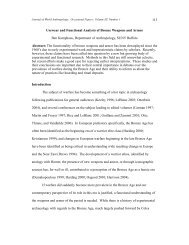
Mechanical Behavior<br />
3a<br />
Human cadaveric<br />
ACL in knee joint
Region 1<br />
“Toe Toe”<br />
Region 2<br />
Region 3<br />
Region 3a<br />
Tensile Response Curve<br />
Crimp: low stiffness; change in slope as collegen fibers<br />
straighten; ligaments become more stiff as more fibers<br />
are recruited<br />
Linear Region: slope = stiffness/Elastic Modulus<br />
Elastic: higher stiffness<br />
Less linear behavior; deformation is permanent<br />
(tearing, stretch); Area of Microfailure;<br />
Microfailure<br />
Ultimate Load: where failure occurs (N)<br />
Energy absorbed to failure: area under the curve<br />
(Nmm Nmm)<br />
Region 4 Ligament ruptures<br />
Region 5<br />
Ligament may appear intact; Fibers to slide under low<br />
loads
Stress Vs. Strain<br />
�� More relevant method of expressing Force vs.<br />
Deformation behavior<br />
�� Region descriptions same as Force vs. Deformation curve<br />
�� Stress (N/mm 2 ) = load per cross-sectional cross sectional area of<br />
sample<br />
�� Strain = percentage change in length
Injuries<br />
�� Occur most frequently during athletic activities<br />
�� Knee injuries<br />
�� ACL<br />
�� Partial or complete tear of ligament caused by quick changes in direction,<br />
slowing down while running, landing a jump, direct contact<br />
�� Symptoms include delayed pain and swelling<br />
�� PCL<br />
�� Sprain of ligament due to overstretching, impact to the front of the knee,<br />
misstep<br />
�� MCL<br />
�� Diagnosis<br />
�� Press gently at knee cap to feel for fluid at the joint<br />
�� X-ray ray<br />
�� MRI<br />
http://orthoinfo.aaos.org/fact/thr_report.cfm?Thread_ID<br />
http:// orthoinfo.aaos.org/fact/thr_report.cfm?Thread_ID=157&topcategory=Knee<br />
=157&topcategory=Knee<br />
http://hcd2.bupa.co.uk/fact_sheets/mosby_factsheets/Knee_ligament_injuries.html<br />
http://hcd2.bupa.co.uk/fact_sheets/mosby_factsheets/Knee_ligament_injuries.html
�� RICE<br />
Healing<br />
�� Rest, Ice, Compression, Elevation<br />
�� Physical therapy<br />
�� Strengthening exercises<br />
�� Range of motion tests<br />
�� Braces<br />
�� Crutches<br />
�� Surgery<br />
http://orthoinfo.aaos.org/fact/thr_report.cfm?Thread_ID<br />
http:// orthoinfo.aaos.org/fact/thr_report.cfm?Thread_ID=157&topcategory=Knee<br />
=157&topcategory=Knee<br />
http://hcd2.bupa.co.uk/fact_sheets/mosby_factsheets/Knee_ligament_injuries.html<br />
http://hcd2.bupa.co.uk/fact_sheets/mosby_factsheets/Knee_ligament_injuries.html
http://12.31.13.115/hwdb/images/hwstd/medical/orthoped/n5550876.jpg
Structure<br />
�� Long cylindrical structures<br />
�� Tightly packed longitudinally running collagen<br />
fibers<br />
�� Nuclei and sparse cytoplasm of fibrocytes<br />
compressed almost flat between them<br />
�� Relatively avascular<br />
�� Slow to heal from trauma injuries<br />
http://adam.about.com/encyclopedia/19089.htm
Attachment<br />
�� Each muscle has two tendons:<br />
�� Proximal: Myotendinous Junction (MTJ)<br />
�� The point of union with a muscle: origin<br />
�� Distal: Osteotendinous Junction (OTJ)<br />
�� The point of union with a bone: insertion
Function<br />
�� Force transmission between muscle and bone<br />
�� Sustain high tensile stresses<br />
�� Conserve substantial muscular energy during<br />
locomotion<br />
�� Energy storage capacity<br />
�� Enables the muscle belly to be at a convenient<br />
distance from joint<br />
�� Satisfies kinematic and damping requirements
Function<br />
�� Withstand tensile forces while retaining<br />
flexibility
Structure<br />
�� Orientations:<br />
�� Parallel to direction of tensile force<br />
�� Larger collagen fibers than in ligaments
Structure of <strong>Tendons</strong>
Collagen Fibers
In Vitro Tensile Test<br />
�� Tissue is elongated to failure<br />
�� Prescribed rate<br />
�� Changes in force are recorded<br />
�� The force is plotted against time<br />
�� Time axis is proportional to elongation<br />
�� Constant strain rate
Response to Tensile Forces<br />
�� Highest tensile strength of any soft tissue<br />
�� Schematic load-elongation load elongation curve with 3 distinct<br />
regions of response to tensile loading:
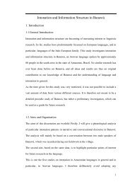
Mechanical Behavior<br />
Energy absorbed to<br />
failure: area under the<br />
curve
Region 1:<br />
“Toe Toe” Region<br />
Region 2:<br />
Linear<br />
Response<br />
Region 3<br />
Region 4:<br />
Macroscopic<br />
Failure<br />
Mechanical Behavior<br />
Collagen fibers straighten (less prominent than in ligaments<br />
because fibers begin more aligned); Continued elongation stiffens stiffens<br />
tissue<br />
Slope represents stiffness; Micro failure occurs at the end;<br />
Elastic recovery at stresses less than 4%<br />
Corresponds to strains of 3-8% 3 8%<br />
Crosslinks fail; Collagen fibers slide past one another; irreversible<br />
changes such as tearing or permanent stretching occurs<br />
Tensile failure of the fibers<br />
Shear failure between the fibers<br />
Once maximum load is surpassed<br />
�� Complete failure occurs rapidly<br />
�� Fibers recoil and blossom<br />
�� Tangled bud at ruptured end<br />
�� Loses Load supporting ability
Mechanical Properties (Cont’d) (Cont d)<br />
�� Greater cross-sectional cross sectional area<br />
�� Larger loads can be applied prior to failure<br />
�� Increased tissue strength<br />
�� Increased Stiffness<br />
�� Longer tissue fibers<br />
�� Greater fiber elongation before failure<br />
�� Decreased tissue stiffness<br />
�� Unaltered tissue strength
Injuries<br />
�� Overuse<br />
�� Spontaneous Rupture<br />
�� Dislocation<br />
�� Thermal Injuries<br />
�� Other Injuries
�� Regeneration<br />
Healing<br />
New tissue identical to normal tissue<br />
�� Structurally<br />
�� New tissue identical to normal tissue<br />
�� Functionally<br />
Soft tissue injury healing<br />
�� Scar repair<br />
�� Soft tissue injury healing<br />
Repair by connective tissue<br />
�� Inferior structural properties<br />
�� Repair by connective tissue<br />
Inferior functional properties<br />
�� Or by their combination<br />
�� Inferior functional properties
Healing Process<br />
�� Inflammation phase<br />
�� From the first day of injury to the fourth<br />
through seventh day<br />
�� Proliferative phase<br />
�� From the seventh through twenty-first twenty first day<br />
�� Maturation or remodeling phase<br />
�� From three weeks to one year
The End
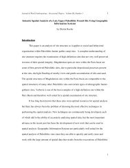
Anterior Cruciate Ligament
ACL: Location
ACL: Flexion<br />
AA--AA’’ –– Anteromedial Anteromedial band band<br />
BB--BB’’ –– Intermediate Intermediate component<br />
component<br />
CC--CC’’ –– Posterolateral aspect aspect of of ligament<br />
ligament
ACL<br />
�� Located between the femur and tibia at the<br />
center of the knee<br />
�� Origin from lateral femoral condyle<br />
�� Insert into the surface of tibial plateau<br />
�� Intracapsular<br />
�� Extrasynovial<br />
Consists of two bundles<br />
�� Anteromedial<br />
�� Posterolateral<br />
�� Consists of two bundles<br />
�� Blood supply originates primarily from femoral<br />
side<br />
http://www.amershamhealth.com/medcyclopaedia/medical/volume%20III%201/CRUCIATE%20LIGAMENT.ASP
Posterior Cruciate Ligament:<br />
Location
PCL: Flexion<br />
A-A’ – Small band<br />
B-B’ – Bulk of the ligament<br />
C-C’ – Anterior meniscofemoral ligament
�� Location<br />
PCL<br />
�� Origin: Medial femoral condyle<br />
�� Insert: Posterior cortical surface of tibia in the sagittal<br />
midline<br />
�� Intimately associated with posterior capsule<br />
�� Covered by Synovium<br />
�� Less susceptible to vascular injury than ACL<br />
�� Blood supply comes from middle geniculate<br />
artery<br />
�� Spiral shape permits tibiofemoral rotation
Medial Collateral Ligament
MCL<br />
�� Primary stabilizer of the medial aspect<br />
�� Location<br />
�� Origin: Medial femoral condyle at the<br />
adductor tubercle<br />
�� Fans out in anterior and posterior directions<br />
�� Insert: Medial side of tibia<br />
�� Has both superficial and deep layer<br />
�� Visually appears like a sailboat
�� Deep Layer<br />
MCL (Cont’d) (Cont d)<br />
�� Originates at adductor tubercle<br />
�� Separates distally<br />
�� Above the joint line<br />
�� Inserts into the medial meniscus<br />
�� Holds the fibro cartilage in place<br />
�� Along the inferior meniscal margin<br />
�� Blends into superficial layer<br />
�� Inserts into the medial tibial diaphysis<br />
�� Has generous blood supply
Lateral Collateral Ligament
Ligament of Humphrey
Ligament of Wrisberg
Other Injuries<br />
�� Tendon Avulsions<br />
�� Tendon Strains<br />
�� Partial Tendon ruptures<br />
�� Lacerations<br />
�� Tendon division<br />
�� Foreign bodies in <strong>Tendons</strong><br />
�� Bite Injuries and Acupunture induced<br />
complications
Tendon Composition
Joint Rotations
Knee Translations
�� Fibrocartilage<br />
(n.) (n.) A kind of<br />
cartilage with a<br />
fibrous matrix and<br />
approaching fibrous<br />
connective tissue in<br />
structure<br />
Fibrocartilage<br />
http://www.kumc.edu/instruction/medicine/anatomy/histoweb/cart/cart12.htm<br />
http://www.brainydictionary.com/words/fi/fibrocartilage164589.html
Fibroblasts<br />
�� Any cell or corpuscle from which connective<br />
tissue is developed<br />
�� Oriented longitudinally with respect to tissue<br />
�� Ovoid or spindle shaped<br />
http://www.digitalnaturopath.com/cond/C136641.html
Fibroblasts<br />
��Secrete Secrete and absorb matrix<br />
elements<br />
��Components:<br />
Components:<br />
��Collagen Collagen<br />
��Proteoglycans<br />
Proteoglycans<br />
��Elastin Elastin (recoil)<br />
��Fibronectin Fibronectin (cell-to (cell to-cell cell<br />
adhesion and migration)
Joint Capsule<br />
�� Fluid sac at joints that holds joints together<br />
�� Creates skeletal system for synovial membrane<br />
�� <strong>Ligaments</strong> or tendons thicken the exterior<br />
�� Protects cartilage, muscles, connective tissue<br />
�� Difficult to identify ligaments and tendons from capsule<br />
in the body<br />
http://physicaltherapy.about.com/cs/disabilities/l/aa111700f.htm<br />
http://web1.tch.harvard.edu/cfapps/A2ZtopicDisplay.cfm?Topic=Anatomy%20of%20a%20Joint
Collagen<br />
�� Different Types:<br />
I>>>III>>V,VI,X,XII<br />
�� Collagen Type I is fibrillar<br />
�� Made up of three<br />
polypeptide chains<br />
�� 2α 1<br />
�� 1α 2<br />
�� Chains are left-handed<br />
left handed<br />
helixes but are wound<br />
together in a right-handed<br />
right handed<br />
helix<br />
http://www.accessexcellence.org/RC/VL/GG/collagen_Elastin.html<br />
http://en.wikipedia.org/wiki/Collagen
�� Hydrogen bonds form<br />
between glycines<br />
(interchain interchain) ) and prolines<br />
and hydroxyprolines<br />
(interchain interchain)<br />
�� Cross-links Cross links between<br />
collagen molecules “head head-<br />
to-tail to tail” and staggered in<br />
parallel<br />
Collagen
�� Hydrogen bonds and<br />
cross-links cross links contribute<br />
to the stability of<br />
each molecule and<br />
aggregation at the<br />
fibril level<br />
�� Result: Structures<br />
extremely resistant<br />
to tensile forces<br />
http://www.orthoteers.co.uk/Nrujp~ij33lm/Orthconntiss.htm<br />
Collagen
Additional Pictures