

World Journal of Clinical Oncology
World Journal of Clinical Oncology
World Journal of Clinical Oncology

Create successful ePaper yourself
Turn your PDF publications into a flip-book with our unique Google optimized e-Paper software.
A B<br />
C D<br />
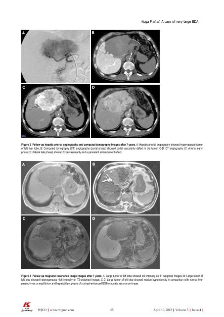
Figure 2 Follow-up hepatic arterial angiography and computed tomography images after 7 years. A: Hepatic arterial angiography showed hypervascular tumor<br />
<strong>of</strong> left liver lobe; B: Computed tomography (CT) angiography (portal phase) showed portal vascularity defect in the tumor; C-D: CT angiography (C: Arterial early<br />
phase; D: Arterial late phase) showed hypervascularity and a persistent enhancement effect.<br />
A B<br />
C D<br />
WJCO|www.wjgnet.com<br />
Koga F et al . A case <strong>of</strong> very large BDA<br />
Figure 3 Follow-up magnatic resonance image images after 7 years. A: Large tumor <strong>of</strong> left lobe showed low intensity on T1-weighted images; B: Large tumor <strong>of</strong><br />
left lobe showed heterogeneous high intensity on T2-weighted images; C-D: Large tumor <strong>of</strong> left lobe showed relative hypointensity in comparison with normal liver<br />
parenchyma on equilibrium and hepatobiliary phase <strong>of</strong> contrast-enhanced EOB magnatic resonance image.<br />
65 April 10, 2012|Volume 3|Issue 4|


