CLINICS IN SPORTS MEDICINE - NYU Langone Medical Center
CLINICS IN SPORTS MEDICINE - NYU Langone Medical Center
CLINICS IN SPORTS MEDICINE - NYU Langone Medical Center

Create successful ePaper yourself
Turn your PDF publications into a flip-book with our unique Google optimized e-Paper software.
Clin Sports Med 25 (2006) 681–702<br />
<strong>CL<strong>IN</strong>ICS</strong> <strong>IN</strong> <strong>SPORTS</strong> MEDIC<strong>IN</strong>E<br />
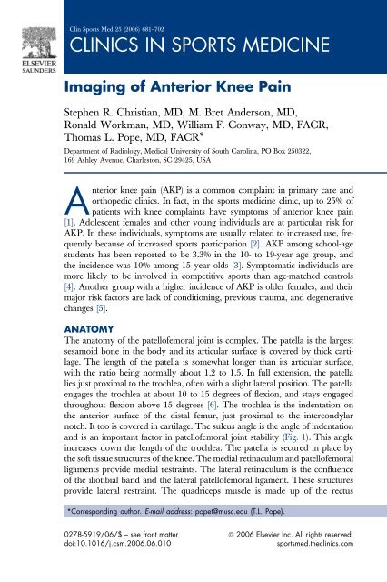
Imaging of Anterior Knee Pain<br />
Stephen R. Christian, MD, M. Bret Anderson, MD,<br />
Ronald Workman, MD, William F. Conway, MD, FACR,<br />
Thomas L. Pope, MD, FACR*<br />
Department of Radiology, <strong>Medical</strong> University of South Carolina, PO Box 250322,<br />
169 Ashley Avenue, Charleston, SC 29425, USA<br />
Anterior knee pain (AKP) is a common complaint in primary care and<br />
orthopedic clinics. In fact, in the sports medicine clinic, up to 25% of<br />
patients with knee complaints have symptoms of anterior knee pain<br />
[1]. Adolescent females and other young individuals are at particular risk for<br />
AKP. In these individuals, symptoms are usually related to increased use, frequently<br />
because of increased sports participation [2]. AKP among school-age<br />
students has been reported to be 3.3% in the 10- to 19-year age group, and<br />
the incidence was 10% among 15 year olds [3]. Symptomatic individuals are<br />
more likely to be involved in competitive sports than age-matched controls<br />
[4]. Another group with a higher incidence of AKP is older females, and their<br />
major risk factors are lack of conditioning, previous trauma, and degenerative<br />
changes [5].<br />
ANATOMY<br />
The anatomy of the patellofemoral joint is complex. The patella is the largest<br />
sesamoid bone in the body and its articular surface is covered by thick cartilage.<br />
The length of the patella is somewhat longer than its articular surface,<br />
with the ratio being normally about 1.2 to 1.5. In full extension, the patella<br />
lies just proximal to the trochlea, often with a slight lateral position. The patella<br />
engages the trochlea at about 10 to 15 degrees of flexion, and stays engaged<br />
throughout flexion above 15 degrees [6]. The trochlea is the indentation on<br />
the anterior surface of the distal femur, just proximal to the intercondylar<br />
notch. It too is covered in cartilage. The sulcus angle is the angle of indentation<br />
and is an important factor in patellofemoral joint stability (Fig. 1). This angle<br />
increases down the length of the trochlea. The patella is secured in place by<br />
the soft tissue structures of the knee. The medial retinaculum and patellofemoral<br />
ligaments provide medial restraints. The lateral retinaculum is the confluence<br />
of the iliotibial band and the lateral patellofemoral ligament. These structures<br />
provide lateral restraint. The quadriceps muscle is made up of the rectus<br />
*Corresponding author. E-mail address: popet@musc.edu (T.L. Pope).<br />
0278-5919/06/$ – see front matter ª 2006 Elsevier Inc. All rights reserved.<br />
doi:10.1016/j.csm.2006.06.010 sportsmed.theclinics.com
682 CHRISTIAN, ANDERSON, WORKMAN, ET AL<br />
Fig. 1. Sulcus angle, A.<br />
femoris and vastus muscles. The patella tendon attaches the patella to the tibial<br />
tubercle. The patella functions by increasing the mechanical advantage of<br />
the quadriceps muscle group [6].<br />
The infrapatellar fad pad, or Hoffa’s fat pad, is an extrasynovial but intracapsular<br />
structure located posterior to the patellar tendon and joint capsule but anterior<br />
to the knee joint synovium. It is directly attached to the anterior meniscal<br />
horns inferiorly. Rests of synovial tissue are present within Hoffa’s fat pad.<br />
PHYSICAL EXAM<strong>IN</strong>ATION<br />
The patient’s history and clinical presentation provide initial clues to the cause of<br />
AKP. Classic symptoms are pain behind the patella brought on by physical activity<br />
such as walking up or down stairs [7]. The physical examination should focus<br />
on evaluating alignment, range of motion, bursitis, and effusion. Multiple maneuvers<br />
can be performed to check for internal derangement. The patella is examined<br />
with the knee in extension. If the examiner can displace the patella laterally by<br />
25% of its diameter or more, it is considered to be ‘‘subluxable’’ [8]. However,<br />
the malalignment often is subtle, and physical exam can be difficult and confusing<br />
in many patients. Imaging plays an important role in evaluation, not only in<br />
searching for malalignment, but for other sources of pain as well.<br />
IMAG<strong>IN</strong>G<br />
The standard radiographic evaluation of the knee includes frontal, lateral, and<br />
axial (sunrise) views of the knee. The axial view is usually obtained in 30 degrees<br />
of flexion. Computed tomography (CT) and MRI are also commonly<br />
used to evaluate anterior knee pain, especially in complex or refractory cases.<br />
Both of these modalities can be performed using standard protocols. CT is useful<br />
for osseous evaluation, such as in trauma or in some cases of possible malalignment.<br />
MRI is a more powerful modality, as it can diagnose cartilage and<br />
soft tissue abnormalities to greater effect than CT or radiography. Both CT<br />
and MRI can be used in dynamic modes, which can be useful for tracking abnormalities<br />
of the patella. Nuclear scinitigraphy is somewhat limited in its
IMAG<strong>IN</strong>G OF ANTERIOR KNEE PA<strong>IN</strong><br />
usefulness for anterior knee pain, however it can have a role, especially in cases<br />
of occult fracture and tumors.<br />
DIFFERENTIAL DIAGNOSIS<br />
The differential for AKP is broad. The major differential considerations are<br />
listed in Box 1.<br />
S<strong>IN</strong>D<strong>IN</strong>G-LARSEN-JOHANSSON SYNDROME<br />
Originally described by Norwegian physician Christian Magnus Falsen Sinding-Larsen<br />
and Swedish surgeon Sven Christian Johansson, Sinding-Larsen-<br />
Johansson syndrome (SLJS) is defined as ‘‘apophysitis of the distal pole of<br />
the patella’’ and is considered one of the osteochondroses [9]. This condition<br />
of the distal patella and proximal patellar tendon is quite similar to Osgood-<br />
Schlatter disease and primarily affects athletically active adolescents between<br />
10 and 14 years of age with prevalence in boys. This entity typically presents<br />
as pain and tenderness with occasional swelling over the inferior pole of the<br />
patella brought on by overuse or trauma [10].<br />
Contusion or tendinopathy of the proximal patellar tendon creates a traction<br />
phenomenon followed by calcification and ossification, and patellar fracture or<br />
Box 1: Differential Considerations for AKP<br />
Patellar tendon causes<br />
Tendinopathy/rupture<br />
Osgood-Slatter<br />
Sindig-Larsen-Johanssen<br />
Patella<br />
Chondromalacia<br />
Patello femoral OA<br />
Stress fracture<br />
Bipartite patella<br />
Osteochondritis<br />
Intra-articular pathology/Hoffa’s fat pad<br />
Meniscal tear/cysts<br />
Plica syndromes<br />
Hoffa’s syndrome<br />
Bursitis<br />
Prepatellar<br />
Pes ancerine<br />
683
684 CHRISTIAN, ANDERSON, WORKMAN, ET AL<br />
avulsion can produce one or multiple ossification sites. SLJS is most likely to occur<br />
in an active adolescent during ‘‘growth spurts’’ when the tendon cannot keep pace<br />
with the growing tibia, resulting in a relative shortening and traction on the immature<br />
lower pole of the patella. The natural duration of the disease is 3 to 12<br />
months and usually requires only rest and conservative management [11].<br />
Radiographs of the knee are frequently normal with varying calcification and<br />
ossification of the lower pole of the patella. Findings of osseous fragmentation<br />
of the inferior patella on knee radiography support the diagnosis in a patient<br />
with history and physical examination suggestive of SLJS (Fig. 2) [11].<br />
JUMPER’S KNEE<br />
Although often referred to as patellar tendonitis, a more accurate description of<br />
‘‘Jumper’s knee,’’ based on histologic studies, is of overuse tendinopathy. This<br />
is the most common tendinopathy in skeletally mature athletes, occurring in up<br />
to 20% of jumping athletes. Jumper’s knee primarily affects the proximal posterior<br />
fibers of the patellar tendon and is a cause of significant functional disability<br />
in professional and recreational athletes [12]. Biomechanically, to squat and<br />
land softly from a jump, the quadriceps muscle lengthens in eccentric contraction<br />
and creates high tension on the patellar tendon. Patellar tendinopathy occurs<br />
secondary to repetitive microtrauma caused by tendon overload without<br />
adequate repair. This overuse can lead to pain, tenderness, swelling, and decreased<br />
performance.<br />
Most commonly, an athlete will present with anterior knee pain of insidious<br />
onset that is aggravated by activity (jumping, squatting, kneeling, and going<br />
down stairs). Symptoms can range from pain after activity to pain that persists<br />
Fig. 2. Sinding-Larsen-Johannsen Syndrome (SLJS). There is fragmentation of the distal aspect<br />
of the patella, consistent with SLJ syndrome in the right clinical setting.
IMAG<strong>IN</strong>G OF ANTERIOR KNEE PA<strong>IN</strong><br />
685<br />
throughout an activity. Many individuals experience no decrease in performance,<br />
while severely affected individuals may suffer a substantial decrease<br />
in athletic ability. On physical examination, localized tenderness over the inferior<br />
patella/proximal patellar tendon is commonly found [13].<br />
Imaging of patellar tendinopathy remains somewhat controversial. Radiographs<br />
are occasionally useful in identifying ossification of the tendon or associated<br />
osseous abnormalities, but ultrasonography and MRI are the modalities<br />
of choice for diagnosis. Ultrasonography, the accepted modality in much of<br />
Europe, demonstrates lower pole irregularity, fragmentation, chondral changes,<br />
and thickening of the tendon insertion at the patella [11]. Ultrasound evaluation<br />
of tendinopathy is quite dependent on equipment and operator experience. MRI<br />
also demonstrates tendon thickening with increased signal particularly on<br />
spin-echo and gradient-echo imaging. T2-weighted imaging best demonstrates<br />
partial tears with high signal intensity in the area of injury. Although the exact<br />
modality of choice for evaluation of this condition is debatable, ultrasound and<br />
MR have both been proven effective (Fig. 3) [13].<br />
PATELLAR AND QUADRICEPS TENDON RUPTURE<br />
Patellar tendon rupture, an overall infrequent occurance, is the third most common<br />
injury to the extensor mechanism of the knee after patellar fracture and<br />
quadriceps rupture. Rupture usually occurs unilaterally as a result of athletic<br />
injury in a patient younger than 40 years of age. Typically, an abrupt eccentric<br />
contraction of the quadriceps as the athlete lands with the knee flexed and foot<br />
planted will tear the tendon at the osseotendinous junction. In the setting of<br />
longstanding systemic inflammatory disease, diabetes mellitus, and chronic renal<br />
failure, bilateral rupture can occur [14]. Patellar tendon rupture can also be<br />
Fig. 3. Jumper’s knee. T2-weighted fat-saturated image shows increased signal intensity in the<br />
proximal patellar tendon.
686 CHRISTIAN, ANDERSON, WORKMAN, ET AL<br />
seen in patients who have had the central third of the tendon used as allograft<br />
for repair of the anterior cruciate ligament.<br />
Acute rupture is associated with immediate debilitating pain accompanied by<br />
a ‘‘pop’’ or tearing sensation with inability to bear weight. Examination reveals<br />
swelling/tenderness of the anterior knee, ecchymosis, hemarthrosis, and patella<br />
alta (see later discussion for definition) with a palpable gap in the extensor<br />
mechanism [15].<br />
Diagnosis can usually be made by physical exam and radiographs. Contralateral<br />
images can be helpful in assessing patellar height. Even if the diagnosis<br />
of patellar tendon rupture is clinically obvious, radiographic evaluation is recommended<br />
to evaluate for concomitant injury. The classic finding is patella<br />
alta on the lateral radiograph. If the diagnosis cannot be made on physical<br />
and plain radiographic examination, MRI is the modality of choice and easily<br />
demonstrates discontinuity of the tendon fibers, hemorrhage and edema<br />
(Fig. 4A) [14].<br />
Quadriceps tendon injury is more often seen on older individuals. This injury<br />
can be difficult to diagnose clinically, and misdiagnoses are common<br />
[16]. Complete tear is often the result of repetitive microtrauma. MR is useful<br />
in demonstrating partial or complete tears. On MR, a complete tear shows discontinuity<br />
of the tendon, hemorrhage, and edema, which is manifested as increased<br />
signal on T2-weighted sequences [17] (Fig. 4B).<br />
CHONDROMALACIA PATELLAE<br />
As indicated by its Greek and Latin roots, chondromalacia patellae (CP) is<br />
a condition characterized by abnormal softening of the cartilage along the<br />
Fig. 4. (A) Patellar tendon rupture. T2-weighted sequence shows patella alta with<br />
discontinuity of the patellar tendon and associated edema. (B) Quadriceps tendon rupture.<br />
T2-weighted fat-saturated sagittal image shows discontinuity of the quadriceps tendon with<br />
associated edema.
IMAG<strong>IN</strong>G OF ANTERIOR KNEE PA<strong>IN</strong><br />
687<br />
undersurface of the patella. Common causes include trauma, repeated stress, and<br />
patellofemoral instability. Also referred to as ‘‘runner’s knee,’’ this problem is<br />
a cause of anterior knee pain and is often seen in young, otherwise healthy individuals.<br />
The disorder affects women more often than men and is thought to be<br />
a result of anatomical differences in which more lateral force is applied to the<br />
female patella. This results in increased lateral tracking of the patella. With proper<br />
therapy, early CP can be reversed; however, if left unchecked its changes become<br />
advanced, chronic, and may progress to patellofemoral osteoarthritis.<br />
Multiple imaging modalities and techniques can be used to evaluate the patient<br />
with suspected chondromalacia patellae. Conventional radiography, CT arthrography,<br />
MR arthrography, and conventional MRI are available options.<br />
Conventional radiographs are relatively insensitive in evaluating for cartilage<br />
loss, except when it is severe. CT arthrography may demonstrate fissuring and<br />
foci of cartilage loss, but this technique is invasive and involves ionizing radiation.<br />
MR arthrography has also been shown to be sensitive and specific, but like CT<br />
arthrography, it is invasive [18]. Conventional MRI can show focal cartilage surface<br />
irregularities, as well as provide excellent soft tissue differentiation and reveal<br />
deeper internal cartilaginous derangement [19]. However, some studies have also<br />
shown relative insensitivity of conventional MRI in detecting early changes of CP<br />
[20]. Although invasive, MR arthrography with spoiled gradient recalled acquisition<br />
(SPGR) has demonstrated the high sensitivity for detecting early stage<br />
CP in multiple studies [21,22]. The MR findings typically show focal signal abnormalities<br />
or focal contour defects along the patellar cartilage on T2WI. These<br />
abnormalities can progress to patellofemoral osteoarthritis (PO) if left untreated<br />
(Fig. 5). A useful four-level grading scheme for CP based on arthroscopic and<br />
MRI appearance is presented in Box 2 and Fig. 5B.<br />
PATELLOFEMORAL OSTEOARTHRITIS<br />
Patellofemoral osteoarthritis is an extremely common cause of anterior knee<br />
pain. This is encountered primarily in older individuals, but can be seen in<br />
younger patients with accelerated degenerative changes brought on by comorbidities<br />
such as obesity. The classic radiographic features of PO include loss of<br />
articular cartilage with joint space narrowing, subchondral sclerosis and/or cyst<br />
formation, and osteophyte formation along the posterior margin of the patella.<br />
The symptoms commonly seen with PO include morning knee joint stiffness,<br />
loss of mobility, pain with ambulation (particularly walking up an incline or<br />
along a flight of stairs), and weakness about the knee joint.<br />
Early clinical signs and symptoms may precede detectable conventional<br />
radiographic abnormalities. MRI, however, is especially sensitive to soft tissue<br />
and bone abnormalities. Fast spin echo T2 fat-saturated sequences are sensitive<br />
and specific for focal cartilage abnormalities [23]. MR findings in PO include<br />
cartilage surface thinning and irregularity, fine delineation of focal articular<br />
cartilage loss, or cartilage fissuring. Osteophytes are also common. There<br />
may be high T2 signal changes in the patella if there is significant marrow<br />
edema (Fig. 6).
688 CHRISTIAN, ANDERSON, WORKMAN, ET AL<br />
Fig. 5. Chondromalacia patella. (A) T2-weighted image shows focal abnormal high signal<br />
within the patellar cartilage. (B) Diagram of CP grading scheme, pathologic findings. (From<br />
Conway WF, Hayes CW, Loughran T, et al. Cross-sectional imaging of the patellofemoral joint<br />
and surrounding structures. Radiographics 1991;11:195–217; with permission.)<br />
PREPATELLAR AND PES ANSER<strong>IN</strong>E BURSITIS<br />
The term ‘‘bursa’’ is Latin for ‘‘pouch’’ and is a synovium-lined sac that helps<br />
lubricate structures that move along one another. Bursae facilitate motion by<br />
reducing friction, and they can become symptomatic when inflamed, damaged,<br />
or infected. Prepatellar and pes anserine bursitis are commonly encountered<br />
causes of anterior knee pain.<br />
The prepatellar bursa is a superficial bursa located between the skin and the<br />
anterior patella. Inflammation of this structure, also known as ‘‘Housemaid’s<br />
knee,’’ results in prepatellar bursitis and is a common cause of AKP in those<br />
individuals who frequently kneel or spend large amounts of time on their knees<br />
Box 2: Grading of Chondromalacia Patella (modified Shahriaree)<br />
[60]<br />
Grade 1. Arthroscopic findings: Softening of articular cartilage; T1-weighted MR<br />
findings: Partial width focal decreased signal areas of patellar cartilage<br />
on T1-weighted sequences, not extending to cartilage surface.<br />
Grade 2. Arthroscopic findings: ‘‘Blistering’’ of articular cartilage with surface<br />
abnormality; T1-weighted MR findings: Focal area of sharply marginated<br />
decreased signal extending to the articular surface.<br />
Grade 3. Arthroscopic findings: Cartilage fibrillation; T1-weighted MR findings:<br />
Indistinct focal areas of decreased signal extending to the articular surface.<br />
Grade 4. Arthroscopic findings: Full thickness cartilage ulceration; T1 weighted<br />
MR fingings: Full-thickness decreased signal abnormalities with associated<br />
subchondral bone low signal changes.
IMAG<strong>IN</strong>G OF ANTERIOR KNEE PA<strong>IN</strong><br />
689<br />
Fig. 6. Patellofemoral osteoarthritis. Radiograph demonstrates osteophytes at the PF articulation.<br />
such as gardeners and carpet layers. Radiography may show prepatellar soft<br />
tissue swelling. Symptoms and physical exam findings may include pain that<br />
increases with ambulation or kneeling, decreased range of motion, and erythema/edema<br />
along the lower pole of the patella. MRI will usually show a prepatellar<br />
fluid collection with low T1/high T2 signal.<br />
Pes anserine bursitis is an inflammation of the conjoined insertion of the sartorius,<br />
gracilis, and semitendinosus muscle tendons along the proximal medial<br />
aspect of the tibia [24]. This entity is commonly associated with degenerative<br />
joint disease of the knee, but can also be seen in younger, active individuals<br />
who engage in sports requiring frequent side-to-side movements. The most specific<br />
physical exam finding is pain over the proximal anterior medial tibia<br />
where the conjoined tendons insert. On T1WI, there is usually a low intensity<br />
fluid collection in the region of the pes anserinus along the medial tibial metaphysis,<br />
which shows relatively high homogeneous signal on T2WI (Fig. 7).<br />
Fig. 7. (A,B) Prepatellar bursitis. T2-weighted images demonstrates fluid signal intensity superficial<br />
to the patellar tendon, consistent with prepatellar bursitis.
690 CHRISTIAN, ANDERSON, WORKMAN, ET AL<br />
BIPARTITE PATELLA AND PATELLAR STRESS FRACTURE<br />
The patella is normally one bone, but in approximately 1% to 2% of the population<br />
the patella develops as two unfused ossification centers. This condition,<br />
known as bipartite patella, is a variant of normal and affects men more than<br />
women. These two bones are not separate, but are connected by thick fibrous tissue.<br />
The patient with a bipartite patella is usually asymptomatic but can experience<br />
pain with standing or jumping. The classic bipartite patella appears as<br />
a small unfused fragment of the upper outer margin of the larger, main patellar<br />
fragment. On radiographs, inexperienced physicians can mistake the bipartite<br />
patella as a patellar fracture. Sometimes stress fracture superimposed on bipartite<br />
patella can occur and is a potentially difficult clinical entity to recognize (Fig. 8).<br />
MRI can aid with diagnosis by showing increased signal within the marrow on<br />
fat-suppressed T2WI compatible with marrow edema in cases of stress fracture.<br />
Rest and strengthening exercises are usually sufficient treatment for uncomplicated<br />
knee pain in patients with bipartite patella; however, if there is avulsion<br />
at the fibrous connection, or a stress fracture, immobilization may be necessary.<br />
ACUTE PATELLAR DISLOCATION<br />
Acute lateral patellar dislocation can occur as a result of knee trauma and is<br />
most often seen in young athletes. The dislocation often reduces spontaneously<br />
without treatment, and the patient may not be aware that it has occurred. After<br />
such an event, the clinical examination is nonspecific, and as many as 75% of<br />
patients are misdiagnosed on initial physical exam and radiographs [25]. MR<br />
has been useful for diagnosis as several specific findings have been described.<br />
These include hemarthrosis/effusion, lateral femoral condyle and medial patellar<br />
facet bone contusions, osteochondral injury, and medial retinacular injury.<br />
The medial patello-femoral ligament, which has been identified as the major<br />
Fig. 8. Bipartite patella. (A) Gradient echo MR image shows cleft in the superior lateral<br />
patella without evidence for edema. Corticated margins suggest the diagnosis. (B) Radiograph<br />
bipartite patella.
IMAG<strong>IN</strong>G OF ANTERIOR KNEE PA<strong>IN</strong><br />
691<br />
restraint to lateral dislocation of the patella, is often injured when patellar<br />
dislocation occurs (Fig. 9) [26].<br />
DISEASES OF HOFFA’S FAT PAD<br />
A variety of disease entities can affect this structure and cause AKP, including<br />
impingement syndromes, postarthroscopy changes, plica syndromes, and mass<br />
lesions [27–30].<br />
Hoffa’s Fat Pad Syndrome<br />
Acute or repetitive trauma to Hoffa’s fat pad can result in edema and hemorrhage.<br />
The resultant changes of enlargement put the fat pad at risk for impingement<br />
between the femur and tibia. Fibrosis and anterior knee pain can result<br />
[27]. This is called Hoffa’s disease or syndrome. Acutely, there is high T2 signal<br />
and mass effect with the fat pad. Chronically, fibrosis appears dark on both<br />
T1- and T2-weighted images [27].<br />
Plica Syndromes<br />
A possibly related entity to Hoffa’s fat pad syndrome is an abnormal infrapatellar<br />
plica. The infrapatellar plica is a synovial fold that runs parallel to the<br />
anterior cruciate ligament (ACL) in the intercondylar notch. It travels over and<br />
through the superior aspect of the Hoffa’s fat pad. Normally it shows low signal<br />
similar to that of ligaments. This structure can be injured, resulting in abnormal<br />
signal in both the plica and the superior aspect of Hoffa’s fat pad. Injury to this<br />
plica is logically associated with injury to the fat pad, and thus the two entities<br />
may appear together and have some similar imaging findings. Clinical differentiation<br />
may also be difficult [31].<br />
Other plica syndromes, including medial, lateral, and suprapatellar, are described.<br />
The mediopatellar plica is most often symptomatic. It extends from<br />
the medial joint wall to the synovium covering Hoffa’s fat pad (Fig. 10).<br />
When it is prominent, it can be impinged upon by the medial condyle of the<br />
Fig. 9. Transient patellar dislocation. T2-weighted fat-saturated images of the knee show<br />
marrow edema in the lateral femoral condyle (A) and the medial patellar facet (B).
692 CHRISTIAN, ANDERSON, WORKMAN, ET AL<br />
Fig. 10. Medial patella plica. T2-weighted sagittal image shows a low signal band in the<br />
suprapatella bursa consistent with a medial plica.<br />
femur and the patella. This can result in chronic irritation and injury, with an<br />
increase in thickening, edema, and further impingement. The plica can then become<br />
fibrotic and cause damage to the articular cartilage and synovitis. Symptoms<br />
range from crepitation and swelling to joint pain medial to the patella [32].<br />
Patellar Tendon Lateral Femoral Condyle Friction Syndrome<br />
Patellar tendon lateral femoral condyle friction syndrome, so named by Chung<br />
and colleagues [33], is related to the clinical disease known as fat pad impindgement<br />
syndrome. Patients present with anterior knee pain, more pronounced at<br />
the inferior aspect of the patella. Abnormal increased T2 signal is seen in the inferolateral<br />
aspect of the patellofemoral joint and with possible involvement of the<br />
lateral fat pad. Cystic changes in the fat pad and enhancement may occur.<br />
Mass Lesions<br />
Several symptomatic mass lesions can occur in Hoffa’s fat pad that may cause<br />
symptoms. Localized nodular synovitis is the localized form of PVNS. It most<br />
commonly occurs outside the knee, but can occur in Hoffa’s fat pad. MR shows<br />
a mass-like lesion with variable signal characteristics. Hemosiderin will often be<br />
present with its associated artifacts on gradient echo sequences [28]. Para-articular<br />
chondroma arises from connective tissue due to cartilaginous metaplasia<br />
and most commonly occurs in or near Hoffa’s fat pad. The MR appearance<br />
is that of a lobulated mass obliterating the normal high T1 signal fat pad inferior<br />
to the patella. The lesion shows increased T2 signal and will enhance after<br />
IV gadolinium administration. Primary intra-articular sarcoma has been reported<br />
but is extremely rare. Imaging appearances are often nonspecific and biopsy<br />
is often required for diagnosis [34].<br />
Postsurgical Changes<br />
The major portals used in arthroscopy are anterolateral, anteromedial, and medial<br />
and all of these can cause fibrosis within Hoffa’s fat pad [28]. The fibrosis<br />
appears as well-defined strands of low signal (on T1 and T2 sequences) tissue<br />
coursing through the high-signal fat. Artifact from metallic fragments can also
IMAG<strong>IN</strong>G OF ANTERIOR KNEE PA<strong>IN</strong><br />
693<br />
be seen after arthroscopy [27]. These changes on MR are not usually thought<br />
of as a cause of pain, but are important to recognize and not be misdiagnosed.<br />
Patient’s who have undergone ACL repair may present with a postoperative<br />
fibrotic complication known as localized anterior arthrofibrosis, or ‘‘Cyclops’’<br />
lesion. This complication can result in pain and limited extension of the knee<br />
and is thought to occur from impingement of the anterior intercondylar notch<br />
on a graft that has been positioned too far anterior. The MR appearance is<br />
a low to intermediate signal intra-articular structure anterior to the graft.<br />
MRI has been shown to be sensitive for detection of the lesion [35].<br />
PATELLOFEMORAL PA<strong>IN</strong> SYNDROME<br />
Patellofemoral pain syndrome has been suggested as a diagnosis of exclusion<br />
reserved for patients with anterior knee pain without one of the conditions described<br />
above. Causes of this variety of anterior knee pain are somewhat controversial.<br />
Fulkerson points out that there are six main tissues to consider when<br />
looking for the etiology of patellofemoral pain. These include subchondral<br />
bone, synovium, retinaculum, skin, muscle, and nerve. He believes that the<br />
most common causes of pain from an orthopedic standpoint are overuse, patellofemoral<br />
malalignment, and trauma [36].<br />
MALALIGNMENT EVALUATION—TRADITIONAL <strong>IN</strong>DICES<br />
Several measurements obtained from the axial, or sunrise, view have traditionally<br />
been used to evaluate for malalignment. There are three main radiographic<br />
patterns of malalignment (described with CT). These include subluxation of<br />
the patella with and without tilt, and tilt without subluxation [37]. Conditions<br />
that lead to malalignment and influence patellofemoral stability, such as depth<br />
of the trochlea, can be measured with imaging. The most common indices described<br />
in the literature are the lateral patellofemoral angle (tilt), the congruence<br />
angle, and lateral patellar displacement [38–40]. Other measurements that have<br />
been shown to potentially have merit are the Q angle and the trochlear-tubercle<br />
distance [41,42].<br />
The lateral PF angle is calculated on an axial radiograph obtained at 20 degrees<br />
of flexion by measuring the angle of the lateral patellar facet compared<br />
with a line drawn across the femoral condyles. A study by Laurin and<br />
colleagues [40] showed that 97% of normal patellae open laterally. If the angle<br />
opens medially, or is parallel, then the patella is tilted (external rotation)<br />
(Fig. 11) [43]. Lateral patellar displacement is measured by comparing the medial<br />
margin of the patella on an axial view to the medial femoral condyle. If is<br />
more than 1 mm lateral in relation to the medial condyle apex, it is considered<br />
subluxed (Fig. 12) [39,43].<br />
The congruence angle, as described by Merchant [38], is measured from a 45<br />
degree of flexion axial film. The measurement is made by bisecting the sulcus<br />
angle to create a zero reference line. Then a line is drawn from the lowest point<br />
on the patella to the sulcus angle point. The angle created is then measured.<br />
The ‘‘normal’’ value is –6 11 degrees. Values outside this range are an
694 CHRISTIAN, ANDERSON, WORKMAN, ET AL<br />
Fig. 11. Lateral patellofemoral angle. The angle of the lateral patellar facet compared with<br />
a line drawn across the femoral condyles. Angle A is the lateral patellofemoral angle.<br />
indicator of patellar subluxation (Fig. 13) [37]. These values may also be obtained<br />
with CT and MR.<br />
The Q angle is a measure of the angle formed between a line drawn from the<br />
tibial tubercle and a line drawn from the middle of the patella and anterior superior<br />
iliac spine. The normal value is 15 degrees [42]. An increased Q angle<br />
implies that the tibial tubercle is more lateral than normal. Thus, the patella experiences<br />
a lateral force with contraction of the quadriceps, predisposing it to<br />
lateral subluxation or dislocation. The Q angle can be measured by scout<br />
CT image or physical exam.<br />
Fig. 12. Lateral patellar displacement. This is measured by comparing the medial margin of<br />
the patella on an axial view to the medial femoral condyle. The distance A is lateral patellar<br />
displacement.
IMAG<strong>IN</strong>G OF ANTERIOR KNEE PA<strong>IN</strong><br />
695<br />
Fig. 13. Congruence angle. (A) The measurement is made by bisecting the sulcus angle to<br />
create a zero reference line. Then a line is drawn from the lowest point on the patella to the<br />
sulcus angle point. The angle created (D) is then measured. (B) CA¼ congruence angle.<br />
The tibial tubercle distance (T-T distance) can be measured with axial imaging,<br />
and can substitute for the Q angle. This is a measure of the tibial tubercle in relation<br />
to the trochlear nadir. Two sagittal lines are drawn, one through the tibial<br />
tubercle, the other through the bottom of the trochlear groove. The lines’ difference<br />
in position in the axial plane is the T-T distance. One study showed high<br />
specificity for maltracking if the tibial tubercle distance was 2 cm or greater [41].<br />
LATERAL RADIOGRAPHS OF THE KNEE<br />
Recently, several studies have described the limitations of the axial view for patellar<br />
alignment evaluation. Walker and colleagues [44] questioned the value of<br />
the axial view and compared the axial view with CT of the patellofemoral joint.<br />
They found that these two modalities often give conflicting results and concluded<br />
the axial view to be of limited value due to lack of sensitivity, as<br />
‘‘even florid examples [of maltracking] must be missed.’’ Many researchers<br />
now advocate using the lateral view of the knee, in various degrees of flexion,<br />
to evaluate for alignment at the patellofemoral joint. The basis of this argument<br />
rests on the fact that in most knees, the patella is fully engaged in the trochlea<br />
by 30 degrees of flexion. Many patients with mild subluxation or tilt are not<br />
diagnosed on axial images as their abnormally aligned patellae have corrected<br />
at the angle the axial images are obtained. It is not possible to obtain an axial<br />
image at less than 20 degrees of flexion; special equipment and elaborate technique<br />
is required to obtain these images at angles less than 30 degrees.<br />
Maldague and Malghem [45] have shown how the lateral radiograph can be<br />
used to evaluate for malposition of the patella. Dupont and Guier [46] added to<br />
their grading scheme (Fig. 14). There is a technical advantage to this technique.<br />
A true lateral view can be obtained in full extension or in varying degrees of
696 CHRISTIAN, ANDERSON, WORKMAN, ET AL<br />
Fig. 14. Schematic representation of patellar position on lateral views, according to the<br />
degree of external rotation (tilt), modified Maldague-Malghem’s classification in 4 stages.<br />
(A) Stage 1 (normal position): both lines (central ridge, CR; lateral edge, LE) are concave<br />
posteriorly and separated by 5 to 10 mm. The anterior line is the lateral edge, the posterior<br />
line the central ridge. (B) Stage 2 (false lateral profile as described by Maldague and<br />
Malghem, or minor lateral patellar subluxation): both lines are superimposed. Only one<br />
straight line is visible. (C) Stage 3 (overwhelming lateral profile, or pronounced lateral subluxation):<br />
the anterior line is the central ridge, the posterior line is the lateral aspect of the patella<br />
and is convex. (D) Stage 4. The central ridge is no longer visible. The patella often covers the<br />
anterior cortex of the femoral metaphysis and appears ovoid in shape [46]. (From Dupont JY,<br />
Guier CA. Comparison of three standard radiologic techniques for screening of patellar subluxations.<br />
Clin Sports Med 2002;21(3):389–401; with permission.)<br />
flexion to the point where the patella engages the trochlea. Murray and colleagues<br />
[47] compared lateral radiographs (taken in 15 to 30 degrees of flexion)<br />
of the knee with axial views and found the lateral films had increased sensitivity<br />
and specificity for correlation to patellofemoral pain, previous dislocation, and<br />
malalignment. Similar results were obtained by Dupont and Guier [46].<br />
The lateral radiograph can also allow assessment of the condition of the trochlea<br />
and patellar height. The assessment of patella height is important as patella alta<br />
is associated with an increased risk for dislocation. A simple method is the modified<br />
Insall-Salvati method, which takes into account the wide variety of patellar<br />
shapes that are encountered [48]. This method is highly reproducible and does<br />
not rely on exact positioning. The patellar articular surface is compared with<br />
the length of the patellar tendon from the tibial tubercle to the most inferior aspect<br />
of the patellar articular surface. This ratio of patellar tendon/patellar length is<br />
measured and a normal value is less than 2 (Fig. 15). Trochlear depth, accurately<br />
measured by the lateral knee radiograph, is an important factor in patellar malalignment.<br />
A shallow trochlea places one at risk for dislocation and subluxation.<br />
Malghem and Maldague [49] demonstrated that a depth in the proximal trochlea<br />
of less than 5 mm increases the risk of instability.
IMAG<strong>IN</strong>G OF ANTERIOR KNEE PA<strong>IN</strong><br />
697<br />
Fig. 15. Modified Insall-Salvati method to determine patellar height. The ratio of B/A should<br />
normally be less than 2.<br />
CT and MR<br />
MR and CT can both be used to study the patellofemoral articulation and each<br />
has intrinsic advantages and disadvantages. Both modalities have the advantage<br />
of being able to study the knee directly in degrees of flexion less than<br />
30 degrees, without overlapping structures adding confusion. A static study involves<br />
acquiring axial images at a fixed degree of flexion, usually less than 20<br />
degrees. Kinematic studies acquire images while the knee moves from flexion<br />
to extension, and the images acquired can be viewed in a cine mode. These modalities<br />
may be useful in patients with symptoms that suggest malalignment but<br />
without evidence of the diagnosis by radiographs or physical exam [50,51].<br />
Schutzer and colleagues [37] in 1986 evaluated the patellofemoral joint with<br />
CT. They described three malalignment patterns. These include subluxation<br />
without tilt, subluxation with tilt, and tilt without subluxation. Their study demonstrated<br />
that in asymptomatic normal controls, the patella is either centered or<br />
slightly medially displaced by 10 degrees of flexion. Therefore, a patella is considered<br />
subluxed if the congruence angle is greater than 0 with the knee in 10 degrees<br />
or more of flexion. The patellar tilt angle in asymptomatic controls always was<br />
greater than 8 degrees and usually was more than 15 degrees. The authors conclude<br />
that 8 degrees is the lower limit of normal tilt on CT [37].<br />
Static MR techniques, such as the one described by Koskinen and colleagues<br />
[52], can be used to obtain accurate traditional patellofemoral indices such as<br />
lateral patellar tilt, lateral patellofemoral angle, lateral patellar displacement,<br />
sulcus angle, and congruence angle. The advantages of static MR over CT include<br />
its ability to characterize the status of the soft tissues of the knee. Patellar<br />
height can also be assessed reliably with MR. Miller and colleagues [53] compared<br />
sagittal MR images and lateral radiographs, and found good correlation
698 CHRISTIAN, ANDERSON, WORKMAN, ET AL<br />
between the two modalities. They compared patellar tendon length to patellar<br />
length ratios of lateral knee radiographs and midline sagittal MR images. CT<br />
sagittal reconstructions theoretically could provide similar information. Pfirrmann<br />
and colleagues [54] compared MR and lateral knee radiographs to demonstrate<br />
that MR is a reliable test for trochlear dysplasia.<br />
Kinematic MR and CT<br />
The goal of kinematic studies is to better evaluate patellar tracking and to image<br />
the PF joint in a manner that more closely mimics physiologic conditions.<br />
Advances in fast imaging technology have allowed the development of practical<br />
kinematic techniques [50]. Earlier kinematic studies were performed with passive<br />
movement of the knee while images were obtained at different degrees of<br />
flexion. More recently, active movement kinematic studies let the patient move<br />
the knee through quadriceps contraction while being imaged [55]. Active contraction<br />
kinematic studies can identify malalignment and maltracking in patients<br />
who otherwise would appear normal. Shellock and colleagues [56]<br />
demonstrated the advantages of stressing the patellofemoral joint with<br />
‘‘loaded’’ kinematic MR studies. This was accomplished by the patient performing<br />
quadriceps contraction against resistance supplied by weights. Their<br />
study showed improved ability to identify alignment abnormalities compared<br />
with unloaded active kinematic exams.<br />
McNally and colleagues [51] described a useful loaded kinematic MR technique<br />
in their study comparing static and kinematic MR. With this technique,<br />
the patient was placed supine in the magnet with the knees strapped loosely together<br />
at about 30 degrees of flexion. Images were obtained while the patient extended<br />
the knee against a balloon, which provided resistance (loading). Multiple<br />
fast gradient echo sequences were acquired while extension took place. The imaging<br />
took about 2 minutes, with the balloon controlling the rate of extension.<br />
The axial slices closest to the center of the patella were selected and compiled<br />
for a cine loop. This loop was viewed on a PACS station and subjectively quantified.<br />
In the study, the authors rated the observed subluxation (as defined by the<br />
lateral movement of the patella in relation to the trochlea) as grade 1 (mild), grade<br />
2 (moderate), or grade 3 (severe). They then compared the given grade of subluxation<br />
to multiple patellofemoral measurements, including femoral sulcus angle,<br />
sulcus depth, lateral patellar angle, patellar lateralization, and patella-patellar tendon<br />
ratio near extension. Their study showed that with increasing grade of maltracking<br />
there was a progressive worsening of the above patellofemoral indices,<br />
suggesting their simple subjective grading method’s utility.<br />
In a separate study, O’Donnell and colleagues [57] compared tracking patterns<br />
in 50 patients with anterior knee pain to 50 asymptomatic controls using<br />
the protocol described by McNally and coworkers. They demonstrated that increasing<br />
degrees of patellar lateralization relate to increasing severity of symptoms<br />
in patients. They also showed that many normal controls show mild<br />
lateralization near full extension, and thus conclude that this phenomenon is<br />
likely a normal variant rather than pathologic.
IMAG<strong>IN</strong>G OF ANTERIOR KNEE PA<strong>IN</strong><br />
699<br />
Fulkerson and colleagues [8] described a technique for unloaded (nonweighted)<br />
kinematic CT of the patellofemoral joint obtained at various degrees<br />
of flexion. This technique allowed for the evaluation of tilt and subluxation at<br />
various degrees of flexion. Tilt was measured by comparing the posterior condylar<br />
line to the lateral patellar facet. Congruence angle was used to measure<br />
subluxation. The examination was performed in about 20 minutes and at<br />
a cost similar to standard knee radiography. The values obtained were compared<br />
with the guidelines set forth by Schutzer and colleagues [37].<br />
Competing Viewpoints<br />
Dye [58] has challenged the idea that malalignment (without subluxation) by<br />
itself causes patellofemoral pain, and has questioned some of the measurements<br />
that have been traditionally used to evaluate patients with anterior knee pain.<br />
In his study there was no statistical difference in Q angle and congruence angle<br />
in patients with patellofemoral pain and asymptomatic controls. He also concluded<br />
that osseous landmarks on radiography often do not match the contour<br />
of the underlying cartilage, which challenges the concept of patellar tilt. Ninety<br />
percent of patients with a diagnosis of malalignment improve with conservative<br />
treatment even though their malalignment is not surgically addressed. Many<br />
patients with bilateral patellar tilt have symptoms in only one knee. Therefore,<br />
Dye asserts that loss of tissue homeastasis, which is the root cause of patellofemoral<br />
pain, is likely due to ‘‘supraphysiologic’’ overload [58]. Other authors,<br />
such as Thomee and colleagues, have also pointed out problems with the theory<br />
of malalignment as the cause of anterior knee pain [43,59].<br />
SUMMARY<br />
A variety of entities may be responsible for AKP. A combination of patient history,<br />
physical examination, and appropriate use of imaging may, in most instances,<br />
result in a reliable diagnosis in most individuals affected by this disorder.<br />
References<br />
[1] Devereaux MD, Lachmann SM. Patello-femoral arthralgia in athletes attending a sports injury<br />
clinic. Br J Sports Med 1984;18(1):18–21.<br />
[2] Patel DR, Nelson TL. Sports injuries in adolescents. Med Clin N Am 2000;84(4):<br />
983–1007.<br />
[3] Hording G. [Chondromalacia of the patella in school children]. Nodisk Medicin<br />
1983;98(8–9):207–8 [in Swedish].<br />
[4] Fairbank JC, Pynsent PB, van Poortvliet JA, et al. Mechanical factors in the incidence of knee<br />
pain in adolescents and young adults. J Bone Joint Surg Br 1984;66(5):685–93.<br />
[5] Fulkerson JP, Arendt EA. The female knee—anterior knee pain. Conn Med 1999;63(11):<br />
661–4.<br />
[6] Grelsamer RP, McConnell J. The patella: a team approach. Gaithersburg, MD: Aspen Publishers;<br />
1998.<br />
[7] Fredericson M, Powers CM. Practical management of patellofemoral pain. Clin J Sport Med<br />
2002;12(1):36–8.<br />
[8] Fulkerson JP, Buuck DA, Post WR. Disorders of the patellofemoral joint. 3rd edition. Baltimore,<br />
MD: Williams & Wilkins; 1997. p. 365.
700 CHRISTIAN, ANDERSON, WORKMAN, ET AL<br />
[9] Medlar RC, Lyne ED. Sinding-Larsen-Johansson disease. Its etiology and natural history.<br />
J Bone Joint Surg Am 1978;60(8):1113–6.<br />
[10] Peck DM. Apophyseal injuries in the young athlete. Am Fam Physician 1995;51(8):1891–<br />
5, 1987–8.<br />
[11] Duri ZA, Patel DV, Aichroth PM. The immature athlete. Clin J Sport Med 2002;21(3):<br />
461–82.<br />
[12] Peers KH, Lysens RJ. Patellar tendinopathy in athletes: current diagnostic and therapeutic<br />
recommendations. Sports Med 2005;35(1):71–87.<br />
[13] DePalma MJ, Perkins RH. Patellar tendinosis. Phys Sportsmed 2004;32(5):41–5.<br />
[14] Kellersman R, Blattert TR, Weckback A. Bilateral patellar tendon rupture without predisposing<br />
systemic disease or steroid use. Arch Orthop Trauma Surg 2005;125(2):127–33.<br />
[15] Rose PS, Frassica FJ. Atraumatic bilateral patellar tendon rupture: a case report and review<br />
of the literature. J Bone Joint Surg Am 2001;89(3):1382–6.<br />
[16] McGrory JE. Disruption of the extensor mechanism of the knee. J Emerg Med 2003;24(2):<br />
163–8.<br />
[17] Sonin AH, Fitzgerald SW, Bresler ME, et al. MR imaging appearance of the extensor mechanism<br />
of the knee: functional anatomy and injury patterns. Radiographics 1995;15:<br />
367–82.<br />
[18] Gagliardi JA, Chung EM, Chandnani VP, et al. Detection and staging of chondromalacia<br />
patellae: relative efficacies of conventional MR imaging, MR arthrography, and CT<br />
arthrography. AJR Am J Roentgenol 1994;163(3):629–36.<br />
[19] Elias DA, White LM. Imaging of patellofemoral disorders. Clin Radiol 2004;59(7):543–57.<br />
[20] Konig H, Sauter R, Deimling M, et al. Cartilage disorders: comparison of spin-echo, CHESS,<br />
and FLASH sequence MR images. Radiology 1987;164(3):753–8.<br />
[21] Rand T, Brossmann J, Pedowitz R, et al. Analysis of patellar cartilage. Comparison of<br />
conventional MR imaging and MR and CT arthrography in cadavers. Acta Radiol<br />
2000;41(5):492–7.<br />
[22] Recht MP, Kramer J, Marcelis S, et al. Abnormalities of articular cartilage in the knee: analysis<br />
of available MR techniques. Radiology 1993;187(2):473–8.<br />
[23] Bredella MA, Tirman PF, Peterfy CG, et al. Accuracy of T2-weighted fast spin-echo MR imaging<br />
with fat saturation in detecting cartilage defects in the knee: comparison with arthroscopy<br />
in 130 patients. AJR Am J Roentgenol 1999;172(4):1073–80.<br />
[24] Rennie WJ, Saifuddin A. Pes anserine bursitis: incidence in symptomatic knees and clinical<br />
presentation. Skeletal Radiol 2005;34(7):395–8.<br />
[25] Kirsch MD, Fitzgerald SW, Friedman H, et al. Transient lateral patellar dislocation: diagnosis<br />
with MR imaging. AJR Am J Roentgenol 1993;161(1):109–13.<br />
[26] Elias DA, White LM, Fithian DC. Acute lateral patellar dislocation at MR imaging: injury<br />
patterns of medial patellar soft-tissue restraints and osteochondral injuries of the inferomedial<br />
patella. Radiology 2002;225(3):736–43.<br />
[27] Saddik D, McNally EG, Richardson M. MRI of Hoffa’s fat pad. Skeletal Radiol 2004;33(8):<br />
433–44.<br />
[28] Jacobson JA, Lenchik L, Ruhoy MK, et al. MR imaging of the infrapatellar fat pad of Hoffa.<br />
Radiographics 1997;17(3):675–91.<br />
[29] Cothran RL, McGuire PM, Helms CA, et al. MR imaging of infrapatellar plica injury. AJR Am<br />
J Roentgenol 2003;180(5):1443–7.<br />
[30] Bui-Mansfield LT, Youngberg RA. Intraarticular ganglia of the knee: prevalence,<br />
presentation, etiology and management. AJR Am J Roentgenol 1997;168(1):123–7.<br />
[31] Kosarek FJ, Helms CA. The MR appearance of the infrapatellar plica. AJR Am J Roentgenol<br />
1999;172(2):481–4.<br />
[32] Garcia-Valtuille R, Abascal F, Cerezal L, et al. Anatomy and MR imaging appearances of<br />
synovial plicae of the knee. Radiographics 2002;22(4):775–84.<br />
[33] Chung CB, Skaf A, Roger B, et al. Patellar tendon-lateral femoral condyle friction syndrome:<br />
MR imaging in 42 patients. Skeletal Radiol 2001;30(12):694–7.
IMAG<strong>IN</strong>G OF ANTERIOR KNEE PA<strong>IN</strong><br />
701<br />
[34] Helpert C, Davies AM, Evans N, et al. Differential diagnosis of tumours and tumour-like<br />
lesions of the infrapatellar (Hoffa’s) fat pad: pictorial review with an emphasis on MR imaging.<br />
Eur Radiol 2004;14(12):2337–46.<br />
[35] McCauley TR. MR imaging evaluation of the postoperative knee. Radiology 2005;234(1):<br />
53–61.<br />
[36] Fulkerson JP. Diagnosis and treatment of patients with patellofemoral pain. Am J Sports Med<br />
2002;30(3):447–56.<br />
[37] Schutzer SF, Ramsby GR, Fulkerson JP. Computed tomographic classification of patellofemoral<br />
pain patients. Orthop Clin North Am 1986;17(2):235–48.<br />
[38] Merchant AC, Mercer RL, Jacobsen RH, et al. Roentgenographic analysis of patellofemoral<br />
congruence. J Bone Joint Surg Am 1974;56(7):1391–6.<br />
[39] Laurin CA, Dussault R, Levesque HP. The tangential x-ray investigation of the patellofemoral<br />
joint: x-ray technique, diagnostic criteria and their interpretation. Clin Orthop Relat Res<br />
1979;144:16–26.<br />
[40] Laurin CA, et al. The abnormal lateral patellofemoral angle: a diagnostic roentgenographic<br />
sign of recurrent patellar subluxation. J Bone Joint Surg Am 1978;60(1):55–60.<br />
[41] McNally EG. Imaging assessment of anterior knee pain and patellar maltracking. Skeletal<br />
Radiol 2001;30:484–95.<br />
[42] Insall J, Falvo KA, Wise DW. Chondromalacia patellae. A prospective study. J Bone Joint<br />
Surg Am 1976;58(1):1–8.<br />
[43] Thomee R, Renstrom P, Karlsson J, et al. Patellofemoral pain syndrome in young women. II.<br />
Muscle function in patients and healthy controls. Scand J Med Sci Sports 1995;5(4):<br />
245–51.<br />
[44] Walker C, Cassar-Pullicino VN, Vaisha R, et al. The patello-femoral joint—a critical<br />
appraisal of its gemetric assessment utilizing conventional axial radiography and computed<br />
arthro-tomography. Br J Radiol 1993;66(789):755–61.<br />
[45] Maldague B, Malghem J. [Significance of the radiograph of the knee profile in the detection<br />
of patellar instability. Preliminary report.]. Rev Chir Orthop Reparatrice Appar Mot<br />
1985;71(Suppl 2):5–13 [in French].<br />
[46] Dupont JY, Guier CA. Comparison of three standard radiologic techniques for screening of<br />
patellar subluxations. Clin Sports Med 2002;21(3):389–401.<br />
[47] Murray TF, Dupont JY, Fulkerson JP. Axial and lateral radiographs in evaluating patellofemoral<br />
malalignment. Am J Sports Med 1999;27(5):580–4.<br />
[48] Grelsamer RP, Meadows S. The modified Insall-Salvati ratio for assessment of patellar<br />
height. Clin Orthop Relat Res 1992;282:170–6.<br />
[49] Malghem J, Maldague B. Depth insufficiency of the porximal trochlear groove on lateral<br />
radiographs of the knee: relation to patellar dislocation. Radiology 1989;170(2):507–10.<br />
[50] Dupuy DE, Hangen DH, Zachazewski JE, et al. Kinematic CTof the patellofemoral joint. AJR<br />
Am J Roentgenol 1997;169:211–5.<br />
[51] McNally EG, Ostlere SJ, Pal C, et al. Assessment of patellar maltracking using combined<br />
static and dynamic MRI. Eur Radiol 2000;10:1051–5.<br />
[52] Koskinen SK, Taimela S, Nelimarkka O, et al. Magnetic resonance imaging of<br />
patellofemoral relationships. Skeletal Radiol 1993;22(6):403–10.<br />
[53] Miller TT, Staron RB, Feldman F. Patellar height on sagittal MR imaging of the knee. AJR Am J<br />
Roentgenol 1996;167:339–41.<br />
[54] Pfirrmann CW, Zanetti M, Romero J, et al. Femoral trochlear dysplasia: MR findings.<br />
Radiology 2000;216:858–64.<br />
[55] Shellock FG, Mink JH, Deutsch AL, et al. Kinematic MR imaging of the patellofemoral joint:<br />
comparison of passive positioning and active movement techniques. Radiology<br />
1992;184(2):574–7.<br />
[56] Shellock FG, Mink JH, Deutsch AL, et al. Patellofemoral joint: identification of abnormalities<br />
with active-movement, ‘‘unloaded’’ versus ‘‘loaded’’ kinematic MR imaging techniques. Radiology<br />
1993;188(2):575–8.
702 CHRISTIAN, ANDERSON, WORKMAN, ET AL<br />
[57] O’Donnell P, Johnstone C, Watson M, et al. Evaluation of patellar tracking in symptomatic<br />
and asymptomatic individuals by magnetic resonance imaging. Skeletal Radiol 2005;34:<br />
130–5.<br />
[58] Dye SF. Patellofemoral pain current concepts: an overview. Sports Medicine and Arthroscopy<br />
Review 2001;9(4):264–72.<br />
[59] Thomee R, Augustsson J, Karlsson J. Patellofemoral pain syndrome: a review of current<br />
issues. Sports Med 1999;28(4):245–62.<br />
[60] Conway WF, Hayes CW, Loughran T, et al. Cross-sectional imaging of the patellofemoral<br />
joint and surrounding structures. Radiographics 1991;11:195–217.