1 Running head: Steryl glycoside mutants Name: Seth DeBolt ...
1 Running head: Steryl glycoside mutants Name: Seth DeBolt ...
1 Running head: Steryl glycoside mutants Name: Seth DeBolt ...
Create successful ePaper yourself
Turn your PDF publications into a flip-book with our unique Google optimized e-Paper software.
with that of wild-type WS-O plants.<br />
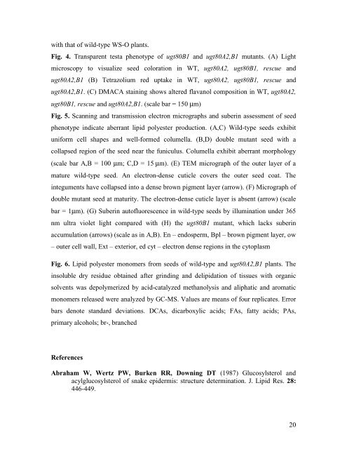
Fig. 4. Transparent testa phenotype of ugt80B1 and ugt80A2,B1 <strong>mutants</strong>. (A) Light<br />
microscopy to visualize seed coloration in WT, ugt80A2, ugt80B1, rescue and<br />
ugt80A2,B1 (B) Tetrazolium red uptake in WT, ugt80A2, ugt80B1, rescue and<br />
ugt80A2,B1. (C) DMACA staining shows altered flavanol composition in WT, ugt80A2,<br />
ugt80B1, rescue and ugt80A2,B1. (scale bar = 150 μm)<br />
Fig. 5. Scanning and transmission electron micrographs and suberin assessment of seed<br />
phenotype indicate aberrant lipid polyester production. (A,C) Wild-type seeds exhibit<br />
uniform cell shapes and well-formed columella. (B,D) double mutant seed with a<br />
collapsed region of the seed near the funiculus. Columella exhibit aberrant morphology<br />
(scale bar A,B = 100 μm; C,D = 15 μm). (E) TEM micrograph of the outer layer of a<br />
mature wild-type seed. An electron-dense cuticle covers the outer seed coat. The<br />
integuments have collapsed into a dense brown pigment layer (arrow). (F) Micrograph of<br />
double mutant seed at maturity. The electron-dense cuticle layer is absent (arrow) (scale<br />
bar = 1μm). (G) Suberin autofluorescence in wild-type seeds by illumination under 365<br />
nm ultra violet light compared with (H) the ugt80B1 mutant, which lacks suberin<br />
accumulation (arrows) (scale as in A,B). En – endosperm, Bpl – brown pigment layer, ow<br />
– outer cell wall, Ext – exterior, ed cyt – electron dense regions in the cytoplasm<br />
Fig. 6. Lipid polyester monomers from seeds of wild-type and ugt80A2,B1 plants. The<br />
insoluble dry residue obtained after grinding and delipidation of tissues with organic<br />
solvents was depolymerized by acid-catalyzed methanolysis and aliphatic and aromatic<br />
monomers released were analyzed by GC-MS. Values are means of four replicates. Error<br />
bars denote standard deviations. DCAs, dicarboxylic acids; FAs, fatty acids; PAs,<br />
primary alcohols; br-, branched<br />
References<br />
Abraham W, Wertz PW, Burken RR, Downing DT (1987) Glucosylsterol and<br />
acylglucosylsterol of snake epidermis: structure determination. J. Lipid Res. 28:<br />
446-449.<br />
20


