

Munich - 17th Annual Meeting German Society for Gene Therapy ...
Munich - 17th Annual Meeting German Society for Gene Therapy ...
Munich - 17th Annual Meeting German Society for Gene Therapy ...

Create successful ePaper yourself
Turn your PDF publications into a flip-book with our unique Google optimized e-Paper software.
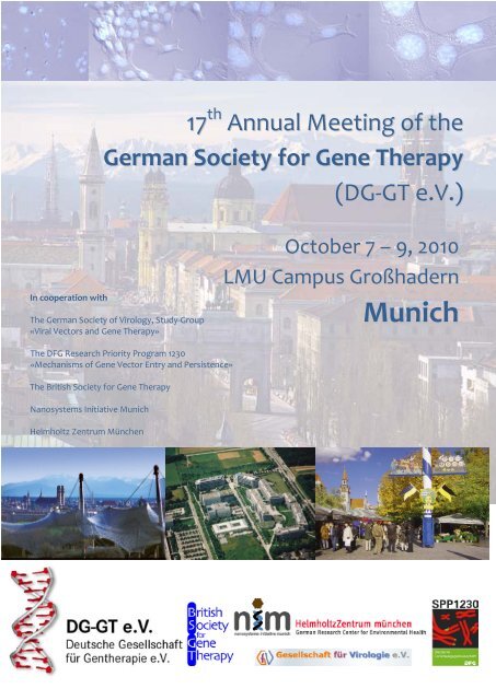
In cooperation with<br />
17 th <strong>Annual</strong> <strong>Meeting</strong> of the<br />
<strong>German</strong> <strong>Society</strong> <strong>for</strong> <strong>Gene</strong> <strong>Therapy</strong><br />
(DG-GT e.V.)<br />
The <strong>German</strong> <strong>Society</strong> of Virology, Study-Group<br />
«Viral Vectors and <strong>Gene</strong> <strong>Therapy</strong>»<br />
The DFG Research Priority Program 1230<br />
«Mechanisms of <strong>Gene</strong> Vector Entry and Persistence»<br />
The British <strong>Society</strong> <strong>for</strong> <strong>Gene</strong> <strong>Therapy</strong><br />
Nanosystems Initiative <strong>Munich</strong><br />
Helmholtz Zentrum München<br />
October 7 – 9, 2010<br />
LMU Campus Großhadern<br />
<strong>Munich</strong>
1 |<br />
Imprint<br />
Editors:<br />
Manfred Ogris<br />
Verena Brand<br />
Pirmin Burth<br />
Cover Layout:<br />
Pirmin Burth<br />
17 th <strong>Annual</strong> <strong>Meeting</strong> of the <strong>German</strong> <strong>Society</strong> <strong>for</strong> <strong>Gene</strong> <strong>Therapy</strong> (DG-GT e.V.), October 7-9, 2010,<br />
<strong>Munich</strong>
Table of Content<br />
Welcome 3<br />
<strong>Gene</strong>ral In<strong>for</strong>mation 4<br />
Organizing Committee 6<br />
Approach and Maps 6<br />
<strong>Meeting</strong> Program 9<br />
Sponsors 15<br />
Abstracts 16<br />
17 th <strong>Annual</strong> <strong>Meeting</strong> of the <strong>German</strong> <strong>Society</strong> <strong>for</strong> <strong>Gene</strong> <strong>Therapy</strong> (DG-GT e.V.), October 7-9, 2010,<br />
<strong>Munich</strong><br />
| 2
3 |<br />
Welcome<br />
Dear members and friends of the <strong>German</strong> <strong>Society</strong> <strong>for</strong> <strong>Gene</strong> <strong>Therapy</strong>,<br />
on behalf of the <strong>German</strong> <strong>Society</strong> <strong>for</strong> <strong>Gene</strong> <strong>Therapy</strong> we cordially welcome you to our 17 th <strong>Annual</strong><br />
<strong>Meeting</strong> held at the Chemistry and Pharmacy Campus of the University of <strong>Munich</strong>. The meeting is<br />
organized in conjunction with the British <strong>Society</strong> <strong>for</strong> <strong>Gene</strong> <strong>Therapy</strong>, the Study Group Viral Vectors of<br />
the <strong>German</strong> <strong>Society</strong> <strong>for</strong> Virology, the Research Priority Program SPP1230, the Nanosystems Initiative<br />
<strong>Munich</strong> (NIM) and the <strong>Munich</strong> Helmholtz <strong>Society</strong>.<br />
We have sought to put together an exciting and diversified program presenting state-of-the-art<br />
advances in the field of gene therapy, a field that has considerably evolved within recent years. The<br />
first day (Thursday, Oct., 7 th ) is dedicated to the Educational Sessions. Out of the portfolio of areas<br />
contributing to the progress of gene therapeutical approaches, we have chosen this year viral and<br />
non-viral vector design, tumor imaging and clinical aspects of gene therapy. The meeting is opened<br />
with an inaugural talk by our key note lecturer Mark Kay (Stan<strong>for</strong>d University) on Thursday evening<br />
and is continued on Friday and Saturday with internationally highly recognized experts in areas like<br />
vector development, cancer gene therapy, treatment of metabolic diseases and others. In addition,<br />
21 oral presentations were selected out of >100 submitted abstracts. During coffee- and lunch<br />
breaks, >60 posters are presented. Also the socializing part will be addressed accordingly with a<br />
Bavarian evening on Thursday offering ‘a saubere Brotzeit’ with classics from the Bavarian cuisine<br />
and brewery and a social event on Friday evening themed ‘Sports Night’. We hope that you will all<br />
enjoy the meeting and take the chance to discuss science and to relish <strong>Munich</strong>.<br />
Thank you all <strong>for</strong> coming to <strong>Munich</strong>!<br />
Manfred Ogris Hildegard Büning Anja Ehrhardt<br />
Andrew Baker Wolfgang Hammerschmidt Ernst Wagner<br />
17 th <strong>Annual</strong> <strong>Meeting</strong> of the <strong>German</strong> <strong>Society</strong> <strong>for</strong> <strong>Gene</strong> <strong>Therapy</strong> (DG-GT e.V.), October 7-9, 2010,<br />
<strong>Munich</strong>
<strong>Gene</strong>ral In<strong>for</strong>mation<br />
Organization and contact<br />
PD Dr. Manfred Ogris<br />
Phone +49 (0)89 2180 77842<br />
Fax +49 (0)89 2180 77791<br />
Email: dg-gt2010@cup.uni-muenchen.de<br />
Butenandtstr. 5-13, 81377 München, <strong>German</strong>y<br />
Venue<br />
Ludwig-Maximilians-Universität München<br />
Dekanat der Fakultät für Chemie und Pharmazie<br />
Butenandtstr. 5-13<br />
81377 München<br />
Registration<br />
The registration desk in the foyer of house F is open on:<br />
Thursday October 7 th from 12:00 pm to 06:30 pm<br />
Friday October 8 th from 08:00 am to 06:30 pm<br />
Saturday October 9 th from 08:00 am to 02:00 pm<br />
The registration fee includes:<br />
- access to scientific sessions<br />
- program and abstract book<br />
- coffee/tea during the morning and afternoon breaks<br />
- lunch during the poster sessions at noon<br />
- Post Oktoberfest event<br />
Oral Presentations<br />
All presentations will take place in the Buchner lecture hall (see map). The lecture hall is equipped<br />
with computers (Windows, NOT Mac) and a projector <strong>for</strong> PowerPoint presentations. For those<br />
people who use Mac please bring your own notebook. For those who use Windows please bring<br />
your presentation on a memory stick.<br />
There is a possibility to prepare the presentations in the Liebig lecture hall (next to Buchner lecture<br />
hall). It’s equipped with the same computers and projectors <strong>for</strong> PowerPoint presentations<br />
The Liebig lecture hall is open:<br />
Thursday October 7 th from 01:00 pm to 06:30 pm<br />
Friday October 8 th from 08:00 am to 10 am, from 1 pm to 06:30 pm<br />
Saturday October 9 th from 08:00 am to 02:00 pm<br />
17 th <strong>Annual</strong> <strong>Meeting</strong> of the <strong>German</strong> <strong>Society</strong> <strong>for</strong> <strong>Gene</strong> <strong>Therapy</strong> (DG-GT e.V.), October 7-9, 2010,<br />
<strong>Munich</strong><br />
| 4
5 |<br />
Posters<br />
The posters will be displayed in the foyer of house F (see map) from Friday 11:00 am to Saturday<br />
03:00 pm.<br />
Poster authors are present at their posters during poster sessions (see program).<br />
The posters can be handed over at the registration desk on Thursday or should be mounted on<br />
Friday until 10:30 am and can be removed on Saturday at 03:00 pm.<br />
Post Oktoberfest Event<br />
The Post Oktoberfest Event will take place in front of the Buchner lecture hall on Thursday, October<br />
7 th .<br />
Speakers Dinner<br />
The speakers dinner will take place at the Restaurant 181 on the top of the Olympiaturm. There will<br />
be a shuttle bus from the campus to the restaurant at 07:30 pm (return at 10:30 pm).<br />
Social Event “Sports Night”<br />
The social event is NOT included in the registration fee. Please wear your name badge!<br />
The social event will take place at Primafila in Laim (near Sportpark Nymphenburg) on Friday,<br />
October 8 th , 08:30 pm.<br />
Approach:<br />
There will be a shuttle bus from the campus to the restaurant at 08:00 pm.<br />
By public transport:<br />
From the campus take the subway U6 in the direction of Garching Forschungszentrum and get off at<br />
Holzapfelkreuth. Then take bus 51 in the direction of Olympia Einkaufszentrum and get off at Laim.<br />
Pass the underground passage and follow Wotanstraße, then turn left and walk along Margarethe-<br />
Danzi-Straße until you reach the Sportpark Nymphenburg and Primafila.<br />
You can also go there by S-Bahn and get off at Laim (see map).<br />
17 th <strong>Annual</strong> <strong>Meeting</strong> of the <strong>German</strong> <strong>Society</strong> <strong>for</strong> <strong>Gene</strong> <strong>Therapy</strong> (DG-GT e.V.), October 7-9, 2010,<br />
<strong>Munich</strong>
Organizing Committee<br />
PD Dr Manfred Ogris<br />
PD Dr Hildegard Büning<br />
PD Dr Anja Ehrhardt<br />
Prof Dr Andrew Baker<br />
Prof Dr Wolfgang Hammerschmidt<br />
Prof Dr Ernst Wagner<br />
Approach and Maps<br />
How to reach us by car<br />
• From the Nürnberg highway: get onto the Mittlerer Ring (direction Autobahn Lindau), then<br />
onto the Lindau highway to the exit Blumenau, keep going to Gräfelfing from where<br />
Würmtalstrasse will lead you to our campus.<br />
• From the Stuttgart highway: from the end of the highway in Obermenzing turn off to Pasing,<br />
from Pasing drive in the direction of Gräfelfing, then turn left to Großhadern.<br />
• From the Salzburg or Garmisch highways: drive onto the "Mittlerer Ring" in the direction of<br />
Großhadern and Stuttgart, then, in Großhadern turn to Gräfelfing and follow Würmtalstrasse<br />
which passes by our campus.<br />
Destination address <strong>for</strong> navigation system and parking<br />
Marchioninistraße, 81377 München<br />
Follow the parking signs at “Klinikum Großhadern”<br />
By train and public transport<br />
By IC, EC, ICE to <strong>Munich</strong> Central Station (München Hauptbahnhof). From there, take the subway U1<br />
or U2 to Sendlinger Tor. Change to U6 in the direction of Klinikum Großhadern and get off at<br />
Großhadern. Take the stairway to your left and keep left. You are now on Würmtalstraße. Now you<br />
can walk along Würmtalstraße until the campus comes into view (large modern buildings) on the left<br />
(about 10 to 15 minutes) or take the bus 266 or 268 in the direction of Planegg to Waldhüterstraße<br />
17 th <strong>Annual</strong> <strong>Meeting</strong> of the <strong>German</strong> <strong>Society</strong> <strong>for</strong> <strong>Gene</strong> <strong>Therapy</strong> (DG-GT e.V.), October 7-9, 2010,<br />
<strong>Munich</strong><br />
| 6
7 |<br />
By plane and public transport<br />
From the <strong>Munich</strong> airport take the S-Bahn S8 (or S1) to Marienplatz. Change to the subway U6 in the<br />
direction of Klinikum Großhadern and get off at Großhadern. Take the stairway to your left and keep<br />
left. You are now on Würmtalstraße. Now you can walk along Würmtalstraße until the campus<br />
comes into view (large modern buildings) on the left (about 10 to 15 minutes) or take the bus 266 or<br />
268 in the direction of Planegg to Waldhüterstraße.<br />
Ticketing <strong>for</strong> public transport<br />
You can use:<br />
- MVV-3-days ticket <strong>for</strong> the inner district (=white zone) from the moment of validation until 6<br />
am on the fourth day (3-Tages-Ticket Innenraum, 12,80€).<br />
- MVV-1-day ticket <strong>for</strong> the inner district (=white zone) from the moment of validation until 6<br />
am the following day (1-Tages-Ticket Innenraum, 5,20€).<br />
All tickets must be validated once in a blue ticket-cancelling machine be<strong>for</strong>e starting with your trip,<br />
except <strong>for</strong> tickets from the bus and tram ticket machines. These are already validated.<br />
17 th <strong>Annual</strong> <strong>Meeting</strong> of the <strong>German</strong> <strong>Society</strong> <strong>for</strong> <strong>Gene</strong> <strong>Therapy</strong> (DG-GT e.V.), October 7-9, 2010,<br />
<strong>Munich</strong>
<strong>Meeting</strong> Program<br />
Educational Session<br />
THURSDAY OCTOBER 7<br />
13:00 – 14:30 Educational Session Part 1<br />
Session Chair: Christian Kupatt, Klinikum Großhadern, LMU <strong>Munich</strong><br />
• Inv 1 Christina Rauschhuber (Department of Virology, Max von Pettenkofer-Institute,<br />
<strong>Munich</strong>, <strong>German</strong>y)<br />
Design of Recombinant Adenoviral Vectors <strong>for</strong> <strong>Gene</strong> <strong>Therapy</strong>: Improvements and<br />
Challenges<br />
• Inv 2 Martina Anton (Klinikum rechts der Isar, TUM, <strong>Munich</strong>, <strong>German</strong>y)<br />
Lentiviral vectors in gene and cell therapy approaches<br />
• Inv 3 Christian Kupatt (Klinikum Großhadern, LMU, <strong>Munich</strong>, <strong>German</strong>y)<br />
AAV based vectors <strong>for</strong> cardiovascular diseases<br />
14:30 – 15:00 Coffee break<br />
15:00 – 17:00 Educational Session Part 2<br />
Session Chair: Carsten Rudolph, Von Haunersches Kinderspital, LMU, <strong>Munich</strong><br />
• Inv 4 Carsten Rudolph (Von Haunersches Kinderspital, LMU, <strong>Munich</strong>)<br />
Introduction into nonviral gene delivery – physical and chemical delivery methods<br />
• Inv 5 Christine Spitzweg (Klinikum Großhadern, LMU, <strong>Munich</strong>)<br />
Imaging in cancer gene therapy<br />
• Inv 6 Len Seymour (University of Ox<strong>for</strong>d, UK)<br />
<strong>Gene</strong> therapy around the globe<br />
• Inv 7 Josef Rosenecker (Von Haunersches Kinderspital, LMU, <strong>Munich</strong>)<br />
Clinical experience with gene therapy in pediatric patients<br />
17:00 – 18:00 Coffee break<br />
17 th <strong>Annual</strong> <strong>Meeting</strong> of the <strong>German</strong> <strong>Society</strong> <strong>for</strong> <strong>Gene</strong> <strong>Therapy</strong> (DG-GT e.V.), October 7-9, 2010,<br />
<strong>Munich</strong><br />
| 8
9 |<br />
<strong>Gene</strong>ral <strong>Meeting</strong><br />
18:00 – 18:15 Opening Remarks and Opening ceremony<br />
18:15 – 19:05 Keynote Lecture<br />
Session Chair: Hildegard Büning, University of Cologne<br />
• Inv 8 Mark Kay (School of Medicine, Stan<strong>for</strong>d University, USA)<br />
<strong>Gene</strong> transfer approaches <strong>for</strong> gene addition, knockdown and cellular reprogramming<br />
in vivo<br />
19:05 – 20:30 Post Oktoberfest Event<br />
20:30 – 22:30 Speakers dinner<br />
FRIDAY OCTOBER 8<br />
9:00 – 11:00 Vector Development<br />
Session Chairs: Florian Kreppel, University of Ulm<br />
Ernst Wagner, Department of Pharmacy, LMU, <strong>Munich</strong><br />
• Inv 9 Len Seymour (University of Ox<strong>for</strong>d, UK)<br />
Delivery issues <strong>for</strong> oncolytic viruses<br />
• Inv 10 Andrew Baker (University of Glasgow, Scotland, UK)<br />
Modification of the adenovirus capsid: integrating virus biology and vector<br />
engineering<br />
• Selected Talk from abstracts:<br />
Or 1: Sigrid Espenlaub (Department of <strong>Gene</strong> <strong>Therapy</strong>, University of Ulm, <strong>German</strong>y)<br />
Analysis of intracellular particle trafficking and bioresponsive bonds <strong>for</strong> Ad vector<br />
shielding by capsomer specific fluorescent labeling<br />
Or 2: Jessica Sallach (Clinic I of Internal Medicine and Center <strong>for</strong> Molecular Medicine<br />
Cologne, University of Cologne, <strong>German</strong>y)<br />
Primary human keratinocyte-selective AAV2-based targeting vectors<br />
Or 3: Nadja Noske (Department of Virology, Max von Pettenkofer-Institute, LMU, <strong>Munich</strong>,<br />
<strong>German</strong>y)<br />
Development of novel PhiC31 integrase fusion proteins <strong>for</strong> improving efficacy and<br />
safety of transgene insertion in therapeutic applications<br />
Or 4: Keiji Itaka (Division of Clinical Biotechnology, Graduate School of Medicine, The<br />
University of Tokyo, Japan)<br />
Biocompatible polyplex nanomicelle <strong>for</strong> safe and effective gene transfer<br />
11:00 – 11:15 Coffee Break and Poster Session<br />
17 th <strong>Annual</strong> <strong>Meeting</strong> of the <strong>German</strong> <strong>Society</strong> <strong>for</strong> <strong>Gene</strong> <strong>Therapy</strong> (DG-GT e.V.), October 7-9, 2010,<br />
<strong>Munich</strong>
11:15 – 12:00 Vector Development<br />
• Inv 11 Michael Gait (MRC Laboratory of Molecular Biology, Cambridge, UK)<br />
Peptide-enhanced delivery of oligonucleotide analogues targeting Duchenne<br />
muscular dystrophy<br />
• Selected Talk from abstracts:<br />
Or 5: Frauke M. Koenig (Department of Chemistry, LMU, <strong>Munich</strong>, <strong>German</strong>y)<br />
EGF receptor targeting of polyplexes with the short artificial peptide GE11 studied by<br />
live cell imaging<br />
12:00 – 13:30 On the Route to Clinical Application<br />
Session Chair: Christoph von Kalle, National Center <strong>for</strong> Tumor Diseases , Heidelberg<br />
• Inv 12 Marinee Chuah (Vesalius Research Center, Leuven, Belgium)<br />
Emerging transposon technology <strong>for</strong> gene transfer and IPS applications<br />
• Inv 13 Simon Waddington (Imperial College London, UK)<br />
Perinatal gene therapy <strong>for</strong> lethal genetic diseases<br />
• Selected Talk from abstracts:<br />
Or 6: Stylianos Michalakis (Department of Pharmacy – Center <strong>for</strong> Drug Research, LMU,<br />
<strong>Munich</strong>, <strong>German</strong>y)<br />
Restoration of cone vision in the CNGA3–/– mouse model of congenital complete lack<br />
of cone photoreceptor function<br />
Or 7: Anna Paruzynski (Department of Translational Oncology, National Center <strong>for</strong> Tumor<br />
Diseases (NCT) and <strong>German</strong> Cancer Research Center (DKFZ), Hannover, <strong>German</strong>y)<br />
High throughput integration site analysis reveals a polyclonal lineage-specific<br />
integration site distribution in a successful WAS gene therapy trial<br />
13:30 – 14:30 Lunch Break and Poster Session<br />
14:30 – 16:00 <strong>Gene</strong> <strong>Therapy</strong><br />
Session Chairs: Boris Fehse, University Medical Centre Hamburg-Eppendorf<br />
Manuel Grez, Georg-Speyer-Haus, Frankfurt<br />
• Inv 14 Robin Ali (Institute of Ophthalmology, London, UK)<br />
<strong>Gene</strong> therapy <strong>for</strong> inherited retinal dystrophies<br />
• Inv 15 Harald Petry (Amsterdam Molecular Therapeutics (AMT), Netherlands)<br />
Alipogene Tiparvovec: the first gene therapy <strong>for</strong> a general metabolic disorder<br />
• Selected Talk from abstracts:<br />
Or 8: Teresa Trenkwalder (Klinikum Großhadern, LMU, <strong>Munich</strong>, <strong>German</strong>y)<br />
Enhanced therapeutic neovascularization via AAV2.9/Thymosin β4: Evidence <strong>for</strong> a<br />
Myovascular Crosstalk<br />
Or 9: Abdullah Cim (King’s College London, School of Medicine, London, UK)<br />
Nonviral Delivery of the rat PDX1 gene to rat liver <strong>for</strong> the in vivo transdifferentiation<br />
of liver cells to pancreatic β-cells<br />
17 th <strong>Annual</strong> <strong>Meeting</strong> of the <strong>German</strong> <strong>Society</strong> <strong>for</strong> <strong>Gene</strong> <strong>Therapy</strong> (DG-GT e.V.), October 7-9, 2010,<br />
<strong>Munich</strong><br />
| 10
11 |<br />
16:00 – 16:30 Coffee Break and Poster Session<br />
16:30 – 17:15 <strong>Gene</strong> <strong>Therapy</strong><br />
• Inv 16 Manuel Grez (Georg-Speyer-Haus, Frankfurt, <strong>German</strong>y)<br />
<strong>Gene</strong> therapy of chronic granulomatous disease: the past and the future<br />
• Selected Talk from abstracts:<br />
Or 10: Ute Modlich (Hannover Medical School, Hannover, <strong>German</strong>y)<br />
Correction of Mpl deficiency by lentiviral vectors with lineage-specific expression<br />
17:15 – 18:45 Cancer <strong>Gene</strong> <strong>Therapy</strong><br />
Session Chairs: Dorothee von Laer, Innsbruck Medical University<br />
Dirk Nettelbeck, DKFZ Heidelberg<br />
• Inv 17 Roberto Cattaneo (Mayo Clinic, Minnesota, USA)<br />
Viruses as cancer therapeutics: three points of attack<br />
• Inv 18 Caroline Breitbach (Jennerex, Inc., San Francisco, USA)<br />
Targeted and armed oncolytic poxviruses: a novel multi-mechanistic<br />
therapeutic class <strong>for</strong> cancer<br />
• Selected Talk from abstracts:<br />
Or 11: Alexander Muik (Georg-Speyer-Haus, Frankfurt am Main, <strong>German</strong>y)<br />
LCMV-Pseudotyped VSV-based systems <strong>for</strong> treatment of malignant glioma<br />
Or 12: Jennifer Altomonte (Klinikum rechts der Isar, TU München, <strong>Munich</strong>, <strong>German</strong>y)<br />
18:45 – 19:00 Award Ceremony DG-GT Forscherpreis<br />
19:00 – 20:00 DGGT <strong>Gene</strong>ral Assembly<br />
20:30 – open end Social Event themed “Sports Night”<br />
17 th <strong>Annual</strong> <strong>Meeting</strong> of the <strong>German</strong> <strong>Society</strong> <strong>for</strong> <strong>Gene</strong> <strong>Therapy</strong> (DG-GT e.V.), October 7-9, 2010,<br />
<strong>Munich</strong>
SATURDAY OCTOBER 9<br />
9:00 – 9:45 Cancer <strong>Gene</strong> <strong>Therapy</strong><br />
• Inv 19 John Bell (Ottawa Hospital Research Institute, Canada)<br />
Oncolytic vaccinia virus <strong>for</strong> the treatment of cancer<br />
• Selected Talk from abstracts:<br />
Or 13: Geoffrey K. Grünwald (Klinikum Grosshadern, LMU, <strong>Munich</strong>, <strong>German</strong>y)<br />
An α-fetoprotein promoter driven, conditionally replicating adenovirus that expresses<br />
the sodium iodide symporter (NIS) <strong>for</strong> radiovirotherapy of HCC<br />
9:45 – 10:45 Tumor Biology and (Cancer) Stem Cells<br />
Session Chair: Axel Schambach, Hannover Medical School<br />
• Inv 20 Norman Maitland (YCR Cancer Research Unit, University of York, UK)<br />
Prostate cancer stem cells: a new target <strong>for</strong> therapy<br />
• Selected Talk from abstracts:<br />
Or 14: Kerstin Knoop (Klinikum Grosshadern, LMU, <strong>Munich</strong>, <strong>German</strong>y)<br />
Tumor stroma-specific NIS gene delivery using mesenchymal stem cells<br />
Or 15: Axel Schambach (Department of Experimental Hematology, Hannover Medical<br />
School, Hannover, <strong>German</strong>y)<br />
Monitoring and excising reprogramming factors: a novel lentiviral expression system<br />
<strong>for</strong> reprogramming strategies<br />
10:45 – 11:15 Coffee Break and Poster Session<br />
11:15 – 13:15 Cancer Immune <strong>Therapy</strong> and T-cell <strong>Therapy</strong><br />
Session Chair: Wolfgang Uckert, Max Delbrück Center <strong>for</strong> Molecular Medicine, Berlin<br />
• Inv 21 Renata Stripecke (Hannover Medical School, <strong>German</strong>y)<br />
Lentiviral vector-induced dendritic cells <strong>for</strong> melanoma immunotherapy<br />
• Inv 22 Hinrich Abken (Uniklinikum Köln, <strong>German</strong>y)<br />
Arming immune cells to fight cancer<br />
• Inv 23 Farzin Farzaneh (Kings College, London, UK)<br />
Immune gene therapy <strong>for</strong> acute myeloid leukaemia<br />
• Selected Talk from abstracts:<br />
Or 16: Matthias Leisegang (Max-Delbrück-Center <strong>for</strong> Molecular Medicine, Berlin, <strong>German</strong>y)<br />
MHC-restricted fratricide of recipient lymphocytes expressing transgenic T-cell<br />
receptors specific <strong>for</strong> the apoptosis-inhibitor protein survivin<br />
Or 17: Eliana Ruggiero (<strong>German</strong> Cancer Research Center and National Center <strong>for</strong> Tumor<br />
Diseases, Heidelberg, <strong>German</strong>y)<br />
Integration site analysis of reprogrammed T-cells in a mouse model of T-cell receptor<br />
gene therapy developing graft versus host disease<br />
17 th <strong>Annual</strong> <strong>Meeting</strong> of the <strong>German</strong> <strong>Society</strong> <strong>for</strong> <strong>Gene</strong> <strong>Therapy</strong> (DG-GT e.V.), October 7-9, 2010,<br />
<strong>Munich</strong><br />
| 12
13 |<br />
13:15 – 14:00 Lunch Break and Poster Session<br />
14:00 – 16:00 Pharmacology and Toxicology<br />
Session Chair: Hildegard Büning, University of Cologne<br />
• Inv 24 Anne Galy (<strong>Gene</strong>thon, Evry, France)<br />
Preclinical safety and efficacy data leading to a clinical trial <strong>for</strong> the gene therapy of<br />
Wiskott Aldrich Syndrome<br />
• Inv 25 Klaus Cichutek (Paul-Ehrlich-Institut, Langen, <strong>German</strong>y)<br />
Lentivector transduction of novel cell targets<br />
• Selected Talk from abstracts:<br />
Or 18: Katarina Farkasova (Pharmaceutical Biotechnology, LMU, <strong>Munich</strong>, <strong>German</strong>y)<br />
Luciferase-based dual bioluminescence imaging of tumor metastases after systemic<br />
transgene delivery with a synthetic gene carrier<br />
Or 19: Niels Heinz (Experimental Hematology, Hannover Medical School, Hannover,<br />
<strong>German</strong>y)<br />
Retroviral and transposon-based Tet-regulated all-in-one vectors with reduced<br />
background expression and improved dynamic range<br />
Or 20: Simone J. Scholz (<strong>German</strong> Cancer Research Center and National Center <strong>for</strong> Tumor<br />
Diseases, Heidelberg, <strong>German</strong>y)<br />
High-throughput integration site analysis <strong>for</strong> vector biosafety assessment in CGD<br />
gene therapy<br />
Or 21: Margaret R. Duffy (Glasgow Cardiovascular Research Centre, University of Glasgow,<br />
Glasgow, UK)<br />
Modification of the FX serine protease domain ablates HSPG engagement by Ad5-FX<br />
complexes<br />
16:00 – 16:15 Poster Award and Concluding Remarks<br />
17 th <strong>Annual</strong> <strong>Meeting</strong> of the <strong>German</strong> <strong>Society</strong> <strong>for</strong> <strong>Gene</strong> <strong>Therapy</strong> (DG-GT e.V.), October 7-9, 2010,<br />
<strong>Munich</strong>
Sponsors<br />
Gold Sponsors Silver Sponsors<br />
Thanks <strong>for</strong> the support to our sponsors!<br />
17 th <strong>Annual</strong> <strong>Meeting</strong> of the <strong>German</strong> <strong>Society</strong> <strong>for</strong> <strong>Gene</strong> <strong>Therapy</strong> (DG-GT e.V.), October 7-9, 2010,<br />
<strong>Munich</strong><br />
| 14
15 |<br />
Abstracts<br />
Invited presentations 16 - 27<br />
Oral presentations 27 - 40<br />
Poster presentations 41 - 78<br />
Abstract author index 79 - 82<br />
All abstract are published in the September 2010 issue of Human <strong>Gene</strong> <strong>Therapy</strong> and can be<br />
downloaded via this link: http://www.liebertonline.com/doi/abs/10.1089/hum.2010.804<br />
17 th <strong>Annual</strong> <strong>Meeting</strong> of the <strong>German</strong> <strong>Society</strong> <strong>for</strong> <strong>Gene</strong> <strong>Therapy</strong> (DG-GT e.V.), October 7-9, 2010,<br />
<strong>Munich</strong>
DG-GT 2010 Invited Presentations<br />
Inv 1<br />
Design of Recombinant Adenoviral Vectors<br />
<strong>for</strong> <strong>Gene</strong> <strong>Therapy</strong>: Improvements and<br />
Challenges<br />
Christina Rauschhuber<br />
Department of Virology, Max von Pettenkofer-<br />
Institute, <strong>Munich</strong>, <strong>German</strong>y<br />
Over the past decade recombinant adenoviral<br />
vectors (rAdVs) became one of the most<br />
prominent gene therapeutic vector systems used<br />
in preclinical and clinical approaches. Adenoviral<br />
vectors exhibit several targets <strong>for</strong> modifications,<br />
thus they are useful <strong>for</strong> a variety of applications.<br />
Different generations of rAdV based on human<br />
adenovirus serotype 5 were generated starting<br />
with first and second generation adenoviruses,<br />
which lack one or two viral genes to more<br />
sophisticated technologies such as high capacity<br />
adenoviral vectors (HD-Ad) deleted <strong>for</strong> all viral<br />
coding sequences. Besides these types of<br />
vectors which will be discussed in detail,<br />
oncolytic adenoviruses exhibiting restricted<br />
replication in tumor tissues have to be mentioned<br />
but are not the major focus of this lecture.<br />
Common <strong>for</strong> all rAdV is their ability to efficiently<br />
transduce a broad range of dividing and nondividing<br />
cells and that they can be produced at<br />
high titers. However, in contrast to first and<br />
second generation adenoviruses, HD-AdVs have<br />
a significantly larger packaging capacity <strong>for</strong> large<br />
trangene expression cassettes and they display<br />
long-term transgene expression in preclinical<br />
studies. Nevertheless, the biggest challenge in<br />
adenoviral gene therapy is to circumvent the<br />
immune response against the vector. As HD-AdV<br />
lacks all viral coding sequences, there is no<br />
response to de novo synthesized viral proteins<br />
but immune response directed against the<br />
incoming viral capsid components may impair the<br />
outcome of a gene therapeutic approach. Thus,<br />
improvements with respect to the administration<br />
route and methods to modify the surface of the<br />
virion are the major focus of ongoing research<br />
and will be discussed within this lecture.<br />
Session: Educational Session<br />
Inv 2<br />
Lentiviral Vectors in <strong>Gene</strong> and Cell <strong>Therapy</strong><br />
Approaches<br />
Martina Anton<br />
17 th <strong>Annual</strong> <strong>Meeting</strong> of the <strong>German</strong> <strong>Society</strong> <strong>for</strong> <strong>Gene</strong> <strong>Therapy</strong> (DG-GT e.V.), October 7-9, 2010,<br />
<strong>Munich</strong><br />
| 16<br />
Institute of Experimental Oncology and <strong>Therapy</strong><br />
Research, Technische Universität München,<br />
<strong>Munich</strong>, <strong>German</strong>y<br />
Retroviruses are evolutionally optimized <strong>for</strong><br />
transfer of their genetic material into cells and<br />
integrating their genome into the host cell<br />
genome. They are thus well suited <strong>for</strong> long-term<br />
stable gene expression. Recombinant vectors<br />
based on gammaretroviruses (MLV) have been<br />
widely used in the past in preclinical research as<br />
well as clinical trials. However, during clinical<br />
trials of SCID, severe side effects occurred<br />
leading to induction of leukaemia in five cases.<br />
More recently researchers have tried to harness<br />
lentiviruses, another genus of the retroviridae and<br />
developed recombinant vectors based on Human<br />
Immunodeficiency Virus (HIV-1), Simian<br />
Immunodeficiency Virus (SIV), Feline (FIV)<br />
Immunodeficiency Virus, equine infectious<br />
anaemia virus (EIAV) and others. Whereas<br />
commonly used gammaretroviruses like MLV<br />
depend on cell division and breakdown of the<br />
nuclear membrane, lentiviruses and their vectors<br />
can also infect and transduce non-proliferating<br />
cells. The general concepts on how these<br />
pathogens can be converted into efficient and<br />
safe gene delivery tools <strong>for</strong> cell modification, the<br />
correction of inherited or acquired diseases will<br />
be introduced. Packaging concepts and targeting<br />
of lentiviral vectors (LV) will be discussed, as well<br />
as self inactivating (SIN) LV that are thought to<br />
be less likely to activate or disrupt neighbouring<br />
genes upon integration. Additionally important<br />
areas of research focus on how lentiviral vectors<br />
can be modified to avoid integration and thus<br />
reduce risk of insertional mutagenesis or to target<br />
integration to specific sites in the genome. An<br />
overview on construction and use of lentiviral<br />
vectors as tools in molecular biology,<br />
transcriptional targeting and de-targeting of LV,<br />
regulation of LV-mediated gene expression, preclinical<br />
as well as recent and future clinical<br />
applications will be presented.<br />
Session: Educational Session
17 |<br />
Inv 3<br />
AAV-Based Vectors <strong>for</strong> Cardiovascular<br />
Diseases: Therapeutic Potential in Chronic<br />
Ischemia<br />
C. Kupatt, R. Hinkel<br />
Klinikum Großhadern, Medizinische Klinik I und<br />
Poliklinik, Ludwig-Maximilians-University <strong>Munich</strong>,<br />
<strong>Munich</strong>, <strong>German</strong>y<br />
Therapeutic neovascularization of chronic<br />
ischemic muscle tissue is a treatment option <strong>for</strong><br />
otherwise no-option patients, which are not<br />
suitable <strong>for</strong> interventional or surgical<br />
interventions. Protein application of proangiogenic<br />
factors, such as VEGF or bFGF<br />
provided vessel growth and perfusion gains in<br />
small and large animal studies. However, in<br />
patient studies no clearcut success was achieved<br />
by pro-angiogenic protein application. Similar<br />
discrepancies were idetified <strong>for</strong> plasmid therapy,<br />
e.g., by liposomal transfection: significant<br />
improvement in preclinical experiments was<br />
followed by mixed results in human. Even<br />
overexpression of pro-angiogenic genes via<br />
adenoviral vector, leading to a transgene<br />
overexpression <strong>for</strong> 5-7 days, failed to significantly<br />
improve perfusion of an ischemic myocardium<br />
(Yla-Herttuala, 2007, Lavu JMCC 2010) Since<br />
prolonged transgene expression currently<br />
appears as a critical variable <strong>for</strong> pro-angiogenic<br />
gene therapy, long-acting adeno associated viral<br />
vectors might no-option patients. Adenoassociated<br />
virus (AAV), a member of the<br />
parvovirus family, is a non-pathogenic DNA virus,<br />
which transduces dividing and non-dividing cells<br />
and leading to a long-term overexpression,<br />
displaying a favorable immunogenic profile<br />
compared to adenoviral vectors (Gruchala,<br />
2004). Recently, we identified the pseudotyped<br />
virus strains AAV2/6 (carrying a partial genome<br />
of AAV2 and the envelope of AAV6) and AAV2/9<br />
as highly efficient vectors in large animals (pigs,<br />
Raake et al., JACC 2008, Kupatt et al., JACC<br />
2010), the latter sufficing to resolve hibernating<br />
myocardium after chronic coronary artery<br />
occlusion. Moreover, the long-lasting transgene<br />
expression after AAV transduction may require<br />
inducible transgene activity. We established the<br />
combination of AAV vectors and a Tet-off<br />
transgene system, where the pro-angiogenic<br />
factor Thymosin β4 was used to provide<br />
neovascularization being active only 2d a week.<br />
In a model of chronic hindlimb ischemia, pulsed<br />
pro-angiogenic activity appeared non-inferior to<br />
constitutive transgene expression up to 4 weeks<br />
after vector application. Taken together, the<br />
combination of of AAV vector and an efficient<br />
pro-angiogenic transgene may offer therapeutic<br />
potential in patients with ischemic<br />
cardiomyopathy or peripheral artery disease and<br />
exhausted conventional options.<br />
Session: Educational Session<br />
Inv 4<br />
Introduction into Nonviral <strong>Gene</strong> Delivery –<br />
Overview of Physical and Chemical Delivery<br />
Methods<br />
Carsten Rudolph<br />
Ludwig Maximilians University, <strong>Munich</strong>, <strong>German</strong>y<br />
Physicochemical methods <strong>for</strong> gene delivery have<br />
been intensively investigated during the last<br />
decades and first products have been approved<br />
<strong>for</strong> veterinary use. Although transfection rates<br />
achieved with nonviral delivery systems are<br />
frequently lower and gene expression is of only<br />
short duration when compared with viral vectors,<br />
they offer the advantage of being less<br />
immunogenic, less restricted to the size of the<br />
delivered transgene and being less expensive<br />
with respect to production. The currently used<br />
repertoire of physicochemical gene delivery<br />
methods have evolved from various basic<br />
concept using either naked DNA or complexed<br />
with gene transfer agents. Moreover, a variety of<br />
methods have been established which make use<br />
of different physical phenomena to introduce<br />
DNA into cells. In this lecture, basic concepts of<br />
various physicochemical gene delivery methods<br />
making use of either nanoparticle <strong>for</strong>mation or<br />
physical <strong>for</strong>ces <strong>for</strong> transfection will be discussed.<br />
In addition, advantages and restrictions of the<br />
physicochemical gene delivery methods will be<br />
discussed.<br />
Session: Educational Session<br />
Inv 5<br />
Imaging in Cancer <strong>Gene</strong> <strong>Therapy</strong><br />
Christine Spitzweg<br />
Department of Internal Medicine II, Klinikum<br />
Großhadern, Ludwig Maximilians University,<br />
<strong>Munich</strong>, <strong>German</strong>y<br />
The field of gene therapy has made considerable<br />
strides in the last decade by the development of<br />
new vectors and an increasing repertoire of<br />
17 th <strong>Annual</strong> <strong>Meeting</strong> of the <strong>German</strong> <strong>Society</strong> <strong>for</strong> <strong>Gene</strong> <strong>Therapy</strong> (DG-GT e.V.), October 7-9, 2010,<br />
<strong>Munich</strong>
therapeutic genes. Non-invasive monitoring of<br />
the in vivo distribution of viral and non-viral<br />
vectors as well as biodistribution, level and<br />
duration of transgene expression have been<br />
recognized as critical elements in the design of<br />
clinical gene therapy trials. Besides<br />
bioluminescence imaging using luciferase,<br />
reporter genes that have been evaluated <strong>for</strong><br />
possible human studies are the herpes simplex<br />
thymidine kinase and the dopamine D2 receptor.<br />
Cloning of the sodium iodide symporter (NIS),<br />
that mediates the active transport of iodide in the<br />
thyroid gland and represents the molecular basis<br />
<strong>for</strong> radioiodine scintigraphy, has provided us with<br />
one of the most promising reporter genes<br />
available today. NIS represents a nonimmunogenic<br />
protein with a well-defined body<br />
distribution that mediates the transport of readily<br />
available radionuclides such as 131 I, 123 I, 124 I,<br />
99m 188 211<br />
Tc, Re or At, which can be used <strong>for</strong><br />
gamma camera scintigraphic imaging, SPECT<br />
and PET imaging. Several research groups<br />
including our own have studied the potential of<br />
NIS as reporter gene in various applications,<br />
demonstrating that in vivo imaging of radioiodine<br />
accumulation correlates well with the results of ex<br />
vivo gamma counter measurements as well as<br />
NIS mRNA and protein analysis. NIS was<br />
successfully used to monitor in vivo<br />
biodistribution of synthetic vectors and<br />
mesenchymal stem cells as gene delivery<br />
vehicles after systemic application as well as<br />
replication-competent viral vectors using<br />
123 99m<br />
conventional I- or Tc-gamma camera<br />
imaging or 99m Tc-SPECT/CT fusion imaging. In<br />
addition, PET imaging using<br />
124 I provides<br />
significant advantages <strong>for</strong> exact localization and<br />
quantitative analysis of NIS-mediated radioiodine<br />
accumulation due to enhanced resolution and<br />
sensitivity. Taken together, currently available<br />
data clearly demonstrate the enormous potential<br />
of NIS as a novel reporter gene, that in its dual<br />
function as reporter and therapy gene also allows<br />
therapeutic radionuclide application.<br />
Session: Educational Session<br />
Inv 6<br />
<strong>Gene</strong> <strong>Therapy</strong> Around the Globe<br />
Len Seymour<br />
Department of Clinical Pharmacology, University<br />
of Ox<strong>for</strong>d, Ox<strong>for</strong>d, UK<br />
<strong>Gene</strong> therapy is a whole new approach to<br />
medicine. Instead of designing drugs to treat the<br />
17 th <strong>Annual</strong> <strong>Meeting</strong> of the <strong>German</strong> <strong>Society</strong> <strong>for</strong> <strong>Gene</strong> <strong>Therapy</strong> (DG-GT e.V.), October 7-9, 2010,<br />
<strong>Munich</strong><br />
| 18<br />
symptoms of a disease, gene therapy seeks to<br />
identify the root genetic cause of a disorder and<br />
to treat it at that level. For diseases caused by<br />
mutations in ‘single genes’, this provides the<br />
possibility of treating the disease very effectively<br />
by providing healthy copies of the mutant gene,<br />
allowing production of normal proteins and<br />
restoring the healthy phenotype. Accordingly this<br />
approach has exciting potential <strong>for</strong> treatment of<br />
many gene-based diseases. Despite its promise,<br />
however, gene therapy is often limited by the<br />
difficulty of delivering therapeutic genes<br />
effectively into the diseased cells. Normally<br />
scientists try to do this using viruses as genevectors;<br />
however, even viruses find it challenging<br />
to access all diseased cells within a body. This is<br />
the central challenge of gene therapy. Where<br />
delivery has been successfully addressed, the<br />
results have been very encouraging. For<br />
example, children with bone marrow-based<br />
immune deficiencies can now be treated very<br />
effectively by introducing normal genes into their<br />
bone marrow, a procedure that is normally<br />
per<strong>for</strong>med in the test tube be<strong>for</strong>e reintroducing<br />
the bone marrow to the patient. This approach<br />
has a success rate comparable to or higher than<br />
bone marrow transplantation, and can be applied<br />
to all patients (with no requirement to identify a<br />
matched bone marrow donor). Similarly a recent<br />
study has shown excellent treatment of<br />
adrenoleukodystrophy (the disease featured in<br />
the film ‘Lorenzo's Oil’) using this approach.<br />
However, the choice of viral vectors is important,<br />
since treatment of these disorders requires lifelong<br />
expression of the therapeutic genes, and<br />
early generations of viruses used have been<br />
found to damage the DNA upon inserting into the<br />
genome, leading to leukaemia. Accordingly<br />
scientists are now seeking to identify safer<br />
viruses, to enable this approach to be used<br />
widely. Another approach to efficient delivery is in<br />
the field of retinal blindness, where viruses<br />
expressing therapeutic genes can be identified<br />
directly into the diseased area. Again, very<br />
encouraging results have been seen, with clear<br />
improvements in sight resulting. <strong>Gene</strong> therapy<br />
can also apply to the use of genetic vaccines,<br />
siRNA, and tumour-killing ‘oncolytic’ viruses, all<br />
areas where we are seeing rapid progress. As<br />
knowledge increases rapidly, so the day when<br />
genetic medicines are a routine part of life comes<br />
steadily closer.<br />
Session: Educational Session
19 |<br />
Inv 7<br />
Clinical Experience with <strong>Gene</strong> <strong>Therapy</strong> in<br />
Pediatric Patients<br />
Josef Rosenecker<br />
Department of Pediatrics, Ludwig Maximilians<br />
University, <strong>Munich</strong>, <strong>German</strong>y<br />
The division of infectious diseases at the<br />
Department of Pediatrics, University of <strong>Munich</strong>,<br />
takes care <strong>for</strong> three patients who were treated<br />
with gene therapy protocols. This presentation<br />
will highlight their clinical follow up after gene<br />
therapy. The first patient was diagnosed with<br />
SCID-X1 in early infancy. No HLA-identical<br />
sibling was avialable and no matched bone<br />
marrow donor was available. There<strong>for</strong>e, the<br />
patient was transferred to Hospital Necker-<br />
Enfants Malades, Dr. Fischer, where an ex vivo<br />
retroviral mediated gene therapy was per<strong>for</strong>med.<br />
The lymphocyte development after gene therapy<br />
showed a successful reconstitution of the<br />
immune system. After gene therapy the patient<br />
developed a clonal T-cell proliferation, but is now<br />
in good clinical condition. The second patient was<br />
diagnosed with ADA-SCID and was transferred to<br />
Hospital San Raffaele, Dr. Aiuti, Milano, where an<br />
ex vivo gene therapy protocol was per<strong>for</strong>med.<br />
After gene therapy the patient showed<br />
reconstitution of the immune system and is now<br />
also in good clinical condition. A third patient was<br />
diagnosed of chronic granulomatous disease and<br />
at the age of 5 years was having severe infection<br />
with Aspergillus nidulans of the lung and of the<br />
thoracic vertebral column. He showed<br />
progressive tetraparesis. Antimycotic therapy<br />
showed only limited success. No HLA-identical<br />
sibling was avialable. No matched unrelated<br />
donor found. In this situatuion the patient was<br />
transferred to Zurich, where an ex vivo gene<br />
therapy protocol has been per<strong>for</strong>med. After gene<br />
therapy the patient showed 20% gene corrected<br />
cells in the PBL <strong>for</strong> about 4 weeks, then less than<br />
1%. He showed progressive recovery and plays<br />
no football and goes to school.<br />
Session: Educational Session<br />
Inv 8<br />
<strong>Gene</strong> Transfer Approaches <strong>for</strong> <strong>Gene</strong> Addition,<br />
Knockdown and Cellular Reprogramming In<br />
Vivo<br />
Mark A. Kay<br />
Departments of Pediatrics and <strong>Gene</strong>tics,<br />
Stan<strong>for</strong>d University, Stan<strong>for</strong>d, CA, USA<br />
Vectors that allow <strong>for</strong> DNA directed-RNA<br />
transcription can be used to treat a broad number<br />
of diseases. Our laboratory has been developing<br />
both minicircle plasmid based vectors and<br />
recombinant adenoassociated viral (rAAV)<br />
vectors <strong>for</strong> the purpose of developing plat<strong>for</strong>ms<br />
<strong>for</strong> gene addition, knockdown, and cellular<br />
reprogramming in vivo. These plat<strong>for</strong>m<br />
technologies are being tested in mice, dogs, and<br />
humans with hemophilia B (Factor IX deficiency),<br />
mouse models of Hepatitis C Virus Infection, and<br />
mouse models of juvenile onset insulin<br />
dependent diabetes mellitus, as examples of<br />
gene addition, knockdown, and cellular<br />
reprogramming strategies, respectively. Minicircle<br />
DNA plasmid vectors are devoid of all bacterial<br />
plasmid backbone DNA providing 20 to 1000<br />
times more persistent transgene expression<br />
compared to routine plasmids when transfected<br />
into quiescent cells in vivo. The mechanistic<br />
differences compared to routine plasmid DNA<br />
vectors are beginning to be elucidated. From a<br />
practical standpoint, we have developed a<br />
simplified method <strong>for</strong> minicircle vector<br />
preparation that is nearly equivalent to a routine<br />
plasmid preparation making it feasible <strong>for</strong> these<br />
DNAs to replace routine plasmids <strong>for</strong> all<br />
mammalian expression studies. Novel<br />
recombinant AAV vectors with altered<br />
transduction properties and/or site-specific<br />
integration into the ribosomal DNA locus are<br />
being derived. These expanded vector properties<br />
increase their utility <strong>for</strong> treating serious diseases<br />
in people. We will present our current studies<br />
using these improved vectors <strong>for</strong> treating the<br />
three plat<strong>for</strong>m diseases stated above.<br />
Session: Keynote Lecture<br />
Inv 9<br />
Delivery Issues <strong>for</strong> Oncolytic Viruses<br />
Len Seymour<br />
Department of Clinical Pharmacology, University<br />
of Ox<strong>for</strong>d, Ox<strong>for</strong>d, UK<br />
While there are several examples where gene<br />
therapy approaches are looking promising, they<br />
all reflect situations where the gene vectors can<br />
be introduced efficiently into target cells – either<br />
by direct injection or by transduction ex vivo.<br />
Indeed, cancer gene therapy can be very<br />
effective if tumour-killing viruses are injected<br />
17 th <strong>Annual</strong> <strong>Meeting</strong> of the <strong>German</strong> <strong>Society</strong> <strong>for</strong> <strong>Gene</strong> <strong>Therapy</strong> (DG-GT e.V.), October 7-9, 2010,<br />
<strong>Munich</strong>
directly into tumours. Un<strong>for</strong>tunately three quarters<br />
of people who develop cancer in the West go on<br />
to die from metastatic disease. In this situation it<br />
is not possible to inject gene therapy vectors into<br />
all the tumour nodules, and intravenous<br />
‘systemic’ therapy is required. However, the<br />
human bloodstream represents a very aggressive<br />
environment <strong>for</strong> most gene therapy vectors.<br />
Therapeutic microbes delivered via the<br />
bloodstream encounter many host defences and<br />
anatomical barriers that must be surmounted to<br />
enable their access to disseminated cancers.<br />
This is particularly true <strong>for</strong> adenovirus type 5 in<br />
humans, where most recipients have powerful<br />
pre-existing adenovirus-neutralising activity. We<br />
have recently shown that human (but not murine)<br />
erythrocytes provide an additional barrier by<br />
sequestering adenovirus onto the Coxsackie and<br />
Adenovirus Receptor and (via antibodies)<br />
complement receptor 1. Coating adenovirus with<br />
a layer of hydrophilic polymer can prevent this<br />
interaction and allow virus to circulate free in the<br />
plasma, showing passive targeting to<br />
disseminated tumours and mediating good<br />
anticancer efficacy. Entry of polymer-coated virus<br />
particles into the tumour mass is a product of<br />
fluid transfer, and is directly proportional to the<br />
area under the plasma concentration-time curve.<br />
Increasing extravasation of fluid through tumourassociated<br />
endothelium using permeabilityenhancers<br />
such as Tumour Necrosis Factor<br />
alpha can improve virus particle entry into<br />
tumours over 100-fold, reaching as high as 10%<br />
injected dose (virus particles) per tumour. This<br />
provides the possibility <strong>for</strong> highly efficient<br />
targeting to tumours and good anticancer<br />
efficacy. An alternative approach is to target<br />
agents to infect tumour-associated vasculature.<br />
However while this provides a vulnerable target<br />
to traditional gene therapy approaches,<br />
endothelial cells do not normally support<br />
‘oncolytic’ viruses. One approach to overcoming<br />
this problem is to encode syncytium-<strong>for</strong>ming<br />
proteins within the endothelial, to enable transcomplementation<br />
of virus replication by tumourassociated<br />
factors.<br />
Session: Vector Development<br />
Inv 10<br />
Adenovirus Vector Engineering <strong>for</strong> <strong>Gene</strong><br />
<strong>Therapy</strong><br />
Andrew H Baker<br />
University of Glasgow, Glasgow, UK<br />
17 th <strong>Annual</strong> <strong>Meeting</strong> of the <strong>German</strong> <strong>Society</strong> <strong>for</strong> <strong>Gene</strong> <strong>Therapy</strong> (DG-GT e.V.), October 7-9, 2010,<br />
<strong>Munich</strong><br />
| 20<br />
Adenoviral vectors are used frequently <strong>for</strong> gene<br />
therapy but much of their potential is limited by<br />
their tropism <strong>for</strong> liver and spleen, leading to<br />
effects on virion, sequestration, limited gene<br />
transfer to alternate tissue and toxicity. Recent<br />
studies have elucidated the mechanism<br />
underlying much of this tropism and the<br />
consequence of this infectivity profile. 1–4 We have<br />
shown the precise role of coagulation factor X<br />
(FX) in mediating liver uptake of adenovirus in<br />
rodent models in recent years. This occurs<br />
through a nM interaction between the FX Gla<br />
domain and the hypervariable regions of the Ad5<br />
hexon trimer. We modeled our cryoelectron<br />
microscopy data from the Ad5:FX interaction. We<br />
observed specific contact points within the hexon<br />
in hypervariable regions 5 and 7. Creation of<br />
novel vectors with deletions and amino acid<br />
mutations in these regions had dramatic effects<br />
on FX-mediated gene delivery in vitro and in vivo.<br />
We have also assessed the impact of HSPG<br />
structure on adenovirus uptake. These findings<br />
highlight the fundamental importance of the<br />
hexon:FX interaction dictating in vivo tropism as<br />
well as novel avenues <strong>for</strong> vector retargeting to<br />
alternate sites in vivo. 1 Parker AL et al., Blood<br />
(2006); 2 Waddington SN et al., Cell, (2008);<br />
3 4<br />
Kalyuzhniy, O et al., PNAS (2008); Di Paola, N<br />
et al., Immunity (2009).<br />
Session: Vector Development<br />
Inv 11<br />
Peptide-Enhanced Delivery of Oligonucleotide<br />
Analogues Targeting Duchenne Muscular<br />
Dystrophy<br />
Michael J Gait 1 , Amer F Saleh 1 , Andrey A<br />
Arzumanov 1 , Haifang Yin 2 , Matthew Wood 2<br />
1Laboratory of Molecular Biology, Medical<br />
Research Council, Cambridge, UK; 2 University of<br />
Ox<strong>for</strong>d, Ox<strong>for</strong>d, UK<br />
Duchenne muscular dystrophy (DMD) is an Xlinked<br />
genetic disease that affects 1 in 3500 male<br />
births. Mutations in the dystrophin gene result in<br />
aberrant splicing of the pre-mRNA and hence a<br />
truncated and inactive protein. Dystrophin<br />
connects actin to the dystrophin-associated<br />
complex at the sarcolemma membrane and the<br />
lack of dystrophin leads to progressive muscle<br />
degeneration and a significantly shorter life span.<br />
There is no effective treatment currently<br />
available. A number of potential gene therapy<br />
approaches are in development. Meanwhile a<br />
promising treatment has reached clinical trials,
21 |<br />
which involves use of antisense oligonucleotides<br />
(ON). ONs are targeted to bind to dystrophin premRNA<br />
and redirect splicing, resulting in “exon<br />
skipping” that restores the correct protein reading<br />
frame. The resultant expressed protein is shorter<br />
than natural dystrophin but can substitute very<br />
effectively. This phenotype is similar to Becker<br />
muscular dystrophy that shows generally only<br />
mild symptoms in patients. Two chemistry types<br />
<strong>for</strong> an exon 51 targeting ON are currently in<br />
clinical trials, ′ 2 -O-methyl phosphorothioates in<br />
Holland and Belgium and phosphoramidate<br />
morpholino oligonucleotides (PMO) in the UK.<br />
Early results in both cases have shown some<br />
dystrophin production in patients following<br />
systemic delivery. However, levels of dystrophin<br />
restored are variable and it is unclear yet whether<br />
a therapeutically useful effect will be reached in<br />
all muscle types at moderate dosage levels. To<br />
enhance activity, Arginine-rich peptides have<br />
been developed as conjugates of PMO targeting<br />
exon 23 that showed in an mdx mouse model of<br />
DMD substantially improved dystrophin<br />
production compared to naked PMO. We have<br />
been developing novel Arginine-rich Pip peptides<br />
that when attached to exon 23-targeted PMO<br />
have shown very high dystrophin generation in<br />
mdx mice, including in hard-to-reach heart<br />
muscle. These and other Arginine-rich peptides<br />
involving muscle-specific targeting domains are<br />
currently being evaluated as candidates <strong>for</strong><br />
possible use as a peptide-PMO conjugate in a<br />
future clinical trial <strong>for</strong> DMD.<br />
Session: Vector Development<br />
Inv 12<br />
Hyperactive Transposons <strong>for</strong> <strong>Gene</strong>tic<br />
Modification of Induced Pluripotent and Adult<br />
Stem Cells: A Novel Non-Viral Paradigm <strong>for</strong><br />
Coaxed Differentiation<br />
Marinee K. L. Chuah 1 , Eyayu Belay 1 , Janka<br />
Mátrai 1 , Abel Acosta-Sanchez 1 , Ling Ma 1 , Mattia<br />
Quattrocelli 2 , Lajos Mátés 3 , Pau Sancho-Bru 2 ,<br />
Martine Geraerts 2 , Bing Yan 1 , Joris Vermeesch 4 ,<br />
Ermira Samara-Kuko 1 , Zoltán Ivics 3,5 , Catherine<br />
Verfaillie 2 , Maurillio Sampaolesi 2 , Zsuzsanna<br />
Izsvák 3,5 , Thierry VandenDriessche 1<br />
1 Vesalius Research Centrum KUL-VIB, Leuven,<br />
Belgium; 2 Stem Cell Institute, University of<br />
Leuven, Leuven, Belgium; 3 Max Delbrück Center<br />
<strong>for</strong> Molecular Medicine, Berlin, <strong>German</strong>y; 4 Center<br />
<strong>for</strong> Human <strong>Gene</strong>tics, University Hospital<br />
Gasthuisberg, Leuven, Belgium; 5 University of<br />
Debrecen, Debrecen, Hungary<br />
Adult stem cells and induced pluripotent stem<br />
cells (iPS) hold great promise <strong>for</strong> regenerative<br />
medicine. The development of robust non-viral<br />
approaches <strong>for</strong> stem cell gene transfer would<br />
facilitate functional studies and potential clinical<br />
applications. We there<strong>for</strong>e generated hyperactive<br />
transposases derived from Sleeping Beauty,<br />
using an in vitro molecular evolution and<br />
selection paradigm. These hyperactive<br />
transposases resulted in superior gene transfer<br />
efficiencies and expression in mesenchymal and<br />
muscle stem/progenitor cells, consistent with<br />
higher expression levels of therapeutically<br />
relevant proteins including coagulation factor IX.<br />
Their differentiation potential and karyotype was<br />
not affected. Most importantly, relatively efficient<br />
stable gene transfer could be obtained in bona<br />
fide hematopoietic stem cells that are capable of<br />
hematopoietic reconstitution and multi-lineage<br />
gene marking (Mates, Chuah et al., Nature<br />
<strong>Gene</strong>tics, 41(6):753–761, 2009). Moreover,<br />
stable transposition could also be achieved in iPS<br />
which retained their ability to differentiate along<br />
neuronal, cardiac and hepatic lineages without<br />
causing cytogenetic abnormalities. Most<br />
importantly, transposon-mediated delivery of the<br />
myogenic PAX3 transcription factor into iPS<br />
coaxed their differentiation into MYOD +<br />
myogenic progenitors and multinucleated<br />
myofibers, suggesting that PAX3 may serve as a<br />
myogenic “molecular switch” in iPS. Hence, this<br />
hyperactive transposon system represents an<br />
attractive non-viral gene transfer plat<strong>for</strong>m with<br />
broad implications <strong>for</strong> regenerative medicine, cell<br />
and gene therapy.<br />
Session: On the Route to Clinical Application<br />
Inv 13<br />
Perinatal <strong>Gene</strong> <strong>Therapy</strong> <strong>for</strong> Lethal <strong>Gene</strong>tic<br />
Diseases<br />
Simon N Waddington 1 , Ahad A. Rahim 2 , Citra<br />
Mattar 3 , Andrew M.S. Wong 4 , Klemens Hoeffer 4 ,<br />
Suzanne M.K. Buckley 5 , Jonathan D. Cooper 4 ,<br />
Jerry Chan 3<br />
1 University College London, London, UK;<br />
2 Institute <strong>for</strong> Women's Health, University College<br />
London, London, UK; 3 National University of<br />
Singapore, Singapore; 4 Paediatric Storage<br />
Disease Laboratory, Kings College London,<br />
London, UK; 5 Department of Haematology,<br />
University College London, London, UK;<br />
A number of inherited neurological diseases are<br />
characterised by neurodegenerative changes at<br />
17 th <strong>Annual</strong> <strong>Meeting</strong> of the <strong>German</strong> <strong>Society</strong> <strong>for</strong> <strong>Gene</strong> <strong>Therapy</strong> (DG-GT e.V.), October 7-9, 2010,<br />
<strong>Munich</strong>
or around birth. Prognosis remains dismal and <strong>for</strong><br />
several of these diseases, including Acute<br />
Neuronopathic Gaucher Disease there is no<br />
treatment and palliative care remains the only<br />
option. Owing to the aggressive nature of this<br />
and related diseases fetal or neonatal gene<br />
therapy may be the only potential means of<br />
treatment. Recently, AAV9 has been shown to<br />
cross the blood brain barrier following<br />
intravenous injection into neonatal mice, cats and<br />
macaques. We have investigated this further by<br />
studying vector tropism after intravenous injection<br />
into fetal mice. In utero intravenous injection of<br />
single-stranded (ss) and self-complimentary (sc)<br />
AAV9 expressing green fluorescent protein<br />
(GFP) into mice (embryonic day 16) resulted in<br />
efficient global transduction of neurons in the<br />
CNS. Furthermore, there was a stark contrast in<br />
cell type transduction when compared to<br />
neonatal administration, confirmed by scanning<br />
confocal microscopy. The efficiency varied<br />
depending upon ss or sc configuration of the<br />
vector. Examination of injected mice by<br />
fluorescent microscopy, immunohistology, and<br />
GFP ELISA revealed extensive and efficient<br />
transduction in the visceral organs in addition to<br />
muscle, bone, eye, and skin. <strong>Gene</strong> expression<br />
was also seen in the peripheral nervous system.<br />
Similarly, extensive transduction was also<br />
observed following intravenous administration of<br />
AAV9 to the fetal macaque. The combination of<br />
CNS and visceral organ transduction is well<br />
suited to the study, and potential treatment <strong>for</strong><br />
diseases such as acute neuronopathic Gaucher<br />
disease where both CNS and visceral pathology<br />
require targeting and a suitable mouse model is<br />
available.<br />
Session: On the Route to Clinical Application<br />
Inv 14<br />
Clinical Trial of <strong>Gene</strong> <strong>Therapy</strong> <strong>for</strong> Early Onset<br />
Severe Retinal Dystrophy Resulting from<br />
Defects in RPE65<br />
Robin R Ali 1 , JWB Bainbridge 1 , AJ Smith 1 , FW<br />
Fitzke 1 , GE Holder 1 , A Stockman 1 , SS<br />
Bhattacharya 1 , GS Rubin 1 , S Yzer 2 , I van den<br />
Born 2 , AT Moore 1<br />
1 Division of Molecular <strong>Therapy</strong>, UCL Institute of<br />
Ophthalmology and UCL/Moorfields Eye Hospital<br />
Biomedical Research Centre <strong>for</strong> Ophthalmology,<br />
London, UK; 2 Rotterdam Eye Hospital,<br />
Rotterdam, The Netherlands<br />
17 th <strong>Annual</strong> <strong>Meeting</strong> of the <strong>German</strong> <strong>Society</strong> <strong>for</strong> <strong>Gene</strong> <strong>Therapy</strong> (DG-GT e.V.), October 7-9, 2010,<br />
<strong>Munich</strong><br />
| 22<br />
Early-onset severe retinal dystrophy caused by<br />
defects in the gene encoding the retinal<br />
isomerase RPE65 is associated with poor vision<br />
at birth and complete loss of vision in early<br />
adulthood. In a phase I/II dose-escalation trial,<br />
we have delivered subretinally recombinant<br />
adeno-associated virus (rAAV) vector expressing<br />
RPE65 under the control of an RPE65 promoter<br />
in 9 human subjects with early onset severe<br />
retinal dystrophy associated with mutations in<br />
RPE65. We have examined systemic vector<br />
dissemination and immune responses following<br />
vector delivery, assessed visual function pre- and<br />
post-vector delivery using a range of<br />
psychophysical techniques, and per<strong>for</strong>med<br />
detailed electrophysiology and retinal imaging<br />
studies. There have been no serious adverse<br />
effects of surgical delivery of vector in the<br />
subjects enrolled to date. We have detected no<br />
systemic dissemination of vector genome.<br />
Although we have detected an increase in<br />
systemic neutralising antibodies to AAV capsid in<br />
two subjects, there have been no evidence of<br />
immune responses to RPE65 protein. We have<br />
measured significant improvements in retinal<br />
sensitivity by microperimetry and dark-adapted<br />
perimetry, and improved per<strong>for</strong>mance in a test of<br />
visually-guided mobility. The outcomes in the first<br />
9 subjects to date suggest that subretinal delivery<br />
of rAAV vector can be safe in humans in the<br />
short term and can improve retinal sensitivity.<br />
These findings support further clinical studies in<br />
subjects with RPE65 deficiency and the<br />
development of gene therapy <strong>for</strong> other inherited<br />
retinal disorders.<br />
Session: <strong>Gene</strong> <strong>Therapy</strong><br />
Inv 15<br />
Alipogene Tiparvovec: the First <strong>Gene</strong> <strong>Therapy</strong><br />
<strong>for</strong> a <strong>Gene</strong>ral Metabolic Disorder<br />
Harald Petry 1 , D. Gaudet 2 , A. Carpentier 3 , E.<br />
Stroes 4 , J. Twisk 1 , S. Greentree 1<br />
1Amsterdam<br />
Molecular Therapeutics (AMT),<br />
Amsterdam, The Netherlands; 2 Department of<br />
medicine, Université de Montreal, Quebec,<br />
Canada; 3 Department of Endocrinology,<br />
University of Sherbrooke, Quebec, Canada;<br />
4<br />
Academisch Medical Center (AMC), Amsterdam,<br />
The Netherlands<br />
<strong>Gene</strong> therapy is coming of age. Correction or<br />
supplementation of genetic defects through gene<br />
therapy is amenable to both monogenic disorders<br />
as well as to diseases with a key protein playing
23 |<br />
a key role in the pathology. Alipogene tiparvovec<br />
(Glybera ® , AMT-011) is the first gene therapy<br />
product <strong>for</strong> a metabolic disease in late stage<br />
clinical development. The product contains a<br />
gain-of-function variant of the human lipoprotein<br />
lipase gene in an AAV1 based vector (AAV1-<br />
LPLS447X). Alipogene tiparvovec has been<br />
developed <strong>for</strong> the long term correction of<br />
lipoprotein lipase deficiency, to control or abolish<br />
symptoms and prevent complications in adult<br />
patients clinically diagnosed with lipoprotein<br />
lipase deficiency (LPLD). LPLD is a rare,<br />
seriously debilitating autosomal inherited<br />
monogenic disorder of lipid metabolism.<br />
Deficiency of LPL function results in<br />
chylomicronaemia. Recurrent pancreatitis is<br />
known as the most frequent, potentially lethal,<br />
complication; other severe sequelae include<br />
diabetes and increased tendency <strong>for</strong><br />
atherosclerosis. During the presentation the<br />
various steps to develop a gene therapy<br />
medicinal product will be discussed. Alipogene<br />
tiparvovec in LPLD will be used to demonstrate<br />
that persistent gene transduction after one-time<br />
intramuscular administration is feasible, and<br />
results in long-term clinical improvements in the<br />
‘chylomicronaemia syndrome’ caused by LPLD.<br />
Session: <strong>Gene</strong> <strong>Therapy</strong><br />
Inv 16<br />
<strong>Gene</strong> <strong>Therapy</strong> <strong>for</strong> Chronic Granulomatous<br />
Disease: the Past and the Future<br />
Manuel Grez 1 , CGD Consortium 2<br />
1Georg-Speyer-Haus, Frankfurt am Main,<br />
<strong>German</strong>y; 2 Frankfurt, Heidelberg, Idar Oberstein,<br />
London, Zürich<br />
In our recent gene therapy trial <strong>for</strong> X-CGD we<br />
demonstrated reconstitution of superoxide activity<br />
in phagocytic cells and elimination of preexisting<br />
infections in two treated patients. However, an<br />
unexpected expansion of myeloid progenitors<br />
occurred five months after transplantation. Both<br />
patients developed a myelodysplastic syndrome<br />
(MDS) caused by insertional activation of<br />
MDS1/EVI1 followed by clonal progression and<br />
the gradual loss of chromosome 7. P1 died 27<br />
months after gene therapy of MDS in<br />
combination with severe septicemia, the latter<br />
resulting from loss of bacterial killing activity in<br />
transduced cells. P2 underwent allogeneic stem<br />
cell transplantation. Forced overexpression of<br />
MDS1/EVI1 or EVI1 in human cells disrupted<br />
normal centrosome duplication, linking<br />
MDS1/EVI1 activation to the development of<br />
genomic instability. Optimized SIN<br />
gammaretroviral vectors have been shown to<br />
have an enhanced safety profile. From several<br />
SIN-gammaretroviral vectors expressing<br />
gp91phox, we selected a construct containing the<br />
promoter of the human c-fes gene <strong>for</strong> detailed<br />
preclinical studies. We used X-CGD mice <strong>for</strong><br />
monitoring safety and functional reconstitution of<br />
NADPH oxidase activity in primary and<br />
secondary animals. The SINfes vector was safe,<br />
was resistant to CpG methylation and<br />
reconstituted superoxide activity to clinical<br />
relevant levels. We plan to use this vector <strong>for</strong> the<br />
next Phase I gene therapy trial.<br />
Session: <strong>Gene</strong> <strong>Therapy</strong><br />
Inv 17<br />
Reprogrammed Measles Viruses as Cancer<br />
Therapeutics Three Points of Attack<br />
Roberto Cattaneo<br />
Department of Molecular Medicine, Mayo Clinic,<br />
and Virology and <strong>Gene</strong> <strong>Therapy</strong> track, Mayo<br />
Graduate School, Rochester, USA<br />
Soon after viruses were recognized more than<br />
100 years ago, tumor regressions occasionally<br />
documented after accidental infections suggested<br />
the idea of using them to fight cancer. Early<br />
virotherapy clinical trials based on wild type<br />
viruses were unsuccessful, but recent ones<br />
based on genetically modified viruses are built on<br />
much stronger foundations. They <strong>for</strong>esee<br />
extensive monitoring of viral replication, gene<br />
expression, and host immunity. Therapeutic<br />
efficacy is being assessed by well-defined<br />
biological end points, and can be improved. For<br />
future clinical trials more specific and potent<br />
oncolytic viruses are developed based on three<br />
types of modification: targeting, arming, and<br />
shielding. Targeting introduces multiple layers of<br />
cancer specificity; arming amplifies locally the<br />
effects of approved cancer therapeutics; and<br />
shielding provides temporary relief from the<br />
immune response. We have shown that the<br />
envelope of measles (MV) and related<br />
paramyxoviruses can be targeted to many<br />
designated receptors. Retargeted MV envelopes<br />
have become preferred tools to pseudotype<br />
lentiviral and other gene transfer vectors. We<br />
have developed protease activation targeting,<br />
and innate immunity control targeting. We have<br />
armed MV with a prodrug convertase, and shown<br />
that this virus synergizes with the<br />
17 th <strong>Annual</strong> <strong>Meeting</strong> of the <strong>German</strong> <strong>Society</strong> <strong>for</strong> <strong>Gene</strong> <strong>Therapy</strong> (DG-GT e.V.), October 7-9, 2010,<br />
<strong>Munich</strong>
chemotherapeutic fludarabine to eliminate mantle<br />
cell lymphoma xenografts. MV-based<br />
therapeutics developed by the Russell and Peng<br />
groups at Mayo are being administered to<br />
patients who, having exhausted other therapeutic<br />
options, have enrolled in clinical trials of ovarian<br />
carcinoma, glioma, and myeloma. Survival and<br />
quality of life data are being collected and will be<br />
critical <strong>for</strong> the approval of new therapeutic<br />
products. Cattaneo, R. (2010) Paramyxovirus<br />
entry and targeted vectors <strong>for</strong> cancer therapy.<br />
PLoS Pathogens 6: e1000973.<br />
Session: Cancer <strong>Gene</strong> <strong>Therapy</strong><br />
Inv 18<br />
Targeted and Armed Oncolytic Poxviruses: A<br />
Novel Multimechanistic Therapeutic Class <strong>for</strong><br />
Cancer<br />
Caroline Breitbach 1 , John C Bell 1,2 , David H Kirn 1<br />
1 Jennerex Inc, San Francisco, USA; 2 Centre <strong>for</strong><br />
Cancer Therapeutics, Ottawa Hospital Research<br />
Institute, Ottawa, Canada<br />
Engineered viruses have been developed <strong>for</strong><br />
cancer therapy both as non-replicating gene<br />
therapy agents and as cancer vaccines.<br />
Oncolytic viruses, in contrast, were developed to<br />
replicate within and subsequently lyse, cancer<br />
cells. Clinical efficacy to date with each of these<br />
approaches has been limited by multiple factors<br />
including the inability to infect enough tumor cells<br />
in vivo locally within a tumor or systemically, and<br />
resistance of complex advanced tumors to a<br />
single mechanism-of-action (MOA). Over the last<br />
several years, however, a novel therapeutic class<br />
has emerged that combines the best features of<br />
all three approaches: targeted and armed<br />
oncolytic poxviruses. Recent preclinical and<br />
clinical results demonstrate convincingly that<br />
products from this therapeutic class can achieve<br />
highly selective and potent cancer destruction<br />
systemically through a multi-pronged MOA.<br />
Given recent clinical validation, we expect this<br />
therapeutic class to expand rapidly.<br />
Session: Cancer <strong>Gene</strong> <strong>Therapy</strong><br />
17 th <strong>Annual</strong> <strong>Meeting</strong> of the <strong>German</strong> <strong>Society</strong> <strong>for</strong> <strong>Gene</strong> <strong>Therapy</strong> (DG-GT e.V.), October 7-9, 2010,<br />
<strong>Munich</strong><br />
| 24<br />
Inv 19<br />
Oncolytic Vaccinia Virus <strong>for</strong> the Treatment of<br />
Cancer<br />
John C. Bell<br />
Centre <strong>for</strong> Cancer Therapeutics, Ottawa Hospital<br />
Research Institute, Ottawa, Canada<br />
The creation of novel “cancer lysing” or<br />
“oncolytic” viruses over the last 10-15 years has<br />
led to the development of a number of innovative<br />
anti-cancer therapeutic products many of which<br />
have entered into clinical testing. We are<br />
examining a number of poxvirus backbones <strong>for</strong><br />
their ability to selectively target and kill tumour<br />
cells. A derivative of the Wyeth Strain called JX-<br />
594 has engineered deletions, natural mutations<br />
and expresses a GM-CSF transgene, all of which<br />
contribute to its ability to infect and kill tumour<br />
tissues while leaving normal tissues unscathed.<br />
The results of our preclinical models and<br />
molecular studies have shown that JX-594<br />
attacks cancers using a multi-pronged approach<br />
that includes tumour vascular targeting, immune<br />
stimulation and direct cancer cell lysis. JX-594<br />
has now been tested in over 70 patients in a<br />
variety of different trial designs and consistently<br />
shows tumour targeting activity and a very strong<br />
safety profile. The results of our ongoing<br />
translational research ef<strong>for</strong>ts and clinical data<br />
from our phase I and II trials will be presented.<br />
Session: Cancer <strong>Gene</strong> <strong>Therapy</strong><br />
Inv 20<br />
Prostate Cancer Stem Cells: A New Target <strong>for</strong><br />
<strong>Therapy</strong><br />
Norman J Maitland<br />
YCR Cancer Research Unit, Dept of Biology,<br />
University of York, Heslington, UK<br />
Human prostate cancer is characterised by<br />
cellular heterogeneity; perhaps one reason <strong>for</strong><br />
the resistance of prostate cancer to many<br />
medical therapies. Once a prostate cancer has<br />
failed on hormone-based therapies, the<br />
prognosis <strong>for</strong> the patients is poor. Our approach<br />
to tissue heterogeneity in the prostate has been<br />
to identify and purify individual cell populations<br />
from human prostate cancers and matched<br />
normal tissues. We have fractionated cancer<br />
epithelium into a majority luminal/secretory<br />
population (which comprises more than 99% of<br />
the cancer mass) and a minor population
25 |<br />
consisting of committed basal cells, transamplifying<br />
cells and a stem cell population. The<br />
latter cells are responsible <strong>for</strong> tumour initiation<br />
and in vitro colony <strong>for</strong>mation. Affymetrix<br />
microarray analysis of these populations confirms<br />
a distinct phenotype <strong>for</strong> each cell type and<br />
functional expression of fate influencing genes.<br />
Further analysis has revealed populations of<br />
genes whose expression is tightly linked to the<br />
differentiation process, while others are<br />
associated with the cancerous properties of the<br />
cells. The tumour origin of the identified cells has<br />
been confirmed by xenografting into a highly<br />
immuno-compromised mouse strain at orthotopic,<br />
kidney capsule and subcutaneous sites with<br />
demonstrable metastases. <strong>Gene</strong>s associated<br />
with prostate cancer are expressed in the<br />
cultures and their functional significance<br />
assessed by SiRNA knock-down in stem cells<br />
and in their daughter cells. Epigenetic control, in<br />
contrast to permanent genetic alterations or<br />
gross alterations involving a reprogramming of<br />
cells to express specific transcription factor sets,<br />
seems to be a predominant mechanism <strong>for</strong> an<br />
adaptable stem cell in prostate. A better<br />
knowledge of the phenotype of the prostate<br />
CSC's should permit therapeutic targeting.<br />
However, whilst stem cell directed therapies (to<br />
treat less than 0.1% of the tumour mass) are<br />
unlikely to have an immediate effect on tumour<br />
volume, one consequence of targeting the wrong<br />
cell types within a heterogeneous tumour is to<br />
perturb the natural history of the tumour and<br />
results in an amplification of CSC's, in agreement<br />
with cancer relapse patterns in man.<br />
Session: Tumor Biology and (Cancer) Stem Cells<br />
Inv 21<br />
A Consortium <strong>for</strong> Clinical Development of<br />
Lentiviral Vector-Induced DCs <strong>for</strong> Melanoma<br />
Immunotherapy<br />
Renata Stripecke 1 , Sandra Kuhs 1 , Ralf Gutzmer 2 ,<br />
Henk Garritsen 3 , Farzin Farzaneh 4<br />
1 Department of Hematology, Hemostasis,<br />
Oncology and Stem Cell Transplantation,<br />
Hannover Medical School, Hannover, <strong>German</strong>y;<br />
2 Department of Dermatology, Hannover Medical<br />
School, Hannover, <strong>German</strong>y; 3 Division of<br />
Transfusion Medicine, Braunschweig City<br />
Hospital, Braunschweig, <strong>German</strong>y; 4 Department<br />
of Haematological Medicine, King's College<br />
London, Rayne Institute, London, UK<br />
Dendritic Cells (DCs) are key players in the<br />
development of adaptive immunity. Their use to<br />
boost anti-tumor responses have reached phase<br />
III clinical trials but ex vivo production is costly<br />
and difficult to standardize. A novel concept <strong>for</strong><br />
DC production consists of overnight lentiviral<br />
vector (LV) transduction of growth factors and<br />
full-length antigens into monocytes that results<br />
into induction of “SMART-DCs” (Selfdifferentiated<br />
Myeloid-derived Antigenpresenting-cells<br />
Reactive against Tumors). This<br />
concept has been validated in the preclinical B16<br />
melanoma mouse model <strong>for</strong> potency and safety<br />
with MART-1 and TRP2 as melanoma-associated<br />
antigens, overexpressed in 90% of the metastatic<br />
melanoma (Koya et al., 2007; Pincha et al.,<br />
submitted). Third generation self-inactivating<br />
tricistronic lentiviral vectors containing<br />
interspacing 2A elements co-expressing human<br />
GM-CSF, IL-4 and MART-1 or TRP-2 were<br />
constructed. Cytokine preconditioning and 16h<br />
transduction of CD14 + monocytes with high titer<br />
LVs resulted in persistent (3 weeks) autocrine<br />
production of GM-CSF (average 10 ng/ml), IL-4<br />
(average 12 ng/ml) and expression of MART-1 or<br />
TRP2 (detectable by intracellular staining).<br />
Transduced monocytes readily self-differentiated<br />
into SMART-DCs (CD209 + , MHCII + , CD80 +<br />
CD86+) and were stable in culture <strong>for</strong> 3 weeks.<br />
SMART-DCs/MART-1 were recognized by T cell<br />
clones specific <strong>for</strong> MART-1 in an HLA-restricted<br />
manner, assessed by IFN-γ-ELISPOT-Assay.<br />
PBMCs obtained from healthy donors that were<br />
primed/boosted in vitro with autologous SMART-<br />
DCs/MART-1 demonstrated the induction of<br />
MART-1-specific T cell responses assayed by<br />
IFN-γ-ELISPOT-Assay. SMART-DCs/MART-1<br />
and SMART-DCs/TRP2 are currently being<br />
tested <strong>for</strong> stimulation of T cell responses in<br />
melanoma patients. A consortium <strong>for</strong> clinical<br />
development of SMART-DCs has been <strong>for</strong>med:<br />
GMP-grade batch of the tricistronic vectors is<br />
planned <strong>for</strong> additional preclinical testing (optimal<br />
vector dose, number of integrated copies,<br />
toxicity), monocytes will be transduced with LVs<br />
in a closed GMP-grade bag system, development<br />
of Standard Operating Procedures and<br />
establishment of identity and potency markers<br />
are underway.<br />
Session: Cancer Immune <strong>Therapy</strong> and T-Cell<br />
<strong>Therapy</strong><br />
17 th <strong>Annual</strong> <strong>Meeting</strong> of the <strong>German</strong> <strong>Society</strong> <strong>for</strong> <strong>Gene</strong> <strong>Therapy</strong> (DG-GT e.V.), October 7-9, 2010,<br />
<strong>Munich</strong>
Inv 22<br />
Arming Immune Cells to Fight Cancer<br />
Hinrich Abken<br />
Zentrum für Molekulare Medizin Köln und Klinik I<br />
für Innere Medizin, Uniklinik Köln, Cologne,<br />
<strong>German</strong>y<br />
Cancer cells frequently escape T cell recognition<br />
by repression of the antigen processing<br />
machinery making them invisible <strong>for</strong> a cytolytic T<br />
cell attack. Strategies in adoptive immunotherapy<br />
during the last decade aimed to overcome the<br />
situation by engineering T cells with a<br />
recombinant chimeric antigen receptor (CAR)<br />
which uses a single chain fragment of variable<br />
region (scFv) antibody <strong>for</strong> target binding and the<br />
intracellular CD3zeta domain <strong>for</strong> T cell activation.<br />
To provide full T cell activation, the CD3zeta<br />
chain was fused with the CD28 costimulatory<br />
domain in one polypeptide chain; other<br />
costimulatory moieties are likewise used. We<br />
here discuss the impact of CD28 costimulation on<br />
the threshold in T cell activation and on the<br />
redirected anti-tumor response in the presence of<br />
TGF-beta and regulatory T cells. We moreover<br />
introduce a novel generation of CARs with<br />
combined CD28-OX40 costimulation in order to<br />
harness central memory T cells <strong>for</strong> a redirected<br />
anti-tumor attack. Finally, we demonstrate the<br />
power of a redirected T cell attack in eradicating<br />
established tumor lesions by eliminating minor<br />
tumor cell subsets.<br />
Session: Cancer Immune <strong>Therapy</strong> and T-Cell<br />
<strong>Therapy</strong><br />
Inv 23<br />
Immune <strong>Gene</strong> <strong>Therapy</strong> <strong>for</strong> Acute Myeloid<br />
Leukaemia<br />
Farzin Farzaneh 1 , Lucas Chan 1 , Nicola<br />
Hardwick 1 , Wendy Ingram 1 , Ghulam Mufti 1 , Mark<br />
Aloysius 2 , Adrian Robins 3 , Nagy Habib 4 , Joti<br />
Bhalla 1 , Oleg Eremin 2 , Tye Gee Jun 1 , David<br />
Oppenheim 1 , James Wells 5 , Christopher<br />
Cowled 5 , Alistair Noble 5<br />
1 Department of Haematological Medicine, King's<br />
College London, Rayne Institute, London, UK;<br />
2 Section of Surgery, Biomedical Research Unit,<br />
Nottingham Digestive Diseases Centre,<br />
University of Nottingham, Nottingham, UK;<br />
3 Institute of Infection and Immunity, School of<br />
Molecular Medical Sciences, Nottingham<br />
University Hospitals, University of Nottingham,<br />
17 th <strong>Annual</strong> <strong>Meeting</strong> of the <strong>German</strong> <strong>Society</strong> <strong>for</strong> <strong>Gene</strong> <strong>Therapy</strong> (DG-GT e.V.), October 7-9, 2010,<br />
<strong>Munich</strong><br />
| 26<br />
Nottingham, UK; 4 Section of Surgery,<br />
Department of Surgical Oncology and<br />
Technology, Imperial College London, London,<br />
UK; 5 MRC and Asthma UK Centre in Allergic<br />
Mechanisms of Asthma, King's College London,<br />
Guy's Hospital, London, UK<br />
Immune mediated rejection of established<br />
tumours needs adequate stimulation of cell<br />
mediated immunity, as well as the suppression of<br />
inhibitory responses that are induced by chronic<br />
exposure to tumour associated antigens.<br />
Preclinical studies have demonstrated that<br />
tumour cells expressing immune costimulatory<br />
molecules and appropriate Th1 cytokines can<br />
induce immune mediated rejection of previously<br />
established tumours. This strategy is now being<br />
assessed in a Phase-I clinical trial <strong>for</strong> the<br />
vaccination of relapsed poor prognosis acute<br />
myeloid leukaemia (AML) with autologous AML<br />
cells that are genetically modified to express B7.1<br />
(CD80) and IL-2. We have also shown that<br />
vaccination of advanced solid tumour patients,<br />
with dendritic cells that are pulsed with<br />
telomerase (hTERT) peptides, can result in the<br />
expansion of antigen specific T cells with some<br />
evidence of a modest and short-lived therapeutic<br />
benefit. In a further recently started Phase-I study<br />
we are now evaluating the safety, and potential<br />
efficacy of, hTERT peptide loaded dendritic cell<br />
vaccination in combination with<br />
cyclophosphamide mediated inhibition of immune<br />
suppression by regulatory T cells (Treg). A much<br />
less demanding alternative to the use of dendritic<br />
or tumour cell vaccines is the direct vaccination<br />
with tumour associated antigens, provided that<br />
the vaccine could indeed induce cell mediated<br />
immunity. With this goal in mind we have recently<br />
developed a new vaccination strategy based on<br />
the combined adjuvants <strong>for</strong> synergistic activation<br />
of cellular immunity (CASAC). CASAC contains<br />
different combinations of defined molecules that<br />
act synergistically to induce dendritic cell<br />
activation. Subcutaneous vaccination with two<br />
doses of a single peptide (OVA or Trp2) plus<br />
CASAC, induces IL-12 secretion, stimulation of<br />
Th1-biased CD4 T-cells, and high levels<br />
(routinely 50% by tetramer staining) of antigen<br />
specific cytolytic CD8 T cells. The magnitude of<br />
CASAC mediated immune stimulation is<br />
substantially greater than can be achieved by<br />
other adjuvants (e.g., about 100-fold greater than<br />
complete Freund's adjuvant). The antigen<br />
specific CTL activity induced by CASAC<br />
mediated peptide vaccination allows the in vivo<br />
lysis of greater than 90% of antigen positive<br />
tumour cells in mouse tumour models, resulting<br />
in long-lasting immunity with a robust recall<br />
response. This strategy will shortly entre clinical
27 |<br />
studies <strong>for</strong> WT1 peptide vaccination of myeloid<br />
leukaemia patients.<br />
Session: Cancer Immune <strong>Therapy</strong> and T-Cell<br />
<strong>Therapy</strong><br />
Inv 24<br />
Vector Safety Data Leading to a Clinical Trial<br />
<strong>for</strong> the <strong>Gene</strong> <strong>Therapy</strong> of Wiskott Aldrich<br />
Syndrome: A Perspective on Future<br />
Developments<br />
Anne Galy, Sabine Charrier, Otto-Wilhelm<br />
Merten, Didier Caizergues<br />
<strong>Gene</strong>thon, Evry, France<br />
Hematopoietic gene therapy currently relies on<br />
the ex vivo gene correction of patient autologous<br />
hematopoietic stem cells. This approach has now<br />
been tested <strong>for</strong> over 10 years in pilot phase I/II<br />
trials in several inherited diseases. The domain is<br />
evolving and the use of lentiviral vectors derived<br />
from HIV-1 provide significant advantages in<br />
terms of genomic stability, biological properties<br />
but also in the area of pharmaceutical product<br />
characterization. The rare primary<br />
immunodeficiency Wiskott Aldrich syndrome<br />
(WAS) is a combined platelet and immune defect.<br />
In the perspective of developing a gene therapy<br />
<strong>for</strong> WAS, we have characterized an advanced<br />
generation rHIV lentiviral vector pseudotyped<br />
with VSVg and implemented a large-scale<br />
industrial process to produce and to control this<br />
vector under good manufacturing practices<br />
(GMP). Recently several European centers were<br />
approved to start gene therapy trials <strong>for</strong> WAS<br />
with this lentiviral vector. With a growing number<br />
of potential disease applications <strong>for</strong> lentiviral<br />
vector-mediated hematopoietic gene therapy, it<br />
seems that there is ground to integrate<br />
in<strong>for</strong>mation on preclinical vector safety features<br />
particularly at the level of design, manufacture<br />
process or controls. We will discuss these<br />
various points through the different aspects<br />
leading to the development of WAS gene<br />
therapy.<br />
Session: Pharmacology and Toxicology<br />
Inv 25<br />
Lentivector Transduction of Novel Target<br />
Cells<br />
Klaus Cichutek<br />
Paul-Ehrlich-Institut, Langen, <strong>German</strong>y<br />
Session: Pharmacology and Toxicology<br />
17 th <strong>Annual</strong> <strong>Meeting</strong> of the <strong>German</strong> <strong>Society</strong> <strong>for</strong> <strong>Gene</strong> <strong>Therapy</strong> (DG-GT e.V.), October 7-9, 2010,<br />
<strong>Munich</strong>
DG-GT 2010 Oral Presentations<br />
Or 1<br />
Analysis of Intracellular Particle Trafficking<br />
and Bioresponsive Bonds <strong>for</strong> Ad Vector<br />
Shielding by Capsomer-Specific Fluorescent<br />
Labeling<br />
Sigrid Espenlaub 1 , Stephanie Corjon 1 , Tatjana<br />
Engler 1 , Carolin Fella 2 , Manfred Ogris 2 , Ernst<br />
Wagner 2 , Stefan Kochanek 1 , Florian Kreppel 1<br />
1Department of <strong>Gene</strong> <strong>Therapy</strong>, University of Ulm,<br />
Ulm, <strong>German</strong>y; 2 Pharmaceutical Biotechnology,<br />
Department of Pharmacy, Ludwig-Maximilians-<br />
University, <strong>Munich</strong>, <strong>German</strong>y<br />
Adenovirus (Ad) vectors are widely used <strong>for</strong> gene<br />
therapy approaches. Due to the high abundance<br />
of the natural adenovirus receptors CAR/integrins<br />
on a wide variety of cells numerous methods<br />
have been developed to redirect the virions to<br />
specific receptors on target cell surfaces. An<br />
increasing number of recent publications<br />
evidenced that the success of targeting strategies<br />
does not only depend on receptor binding and<br />
cellular uptake, but also on intracellular trafficking<br />
processes. There<strong>for</strong>e, improved knowledge on<br />
the intracellular fate of targeted Ad vector<br />
particles is mandatory <strong>for</strong> a rational design of<br />
targeted Ad vectors. However, while in principle<br />
potent tools are available <strong>for</strong> fluorescent labeling<br />
of Ad vectors in order to analyze post-receptor<br />
trafficking, these tools suffer from significant<br />
limitations: (i) capsids can so far only be labeled<br />
upon amino groups present in all proteins all over<br />
the particle surface and this imposes the risk of<br />
interference with particle infectivity, (ii)<br />
capsomere-specific labeling requires extensive<br />
genetic modifications and has only been<br />
demonstrated at protein IX, (iii) two-color labeling<br />
approaches are not available. Here we present a<br />
robust and straight <strong>for</strong>ward labeling procedure<br />
based on a genetic-chemical approach that<br />
overcomes these limitations. It allows <strong>for</strong> specific<br />
labeling of the capsomeres fiber, protein IX, or<br />
hexon and permits two-color labeling. Using this<br />
technology we analyzed by means of live cell<br />
imaging two different bioresponsive bonds that<br />
potentially can be used <strong>for</strong> the reversible<br />
attachment of shielding/targeting moieties to the<br />
capsids: disulfide and hydrazone bonds. In our<br />
experiments we could not observe significant<br />
17 th <strong>Annual</strong> <strong>Meeting</strong> of the <strong>German</strong> <strong>Society</strong> <strong>for</strong> <strong>Gene</strong> <strong>Therapy</strong> (DG-GT e.V.), October 7-9, 2010,<br />
<strong>Munich</strong><br />
| 28<br />
reduction of disulfides within the observation time<br />
of 80 minutes post cell entry. Acid-responsive<br />
hydrazone bonds in contrast became quickly<br />
hydrolyzed after uptake of the virions (30 min)<br />
and may thus be favorable <strong>for</strong> the generation of<br />
bioresponsive vectors and potent tools to<br />
overcome the limitations in intracellular trafficking<br />
processes of targeted Ad vectors.<br />
Session: Vector Development<br />
Or 2<br />
Primary Human Keratinocyte-Selective AAV2-<br />
Based Targeting Vectors<br />
Jessica Sallach 1 , Fernando Laguzzi Larcher 2 ,<br />
David Gomez Almarza 2 , Luca Perabo 1 , Michael<br />
Hallek 1 , Hildegard Büning 1<br />
1 Clinic I of Internal Medicine and Center <strong>for</strong><br />
Molecular Medicine Cologne (CMMC), University<br />
Hospital Cologne, University of Cologne,<br />
Cologne, <strong>German</strong>y; 2 Ciemat-Centro de<br />
Investigacion Energetica Medioambiental y<br />
Tecnologica, Moncloa-Aravaca, Madrid, Spain<br />
Vectors based on the adeno-associated virus<br />
(AAV), a non-enveloped DNA virus, have<br />
emerged as one of the leading gene transfer<br />
systems in gene therapy. However, transduction<br />
efficiency of clinical relevant cell types such as<br />
keratinocytes is insufficient and seems to depend<br />
on donor-specific factors. We there<strong>for</strong>e<br />
conducted AAV peptide display selection on<br />
human primary <strong>for</strong>eskin keratinocytes in order to<br />
identify AAV2-based targeting mutants with<br />
improved efficacy and specificity of gene transfer<br />
into keratinocytes. Aiming to <strong>for</strong>ce a ligandreceptor<br />
interaction that is independent of<br />
heparan sulphate proteoglycan (HSPG) binding,<br />
a cell surface molecule that is expressed on a<br />
wide range of cell types, we depleted our AAV<br />
peptide library of mutants capable of HSPG<br />
binding. Following five selection rounds on<br />
human primary keratinocytes obtained from<br />
different donors, viral genomes were sequenced<br />
to identify peptide sequence(s) that mediated<br />
viral infection. Interestingly, all selected mutants
29 |<br />
displayed an integrin binding motif as peptide<br />
insertion at amino acid position 587 of the AAV2<br />
capsid. Selected peptides were then introduced<br />
into the capsid of recombinant AAV vectors<br />
(rAAV) which were subsequently assayed <strong>for</strong><br />
their transduction efficiency and cell type<br />
specificity. Compared to rAAV2 the targeting<br />
vectors showed an increased transduction<br />
efficiency of up to 15-fold in primary human<br />
keratinocytes and a dramatic decrease (up to 30fold)<br />
in off-target transduction. Inhibitor studies<br />
confirmed HSPG-independent, ligand-mediated<br />
cell entry. Further characterization of the entry<br />
mode and its consequences of host-cell<br />
interaction is ongoing and results will be<br />
discussed.<br />
Session: Vector Development<br />
Or 3<br />
Development of Novel PhiC31 Integrase<br />
Fusion Proteins <strong>for</strong> Improving Efficacy and<br />
Safety of Transgene Insertion in Therapeutic<br />
Applications<br />
Nadja Noske 1 , Nina Harms 1 , Toni Cathomen 2 ,<br />
Anja Ehrhardt 1<br />
1 Department of Virology, Max von Pettenkofer-<br />
Institute, LMU, <strong>Munich</strong>, <strong>German</strong>y; 2 Department of<br />
Experimental Hematology, Hannover Medical<br />
School, Hannover, <strong>German</strong>y<br />
Integration systems are promising and attractive<br />
tools <strong>for</strong> the development of new gene<br />
therapeutic strategies. Based on site-directed<br />
recombination the bacteriophage derived PhiC31<br />
system provides outstanding opportunities <strong>for</strong><br />
transgene integration into the host genome.<br />
However, due to unpredicted insertion events in<br />
the genome mediated by PhiC31, the<br />
optimization of activity and directing integration<br />
into specific target sites are important goals <strong>for</strong><br />
the usage in therapeutic applications. There<strong>for</strong>e,<br />
we constructed several fusion proteins of the<br />
integrase and different DNA binding domains<br />
(DBD) like the synthetic polydactyl zinc finger<br />
E2C and the AAV Rep protein. The motifs were<br />
either fused N- or C-terminal to the integrase<br />
separated by a short TS or a long GGS5 linker.<br />
Immunoblot and flow cytometry analysis revealed<br />
that the fusion proteins were expressed in<br />
comparable amounts after transfection into 293<br />
cells. Excision and integration activity retained up<br />
to 80% of the wildtype integrase (WT) when the<br />
binding motifs were attached to the C-terminus of<br />
the integrase separated by the 45-bp long GGS5<br />
interdomain. As a further step we generated<br />
fusion mutants lacking the native DBD of the<br />
integrase (ΔDBD). As expected, these were<br />
catalytically inactive when measuring<br />
recombination efficiencies between wild type<br />
PhiC31 integrase recognition sites. However,<br />
after per<strong>for</strong>ming initial colony <strong>for</strong>ming assays to<br />
measure integration events mediated by the<br />
ΔDBD fusion constructs, we observed 31%<br />
activity of the WT. To further improve the PhiC31<br />
integrase system we plan to build up on our<br />
previous study in which we identified hyperactive<br />
variants of the integrase showing up to 5.5-fold<br />
increased integration efficiency (Liesner et al.,<br />
2010). We also characterized critical amino acids<br />
involved either in attaching DNA or in the<br />
insertion process. There<strong>for</strong>e, mutants which lost<br />
their native DNA binding properties and their<br />
restoration by fusing known binding motifs would<br />
be an interesting approach to develop novel tools<br />
<strong>for</strong> site-specific integration. In combination with<br />
hyperactive PhiC31 variants this approach could<br />
lead to a more specific and efficient PhiC31<br />
integrase system.<br />
Session: Vector Development<br />
Or 4<br />
Biocompatible Polyplex Nanomicelle <strong>for</strong> Safe<br />
and Effective <strong>Gene</strong> Transfer<br />
Keiji Itaka 1 , Kazunori Kataoka 2<br />
1 Division of Clinical Biotechnology, CDBIM,<br />
Graduate School of Medicine, The University of<br />
Tokyo, Tokyo, Japan; 2 Department of Materials<br />
Science and Engineering, Graduate School of<br />
Engineering, The University of Tokyo, Tokyo,<br />
Japan<br />
<strong>Gene</strong> delivery using cationic polymers has<br />
attracted much attention due to their potential<br />
advantages, such as large DNA loading capacity,<br />
ease of large-scale production, and reduced<br />
immunogenicity. We are developing a polyplex<br />
system <strong>for</strong>ming micelle from polyethyleneglycol<br />
(PEG) - poly[N-[N-(2-aminoethyl)-2aminoethyl]aspartamide]<br />
(PEG-PAsp(DET)) and<br />
plasmid DNA. By highly-regulated structure of a<br />
diameter of ≈ 100 nm with a PEG palisade, the<br />
nanomicelle shows increased tolerance under<br />
physiologic conditions with the remarkably low<br />
cytotoxicity. A key feature <strong>for</strong> safe and effective<br />
gene transfer is the biodegradability of<br />
PAsp(DET). PAsp(DET) was revealed to undergo<br />
rapid degradation by Gel permeation<br />
chromatography (GPC) and electrospray<br />
17 th <strong>Annual</strong> <strong>Meeting</strong> of the <strong>German</strong> <strong>Society</strong> <strong>for</strong> <strong>Gene</strong> <strong>Therapy</strong> (DG-GT e.V.), October 7-9, 2010,<br />
<strong>Munich</strong>
ionization mass spectrometry (ESI-MS)<br />
measurements. In contrast, a derivative<br />
polycation, N-substituted polyglutamide<br />
(PGlu(DET)), showed no degradability, indicating<br />
that the degradation of PAsp(DET) was induced<br />
by a specific self-catalytic reaction between the<br />
PAsp backbone and the sidechain amide<br />
nitrogen. Degradation products of PAsp(DET)<br />
caused no cytotoxicity, even at high<br />
concentrations in the culture medium. Repeated<br />
transfection by administering the polyplexes <strong>for</strong><br />
every 24 h showed that biodegradable<br />
PAsp(DET) provided a continuous increase in<br />
transgene expression, while non-degradable<br />
PGlu(DET) showed a decrease in transgene<br />
expression after 48 h, coupled with fluctuations in<br />
expression profiles of endogenous genes. The<br />
biocompatibility of nanomicelles plays a<br />
significantly role in in vivo applications. By<br />
obtaining sustained transgene expressions with<br />
minimized toxicity and inflammatory responses<br />
such as cytokine induction, we achieved<br />
successful therapeutic outcomes by gene<br />
introduction using nanomicelles, including in vivo<br />
bone regeneration by introducing osteogenic<br />
factors to bone defect area and intratracheal<br />
gene transfer of adrenomedullin to treat<br />
pulmonary hypertension. Hydrodynamic gene<br />
introduction to skeletal muscle was also well<br />
achieved compared with naked pDNA. We are<br />
now trying various applications <strong>for</strong> disease<br />
treatment and tissue regeneration using model<br />
animals.<br />
Session: Vector Development<br />
Or 5<br />
EGF Receptor Targeting of Polyplexes with<br />
the Short Artificial Peptide GE11 Studied by<br />
Live Cell Imaging<br />
Frauke M Koenig 1 , Nadia Ruthardt 1 , Ernst<br />
Wagner 2,3 , Manfred Ogris 2,3 , Christoph<br />
Braeuchle 1,3<br />
1Physical Chemistry, Department of Chemistry,<br />
Ludwig-Maximilians-University, <strong>Munich</strong>,<br />
<strong>German</strong>y; 2 Pharmaceutical Biotechnology,<br />
Department of Pharmacy, Ludwig-Maximilians-<br />
University, <strong>Munich</strong>, <strong>German</strong>y; 3 Center <strong>for</strong><br />
Nanoscience (CeNS), Ludwig-Maximilians-<br />
University, <strong>Munich</strong>, <strong>German</strong>y<br />
For the selective gene therapy of cancer cells in<br />
the human body, a tumor specific targeting is<br />
required. The EGF receptor provides a promising<br />
target as it is overexpressed in many human<br />
17 th <strong>Annual</strong> <strong>Meeting</strong> of the <strong>German</strong> <strong>Society</strong> <strong>for</strong> <strong>Gene</strong> <strong>Therapy</strong> (DG-GT e.V.), October 7-9, 2010,<br />
<strong>Munich</strong><br />
| 30<br />
cancers. A number of anticancer therapeutics<br />
directed against the EGF receptor have been<br />
successfully developed so far. Polyplexes<br />
consisting of DNA complexed with a cationic<br />
polymer can be equipped with EGF as a ligand<br />
resulting in their fast, receptor-dependent uptake<br />
into cancer cells. However, full-length EGF has a<br />
high tendency to aggregate and is expensive in<br />
production. Furthermore, it activates the<br />
mitogenic signaling cascade of target cells,<br />
leading to undesirable cell proliferation. In this<br />
study, we there<strong>for</strong>e examined the alternative<br />
usage of GE11, a short artificial peptide with high<br />
affinity to the EGF receptor, <strong>for</strong> the specific<br />
targeting of polyplexes HUH7 cells. By live-cell<br />
imaging with highly sensitive fluorescence<br />
microscopy, we directly compared the<br />
internalization kinetics of GE11 polyplexes to the<br />
uptake of EGF coupled and untargeted<br />
polyplexes. To determine the extent of mitogenic<br />
signaling after polyplex binding to the receptor,<br />
phosphorylated signaling molecules were<br />
detected by western blotting. The correlation<br />
between receptor signaling and polyplex uptake<br />
was then examined in more detail by inhibitor<br />
studies with Erlotinib, a specific inhibitor of EGF<br />
receptor phosphorylation. Our results reveal that<br />
the uptake of GE11 polyplexes is slow compared<br />
to EGF polyplexes but much more efficient<br />
compared to untargeted polyplexes. After two<br />
hours of incubation, GE11- and EGF polyplexes<br />
reach similar internalization levels resulting in<br />
comparable transfection efficiencies on a long<br />
time scale. The activation of mitogenic signaling<br />
is significantly reduced <strong>for</strong> GE11- compared to<br />
EGF polyplexes. Further studies reveal that the<br />
different uptake kinetics of GE11 and EGF can<br />
be correlated to the activation of receptor<br />
signaling. Taken together, GE11 polyplexes are a<br />
promising candidate <strong>for</strong> clinical therapy as they<br />
show a high transfection efficiency, low<br />
aggregation properties and a reduced mitogenic<br />
activity.<br />
Session: Vector Development
31 |<br />
Or 6<br />
Restoration of Cone Vision in the CNGA3 −/−<br />
Mouse Model of Congenital Complete Lack of<br />
Cone Photoreceptor Function<br />
Stylianos Michalakis 1,2 , Regine Mühlfriedel 3 ,<br />
Naoyuki Tanimoto 3 , Vidhyasankar<br />
Krishnamoorthy 4 , Susanne Koch 1,2 , M. Dominik<br />
Fischer 3 , Elvir Becirovic 1,2 , Lin Bai 1,2 , Gesine<br />
Huber 3 , Susanne C Beck 3 , Edda Fahl 3 , Hildegard<br />
Büning 4 , François Paquet-Durand 3 , Xiangang<br />
Zong 1,2 , Tim Gollisch 5 , Martin Biel 1,2 , Mathias W.<br />
Seeliger 3<br />
1 Department of Pharmacy – Center <strong>for</strong> Drug<br />
Research, Ludwig-Maximilians-Universität<br />
München, <strong>Munich</strong>, <strong>German</strong>y; 2 Center <strong>for</strong><br />
Integrated Protein Science <strong>Munich</strong> (CIPSM),<br />
Department of Pharmacy – Center <strong>for</strong> Drug<br />
Research, Ludwig-Maximilians-Universität<br />
München, <strong>Munich</strong>, <strong>German</strong>y; 3 Division of Ocular<br />
Neurodegeneration and Division of Experimental<br />
Ophthalmology, Institute <strong>for</strong> Ophthalmic<br />
Research, Centre <strong>for</strong> Ophthalmology, Eberhard<br />
Karls-Universität, Tübingen, <strong>German</strong>y; 4 Clinic I of<br />
Internal Medicine and Center <strong>for</strong> Molecular<br />
Medicine Cologne (CMMC), University Hospital<br />
Cologne, University of Cologne, Cologne,<br />
<strong>German</strong>y; 5 Visual Coding Group, Max Planck<br />
Institute of Neurobiology, Martinsried, <strong>German</strong>y<br />
Congenital absence of cone photoreceptor<br />
function is associated with strongly impaired<br />
daylight vision and loss of color discrimination in<br />
human achromatopsia. Here, we introduce rAAVmediated<br />
gene replacement therapy as a<br />
potential treatment <strong>for</strong> this disease in the<br />
CNGA3 −/− mouse model. We show that such<br />
therapy can restore cone-specific visual<br />
processing in the CNS even if cone<br />
photoreceptors had been nonfunctional from<br />
birth. The restoration of cone vision was<br />
assessed at different stages along the visual<br />
pathway. Treated CNGA3 −/− mice became able to<br />
generate cone photoreceptor responses and to<br />
transfer these signals to bipolar cells. In support,<br />
we found morphologically that treated cones<br />
expressed regular CNG channel complexes and<br />
opsins in outer segments, which previously they<br />
did not. Moreover, expression of CNGA3<br />
normalized cGMP levels in cones, reduced the<br />
inflammatory response of Müller glia cells that is<br />
typical of retinal degenerations and delayed cone<br />
cell loss. Furthermore, ganglion cells from<br />
treated, but not from untreated CNGA3 −/− mice<br />
displayed cone-driven light-evoked spiking<br />
activity, indicating that signals generated in the<br />
outer retina are transmitted to the brain. Finally,<br />
we demonstrate that this newly acquired sensory<br />
in<strong>for</strong>mation was translated into cone-mediated<br />
vision-guided behavior.<br />
Session: On the Route to Clinical Application<br />
Or 7<br />
High Throughput Integration Site Analysis<br />
Reveals a Polyclonal Lineage-Specific<br />
Integration Site Distribution in a Successful<br />
WAS <strong>Gene</strong> <strong>Therapy</strong> Trial<br />
Anna Paruzynski 1 , Kaan Boztug 2 , Ali Nowrouzi 1 ,<br />
Claudia R. Ball 1 , Anne Arens 1 , Christina Lulay 1 ,<br />
Marie Böhm 2 , Sonja Naundorf 3 , Klaus Kühlcke 3 ,<br />
Rainer Blasczyk 4 , Irina Kondratenko 5 , Laszló<br />
Maródi 6 , Hanno Glimm 1 , Christoph Klein 2 ,<br />
Christof von Kalle 1 , Manfred Schmidt 1<br />
1Department of Translational Oncology, National<br />
Center <strong>for</strong> Tumor Diseases (NCT) and <strong>German</strong><br />
Cancer Research Center (DKFZ), Hannover,<br />
<strong>German</strong>y; 2 Department of Pediatric<br />
Hematology/Oncology, Hannover Medical<br />
School, Hannover, <strong>German</strong>y; 3 EUFETS AG, Idar-<br />
Oberstein, <strong>German</strong>y; 4 Institute <strong>for</strong> Transfusion<br />
Medicine, Hannover Medical School, Hannover,<br />
<strong>German</strong>y; 5 Department of Clinical Immunology,<br />
Russian Clinical Children's Hospital, Moscow,<br />
Russia; 6 Department of Infectious and Pediatric<br />
Immunology, Medical and Health Science Center,<br />
University of Debrecen, Debrecen, Hungary<br />
The Wiskott Aldrich Syndrome (WAS) is a<br />
complex primary immunodeficiency disease<br />
affecting both the lymphoid and myeloid<br />
compartment and leading to the development of<br />
thrombocytopenia, eczema, autoimmunity and an<br />
increased risk of malignancies. In a <strong>German</strong><br />
WAS clinical gene therapy trial, two patients were<br />
successfully treated by receiving autologous<br />
CD34 + cells transduced with a MLV-based vector<br />
encoding the WAS gene. By per<strong>for</strong>ming linear<br />
amplification-mediated PCR (LAM-PCR)<br />
combined with high-throughput sequencing, we<br />
were able to retrieve > 5700 unique integration<br />
sites (IS) <strong>for</strong> patient 1 (P1) and > 9500 unique IS<br />
<strong>for</strong> patient 2 (P2). In both patients, the genecorrected<br />
hematopoiesis showed a polyclonal IS<br />
pattern up to 3 years after gene therapy. The<br />
analysis of the clonal composition revealed a<br />
high clustering of IS in specific genomic regions<br />
with more than 70 common integration sites (CIS)<br />
of ≥ 10th order. Strikingly, this high clustering<br />
could be found amongst others within or close to<br />
the proto-oncogenes MDS1-EVI1 (P1: 81 IS; P2:<br />
94 IS), PRDM16 (P1: 10 IS; P2: 28 IS), LMO2<br />
(P1: 13 IS; P2: 29 IS) and CCND2 (P1: 11 IS; P2:<br />
17 th <strong>Annual</strong> <strong>Meeting</strong> of the <strong>German</strong> <strong>Society</strong> <strong>for</strong> <strong>Gene</strong> <strong>Therapy</strong> (DG-GT e.V.), October 7-9, 2010,<br />
<strong>Munich</strong>
18 IS). A quantitative analysis further revealed<br />
that the most active cell clones showed a vector<br />
integration upstream of CCND2 in P1 and within<br />
the MDS1 locus in P2 constantly contributing to<br />
the gene-corrected hematopoiesis. The analysis<br />
of sorted cell fractions showed a distinct IS<br />
distribution with the appearance of IS within or<br />
close to the MDS1-EVI1 and PRDM16 gene loci<br />
almost exclusively in the myeloid fraction,<br />
whereas IS within or close to the LMO2 and<br />
CCND2 gene loci were preferentially detected in<br />
the lymphoid compartment. Even though we<br />
detected several IS within or close to already<br />
known proto-oncogenes, our IS analysis shows a<br />
highly polyclonal reconstitution of the<br />
hematopoietic system until the latest time point<br />
analyzed (1108 and 1071 days after gene<br />
therapy, respectively) without any apparent signs<br />
of clonal outgrowth. The monitoring of the clonal<br />
inventory and a large scale tracking of individual<br />
clones is ongoing to further study the efficacy and<br />
safety of the vector used <strong>for</strong> this clinical WAS<br />
gene therapy trial.<br />
Session: On the Route to Clinical Application<br />
Or 8<br />
Enhanced Therapeutic Neovascularization via<br />
AAV2.9/Thymosin β4: Evidence <strong>for</strong> a<br />
Myovascular Crosstalk<br />
Teresa Trenkwalder 1 , Rabea Hinkel 1 , Shahana<br />
Sultana 1 , Georg Stachel 1 , Achim Pfosser 1 ,<br />
Corinna Lebherz 1 , Ildiko Bock-Marquette 2 ,<br />
Elisabeth Deindl 3 , Christian Kupatt 1<br />
1Internal Medicine I, Klinikum Großhadern, LMU<br />
München, <strong>Munich</strong>, <strong>German</strong>y; 2 Southwestern<br />
Medical Center, Dallas, USA; 3 Walter-Brendel-<br />
Zentrum für Experimentelle Medizin, LMU<br />
München, <strong>Munich</strong>, <strong>German</strong>y<br />
Thymosin β4 (Tβ4) is known to be involved in<br />
wound healing, coronary vessel <strong>for</strong>mation and<br />
cardioprotection, alltogether implying an activated<br />
Protein Kinase B. We investigated the role of<br />
Thymosin β4 and AKT activation in angiogenesis<br />
and arteriogenesis using recombinant AAV2/9<br />
(rAAV) gene transfer in a model of chronic<br />
hindlimb ischemia. We hypothesise a<br />
myovascular crosstalk in which the Tβ4 induced<br />
stimulation of myocytes results in an enhanced<br />
neovascularisation. Methods: Mice (n = 8) were<br />
subjected to 3 × 10 12 r.AAV encoding <strong>for</strong> Tβ4, wt-<br />
AKT, AKT-DN, AKT-DN-Tet-off, GFP-Tet-off or<br />
Lac-Z 14 days be<strong>for</strong>e femoral artery ligation (d0).<br />
Recovery of blood flow was assessed by Laser<br />
17 th <strong>Annual</strong> <strong>Meeting</strong> of the <strong>German</strong> <strong>Society</strong> <strong>for</strong> <strong>Gene</strong> <strong>Therapy</strong> (DG-GT e.V.), October 7-9, 2010,<br />
<strong>Munich</strong><br />
| 32<br />
Doppler flowmetry on day 3, 7 and 14 after<br />
ligation. To further study the reversibility of the<br />
AAV system, we generated Tet-off vectors<br />
encoding <strong>for</strong> GFP and AKTDN. Doxycycline<br />
(Dox) was applied from day 3 to day 14 after<br />
femoral ligation and thereby disabeling gene<br />
transcription. Capillary density was assessed via<br />
immunostaining <strong>for</strong> PECAM-1 (c/mf). Results:<br />
rAAV-based overexpression of Tβ4 (0.80 ± 0.13)<br />
or wt-AKT (0.79 ± 0.02) showed an increase in<br />
perfusion in flow measurements on day 7 in<br />
comparison to the control group (0.59 ± 0.1). The<br />
application of AKT-DN (0.43 ± 0.12) resulted in<br />
distinctly lower perfusion. Furthermore, capillary<br />
density was significantly increased in mice<br />
treated with Tβ4 (1.71 ± 0.05 c/mf) or wt-AKT<br />
(1.67 ± 0.06 c/mf) in comparison to the AKT-DN<br />
group (1.14 ± 0.08 c/mf) which stayed on control<br />
level (1.1 ± 0.01 c/mf). Using the inducible Tet-<br />
System GFP-positive myocytes were only<br />
detectable in the hindlimb muscles in the<br />
absence of Dox. Injection of rAAV.Tet-off-AKT<br />
reduced perfusion in the ischemic limb (0.3 ±<br />
0.01) in the absence of Dox, whereas application<br />
of Doxycyline from day 3 on normalized recovery<br />
(0.73 ± 0.04). We conclude that rAAV-Tβ4 or<br />
rAAV.AKT-transduction of skeletal myoctes<br />
activates a myovascular crosstalk resulting in<br />
enhanced angiogenesis and arteriogenesis. The<br />
Tet-off system, switching off gene transcription in<br />
the presence of Doxycycline, elucidates the<br />
relevance and reversibility of early AKT-inhibition<br />
<strong>for</strong> functional neovascularization.<br />
Session: <strong>Gene</strong> <strong>Therapy</strong>
33 |<br />
Or 9<br />
Nonviral Delivery of the Rat PDX1 <strong>Gene</strong> to Rat<br />
Liver For the In Vivo Transdifferentiation of<br />
Liver Cells to Pancreatic Beta-Cells<br />
Abdullah Cim 1 , Greta Sawyer 1 , Xiahong Zhang 1 ,<br />
Haibin Su 1 , Louise Collins 1 , Peter Jones 2 ,<br />
Michael Antoniou 3 , Hans J. Lipps 4 , John Fabre 4<br />
1 Department of Hepatology and Transplantation,<br />
King's College London, School of Medicine,<br />
London, UK; 2 Beta Cell Development & Function<br />
Group, King's College London, School of<br />
Biomedical & Health Sciences, London, UK;<br />
3 Department of Medical and Molecular <strong>Gene</strong>tics,<br />
King's College London, School of Medicine,<br />
London, UK; 4 Department of Cell Biology,<br />
University of Witten/Herdecke, Witten/Herdecke,<br />
<strong>German</strong>y<br />
Adenoviral vectors delivering single or multiple<br />
pancreatic transcription factors can induce liver<br />
cells or pancreatic exocrine cells to<br />
transdifferentiate towards pancreatic beta-cells.<br />
Nonviral approaches <strong>for</strong> gene delivery would be<br />
much more clinically applicable, but the level and<br />
longevity of gene expression necessary are<br />
unknown. We there<strong>for</strong>e evaluated five DNA<br />
expression constructs with different functional<br />
elements <strong>for</strong> the level and longevity of expression<br />
of the rat transcription factor pdx1 delivered<br />
hydrodynamically to rat liver. We also evaluated<br />
the degree of transdifferentiation towards a<br />
pancreas phenotype. Groups of three or four rats<br />
were harvested at days 1, 3, 7, 14 and 28. Pdx1<br />
from the plasmids and insulin 2 as a marker of<br />
transdifferentiation were assayed quantitatively<br />
using Taqman PCR. The pEPI plasmid, with a<br />
CMV promoter and a ≈2kb Scaffold/Matrix<br />
Attachment Region, gave strong pdx1 expression<br />
at day 1, but levels at day 3 were ≈150 fold lower.<br />
Substituting alpha1-antitrypsin promoters, with or<br />
without a ubiquitous chromatin opening element<br />
(UCOE), gave sustained pdx1 expression to day<br />
3, with a more gradual decline thereafter.<br />
However, actual levels of expression were 20 to<br />
300-fold lower than with the pEPI with CMV<br />
promoter. A CpG-depleted plasmid (InvivoGen)<br />
gave both high levels and sustained expression.<br />
Only the CpG-depleted plasmid induced a<br />
pancreatic phenotype in rat liver. Insulin 2<br />
expression 30 to 70-fold higher than day 1 liver<br />
was found in two of three rats at day 14. To<br />
conclude, nonviral delivery of pdx1 alone to liver<br />
in a plasmid giving sustained pdx1 expression<br />
gave weak insulin expression in some rats.<br />
Multiple transcription factors are currently under<br />
investigation.<br />
Session: <strong>Gene</strong> <strong>Therapy</strong><br />
Or 10<br />
Correction of Mpl Deficiency by Lentiviral<br />
Vectors with Lineage-Specific Expression<br />
Ute Modlich 1 , Dirk Heckl 1 , Daniel Wicke 1 ,<br />
Guntram Büsche 2 , Martijn Brugman 1 , Johann<br />
Meyer 1 , Axel Schambach 1 , Matthias Ballmaier 3 ,<br />
Christopher Baum 1<br />
1Department of Experimental Hematology,<br />
Hannover Medical School, Hannover, <strong>German</strong>y;<br />
2 Department of Pathology, Hannover Medical<br />
School, Hannover, <strong>German</strong>y; 3 Department of<br />
Pediatric Hematology, Hannover Medical School,<br />
Hannover, <strong>German</strong>y<br />
Congenital amegakaryocytic thrombocytopenia<br />
(CAMT) is a rare genetic disorder that is caused<br />
by inactivating mutations in the Thrombopoietin<br />
receptor MPL. Children with CAMT develop<br />
severe thrombocytopenia and aplastic anemia<br />
leading to early death. With the aim to develop a<br />
gene therapy <strong>for</strong> CAMT we retrovirally<br />
overexpressed Mpl. First experiments expressing<br />
Mpl C57Bl/6 wt mice caused severe adverse<br />
reactions (SAE), e.g., uncontrolled expansion of<br />
stem and progenitor cells (CMPD), stem cell<br />
alterations (MDS like phenotype) and increased<br />
frequency of insertional leukemias. To avoid<br />
those SAE, we developed self-inactivating<br />
lentiviral vectors that expressed Mpl by the<br />
lineage-specific human and murine Mpl or human<br />
GPIba promoter. For comparison we used the<br />
ubiquitously active PGK promoter. In vivo<br />
expression was restricted to megakaryocytes<br />
(CD41 + ) and hematopoietic stem cells (LSK<br />
CD34 − ). We established a murine model <strong>for</strong><br />
CAMT gene therapy based on the transplantation<br />
of Mpl −/− hematopoietic cells into Mpl −/− mice. Mpl<br />
expressed by the PGK promoter caused death 6<br />
days post transplantation (6/7 mice) due to<br />
spleen rupture after erythroid hyperproliferation<br />
showing the extreme sensitivity to ectopic Mpl<br />
expression. Mice transplanted with bone marrow<br />
(BM) that express Mpl from the lineage specfic<br />
promoters survived long term while mice that<br />
were transplanted with eGFP transduced Mpl −/−<br />
BM cells showed increased lethality during<br />
establishment of long term hematopoiesis (>12<br />
weeks). Both early post-transplant complications<br />
were prevented when using the lineage-specific<br />
promoters. Furthermore platelet production was<br />
restored in ≈ 50% of transplanted mice (n = 23)<br />
and multinucleated megakaryocytes were found<br />
in the BM 6-7 months after transplantation. In<br />
addition, the number LSK cells was significantly<br />
increased in Mpl-corrected animals. The<br />
17 th <strong>Annual</strong> <strong>Meeting</strong> of the <strong>German</strong> <strong>Society</strong> <strong>for</strong> <strong>Gene</strong> <strong>Therapy</strong> (DG-GT e.V.), October 7-9, 2010,<br />
<strong>Munich</strong>
engraftment of secondary Mpl −/− recipients with<br />
Mpl corrected BM of primary mice gave further<br />
evidence <strong>for</strong> the correction of long term HSC. Our<br />
results show the potential of newly developed<br />
transcriptionally regulated lentiviral vectors to<br />
circumvent genotoxic and transgene related,<br />
potentially lethal adverse reactions.<br />
Session: <strong>Gene</strong> <strong>Therapy</strong><br />
Or 11<br />
LCMV-Pseudotyped VSV-Based Systems <strong>for</strong><br />
Treatment of Malignant Glioma<br />
Alexander Muik 1 , Inna Kneiske 1 , Tsanan<br />
Giroglou 1 , Gert Zimmer 2 , Stefan Momma 3 ,<br />
Dorothee von Laer 4<br />
1Applied Virology and <strong>Gene</strong> <strong>Therapy</strong> Unit, Georg-<br />
Speyer-Haus, Frankfurt am Main, <strong>German</strong>y;<br />
2 Institute of Virology and Immunoprophylaxis,<br />
Mittelhäusern, Switzerland; 3 Institute of<br />
Neurology, Johann Wolfgang Goethe University,<br />
Frankfurt am Main, <strong>German</strong>y; 4 Section of<br />
Virology, Innsbruck Medical University,<br />
Innsbruck, Austria<br />
Malignant glioma is the most frequent primary<br />
brain tumor and still has a very poor prognosis<br />
despite advances in neurosurgical resection and<br />
adjuvant radio- and chemotherapy. A promising<br />
new approach is the use of vesicular stomatitis<br />
virus (VSV)-based systems <strong>for</strong> glioma-targeted<br />
oncolytic virotherapy and/or gene therapy. To<br />
improve glioma specificity and to abrogate the<br />
VSV G-mediated VSV-inherent neurotropism, we<br />
pseudotyped VSV with the LCMV glycoprotein<br />
(LCMV GP). Retargeting to glioma cells while<br />
sparing healthy neurons was successfully shown<br />
in vitro, using a panel of different brain tumor cell<br />
lines, human neural as well as brain tumor stem<br />
cells and human primary neurons. The reduction<br />
of neurotropism was confirmed in vivo in a rat<br />
model after intracranial injection of replicationdeficient<br />
VSV-WT and LCMV-GP pseudotype<br />
vectors, encoding the eGFP gene. Furthermore,<br />
we investigated different systems to address viral<br />
spread and microdistribution of LCMV GPpseudotyped<br />
VSV within the tumor. Besides the<br />
generation of a replication-competent LCMV GP<br />
pseudotype rVSV(GP) as a pure virotherapy<br />
approach, we were able to show that tumorinfiltrating<br />
adult stem cells can be used as a<br />
delivery system and packaging cell line <strong>for</strong> noncytopathic,<br />
replication-deficient VSV vectors<br />
encoding the HSV-TK as suicide gene. Efficacy<br />
of rVSVΔG–TK(GP) producing progenitor cells in<br />
17 th <strong>Annual</strong> <strong>Meeting</strong> of the <strong>German</strong> <strong>Society</strong> <strong>for</strong> <strong>Gene</strong> <strong>Therapy</strong> (DG-GT e.V.), October 7-9, 2010,<br />
<strong>Munich</strong><br />
| 34<br />
mediating tumor killing after ganciclovir treatment<br />
was shown in vitro in a G62 spheroid model. If<br />
the reduced neurotoxicity can be confirmed in our<br />
current studies in vivo, rVSV(GP) would be an<br />
extremely promising candidate <strong>for</strong> oncolytic<br />
virotherapy of brain cancer.<br />
Session: Cancer <strong>Gene</strong> <strong>Therapy</strong><br />
Or 12<br />
TBA<br />
Jennifer Altomonte 1 , Enrico DeToni 2 , Irene<br />
Esposito 1 , Claus Hellerbrand 3 , Sabrina Marozin 1 ,<br />
Oliver Ebert 1<br />
1II. Medizinische Klinik und Poliklinik, Klinikum<br />
rechts der Isar, TU München, <strong>Munich</strong>, <strong>German</strong>y;<br />
2 Klinikum Großhadern, LMU; München, <strong>Munich</strong>,<br />
<strong>German</strong>y; 3 Universita¨tsklinikum Regensburg,<br />
<strong>Munich</strong>, <strong>German</strong>y<br />
Session: Cancer <strong>Gene</strong> <strong>Therapy</strong><br />
Or 13<br />
An α-Fetoprotein Promoter Driven,<br />
Conditionally Replicating Adenovirus that<br />
Expresses the Sodium Iodide Symporter (NIS)<br />
<strong>for</strong> Radiovirotherapy of HCC<br />
Geoffrey K Grünwald 1 , Kathrin Klutz 1 , Michael J<br />
Willhauck 1 , Nathalie Wunderlich 1 , Reingard<br />
Senekowitsch-Schmidtke 2 , Manfred Ogris 3 , Per S<br />
Holm 4 , Burkhard Göke 1 , Christine Spitzweg 1<br />
1 Department of Internal Medicine II, Klinikum<br />
Grosshadern, Ludwig-Maximilians-University<br />
<strong>Munich</strong>, <strong>Munich</strong>, <strong>German</strong>y; 2 Department of<br />
Nuclear Medicine, Technical University <strong>Munich</strong>,<br />
<strong>Munich</strong>, <strong>German</strong>y; 3 Pharmaceutical<br />
Biotechnology, Department of Pharmacy,<br />
Ludwig-Maximilians-University, <strong>Munich</strong>,<br />
<strong>German</strong>y; 4 Institute of Experimental Oncology<br />
and <strong>Therapy</strong> Research, Klinikum rechts der Isar,<br />
TU <strong>Munich</strong>, <strong>Munich</strong>, <strong>German</strong>y<br />
We recently reported iodide accumulation and<br />
therapeutic efficacy of 131 I in hepatocellular<br />
carcinoma (HCC) cells stably expressing the NIS<br />
gene under control of the tumor-specific αfetoprotein<br />
promoter (AFP) <strong>for</strong> transcriptional<br />
targeting. As a next crucial step towards clinical<br />
application of this promising targeting technology
35 |<br />
we investigated in the current study the in vitro<br />
and in vivo efficacy of NIS gene transfer using a<br />
replication-selective oncolytic adenovirus. For<br />
this purpose NIS gene transfer was per<strong>for</strong>med in<br />
vitro and in vivo in human HCC cell (HepG2)<br />
xenografts, using a replication-selective oncolytic<br />
adenoviral vector carrying the NIS gene linked to<br />
a mouse AFP-promoter construct (Ad5-E3-AFP-<br />
NIS). In vitro experiments with HepG2 cells and<br />
control cell lines demonstrated high transduction<br />
efficiency and tumor selectivity of Ad5-E3-AFP-<br />
NIS. Functional NIS protein expression was<br />
confirmed by measurement of in vitro uptake of<br />
125<br />
I in HCC cells showing a 30-fold increase in<br />
perchlorate-sensitive iodide uptake activity as<br />
compared to control cell lines. A clonogenic<br />
assay with HCC cells infected with Ad5-E3-AFP-<br />
NIS (20 MOI) showed only 6.45 % survival after<br />
incubation with 0.8 mCi (29.6 MBq)<br />
131 I as<br />
compared to 100% survival of untreated HCC<br />
cells representing the high therapeutic potential<br />
of this gene therapy strategy. qPCR of NIS and<br />
fiber protein of the infected cells, a cytopathic<br />
effect assay and a spread assay showed<br />
corresponding results clearly demonstrating the<br />
replication selectivity due to the AFP promoter. In<br />
first in vivo experiments functional NIS protein<br />
expression after adenoviral NIS gene transfer<br />
was confirmed by 123 I gamma-camera imaging as<br />
well as qPCR. HepG2 cell xenografts in nude<br />
mice injected intratumorally with 5 × 10 8 PFU<br />
Ad5-E3-AFP-NIS accumulated approx. 11% ID/g<br />
(percentage injected dose per gram tumor tissue)<br />
48 h after virus injection with an average<br />
biological half-life of 5.5 h. These results clearly<br />
demonstrate the high potential of an α-fetoprotein<br />
promoter driven, conditionally replicating<br />
adenovirus <strong>for</strong> tumor-selective NIS gene delivery<br />
in liver cancer.<br />
Session: Cancer <strong>Gene</strong> <strong>Therapy</strong><br />
Or 14<br />
Tumor Stroma-Specific NIS <strong>Gene</strong> Delivery<br />
Using Mesenchymal Stem Cells<br />
Kerstin Knoop 1 , Marie Kolokythas 1 , Kathrin<br />
Klutz 1 , Michael J Willhauck 1 , Nathalie<br />
Wunderlich 1 , Dan Draganovici 2 , Christian Zach 3 ,<br />
Franz Josef Gildehaus 3 , Guido Böning 3 , Burkhard<br />
Göke 1 , Ernst Wagner 4 , Peter J Nelson 2 , Christine<br />
Spitzweg 1<br />
1Department of Internal Medicine II, Klinikum<br />
Grosshadern, Ludwig-Maximilians-University<br />
<strong>Munich</strong>, <strong>Munich</strong>, <strong>German</strong>y; 2 Clinical Biochemistry<br />
Group, Medical Policlinic, Ludwig-Maximilians-<br />
University <strong>Munich</strong>, <strong>Munich</strong>, <strong>German</strong>y;<br />
3 Department of Nuclear Medicine, Klinikum<br />
Großhadern, Ludwig-Maximilians-University<br />
<strong>Munich</strong>, <strong>Munich</strong>, <strong>German</strong>y; 4 Pharmaceutical<br />
Biotechnology, Department of Pharmacy,<br />
Ludwig-Maximilians-University, <strong>Munich</strong>, <strong>German</strong>y<br />
The tumor-homing property of mesenchymal<br />
stem cells (MSC) has suggested their potential<br />
use as a delivery vehicle <strong>for</strong> cancer therapeutics.<br />
Tracking the delivery and engraftment of MSCs<br />
into human tumors is a crucial prerequisite <strong>for</strong><br />
safety and effectiveness of MSCs as promising<br />
gene delivery vehicles. Due to its dual role as<br />
reporter and therapy gene, the sodium iodide<br />
symporter (NIS) allows non-invasive imaging of<br />
functional NIS expression by 123 I-scintigraphy and<br />
124 I-PET imaging, and thereby provides an<br />
excellent means to elucidate the capacity of<br />
MSCs <strong>for</strong> tumor targeting of therapeutic genes. In<br />
the current study we stably transfected human<br />
bone marrow derived CD34 − MSCs with a NISexpressing<br />
plasmid (CMV-NIS-pcDNA3) (NIS-<br />
MSC) followed by analysis of NIS expression and<br />
cytotoxicity of 131 I. In a human hepatocellular<br />
cancer (HCC) xenograft model (Huh7) we further<br />
investigated distribution and tumor recruitment of<br />
NIS-MSCs by I-123-scintigraphy and 124 I-PET<br />
imaging. NIS-MSCs revealed a 12-fold increase<br />
in perchlorate-sensitive iodide uptake activity as<br />
compared to wild-type MSCs. Further, Western<br />
Blot analysis confirmed NIS protein expression in<br />
NIS-MSCs. In mixed populations of NIS-MSC<br />
and HCC cells (ratio 1:1) a clonogenic assay<br />
showed a 50% reduction of HCC cell survival<br />
after 131 I application induced by its crossfire<br />
effect. After establishment of subcutaneous Huh7<br />
xenografts in nude mice, NIS-MSCs were<br />
injected via the tail vein. Gamma camera and<br />
PET imaging revealed active MSC recruitment<br />
into the tumor stroma as shown by tumorselective<br />
iodide accumulation (9.5% ID/g 123 I,<br />
biol. half-life 4h). Immunhistochemistry and ex<br />
vivo 123 I biodistribution analysis by gamma<br />
counter analysis confirmed active recruitment of<br />
17 th <strong>Annual</strong> <strong>Meeting</strong> of the <strong>German</strong> <strong>Society</strong> <strong>for</strong> <strong>Gene</strong> <strong>Therapy</strong> (DG-GT e.V.), October 7-9, 2010,<br />
<strong>Munich</strong>
NIS-MSCs into the tumor stroma while non-target<br />
organs showed no significant MSC recruitment.<br />
Taken together, our results convincingly show<br />
tumor-specific radioiodine accumulation after<br />
MSC-mediated NIS gene delivery demonstrating<br />
tumor stroma-selective MSC recruitment. These<br />
results open the exciting prospect of tumorspecific<br />
NIS-mediated radionuclide therapy of<br />
thyroidal or extrathyroidal tumors using MSCs as<br />
gene delivery vehicles.<br />
Session: Tumor Biology and (Cancer) Stem Cells<br />
Or 15<br />
Monitoring and Excising Reprogramming<br />
Factors: A Novel Lentiviral Expression<br />
System <strong>for</strong> Reprogramming Strategies<br />
Axel Schambach 1 , Eva Warlich 1 , Johannes<br />
Kühle 1 , Tobias Cantz 2 , Martijn Brugman 1 , Tobias<br />
Mätzig 1 , Melanie Galla 1 , Adam Filipczyk 3 ,<br />
Hannes Klump 4 , Hans Schöler 5 , Timm<br />
Schroeder 3 , Christopher Baum 1<br />
1 Department of Experimental Hematology,<br />
Hannover Medical School, Hannover, <strong>German</strong>y;<br />
2 Junior Research Group Stem Cell Biology,<br />
Hannover Medical School, Hannover, <strong>German</strong>y;<br />
3 Institute of Stem Cell Research, Helmholtz<br />
Zentrum München, <strong>Munich</strong>, <strong>German</strong>y; 4 Institute<br />
<strong>for</strong> Transfusion Medicine, University Hospital<br />
Essen, Essen, <strong>German</strong>y; 5 Department of Cell<br />
and Developmental Biology, MPI <strong>for</strong> Molecular<br />
Biomedicine, Münster, Münster, <strong>German</strong>y<br />
Cellular identity is largely determined by<br />
transcription factor networks. The concerted<br />
action of 4 reprogramming factors (RFs: Oct4,<br />
Sox2, Klf4, c-Myc) leads to generation of induced<br />
pluripotent stem cells (iPSC), a promising<br />
resource <strong>for</strong> regenerative medicine and disease<br />
modelling. Here we constructed a modular<br />
versatile lentiviral vector system <strong>for</strong> mono- and<br />
polycistronic expression of these RFs (of either<br />
murine or human origin and optionally codonoptimized).<br />
To drive the reprogramming factors,<br />
we used the strong retroviral promoter SFFV,<br />
which enables high RF expression in most<br />
somatic cells and is frequently silenced in ES<br />
cells. To avoid the risks of residual permanent RF<br />
expression and insertional mutagenesis, we<br />
further improved our system by the inclusion of<br />
FRT, lox or rox sites to be able to remove the RF<br />
cassette through transient recombinase<br />
expression. Having the choice of 3 different<br />
recombinases offers the targeted shut off of RF<br />
at multiple desired time points. Using this system<br />
17 th <strong>Annual</strong> <strong>Meeting</strong> of the <strong>German</strong> <strong>Society</strong> <strong>for</strong> <strong>Gene</strong> <strong>Therapy</strong> (DG-GT e.V.), October 7-9, 2010,<br />
<strong>Munich</strong><br />
| 36<br />
and taking advantage of an Oct4-GFP reporter<br />
model, we were able to generate murine and<br />
human iPS cells with high efficiency. To learn<br />
more about the kinetics of iPSC reprogramming<br />
and to understand better the underlying<br />
mechanisms, we developed color-coded RF<br />
vectors to follow expression kinetics qualitatively<br />
and quantitatively in relation to Oct4-GFP.<br />
Thereby, we could document the dynamic<br />
conversion of fibroblasts to pluripotent cells by<br />
fluorescence microscopy combined with longterm<br />
single cell tracking and alternatively highdefinition<br />
structural analysis of single iPSC<br />
colonies. Filming the “birth” of iPSC generation,<br />
we obtained movies, which show the potential<br />
genetic mosaic of early iPSC colonies and also<br />
indicate the necessity of stochastic epigenetic<br />
changes during the process of iPSC<br />
development. With the growing insights in cellspecific<br />
transcription factor networks our versatile<br />
vector system will be a good candidate <strong>for</strong> direct<br />
reprogramming approaches into somatic cells<br />
(e.g., neurons). In summary, with the described<br />
vector system we have an efficient tool in hand<br />
<strong>for</strong> “excisable” and safe reprogramming<br />
strategies in regenerative medicine.<br />
Session: Tumor Biology and (Cancer) Stem Cells<br />
Or 16<br />
MHC-Restricted Fratricide of Recipient<br />
Lymphocytes Expressing Transgenic T Cell<br />
Receptors Specific <strong>for</strong> the Apoptosis-Inhibitor<br />
Protein Survivin<br />
Matthias Leisegang 1 , Susanne Wilde 2 , Stefani<br />
Spranger 2 , Slavoljub Milosevic 2 , Bernhard<br />
Frankenberger 2 , Dolores J Schendel 2 , Wolfgang<br />
Uckert 1<br />
1 Max-Delbrück-Center <strong>for</strong> Molecular Medicine,<br />
Berlin, <strong>German</strong>y; 2 Helmholtz Zentrum München,<br />
<strong>Munich</strong>, <strong>German</strong>y<br />
Expression of T cell receptors (TCR) as<br />
transgenic proteins in peripheral blood<br />
lymphocytes (PBL) enables T cells with defined<br />
tumor specificity to be generated in high numbers<br />
<strong>for</strong> patient-individualized therapy (TCR gene<br />
therapy). Hereby, the selection of tumorassociated<br />
antigens (TAA) that can be effectively<br />
targeted on tumor cells is of central importance.<br />
Survivin, a well-characterized inhibitor of<br />
apoptosis, has been suggested as a candidate<br />
TAA because of its potential role in oncogenicity<br />
and its broad expression in most tumors but<br />
absence from most normal adult tissues. To
37 |<br />
explore use of survivin as a target antigen <strong>for</strong><br />
TCR gene therapy, we generated HLA-A2 allorestricted<br />
survivin-specific T cells with high<br />
functional avidity. We isolated several high<br />
affinity TCR recognizing peptides derived from<br />
survivin presented by HLA-A2 molecules.<br />
Following transfer of these TCR into HLA-A2-<br />
recipient lymphocytes, we found that the effector<br />
cells displayed excellent specific killing of HLA-<br />
A2 + survivin + tumor cells, indicating their<br />
therapeutic potential. Surprisingly, when these<br />
TCR were expressed as transgenic proteins in<br />
HLA-A2 + recipient lymphocytes, as they would<br />
be applied in the clinical setting, we discovered<br />
that the recipient lymphocytes underwent<br />
extensive apoptosis over time. This demise was<br />
caused by HLA-A2-restricted fratricide that<br />
occurred due to survivin expression in recipient<br />
lymphocytes, which created ligands <strong>for</strong><br />
transgenic TCR recognition. Furthermore, we<br />
could demonstrate that cytotoxic T cell clones of<br />
various specificities derived from HLA-A2 +<br />
donors were directly killed by survivin-specific<br />
TCR-modified PBL. There<strong>for</strong>e, survivin-specific<br />
TCR gene therapy would be limited to application<br />
in HLA-A2-mismatched stem cell transplantation.<br />
However, these results raise a general question<br />
regarding cancer vaccines targeting proteins that<br />
are also expressed in activated lymphocytes,<br />
since induction of high avidity T cells that expand<br />
in lymph nodes following vaccination or later<br />
accumulate at tumor sites might limit themselves<br />
by self-MHC-restricted fratricide and also<br />
eliminate neighboring T cells of other<br />
specificities.<br />
Session: Cancer Immune <strong>Therapy</strong> and T-Cell<br />
<strong>Therapy</strong><br />
Or 17<br />
Integration Site Analysis of Reprogrammed T<br />
Cells in a Mouse Model of T Cell Receptor<br />
<strong>Gene</strong> <strong>Therapy</strong> Developing Graft Versus Host<br />
Disease<br />
Eliana Ruggiero 1 , Gavin M Bendle 2 , Ton N.<br />
Schumacher 2 , Hanno Glimm 1 , Christof von<br />
Kalle 1 , Manfred Schmidt 1 , Carsten Linnemann 1 ,<br />
Laura Bies 1<br />
1<strong>German</strong> Cancer Research Center and National<br />
Center <strong>for</strong> Tumor Diseases, Heidelberg,<br />
<strong>German</strong>y; 2 The Netherlands Cancer Institute,<br />
Amsterdam, The Netherlands, Amsterdam, The<br />
Netherlands<br />
The genetic modification of immune cells and the<br />
generation of recombinant vectors encoding<br />
cancer antigens have highlighted the great<br />
potential of immunotherapy in treating cancer.<br />
Since the ability of a T cell to specifically<br />
recognize an antigen relies on the T cell receptor<br />
(TCR), a tumor reactive T cell repertoire can be<br />
generated by the genetic introduction of<br />
transgenes encoding TCR directed towards<br />
specific tumor antigens. We applied our<br />
experience in the comprehensive genome wide<br />
retrieval of gammaretroviral insertions sites (IS)<br />
to analyze the integration site distribution and the<br />
potential influences on the clonal fate of<br />
transduced cells after TCR gene transfer in order<br />
to assess their safety <strong>for</strong> clinical trials. We<br />
per<strong>for</strong>med IS analysis combined with highthroughput<br />
sequencing on mouse CD4 and CD8<br />
T cells transduced with a gamma retroviral vector<br />
encoding an ovalbumin-specific OT-I TCR. Mice<br />
receiving IL-2 administration after OT-I TCR<br />
transplantation developed Graft-versus-Host-<br />
Disease (GvHD). In total, we found 841 unique IS<br />
in OT-I transduced mice and 1063 unique IS in<br />
control GFP transduced mice with a higher<br />
clustering of IS in genes involved in the immune<br />
response (e.g., Cd28, Ctla4) in OT samples<br />
compared to the control samples. Results from<br />
the Ingenuity Pathways Analysis indicated the<br />
presence of specific gene classes, Hematological<br />
disease and Inflammatory response,<br />
preferentially targeted in mice transduced with<br />
the OT-I encoding vector compared to the mice<br />
transduced with the GFP vector (p = 7.84*10 −4<br />
and p = 2.13*10 −2 , respectively). In GvHD<br />
developing mice we observed a higher frequency<br />
of IS located in gene and in the surrounding 10 kb<br />
compared to the control counterpart (74% and<br />
65%, respectively; p = 3*10 −2 ) and an increased<br />
contribution of integration clones to genecorrected<br />
hematopoiesis, indicating the<br />
occurrence of a potential in vivo skewing. Further<br />
sequencing and statistical analyses are ongoing.<br />
17 th <strong>Annual</strong> <strong>Meeting</strong> of the <strong>German</strong> <strong>Society</strong> <strong>for</strong> <strong>Gene</strong> <strong>Therapy</strong> (DG-GT e.V.), October 7-9, 2010,<br />
<strong>Munich</strong>
Our results provide important insights in IS<br />
distribution, clonal diversity and identification of<br />
genes preferentially targeted by TCR specific<br />
gamma retroviral vectors.<br />
Session: Cancer Immune <strong>Therapy</strong> and T-Cell<br />
<strong>Therapy</strong><br />
Or 18<br />
Luciferase-Based Dual Bioluminescence<br />
Imaging of Tumor Metastases After Systemic<br />
Transgene Delivery with a Synthetic <strong>Gene</strong><br />
Carrier<br />
Katarina Farkasova 1 , Arzu Cengizeroglu 1 , Rudolf<br />
Haase 1 , Martina Anton 2 , Joachim Ellwart 3 , Ernst<br />
Wagner 1,4 , Manfred Ogris 1,4<br />
1 Pharmaceutical Biotechnology, Department of<br />
Pharmacy, Ludwig-Maximilians-University,<br />
<strong>Munich</strong>, <strong>German</strong>y; 2 Institute of Experimental<br />
Oncology and <strong>Therapy</strong> Research, Klinikum<br />
rechts der Isar, TU <strong>Munich</strong>, <strong>Munich</strong>, <strong>German</strong>y;<br />
3 Institute of Molecular Immunology, Helmholtz<br />
Center <strong>Munich</strong>, <strong>Munich</strong>, <strong>German</strong>y; 4 Center <strong>for</strong><br />
Nanoscience (CeNS), Ludwig-Maximilians-<br />
University, <strong>Munich</strong>, <strong>German</strong>y<br />
Noninvasive, biooptical in vivo imaging is a<br />
powerful method to study the biodistribution and<br />
pharmacodynamics of labeled drugs in the living<br />
organism. For studies on nucleic acid delivery,<br />
the use of luciferase gene allows the highly<br />
sensitive detection of transgene expression by<br />
bioluminescence imaging (BLI). Here we<br />
describe a method, which allows both monitoring<br />
the growth of tumor metastases and the<br />
visualization of transgene expression after<br />
intravenous delivery of nonviral gene vectors in a<br />
syngeneic mouse tumor model. Murine<br />
neuroblastoma cells (Neuro2A) were stably<br />
transduced with a firefly luciferase and enhanced<br />
green fluorescent protein (EGFP) encoding<br />
lentiviral vector. EGFP sorted cells were injected<br />
either intravenously or intrasplenic into A/J mice<br />
and the tumor growth was monitored by BLI after<br />
intraperitoneal injection of luciferin, the substrate<br />
<strong>for</strong> firefly luciferase. After development of well<br />
vascularized tumor metastases, polyplexes<br />
containing a novel plasmid encoding <strong>for</strong><br />
membrane-bound gaussia luciferase (Santos et<br />
al., 2009, Nature Medicine 15, 338–344) and a<br />
polycationic, biodegradable gene carrier were<br />
applied via the tail vein. Gaussia transgene<br />
expression was monitored after intravenous<br />
injection of its substrate coelenterazine. Due to<br />
the different substrates applied <strong>for</strong> firefly and<br />
17 th <strong>Annual</strong> <strong>Meeting</strong> of the <strong>German</strong> <strong>Society</strong> <strong>for</strong> <strong>Gene</strong> <strong>Therapy</strong> (DG-GT e.V.), October 7-9, 2010,<br />
<strong>Munich</strong><br />
| 38<br />
gaussia luciferases, we could clearly distinguish<br />
between the firefly luciferase signal from the<br />
labeled tumor cells and the gaussia luciferase<br />
signal from cells expressing the gaussia<br />
transgene. After intravenous injection of<br />
polyplexes, gaussia transgene was only<br />
observed in areas were firefly luciferase labeled<br />
tumors were located, whereas in tumor-free<br />
animals no significant gaussia signal was<br />
observed. Due to a time lag between luciferase<br />
and coelenterazine injection, crosstalk between<br />
firefly and gaussia BLI signal was avoided. This<br />
study clearly shows, that polyplexes based on<br />
biodegradable polycations can selectively<br />
transfect well vascularized tumor metastases and<br />
dual BLI allows to distinguish between tumor<br />
localization and transgene expression.<br />
Session: Pharmacology and Toxicology<br />
Or 19<br />
Retroviral and Transposon-Based Tet-<br />
Regulated All-In-One Vectors with Reduced<br />
Background Expression and Improved<br />
Dynamic Range<br />
Niels Heinz 1 , Rainer Loew 2 , Christopher Baum 1 ,<br />
Bernhard Schiedlmeier 1<br />
1 Experimental Hematology, Hannover Medical<br />
School, Hannover, <strong>German</strong>y; 2 EUFETS GmbH,<br />
Idar-Oberstein, <strong>German</strong>y<br />
The constitutive expression of therapeutic genes<br />
might interfere with cell fate in vivo. There<strong>for</strong>e,<br />
regulated transgene expression is of major<br />
interest <strong>for</strong> gene therapy. Such a system needs<br />
to provide tightly controlled inducible promoters,<br />
as shown <strong>for</strong> the tetracycline regulatory system<br />
(tet-system). However, its application requires the<br />
introduction of two components into the target<br />
cell genome: the tet-responsive transactivator<br />
and the regulated expression cassette. In order<br />
to render the tet-system valuable <strong>for</strong> approaches<br />
in gene therapy, preselection of transactivator<br />
expressing cells has to be avoided, thus both<br />
components <strong>for</strong> regulated transgene expression<br />
have to be provided by a single vector. Up to<br />
date, published All-In-One vectors led to<br />
regulatory windows of around 500-fold, even after<br />
selection <strong>for</strong> single cell clones, thereby displaying<br />
a relatively high signal-to-noise ratio. In this<br />
study, we develop optimized All-In-One vectors<br />
which display tight transgene regulation without<br />
the need <strong>for</strong> clonal selection. Improvements in<br />
the dynamic range could be achieved through<br />
modifications of the vector architecture combined
39 |<br />
with the introduction of newly developed tetresponsive<br />
promoters, Ptet. These optimizations<br />
increased the dynamic range to levels up to<br />
14,000-fold <strong>for</strong> retroviral and 25,000-fold <strong>for</strong><br />
nonviral transposon vectors in non-clonal human<br />
cell lines, and up to 2,800-fold in murine<br />
hematopoietic cell lines. This improved regulation<br />
was based on a strong reduction of background<br />
expression in the off-state, even if cells were<br />
transduced at high multiplicity of infection, while<br />
induction remained at high levels. Our results<br />
indicate that an optimized signal-to-noise ratio of<br />
transgene expression in different target cells<br />
depends on vector architecture, the choice of the<br />
inducible promoter and the integration pattern.<br />
Session: Pharmacology and Toxicology<br />
Or 20<br />
High-Throughput Integration Site Analysis <strong>for</strong><br />
Vector Biosafety Assessment in CGD <strong>Gene</strong><br />
<strong>Therapy</strong><br />
Simone J. Scholz 1 , Margarita Diaz 2 , Stefan<br />
Stein 2 , Axel Schambach 3 , Hanno Glimm 1 ,<br />
Christof von Kalle 1 , Manuel Grez 2 , Manfred<br />
Schmidt 1<br />
1<strong>German</strong> Cancer Research Center and National<br />
Center <strong>for</strong> Tumor Diseases, Heidelberg,<br />
<strong>German</strong>y; 2 Institute <strong>for</strong> Biomedical Research,<br />
Georg-Speyer-Haus, Frankfurt am Main,<br />
<strong>German</strong>y; 3 Experimental Hematology, Hannover<br />
Medical School, Hannover, <strong>German</strong>y<br />
In several (pre)clinical gene therapy trials<br />
insertional mutagenesis by integrating<br />
gammaretroviral vectors was observed. In a<br />
clinical trial <strong>for</strong> the treatment of X-linked chronic<br />
granulomatous disease (X-CGD) insertional<br />
activation of MDS1/EVI1 has led to the clonal<br />
expansion of gene marked myeloid progenitor<br />
cells, triggering the development of monosomy 7<br />
and a myelodysplastic syndrome in both patients<br />
enrolled. There<strong>for</strong>e, a new self-inactivating (SIN)<br />
vector containing an internal cellular promoter (cfes)<br />
and a codon-optimized transgene<br />
(SINfes.gp91s) was developed <strong>for</strong> the treatment<br />
of X-CGD improving the safety and efficacy of<br />
gene delivery (Manuel Grez, Georg-Speyer-<br />
Haus, Frankfurt). As part of the preclinical testing<br />
we per<strong>for</strong>med high-throughput integration site<br />
(IS) analysis and deep sequencing (454/Roche)<br />
with associated bioin<strong>for</strong>matical tools on mice<br />
transplanted with gene modified lineage markernegative<br />
hematopoietic stem cells. As a control a<br />
full LTR SFFV-promoter driven vector<br />
(SF91eGFP) – as it was used in the previous trial<br />
– was analyzed in parallel. In total, we found<br />
1159 unique IS in SF91eGFP transduced mice (n<br />
= 4) and 2261 unique IS in SINfes.gp91s<br />
transduced mice (n = 5). While we did not<br />
observe any significant differences in the<br />
integration site distribution between the vectors,<br />
high-throughput sequencing revealed a less<br />
polyclonal IS pattern in SF91eGFP mice<br />
compared to the SINfes.gp91s transplanted mice<br />
(in average 281 vs. 442 unique IS/mouse).<br />
Furthermore, we found an increased frequency of<br />
(pre)dominant clones in mice transduced with the<br />
SF91eGFP vector than with SINfes.gp91s.<br />
Moreover, SF91eGFP integrants are more<br />
frequently found in/near EVI1/MDS1 than<br />
SINfes.gp91s integrants (9 of 1065 and 2 of<br />
2115, respectively; p = 6.7*10 −4 ). Finally, we could<br />
observe an increased in vivo skewing in<br />
secondary recipients of SF91eGFP transduced<br />
mice compared to SINfes.gp91s (p = 2.9*10 −6 ).<br />
Overall, our results showed no signs of obvious<br />
vector-induced side-effects (no loss of<br />
polyclonality, no obvious skewing) <strong>for</strong> the<br />
SINfes.gp91s vector. * contributed equally<br />
Session: Pharmacology and Toxicology<br />
Or 21<br />
Modification of the Factor X Serine Protease<br />
Domain Ablates Heparan Sulfate<br />
Proteoglycan Engagement by Ad5-FX<br />
Complexes<br />
Margaret R. Duffy 1 , A.C. Bradshaw 1 , A.L.<br />
Parker 1 , S.A. Nicklin 1 , J.H. McVey 2 , A.H. Baker 1<br />
1Glasgow Cardiovascular Research Centre,<br />
University of Glasgow, Glasgow, UK;<br />
2 Thrombosis Research Institute, London, UK<br />
Adenoviruses are commonly used vectors in<br />
gene therapy clinical trials, the majority of which<br />
are based on serotype 5. Following intravascular<br />
administration in vivo Ad5 exhibits high<br />
sequestration to the liver and spleen. Recent<br />
studies show that hepatocyte transduction by<br />
Ad5 is mediated by a high affinity interaction<br />
between coagulation factor X (FX) and the<br />
adenovirus 5 hexon proteins bridging the<br />
adenovirus capsid to the cell hepatocyte surface<br />
putatively through interaction with heparan<br />
sulfate proteoglycan receptors (HSPGs).<br />
Pharmacological blockade of the heparin-binding<br />
exosite in the serine protease (SP) domain of FX<br />
prevents cell surface binding and gene transfer<br />
mediated through FX1. However the exact<br />
17 th <strong>Annual</strong> <strong>Meeting</strong> of the <strong>German</strong> <strong>Society</strong> <strong>for</strong> <strong>Gene</strong> <strong>Therapy</strong> (DG-GT e.V.), October 7-9, 2010,<br />
<strong>Munich</strong>
HSPG-interacting residues in the SP domain of<br />
FX require further elucidation. Previous studies<br />
have indicated that the basic residues Arg-316,<br />
Lys-319, Arg-349, Arg-390, Lys-394, Lys-463 and<br />
Arg-467 of FX constitute the exosite in the SP<br />
domain2. We there<strong>for</strong>e sought to generate a FX<br />
mutant and assess the importance of this exosite.<br />
A human FX plasmid construct with mutations at<br />
Arg-316, Lys-319, Arg-349, Arg-390, Lys-394,<br />
Lys-463 and Arg-467 was generated and<br />
confirmed by sequence analysis. Recombinant<br />
FX (mutant and wildtype) was produced by<br />
plasmid transfection into 293T cells in the<br />
presence of vitamin K with collection of the<br />
supernatant. Enzyme-linked immunosorbent<br />
assay was used to quantify the rFX. In vitro<br />
assays demonstrated that the exosite mutations<br />
blocked the ability of FX to enhance transduction<br />
of Ad5 thus confirming the importance of this<br />
exosite in HSPG engagement. [1] Waddington<br />
2008 Cell 132,397-409; [2] Rezaie 2000 JBC<br />
132,3320-3327.<br />
Session: Pharmacology and Toxicology<br />
17 th <strong>Annual</strong> <strong>Meeting</strong> of the <strong>German</strong> <strong>Society</strong> <strong>for</strong> <strong>Gene</strong> <strong>Therapy</strong> (DG-GT e.V.), October 7-9, 2010,<br />
<strong>Munich</strong><br />
| 40
41 |<br />
DG-GT 2010 Poster Presentations<br />
P 1<br />
Development of a Stable Lyophilized<br />
Formulation <strong>for</strong> LPEI Polyplexes<br />
Julia C Kasper 1 , David Schaffert 2 , Manfred<br />
Ogris 2 , Ernst Wagner 2 , Wolfgang Frieß 1<br />
1 Department of Pharmacy, Pharmaceutical<br />
Technology and Biopharmaceutics, Ludwig-<br />
Maximilians-University <strong>Munich</strong>, <strong>Munich</strong>,<br />
<strong>German</strong>y; 2 Department of Pharmacy,<br />
Pharmaceutical Biotechnology, Ludwig-<br />
Maximilians-University <strong>Munich</strong>, <strong>Munich</strong>, <strong>German</strong>y<br />
Polyplexes based on linear polyethylenimine<br />
(LPEI) are efficient non-viral gene delivery<br />
systems but the requirement <strong>for</strong> freshly prepared<br />
<strong>for</strong>mulations prior to administration due to the<br />
instability in aqueous suspension is a major<br />
drawback. There<strong>for</strong>e, the aim was to establish an<br />
up-scaled and reproducible method <strong>for</strong> the<br />
preparation of standardized batches of welldefined<br />
LPEI polyplexes and to develop isotonic<br />
lyophilized polyplex <strong>for</strong>mulations with long-term<br />
stability. Polyplexes consisting of pCMVLuc<br />
plasmid and 22 kDa LPEI were prepared by<br />
classical hand pipetting or using an up-scaled<br />
micro-mixer method at different mixing speeds.<br />
Freeze-thawing and lyophilization were<br />
per<strong>for</strong>med on a laboratory scale freeze-drier.<br />
Several excipients at varying concentrations were<br />
tested <strong>for</strong> their cryo- and lyoprotectant potential.<br />
To evaluate long-term stability, lyophilized<br />
samples were stored at 2-8°C, 20°C and 40°C <strong>for</strong><br />
6 weeks. The z-average diameter and<br />
polydispersity index (PdI) of the polyplexes was<br />
measured by dynamic light scattering.<br />
Transfection efficiency was evaluated in murine<br />
neuroblastoma (Neuro 2A) cells and metabolic<br />
activity as an indicator <strong>for</strong> cell toxicity was<br />
analyzed. By using the up-scaled micro-mixer<br />
method with varied mixing speeds, size (59–197<br />
nm) and PdI (0.05-0.19) of the polyplexes can be<br />
directly controlled. When <strong>for</strong>mulated with 14%<br />
lactosucrose, 10% HP-beta-CD/6.5% sucrose or<br />
10% povidone/6.3% sucrose at isotonic<br />
concentrations, the increase in polyplex size was<br />
less than 50% after freeze-thawing, freeze-drying<br />
and during storage. Transfection efficiency and<br />
metabolic activity was high when polyplexes were<br />
<strong>for</strong>mulated with lactosucrose or HP-beta-<br />
CD/sucrose but decreased when PVP/sucrose<br />
was used as stabilizer. The possibility to<br />
reproducibly manufacture large standardized<br />
batches of well-defined, transfection efficient<br />
polyplexes with long-term stability by using the<br />
established up-scaled preparation method<br />
followed by lyophilization is an important step<br />
closer from promising technology to clinical<br />
application.<br />
Session: Vector Development<br />
P 2<br />
Recombinant Influenza C Virus: A New<br />
Prototype of a Melanoma-Specific Oncolytic<br />
Virus<br />
Karin Pachler, Juliane Mayr, Reinhard Vlasak<br />
University of Salzburg, Department of Molecular<br />
Biology, Salzburg, Austria<br />
The ability to genetically engineer viruses<br />
contributes greatly to our understanding of their<br />
life cycle and allows creation of virus-derived<br />
vectors <strong>for</strong> vaccine development, gene therapy or<br />
oncolysis. In the past decade, several reversegenetics<br />
systems have been developed to<br />
manipulate the genomes of influenza A and B<br />
viruses. Here, we introduce a system <strong>for</strong> the<br />
generation of recombinant influenza C viruses<br />
from seven plasmids.The seven full-length viral<br />
cDNAs were cloned into an RNA pol I/pol IIbased<br />
bidirectional vector. Transfection of Vero<br />
cells and subsequent amplification on MDCK<br />
cells yielded viral HA titres of 128. With this<br />
efficient method, mutations or <strong>for</strong>eign genes may<br />
be easily incorporated into the influenza C virus<br />
genome. Next, the influenza C virus protein NS1<br />
was identified as a factor antagonising the<br />
cellular interferon (IFN) signalling, and the<br />
regions of NS1 necessary <strong>for</strong> IFN inhibition were<br />
mapped. Several melanoma cell lines have a<br />
defective IFN signalling and express the cellular<br />
receptor <strong>for</strong> influenza C virus, which is 9-O-acetyl<br />
sialic acid. Thus NS1-deficient recombinant<br />
influenza C viruses could be engineered to serve<br />
17 th <strong>Annual</strong> <strong>Meeting</strong> of the <strong>German</strong> <strong>Society</strong> <strong>for</strong> <strong>Gene</strong> <strong>Therapy</strong> (DG-GT e.V.), October 7-9, 2010,<br />
<strong>Munich</strong>
as gene delivery vectors and/or tumour-specific<br />
oncolytic viruses in the future.<br />
Session: Vector Development<br />
P 3<br />
Live-Cell Imaging and Single-Particle<br />
Tracking of Polyplex Internalization<br />
Nadia Ruthardt 1 , Karla de Bruin 1 , Ernst<br />
Wagner 2,3 , Christoph Bräuchle 1,3<br />
1Physical Chemistry, Department of Chemistry,<br />
Ludwig-Maximilians-University, <strong>Munich</strong>,<br />
<strong>German</strong>y; 2 Pharmaceutical Biotechnology,<br />
Department of Pharmacy, Ludwig-Maximilians-<br />
University, <strong>Munich</strong>, <strong>German</strong>y; 3 Center <strong>for</strong><br />
Nanoscience (CeNS), Ludwig-Maximilians-<br />
University, <strong>Munich</strong>, <strong>German</strong>y<br />
We investigate the uptake of targeted and<br />
untargeted polymeric gene vectors (polyplexes)<br />
by highly sensitive fluorescence microscopic<br />
methods by live cell imaging on a single cell<br />
level. The epidermal growth factor receptor<br />
(EGFR) is overexpressed on a high percentage<br />
of human carcinomas and is there<strong>for</strong>e an<br />
attractive therapeutic target <strong>for</strong> tissue-specific<br />
targeting by non-viral vectors in cancer gene<br />
therapy. By comparing uptake kinetics and<br />
internalization dynamics, single particle tracking<br />
in combination with quenching experiments<br />
revealed typical three-phase dynamics of the<br />
uptake process independent of targeting. Phase I<br />
was characterized by slow, actin-cytoskeletonmediated<br />
movement of the particles with drift and<br />
included the internalization process. During<br />
phase II, particles displayed increased velocities<br />
with confined and anomalous diffusion in the<br />
cytoplasm. Phase III was characterized by fast<br />
active transport along microtubules. Targeting of<br />
polyplexes <strong>for</strong> receptor-mediated endocytosis by<br />
the EGF receptor resulted in shortening of phase<br />
I and strongly accelerated internalization.<br />
Targeted as well as untargeted particles were<br />
transported in early endosomes marked by Rab5-<br />
GFP and accumulated in late endosomes marked<br />
by Rab9-GFP. The endosomal release dynamics<br />
of polyplexes consisting of DNA condensed with<br />
the cationic polymers linear polyethyleneimine<br />
(LPEI), poly-(l)-lysine (PLL) or poly-(d)-lysine<br />
(PDL) were studied by photochemical release in<br />
living cells. Using double-labeled polyplexes,<br />
DNA and polymer were imaged simultaneously<br />
by dual-color fluorescence microscopy. Our<br />
results demonstrate that the characteristics of the<br />
cationic polymer significantly influence the<br />
17 th <strong>Annual</strong> <strong>Meeting</strong> of the <strong>German</strong> <strong>Society</strong> <strong>for</strong> <strong>Gene</strong> <strong>Therapy</strong> (DG-GT e.V.), October 7-9, 2010,<br />
<strong>Munich</strong><br />
| 42<br />
release behavior of the polyplexes. For<br />
LPEI/DNA particles, LPEI quickly spread<br />
throughout the cytosol, whereas DNA was<br />
released slowly and remained immobile<br />
thereafter. In case of PLL particles, both DNA<br />
and polymer showed quick release. PDL particles<br />
remained condensed upon photosensitizer<br />
activation.<br />
Session: Vector Development<br />
P 4<br />
<strong>Gene</strong>ration of the Novel, Synthetic Hybrid<br />
SCE-Promoter <strong>for</strong> <strong>Gene</strong>-Therapeutical<br />
Applications<br />
Rudolf Haase, Terese Magnusson, Ernst<br />
Wagner, Manfred Ogris<br />
Pharmaceutical Biotechnology, Department of<br />
Pharmacy, Ludwig-Maximilians-University,<br />
<strong>Munich</strong>, <strong>German</strong>y<br />
A major factor <strong>for</strong> successful gene therapy is<br />
strong and enduring expression of the transgene.<br />
Commonly used promoters <strong>for</strong> expression of<br />
transgenes are the CMV-immediate early (CMV-<br />
IE), chicken β-actin, ubiquitin C or the EF1α<br />
promoter, each with their own advantages and<br />
disadvantages. Here we present the generation<br />
of novel synthetic hybrid promoter, based on the<br />
CMV-IE and the EF1α promoter. It was obtained<br />
by generating shuffled consensus sequences of<br />
both promoters, which were analyzed by<br />
promoter prediction software. One sequence, the<br />
shuffle CMV/EF1α promoter (SCE), was<br />
synthesized and tested in vitro and in vivo. The<br />
SCEP sequence features an identity of 83% with<br />
the CMV and 85% with the EF1α promoter. The<br />
sequence was CpG-free designed to circumvent<br />
inflammatory reactions towards CpG isles and<br />
promoter methylation leading to silencing. In<br />
several cell lines a 2-7 fold stronger expression of<br />
the SCE promoter compared to the EF1α was<br />
observed; when compared to CMV-IE, SCE<br />
promoter activity was almost similar. All<br />
promoters were within a CpG-free plasmid<br />
featuring a human CMV-IE enhancer and<br />
luciferase as transgene. In immunocompetent<br />
Balb/c mice we observed a ≈3 fold stronger<br />
transgene expression of SCEP compared to the<br />
EF1α promoter after hydrodynamic gene delivery<br />
to the liver <strong>for</strong> a time period of 30 days. Hence<br />
the SCE promoter is qualified <strong>for</strong> further<br />
applications in biotechnology or gene therapy,<br />
when strong and enduring transgene expression<br />
and evasion of promoter silencing is needed.
43 |<br />
Session: Vector Development<br />
P 5<br />
pH-Labile Degradable Pseudodendritic<br />
Polycations <strong>for</strong> In Vitro and In Vivo DNA<br />
Delivery into Cancer Cells<br />
Christian Dohmen, Thomas Fröhlich, Ruggero<br />
Foralosso, Ernst Wagner<br />
Pharmaceutical Biotechnology, Department of<br />
Pharmacy, Ludwig-Maximilians-University,<br />
<strong>Munich</strong>, <strong>German</strong>y<br />
Polycationic dendritic structures show good<br />
properties <strong>for</strong> DNA delivery into cancer cells. For<br />
example branched polyethylenimines (bPEI) are<br />
known to have a good transfection ability but also<br />
high toxicity. Former work of our group (Russ et<br />
al., <strong>Gene</strong> <strong>Therapy</strong> 2008) indicated that<br />
pseudodendrimers containing degradable ester<br />
linkages have similar transfection efficiency but<br />
lower toxicity. Within this work we designed novel<br />
pseudodendrimers based on different polyamines<br />
such as polypropylenimine and oligoethylenimine<br />
as core structure. They were modified with the<br />
acid degradable bisacrylate acetal linker BAA<br />
and oligoethylenimine (OEI 800Da) as<br />
polycationic surface structure. We could show<br />
that these dendritic molecules have the ability to<br />
<strong>for</strong>m stable polyplexes with DNA and are<br />
degradable under incubation at endosomal pH.<br />
Cell testing confirmed that this degradability<br />
leads to a lower intracellular toxicity, while<br />
keeping the transfection ability both in vitro as<br />
well as in vivo in a neuroblastoma mouse tumor<br />
model.<br />
Session: Vector Development<br />
P 6<br />
Biodegradable Pseudodendritic Oligoamines<br />
<strong>for</strong> siRNA Delivery<br />
Thomas Fröhlich, Alexander Philipp, Verena<br />
Ruß, Ernst Wagner<br />
Pharmaceutical Biotechnology, Department of<br />
Pharmacy, Ludwig-Maximilians-University,<br />
<strong>Munich</strong>, <strong>German</strong>y<br />
Searching <strong>for</strong> efficient synthetic vectors <strong>for</strong> siRNA<br />
delivery, a series of pseudodendritic oligoamines<br />
with different hydrophilic/lipophilic properties was<br />
synthesized. The polymers consist of an 800 Da<br />
oligoethylenimine (OEI) core and various surface<br />
molecules (spermine, spermidine, OEI, etc.),<br />
attached to the core via ester bond containing<br />
linkers. Out of this series, the HD-S polymer most<br />
effectively facilitates siRNA delivery, silencing<br />
luciferase expression in stably transfected<br />
Neuro2A-luc cells. All presented polymers are<br />
biodegradable because of the incorporated ester<br />
bonds, which can be cleaved by esterases<br />
ubiquitously present in living organisms.<br />
Ineffective polymers like the HD-O can be<br />
trans<strong>for</strong>med into successful delivery agents by<br />
crosslinking the surface amines of the polyplexes<br />
with DSP (Dithiobis-succinimidyl-propionate).<br />
This lateral stabilization of polyplexes boosts the<br />
delivery efficiency of HD-O, which is<br />
demonstrated on Neuro2A-luc cells and HUH-7luc<br />
hepatoma cells. Enhanced cellular uptake of<br />
the crosslinked particles is proven by FACS<br />
analysis and gene down regulation is analyzed<br />
on mRNA level by qPCR. The disulfide bond in<br />
DSP maintains the biodegradability of the<br />
polymers and ensures sufficient release of the<br />
siRNA out of the stabilized polyplexes due to<br />
cleavage of disulfide bonds in the reductive<br />
cytosolic environment.<br />
Session: Vector Development<br />
P 7<br />
Succinylated PEI as Carrier <strong>for</strong> siRNA<br />
Delivery in Tumor Xenograft Models:<br />
Evaluation by RT-qPCR<br />
Daniel Edinger, Alexander Philipp, Katarina<br />
Farkasova, Ernst Wagner<br />
Pharmaceutical Biotechnology, Department of<br />
Pharmacy, Ludwig-Maximilians-University,<br />
<strong>Munich</strong>, <strong>German</strong>y<br />
Developing polymers <strong>for</strong> tumor targeted delivery<br />
of siRNA, succinylated 25 kDa PEI has emerged<br />
as highly effective carrier system. Mechanistic in<br />
vitro study demonstrated that lower toxicity of the<br />
polymer compared to standard 25 kDa PEI is<br />
mainly responsible <strong>for</strong> the enhanced siRNA<br />
delivery efficacy. Higher amounts of polymer can<br />
be applied, which result in improved endosomal<br />
escape characteristics. For in vivo knockdown<br />
studies in xenograft tumor models the<br />
establishment of accurate and reliable mRNA<br />
detection methods is important. For this purpose<br />
we optimized Reverse Transcription (RT)<br />
quantitative Polymerase Chain Reaction (qPCR).<br />
Variations in expression levels of reporter genes<br />
17 th <strong>Annual</strong> <strong>Meeting</strong> of the <strong>German</strong> <strong>Society</strong> <strong>for</strong> <strong>Gene</strong> <strong>Therapy</strong> (DG-GT e.V.), October 7-9, 2010,<br />
<strong>Munich</strong>
(luciferase), therapeutic genes (RAN, KSP) and<br />
housekeeping genes (ACTB, GAPDH, AHA)<br />
were evaluated in tumor samples in vitro and in<br />
vivo. Human and mouse specific primer probe<br />
sets were designed. Those species specific<br />
primer probe sets offer the possibility to<br />
accurately detect levels of the human tumor<br />
target RNA in xenograft tumor models in the<br />
presence of mouse tissue. In our studies ACTB<br />
has emerged to be a more reliable housekeeping<br />
gene than GAPDH due to less variation in mRNA<br />
levels. Knockdown of therapeutic genes (such as<br />
KSP), which are cell cycle dependant, showed<br />
higher variations in analysis of cell culture and in<br />
vivo tumor samples. In addition silencing target<br />
genes which affect tumor survival complicate the<br />
evaluation due to cell death of transfected cells.<br />
Thus, targeting of non-essential gene targets<br />
seems to be more feasible <strong>for</strong> measuring<br />
silencing efficiency.<br />
Session: Vector Development<br />
P 8<br />
Magnetic Microbubbles: Magnetically<br />
Targeted and Ultrasound-Triggered Vectors<br />
<strong>for</strong> <strong>Gene</strong> Delivery In Vitro<br />
Dialechti Vlaskou 1 , Olga Mykhaylyk 1 , Pallab<br />
Pradhan 1 , Christian Bergemann 2 , Alexander L.<br />
Klibanov 3 , Karin Hensel 4 , Georg Schmitz 4 ,<br />
Christian Plank 1<br />
1 Klinikum rechts der Isar, TU München, Institute<br />
für Experimentelle Onkologie, <strong>Munich</strong>, <strong>German</strong>y;<br />
2 Chemicell GmbH, Berlin, Berlin, <strong>German</strong>y;<br />
3 Department of Biomedical Engineering, Division<br />
of Cardiovascular Medicine, University of<br />
Virginia, Charlottesville, USA; 4 Institute of<br />
Medical Engineering, Dept. <strong>for</strong> Electrical<br />
Engineering and In<strong>for</strong>mation Sciences, Ruhr-<br />
University Bochum, Bochum, <strong>German</strong>y<br />
In recent years microbubbles technology has<br />
gained lot of interest in the field of gene and drug<br />
delivery. Based on the “Magnetofection” concept<br />
[1], besides the development of magnetic<br />
acoustically active lipospheres [2, 3], we have<br />
been able to prepare lipid monolayer shelled<br />
microbubbles loaded with highly positively<br />
charged naked magnetic nanoparticles<br />
(composed of iron oxide) through electrostatic<br />
and matrix affinity interactions. These magnetic<br />
microbubbles show strong ultrasound contrast.<br />
Treatment of cancer cells with these<br />
microbubbles using ultrasound exhibited strong<br />
dose-dependent cytotoxic effects, although<br />
17 th <strong>Annual</strong> <strong>Meeting</strong> of the <strong>German</strong> <strong>Society</strong> <strong>for</strong> <strong>Gene</strong> <strong>Therapy</strong> (DG-GT e.V.), October 7-9, 2010,<br />
<strong>Munich</strong><br />
| 44<br />
ultrasound alone, lipid microbubbles alone,<br />
magnetic nanoparticles or magnetic<br />
microbubbles alone at the corresponding<br />
concentrations did not affect the cell viability. On<br />
the other hand, when these magnetic<br />
microbubbles were mixed with plasmid DNA<br />
encoding a reporter gene, we achieved gene<br />
delivery to cultured adherent cells only when<br />
ultrasound was applied. <strong>Gene</strong> transfer efficiency<br />
was strongly dependent on the application of a<br />
gradient magnetic field to sediment the<br />
microbubbles on the target cell membranes.<br />
From the preliminary experiments we conclude<br />
that magnetic microbubbles could be used as<br />
magnetically targeted diagnostic agents <strong>for</strong> realtime<br />
ultrasound as well as magnetic resonance<br />
imaging. At the same time, such magnetic<br />
microbubbles may be useful <strong>for</strong> therapeutic<br />
purposes such as in cancer therapy, vascular<br />
thrombolysis and gene therapy. However, further<br />
improvements are required to control their<br />
cytotoxicity. References [1] C. Plank, U.<br />
Schillinger, F. Scherer, C. Bergemann, J-S.<br />
Remy, F. Krötz, M. Anton, J. Lausier, J.<br />
Rosenecker. The magnetofection method: using<br />
magnetic <strong>for</strong>ce to enhance gene delivery. Biol.<br />
Chem. 2003; 384 (5): 737–747. [2] Plank C,<br />
Vlaskou D, Schillinger U, Mykhaylyk O, Brill T,<br />
Rudolph C, Huth S, Krötz F, Hirschberger J,<br />
Bergemann C. Localized nucleic acid delivery<br />
using magnetic nanoparticles. European Cells<br />
and Materials. 2005; 10(Suppl. 5):8. [3] Vlaskou<br />
D, Mykhaylyk O, Giunta R, Neshkova I, Hellwig<br />
N, Krötz F, Bergemann C, Plank C. Magnetic<br />
Microbubbles: New Carriers <strong>for</strong> Localized <strong>Gene</strong><br />
and Drug Delivery. Mol Ther. 2006; 13(Suppl.<br />
1):S290.<br />
Session: Vector Development<br />
P 9<br />
Sleeping Beauty-Mediated Transposition in<br />
Human Cells Is Enhanced in RNA Interference<br />
Knockdown Cells<br />
Christina Rauschhuber, Anja Ehrhardt<br />
Department of Virology, Max von Pettenkofer-<br />
Institute, LMU, <strong>Munich</strong>, <strong>German</strong>y<br />
Integrating nonviral vectors based on<br />
transposable elements and bacteriophage<br />
derived integrases are widely used <strong>for</strong> genetically<br />
engineering mammalian cells, in functional<br />
genomics and therapeutic gene transfer. For the<br />
Sleeping Beauty (SB) transposase system it was<br />
demonstrated that convergent transcription
45 |<br />
driven by the SB transposase inverted repeats<br />
(IRs) in eukaryotic cells occurs. This may lead to<br />
<strong>for</strong>mation of double-stranded RNAs potentially<br />
presenting targets <strong>for</strong> the RNA interference<br />
(RNAi) machinery and finally to silencing of the<br />
transgene. There<strong>for</strong>e, in the present study we<br />
generated and characterized RNAi knockdown<br />
mammalian cell lines and analyzed transposition<br />
in these cells. RNAi knockdown HEK293 cell lines<br />
were generated by stably introducing the RNAi<br />
suppressor protein P19 from the tomato bushy<br />
stunt virus which binds and inhibits 21 nucleotide<br />
long double stranded siRNAs. In order to<br />
characterize our RNAi knockdown cell lines we<br />
used the marker gene HoxB8 which, in<br />
differentiated cells, is suppressed by the miRNA<br />
miR196a. Using a quantitative Real-Time PCR<br />
based approach we detected up to 10-fold higher<br />
HoxB8 mRNA levels in the RNAi knockdown cell<br />
lines compared to the parental cell line clearly<br />
demonstrating that P19 is a sufficient inhibitor of<br />
the RNAi pathway. After per<strong>for</strong>ming colony<br />
<strong>for</strong>ming assays measuring SB transposition<br />
events we could show that transposition was<br />
increased up to 4.5-fold in our RNAi knockdown<br />
cell lines compared to parental HEK293 cells,<br />
providing clear evidence <strong>for</strong> the first time that SB<br />
transposition is regulated by the RNAi machinery<br />
in mammalian cells. In contrast another<br />
transposable element, the Frog Prince<br />
transposon that displays only modest<br />
transcriptional activity, a 1.2-fold increase in<br />
transposition events was observed. To<br />
investigate integration of non-viral vectors in a<br />
more global manner, we also quantified<br />
bacteriophage-derived integrase PhiC31<br />
mediated integration events, and found that<br />
integration was enhanced up to 3-fold. In<br />
conclusion, our results indicate that transposition<br />
in mammalian cells and potentially also the<br />
PhiC31 integrase system are regulated by the<br />
endogenous RNAi machinery.<br />
Session: Vector Development<br />
P 10<br />
Micro RNA Knockdown Significantly<br />
Enhances Adenovirus Replication and Vector<br />
Production<br />
Christina Rauschhuber, Martin Hausl, Anja<br />
Ehrhardt<br />
Department of Virology, Max von Pettenkofer-<br />
Institute, LMU, <strong>Munich</strong>, <strong>German</strong>y<br />
The use of microRNAs (miRNAs) fundamentally<br />
improved the generation of viral vectors <strong>for</strong> gene<br />
therapy. For instance the introduction of miRNA<br />
target sequences into lentiviral vectors excluded<br />
expression of the respective protein from an<br />
unwanted tissue (Brown et al., 2006). However,<br />
the effect of the RNA interference (RNAi)<br />
pathway itself on gene therapy vectors remains<br />
an obstacle. In this study we used our recently<br />
established RNAi knockdown cell line B6<br />
(Rauschhuber and Ehrhardt, submitted) to<br />
analyze the influence of the RNAi pathway on<br />
adenoviral vectors. We first investigated<br />
replication of wildtype adenovirus 5 (wtAd5) and<br />
first generation adenovirus (FgAd) on a genome<br />
level. An up to 10-fold increase in viral genome<br />
copy numbers 24 hours and 48 hours post<br />
infection could demonstrate that there is<br />
significantly enhanced viral DNA replication<br />
under RNAi knockdown conditions. To analyze<br />
whether we can also achieve higher viral titers in<br />
the B6 cell line, we infected with a commonly<br />
used FgAd expressing firefly luciferase<br />
(FgAdluc). After re-infection of HEK293 cells we<br />
measured up to 5-fold increased luciferase<br />
activity indicating a higher titer of FgAdluc<br />
derived from B6 cells. To reach even stronger<br />
effects on virus replication we hypothesized that<br />
the amount of the RNAi suppressor protein P19<br />
may be the limiting factor. Thus, we applied our<br />
novel adenovirus cloning technology based on<br />
bacterial artificial chromosomes and generated a<br />
recombinant adenovirus expressing p19 under<br />
the control of the fiber promoter <strong>for</strong> which the<br />
amount of P19 directly correlates with adenovirus<br />
genome replication. We detected up to 10-fold<br />
increased genome copy numbers compared to<br />
wtAd5 and the corresponding control virus.<br />
Following our new method of adenovirus type<br />
switch we also generated a helper-virus<br />
expressing p19 <strong>for</strong> helper-dependent adenovirus<br />
(HC-AdV) production. In contrast to a<br />
conventionally used helper-virus we detected up<br />
to 12-fold increased HC-AdV titers. In conclusion,<br />
we provide new insights into regulation of<br />
adenoviral vectors by RNAi and our RNAi<br />
knockdown cell line B6 could be used as a novel<br />
producer cell line <strong>for</strong> adenoviral vectors.<br />
Session: Vector Development<br />
17 th <strong>Annual</strong> <strong>Meeting</strong> of the <strong>German</strong> <strong>Society</strong> <strong>for</strong> <strong>Gene</strong> <strong>Therapy</strong> (DG-GT e.V.), October 7-9, 2010,<br />
<strong>Munich</strong>
P 11<br />
A Transient Cell Cycle Arrest Enhances AAV<br />
and ZFN-Mediated <strong>Gene</strong> Targeting<br />
Shamim H Rahman, Katharina Gellhaus, Kaweh<br />
Montazami-Astaneh, Regine Heilbronn, Toni<br />
Cathomen<br />
Department of Experimental Hematology,<br />
Hannover Medical School, Hannover, <strong>German</strong>y<br />
Vectors based on adeno-associated virus (AAV)<br />
are efficient tools to modify complex genomes by<br />
gene targeting. <strong>Gene</strong> targeting is based on the<br />
homologous recombination (HR) pathway, which<br />
is mainly available during the G2 phase of the<br />
cell cycle. We have shown that insertion of a<br />
DNA double strand break (DSB) in the target<br />
locus stimulates gene targeting up to 1000-fold,<br />
most likely by activating the HR repair pathway.<br />
To assess the effect of the cell cycle on AAV and<br />
DSB-mediated gene targeting, we established<br />
three human cell lines (HeLa, HT-1080, U-2 OS)<br />
that carry a copy of a mutated eGFP gene<br />
flanked by recognition sites <strong>for</strong> the meganuclease<br />
I-SceI and zinc-finger nucleases (ZFNs). After<br />
treatment with different cytostatic drugs (including<br />
hydroxyurea, vinblastine, indirubin), cells were<br />
transduced with AAV vectors that encode the<br />
nuclease and an HR donor with the purpose of<br />
rescuing eGFP expression by gene targeting.<br />
The cell cycle profile, the extent of cytotoxicity<br />
and the frequency of gene targeting were<br />
assessed by flow cytometry. We show that a<br />
transient cell cycle arrest be<strong>for</strong>e the creation of a<br />
DSB in the target locus increased AAV-mediated<br />
gene targeting up to 6-fold. Although this effect<br />
could be partially attributed to an increase in AAV<br />
transduction, a significant increase in gene<br />
targeting was also observed by plasmid-based<br />
eGFP rescue, implying that a drug-induced<br />
transient cell cycle arrest is a general means to<br />
augment DSB-mediated gene targeting.<br />
Depending on the cell line and the AAV vector<br />
dose, we achieved targeted gene editing in up to<br />
12% of transduced cells. Interestingly, the ratio<br />
between AAV vectors used <strong>for</strong> gene targeting vs.<br />
illegitimate integration was strongly cell type<br />
dependent and ranged between 30:1 and 1:9.<br />
Also, drug-induced transient cell cycle arrest had<br />
only minor effects on this ratio. In conclusion,<br />
cytostatic drugs can be used to enhance DSBmediated<br />
gene targeting in different cell lines.<br />
The combined beneficial effects of site-specific<br />
DSBs and transient cell cycle arrest allowed us to<br />
lower the AAV vector dose – and hence<br />
illegitimate integration of the vectors – without<br />
compromising on the gene targeting efficacy.<br />
Session: Vector Development<br />
P 12<br />
Novel Heterospecific FRTs Enable <strong>Gene</strong><br />
Targeting of Several Loci by Flp-Multiplex-<br />
RMCE<br />
Sören Turan, Juergen Bode, Johannes Kuehle,<br />
Axel Schambach<br />
Hannover Medical School (MHH), Experimental<br />
Hematology, Hannover, <strong>German</strong>y<br />
17 th <strong>Annual</strong> <strong>Meeting</strong> of the <strong>German</strong> <strong>Society</strong> <strong>for</strong> <strong>Gene</strong> <strong>Therapy</strong> (DG-GT e.V.), October 7-9, 2010,<br />
<strong>Munich</strong><br />
| 46<br />
Since 48 bp Flp-recombinase sites (FRTs)<br />
represent unique targets in all eukaryotic<br />
genomes recombinase-mediated cassette<br />
exchange (Flp-RMCE) is not hampered by the<br />
occurence of genomic pseudosites. We explored<br />
the feasibility of exchanging two distinct<br />
genomically anchored cassettes, each flanked by<br />
an unique pair of heterospecific FRT sites. For<br />
successful RMCE novel constructed mutant<br />
FRTs had to meet two major prerequisites (a)<br />
given self-recognition potential comparable to a<br />
pair of FRT wildtype sites (b) negligible<br />
crossinteraction among heterospecific sites. We<br />
applied a two-step strategy to characterize<br />
various newly created FRT spacer mutants <strong>for</strong><br />
these properties. As a result of our screening<br />
steps we identified combinations F3/F and<br />
F13/F14 of sites, which were successfully applied<br />
to simultaneous Flp-mediated genomic targeting<br />
(“multiplexing”) reactions. The emergence of<br />
novel heterospecific FRTs can play a key role <strong>for</strong><br />
specified gene targeting approaches, <strong>for</strong> instance<br />
the sequential elaboration of larger gene domain<br />
subunits by RMCE. Reference: Turan S, Kuehle<br />
J, Schambach A, Baum, C and Bode, J (2010)<br />
RMCE-multiplexing: versatile extensions of the<br />
Flp-Recombinase-Mediated Cassette-Exchange<br />
technology. J. Mol. Biol. In press.<br />
Session: Vector Development
47 |<br />
P 13<br />
Chemical Modification of the Adenovirus<br />
Capsid <strong>for</strong> Enhanced Viral Uptake into Target<br />
Cells<br />
Alexandra Vetter 1 , Kulpreet Singh Virdi 2 , Sigrid<br />
Espenlaub 3 , Wolfgang Rödl 1 , Ernst Wagner 1,4 ,<br />
Per S. Holm 5 , Florian Kreppel 3 , Christina Scheu 2 ,<br />
Christine Spitzweg 6 , Manfred Ogris 1,4<br />
1 Pharmaceutical Biotechnology, Department of<br />
Pharmacy, Ludwig-Maximilians-University,<br />
<strong>Munich</strong>, <strong>German</strong>y; 2 Physical Chemistry,<br />
Department of Chemistry, Ludwig-Maximilians-<br />
University, <strong>Munich</strong>, <strong>German</strong>y; 3 Division of <strong>Gene</strong><br />
<strong>Therapy</strong>, University of Ulm, Ulm, <strong>German</strong>y;<br />
4 Center <strong>for</strong> Nanoscience (CeNS), Ludwig-<br />
Maximilians-University, <strong>Munich</strong>, <strong>German</strong>y;<br />
5 Institute of Experimental Oncology and <strong>Therapy</strong><br />
Research, Technische Universität München,<br />
<strong>Munich</strong>, <strong>German</strong>y; 6 Department of Internal<br />
Medicine II, Ludwig-Maximilians-University,<br />
<strong>Munich</strong>, <strong>German</strong>y<br />
Recombinant adenovirus type 5 (Ad5) is a highly<br />
efficient vector <strong>for</strong> cancer gene therapy<br />
approaches, but due to its natural tropism <strong>for</strong> the<br />
coxsackie- and adenovirus receptor (CAR) it<br />
lacks specificity <strong>for</strong> tumor cells and can induce<br />
severe inflammatory responses. Although<br />
present on a broad range of tissues, CAR is often<br />
down-regulated in several tumor types.<br />
Combining the high transfection efficiency of Ad<br />
with synthetic polymers can potentially allow detargeting<br />
from CAR positive tissues and retargeting<br />
to tumor cells. We have coated Ad5<br />
with cationic polymers like PAMAM dendrimer or<br />
linear polyethylenimine (LPEI) by electrostatic<br />
interaction with the negatively charged Ad capsid.<br />
Size and surface charge of polymer coated Ad5<br />
were measured by laser light scattering, particle<br />
shape by transmission electron microscopy<br />
(TEM). Uptake and intracellular transport was<br />
studied by fluorescence microscopy and flow<br />
cytometry using fluorescently labeled Ad<br />
(Alexa488) and polymer (Cy5). Transgene<br />
expression studies in vitro were carried out using<br />
either luciferase gene or the sodium iodide<br />
symporter gene (NIS), were the latter leads to<br />
cellular uptake of radioactive iodine. With TEM it<br />
was possible to show that the virus is<br />
encapsulated by the polymer <strong>for</strong>ming a shell<br />
around the virus with a rough surface structure.<br />
The virus surface charge changed from a net<br />
negative value of naked virus −17mV) ( to + 4 mV<br />
when coated with PAMAM dendrimer, and the<br />
average particle size increased with increasing<br />
amounts of polycation added. Recharging the<br />
virus led to pronounced cellular binding and<br />
uptake of virus. On high CAR expressing cells<br />
transfection efficiency was reduced after polymer<br />
coating, whereas on cell lines with low CAR<br />
expression increased transgene expression was<br />
measured. In addition, we covalently attached a<br />
peptide ligand to the polymer binding to the<br />
epidermal growth factor receptor (EGFR). This<br />
further promoted uptake in EGFR positive tumor<br />
cells. The obtained data suggests that a<br />
combination of viral and nonviral gene vector<br />
systems could compensate the limitations each<br />
approach has on its own and lead to superior<br />
success in cancer gene therapy.<br />
Session: Vector Development<br />
P 14<br />
<strong>Gene</strong>ration of microRNA-Sensitive VSV<br />
Vectors <strong>for</strong> Safer Treatment of Hepatocellular<br />
Carcinoma and Hepatic Metastasis of<br />
Colorectal Cancer<br />
Sibylle Apfel 1 , Jennifer Altomonte 1 , Michael<br />
Stiess 2 , Frank Bradke 2 , Oliver Ebert 1<br />
1 II. Medizinische Klinik und Poliklinik, Klinikum<br />
rechts der Isar, TU München, <strong>Munich</strong>, <strong>German</strong>y;<br />
2 Independent Junior Research Group Axonal<br />
Growth and Regeneration, Max Planck Institute<br />
of Neurobiology, Martinsried, <strong>German</strong>y<br />
Vesicular stomatits virus (VSV) is an oncolytic<br />
virus, which shows promise as a therapeutic<br />
agent <strong>for</strong> cancer, however, toxic collateral effects<br />
on the brain and the liver occur when it is<br />
administered at high doses. We hypothesized<br />
that by employing a novel strategy, termed<br />
microRNA-engineering, we could eliminate offtarget<br />
replication and improve the therapeutic<br />
index. This approach involves the incorporation<br />
of target sequences of a specific miRNA (miRTs)<br />
into the viral genome, resulting in degradation of<br />
viral gene transcripts in cells in which this miRNA<br />
is highly expressed, without interfering with virus<br />
replication in cells not expressing the relevant<br />
miRNA. We screened a panel of candidate<br />
miRNAs to select <strong>for</strong> those with high expression<br />
levels in the brain and/or liver and low levels in<br />
primary human HCC and CRC and cell lines. We<br />
then confirmed the miRNA-mediated regulation of<br />
the candidates in a reporter gene assay. Four<br />
tandem repeats of perfect complementary miRTs<br />
were cloned into the ′ 3 -UTR of a luciferase<br />
reporter gene, and these plasmids were<br />
transfected into a panel of normal and tumor<br />
cells. We then selected only those miRTs which<br />
resulted in a reduction of luciferase expression in<br />
primary hepatocytes and neurons but not in<br />
17 th <strong>Annual</strong> <strong>Meeting</strong> of the <strong>German</strong> <strong>Society</strong> <strong>for</strong> <strong>Gene</strong> <strong>Therapy</strong> (DG-GT e.V.), October 7-9, 2010,<br />
<strong>Munich</strong>
tumor cells. Based on these preliminary<br />
experiments, we chose three miRTs which best<br />
fit our selection criteria, and created recombinant<br />
VSV vectors containing four tandem repeats of<br />
the miRTs in the 3′ -UTRs of crucial endogenous<br />
viral genes. After testing of these vectors in tumor<br />
and non-tumor cells in vitro, thorough toxicity and<br />
efficacy studies will be per<strong>for</strong>med in mouse<br />
models of HCC and CRC. We ultimately aim to<br />
engineer next-generation viruses with one or<br />
more viral gene regulated by multiple miRTs.<br />
Because saturation of endogenous miRNAs in<br />
the target tissues could result in “leaky” virus<br />
replication, we hypothesize that the incorporation<br />
of multiple miRTs could overcome this limitation<br />
and result in an even safer vector. Thus, this<br />
strategy has the potential to provide a basis <strong>for</strong><br />
clinical application of VSV vectors <strong>for</strong> the<br />
treatment of primary and metastatic liver disease,<br />
as well as other cancers, in the future.<br />
Session: Vector Development<br />
P 15<br />
Multifunctional Magnetic Liposomes <strong>for</strong><br />
Cancer <strong>Therapy</strong><br />
Pallab Pradhan 1 , Jyotsnendu Giri 2 , Andreas<br />
Steingoetter 3 , Christian Koch 1 , Olga Mykhaylyk 1 ,<br />
Rinti Banerjee 4 , Dhirendra Bahadur 4 , Christian<br />
Plank 1<br />
1Institut für Experimentelle Onkologie und<br />
Therapie<strong>for</strong>schung, Klinikum rechts der Isar,<br />
TUM, <strong>Munich</strong>, <strong>German</strong>y; 2 Dept. of Chemical<br />
Engineering, Cali<strong>for</strong>nia Institute of Technology,<br />
Pasadena, USA; 3 Dept. of Nuclear Medicine,<br />
Klinikum rechts der Isar, Technical University of<br />
<strong>Munich</strong>, <strong>Munich</strong>, <strong>German</strong>y; 4 Indian Institute of<br />
Technology, Bombay, Powai, Mumbai, India<br />
A multifunctional magnetic liposome <strong>for</strong>mulation,<br />
which is designed to combine features of<br />
biological (folate receptor mediated) and physical<br />
(magnetic field assisted) drug targeting <strong>for</strong> use in<br />
magnetic hyperthermia triggered drug release <strong>for</strong><br />
cancer therapy has been developed. The<br />
magnetic liposomes (MagFolDox) composed of<br />
DPPC/Cholesterol/DSPE-PEG2000/DSPE-<br />
PEG2000-Folate at 80:20:4.5:0.5 molar ratio<br />
released 52% doxorubicin at 43°C after 60<br />
minutes incubation in 50% fetal bovine serum.<br />
The MagFolDox, when physically targeted to<br />
tumor cells in culture by a permanent magnetic<br />
field yielded a several fold increase in cellular<br />
uptake of doxorubicin as compared to Caelyx ® (a<br />
stealth liposomal <strong>for</strong>mulation of doxorubicin),<br />
17 th <strong>Annual</strong> <strong>Meeting</strong> of the <strong>German</strong> <strong>Society</strong> <strong>for</strong> <strong>Gene</strong> <strong>Therapy</strong> (DG-GT e.V.), October 7-9, 2010,<br />
<strong>Munich</strong><br />
| 48<br />
nonmagnetic folate-targeted liposomes and free<br />
doxorubicin in folate receptor expressing tumor<br />
cell lines. Consequently, the MagFolDox<br />
liposomes enabled improved tumor cell killing.<br />
Moreover, MagFolDox and magnetic<br />
hyperthermia (at 42.5-43.5°C) showed synergistic<br />
cytotoxic effect in KB cells. Also, the distribution<br />
of magnetic liposomes in vivo can be monitored<br />
by MRI due to their contrast enhancement<br />
properties. In vivo real time MRI monitoring in<br />
mice confirmed significantly higher blood<br />
circulation time <strong>for</strong> MagFolDox compared with<br />
free magnetic particles. This also confirmed that<br />
the biodistribution of the MagFolDox can be<br />
monitored in vivo by MRI. Thus, the<br />
multifunctionality of the magnetic liposomes has<br />
been successfully demonstrated.<br />
Session: Vector Development<br />
P 16<br />
Melanoma-Killing Adenovirus Derived by<br />
Direct Evolution<br />
Inés Fernández Ulibarri, Dominik Dorer, Dirk M<br />
Nettelbeck<br />
DKFZ, Heidelberg, <strong>German</strong>y<br />
Viral oncolysis is a strategy <strong>for</strong> targeted treatment<br />
of cancer by tumor-restricted virus infection.<br />
Human Adenoviruses (Ad) are promising<br />
oncolytic agents, but showed insufficient<br />
therapeutic efficiency in clinical trials. There<strong>for</strong>e,<br />
the key challenge <strong>for</strong> virotherapy research is to<br />
develop Ad with enhanced viral lysis. In this<br />
regard, I focus on developing effective oncolytic<br />
Ad <strong>for</strong> malignant melanoma by directed evolution<br />
in order to derive adenoviruses with increased<br />
lytic activity. First, we investigated the efficiency<br />
of a panel of Ad serotypes from species B, C,<br />
and D in melanoma cells in comparison to the<br />
most potent virus <strong>for</strong> this type of cancer, Ad5/3<br />
(Ad5 with the cell-binding domain of Ad3). Our<br />
results indicate that none of those serotypes are<br />
more potent than Ad5/3 in melanoma cells.<br />
However, the oncolysis of the melanoma cells is<br />
still strongly attenuated in comparison with<br />
bronchial epithelial cells, which are the natural<br />
host cells. There<strong>for</strong>e, although Ad5/3 is the most<br />
potent Ad in melanoma an improvement is still<br />
needed. To enhance the lytic activity of Ad5/3,<br />
the viruses were treated with nitrous acid sodium<br />
(NaNO2) which induces random mutations in the<br />
virus genome. Subsequently, the originated<br />
mutations were fixed and the individual mutants<br />
were amplified in melanoma cells. Finally, with
49 |<br />
the objective of harvesting the most rapid spread<br />
viruses, the bioselection rounds of the<br />
mutagenized Ad5/3 were per<strong>for</strong>med by<br />
passaging the supernatant (at early stage of<br />
infection) repeatedly on the melanoma cells. After<br />
the 20th round of bioselection, our results show<br />
that mutagenized Ad5/3 were 10-fold more potent<br />
in melanoma cells in comparison with the<br />
parenteral virus. This result suggests that the<br />
most potent virus generated after NaNO2<br />
treatment replicate, lyse the host and are<br />
released into the supernatant be<strong>for</strong>e the majority<br />
of virus population. Currently, I am characterizing<br />
the mutants to restrict their replication in<br />
melanoma cells by a tumor-specific promoter. In<br />
summary, our results indicated that directed<br />
evolution is a strategy which allows the<br />
enrichment of viruses' mutants with enhanced<br />
replication and spread <strong>for</strong> malignant melanoma<br />
cells.<br />
Session: Vector Development<br />
P 17<br />
Versatile and Efficient Genome Editing in<br />
Human Cells by Combining Zinc-Finger<br />
Nucleases with AAV Vectors<br />
Eva M Händel 1 , Katharina Gellhaus 2 , Kafaitullah<br />
Khan 1 , Christien Bednarski 1 , Tatjana I Cornu 2 ,<br />
Felix Müller-Lerch 2 , Regine Heilbronn 2 , Toni<br />
Cathomen 1<br />
1 Department of Experimental Hematology,<br />
Hannover Medical School, Hannover, <strong>German</strong>y;<br />
2 Institute of Virology (CBF), Charité Medical<br />
School, Berlin, <strong>German</strong>y<br />
Zinc-finger nucleases (ZFNs) are an innovative<br />
tool <strong>for</strong> targeted genome engineering. They<br />
consist of a catalytic nuclease domain and a<br />
designed DNA binding domain and can be<br />
employed <strong>for</strong> either targeted deletion or gene<br />
disruption by harnessing the non-homologous<br />
end joining DNA repair pathway or, in the<br />
presence of a properly designed donor DNA,<br />
gene targeting by activating homology directed<br />
repair (HDR). Here, we demonstrate the<br />
versatility of ZFN-mediated genome editing when<br />
combined with vectors derived from adenoassociated<br />
virus type 2 (AAV2). For proof-ofconcept<br />
studies, we generated four different<br />
human reporter cell lines. For assessing gene<br />
disruption and targeted deletion of an integrated<br />
provirus, a lentiviral EGFP vector containing ZFN<br />
target sites in the LTRs was used to generate an<br />
EGFP-positive cell line. To evaluate therapeutic<br />
gene correction, cells harboring a mutated EGFP<br />
or Luciferase reporter were produced. The<br />
frequencies of targeted genome editing and the<br />
extent of cytotoxicity were determined by flow<br />
cytometry and/or luminometry at different time<br />
points after transduction. Disruption of the EGFP<br />
locus was achieved in 18% of cells infected with<br />
vectors encoding EGFP-specific ZFNs. Upon<br />
transduction with AAV vectors coding <strong>for</strong> LTRspecific<br />
ZFNs, the lentiviral provirus was excised<br />
in 10% of cells. Furthermore, after co-infection of<br />
cells with vectors that encode locus-specific<br />
ZFNs or serve as donors <strong>for</strong> gene targeting,<br />
AAV-mediated correction of the mutated marker<br />
genes restored reporter activity in up to 6% of<br />
cells. The frequency of genome editing was<br />
depending on the vector dose and the specificity<br />
of the ZFNs. Significant signs of cell death were<br />
not observed, suggesting minor toxicity<br />
associated with these approaches. In conclusion,<br />
the combined ZFN–AAV vector technology is a<br />
flexible tool to efficiently modify the human<br />
genome ad libitum. Because AAV vectors can<br />
transduce multiple human tissues, including<br />
induced pluripotent stem cells and mesenchymal<br />
stem cells, the ex vivo delivery of ZFNs and HDR<br />
donors via AAV vectors will be of great interest<br />
<strong>for</strong> the treatment of several disorders in<br />
regenerative medicine.<br />
Session: Vector Development<br />
P 18<br />
Progress in Minicircle Manufacturing and<br />
Per<strong>for</strong>mance Testing<br />
Marco Schmeer, Markus Blaesen, Ruth Baier,<br />
Martin Schleef<br />
PlasmidFactory GmbH & Co. KG, Bielefeld,<br />
<strong>German</strong>y<br />
Plasmid DNA is commonly used in vaccination,<br />
gene- or cell therapy but also as a basic<br />
substance in viral vector production. The<br />
dissemination of antibiotic resistance genes, as<br />
well as the uncontrolled expression of backbone<br />
sequences present in such plasmids may have<br />
profound detrimental effects. Additionally,<br />
unmethylated CpG motifs have been shown to<br />
contribute to silencing of episomal transgene<br />
expression. There<strong>for</strong>e, an important goal in<br />
vector development is to produce supercoiled<br />
DNA lacking bacterial backbone sequences:<br />
Minicircle DNA. PlasmidFactory's minicircle<br />
production technology facilitates the large-scale<br />
production of highly pure minicircle DNA <strong>for</strong><br />
17 th <strong>Annual</strong> <strong>Meeting</strong> of the <strong>German</strong> <strong>Society</strong> <strong>for</strong> <strong>Gene</strong> <strong>Therapy</strong> (DG-GT e.V.), October 7-9, 2010,<br />
<strong>Munich</strong>
applications in gene therapy and vaccination as<br />
well as, e.g., virus production. The production<br />
technology is based on two processes: (1) An<br />
inducible, sequence specific, and very efficient in<br />
vivo recombination process; (2) A<br />
chromatographic purification technology <strong>for</strong> the<br />
isolation of the minicircle DNA. The resulting<br />
minicircle DNA only consists of the gene of<br />
interest and a tiny residual sequence stretch<br />
including one of the two recombination sites. The<br />
chromatographic purification results in an<br />
exceptional purity, proven by various QC tests,<br />
which is extremely important since even small<br />
amounts of contaminants can produce dramatic<br />
experimental artefacts. For first efficacy studies,<br />
reporter genes <strong>for</strong> different types of analyses<br />
within various tissues, cells, animals and <strong>for</strong><br />
testing the mode of administration (e.g., electro<br />
gene transfer, sonoporation, lipofection,<br />
magnetofection, etc.) have been used. Also<br />
biodistribution studies using these constructs<br />
have been per<strong>for</strong>med. Additionally, minicircle<br />
constructs <strong>for</strong> vaccines, virus production, etc., are<br />
currently investigated. The results demonstrate a<br />
significant increase of gene expression of<br />
minicircle DNA in comparison to a standard<br />
plasmid. As a result of the work presented here,<br />
PlasmidFactory has launched several<br />
commercially available minicircle DNA products.<br />
Session: Vector Development<br />
P 19<br />
Minicircle – An Overview<br />
Martin Schleef, Marco Schmeer, Ruth Baier,<br />
Markus Blaesen<br />
PlasmidFactory GmbH & Co. KG, Bielefeld,<br />
<strong>German</strong>y<br />
For future gene therapy and even genetic<br />
vaccination approaches it is crucial to develop<br />
safe and highly efficient vector systems to be<br />
transferred into the target cells. Currently, viral<br />
and non-viral vectors are used, both having their<br />
advantages and limitations. The dissemination of<br />
antibiotic resistance genes, as well as the<br />
uncontrolled expression of backbone sequences<br />
present in plasmid DNA may have profound<br />
detrimental effects. Additionally, unmethylated<br />
CpG motifs have been shown to contribute to<br />
silencing of episomal transgene expression.<br />
Hence, it seems obvious that the removal of<br />
bacterial backbone DNA can greatly improve the<br />
safety and efficiency of plasmid DNA used in<br />
gene therapy and vaccination. Here, we give an<br />
17 th <strong>Annual</strong> <strong>Meeting</strong> of the <strong>German</strong> <strong>Society</strong> <strong>for</strong> <strong>Gene</strong> <strong>Therapy</strong> (DG-GT e.V.), October 7-9, 2010,<br />
<strong>Munich</strong><br />
| 50<br />
overview on earlier and recent approaches <strong>for</strong><br />
the production, purification, and application of<br />
such minimal constructs, devoid of bacterial<br />
backbone sequences. Different approaches have<br />
been described so far, from plasmids where the<br />
antibiotics resistance gene has been replaced by<br />
another marker to minicircle DNA consisting only<br />
of the gene of interest and a tiny residual<br />
sequence stretch from the recombination.<br />
Minicircles can be produced by in vivo sitespecific<br />
recombination of a so-called parental<br />
plasmid resulting in a miniplasmid and the<br />
minicircle. This recombination can be achieved<br />
using different enzymes which need specific<br />
target sequences as recombination sites. The<br />
most important difference between these<br />
approaches is the efficiency of the recombination<br />
step as well as the purification procedure used in<br />
order to remove the miniplasmid (with the<br />
bacterial sequence motifs) and residual amounts<br />
of the parental plasmid if necessary. In addition<br />
to their improved safety profile, minicircles have<br />
been shown to greatly increase the efficiency of<br />
transgene expression in various in vitro and in<br />
vivo studies. Scale-up of such technology was<br />
recently possible in fermentation and results in an<br />
extremely pure DNA product.<br />
Session: Vector Development<br />
P 20<br />
<strong>Gene</strong>tic Manipulation of High-Capacity<br />
Adenoviral Vectors Utilizing Bacterial<br />
Artificial Chromosomes and Rapid Small-<br />
Scale Vector Preparations<br />
Martin Hausl, Christina Rauschhuber, Richard<br />
Voigtlander, Zsolt Ruzsics, Anja Ehrhardt<br />
Department of Virology, Max von Pettenkofer-<br />
Institute, LMU, <strong>Munich</strong>, <strong>German</strong>y<br />
First-generation adenoviral vectors (FG-AdVs)<br />
are easy to produce and there<strong>for</strong>e representing<br />
the best option to analyse capsid-modifications or<br />
new expression systems. On the other hand<br />
high-capacity adenoviral vectors (HC-AdVs)<br />
lacking all viral coding sequences are the most<br />
attractive option <strong>for</strong> therapeutic approaches.<br />
There<strong>for</strong>e, there is a great interest in developing<br />
a method <strong>for</strong> adoption of evaluated vectormodifications<br />
or expression systems in FG-AdVs<br />
towards HC-AdVs including fast generation of<br />
small scales of purified HC-AdV <strong>for</strong> initial<br />
experiments and subsequent large-scale<br />
amplification. Utilizing a novel plat<strong>for</strong>m based on<br />
homologous recombination of bacterial artificial
51 |<br />
chromosomes (BACs) we generated a BAC from<br />
a plasmid containing an HC-AdV genome by<br />
replacing the plasmid-backbone with the BACbackbone<br />
in a single cloning step. After this<br />
backbone-exchange we incorporated an eGFPexpression<br />
cassette, generated the respective<br />
HC-AdV and showed eGFP-expression. To adopt<br />
FG-AdVs to the BAC-plat<strong>for</strong>m we demonstrated<br />
that BACs with FG-AdV genomes can be<br />
constructed either by homologous recombination<br />
with adenoviral DNA isolated from purified virions<br />
or by backbone-exchange. Furthermore, we<br />
showed that our BAC technology can be used to<br />
switch from FG-AdVs to BACs with a helper virus<br />
(HV) genome <strong>for</strong> HC-AdV amplification, wild-type<br />
adenovirus or oncolytic adenovirus in a single<br />
cloning step. To highlight the impact of our BAC<br />
plat<strong>for</strong>m on adenovirus vectorology we<br />
exemplified adenovirus vector type switches by<br />
generating a HV with the established fiber mutant<br />
fib5/35 as well as a HV with an eGFP/Fluc<br />
expression cassette in the E3 region and a new<br />
hexon mutant with precisely exchanged<br />
hypervariable regions from human adenovirus<br />
serotype 12. All viruses were reconstituted and<br />
amplified. Notably, we observed that HC-AdVs<br />
can be first amplified in small amounts (4-6 × 20<br />
ml) and purified by column (Vivapure<br />
AdenoPACK 20, Sartorius). Yielded HC-AdV was<br />
used <strong>for</strong> initial proof-of-principle experiments and<br />
to optimize large-scale amplification making this<br />
technology amenable also <strong>for</strong> researchers<br />
without experiences in HC-AdV production.<br />
Session: Vector Development<br />
P 21<br />
A Novel Adenoviral Hybrid-Vector System<br />
Carrying a Plasmid Replicon <strong>for</strong> the Safe and<br />
Efficient <strong>Gene</strong> <strong>Therapy</strong> of Human Diseases<br />
Richard Voigtlander, Rudolf Haase, Martin Hausl,<br />
Armin Baiker, Anja Ehrhardt<br />
Department of Virology, Max von Pettenkofer-<br />
Institute, LMU, <strong>Munich</strong>, <strong>German</strong>y<br />
In dividing cells the two primary aims a gene<br />
therapy approach should accomplish are defined<br />
as the nuclear distribution and retention of<br />
therapeutic DNA. Because conventional<br />
monosystems fail to fulfil both tasks with equal<br />
efficiency, hybrid-vector systems are most<br />
promising in facing the challenges of modern<br />
molecular medicine. Our hybrid-vector system<br />
HDAdV-pEPito synergizes the helper-dependent<br />
adenoviral vector technology (HDAdV) with the<br />
plasmid pEPito containing the therapeutic gene<br />
and special DNA sequence<br />
SMAR(Scaffold/Matrix Attachment Region) <strong>for</strong><br />
episomal retention and replication. Thus, our<br />
technique provides a powerful tool <strong>for</strong> stable<br />
maintenance of the transgene reducing the risk of<br />
insertional mutagenesis. Adenoviral hybridvectors<br />
HDAdV-pEPito and the respective SMAR<br />
deleted control (HDAdV-pEPito-ΔSMAR) were<br />
generated with a novel BAC-technology<br />
established in our laboratory (Hausl et al., in<br />
preparation). Both contain an eGFP-IRES-BSDcassette<br />
under control of promoter hCMV/EF1. In<br />
FACS analysis 83% of A549-cells infected with a<br />
MOI of 50 were positive <strong>for</strong> eGFP-expression.<br />
Since adenoviral vectors carry linear DNA,<br />
pEPito has to be released from their genomes<br />
with Flpe-recombinase to function as a plasmid.<br />
A specific PCR proved this recombination event<br />
<strong>for</strong> both constructs. The episomal status of<br />
recombined plasmids could be verified with<br />
bacterial rescue experiments at day 4 after<br />
coinfecting A549-cells with our constructs and<br />
Flpe-expressing virus HDAdV-mSB-Flpe. More<br />
bacterial colonies grew after trans<strong>for</strong>mation of<br />
genomic DNA containing HDAdV-pEPito with<br />
Flpe (n = 281) than of DNA containing HDAdVpEPito-ΔSMAR<br />
with Flpe (n = 25), indicating<br />
replication of excised plasmids. Ongoing<br />
experiments are the comparison of HDAdVpEPito<br />
and HDAdV-pEPito-ΔSMAR concerning<br />
their long-term persistence in mice and their in<br />
vitro establishment efficiency in a colony <strong>for</strong>ming<br />
assay (CFA). Previous CFAs per<strong>for</strong>med with the<br />
original plasmids inserted into our two viruses<br />
seemed to show a tendency towards SMARpositive<br />
pEPito (48 surviving colonies) compared<br />
to SMAR deleted pEPito-ΔSMAR (27 surviving<br />
colonies) after 4 weeks of blasticidin selection.<br />
Session: Vector Development<br />
P 22<br />
Transient Cold Shock Enhances Zinc-Finger<br />
Nuclease-Mediated <strong>Gene</strong> Disruption – Rule or<br />
Exception?<br />
Nadine Dannemann, Christien Bednarski, Anne-<br />
Kathrin Dreyer, Toni Cathomen<br />
Department of Experimental Hematology,<br />
Hannover Medical School, Hannover, <strong>German</strong>y<br />
Zinc-finger nucleases (ZFNs) are a promising tool<br />
<strong>for</strong> precise editing of the human genome and<br />
hence of great interest in the field of gene<br />
therapy. ZFNs are designer nucleases consisting<br />
17 th <strong>Annual</strong> <strong>Meeting</strong> of the <strong>German</strong> <strong>Society</strong> <strong>for</strong> <strong>Gene</strong> <strong>Therapy</strong> (DG-GT e.V.), October 7-9, 2010,<br />
<strong>Munich</strong>
of the catalytic domain of the FokI endonuclease<br />
and an engineered DNA binding domain. Three<br />
major plat<strong>for</strong>ms are available to generate zincfinger<br />
based DNA-binding domains: modular<br />
assembly, context-specific selection (OPEN), and<br />
the proprietary Sangamo plat<strong>for</strong>m. Recently,<br />
Doyon et al. (Nat Methods 7, 459-60; 2010)<br />
reported that transient hypothermia generally and<br />
robustly increased the frequency of ZFN-induced<br />
gene disruption. Here, we aimed to work out the<br />
mechanism behind enhanced ZFN activity at<br />
30°C and to elucidate whether enhanced ZFN<br />
activity upon cold shock is a generally applicable<br />
phenomenon. To this end, we employed six<br />
different ZFN pairs – Sangamo-based ZFNs<br />
directed against the human AAVS1, CCR5 and<br />
IL2RG loci and OPEN-based ZFN that recognize<br />
the CFTR, EGFP and HOXB13 genes – and<br />
compared their activities at 30°C and 37°C in<br />
vitro and in human cell lines. In vitro cleavage<br />
assays revealed no difference in ZFN activity<br />
when comparing ZFNs of the different plat<strong>for</strong>ms<br />
at 30°C vs. 37°C, implying that the temperature<br />
does neither affect DNA binding nor the catalytic<br />
activity. On the other hand, incubation of the cells<br />
at 30°C induced an M phase arrest and<br />
assessment of the ZFN levels by immunoblotting<br />
revealed increased protein levels, suggesting that<br />
the cold shock either enhances ZFN expression<br />
or stabilizes the enzyme. However, the higher<br />
ZFN levels in transfected cells did not correlate<br />
with a significant increase in gene disruption at<br />
30°C at the analyzed genomic loci, neither in<br />
HeLa nor in 293T cells. Moreover, analysis of<br />
ZFN-mediated EGFP disruption by flow<br />
cytometry in a U2OS.EGFP cell line did not<br />
reveal augmented knockout efficiency <strong>for</strong> cells<br />
that underwent transient cold shock treatment. In<br />
summary, our data suggest that although mild<br />
hypothermic conditions let to a robust increase in<br />
ZFN protein levels, enhanced gene disruption<br />
activity of ZFNs at 30°C may not be a general<br />
phenomenon but rather dependent on the cell<br />
type and the genomic target locus.<br />
Session: Vector Development<br />
P 23<br />
Development of Scaffold/Matrix Attachment<br />
Region (S/MAR) Minicircles <strong>for</strong> Enhanced and<br />
Persistent Transgene Expression in the<br />
Mouse Liver<br />
Orestis Argyros 1 , Suet-Ping Wong 1 , Constantinos<br />
Fedonidis 1 , Oleg Tolmachov 1 , Simon<br />
Waddington 2 , Steven Howe 3 , Charles Coutelle 1 ,<br />
Richard Harbottle 1<br />
17 th <strong>Annual</strong> <strong>Meeting</strong> of the <strong>German</strong> <strong>Society</strong> <strong>for</strong> <strong>Gene</strong> <strong>Therapy</strong> (DG-GT e.V.), October 7-9, 2010,<br />
<strong>Munich</strong><br />
| 52<br />
1 <strong>Gene</strong> <strong>Therapy</strong> Research Group, Section of<br />
Molecular and Medicine, National Heart and Lung<br />
Institute, Imperial College London, London, UK;<br />
2 Department of Haematology, Haemophilia<br />
Centre and Haemostasis Unit, Royal Free and<br />
University College Medical School, London, UK;<br />
3 Molecular Immunology Unit, Institute of Child<br />
Health, University College London, London, UK<br />
We previously developed a Scaffold Matrix<br />
Attachment Region (S/MAR) plasmid system <strong>for</strong><br />
in vivo application (1). We demonstrated about<br />
40% hepatocyte transfection and sustained in<br />
vivo luciferase transgene expression in the<br />
mouse liver <strong>for</strong> at least six months following a<br />
single hydrodynamic administration.<br />
Subsequently, we observed sustained transgene<br />
expression <strong>for</strong> the lifetime of the animal, but this<br />
dropped appreciably over time. It has been<br />
shown that bacterial sequences present in<br />
plasmid DNA are responsible <strong>for</strong> vector toxicity<br />
and silencing of expression in vivo. We there<strong>for</strong>e<br />
hypothesised that by eliminating these<br />
extraneous bacterial components from our<br />
vectors we might improve their per<strong>for</strong>mance in<br />
vivo. We describe here the development of a<br />
minimally sized S/MAR vector using Cre/Lox<br />
recombinase (2), to generate a minicircle devoid<br />
of bacterial sequences and comprising only an<br />
expression cassette and an S/MAR moiety. Upon<br />
administration to mice by hydrodynamic delivery<br />
we found it to be maintained episomally<br />
(Southern blot analysis) and to provide sustained<br />
levels of luciferase expression <strong>for</strong> up to three<br />
months (end of experiment), as measured by a<br />
bioluminescent imager (XENOGEN). In addition,<br />
luciferase expression did not decline over time,<br />
as seen with our original S/MAR vector (1)<br />
containing the bacterial backbone. At the final<br />
time point, luciferase expression from the<br />
minicircle vector was approximately two orders of<br />
magnitude higher than both the control minicircle<br />
vector (no S/MAR) and the original S/MAR vector<br />
containing the bacterial backbone. In a further<br />
experiment, we per<strong>for</strong>med partial hepatectomy<br />
on S/MAR minicircle treated mice, and observed<br />
a rapid drop of expression, indicative of absence<br />
of plasmid replication. These promising results<br />
demonstrate the utility of minimally sized S/MAR<br />
vectors <strong>for</strong> persistent, atoxic gene expression in<br />
the liver. (1) Argyros et al., <strong>Gene</strong> Ther. (2008),<br />
15:1593-1605. (2) Bigger et al., J. Biol. Chem.<br />
(2001), Jun 22;276(25):23018-23027.<br />
Session: Vector Development
53 |<br />
P 24<br />
Systemic Administration of Nonviral Vectors<br />
to Neonatal Mice<br />
Suet-Ping Wong 1 , Argyros Orestis 1 , Steven<br />
Howe 2 , Charles Coutelle 1 , Richard Harbottle 1<br />
1 <strong>Gene</strong> <strong>Therapy</strong> Research Group, Section of<br />
Molecular and Medicine, National Heart and Lung<br />
Institute, Imperial College London, London, UK;<br />
2 Molecular Immunology Unit, Institute of Child<br />
Health, University College London, London, UK<br />
Nonviral vectors have not been extensively<br />
investigated in neonatal mice due to the poor<br />
efficiency of the delivery methods available.<br />
Understanding the effects of nonviral vectors<br />
during early development is vital to develop safe<br />
gene therapy treatments where irreversible<br />
pathological processes may be avoided by early<br />
gene reconstitution. Here we describe a simple<br />
and effective method <strong>for</strong> the systemic<br />
administration of nonviral vectors via the superior<br />
temporal vein of mouse pups at 1.5 days of age.<br />
We show that injection of polyethylenimine (PEI)<br />
complexed plasmid DNA (pDNA) intravenously<br />
results in effective transfection in the liver, lung,<br />
heart and spleen. We also investigate the specific<br />
targeting of transgene expression to the<br />
proliferating neonate liver using a liver-specific<br />
S/MAR plasmid, which has previously been<br />
shown to confer long-term expression in adult<br />
mouse liver. Using bioluminescent imaging, a<br />
gradual increase in transgene expression was<br />
observed peaking at days 11–12, be<strong>for</strong>e the<br />
reduction of expression to background levels by<br />
day 25, suggestive of vector copy number loss.<br />
We conclude that nonviral vectors can<br />
successfully be used <strong>for</strong> systemic delivery to<br />
neonatal mice but that further optimization of the<br />
S/MAR nonviral systems is required <strong>for</strong> longerterm<br />
maintenance of expression during neonatal<br />
growth and development.<br />
Session: Vector Development<br />
P 25<br />
Therapeutic Angiogenesis – Nonviral <strong>Gene</strong><br />
<strong>Therapy</strong> <strong>for</strong> Patients with Critical Limb<br />
Ischemia<br />
Alex Slobodianski 1 , Astrid Kathöfer 2 , Christoph<br />
Hartog 2 , Ziyang Zhang 1 , Jessica Frenz 2 , Peter<br />
Mailänder 2 , Hans-Günther Machens 1<br />
1 Klinikum rechts der Isar, TU München, <strong>Munich</strong>,<br />
<strong>German</strong>y; 2 University Hospital of Schleswig-<br />
Holstein, Kiel, <strong>German</strong>y<br />
Background: Therapeutic angiogenesis using<br />
angiogenic growth factors is expected to be a<br />
new treatment <strong>for</strong> patients with chronic limb<br />
ischemia. In our studies, we designed a GMPadapted<br />
process of therapeutic angiogenesis <strong>for</strong><br />
medicinal purposes using fibroblasts to<br />
temporarily produce bFGF and VEGF165 local in<br />
ischemic tissue. Two different animal models of<br />
ischemia were used <strong>for</strong> evaluations. We also<br />
considered some important aspects <strong>for</strong> preclinical<br />
evaluation of gene transfer medical products.<br />
Methods: The eukaryotic expression vectors<br />
harboring VEGF and bFGF cDNAs were<br />
transfected into rat primary fibroblasts mediated<br />
by Nucleofection. The transfection efficiency as<br />
well as expression of genes in the transfected<br />
fibroblasts were detected by means of flow<br />
cytometry, immunohistochemistry, enzyme-linked<br />
immunosorbent assay (ELISA) and real-time<br />
PCR. Effects on clinical outcome after<br />
transfected cells administration were determined<br />
in an ischemic flap model and in a hindlimb<br />
ischemia model in rat.<br />
Results: Transfection efficiency of about 60%<br />
and viability nearly 80% were observed by<br />
transfection of rat primary fibroblasts. Specific<br />
assays and real-time PCR af<strong>for</strong>d to determine<br />
any correlation between protein expression in<br />
vitro and local therapeutic effect of angiogenesis<br />
in target tissue. The established reproducible<br />
monitoring system allows to detect the<br />
distribution of gene therapeutics in the target<br />
tissue and to trace it in rats. A reduction in flap<br />
necrosis in flap model by nearly 40% was<br />
detected by planimetric measurements after two<br />
weeks if transfected cells were applied.<br />
Arteriogenesis and angiogenesis were improved<br />
in the rat hindlimb ischemia model two weeks<br />
after the transfected cells administration as it was<br />
confirmed by the computer analysis.<br />
Summary: Thus, in our studies we established a<br />
healing method Therapeutic Angiogenesis and<br />
demonstrated that transient expression of bFGF<br />
and VEGF165 induced therapeutic effects in<br />
iscemic tissue and improved tissue survival. Use<br />
of monitoring systems helps to recognize<br />
potential risks of gene therapeutics and to identify<br />
relations between the dosage and therapeutic<br />
effects in the target tissue <strong>for</strong> dose-finding<br />
studies.<br />
Session: On the Route to Clinical Application<br />
17 th <strong>Annual</strong> <strong>Meeting</strong> of the <strong>German</strong> <strong>Society</strong> <strong>for</strong> <strong>Gene</strong> <strong>Therapy</strong> (DG-GT e.V.), October 7-9, 2010,<br />
<strong>Munich</strong>
P 26<br />
Lentiviral Vector-Induced Dendritic Cells<br />
Coexpressing GM-CSF, IFN-α and NS3 <strong>for</strong><br />
Immunotherapy of Hepatitis C<br />
Anusara Daenthanasanmak 1 , Rakesh Bakshi 1 ,<br />
Sandra Kuhs 1 , Bala Sai Sundarasetty 1 , Gustavo<br />
Salguero 1 , Helmut Diepolder 2 , Michael Ott 3 ,<br />
Heiner Wedemeyer 3 , Renata Stripecke 1<br />
1Department of Hematology, Hemostasis,<br />
Oncology and Stem Cell Transplantation,<br />
Hannover Medical School, Hannover, <strong>German</strong>y;<br />
2 Klinikum der Universitaet Muenchen, <strong>Munich</strong>,<br />
<strong>German</strong>y; 3 Department of Gastroenterology,<br />
Hannover Medical School, Hannover, <strong>German</strong>y<br />
Hepatitis C virus (HCV) is the main cause of<br />
chronic hepatitis and an effective therapeutic<br />
approach should effectively result in long-term<br />
immunologic eradication of the virus.<br />
Recombinant viruses and DNA vaccines are in<br />
the development <strong>for</strong> adjunct therapy along with<br />
pegylated interferon alpha (IFN-α) plus ribavirin.<br />
We propose to develop potent antigen-presenting<br />
cells (APCs), such as dendritic cells (DCs)<br />
capable to directly mimic the IFN-α therapy<br />
concurrently with optimized antigen presentation.<br />
We developed a novel technology <strong>for</strong> production<br />
of DC consisting of one overnight lentiviral vector<br />
(LV) transduction of GM-CSF and IL4 plus fulllength<br />
antigens into DC precursors that results<br />
into induction of “SMART-DCs” capable to selfdifferentiate<br />
directly into long-lived DCs in vivo,<br />
and thus bypass in vitro culturing. This concept<br />
has been tested and validated in preclinical<br />
human and mouse tumor models <strong>for</strong> potency<br />
(Koya et al., Molecular <strong>Therapy</strong>, 2007). Recently,<br />
we have demonstrated that monocyte-derived<br />
DCs transduced with lentiviral vectors expressing<br />
the HCV nonstructural cluster result into<br />
multiantigenic anti-HCV responses in vitro (Jirmo<br />
et al., Vaccine, 2010). There<strong>for</strong>e, we are currently<br />
validating multicistronic LVs that coexpress GM-<br />
CSF, IFN-α and a conserved and potent HCV<br />
antigen (NS3) in inducing a novel modality of<br />
engineered APCs, namely self-differentiated<br />
myeloid derived and lentivirus-induced DCs<br />
(SMYLE-DCs). Cotransduction of GM-CSF and<br />
IFN-α genes into CD14 + monocytes with a<br />
bicistronic LV resulted high levels of GM-CSF (1-<br />
2 ng/ml) and IFN-α (4 ng/ml) production, which<br />
were persistently detectable <strong>for</strong> 21 days. The<br />
SMYLE-DCs showed similar morphological and<br />
immunophenotypic features of DCs (MHCII + ,<br />
CD80 + CD86 + and CCR5+) and autonomous<br />
viability <strong>for</strong> 21 days of culture. The SMYLE-DCs<br />
coexpressing NS3 will be subsequently tested in<br />
in vitro assays and in humanized mouse models<br />
<strong>for</strong> the stimulation of CD4 + T cell responses<br />
17 th <strong>Annual</strong> <strong>Meeting</strong> of the <strong>German</strong> <strong>Society</strong> <strong>for</strong> <strong>Gene</strong> <strong>Therapy</strong> (DG-GT e.V.), October 7-9, 2010,<br />
<strong>Munich</strong><br />
| 54<br />
against NS3 class II epitopes, which have been<br />
correlated with virus clearance. Ultimately, we<br />
propose clinical development of programmed DC<br />
vaccines <strong>for</strong> HCV immunotherapy with high<br />
capabilities of standardization.<br />
Session: On the Route to Clinical Application<br />
P 27<br />
Resolution of Chronic Ischemia by Regional<br />
Application of Embryonic eEPCs or Thymosin<br />
β4 in a Preclinical Pig Model<br />
Rabea Hinkel 1 , Alexander Wuchrer 1 , Jan C<br />
Horstkotte 1 , Corinna Lebherz 1 , Antonis K<br />
Hatzopoulos 1 , Ildiko Bock-Marquette 2 , Michael<br />
DiMaio 2 , Peter Boekstegers 1 , Christian Kupatt 1<br />
1Internal medicine I, Klinikum Großhadern, LMU<br />
München, <strong>Munich</strong>, <strong>German</strong>y; 2 Southwestern<br />
Medical Center, Dallas, USA<br />
In patients suffering from ischemic<br />
cardiomyopathy, lacking interventional or surgical<br />
treatment options, induction of neovascularisation<br />
via vascular growth factor application and cell<br />
therapy is a novel therapeutic approach. In a<br />
preclinical pig model model of chronic ischemia,<br />
we investigated the efficacy of regional<br />
application of murine eEPCs or an AAV-mediated<br />
longterm Thymosin β4 (Tβ4) overexpression.<br />
Methods: In vitro, human microvascular<br />
endothelial cells (HMECs) were embedded in<br />
matrigel and tube <strong>for</strong>mation was obtained. Cells<br />
were stimulated with conditioned media (CM)<br />
from eEPCs ± Tβ4shRNA. Tubes like structures<br />
per low power field were evaluated. In pigs (n = 6)<br />
a reduction stent graft was implanted into the<br />
circumflex artery, leading to complete occlusion<br />
at day 28. Retroinfusion of saline, 5 × 10 6 murine<br />
eEPCs or recombinant AAV2/9 rAAV.Tβ4 (5 ×<br />
10 12 viral particles) was per<strong>for</strong>med at d28. At<br />
days 28 and 56, global myocardial function<br />
(LVEDP) was assessed. Regional myocardial<br />
function (SES) and post mortem angiography<br />
were obtained at day 56. Tissue samples from<br />
the ischemic and non-ischemic tissue were<br />
harvested <strong>for</strong> capillary staining. Results: In vitro,<br />
HMECs incubated with conditioned media of the<br />
eEPCs revealed an increase of tube <strong>for</strong>mation<br />
which was comparable to the stimulation with<br />
VEGF (16 ± 1 control vs. 34 ± 2 VEGF stimulation<br />
and 31 ± 2 with eEPC CM tube-like structures/low<br />
power field). The reduction of Tβ4 expression in<br />
the eEPCs via shRNA abolished this effect (11 ±<br />
1). In the pig model, retroinfusion of eEPCs<br />
induced an increase of capillaries (200 ± 12 vs.
55 |<br />
112 ± 6 C/F in controls) and collaterals (4.4 ± 0.4<br />
vs. 1.2 ± 0.2 in controls), followed by improvement<br />
of the global and regional myocardial function<br />
(LVEDP: 14 ± 1 vs. 19 ± 1.2 mmHg in control<br />
group; SES 44 ± 4 vs. 10 ± 6% in control group).<br />
rAAV.Tβ4 displayed a similar gain of<br />
neovascularization and myocardial function. (223<br />
± 14 C/F; 6.4 ± 0.5 collaterals; 14 ± 0.5mmHg<br />
LVEDP; 56 ± 7% SES). Murine embryonic EPCs<br />
are capable of inducing angiogenesis and<br />
arteriogenesis in a Tβ4 dependent manner. The<br />
combination of AAV vector and Tβ4 may offer<br />
therapeutic potential in patients with ischemic<br />
cardiomyopathy.<br />
Session: On the Route to Clinical Application<br />
P 28<br />
AAV-Mediated VEGF-A and PDGF-B<br />
Overexpression Resolves Mal-perfusion in<br />
Chronic Ischemia: Role of Vessel Maturation<br />
Rabea Hinkel 1 , Achim Pfosser 1 , Michael<br />
Thormann 1 , Corinna Lebherz 1 , Alexander<br />
Wuchrer 1 , Franziska Globisch 1 , Chiraz El-Aouni 1 ,<br />
Andrea Banfi 2 , Peter Boekstegers 1 , Christian<br />
Kupatt 1<br />
1 Internal medicine I, Klinikum Groβhadern, LMU<br />
München, <strong>Munich</strong>, <strong>German</strong>y; 2 Cell and <strong>Gene</strong><br />
<strong>Therapy</strong>, Institute <strong>for</strong> surgical Research and<br />
Hospital Management, Basel, Switzerland<br />
In patients suffering from ischemic<br />
cardiomyopathy or peripheral artery disease,<br />
lacking interventional or surgical treatment<br />
options, induction of neovascularization via<br />
vascular growth factor application is a novel<br />
therapeutic concept. Here we tested an approach<br />
by combining hVEGF-A with hPDGF-B and<br />
prolonging the expression period by using a AAV<br />
as viral vector.<br />
Methods: In rabbits (n = 5/group), 0.5 × 10 11<br />
rAAV.VEGF-A ± 1 × 10 12 rAAV.hPDGF-B were<br />
retroinfused at d7 of femoral artery excision.<br />
Angiography of both hindlimbs was per<strong>for</strong>med <strong>for</strong><br />
collateral score and frame count score (% d7).<br />
Capillary density was determined at d35. In pigs,<br />
a reduction stent graft was implanted into the<br />
circumflex artery, leading to complete occlusion<br />
at day 28, when retroinfusion of rAAV.VEGF-<br />
A/PDGF-B (2 × 10 12 particles each), rAAV.VEGF-<br />
A (1 × 10 13 particles) or rAAV.LacZ was<br />
per<strong>for</strong>med. Global and regional myocardial<br />
function and perfusion were assessed at d28 and<br />
d56.<br />
Results: In rabbits, rAAV.hVEGF-A strongly<br />
induced angiogenesis (C/MF 1.32 ± 0.07 vs. 0.96<br />
± 0.08 in controls), but neither collateral growth<br />
(125 ± 7% d7 vs. 95 ± 6% d7) nor perfusion (136 ±<br />
12% vs. 107 ± 9%). hVEGF-A/hPDGF-B<br />
cotransfection, enhanced collateral growth (146 ±<br />
9%) and perfusion (163 ± 8%) at a similar<br />
capillary density (1.44 ± 0.10 CM/F). NG2 staining<br />
revealed a significant increase of pericytes after<br />
VEGFA/PDGF-B overexpression, in contrast to<br />
the VEGF-A group. In the pig model, retroinfusion<br />
of rAAV-VEGF-A/PDGF-B increased myocardial<br />
blood flow (1.5 ± 0.1ml/g vs. 1.0 ± 0,1ml/g in<br />
controls), based on collateral growth (9.5 ± 0.3 vs.<br />
5.5 ± 0.5 in controls) and capillary density (257 ±<br />
23 vs. 135 ± 6/field in controls). VEGF-A alone<br />
failed to enhance myocardial perfusion by the<br />
same capillary density (274 ± 12/field).<br />
rAAV.VEGF-A/PDGF-B improved global<br />
myocardial function (LVEDP: 14.7 ± 0.9 vs. 19.6 ±<br />
0.8mmHg in controls) and regional myocardial<br />
function in the RCx area (11 ± 3% SES vs. 2 ±<br />
6%, controls), an effect lacking in the<br />
rAAV.VEGF-A group (EF 37 ± 2%, SES 6 ± 3%).<br />
Long-term overexpression of hVEGF and PDGF<br />
substantially enhanced angio- and arteriogenesis<br />
in the ischemic region followed by an improved<br />
function, which is superior to hVEGF alone.<br />
Session: On the Route to Clinical Application<br />
P 29<br />
Inhibition of G-Protein Coupled Receptor<br />
Kinase 2 Prevents Myocardial Hypertrophy in<br />
a Mouse Model <strong>for</strong> Dystrophin-Deficient<br />
Cardiomyopathy<br />
Ralf Bauer, Stefanie Schinkel, Christian Volz,<br />
Hugo A Katus, Oliver J Müller<br />
Medizinische Klinik Heidelberg, Abteilung<br />
Kardiologie, Angiologie und Pneumologie,<br />
Heidelberg, <strong>German</strong>y<br />
Severe cardiomyopathy is a common feature in<br />
Duchenne muscular dystrophy (DMD) and has<br />
critical impact on mortality of these patients.<br />
Although, to date efficient treatment strategies to<br />
prevent cardiac complications in DMD remain<br />
elusive, cardiac-specific adeno-associated virus<br />
(AAV)-based gene therapy strategies represent a<br />
promising tool. In DMD, absence of dystrophin<br />
causes instability of the dystrophin-glycoproteincomplex<br />
and increased vulnerability of<br />
cardiomyocytes towards contraction-induced<br />
damage. It is unclear to what extend impairment<br />
of the cardiac beta-adrenergic receptor (BAR)<br />
17 th <strong>Annual</strong> <strong>Meeting</strong> of the <strong>German</strong> <strong>Society</strong> <strong>for</strong> <strong>Gene</strong> <strong>Therapy</strong> (DG-GT e.V.), October 7-9, 2010,<br />
<strong>Munich</strong>
system contributes to progressive cardiac<br />
damage in DMD. It has been shown in transgenic<br />
mice that the inhibition of G-protein coupled<br />
receptor kinase 2 (GRK2) through the carboxylterminus<br />
of GRK2 (βARKct) has beneficial effects<br />
with an attenuation of heart failure symptoms.<br />
There<strong>for</strong>e, we have investigated the efficiency of<br />
AAV9- mediated cardiac overexpression of<br />
βARKct to prevent the development of<br />
cardiomyopathy in dystrophin-deficient (mdx)<br />
mice. AAV-9 vectors containing βARKct cDNA<br />
under transcriptional control of the CMV-MLC<br />
promoter (AAV9/βARKct) were created. 10 12<br />
AAV9/βARKct vector particles were intravenously<br />
injected into the tail vein of 8 week-old mdx mice<br />
be<strong>for</strong>e the onset of any signs of cardiomyopathy.<br />
In some mice AAV9 with enhanced green<br />
fluorescent protein (AAV9/EGFP) was injected as<br />
control vector. At the age of 12 months<br />
echocardiography to assess contractility<br />
(fractional shortening (FS)) and diastolic left<br />
ventricular posterior wall thickness (PWTd) was<br />
per<strong>for</strong>med. Untreated and AAV9/EGFP-treated<br />
mdx mice showed extensive myocardial<br />
hypertrophy and reduced contractility (PWTd<br />
16.5 + 0.04 and 16.3 + 0.14 mm; FS 45 + 9% and<br />
45 + 8%, respectively (values + standard error)).<br />
Treatment with AAV9/βARKct attenuated<br />
myocardial hypertrophy and improved<br />
contractility (PWTd 12.6 + 0.08 mm and FS 71 +<br />
3%) in mdx mice. We suggest that cardiac<br />
inhibition of GRK2 through AAV-mediated<br />
overexpression of βARKct is a valuable strategy<br />
limiting cardiac damage in muscular dystrophyassociated<br />
cardiomyopathy.<br />
Session: On the Route to Clinical Application<br />
P 30<br />
Non-Integrating Lentiviral <strong>Gene</strong> Transfer of<br />
E2F2 to Induce Corneal Endothelial Cell<br />
Replication<br />
Daniel Kampik, Alexander Smith, Ulrich F.O.<br />
Luhmann, Prateek Buch, Daniel F.P. Larkin,<br />
Robin R. Ali<br />
UCL Institute of Ophthalmology, London, UK<br />
The osmotic activity of corneal endothelial cells<br />
(CEC) maintains the clarity of the eye's cornea.<br />
CECs being non-replicative in humans, a<br />
decrease of CEC density due to aging, injury or<br />
inherited disease causes corneal swelling and<br />
loss of transparency. E2F2 is a transcription<br />
factor regulating progression from G1 to S phase<br />
of the cell cycle. We investigate whether gene<br />
17 th <strong>Annual</strong> <strong>Meeting</strong> of the <strong>German</strong> <strong>Society</strong> <strong>for</strong> <strong>Gene</strong> <strong>Therapy</strong> (DG-GT e.V.), October 7-9, 2010,<br />
<strong>Munich</strong><br />
| 56<br />
transfer of E2F2 is able to induce cell proliferation<br />
in CEC. We constructed a non-integrating HIVbased<br />
lentiviral vector delivering E2F2 under a<br />
CMV promoter (LNT-E2F2). A lentiviral vector<br />
delivering GFP (LNT-GFP) served as control. In<br />
293T cells, exposure to LNT-E2F2 resulted in a ><br />
200-fold increase of E2F2 mRNA compared to<br />
uninfected or LNT-GFP infected controls, as<br />
detected by quantitative PCR. E2F2<br />
overexpression was detected by Western blot<br />
and immunohistochemistry. Samples of human<br />
corneal tissue were exposed to either LNT-E2F2<br />
or LNT-GFP or left uninfected. Exogenous E2F2<br />
protein was detectable only in LNT-E2F2 infected<br />
samples by Western blot and<br />
immunohistochemistry. 5-bromodeoxyuridine<br />
(BrdU) incorporation and Ki-67 immunostaining<br />
was used to detect G1 to S phase progression or<br />
cells in any active cell cycle phase, respectively.<br />
CECs infected with LNT-E2F2 showed both BrdU<br />
and Ki67 nuclear staining. Lentivirus mediated<br />
E2F2 overexpression could be used to increase<br />
CEC density in donor corneas ex vivo be<strong>for</strong>e<br />
transplantation. Safety measures are now being<br />
investigated to enable clinical use.<br />
Session: On the Route to Clinical Application<br />
P 31<br />
Membrane-Anchored and Secreted C<br />
Peptides as HIV Entry Inhibitors<br />
Lisa Egerer 1 , Janine Kimpel 1 , Andreas Volk 2 ,<br />
Joerg Kahle 3 , Marianne Hartmann 2 , Felix G.<br />
Hermann 3 , Sebastian Newrzela 2 , Dorothee von<br />
Laer 1<br />
1 Dept. of Hygiene, Microbiology and Social<br />
Medicine, Section Virology, Medical University<br />
Innsbruck, Innsbruck, Austria; 2 Applied Virology<br />
and <strong>Gene</strong> <strong>Therapy</strong>, Institute <strong>for</strong> Biomedical<br />
Research, Georg-Speyer-Haus, Frankfurt am<br />
Main, <strong>German</strong>y; 3 Vision 7 GmbH, Frankfurt am<br />
Main, <strong>German</strong>y<br />
C peptides derived from the HIV envelope<br />
glycoprotein gp41 (e.g., T-20, C46) are highly<br />
efficient inhibitors of virus entry. Here, we<br />
analysed a membrane-anchored (maC46) as well<br />
as a secreted (sC46) variant of the peptide C46<br />
<strong>for</strong> gene therapy of HIV infection. Membraneanchored<br />
or secreted C46 peptides were<br />
expressed from gammaretroviral or lentiviral<br />
vectors in B and T cell lines as well as in primary<br />
human T cells. Cultures were analysed <strong>for</strong><br />
expression of the peptides and antiviral effect.<br />
Furthermore, a humanized mouse model was
57 |<br />
estabished to test these antiviral peptides in vivo.<br />
The sC46 peptides were expressed and secreted<br />
in different lymphoid cells and exerted high<br />
antiviral activity against a variety of HIV envelope<br />
glycoproteins. In mixed cell cultures peptides<br />
secreted from transduced cells produced a<br />
bystander effect and prevented infection of nonmodified<br />
cells. In the mouse model, we observed<br />
a substantial increase of maC46 + CD4 + T cells<br />
in blood as well as in spleen, apparently due to<br />
the selective pressure of ongoing HIV infection.<br />
This increase of CD4 + T cells was neither seen<br />
in uninfected control mice nor with a control<br />
vector. The clear accumulation of maC46 + cells<br />
in HIV-infected humanized mice indicates that<br />
these cells are protected from HIV infection in<br />
vivo. Further studies in this model with maC46<br />
and sC46 will allow us to analyze the conditions<br />
that determine efficacy of T cell based<br />
immuno/gene therapy <strong>for</strong> HIV-infection.<br />
Session: <strong>Gene</strong> <strong>Therapy</strong><br />
P 32<br />
Self-Complementary rAAV8-iSAVE Vectors <strong>for</strong><br />
Liver-Targeted Entry-Inhibitory HIV-1 <strong>Gene</strong><br />
<strong>Therapy</strong><br />
Andreas Volk 1 , Janine Kimpel 1 , Lisa M Egerer 1 ,<br />
Hanna Janicki 2 , Hildegard Büning 2 , Dorothee von<br />
Laer 1<br />
1 Department of Virology, Medical University<br />
Innsbruck, Innsbruck, Austria; 2 Department I of<br />
Internal Medicine and Center <strong>for</strong> Molecular<br />
Medicine Cologne, Cologne, <strong>German</strong>y<br />
Antiviral entry-inhibitory C-peptides, a novel class<br />
of antivirals in HIV standard therapy (HAART),<br />
are derived from the C-heptad motif in the<br />
extracellular domain of the gp41 transmembrane<br />
protein. C-peptides (such as T-20 or C46) bind to<br />
a fusion intermediate state of gp41 and thereby<br />
block viral entry with an IC50 in the low nM range.<br />
T-20 is the only marketed C-peptide and is<br />
administered twice daily via subcutaneous<br />
injection. Major drawbacks of this drug are (i) the<br />
inconvenient route of administration and (ii) rapid<br />
viral resistance emergence. To circumvent these<br />
drawbacks, our group focuses on the<br />
development of an entry-inhibitory gene therapy<br />
based on an elongated variant of T-20, the ‘in<br />
vivo secreted antiviral entry inhibitory C-peptide’<br />
(iSAVE). In comparison to T-20, iSAVE also<br />
targets a very conserved membrane proximal<br />
region, which cannot be mutated without massive<br />
viral replication deficiencies. Additionally, a gene<br />
therapy ideally requires only one administration<br />
of antiviral and thus reduces the need <strong>for</strong><br />
constant patient adherence. In our gene<br />
therapeutical model, iSAVE expression can be<br />
targeted to several sites of production. Besides<br />
the lymphatic tissue as sites of viral replication,<br />
the liver is a very promising organ <strong>for</strong> iSAVE<br />
expression, since (i) hepatocytes are prone to<br />
secrete high amounts of protein and (ii) the liver<br />
is known to be able to induce a tolerogenic<br />
response towards transgenes. Here, we present<br />
first results of iSAVE delivered by a selfcomplementary<br />
rAAV8 vector to the liver. iSAVE<br />
is expressed to moderate nM plasma<br />
concentrations, while its IC50 is in the low nM<br />
range promising a sustained antiviral effect upon<br />
in vivo challenge. Furthermore, no humoral<br />
response against the iSAVE peptide could be<br />
detected in immunocompetent mice, pointing to<br />
an potential tolerogenic transgene response.<br />
Currently, we are investigating the antiviral effect<br />
in a humanized mouse model <strong>for</strong> HIV-infection. In<br />
conclusion, scAAV8-iSAVE is a promising<br />
candidate <strong>for</strong> HIV gene therapy and will be<br />
further analyzed <strong>for</strong> its antiviral activity and<br />
transgene-induced immunologic responses.<br />
Session: <strong>Gene</strong> <strong>Therapy</strong><br />
P 33<br />
<strong>Gene</strong> Vector Loaded Biphasic Calcium<br />
Phosphate Ceramic Induces Ectopic Bone<br />
Formation in Rats<br />
Christian Koch 1 , Andreas Kolk 2 , Balasz Vigh 1 ,<br />
Bernd Gänsbacher 1 , Christian Plank 1<br />
1Institute of Experimental Oncology and <strong>Therapy</strong><br />
Research, Technische Universität München,<br />
<strong>Munich</strong>, <strong>German</strong>y; 2 Department of Oral and<br />
Maxillofacial Surgery, Technische Universität<br />
München, <strong>Munich</strong>, <strong>German</strong>y<br />
TricOs is a resorbable bone substitute which is<br />
already used in clinical applications. This material<br />
is a biphasic calcium phosphate ceramic<br />
composed of a mixture of hydroxyapatite and βtricalcium<br />
phosphate which combines structural<br />
stability with osteogenic capacity. For faster and<br />
better healing of large bone defects the<br />
optimization of the biological properties of this<br />
substitute would be an advantage. This could be<br />
achieved by localized gene delivery using vectors<br />
coding, <strong>for</strong> e.g., a growth factor such as BMP-2.<br />
Based on our knowledge of localized gene<br />
delivery systems using copolymer protected gene<br />
vectors (COPROGs) and organic matrices such<br />
17 th <strong>Annual</strong> <strong>Meeting</strong> of the <strong>German</strong> <strong>Society</strong> <strong>for</strong> <strong>Gene</strong> <strong>Therapy</strong> (DG-GT e.V.), October 7-9, 2010,<br />
<strong>Munich</strong>
as collagen sponges or fibrin glue, we examined<br />
whether a combination of COPROGs and the<br />
inorganic calcium ceramic <strong>for</strong> matrix mediated<br />
gene delivery is possible. For the in vitro studies<br />
we used a novel reporter gene, Metridia<br />
luciferase, a protein which is secreted in the<br />
tissue culture supernatant. We could<br />
demonstrate that our gene vector <strong>for</strong>mulation<br />
immobilized on TricOs revealed Metridia<br />
luciferase expression over a period of 48 days<br />
with a maximum at day 5. Detection of GFP<br />
expression exhibited a bright fluorescene of cells<br />
which colonized the TricOs granules. For in vivo<br />
studies we implanted TricOs granules loaded<br />
with COPROGs coding either <strong>for</strong> BMP-2 or firefly<br />
luciferase as control in the musculus latisimus<br />
dorsi in rats. At different time points the bone<br />
substitutes were explanted and the <strong>for</strong>mation of<br />
newly <strong>for</strong>med bone was examined by micro-CT,<br />
conventional CT and PETscan. In contrast to the<br />
luciferase implants BMP-2 gene vector loaded<br />
granules exhibited a better turnover of the bone<br />
substitute into newly <strong>for</strong>med bone and a higher<br />
bone density. The finding that ectopic bone<br />
<strong>for</strong>mation in the muscle of rats suggests that<br />
matrix mediated gene delivery is useful in bone<br />
regeneration applications.<br />
Session: <strong>Gene</strong> <strong>Therapy</strong><br />
P 34<br />
Bioactivation of Collagen Scaffolds with<br />
Nonviral <strong>Gene</strong> Vectors to Enhance the<br />
Release of Proangiogenic Molecules in Tissue<br />
Regeneration<br />
Ann K Reckhenrich 1 , Christian Koch 1 , Ursula<br />
Hopfner 2 , Hans G Machens 2 , Christian Plank 1 ,<br />
Tomas J Egana 2<br />
1Institute für Experimentelle Onkologie, Klinikum<br />
rechts der Isar, TU München, <strong>Munich</strong>, <strong>German</strong>y;<br />
2 . Klinik für Plastische Chirurgie und<br />
Handchirurgie, Klinikum rechts der Isar, TU<br />
München, <strong>Munich</strong>, <strong>German</strong>y<br />
Background: Biodegradable scaffolds serve as<br />
structural matrix <strong>for</strong> cell guiding, promoting cell<br />
growth and proliferation. Moreover, scaffolds can<br />
also be used as carriers <strong>for</strong> bioactive molecules.<br />
The combination of nanobiotechnology and<br />
tissue engineering offers the opportunity to<br />
temporarily modify cells by nucleic acid<br />
transporting nanoparticles. Under such<br />
conditions, infiltrating cells will be temporarily<br />
modified to express genes of interest. In this<br />
study we developed a new approach to activate<br />
17 th <strong>Annual</strong> <strong>Meeting</strong> of the <strong>German</strong> <strong>Society</strong> <strong>for</strong> <strong>Gene</strong> <strong>Therapy</strong> (DG-GT e.V.), October 7-9, 2010,<br />
<strong>Munich</strong><br />
| 58<br />
biomaterials to release proangiogenic molecules<br />
by combining their use with a new class of nonviral<br />
gene vectors so called Copolymer-protected<br />
gene vectors (COPROGs).<br />
Methods: COPROGs were incorporated into<br />
commercially available collagen scaffolds, and<br />
the optimal concentration of DNA was<br />
determined in vitro by following the release of<br />
reporter proteins. After cell seeding into the<br />
scaffolds, distribution and attachment of the cells<br />
were evaluated by fluorescence microscopy and<br />
cell viability and proliferation by metabolic MTT<br />
assays. Finally, scaffolds were activated with<br />
vectors encoding <strong>for</strong> relevant growth factors (e.g.,<br />
VEGF165) and their release was quantified by<br />
ELISA.<br />
Results: We have determined that the optimal<br />
amount of COPROGs per scaffold was 0.4 μg<br />
(relative to DNA) per milligram of scaffold. After<br />
seeding, results showed that cells were<br />
homogeneously distributed <strong>for</strong>ming focal<br />
adhesions with the scaffold. Moreover, the<br />
presence of COPROGs did not affect cell viability<br />
and proliferation of seeded cells. The release of a<br />
reporter protein was detected <strong>for</strong> at least 2 weeks<br />
in vitro, with a peak of expression between days<br />
3 to 5. Similar results were obtained by the use of<br />
DNA encoding <strong>for</strong> VEGF, where a transient<br />
release of the protein was observed <strong>for</strong> at least 2<br />
weeks with a peak of expression at day 5.<br />
Conclusions: Combine use of collagen scaffolds<br />
and COPROGs, allows inducing local release of<br />
therapeutic proteins without cell toxicity in vitro.<br />
Such technologies may have tremendous impact<br />
in the field of tissue engineering and regenerative<br />
medicine. Preclinical studies are under current<br />
evaluation.<br />
Session: <strong>Gene</strong> <strong>Therapy</strong>
59 |<br />
P 35<br />
Evaluation of a Novel AAV/Transposase<br />
Hybrid-Vector System <strong>for</strong> Somatic Integration<br />
In Vitro and In Vivo<br />
Nadine Müther 1 , Lajos Mates 2 , Zsuzsanna<br />
Izsvak 2 , Zoltan Ivics 2 , Wenli Zhang 1 , Martin<br />
Hausl 1 , Nina Harms 1 , Nicola M. Wolf 1 , Anja<br />
Ehrhardt 1<br />
1Max von Pettenkofer-Institute, Department of<br />
Virology, Ludwig-Maximilians-University of<br />
<strong>Munich</strong>, <strong>Munich</strong>, <strong>German</strong>y; 2 Max Delbrück<br />
Center <strong>for</strong> Molecular Medicine, Berlin, <strong>German</strong>y<br />
Recombinant adeno-associated viral (rAAV)<br />
vectors predominantly persist as<br />
extrachromosomal genomes. However, in<br />
dividing cells, rAAV vector genome copy<br />
numbers and transgene expression levels decline<br />
rapidly. Herein, we inserted the hyperactive<br />
Sleeping Beauty (SB) transposase variants HSB5<br />
and the novel mutant SB100 into our AAV-based<br />
two-viral-vector system displaying 10- and 100fold<br />
increased integration efficiencies compared<br />
to wild-type SB, respectively. Our previous work<br />
showed that transposition only works sufficiently<br />
from circular substrates. However, once inside<br />
the cell rAAV genomes <strong>for</strong>m various episomal<br />
DNA <strong>for</strong>ms including circular and linear DNA<br />
molecules. Thus, in initial experiments we<br />
addressed whether Flp recombinase-mediated<br />
circularization of the transposon from rAAV<br />
genome is a precondition of our system. Our data<br />
suggested that Flp-mediated circularization may<br />
not be required <strong>for</strong> AAV-delivered transposase<br />
activity significantly reducing the complexity of<br />
our system. In order to avoid co-transduction of<br />
two AAV-vectors into the same cell we generated<br />
a stably SB100 expressing cell line. To analyze<br />
integration efficiencies we per<strong>for</strong>med colony<br />
<strong>for</strong>ming assays and found that after infection with<br />
an rAAV with a neomycin encoding transposon<br />
(MOI 10.000), the integration efficiencies in<br />
SB100 cells was 5-fold increased compared to<br />
the control cell line with inactive SB (mSB). This<br />
demonstrated that inserting the SB transposase<br />
system into rAAV increases integration efficiency<br />
significantly. We are in the process to determine<br />
sites of insertion after SB100-mediated<br />
integration from the rAAV vector into the host<br />
genome by using a plasmid rescue strategy. To<br />
address the question whether the new version of<br />
the rAAV hybrid-vector system also results into<br />
stable transgene expression levels in vivo, we<br />
coinjected C57Bl/6 mice with the rAAV<br />
transposon donor vector expressing the<br />
coagulation factor IX and HSB5 delivered by an<br />
adenoviral vector. After induction of rapid cell<br />
cycling in mouse liver, transgene expression<br />
levels were more stable in mice which received<br />
HSB5 compared to the control mSB group<br />
indicating that transposition works from rAAV.<br />
Session: <strong>Gene</strong> <strong>Therapy</strong><br />
P 36<br />
AAV2.9 Thymosin β4 Regional Application<br />
Enhances Therapeutic Neovascularization<br />
and Requires AKT Activation and Capillary<br />
Sprouting in the Calf Muscle<br />
Rabea Hinkel 1 , Teresa Trenkwalder 1 , Elena<br />
Gottlieb 1 , Achim Pfosser 1 , Shahana Sultana 1 ,<br />
Franziska Globisch 1 , Georg Stachel 1 , Corinna<br />
Lebherz 1 , Ildiko Bock-Marquette 2 , Christian<br />
Kupatt 1<br />
1<br />
Internal medicine I, Klinikum Großhadern, LMU<br />
2<br />
München, <strong>Munich</strong>, <strong>German</strong>y; Southwestern<br />
Medical Center, Dallas, Texas, USA<br />
Thymosin β4, an endogenously occurring peptide<br />
of 5 kDa, is cardioprotective after ischemia and<br />
reperfusion in a Protein Kinase B (AKT)<br />
dependent manner. Furthermore it is essential <strong>for</strong><br />
coronary vessel development and provides<br />
angiogenesis during wound healing of the adult<br />
organism. Here we tested if long-term<br />
overexpression of Thymosin β4 with an adenoassociated<br />
virus is beneficial <strong>for</strong> therapeutic<br />
neovascularisation in a chronic in vivo model of<br />
hindlimb ischemia.<br />
Methods: In rabbits (n = 5/group), femoral artery<br />
excision was per<strong>for</strong>med at d0 and 5 × 10 12<br />
AAV2/9 expressing Tβ4 or Lac-Z were applied<br />
through intramuscular injection. Tβ4 was either<br />
injected over the whole limb or only into the calf<br />
muscles. An additional group received Tβ4 whole<br />
limb and 1 × 10 13 AAV 2/9 AKT-DN into the lower<br />
limb only (i.m.). At day 7 and day 35 angiography<br />
was per<strong>for</strong>med <strong>for</strong> collateral quantification (% of<br />
day 7) and frame count score (% of day 7) and<br />
fluorescent microspheres were applied to<br />
determine the regional blood flow. At day 35<br />
tissue was harvested <strong>for</strong> determination of<br />
capillary density via immunostaining <strong>for</strong> PECAM-<br />
1 (c/mf).<br />
Results: rAAV-Tβ4 significantly increasd<br />
capillary density in the ischemic limb (1.6 ± 0.2<br />
c/mf) compared to the control group (0.96 ±<br />
0.1c/mf). Quantification of collateral growth (175 ±<br />
15% vs. 95 ± 6% in controls) and microspheres<br />
(157 ± 10 vs. 110 ± 6 in controls) yielded similar<br />
results after Tβ4 transfection. However<br />
17 th <strong>Annual</strong> <strong>Meeting</strong> of the <strong>German</strong> <strong>Society</strong> <strong>for</strong> <strong>Gene</strong> <strong>Therapy</strong> (DG-GT e.V.), October 7-9, 2010,<br />
<strong>Munich</strong>
cotransfection of AKT-DN blunted the effect (c/mf<br />
1.3 ± 0.2, collaterals 126 ± 11%; microspheres<br />
107 ± 3%). Moreover, calf muscle transduction of<br />
Tβ4 sufficed to induce collateral growth (176.84 ±<br />
4.1%) and gain of perfusion (160 ± 12.1%) to the<br />
same extent as whole limb Tβ4 transduction.<br />
Conclusion: Thymosin β4 long-term<br />
overexpression exerts therapeutic<br />
neovascularisation in chronic ischemia. The<br />
down-regulation of activated AKT in the lower<br />
limb only suppresses capillary <strong>for</strong>mation and<br />
subsequent collateral growth. In contrast,<br />
stimulation of microcirculatory growth using Tβ4<br />
in calf muscles only sufficed to promote<br />
arteriogenesis in the upper leg. These results<br />
suggest a backward signalling from<br />
microcirculation to conductance vessel <strong>for</strong>mation.<br />
Session: <strong>Gene</strong> <strong>Therapy</strong><br />
P 37<br />
Extensive Methylation of SFFV Promoter<br />
Sequences Does Not Allow Lentiviral<br />
Transgene Expression In Vivo<br />
Friederike Herbst 1 , Claudia R Ball 1 , Francesca<br />
Tuorto 2 , Wei Wang 2 , Ulrich Kloz 2 , Franciscus van<br />
der Hoeven 2 , Frank Lyko 2 , Manfred Schmidt 1 ,<br />
Christof von Kalle 1 , Hanno Glimm 1<br />
1 National Center <strong>for</strong> Tumour Diseases (NCT) &<br />
<strong>German</strong> Cancer Research Center (DKFZ),<br />
Heidelberg, <strong>German</strong>y; 2 <strong>German</strong> Cancer<br />
Research Center (DKFZ), Heidelberg, <strong>German</strong>y<br />
Lentiviral vectors (LV) possess a high stability of<br />
expression in vivo and are often used to<br />
investigate genes and their functions. Lentiviral<br />
gene transfer is considered to be a promising<br />
less time consuming technology to facilitate the<br />
generation of transgenic mice with a higher yield<br />
of transgenic offspring as compared to the<br />
commonly used DNA microinjection. We applied<br />
LV to generate a mouse model transgenic <strong>for</strong><br />
SETBP1 and eGFP. Lentiviral particles were<br />
injected into the perivitelline space of early stage<br />
embryos. LV integration was detected in newborn<br />
animals (F0) using PCRs specific <strong>for</strong> either the<br />
SETBP1 transgene or <strong>for</strong> the WPRE element of<br />
the construct. Lentiviral integration sites were<br />
detected in 65% of 31 analyzed F0 mice by<br />
highly sensitive LAM-PCR. 5 out of 9 F0 mice<br />
showed germline transmission, revealing a total<br />
of 33% vector positive offspring. However, no<br />
ectopic transcription and overexpression of<br />
neither SETBP1 nor eGFP could be detected in<br />
17 th <strong>Annual</strong> <strong>Meeting</strong> of the <strong>German</strong> <strong>Society</strong> <strong>for</strong> <strong>Gene</strong> <strong>Therapy</strong> (DG-GT e.V.), October 7-9, 2010,<br />
<strong>Munich</strong><br />
| 60<br />
transgenic mice. We there<strong>for</strong>e analyzed the<br />
methylation status of the internal SFFV promoter<br />
(SFFVp) by bisulfite sequencing. 18 of 18<br />
analyzed CpGs within the promoter region were<br />
extensively methylated in F0 animals and in all<br />
progeny determined (n = 12). To exclude<br />
transgene effects on epigenetic silencing of<br />
SFFVp sequences in self-inactivating LVs, we<br />
transduced mES cells with<br />
LV.SFFV.Setbp1.IRES.eGFP or the<br />
corresponding eGFP-expressing control vector.<br />
eGFP expression decreased 1.8 fold and 3.5 fold<br />
after differentiation of ES cells infected with the<br />
transgene vector and SFFV driven control vector,<br />
respectively. Further, we compared the<br />
methylation status of SFFVp sequences of<br />
gammaretroviral (RV) and LV in peripheral blood<br />
of bone marrow transplanted mice (n = 7) 3<br />
months after transplantation. Strikingly, LV<br />
transplanted mice showed a higher degree of<br />
methylation. Here, we demonstrate that the<br />
commonly used SFFV promoter is highly<br />
methylated with remarkable strength and<br />
frequency during development in vivo and<br />
differentiation in vitro. We conclude that LV using<br />
an internal SFFV promoter are not suitable <strong>for</strong><br />
the generation of transgenic mice or constitutive<br />
expression studies in hematopoietic cells.<br />
Session: <strong>Gene</strong> <strong>Therapy</strong><br />
P 38<br />
Effect of Nogo-B Overexpression in Vascular<br />
Smooth Muscle Cell Migration and<br />
Proliferation Mediated by Integration-Deficient<br />
Lentiviral Vectors<br />
Helen Chick 1 , RA McDonald 1 , AB Kritz 1 , NM<br />
Kane 1 , R Alba 1 , WC Sessa 2 , AJ Thrasher 3 , AH<br />
Baker 1<br />
1BHF Glasgow Cardiovascular Research Centre,<br />
Division of Cardiovascular and Medical Sciences,<br />
University of Glasgow, Glasgow, UK;<br />
2 Department of Pharmacology and Program in<br />
Vascular Signaling and Therapeutics, Boyer<br />
Centre <strong>for</strong> Molecular Medicine, Yale University<br />
School of Medicine, New Haven, Connecticut,<br />
USA; 3 Molecular Immunology Unit, UCL, Institute<br />
of Child Health, London, UK<br />
Proliferation and migration of vascular smooth<br />
muscle cells (VSMCs) is a hallmark of neointima<br />
<strong>for</strong>mation (NI) associated with acute vascular<br />
injury. Nogo-B is a member of the reticulon 4<br />
family of proteins. Nogo-B is highly expressed in<br />
vascular endothelial cells and VSMCs and
61 |<br />
regulates vascular remodeling. Previous studies<br />
have demonstrated that adenoviral-mediated<br />
overexpression of Nogo-B reduced NI after acute<br />
vascular injury in animal models. This effect was<br />
mediated through inhibition of VSMC migration<br />
and proliferation. Lentiviral vectors (LVs) are<br />
efficient at targeting VSMCs and the recent<br />
development of integration-deficient lentiviral<br />
vectors (IDLVs) offers additional potential <strong>for</strong> LVs<br />
in vascular gene delivery. Here, we have<br />
assessed Nogo-B overexpression in VSMCs<br />
mediated by IDLVs in vitro, as a potential<br />
therapeutic strategy <strong>for</strong> the prevention of NI in<br />
vivo. Immunofluorescence and western blot<br />
analysis indicated that IDLVs expressing Nogo-B<br />
efficiently increased Nogo-B expression in<br />
VSMCs, compared to IDLVs expressing GFP and<br />
no virus controls. Assessment of proliferation<br />
demonstrated that IDLVs overexpressing Nogo-<br />
B, led to a significant decrease in VSMC<br />
proliferation compared to IDLVs expressing GFP<br />
(multiplicity of infection (MOI) 25: 0.14 ± 0.0084<br />
absorbance (abs) at 570 nm vs. 0.28 ± 0.01 abs, p<br />
< 0.05). A wound-mediated cell migration assay<br />
demonstrated that IDLV delivery of Nogo-B<br />
significantly reduced the migration of VSMCs,<br />
compared to the IDLVs expressing GFP (MOI 25:<br />
55 ± 4.9 μm vs. 113 ± 7.6 μm, p < 0.05). Taken<br />
together, our study demonstrates that IDLVs are<br />
efficient in mediating overexpression of Nogo-B<br />
in VSMCs, leading to phenotypic effects on<br />
migration and proliferation. This has important<br />
implications <strong>for</strong> the use of IDLVs as a potential<br />
therapeutic vector <strong>for</strong> the prevention of NI during<br />
acute vascular injury.<br />
Session: <strong>Gene</strong> <strong>Therapy</strong><br />
P 39<br />
Combining Oncolytic Adenoviruses with<br />
Expression of Therapeutic Antibodies<br />
Dominik E Dorer 1 , Roland Kontermann 2 , Dirk M<br />
Nettelbeck 1<br />
1 Helmholtz University Group Oncolytic<br />
Adenoviruses, DKFZ and Department of<br />
Dermatology, Heidelberg University, Heidelberg,<br />
<strong>German</strong>y; 2 Institute <strong>for</strong> Cell Biology and<br />
Immunology, University of Stuttgart, Stuttgart,<br />
<strong>German</strong>y<br />
Oncolytic viruses which selectively kill tumor cells<br />
are emerging tools <strong>for</strong> cancer treatment. We seek<br />
to combine adenoviral oncolysis and therapeutic<br />
antibody expression in order to further increase<br />
anti-cancer efficacy through antibody-mediated<br />
toxicity. There<strong>for</strong>e, we created a model system<br />
by arming our adenoviruses with a recombinant<br />
single-chain antibody directed against the well<br />
established carcinoembryonic cancer antigen<br />
(CEA) fused to the constant domain of IgG2a<br />
(scFvCEA-Fc). To ensure cancer cell specific<br />
replication, replication-competent viruses were<br />
engineered to bear a 24 bp deletion in the E1A<br />
region and a chimeric 5/3 fiber to further increase<br />
infectivity <strong>for</strong> various cancer entities.<br />
Furthermore, we used different genomic positions<br />
and genetic tools <strong>for</strong> our scFvCEA-Fc transgene<br />
insertion. Among these we investigated splice<br />
acceptor sites derived from the beta-actin gene,<br />
the adenoviral pIII gene, and the Ad40 long fiber<br />
gene and compared resulting transgene levels to<br />
expression via established tools such as an<br />
internal ribosomal entry site or a nonreplicating<br />
adenovirus containing a cytomegalovirus early<br />
promoter. Infection of various CEA + and CEA −<br />
cell lines with any of our recombinant<br />
adenoviruses led to detectable amounts of<br />
scFvCEA-Fc in cell culture supernatants and cell<br />
pellets. However, transgene levels were strongly<br />
varying depending on the genomic loci and<br />
genetic tool used, giving the highest amount with<br />
the Ad40 splice acceptor. The expressed<br />
scFvCEA-Fc was then further characterized in<br />
flow cytometry, SDS-page, ELISA, and<br />
cytotoxicity assays. Our results show that<br />
scFvCEA-Fc binds only to cells expressing CEA<br />
or to purified antigen in a dose-dependent way<br />
and that transgene expression/antibody mediated<br />
cytotoxicity does not interfere with adenoviral<br />
replication and hence oncolysis. Currently, we<br />
are investigating anti-tumoral efficacy of immune<br />
cells in presence of scFvCEA-Fc in vitro and<br />
eventually in vivo. In the end, we want to create a<br />
highly efficient oncolytic adenovirus plat<strong>for</strong>m that<br />
combines virus-induced tumor cell killing and<br />
expression of therapeutic antibodies with<br />
systemic activity directed against various cancer<br />
markers.<br />
Session: Cancer <strong>Gene</strong> <strong>Therapy</strong><br />
17 th <strong>Annual</strong> <strong>Meeting</strong> of the <strong>German</strong> <strong>Society</strong> <strong>for</strong> <strong>Gene</strong> <strong>Therapy</strong> (DG-GT e.V.), October 7-9, 2010,<br />
<strong>Munich</strong>
P 40<br />
TNF Alpha <strong>Gene</strong> <strong>Therapy</strong> Synergizes with<br />
Liposomal Doxorubicin in the Treatment of<br />
Experimental Neuroblastoma and Hepatoma<br />
in Mice<br />
Baowei Su 1 , Ernst Wagner 1,2 , Manfred Ogris 1,2<br />
1 Pharmaceutical Biotechnology, Department of<br />
Pharmacy, Ludwig-Maximilians-University,<br />
<strong>Munich</strong>, <strong>German</strong>y; 2 Center <strong>for</strong> Nanoscience<br />
(CeNS), Ludwig-Maximilians-University, <strong>Munich</strong>,<br />
<strong>German</strong>y<br />
Tumor necrosis factor alpha (TNF) is a highly<br />
potent cytokine involved in endothelial cell<br />
activation, inflammation and tumor cell killing. As<br />
systemic application of TNF protein can be highly<br />
toxic, tumor localized expression of the cytokine<br />
after gene delivery is a potentially suited<br />
approach. We could recently show that systemic<br />
TNF gene delivery promoted tumor accumulation<br />
of liposomally encapsulated doxorubicine<br />
(Caelyx). Here we present the therapeutic effect<br />
of this treatment on subcutaneous Neuro2A<br />
murine neuroblastoma and enhanced Caelyx<br />
accumulation in HUH7 human hepatocellular<br />
carcinoma. A/J mice bearing subcutaneous,<br />
syngeneic Neuro2A tumors were treated with<br />
polyplexes based on a biodegradable polymer<br />
(G3-HD-OEI), HUH7 hepatoma xenografts in<br />
SCID mice received TNF polyplexes based on<br />
linear polyethylenimine carrying a peptide (GE11)<br />
selectively binding to the epidermal growth factor<br />
receptor, which is highly overexpressed in HUH7.<br />
When pretreating mice with TNF polyplexes,<br />
improved tumor accumulation of Caelyx was<br />
observed in both tumor models as compared to<br />
control polyplexes plus Caelyx. With the help of<br />
live bioluminescense imaging, enhanced tumor<br />
accumulation of Caelyx fluorescently labeled with<br />
the lipid DiR emitting in the near infrared was<br />
observed in living animals. This suggests a TNF<br />
mediated increase of tumor blood vessel<br />
permeability augmenting the accumulation of<br />
liposomal chemotherapeutics. In the Neuro2A<br />
model, three rounds of systemic TNF polyplex<br />
application and subsequent Caelyx treatment led<br />
to tumor growth inhibition lasting until eight days<br />
after the last treatment. Regrowth of treated<br />
tumors could be inhibited by a second treatment<br />
cycle indicating that no resistance towards<br />
combined TNF/Caelyx treatment occurred.<br />
Histological staining of cryosection showed<br />
significant decrease of CD31 expression in the<br />
re-treated group, which indicates a decreased<br />
vessel density caused by TNF/Caelyx treatment.<br />
In conclusion, combining liposomal<br />
chemotherapeutic with TNF gene therapy leads<br />
to a synergistic effect on tumor reduction in vivo.<br />
Session: Cancer <strong>Gene</strong> <strong>Therapy</strong><br />
17 th <strong>Annual</strong> <strong>Meeting</strong> of the <strong>German</strong> <strong>Society</strong> <strong>for</strong> <strong>Gene</strong> <strong>Therapy</strong> (DG-GT e.V.), October 7-9, 2010,<br />
<strong>Munich</strong><br />
| 62<br />
P 41<br />
Harnessing Molecular Targets in Human<br />
Colon Cancer Cells <strong>for</strong> Tumor Specific RNAi<br />
and pDNA-Based Therapies<br />
Arzu Cengizeroglu 1 , Katarina Farkasova 1 ,<br />
Daniela Deutsch 1 , Rudolf Haase 1 , Martina<br />
Anton 2 , Ernst Wagner 1,3 , Manfred Ogris 1,3<br />
1Pharmaceutical Biotechnology, Department of<br />
Pharmacy, Ludwig-Maximilians-University,<br />
<strong>Munich</strong>, <strong>German</strong>y; 2 Institute of Experimental<br />
Oncology and <strong>Therapy</strong> Research, Technische<br />
Universität München, <strong>Munich</strong>, <strong>German</strong>y; 3 Center<br />
<strong>for</strong> Nanoscience (CeNS), Ludwig-Maximilians-<br />
University, <strong>Munich</strong>, <strong>German</strong>y<br />
A major challenge in cancer therapy is to achieve<br />
high levels of specificity and efficacy preventing<br />
cancer cells to escape apoptosis, which<br />
otherwise leads to uncontrolled proliferation. For<br />
targeted cancer gene therapy a tumor cell<br />
specific promoter would be essential, which is<br />
inactive in nontrans<strong>for</strong>med tissue and at the<br />
same allows <strong>for</strong> high levels of transgene<br />
expression in tumor cells. Moreover, the tumorspecific<br />
activity of such a promoter can be utilized<br />
<strong>for</strong> tumor detection as well as measuring antitumoral<br />
effects. In gastrointestinal tumors, the<br />
constitutive activation of the wnt-pathway leads to<br />
malignancy due to high levels of the transcription<br />
factor beta-catenin in the nucleus, which in turn<br />
constitutively activates target genes causing<br />
enhanced proliferation, invasion and metastasis.<br />
Here we describe the use of the synthetic betacatenin<br />
dependent promoter CTP4 <strong>for</strong> specific<br />
expression of toxic transgenes, like the VP3<br />
subunit of the chicken anemia virus (Apoptin). In<br />
addition, CTP4 driven luciferase expression<br />
allowed us to evaluate the effect of RNAi<br />
mediated knockdown of wnt components on<br />
beta-catenin mediated transcription. Early<br />
passages of human colorectal cancer cell lines<br />
and control cell lines (HeLa and U87MG<br />
glioblastoma) were either transiently transfected<br />
with CTP4-luciferase plasmid or stably<br />
transduced with a lentiviral vector encoding <strong>for</strong><br />
CTP4 driven luciferase. Only cell lines with a<br />
deregulated wnt/beta-catenin signaling showed<br />
transgene expression, whereas in control cell<br />
lines transgene expression was below the level of<br />
the control vector without promoter. Knockdown<br />
of beta-catenin with siRNA clearly reduced CTP4<br />
driven luciferase expression, and in a similar way<br />
other factors were targeted by siRNA
63 |<br />
transfection. For plasmid based therapies we<br />
were able to show that in wnt/beta-catenin<br />
deregulated colon carcinoma cells CTP4 driven<br />
apoptin expression led to more prominent cell<br />
killing when compared to control cells. Taken<br />
together, these results provide rationales <strong>for</strong><br />
further preclinical in vivo testing of<br />
transcriptionally targeted plasmid vectors and<br />
siRNA based therapies <strong>for</strong> colorectal cancer.<br />
Session: Cancer <strong>Gene</strong> <strong>Therapy</strong><br />
P 42<br />
Liver Cancer Protease Activity Supports MMP<br />
Activatable Oncolytic Measles Virus<br />
Michael D. Muehlebach 1 , Thomas Schaser 1 ,<br />
Martina Zimmermann 2 , Sorin Armeanu 2 , Kay-<br />
Martin O. Hanschmann 3 , Michael Bitzer 2 , Ullrich<br />
Lauer 2 , Roberto Cattaneo 4 , Klaus Cichutek 1 ,<br />
Christian J. Buchholz 1<br />
1 Division of Medical Biotechnology, Paul-Ehrlich-<br />
Insitut, Langen, <strong>German</strong>y; 2 Innere Medizin,<br />
Medizinische Universitätsklinik, Tübingen,<br />
<strong>German</strong>y; 3 Biostatistics Section, Paul-Ehrlich-<br />
Institut, Langen, <strong>German</strong>y; 4 Molecular Medicine,<br />
Mayo Clinic, Rochester (MN), USA<br />
Primary and secondary cancers of the liver are a<br />
significant health problem with limited treatment<br />
options. We sought to develop an oncolytic<br />
measles virus (MV) preferentially activated in<br />
liver tumor tissue thus reducing infection and<br />
destruction of healthy tissue. Making cell entry of<br />
measles virus (MV) dependent on tumourassociated<br />
matrix metalloproteases (MMPs)<br />
restricts virus propagation to tumour cells and<br />
improves the virus safety in xenograft tumour<br />
models. We documented with specimen of 44<br />
patients that in primary tumor tissue urokinase<br />
type plasminogen activator (uPA) and especially<br />
matrix metallproteinase-2 (MMP-2) are<br />
significantly more active than in adjacent nontumorous<br />
tissue. When allocated to specific<br />
tumor entities, the overall tendency observed in<br />
the whole collective was confirmed. We then<br />
generated variants of the MV fusion protein by<br />
inserting different MMP-substrate motifs at the<br />
protease cleavage site, and identified the motif<br />
PQGLYA as the most efficient cleavage site as<br />
determined by syncytia <strong>for</strong>mation on protease<br />
positive tumor cells. The corresponding MMPactivatable<br />
oncolytic MV-MMPA1 virus was<br />
rescued and shown to be strongly restricted on<br />
primary human hepatocytes and healthy human<br />
liver tissue, while remaining as effective as the<br />
parental MV in tumor tissue sections on tissue<br />
slices of 10 patients. Thereby, a strong trend (p =<br />
0.09) <strong>for</strong> improved tumor targeting of MV-MMPA1<br />
was revealed, which correlated to the upregulation<br />
of MMP-2 activity in tumorous tissue (r<br />
= 0.7388). These data underline the clinical<br />
potency of the MMP-activation concept to<br />
generate safer oncolytic viruses <strong>for</strong> the treatment<br />
of primary and secondary cancers of the liver.<br />
Session: Cancer <strong>Gene</strong> <strong>Therapy</strong><br />
P 43<br />
Specific LNA-ASOs Against Oncogenic p73<br />
Iso<strong>for</strong>ms Permit Therapeutic Cancer<br />
Targeting In Vivo<br />
Stephan Emmrich 1 , Weiwei Wang 2 , Katja John 1 ,<br />
Wenzhong Li 2 , Brigitte M Pützer 1<br />
1 Department of Vectorology and Experimental<br />
<strong>Gene</strong> <strong>Therapy</strong>, Biomedical Research Center,<br />
University of Rostock, Rostock, <strong>German</strong>y;<br />
2 Department of Cardiac Surgery, University of<br />
Rostock, Rostock, <strong>German</strong>y<br />
Differential mRNA splicing and alternative<br />
promoter usage of the TP73 tumor suppressor<br />
gene results in the expression of multiple NH2truncated<br />
iso<strong>for</strong>ms that act as oncogenes.<br />
Abundant levels of these p73 variants in a variety<br />
of human cancers correlate with adverse clinical<br />
prognosis and response failure to conventional<br />
therapies, underscoring their relevance as<br />
marker <strong>for</strong> disease severity and target <strong>for</strong> cancer<br />
intervention. With respect to an equally important<br />
role <strong>for</strong> amino-truncated p73 splice <strong>for</strong>ms<br />
(DeltaTAp73) and DeltaNp73 (summarized as<br />
DNp73) in the tumorigenic process, we designed<br />
locked nucleic acid (LNA) antisense<br />
oligonucleotide (ASO) gapmers against individual<br />
species that were complementary to DeltaEx2<br />
and DeltaEx2/3 splice junctions and a region in<br />
exon 3B unique <strong>for</strong> ′ DeltaN and DeltaN.<br />
Treatment of cancer cells with these ASOs<br />
resulted in a strong and specific reduction of<br />
tumorigenic p73 transcripts and proteins,<br />
importantly, without abolishing the wild-type p73<br />
tumor suppressor <strong>for</strong>m as observed with p73shRNA.<br />
The specific antisense oligonucleotides<br />
rescued cells from apoptosis inhibition due to<br />
overexpression of their corresponding aminotruncated<br />
p73 iso<strong>for</strong>m and decreased tumor cell<br />
proliferation. Furthermore, ASO-116 against<br />
DeltaEx2/3 coupled to magnetic nanobead<br />
polyethyleneimine (MNB/PEI) carriers<br />
significantly inhibited malignant melanoma<br />
17 th <strong>Annual</strong> <strong>Meeting</strong> of the <strong>German</strong> <strong>Society</strong> <strong>for</strong> <strong>Gene</strong> <strong>Therapy</strong> (DG-GT e.V.), October 7-9, 2010,<br />
<strong>Munich</strong>
growth, which correlated with a shift in the<br />
balance between endogenous TAp73 and<br />
DeltaEx2/3 towards apoptotic full-length p73. Our<br />
study demonstrates the successful development<br />
of LNA-ASOs that selectively differentiate<br />
between the closely related p73 oncoproteins,<br />
and provide new tools to further delineate their<br />
biological properties in different human<br />
malignancies and <strong>for</strong> therapeutic cancer<br />
targeting.<br />
Session: Cancer <strong>Gene</strong> <strong>Therapy</strong><br />
P 44<br />
Oncolytic Enhancement of VSV Through<br />
Inhibition of Host Proinflammatory Innate<br />
Immune Responses by Vector-Mediated<br />
Expression of NFκB Super-Repressor<br />
Jennifer Altomonte, Sabrina Marozin, Oliver<br />
Ebert<br />
II. Medizinische Klinik und Poliklinik, Klinikum<br />
rechts der Isar, TU München, <strong>Munich</strong>, <strong>German</strong>y<br />
Vesicular stomatitis virus (VSV) vectors harboring<br />
the MD51 mutation in the endogenous matrix (M)<br />
protein are potent inducers of interferon (IFN) in<br />
non-neoplastic cells, making them safer oncolytic<br />
agents than the wild-type virus. However, we<br />
have observed substantial attenuation of<br />
intratumoral replication of these vectors in our<br />
immune-competent, orthotopic rat model of HCC,<br />
significantly limiting the therapeutic efficacy.<br />
Following a peak in rVSV(MD51) titers in infected<br />
tumors after only 24 hours, a rapid and<br />
logarithmic reduction occurs, which is<br />
coincidental with the massive infiltration of<br />
inflammatory cells to the infected tumor sites. We<br />
hypothesized that intratumoral virus replication,<br />
and hence the oncolytic potency of rVSV(MD51)<br />
could be significantly enhanced by blocking antiviral<br />
inflammatory responses triggered by<br />
induction of the NF-κB pathway following viral<br />
infection without substantially interfering with the<br />
vector's intrinsic interferon (IFN) induction<br />
capacity to protect non-tumor cells. To test this<br />
hypothesis, we incorporated srIkBalpha, the socalled<br />
NF-κB super-repressor into the full-length<br />
VSV(MD51) vector. In vitro characterization<br />
revealed that expression of srIκBalpha as an<br />
additional transcription unit in the rVSV(MD51)<br />
backbone successfully blocked virus-mediated<br />
activation of NF-κB in primary human<br />
hepatocytes. Although reporter assays<br />
demonstrated that srIκBalpha expression partially<br />
interferes with the robust IFN induction and<br />
17 th <strong>Annual</strong> <strong>Meeting</strong> of the <strong>German</strong> <strong>Society</strong> <strong>for</strong> <strong>Gene</strong> <strong>Therapy</strong> (DG-GT e.V.), October 7-9, 2010,<br />
<strong>Munich</strong><br />
| 64<br />
response to the rVSV(MD51) vector, the<br />
rVSV(MD51)-srIkB vector is nevertheless able to<br />
induce IFN significantly better than wild-type<br />
VSV. The advantage of such an intermediate<br />
vector is that it would maintain an improved<br />
safety profile over wild-type, while maintaining<br />
high levels of replication within the tumor. We<br />
predict that this strategy will prolong the kinetics<br />
of rVSV(M51R) replication, resulting in an<br />
improved oncolytic agent. We further hypothesize<br />
that a general reduction of inflammation could<br />
potentially allow us to safely administer higher<br />
doses of virus. These questions will be answered<br />
through comprehensive toxicity and efficacy<br />
studies, which are underway in HCC tumorbearing<br />
immune-competent rats.<br />
Session: Cancer <strong>Gene</strong> <strong>Therapy</strong><br />
P 45<br />
Noninvasive In Vivo Monitoring of Tumor<br />
Responses to Oncolytic Viral <strong>Therapy</strong> of<br />
Hepatocellular Carcinoma in Rats by [ 18 F]-<br />
FDG-PET and MRI<br />
Jennifer Altomonte 1 , Andreas K Buck 2 , Rickmer<br />
Braren 3 , Oliver Ebert 1<br />
1 II. Medizinische Klinik und Poliklinik, Klinikum<br />
rechts der Isar, TU München, <strong>Munich</strong>, <strong>German</strong>y;<br />
2 Nuklearmedizinische Klinik und Poliklinik,<br />
Klinikum rechts der Isar, TU München, <strong>Munich</strong>,<br />
<strong>German</strong>y; 3 Institut für Röntgendiagnostik,<br />
Klinikum rechts der Isar, TU München, <strong>Munich</strong>,<br />
<strong>German</strong>y<br />
Oncolytic viruses are currently under intense<br />
development as alternative cancer therapies.<br />
Preclinical and clinical studies will benefit<br />
significantly from the development of noninvasive<br />
imaging modalities to repetitively monitor key<br />
parameters of tumor response. As oncolytic<br />
viruses can kill tumor cells by distinct<br />
mechanisms from conventional therapies, they<br />
require new approaches to accurately evaluate<br />
responses. We hypothesized that the<br />
combination of positron emission tomography<br />
(PET) and magnetic resonance (MR) imaging<br />
could in<strong>for</strong>matively monitor the effects of<br />
vesicular stomatitis virus (VSV) treatment of HCC<br />
tumors. To this end, VSV or control buffer were<br />
infused through the hepatic arteries of multifocal<br />
HCC-bearing rats, and the animals were imaged<br />
by PET and MRI on days − 1, 1, and 3 posttreatment.<br />
In addition, a subset of animals was<br />
euthanized at each time-point in order to validate<br />
the imaging data with histology. For PET
65 |<br />
imaging, we utilized the 18 F-fluorodeoxyglucose<br />
([ 18 F]-FDG) tracer, which is an analog of glucose<br />
that is taken up by metabolically active cells,<br />
such as tumor cells. Analysis of the isocontour 50<br />
(50% of maximum) of tumor intensities revealed<br />
a significant decrease in [ 18 F]-FDG uptake<br />
following VSV treatment, indicating a reduction of<br />
metabolic activity, as compared to buffer controls.<br />
To determine whether this reduction correlated<br />
with increased tumor necrosis, histological<br />
sections of corresponding tumors were subjected<br />
to morphometric analysis of necrotic areas.<br />
Interestingly, a strong correlation of the two<br />
parameters existed only <strong>for</strong> the buffer control<br />
group. Further investigation by analysis of MRI<br />
and histological data revealed complex<br />
processes underway at early time-points postviral<br />
therapy, such as necrosis, inflammation, and<br />
repair of necrotic tissue, all of which result in<br />
changes in [ 18 F]-FDG uptake. Together, these<br />
experiments indicate that [ 18 F]-FDG-PET data<br />
should be carefully considered at early timepoints<br />
after VSV therapy, as changes in tracer<br />
uptake can be easily misinterpreted. This data<br />
strongly supports the rationale <strong>for</strong> multimodal<br />
imaging to assess tumor responses to oncolytic<br />
viral therapy.<br />
Session: Cancer <strong>Gene</strong> <strong>Therapy</strong><br />
P 46<br />
Functional Analysis of Armed Oncolytic<br />
Adenoviruses <strong>for</strong> Nuclear Reporter <strong>Gene</strong><br />
Imaging<br />
Youlia Kostova 1 , Klaus Mantwill 1 , Katja Dumler 1 ,<br />
Anja Wolf 1 , Hans-Jürgen Wester 2 , Bernd<br />
Gansbacher 1 , Per S Holm 1 , Martina Anton 1<br />
1 Klinikum rechts der Isar der TUM, Institut für<br />
Experimentelle Onkologie und<br />
Therapie<strong>for</strong>schung, <strong>Munich</strong>, <strong>German</strong>y; 2 Klinikum<br />
rechts der Isar der TUM, Nuklearmedizinische<br />
Klinik und Poliklinik, <strong>Munich</strong>, <strong>German</strong>y<br />
Oncolytic adenoviruses (AdV) are recently<br />
gaining interest in gene therapy, as they have the<br />
ability to lyse tumour cells. Hence, we<br />
investigated whether a YB-1-dependent oncolytic<br />
AdV armed with the reporter and suicide gene<br />
herpes simplex virus thymidine kinase (HSV1sr39tk)<br />
displays an enhanced tumour cell killing<br />
through the combined action of oncolysis and<br />
HSV1-sr39tk-Ganciclovir (GCV) mediated<br />
cytotoxicity in human U87MG glioma cells, and if<br />
this AdV is suitable <strong>for</strong> in vivo imaging studies. As<br />
a nuclear reporter gene HSV1-sr39tk is favoured<br />
over the wild-type <strong>for</strong>m because of its improved<br />
in vivo imaging properties. YB-1-dependent<br />
oncolytic AdV have so far not been combined<br />
with suicide genes in attempt to boost their<br />
therapeutic effect. U87MG cells were infected<br />
with armed oncolytic AdV expressing HSV1sr39tk<br />
in the E3 region and control AdV under<br />
varying conditions. DNA, RNA and protein levels<br />
of HSV1-sr39tk were determined by Southern<br />
blot, qRT-PCR and Western blot analyses,<br />
respectively. The oncolytic and cytotoxic effects<br />
of the virus were analysed by Sul<strong>for</strong>hodamine B<br />
staining or XTT assay and compared to the<br />
activity of other oncolytic and replication-deficient<br />
AdV. The enzymatic function of HSV1-sr39tk was<br />
measured by uptake of the [18F]-FHBG, a<br />
radiolabelled prodrug-analogue, 2 days after<br />
infection. Increased AdV DNA and HSV1-sr39TK<br />
DNA and protein levels over time were observed.<br />
The virus displayed an enhanced overlapping<br />
oncolytic and HSV1-sr39tk-GCV-mediated<br />
cytotoxic effect (>95%) in glioma cells, even at a<br />
low viral dosage and at GCV concentrations < 0.1<br />
μg/ml. The strongest cell killing was achieved if<br />
GCV was applied 2–3 days after infection. We<br />
also observed an increased bystander effect<br />
when infected cells were mixed with uninfected<br />
cells. Furthermore, HSV1-sr39TK displayed<br />
enhanced accumulation of radioactive tracer as<br />
compared to control AdV expressing HSV1sr39tk<br />
in the E3 region. The results of this study<br />
indicate that the HSV1-sr39TK expressing<br />
oncolytic adenovirus effectively induces tumour<br />
cell killing and radiotracer accumulation ([ 18 F]-<br />
FHBG) and might allow <strong>for</strong> in vivo PET imaging<br />
within a limited time frame.<br />
Session: Cancer <strong>Gene</strong> <strong>Therapy</strong><br />
17 th <strong>Annual</strong> <strong>Meeting</strong> of the <strong>German</strong> <strong>Society</strong> <strong>for</strong> <strong>Gene</strong> <strong>Therapy</strong> (DG-GT e.V.), October 7-9, 2010,<br />
<strong>Munich</strong>
P 47<br />
Radioiodine <strong>Therapy</strong> of HCC Following<br />
Systemic Sodium Iodide Symporter (NIS)<br />
<strong>Gene</strong> Transfer Using EGF-Receptor Targeted<br />
Nonviral <strong>Gene</strong> Delivery Vectors<br />
Kathrin Klutz 1 , David Schaffert 2 , Geoffrey k<br />
Grünwald 1 , Michael J Willhauck 1 , Wolfgang<br />
Rödl 2 , Nathalie Wunderlich 1 , Christian Zach 3 ,<br />
Franz Josef Gildehaus 3 , Reingard Senekowitsch-<br />
Schmidtke 4 , Ernst Wagner 2 , Burkhard Göke 1 ,<br />
Manfred Ogris 2 , Christine Spitzweg 1<br />
1Department of Internal Medicine II, Klinikum<br />
Großhadern, Ludwig-Maximilians-University<br />
<strong>Munich</strong>, <strong>Munich</strong>, <strong>German</strong>y; 2 Pharmaceutical<br />
Biotechnology, Department of Pharmacy,<br />
Ludwig-Maximilians-University, <strong>Munich</strong>,<br />
<strong>German</strong>y; 3 Department of Nuclear Medicine,<br />
Klinikum Groβhadern, Ludwig-Maximilians-<br />
University <strong>Munich</strong>, <strong>Munich</strong>, <strong>German</strong>y;<br />
4 Department of Nuclear Medicine, Technical<br />
University, <strong>Munich</strong>, <strong>Munich</strong>, <strong>German</strong>y<br />
We have recently demonstrated induction of<br />
significant tumor-selective uptake and therapeutic<br />
efficacy of radioiodine in neuroblastoma tumors<br />
after systemic nonviral polyplex-mediated NIS<br />
gene delivery. The aim of the current study was<br />
to evaluate the efficacy of novel nanoparticle<br />
vectors based on linear polyethylenimine (LPEI),<br />
shielded by polyethylene glycol (PEG), and<br />
coupled with the synthetic peptide GE11 as an<br />
epidermal growth factor (EGF) receptor-specific<br />
ligand <strong>for</strong> targeting the NIS gene to EGFRexpressing<br />
human HCC (Huh7) cells. We used<br />
LPEI-PEG-GE11 to <strong>for</strong>m targeted polyplexes with<br />
a NIS-expressing plasmid (pCpG-EF1-NIS) to<br />
transfect Huh7 cells followed by analysis of<br />
functional NIS expression in vitro and in vivo. In<br />
vitro incubation of Huh7 cells with NIS-conjugated<br />
LPEI-PEG-GE11 resulted in a 22-fold increase in<br />
iodide uptake activity. After establishment of<br />
subcutaneous Huh7 tumors in nude mice, NISconjugated<br />
nanoparticle vectors or control<br />
vectors were injected via the tail vein followed by<br />
analysis of radioiodine biodistribution after i.p.<br />
injection of 123 I using γ-camera imaging and ex<br />
vivo γ-counting. While injection of control vectors<br />
did not result in tumoral iodide accumulation,<br />
Huh7 tumors showed a perchlorate-sensitive<br />
iodide uptake of 6–9 % ID/g 123 I with an eff. halflife<br />
of approx. 6 h. In contrast, non-target organs<br />
like liver, lungs and kidneys showed no<br />
significant iodide uptake activity. After application<br />
of the EGFR-specific antibody cetuximab 24 h<br />
prior to administration of NIS-conjugated LPEI-<br />
PEG-GE11 tumoral iodide uptake activity and<br />
NIS mRNA expression were markedly reduced<br />
confirming the specificity of EGFR-targeted<br />
17 th <strong>Annual</strong> <strong>Meeting</strong> of the <strong>German</strong> <strong>Society</strong> <strong>for</strong> <strong>Gene</strong> <strong>Therapy</strong> (DG-GT e.V.), October 7-9, 2010,<br />
<strong>Munich</strong><br />
| 66<br />
nanoparticle vectors. After 4 cycles of polymer<br />
application followed by therapeutic application of<br />
131 I, tumor growth was significantly reduced as<br />
compared to control groups. In conclusion, these<br />
results clearly demonstrate that systemic in vivo<br />
NIS gene transfer using novel synthetic<br />
nanoparticle vectors coupled with an EGFRtargeting<br />
ligand is capable of inducing tumorspecific<br />
iodide uptake, which represents a<br />
promising innovative strategy <strong>for</strong> systemic NIS<br />
gene therapy in metastatic cancers.<br />
Session: Cancer <strong>Gene</strong> <strong>Therapy</strong><br />
P 48<br />
MSC-Mediated sTRAIL and CXCR4 Delivery in<br />
Combination with XIAP Knockdown in<br />
Pancreatic Cancer Cells Leads to Inhibition of<br />
Metastatic Growth of Pancreatic Carcinoma<br />
Sina Rupprecht 1 , Andrea Mohr 1 , Stella<br />
Albarenque 1 , Rui Yu 1 , Laura Deedigan 1 , Mairead<br />
Reidy 1 , Simone Fulda 2 , Ralf Zwacka 2<br />
1National Centre <strong>for</strong> Biomedical Engineering<br />
Science, Molecular Therapeutics Group, National<br />
University of Ireland, Galway, Ireland; 2 Children's<br />
Hospital Ulm, Ulm University, Ulm, <strong>German</strong>y<br />
Disseminating tumour cells appear to be one of<br />
the biggest problems in oncological medicine.<br />
Here, we combined the tumour-specific<br />
apoptosis-inducing activity of TRAIL with the<br />
ability of mesenchymal stem cells (MSCs) to<br />
infiltrate both tumour and lymphatic tissues.<br />
Using this approach it was our aim to target<br />
primary tumours as well as disseminated cancer<br />
cells in a human pancreatic cancer mouse model.<br />
Targeting XIAP by applying RNAi inside the<br />
cancer cells further optimized this approach,<br />
because it is known that knocking down XIAP<br />
has an apoptosis sensitizing and anti-metastatic<br />
effect on targeted cancer cells. Furthermore, it<br />
has recently been published that the chemokine<br />
receptor CXCR4 and its ligand, the stromal-cellderived<br />
factor (SDF-1), enhanced the migratory<br />
abilities of MSCs, following the tumour-derived<br />
SDF-1 chemotactic axis. We generated MSCs<br />
expressing a trimeric soluble wild-type TRAIL<br />
(MSC.sTRAIL). MSC.sTRAIL triggered limited<br />
apoptosis in pancreatic cancer cells that were<br />
resistant to soluble recombinant TRAIL, which is<br />
most likely due to the enhanced effect of the<br />
direct, cell-mediated delivery of trimeric TRAIL.<br />
MSC.sTRAIL-mediated cell death was markedly<br />
increased by concomitant knockdown of XIAP by<br />
RNAi in the cancer cells. These findings were
67 |<br />
confirmed in xenograft models. Using<br />
MSC.sTRAIL on XIAP silenced xenografts it was<br />
possible to <strong>for</strong>ce the primary tumour to go into<br />
remission and to block the growth of lung<br />
metastasis in MSC.sTRAIL treated animals.<br />
Furthermore, to enhance the directed migration<br />
of sTRAIL-loaded MSCs towards tumour cells,<br />
these cells were adenovirally transducted to<br />
overexpress the chemokine receptor CXCR4.<br />
The natural secretion of SDF-1 by tumour cells<br />
builds up a chemotactic axis and it could be<br />
demonstrated that CXCR4 overexpressing<br />
MSC.sTRAIL utilize this gradient <strong>for</strong> enhanced<br />
migration towards the tumour cells. In summary,<br />
this is the first demonstration that a combined<br />
approach using systemic MSC-mediated delivery<br />
of sTRAIL and overexpression of CXCR4<br />
together with XIAP inhibition suppresses<br />
metastatic growth of pancreatic carcinoma.<br />
Session: Tumor Biology and (Cancer) Stem Cells<br />
P 49<br />
YB-1 Dependent Oncolytic Adenovirus as a<br />
Strategy <strong>for</strong> the Treatment of Glioma Cancer<br />
Stem Cells<br />
Ulrike Naumann 1 , Per Sonne Holm 2 , Klaus<br />
Mantwill 2 , Janina Seznec 1<br />
1Hertie Institute <strong>for</strong> Clinical Brain Research and<br />
Center Neurology, Laboratory <strong>for</strong> Molecular<br />
Neuro-Oncology, Tübingen, <strong>German</strong>y; 2 Institute<br />
of Experimental Oncology and <strong>Therapy</strong><br />
Research, Klinikum rechts der Isar, TU <strong>Munich</strong>,<br />
<strong>Munich</strong>, <strong>German</strong>y<br />
Recent studies have demonstrated the existence<br />
of a small fraction of glioma cells endowed with<br />
features of primitive progenitor cells and tumorinitiating<br />
function. Such cells have been defined<br />
as glioma-like cancer stem cells (GCSC). GCCS<br />
contribute to glioma progression due o their<br />
properties to overcome cell death induced by<br />
chemo- and radiation-therapy, to self-renew and<br />
to promote angiogenesis. For this reason, GCSC<br />
might never be completely eradicated using<br />
standard therapy and will be responsible <strong>for</strong> the<br />
recurrence of glioma. It has been shown that the<br />
Y-box protein YB-1 is an oncogenic<br />
transcription/translation factor associated with the<br />
development of cancer. By upregulation of the<br />
detoxification membrane channel protein MRP<br />
and MDR, YB-1 induces drug resistance. Again,<br />
YB-1 itself is regulated via the stem cell-specific<br />
transcription factors SLUG, SNAIL and TWIST.<br />
Third, YB-1 is overexpressed in GCSC, but is<br />
barely detectable in normal human stem cells.<br />
Fourth, YB-1 plays an important role in the<br />
adenovirus half-life by targeting the adenoviral<br />
E2-late promoter. This knowledge promoted us to<br />
develop an YB-1 based glioma virotherapy. We<br />
investigated the therapeutic potential of the YB-1<br />
dependent oncolytic adenovirus Ad-Delo3-RGD<br />
in GCSC. We demonstrated that in vitro Ad-<br />
Delo3-RGD replicates in GCSC, kills highly<br />
temozolomide (TMZ)-resistant GCSC and<br />
moderately rendered these cells susceptible<br />
towards TMZ-treatment. In vivo, using an<br />
orthotopic mouse xenograft glioma model,<br />
infection of GCSC-derived tumors with Ad-Delo3-<br />
RGD significantly prolonged survival. Taken<br />
together all data, an YB-1 based adenovirotherapy<br />
might be a promising treatment<br />
strategy against glioma.<br />
Session: Tumor Biology and (Cancer) Stem Cells<br />
P 50<br />
Tet-Regulated, Lentivirally Mediated BMP-2<br />
Expression in Primary Cells<br />
Daniela Wübbenhorst, Katja Dumler, Gabriele<br />
Wexel, Andreas Imhoff, Bernd Gansbacher,<br />
Stephan Vogt, Martina Anton<br />
Institute of Experimental Oncology and <strong>Therapy</strong><br />
Research, Klinikum rechts der Isar, TU <strong>Munich</strong>,<br />
<strong>Munich</strong>, <strong>German</strong>y<br />
<strong>Therapy</strong> of cartilage defects is challenging due to<br />
poor self-healing capacity. A combined gene and<br />
cell therapeutic approach using transduced<br />
primary chondrocytes and pluripotent<br />
mesenchymal stem cells (MSC) was chosen.<br />
MSC are able to differentiate into a variety of cell<br />
types among them osteocytes and chondrocytes<br />
and are there<strong>for</strong>e considered <strong>for</strong> the treatment of<br />
osteochondral defects. Regulated gene<br />
expression of growth factor bone morphogenetic<br />
protein 2 (BMP-2) was achieved using the Tet-on<br />
System, delivered by VSV-G pseudotyped<br />
lentiviral SIN vectors (LV). Primary rabbit<br />
chondrocytes were infected with 1-vector Tet-on<br />
constructs, expressing the reverse Transactivator<br />
(rtTA) and eGFP or BMP 2 under control of the<br />
Tet-responsive element (TRE). Transgene<br />
expression was induced by doxycycline (dox).<br />
The efficacy of the Tet-on induction system was<br />
optimized using eGFP. Using BMP-2-expressing<br />
vectors resulted in secretion of 15–16 ng/ml<br />
BMP-2 into the medium. After withdrawal of dox<br />
BMP-2 expression decreased to background<br />
levels and was re-inducible <strong>for</strong> various cycles<br />
17 th <strong>Annual</strong> <strong>Meeting</strong> of the <strong>German</strong> <strong>Society</strong> <strong>for</strong> <strong>Gene</strong> <strong>Therapy</strong> (DG-GT e.V.), October 7-9, 2010,<br />
<strong>Munich</strong>
without loss of efficacy. Additionally, prolonged<br />
tet-regulated BMP-2 expression was feasible.<br />
Produced BMP-2 was functional as evidenced by<br />
proteoglycan synthesis. Transduction efficiency<br />
of rabbit bone marrow derived MSC by eGFP<br />
expressing LV was 65%. After lentiviral infection<br />
cells retained full differentiation potential.<br />
Transduction of MSC with the BMP-2 Tet-on<br />
vectors resulted in inducible BMP-2 expression<br />
with a 36-fold increase of BMP-2 expression on<br />
RNA level as well as a 113-fold increase on<br />
protein level compared to uninduced cells. The<br />
lentivirally delivered Tet-on System allows <strong>for</strong><br />
regulated expression of BMP-2 in primary rabbit<br />
chondrocytes as well as MSCs. The amount of<br />
BMP-2 produced by chondrocytes after induction<br />
in vitro is sufficient <strong>for</strong> proteoglycan synthesis, a<br />
marker <strong>for</strong> chondrogenesis and levels are<br />
comparable to results achieved using a<br />
constitutively expressing retroviral vector.<br />
Session: Tumor Biology and (Cancer) Stem Cells<br />
P 51<br />
Neutrality and Non-Neutrality in Clonal<br />
Development<br />
Lars Thielecke, Ingmar Glauche, Sebastian<br />
Gerdes, Ingo Roeder<br />
Institut für Medizinische In<strong>for</strong>matik und Biometrie,<br />
TU-Dresden, Dresden, <strong>German</strong>y<br />
Tissue regeneration and maintenance are<br />
generally driven by a population of stem cells.<br />
These cells are considered to have closely<br />
similar functional capabilities and contribute to<br />
the production of mature somatic cells by<br />
differentiation and continuous proliferation. Clonal<br />
marking studies in the hematopoietic system<br />
revealed that the peripheral blood is generally<br />
composed of the ancestry of multiple<br />
hematopoietic stem cells at each point in time.<br />
However, in certain pathological situations (i.e., in<br />
leukemias) this balance is upset and the system<br />
converts to a monoclonal, often malign situation.<br />
Similar results are confirmed in vivo and in vitro<br />
using a wide range of viral markers. However, the<br />
general questions remain how this clonal<br />
dominance is generated and how it can be<br />
potentially detected at early stages. Using a<br />
range of mathematical modeling approaches we<br />
characterize the temporal development of<br />
multiple clones under different assumptions<br />
about the relative fitness. In particular we use<br />
these models to study the case of neutral<br />
competition <strong>for</strong> certain in vitro and in vivo settings<br />
17 th <strong>Annual</strong> <strong>Meeting</strong> of the <strong>German</strong> <strong>Society</strong> <strong>for</strong> <strong>Gene</strong> <strong>Therapy</strong> (DG-GT e.V.), October 7-9, 2010,<br />
<strong>Munich</strong><br />
| 68<br />
and extend our studies to the characterization of<br />
clonal dominance. Based on our model<br />
simulations we make predictions about the<br />
variability of clone sizes as a function of time <strong>for</strong><br />
the situation of neutral and non-neutral clonal<br />
competition. Our results suggest that clonal<br />
conversion is inevitable even in the neutral<br />
situation and is only accelerated if dominating<br />
clones exist. We furthermore suggest<br />
experimental strategies <strong>for</strong> a quantitative<br />
characterization of the conversion process.<br />
Session: Tumor Biology and (Cancer) Stem Cells<br />
P 52<br />
Efficient Zinc-Finger Nucleases-Mediated<br />
<strong>Gene</strong> Knockout in Pluripotent Stem Cells<br />
Anna Osiak 1 , Frank Radecke 2 , Eva Guhl 1 , Sarah<br />
Radecke 2 , Kafaitullah Khan 3 , Fabienne Lütge 3 ,<br />
Tobias Cantz 4 , Regine Heilbronn 1 , Klaus<br />
Schwarz 2 , Toni Cathomen 3<br />
1 Institute of Virology (CBF), Charité Medical<br />
School, Berlin, <strong>German</strong>y; 2 Institute <strong>for</strong><br />
Transfusion Medicine, University of Ulm, Ulm,<br />
<strong>German</strong>y; 3 Department of Experimental<br />
Hematology, Hannover Medical School,<br />
Hannover, <strong>German</strong>y; 4 REBIRTH Cluster of<br />
Excellence, JRG Group Stem Cell Biology,<br />
Hannover Medical School, Hannover, <strong>German</strong>y<br />
Murine embryonic stem cells (mESCs) have been<br />
an excellent model system <strong>for</strong> introducing<br />
specific modifications into the genome to study<br />
gene function in vitro or to generate knockout<br />
mouse models. However, gene targeting in<br />
mESCs using standard techniques is quite<br />
inefficient and typically reaches frequencies of<br />
10-7 to 10-6. In consequence, the usage of<br />
complex positive/negative selection strategies to<br />
isolate gene targeted clones has been crucial. A<br />
promising strategy to introduce specific changes<br />
into complex genomes is based on zinc-finger<br />
nucleases (ZFNs). A ZFN subunit consists of a<br />
nonspecific endonuclease domain fused to a<br />
specific DNA-binding domain composed of<br />
engineered zinc-finger motifs that tether the<br />
nuclease domain to a preselected chromosomal<br />
site. Upon dimerization of two ZFN subunits at<br />
the target site, the ZFN pair specifically cleaves<br />
the DNA to trigger the ensuing DNA damage<br />
response, which can be harnessed <strong>for</strong> targeted<br />
genome engineering. Here, we used an EGFPpositive<br />
mESC line in order to explore the<br />
potential of ZFNs to generate a genetic knockout<br />
in the absence of antibiotic selection. EGFP-
69 |<br />
specific ZFNs carrying different previously<br />
described endonuclease variants were initially<br />
tested in an EGFP-positive human cell line and in<br />
EGFP + mESC to identify ZFN variants that<br />
combine high activity with minimal toxicity. Under<br />
optimized conditions, the single copy EGFP locus<br />
was disrupted in 9% of EGFP + mESC after<br />
lipofection of the ZFN expression vectors and 3%<br />
upon electroporation. Importantly, the percentage<br />
of knockout cells remained stable over time,<br />
suggesting minor ZFN-associated toxicity. For<br />
two independent clones the EGFP knockout<br />
status was verified on the genome level.<br />
Currently, these clones are being tested <strong>for</strong> their<br />
pluripotent status by a teratoma <strong>for</strong>mation assay.<br />
In the advent of applying patient-derived induced<br />
pluripotent stem cells in regenerative medicine,<br />
this approach serves as a paradigm <strong>for</strong> knocking<br />
out a dominant mutant allele with minimal<br />
genomic intervention and without the need of<br />
introducing an antibiotic selection marker.<br />
Session: Tumor Biology and (Cancer) Stem Cells<br />
P 53<br />
Optimizing Virus-Magnetic Nanoparticle<br />
Complexes <strong>for</strong> <strong>Gene</strong> Transfer in Cell Lines<br />
and Stem Cells<br />
Olga M Mykhaylyk 1 , Yolanda Sanchez-<br />
Antequera 1 , Markus Döblinger 2 , Stefan<br />
Thalhammer 3 , Per Sonne Holm 1 , Christian Plank 1<br />
1Institute of Experimental Oncology and <strong>Therapy</strong><br />
Research, Klinikum rechts der Isar, TU <strong>Munich</strong>,<br />
<strong>Munich</strong>, <strong>German</strong>y; 2 Department of Chemistry and<br />
Biochemistry, Ludwig-Maximilians-Universität<br />
München, <strong>Munich</strong>, <strong>German</strong>y; 3 Helmholtz Zentrum<br />
München, Institute of Radiation Protection,<br />
NanoAnalytics, <strong>Munich</strong>, <strong>German</strong>y<br />
The goal of this work was providing magnetic<br />
nanoparticles (MNPs) and their <strong>for</strong>mulations <strong>for</strong><br />
efficient viral genetic modification of cell lines and<br />
primary cells in vitro and ex vivo. MNPs of the<br />
core-shell type with magnetite cores of about 10<br />
nm stabilized and decorated with surfactants and<br />
charged polymers were selected from our library<br />
of in-house synthesized MNPs <strong>for</strong> self-assembly<br />
with viral particles. To achieve maximal vector<br />
binding magnetic vectors were optimized with<br />
account <strong>for</strong> the vector association and magnetic<br />
sedimentation with MNPs. Measuring the time<br />
course of the turbidity of suspensions of the<br />
virus-MNP complexes in defined magnetic fields<br />
was used <strong>for</strong> the evaluation of the<br />
magnetophoretic mobility. We have learned that<br />
<strong>for</strong> magnetic viral vectors, it is reasonable to<br />
express the composition in terms of iron weight<br />
per PHYSICAL virus particle, and NOT per<br />
infectious virus particle, taking into account that<br />
both infectious and non-infectious virus particles<br />
are associated with appropriate MNPs. We<br />
suggest a “rule” to <strong>for</strong>mulate virus magnetic<br />
complexes with suitable MNPs, based on the<br />
association and magnetic sedimentation of the<br />
virus with MNPs as well as with account <strong>for</strong> the<br />
functionality of gene delivery. The optimal<br />
complex composition of 2.5-20 fg iron per<br />
physical virus particle, depending on the MNPs<br />
used, is applicable to both adenoviral and<br />
lentiviral vectors. Optimized magnetic virus<br />
complexes were stable in 50% FCS. Electron and<br />
atomic <strong>for</strong>ce microscopy data showed structurally<br />
intact viruses surrounded by multiple MNPs.<br />
<strong>Gene</strong>tic modification of cells on a cell separation<br />
column modified with <strong>for</strong>mulated magnetic viral<br />
vectors (magselectofection) is highly efficient in<br />
hematopoietic stem cells and mesenchymal stem<br />
cells from human umbilical cord, hUC-HSCs and<br />
hUC-MSCs, respectively. Under optimized<br />
transduction conditions, viral magselectofection<br />
of hUC-MSCs with SO-Mag2 lentivirus<br />
complexes at MOI as low as 0.5 pfu/cell resulted<br />
in 60-100% transduced cells. We have also found<br />
a two-fold increase in the percentage of the<br />
reporter gene expressing cells postmagselectofection<br />
of the hUC-MSCs prelabelled<br />
with 20 pg Fe/cell of MNPs.<br />
Session: Tumor Biology and (Cancer) Stem Cells<br />
P 54<br />
T Cell-Based Immunotherapy <strong>for</strong> Renal Cell<br />
Carcinoma<br />
Matthias Leisegang 1 , Adriana Turqueti-Neves 2 ,<br />
Boris Engels 1 , Thomas Blankenstein 1 , Dolores J<br />
Schendel 2 , Elfriede Nößner 2 , Wolfgang Uckert 1<br />
1 Max-Delbrück-Center <strong>for</strong> Molecular Medicine,<br />
Berlin, <strong>German</strong>y; 2 Helmholtz Zentrum München,<br />
<strong>Munich</strong>, <strong>German</strong>y<br />
Adoptive therapy with genetically engineered T<br />
cells carrying redirected antigen specificity (TCR<br />
gene therapy) is a new option to treat cancer,<br />
especially melanoma. However, this treatment is<br />
not yet available <strong>for</strong> metastatic renal cell<br />
carcinoma (RCC), due to the scarcity of<br />
therapeutically useful reagents. We analyzed<br />
tumor-infiltrating lymphocytes (TIL) from RCC to<br />
identify T cells with shared tumor-specific<br />
recognition <strong>for</strong> the generation of T cell receptor<br />
17 th <strong>Annual</strong> <strong>Meeting</strong> of the <strong>German</strong> <strong>Society</strong> <strong>for</strong> <strong>Gene</strong> <strong>Therapy</strong> (DG-GT e.V.), October 7-9, 2010,<br />
<strong>Munich</strong>
(TCR)-engineered T lymphocytes <strong>for</strong> TCR gene<br />
therapy of RCC. We established a T cell clone<br />
from TIL that recognized an HLA-A2-restricted<br />
tumor antigen. The TCRalpha- and beta-chain<br />
genes (TCR53) were isolated, modified by codon<br />
optimization and murinization, and retrovirally<br />
transduced into peripheral blood lymphocytes<br />
(PBL). TCR53-engineered PBL recapitulated the<br />
specificity of the TIL and demonstrated tumorspecific,<br />
HLA-A2-restricted effector activites (IFNγ,<br />
TNF-α, IL-2, MIP-1β, cytotoxicity). TCR53engineered<br />
PBL of healthy donors and RCCpatients<br />
exhibited similar TCR expression levels,<br />
expansion, and polyfunctional profile. Based on<br />
murine B3Z cells, a TCR53-expressing indicator<br />
line (B3Z-TCR53) was established <strong>for</strong> the<br />
screening of the antigen prevalence in RCC, in<br />
other malignancies, and in normal cell<br />
counterparts. Using B3Z-TCR53 cells, 130 tumor<br />
and normal cells were analyzed and shared<br />
TCR53 peptide:MHC expression was found in<br />
more than 60% of RCC and 25% of tumor cell<br />
lines of other histology, while normal tissue cells<br />
were not recognized. To date, TCR53 is the only<br />
TCR with shared HLA-A2-restricted recognition of<br />
RCC. It fulfills important criteria <strong>for</strong> utilization in<br />
TCR gene therapy and could advance T cellbased<br />
immunotherapy to patients with RCC and<br />
other malignancies expressing the TCR-ligand.<br />
Session: Cancer Immune <strong>Therapy</strong> and T-Cell<br />
<strong>Therapy</strong><br />
P 55<br />
Novel Immunostimulatory <strong>Therapy</strong> by<br />
Adjuvant Magnetofection Prolongs Relapse-<br />
Free Survival of Fibrosarcoma Bearing Cats:<br />
A Veterinary Clinical Study<br />
Ulrike Schillinger 1 , Miriam Rutz 2 , Cornelia<br />
Fischer 2 , Anika Jahnke Jahnke 2 , Florian Walsch 2 ,<br />
Mehrije Ferizi 1 , Veronika Benda 1 , Johannes<br />
Hirschberger 2 , Roberto Köstlin 3 , Bernd<br />
Gänsbacher 1 , Thomas Brill 1 , Christian Plank 1<br />
1 Institute of Experimental Oncology and <strong>Therapy</strong><br />
Research, Klinikum rechts der Isar, TU <strong>Munich</strong>,<br />
<strong>Munich</strong>, <strong>German</strong>y; 2 Department of Small Animal<br />
Medicine, Ludwig-Maximilians-University <strong>Munich</strong>,<br />
<strong>Munich</strong>, <strong>German</strong>y; 3 Department of Small Animal<br />
Surgery and Reproduction, Ludwig-Maximilians-<br />
University <strong>Munich</strong>, <strong>Munich</strong>, <strong>German</strong>y<br />
Feline fibrosarcoma is an everyday challenge in<br />
veterinary practice. Despite aggressive pre or<br />
post-operative treatment it has a high relapse<br />
rate of aprox. 75 % within 6 months after surgical<br />
17 th <strong>Annual</strong> <strong>Meeting</strong> of the <strong>German</strong> <strong>Society</strong> <strong>for</strong> <strong>Gene</strong> <strong>Therapy</strong> (DG-GT e.V.), October 7-9, 2010,<br />
<strong>Munich</strong><br />
| 70<br />
resection. To obtain a better therapeutic<br />
outcome, novel strategies are necessary. Hence,<br />
we established immunostimulatory therapy by<br />
magnetofection. Here we report preliminary<br />
results from a comparative clinical study where<br />
the genes <strong>for</strong> feline GM-CSF, IFN-γ and IL-2,<br />
feline GM-CSF alone or human GM-CSF were<br />
administered. The study design is prospective,<br />
randomized, placebo-controlled ( = standard<br />
therapy) and includes four arms: (1) standard<br />
therapy, i.e., surgery alone; (2) nonviral<br />
magnetofection of the triple-combination of feline<br />
GM-CSF, IFN-γ and IL-2 genes into the tumor<br />
be<strong>for</strong>e surgery or nonviral administration by<br />
magnetofection of feline (3) or human GM-CSF<br />
(4) gene alone. Preliminary clinical endpoints of<br />
the studies are relapse-free survival. Nonviral<br />
magnetofection, a procedure developed in our<br />
laboratory, is gene therapy by plasmids<br />
associated with magnetic nanoparticles under the<br />
influence of a magnetic field. The magnetic field<br />
was applied to achieve improved retention of the<br />
injected vector dose in the tumor. All genetherapeutic<br />
treatments were well tolerated and<br />
led to significantly prolonged relapse-free<br />
survival. Recent results from FACS-analysis of<br />
the primary tumor cells from the treated groups<br />
showed increased MHC-II expression compared<br />
to those of control cats. This is encouraging<br />
concerning future use in veterinary practice as<br />
this treatment can be easily administered. The<br />
results are promising with respect to their<br />
potential in human medicine, as they have been<br />
obtained in real patients instead of experimental<br />
tumor models.<br />
Session: Cancer Immune <strong>Therapy</strong> and T-Cell<br />
<strong>Therapy</strong>
71 |<br />
P 56<br />
Targeting the Epidermal Growth Factor<br />
Receptor (HER) Family by T Cell Receptor<br />
<strong>Gene</strong>-Modified T Lymphocytes<br />
Peter Meyerhuber 1 , Heinke Conrad 2 , Lilian<br />
Stärck 1 , Matthias Leisegang 1 , Dirk H. Busch 3 ,<br />
Helga Bernhard 4 , Wolfgang Uckert 1<br />
1Max-Delbrück-Center <strong>for</strong> Molecular Medicine,<br />
Berlin, <strong>German</strong>y; 2 Department of<br />
Hematology/Oncology, Technical University of<br />
<strong>Munich</strong>, Klinikum rechts der Isar, <strong>Munich</strong>,<br />
<strong>German</strong>y; 3 Institute of Microbiology, Immunology<br />
and Hygiene, Technical University of <strong>Munich</strong>,<br />
<strong>Munich</strong>, <strong>German</strong>y; 4 Department of<br />
Hematology/Oncology, Klinikum Darmstadt,<br />
Darmstadt, <strong>German</strong>y<br />
HER2 is over-expressed in 25–40% of all breast<br />
cancers and in a variety of other tumors such as<br />
ovarian cancer and hematological malignancies.<br />
Due to the selective over-expression in malignant<br />
tissue, HER2 is considered one of the most<br />
attractive targets <strong>for</strong> therapeutic interventions,<br />
such as monoclonal antibodies, kinase inhibitors,<br />
and cancer vaccines. However, tumor regression<br />
following immunization with vaccines is rare,<br />
likely a consequence of HER2 being a self<br />
antigen and there<strong>for</strong>e inducing tolerance. To<br />
overcome tolerance, adoptive T cell transfer<br />
(ATT) strategies have been developed. However,<br />
clinical application of ATT is limited, because<br />
isolation and characterization of HER2-reactive T<br />
cells is laborious. Yet, a promising approach is<br />
the transfer of TCRαβ genes, which have been<br />
isolated from non-tolerant settings, into T cells. In<br />
this study, we isolated the TCR genes of a<br />
HER2-reactive, allo-HLA-A2-restricted CTL clone<br />
and introduced the genes into a retroviral vector.<br />
Efficient cell surface expression of the TCR and<br />
improved functional avidity of the gene-modified<br />
T cells were achieved after murinization<br />
(replacement of the human TCR constant regions<br />
by mouse counterparts), codon-optimization and<br />
application of the P2A gene linker (HER2-TCRopt).<br />
Thus, the TCR expression in transduced T<br />
cells increased from 1.5% to 41% measured by<br />
A2/HER2-multimer staining. The ability to secrete<br />
IFN-γ of HER2-TCR-opt transduced T cells was<br />
comparable to that of the CTL clone, which<br />
showed a half maximum IFN-γ secretion at 10-7<br />
M towards HER2 peptide-loaded T2 cells.<br />
Furthermore, HER2-TCR-opt-transduced T cells<br />
lysed HER2-expressing tumor cell lines as<br />
efficient as the parental clone (lysis of 58% at a<br />
E:T of 30:1). The TCR showed a cross-reactivity<br />
to HER3 and HER4 that was similar to the<br />
parental CTL clone. Extensive testing with<br />
HER2/3/4 negative cell lines did not reveal any<br />
further epitopes that were recognized by the<br />
TCR. Our results contribute to the development<br />
of a TCR-based approach <strong>for</strong> the treatment of<br />
HER2-positive breast cancer, as well as of other<br />
malignancies expressing HER2, HER3 and/or<br />
HER4.<br />
Session: Cancer Immune <strong>Therapy</strong> and T-Cell<br />
<strong>Therapy</strong><br />
P 57<br />
Humanized Mouse Models <strong>for</strong> the Pathogenic<br />
Analysis of T-Cell Leukemia/Lymphoma<br />
Development<br />
Benjamin Rengstl 1 , Sebastian Newrzela 1 , Sylvia<br />
Hartmann 1 , Alexander Benz 1 , Tim Heinrich 1 ,<br />
Martin-Leo Hansmann 1 , Dorothee von Laer 2<br />
1<br />
Department of Pathology, University of<br />
Frankfurt, Frankfurt am Main, <strong>German</strong>y;<br />
2<br />
Department of Virology, University of Innsbruck,<br />
Innsbruck, Austria<br />
The oncogenic potential of human hematopoietic<br />
stem cells (HSCs) in the context of retroviral<br />
gene therapy, as reported in patients treated <strong>for</strong><br />
X-linked severe combined immunodeficiency (X-<br />
SCID) or X-linked chronic granulomatous disease<br />
(X-CGD), is now well described. Syngeneic<br />
mouse models <strong>for</strong> the development of<br />
leukemia/lymphoma are established as well as<br />
the reconstitution of immunodeficient mice with<br />
human stem cells. The combination of both<br />
systems, a mouse model <strong>for</strong> the development of<br />
human leukemia/lymphoma is still missing. To<br />
address this issue, we aim to test different<br />
humanized mouse models. Currently, the most<br />
promising mouse strains available <strong>for</strong> the<br />
engraftment of human cells are NOD-SCID γc −/−<br />
(NOG) and Rag2 −/− γc −/− mice. These strains will<br />
be repopulated with human HSCs or mature T<br />
cells expressing potent (proto-)oncogenes after<br />
retroviral/lentiviral gene transfer. To further<br />
enhance the repopulation dynamics of mature T<br />
cells, human HSCs will be transduced with<br />
mouse strain-specific murine T-cell receptors<br />
(TCRs). After successful repopulation the strainadapted<br />
human, mature T cells will be isolated, in<br />
vitro transduced with potent (proto-)oncogenes<br />
and transplanted into secondary recipients. A<br />
second strategy to overcome the mismatch<br />
between the human TCR and the murine major<br />
histocompatibility complex (MHC) is to utilize<br />
huMHC-transgene mice. Due to the lack of<br />
functional lymph nodes in the immunodeficient<br />
mouse strains in a third strategy we want to use<br />
17 th <strong>Annual</strong> <strong>Meeting</strong> of the <strong>German</strong> <strong>Society</strong> <strong>for</strong> <strong>Gene</strong> <strong>Therapy</strong> (DG-GT e.V.), October 7-9, 2010,<br />
<strong>Munich</strong>
the so-called trimera mouse model: lethally<br />
irradiated and cyclophosphamid treated<br />
immunocompetent mice will be reconstituted with<br />
bone marrow of highly immunodeficient mice to<br />
generate an optimal environment <strong>for</strong> the<br />
repopulation with retrovirally/lentivirally modified<br />
human, mature lymphocytes. Moreover, all<br />
described mouse models will be used to<br />
investigate established human<br />
leukemia/lymphoma cell lines in vivo. The future<br />
goals of the project are on the one hand to<br />
compare expression profiles of healthy and<br />
neoplastic lymphocytes and on the other hand<br />
the identification of leukemic stem cells (LSCs) of<br />
leukemia/lymphoma cell lines.<br />
Session: Cancer Immune <strong>Therapy</strong> and T-Cell<br />
<strong>Therapy</strong><br />
P 58<br />
Clonal Dynamics Within Mature T-Cell<br />
Populations in the Preleukemic Phase of T-<br />
Cell Leukemia/Lymphoma<br />
Tim Heinrich 1 , Dorothee von Laer 2 , Sebastian<br />
Newrzela 1 , Benjamin Rengstl 1 , Sylvia Hartmann 1 ,<br />
Martin-Leo Hansmann 1<br />
1 Department of Pathology, University of<br />
Frankfurt, Frankfurt am Main, <strong>German</strong>y; 2 Medical<br />
University of Innsbruck, Department of Virology,<br />
Innsbruck, Austria<br />
Retroviral insertional mutagenesis in<br />
hematopoietic progenitor cells can activate<br />
neighboring proto-oncogenes and thus contribute<br />
to leukemia development. This genotoxic activity<br />
of integrating viruses is highly relevant <strong>for</strong> the risk<br />
assessment of stem cell gene therapy and also<br />
has been the basis <strong>for</strong> the identification of several<br />
proto-oncogenes. In previous studies, we<br />
analyzed the susceptibility of mature T cells to<br />
trans<strong>for</strong>mation (Newrzela et al., Blood, 2008).<br />
Surprisingly, T-cell transplanted animals showed<br />
no sign of leukemia/lymphoma development<br />
during a follow-up of more than 500 days. These<br />
results show that polyclonal mature T cells are<br />
less susceptible to trans<strong>for</strong>mation in vivo by<br />
known T-cell oncogenes than progenitor cells. In<br />
contrast, TCR-monoclonal T lymphocytes<br />
expressing oncogenes developed T-cell<br />
leukemia/lymphoma in recipient mice. We thus<br />
postulate that clonal competition may control<br />
leukemogenesis in mature T-cell populations. To<br />
analyze the mechanisms that control the<br />
outgrowth of malignant clones in TCR polyclonal,<br />
but not in TCR monoclonal mice, clonal<br />
17 th <strong>Annual</strong> <strong>Meeting</strong> of the <strong>German</strong> <strong>Society</strong> <strong>for</strong> <strong>Gene</strong> <strong>Therapy</strong> (DG-GT e.V.), October 7-9, 2010,<br />
<strong>Munich</strong><br />
| 72<br />
fluctuation within the T-cell population will be<br />
analyzed during the manifestation of mature Tcell<br />
leukemia/lymphoma. DNA samples from<br />
peripheral blood T cells will be frozen in regular<br />
intervals from mice transplanted with polyclonal<br />
or monoclonal T cells expressing specific (proto-<br />
)oncogenes or a control gene. As the<br />
gammaretroviral vector used <strong>for</strong> gene transfer<br />
integrates randomly into the host genome,<br />
individual T-cell clones are marked by their<br />
specific integration sites. Integration site analysis<br />
by LM-PCR will reveal dominant clones within the<br />
T-cell population. In order to visualize the clonal<br />
fluctuation and progressing dominance of certain<br />
clones with a growth advantage due to insertional<br />
mutagenesis, these T-cell clones will be<br />
investigated retrospectively by integration-site<br />
specific PCR. In addition, the TCR diversity in the<br />
polyclonal setting will be analyzed on a genomic<br />
level during leukemia/lymphoma<br />
development/suppression to correlate the<br />
homeostatic clonal competition and the outgrowth<br />
of malignant T-cell clones.<br />
Session: Cancer Immune <strong>Therapy</strong> and T-cell<br />
<strong>Therapy</strong><br />
P 59<br />
A Hexon Modification in hAd5-Based Vectors<br />
Results in an Increased Transduction Efficacy<br />
of Pancreatic Cancer and Stellate Cells<br />
Tanja Lucas 1 , Karim Benihoud 2 , Andreas<br />
Wortmann 3 , Max G Bachem 4 , Stefan Kochanek 1<br />
1Department of <strong>Gene</strong> <strong>Therapy</strong>, University of Ulm,<br />
Ulm, <strong>German</strong>y; 2 CNRS UMR 8121, Vectorology<br />
and <strong>Gene</strong> Transfer, Gustave Roussy Institute,<br />
Villejuif, France; 3 Tier<strong>for</strong>schungszentrum,<br />
University of Ulm, Ulm, <strong>German</strong>y; 4 Department of<br />
Clinical Chemistry, University of Ulm, Ulm,<br />
<strong>German</strong>y<br />
Pancreatic cancer is the fifth leading cause of<br />
cancer-related death in <strong>German</strong>y. Pancreatic<br />
adenocarcinomas (PDACs) are characterised by<br />
rapid progression, early metastasis, diagnosis at<br />
an advanced stage, and high resistance to<br />
standard therapy. Thus, patients suffering from<br />
PDACs have a very poor prognosis. PDACs are<br />
composed of infiltrating tumor cells surrounded<br />
by extracellular components and other nonneoplastic<br />
cell types. Very frequently, tumor cells<br />
can be acounted <strong>for</strong> significantly less than half of<br />
the cellular mass within the tumor. The complex<br />
mutual interaction between tumor cells and nonneoplastic<br />
human pancreatic stellate cells
73 |<br />
(hPSCs – the major stromal cell type present in<br />
pancreatic cancer) is highly regulated through<br />
secretion of different growth factors (e.g., TGF-β),<br />
strongly influencing growth and malignancy of<br />
PDACs. In mouse experiments coinjection of<br />
tumor cells and hPSCs resulted in an advanced<br />
tumor progression. For treatment of pancreatic<br />
cancer with adenoviral (Ad) vectors, it would be<br />
there<strong>for</strong>e of advantage to target these vectors to<br />
both cell types. To date the systemic<br />
admininstration of Ad vectors, however, is<br />
characterised by two major drawbacks: the low<br />
transduction of tumor cells and the severe<br />
damage of healthy tissues (e.g., liver). To<br />
address these obstacles a hexon-modified Ad<br />
vector has been developed displaying the TGF-β<br />
receptor binding peptide CSK17 on the<br />
hypervariable region 5 of hexon resulting in<br />
AdhCKS17. Subsequent in vitro studies have<br />
shown a significantly enhanced transduction of<br />
early tumor cells and primary hPSCs after<br />
infection with AdhCKS17. As a consequence the<br />
hexon-modification resulted in an increased burst<br />
size and cytotoxicity in both cell types. A<br />
preliminary biodistribution study indicates a<br />
decreased liver uptake of this hexon-modified<br />
vector compared to the control vector indicating<br />
detargeting from liver, thereby potentially<br />
reducing liver damage. Subsequent in vivo<br />
investigations are directed at analysing this<br />
vector within the human tumor in an ex vivo<br />
culture system and in murine xenotransplantation<br />
models bearing composite tumors consisting of<br />
human pancreatic cancer cells and stroma cells.<br />
Session: Pharmacology and Toxicology<br />
P 60<br />
Controlled Removal of a Nonviral Episome<br />
Claudia Hagedorn 1 , Sina Rupprecht 2 , Hans J.<br />
Lipps 2<br />
1 Universität Witten/Herdecke, Institut für<br />
Zellbiologie, Witten/Herdecke, <strong>German</strong>y; 2 Institut<br />
für Zellbiologie ZBAF – Zentrum für<br />
biomedizinische Ausbildung und Forschung<br />
Universität Witten/Herdecke, Witten/Herdecke,<br />
<strong>German</strong>y<br />
The vector pEPI is the first non-viral autonomous<br />
replicon that was constructed <strong>for</strong> mammalian<br />
cells. It represents a minimal model system to<br />
study the epigenetic regulation of replication and<br />
transcription. Its function relies on a transcription<br />
unit linked to an S/MAR sequence. In gene<br />
therapy, situations exist in which transgenes<br />
have to be expressed <strong>for</strong> a limited time and thus<br />
it would be of considerable interest not only to<br />
silence the expression of the transgene but to<br />
construct a vector which can be removed from<br />
the cell at any time. There<strong>for</strong>e we followed two<br />
approaches: (I) We constructed the inducible<br />
episomal vector pEPI-Tet in which transcription<br />
running into the S/MAR is regulable with<br />
doxycycline. We found that <strong>for</strong> vector replication<br />
and long-term maintenance an ongoing<br />
transcription running into the S/MAR element is<br />
required. Once established, the vector is lost<br />
from the cell upon switching off transcription from<br />
the gene linked to the S/MAR. (II) Secondly, we<br />
used the siRNA approach: Cells in which pEPIeGFP<br />
has been stably established were<br />
transfected with a vector expressing siRNA<br />
directed against eGFP. As a consequence, due<br />
to the abrogated transcription of the transgene<br />
running in to the S/MAR, reduced plasmid copies<br />
per cell were detected using quantitative PCR.<br />
Furthermore, when comparing cells transfected<br />
with an eGFP-siRNA with those transfected with<br />
a nonsense-siRNA, we found that active<br />
chromatin markers, e.g., H3K36me or H3K4me3,<br />
got lost.<br />
Session: Pharmacology and Toxicology<br />
P 61<br />
Transposition from a High-Capacity<br />
Adenoviral Vector: Optimization of the<br />
System and Mapping of Transposon<br />
Integration Sites in Murine Liver<br />
Wenli Zhang 1 , Martin Hausl 1 , Anja Ehrhardt 1<br />
1 Department of Virology, Max von Pettenkofer-<br />
Institute, LMU, <strong>Munich</strong>, <strong>German</strong>y<br />
High-capacity adenoviral vectors (HC-AdVs)<br />
devoid of all viral coding sequences offer high<br />
transduction efficiencies, large packaging<br />
capacities (up to 35 kb) and reduced toxicity. To<br />
further improve the persistence of transgene<br />
expression and the maintenance of the vector<br />
genome in cycling cells, we recently developed<br />
an adeno/transposase hybrid-vector-system<br />
utilizing a hyperactive Sleeping Beauty<br />
transposase HSB5 <strong>for</strong> somatic integration (Hausl,<br />
Zhang et al., Molecular <strong>Therapy</strong>, accepted).<br />
However, with respect to preclinical evaluation<br />
this hybrid-vector system needs to be<br />
characterized in more detail. Herein, we<br />
optimized the virus dose required <strong>for</strong> long-term<br />
effects without toxicity and we per<strong>for</strong>med a vector<br />
fate analysis. Based on our established hybrid-<br />
17 th <strong>Annual</strong> <strong>Meeting</strong> of the <strong>German</strong> <strong>Society</strong> <strong>for</strong> <strong>Gene</strong> <strong>Therapy</strong> (DG-GT e.V.), October 7-9, 2010,<br />
<strong>Munich</strong>
vector system, female and male C57Bl/6 mice<br />
were co-injected at three dosages [1.6 × 10 8 8 ×<br />
10 8 and 4 × 10 9 transducing units (TU) total per<br />
mouse] with a canine factor IX (cFIX) transposon<br />
donor vector and a second vector encoding the<br />
hyperactive SB transposase HSB5 and Flp<br />
recombinase (n = 5 per group). In groups which<br />
received the highest dose, toxicity was observed<br />
in mice of both genders. At the lowest dose, only<br />
low levels of serum cFIX were detected ( ≈ 100<br />
ng/ml). This was in sharp contrast to the middledose<br />
group in which we measured ≈ 1000 ng/ml<br />
serum cFIX levels without toxicity. We concluded<br />
that an efficient non-toxic dose <strong>for</strong> our hybridvector<br />
lies between 8 × 10 8 to 4 × 10 9 TUs per<br />
mouse. Notably, at the middle dose we observed<br />
a gender influence on transgene expression<br />
levels. In order to address issues related to<br />
genotoxicity after HSB5-mediated somatic<br />
integration, transposition events were analyzed<br />
by a PCR-based high-throughput sequencing<br />
method. For each individual we generated 4<br />
genomic DNA libraries using different restriction<br />
enzymes. Of the 73 transposition events<br />
identified in liver of HSB treated mice, 74%<br />
(54/73) were found in non-gene areas and 26%<br />
(19/73) in genes indicating a random integration<br />
pattern. Of the transposition events detected in<br />
genes, 17 insertion sites were in introns and only<br />
2 in exons. From all animals, no integration site<br />
was found in or near putative oncogenes.<br />
Session: Pharmacology and Toxicology<br />
P 62<br />
Insertional Mutagenesis in Mature T Cells – A<br />
Mathematical Modelling Approach<br />
Sebastian Gerdes 1 , Sebastian Newrzela 2,3 , Tim<br />
Heinrich 2,3 , Dorothee von Laer 2,4 , Ingmar<br />
Glauche 1 , Ingo Roeder 1<br />
1 Institute <strong>for</strong> Medical In<strong>for</strong>matics and Biometry,<br />
Faculty of Medicine “Carl Gustav Carus”,<br />
Dresden University of Technology, Dresden,<br />
<strong>German</strong>y; 2 Frankfurter Stiftung für krebskranke<br />
Kinder, Frankfurt am Main, <strong>German</strong>y; 3 Pathology<br />
of the Goethe-University Hospital Frankfurt,<br />
Frankfurt am Main, <strong>German</strong>y; 4 Innsbruck Medical<br />
University, Innsbruck, Austria<br />
Leukemia caused by insertional mutagenesis is a<br />
major risk of gene therapy with HSCs. In<br />
contrast, polyclonal mature T cells have turned<br />
out to be surprisingly resistant to leukemogenic<br />
insertional mutagenesis. Newrzela et al. showed<br />
that leukemia/lymphoma did not occur upon<br />
17 th <strong>Annual</strong> <strong>Meeting</strong> of the <strong>German</strong> <strong>Society</strong> <strong>for</strong> <strong>Gene</strong> <strong>Therapy</strong> (DG-GT e.V.), October 7-9, 2010,<br />
<strong>Munich</strong><br />
| 74<br />
transplantation of wild-type mature T cells with<br />
polyclonal T-cell receptors (TCR) being<br />
transduced with high copy numbers of<br />
gammaretroviral vectors encoding potent T- cell<br />
oncogenes into RAG1-1-deficient recipients [1].<br />
However, further studies demonstrated that the<br />
transplantation of T cells from TCR-monoclonal<br />
OT1 mice that were transduced with the same<br />
protocol resulted in leukemia/lymphoma. The<br />
underlying mechanisms that prevent oncogenesis<br />
in the polyclonal situation and permit the<br />
outbreak of leukemia in the monoclonal situation<br />
are currently unclear. Using a mathematical<br />
modelling approach, we challenge the arising<br />
hypothesis that polyclonality induces competition<br />
within the T-cell repertoire, which in turn<br />
suppresses the emergence of a leukemic clone.<br />
As a first step, we developed a mathematical<br />
model of T-cell homeostasis that is derived from<br />
a similar niche-based based model of<br />
hematopoiesis [2]. The key assumption of the<br />
novel model is that T-cell survival is critically<br />
dependant on the interaction of the clone-specific<br />
TCR with self-peptide-MHC-complexes and<br />
there<strong>for</strong>e subject to competition between different<br />
T-cell clones. Our model consistently reproduces<br />
the polyclonal pattern observed in T-cell<br />
homeostasis, and responds in an adequate<br />
manner to perturbations of the system such as<br />
infection. Furthermore, based on our modelling<br />
results, we speculate how the deregulation of Tcell<br />
receptor affinities supports the <strong>for</strong>mation of<br />
dominant clones. The modelling results<br />
underscore the possibility that clonal competition<br />
can prevent the outbreak of overt mature T-cell<br />
leukemia/lymphoma. References: [1] Newrzela S<br />
et al. Resistance of mature T cells to oncogene<br />
trans<strong>for</strong>mation. Blood. 2008;112(6):2278–2286.<br />
[2] Roeder I., Loeffler M. A novel dynamic model<br />
of hematopoietic stem cell organization based on<br />
the concept of within-tissue plasticity. Exp.<br />
Hematol. 2002;30(8):853-861.<br />
Session: Pharmacology and Toxicology
75 |<br />
P 63<br />
Zinc-Finger Nucleases <strong>for</strong> Targeted <strong>Gene</strong><br />
Repair: Studying Off-Target Activities<br />
Frank Radecke 1 , Sarah Radecke 2 , Toni<br />
Cathomen 3 , Klaus Schwarz 1,4<br />
1 Institute <strong>for</strong> Transfusion Medicine, University of<br />
Ulm, Ulm, <strong>German</strong>y; 2 Institute <strong>for</strong> Clinical<br />
Transfusion Medicine and Immunogenetics Ulm,<br />
Ulm, <strong>German</strong>y; 3 Department of Experimental<br />
Hematology, Hannover Medical School,<br />
Hannover, <strong>German</strong>y; 4 Institute <strong>for</strong> Clinical<br />
Transfusion Medicine and Immunogenetics,<br />
University of Ulm, Ulm, <strong>German</strong>y<br />
Targeted gene repair with single-stranded<br />
oligodeoxynucleotides as repair matrix often<br />
yields low rates of corrected cells, but these rates<br />
can be enhanced by the concomitant induction of<br />
a DNA double-strand break (DSB) close to the<br />
mutation. As a tool to achieve defined DSBs,<br />
customisable zinc-finger nucleases (ZFNs) are<br />
studied. They consist of a specific DNA-binding<br />
domain (designed zinc-finger motifs) linked to a<br />
nonspecific FokI-derived dimer-<strong>for</strong>ming nuclease<br />
domain. Here, off-target activities of two ZFNs<br />
(GZF1 and GZF3) were investigated using three<br />
complementing approaches. To reduce toxicity,<br />
both ZFNs already harbour a variant dimer<br />
interface to favour heterodimerisation. For in vitro<br />
cleavage assays, the ZFN proteins were<br />
incubated with target locus DNA. Off-target<br />
activity was detected at two positions due to<br />
unwanted GZF1 homodimerisation, where one<br />
ZFN bound its perfect 9-mer site while the<br />
partner functionally attached to a mismatched<br />
sequence. To examine whether this might<br />
happen in a 293 cell line with a transgenic singlecopy<br />
target locus, one expression vector (<strong>for</strong><br />
GZF1 or GZF3) was transfected to then analyse<br />
target locus DNAs <strong>for</strong> signatures of DSB repair<br />
(e.g., nonhomologous end joining (NHEJ)). At the<br />
regular GZF1/GZF3 site, one NHEJ-derived<br />
modification per ≈ 15,000 alleles was observed<br />
when only GZF1 was present. Since similar<br />
binding site variants were found also <strong>for</strong> GZF3<br />
homodimers, related off-target activity—not yet<br />
detected—might also occur. In silico analyses<br />
(Ensembl BLASTN on haploid genome) predicted<br />
28,100 and 12,695 additional perfect sites <strong>for</strong><br />
GZF1 and GZF3, respectively. Based on<br />
sequence analyses from the in vitro and in cellula<br />
assays, ≈ 6% of these sites were classified as<br />
potential off-targets with canonical 5–7 base-pair<br />
spacers and second binding sites (9-mers) with<br />
6–8 matches. At half of these sites, the favoured<br />
heterodimers might <strong>for</strong>m. In conclusion, our<br />
multifaceted plat<strong>for</strong>m consisting of genome-wide<br />
in silico predictions combined with in vitro and in<br />
cellula assays represents a potent tool to<br />
evaluate ZFN specificities and can be used to<br />
estimate biological activities of any ZFN with<br />
known DNA binding parameters.<br />
Session: Pharmacology and Toxicology<br />
P 64<br />
Control Mechanisms in Mature T Cell<br />
Leukemia/Lymphoma<br />
Sebastian Newrzela 1 , Nabil Al-Ghaili 2 , Tim<br />
Heinrich 1 , Mina Petkova 2 , Kerstin Cornils 3 , Sylvia<br />
Hartmann 1 , Martin-Leo Hansmann 1 , Boris<br />
Fehse 3 , Sebastian Gerdes 4 , Ingo Roeder 4 ,<br />
Dorothee von Laer 5<br />
1Pathology of the Goethe-University Frankfurt,<br />
Frankfurt am Main, <strong>German</strong>y; 2 Frankfurter<br />
Stiftung für krebskranke Kinder, Frankfurt am<br />
Main, <strong>German</strong>y; 3 University Medical Center<br />
Hamburg-Eppendorf, Hamburg, <strong>German</strong>y;<br />
4 Institute <strong>for</strong> Medical In<strong>for</strong>matics and Biometry,<br />
Faculty of Medicine “Carl Gustav Carus”,<br />
Dresden University of Technology, Dresden,<br />
<strong>German</strong>y; 5 Innsbruck Medical University,<br />
Innsbruck, Austria<br />
In safety studies in a mouse model, we found that<br />
in hematopoietic stem cells high copy numbers of<br />
gammaretroviral vectors expressing T-cell<br />
oncogenes readily induced T-cell<br />
leukemia/lymphoma in recipient mice. However,<br />
mature polyclonal T cells could not be<br />
trans<strong>for</strong>med (Newrzela et al., Blood 2008). Yet,<br />
and quite surprisingly, T-cell receptor (TCR)<br />
monoclonal T lymphocytes derived from OT-I<br />
mice were trans<strong>for</strong>med with the same kinetics<br />
and efficiency as stem cells. This propensity of<br />
monoclonal T-cell populations to develop<br />
malignancies is highly relevant <strong>for</strong> evaluating the<br />
safety of therapeutic gene transfer into mature T<br />
cells, especially the adoptive transfer of<br />
therapeutic TCRs. On the other hand, these<br />
observations imply that polyclonal T-cell<br />
populations can control the outgrowth of<br />
malignant T-cell clones. The population-dynamics<br />
of T cells will be analyzed with respect to the<br />
diversity of the T-cell repertoire. The generated<br />
data on T-cell dynamics will furthermore help to<br />
validate mathematical models on the normal and<br />
malignant T-cell homeostasis, which will be<br />
developed in the group of Ingo Roeder, Dresden.<br />
In addition, the molecular mechanisms that<br />
control T-cell malignancy in the polyclonal, but<br />
enhance leukemogenesis in the monoclonal<br />
setting will be identified. The mechanisms<br />
17 th <strong>Annual</strong> <strong>Meeting</strong> of the <strong>German</strong> <strong>Society</strong> <strong>for</strong> <strong>Gene</strong> <strong>Therapy</strong> (DG-GT e.V.), October 7-9, 2010,<br />
<strong>Munich</strong>
involved in the pathogenesis of mature T-cell<br />
malignancies are addressed in this study.<br />
Session: Pharmacology and Toxicology<br />
P 65<br />
Characterization of rAAV Genomes After<br />
Delivery of ssAAV or scAAV in Rats by LAM-<br />
PCR and Deep Sequencing<br />
Christine Kaeppel 1 , Ali Nowrouzi 1 , Uwe Appelt 1 ,<br />
Anne Arens 1 , Christian Weber 1 , Maude Flageul 2 ,<br />
Paula Miranda 3 , Nicolas Ferry 2 , Piter Bosma 3 ,<br />
Christof von Kalle 1 , Manfred Schmidt 1<br />
1National Center <strong>for</strong> Tumor Diseases Heidelberg,<br />
Department of Translational Oncology, <strong>German</strong><br />
Cancer Research Center, Heidelberg, <strong>German</strong>y;<br />
2 INSERM UMR 948, Nantes, France; 3 Academic<br />
Medical Center Liver Center, Amsterdam, The<br />
Netherlands<br />
Long-term gene expression can be achieved<br />
following recombinant adeno-associated virus<br />
(rAAV) vector delivery in vivo. Previously, we<br />
have shown that rAAV8 delivery to newborn rat<br />
liver results in low frequent integrated rAAV<br />
genomes in a minority of cells. However, even<br />
the rare integration frequency of the rAAV vector<br />
raises concerns about its clinical safety. To<br />
evaluate the safety of different AAV-serotypes<br />
and vector doses in preclinical settings we<br />
estimated rAAV integration characteristics in rat<br />
liver after single-stranded (ss)AAV or selfcomplementary<br />
(sc)AAV injection of different<br />
vector doses. The persistence of ssAAV and<br />
scAAV was analysed in transduced rat liver 14<br />
months and 13 months post-rAAV injection. In<br />
rats transduced with scAAV partial hepatectomy<br />
(HP) was per<strong>for</strong>med 13 months post-injection<br />
and the integration frequency was evaluated 3<br />
months post HP. LAM-PCR was per<strong>for</strong>med on<br />
250-500 ng of total DNA derived from 12 ssAAV1,<br />
6 scAAV1 and 6 scAAV8 transduced rats and<br />
combined with deep sequencing allowing<br />
simultaneous and efficient identification of<br />
complex rAAV concatemeric <strong>for</strong>ms and<br />
proviruses. Sequencing of > 68.000 LAMamplicons<br />
revealed 67 unique insertion sites (IS),<br />
in ssAAV transduced rats up to seven per sample<br />
and in scAAV transduced ones up to eight per<br />
sample. The averaged number of IS was higher<br />
in rat liver transduced with 1.2 × 10 12 TU/kg<br />
ssAAV1 compared to a vector dose of 8 × 10 12<br />
TU/kg (4.6 IS vs. 0.5 IS). Semi-quantitative<br />
measurements of rAAV concatemers showed<br />
that in rat liver transduced with 8 × 10 12 TU/kg<br />
17 th <strong>Annual</strong> <strong>Meeting</strong> of the <strong>German</strong> <strong>Society</strong> <strong>for</strong> <strong>Gene</strong> <strong>Therapy</strong> (DG-GT e.V.), October 7-9, 2010,<br />
<strong>Munich</strong><br />
| 76<br />
ssAAV1 up to 100% of the analysed sequences<br />
were due to concatemers while in rat liver<br />
transduced with 1.2 × 10 12 TU/kg up to 3/4 of all<br />
analysed sequences could be referred to IS. In<br />
addition, sequencing of rAAV LAM-amplicons<br />
derived from ssAAV and scAAV transduced rat<br />
liver was used to show the feasibility of complete<br />
ITR sequencing in vivo. In partial ITR no<br />
preferential ITR breakpoints were detected in<br />
ssAAV transduced rat liver while in scAAV<br />
transduced ones preferential ITR breakpoints<br />
were detected. These insights are of clinical<br />
relevance <strong>for</strong> potential vector genotoxicity of<br />
different rAAV serotypes and doses.<br />
Session: Pharmacology and Toxicology<br />
P 66<br />
Improved Drug-Selectable Fluorescent<br />
Proteins as Bifunctional Marker <strong>Gene</strong>s<br />
Kristoffer Weber 1 , Ellen Preuß 1,2 , Lisa<br />
Weingarten 1 , Boris Fehse 1<br />
1 Universitätsklinikum Hamburg-Eppendorf,<br />
Hamburg, <strong>German</strong>y; 2 Frankfurter Stiftung für<br />
krebskranke Kinder, Hamburg, <strong>German</strong>y<br />
The use of fluorescent proteins (FPs) as markers<br />
expressed by gene transfer vectors facilitates fast<br />
and unambiguous identification of gene-modified<br />
cells. However, to select cells based on their<br />
expression of FPs, fluorescence-activated cell<br />
sorting would have to be used. This complex and<br />
expensive technique is not available in all<br />
laboratories, bears a certain contamination risk<br />
and might be too stressful <strong>for</strong> some delicate cell<br />
types. To overcome these limitations we have<br />
recently developed ten fusion proteins as<br />
bifunctional markers composed of a variety of<br />
fluorescent proteins (Cerulean, eGFP, Venus,<br />
dTomato, mCherry) fused to drug resistance<br />
proteins (NeomycinR, ZeocinR, PuromycinR,<br />
HygromycinR, BlasticidinR). Now we present the<br />
second generation of bifunctional markers which<br />
are greatly improved in brightness by inserting a<br />
2A peptide in between the fluorescent protein<br />
and the resistance protein. In fact, the brightness<br />
of the so far dimmest marker (eGFP fused to<br />
PuromycinR) was increased by a factor of seven,<br />
now reaching 90% of the brightness of unfused<br />
eGFP. All bifunctional markers have been tested<br />
in lentiviral vectors (www.LentiGO-Vectors.de)<br />
and were cloned as “building blocks” to easily<br />
interchange the resistances in a single cloning<br />
step. Importantly, working with the new<br />
bifunctional markers revealed that the efficacy of
77 |<br />
cell selection could be overestimated with<br />
standard antibiotic-resistance genes. In fact, in<br />
different adherent cell lines we found by flow<br />
cytometry that antibiotic-mediated selection of<br />
cells was by far not complete, even though all<br />
control cells without the respective antibiotic<br />
resistance were dead. This data not only has<br />
important implications <strong>for</strong> assays with antibioticsselected,<br />
apparently pure cell populations, but<br />
also indicates some so far unknown bystander<br />
protection of nontransduced cells in adherent<br />
cultures. In conclusion we suppose the novel<br />
bifunctional markers are well suited <strong>for</strong> numerous<br />
applications and different vector systems.<br />
Session: Pharmacology and Toxicology<br />
P 67<br />
From Insertion Site Profiles to Clonal<br />
Contributions<br />
J. Uwe Appelt, Frank A. Giordano, Anne Arens,<br />
Cynthia C Bartholomae, Manfred Schmidt,<br />
Christof von Kalle, Stephanie Laufs<br />
Department of Translational Oncology, National<br />
Center <strong>for</strong> Tumor Diseases and <strong>German</strong> Cancer<br />
Research Center, Heidelberg, <strong>German</strong>y<br />
Initially, our goals were to test if viral insertion is<br />
nonrandom and, specifically, to clarify whether<br />
host and disease-specific integration patterns<br />
exist. We developed the bioin<strong>for</strong>matic<br />
applications IntegrationSeq, IntegrationMap and<br />
QuickMap, and others (manuscript in revision)<br />
which allow high throughput analyses (next<br />
generation sequencing) of insertion sites of viral<br />
vectors (QuickMap available at<br />
http://www.gtsg.org). Furthermore, we designed a<br />
first version of a database, which to date contains<br />
more than 77,334 different vector insertion sites<br />
derived from previous analyses on different<br />
vectors (ASLV, FIV, MMTV, HTLV, HIV, EIAV,<br />
FV, MLV, and SIV) and different host cells. We<br />
subjected all insertion sites stored in this<br />
database to our analysis pipeline and<br />
characterized insertions with regard to<br />
localization on chromosomes, in or next to a<br />
genes, cancer genes, fragile sites, transcription<br />
factor binding sites, CpGs, or repetitive elements<br />
(SINE, LINE, LTR). A random insertion site set<br />
(10 6 sequences) reliably reflects random<br />
distribution and serves as our reference <strong>for</strong> all<br />
analyses of experimental data. We saw that the<br />
distinct insertion profiles of the different<br />
retroviruses resemble genomic finger prints,<br />
which allow distinguishing between different<br />
retroviruses. Further, a high-resolution metaanalysis<br />
HIV vector insertions revealed that HIV<br />
significantly spare a region of 1 kb+ / − to<br />
transcription start sites (Giordano et al., AIDS<br />
2009). Using the insertion site profiles and the<br />
in<strong>for</strong>mation on redundancy of recovered<br />
sequences (read counts), both provided by<br />
QuickMap and our other integration site<br />
sequence data mining tools, we could reliably<br />
estimate clonal compositions of complex samples<br />
(preclinical and clinical studies), thus allowing<br />
monitoring of transduced cells and assessment<br />
<strong>for</strong> the safety of gene transfer. We are now<br />
building up a database that will allow the dynamic<br />
storage of integration site sequences in the Terra<br />
range and comprehensively fulfill the needs <strong>for</strong> (i)<br />
more specialized features of vectors (e.g., IDLV,<br />
AAV), (ii) genomic and transcriptomic features<br />
near the integration site and (iii) new powerful<br />
sequencing technologies.<br />
Session: Pharmacology and Toxicology<br />
P 68<br />
Next <strong>Gene</strong>ration Sequencing Data<br />
Management <strong>for</strong> Integration Site Analyses<br />
Anne Arens, Derek Gustafson, Cynthia C<br />
Bartholomae, Uwe Appelt, Frank Giordano,<br />
Annette Deichmann, Hanno Glimm, Stephanie<br />
Laufs, Christof von Kalle, Manfred Schmidt<br />
Department of Translational Oncology, National<br />
Center <strong>for</strong> Tumor Diseases (NCT) and <strong>German</strong><br />
Cancer Research Center (DKFZ), Heidelberg,<br />
<strong>German</strong>y<br />
Integration site analyses have become a valuable<br />
and powerful tool to detect clonal repopulation<br />
kinetics in clinical gene therapy, to address the<br />
biosafety of a specific gene transfer approach or<br />
to uncover vector-induced side effects. With a<br />
combinatorial approach of standard or<br />
nonrestrictive (nr) linear amplification-mediated<br />
(LAM) PCR followed by next generation<br />
sequencing, time- and cost-efficiency of<br />
integration site sequence retrieval can be<br />
improved by several logs compared to standard<br />
Sanger sequencing technologies. Using our<br />
implemented analyses tools, such as Quickmap<br />
(www.gtsg.org) and others (Paruzynski A et al.,<br />
Nature Protocols 2010), sequencing data output<br />
provides relevant integration site in<strong>for</strong>mation<br />
such as chromosome, chromosomal locus,<br />
distance and orientation to the next RefSeq<br />
genes and retrieval frequency of individual<br />
integration sites. However, integration site data<br />
17 th <strong>Annual</strong> <strong>Meeting</strong> of the <strong>German</strong> <strong>Society</strong> <strong>for</strong> <strong>Gene</strong> <strong>Therapy</strong> (DG-GT e.V.), October 7-9, 2010,<br />
<strong>Munich</strong>
mining nowadays not only requires setting up<br />
certain software that allow an automated and<br />
standardized readout which can be handled by<br />
non-bioin<strong>for</strong>maticians. The vast amount of<br />
different vector types used in various areas of<br />
gene transfer approaches (e.g., high frequent<br />
integrating oncoretro- and lentiviral vectors, lowfrequent<br />
integrating integrase deficient lentiviral<br />
and AAV vectors as well as transposons such as<br />
Sleeping Beauties) <strong>for</strong> different cell types and<br />
species also requires a hitherto unknown<br />
flexibility of relevant downstream programs to<br />
answer the specific scientific questions beyond.<br />
Here, we will present some aspects of our<br />
‘downstream’ bioin<strong>for</strong>matical data analysis<br />
system that meet these requirements, including<br />
among others (i) therapeutically relevant<br />
in<strong>for</strong>mations on clonal constitution and dynamics<br />
of gene-modified cell populations in preclinical<br />
and clinical gene transfer settings<br />
(‘pharmacokinetics'), (ii) precision of vector<br />
integration and (iii) sequence homology<br />
comparison and others.<br />
Session: Pharmacology and Toxicology<br />
P 69<br />
Investigation of the Localisation and<br />
Interactions of Adenoviral Vectors with<br />
Immune Cell Populations in the Spleen<br />
Following Intravascular Delivery<br />
Lynda Coughlan 1 , Angela C. Bradshaw 1 , Raoul<br />
Alba 1 , Robert A. McDonald 1 , Nico van Rooijen 2 ,<br />
James M. Brewer 3 , Paul Garside 3 , Stuart A.<br />
Nicklin 1 , Andrew H. Baker 1<br />
1 BHF GCRC, University of Glasgow, Glasgow,<br />
UK; 2 Department of Molecular Cell Biology, Vrije<br />
Universiteit Medical Centre (VUMC), Amsterdam,<br />
The Netherlands; 3 Division of Immunology,<br />
Infection and Inflammation, GBRC, University of<br />
Glasgow, Glasgow, UK<br />
The spleen is an important site <strong>for</strong> the induction<br />
of immune responses to adenoviral (Ads)<br />
vectors, especially following intravascular<br />
delivery. Ads have been shown to interact with<br />
macrophage populations and dendritic cells in the<br />
spleen, and high level splenic uptake has been<br />
associated with vector-induced toxicity in vivo.<br />
However, the precise immune effectors which<br />
determine the release of characteristic<br />
inflammatory cytokines in response to Ad remain<br />
to be fully identified. Using a panel of viruses,<br />
including Ad5, Ad48 and hexon-modified Ad5, we<br />
assessed their interactions with cell markers in<br />
17 th <strong>Annual</strong> <strong>Meeting</strong> of the <strong>German</strong> <strong>Society</strong> <strong>for</strong> <strong>Gene</strong> <strong>Therapy</strong> (DG-GT e.V.), October 7-9, 2010,<br />
<strong>Munich</strong><br />
| 78<br />
the spleen (CD11b, CD11c, SIGNR1, MARCO,<br />
CD169 and F4/80) at low and high doses of Ad,<br />
and at early and late time-points post-injection. In<br />
order to fully analyse these interactions and their<br />
contribution to inflammatory responses, we<br />
administered a cohort of mice with clodronateencapsulated<br />
liposomes and assessed the<br />
effects on macrophage and other immune cell<br />
populations in the spleen. We quantified serum<br />
cytokines 6 hr post-injection using a 20-plex<br />
luminex assay, <strong>for</strong> clodronate treated and<br />
untreated animals. Fluorescently labeled Ads<br />
(Ad5, Ad48 and hexon-modified Ad5) were<br />
detected by immunohistochemistry in colocalisation<br />
with MARCO + and Moma-1 + cells,<br />
within the marginal zone surrounding the white<br />
pulp of the spleen, and with F4/80 + red pulp<br />
macrophages (1 h). These macrophage<br />
populations were almost completely absent in<br />
animals treated with clodronate liposomes and<br />
subsequently the spatial distribution of virus<br />
within the spleen was altered. The defining<br />
splenic cell markers <strong>for</strong> virus transgeneexpressing<br />
cell types at later time-points (48 h)<br />
were not the same as those detected at early<br />
time-points. These currently remain to be<br />
identified. Further studies will be required to fully<br />
characterise the time-course of adenoviral uptake<br />
in the spleen and the contribution of the various<br />
cellular effectors to the induction of inflammation.<br />
Session: Pharmacology and Toxicology
79 |<br />
DGGT 2010 Abstract author index<br />
Abken, Hinrich, Inv 22<br />
Acosta-Sanchez, Abel, Inv 12<br />
Al-Ghaili, Nabil, P 64<br />
Alba, Raoul, P 38, P 69<br />
Albarenque, Stella, P 48<br />
Ali, Robin R, Inv 14, P 30<br />
Almarza, David Gomez, Or 2<br />
Aloysius, Mark, Inv 23<br />
Altomonte, Jennifer, P 14, P 44, P 45<br />
Anton, Martina, Inv 2, Or 18, P 41, P 46, P 50<br />
Antoniou, Michael, Or 9<br />
Apfel, Sibylle, P 14<br />
Appelt, Uwe, P 65, P 67, P 68<br />
Arens, Anne, Or 7, P 65, P 67, P 68<br />
Argyros, Orestis, P 23, P 24<br />
Armeanu, Sorin, P 42<br />
Arzumanov, Andrey A, Inv 11<br />
Bachem, Max G, P 59<br />
Bahadur, Dhirendra, P 15<br />
Bai, Lin, Or 6<br />
Baier, Ruth, P 18, P 19<br />
Baiker, Armin, P 21<br />
Bainbridge, JWB, Inv 14<br />
Baker, Andrew H, Inv 10, Or 21, P 38, P 69<br />
Bakshi, Rakesh, P 26<br />
Ball, Claudia R, Or 7, P 37<br />
Ballmaier, Matthias, Or 10<br />
Banerjee, Rinti, P 15<br />
Banfi, Andrea, P 28<br />
Bartholomae, Cynthia C, P 67, P 68<br />
Bauer, Ralf, P 29<br />
Baum, Christopher, Or 10, Or 15, Or 19<br />
Becirovic, Elvir, Or 6<br />
Beck, Susanne C, Or 6<br />
Bednarski, Christien, P 17, P 22<br />
Belay, Eyayu, Inv 12<br />
Bell, John C, Inv 18, Inv 19<br />
Benda, Veronika, P 55<br />
Bendle, Gavin M, Or 17<br />
Benihoud, Karim, P 59<br />
Benz, Alexander, P 57<br />
Bergemann, Christian, P 8<br />
Bernhard, Helga, P 56<br />
Bhalla, Joti, Inv 23<br />
Bhattacharya, SS, Inv 14<br />
Biel, Martin, Or 6<br />
Bies, Laura, Or 17<br />
Bitzer, Michael, P 42<br />
Blaesen, Markus, P 18, P 19<br />
Blankenstein, Thomas, P 54<br />
Blasczyk, Rainer, Or 7<br />
Bock-Marquette, Ildiko, Or 8, P 27, P 36<br />
Bode, Juergen, P 12<br />
Boekstegers, Peter, P 27, P 28<br />
Böhm, Marie, Or 7<br />
Böning, Guido, Or 14<br />
Bosma, Piter, P 65<br />
Boztug, Kaan, Or 7<br />
Bradke, Frank, P 14<br />
Bradshaw, Angela C, Or 21, P 69<br />
Braren, Rickmer, P 45<br />
Bräuchle, Christoph, Or 5, P 3<br />
Breitbach, Caroline, Inv 18<br />
Brewer, James M., P 69<br />
Brill, Thomas, P 55<br />
Brugman, Martijn, Or 10, Or 15<br />
Buch, Prateek, P 30<br />
Buchholz, Christian J, P 42<br />
Buck, Andreas K, P 45<br />
Buckley, Suzanne MK, Inv 13<br />
Büning, Hildegard, Or 2, Or 6, P 32<br />
Busch, Dirk H, P 56<br />
Büsche, Guntram, Or 10<br />
Caizergues, Didier, Inv 24<br />
Cantz, Tobias, Or 15, P 52, P 52<br />
Carpentier, A, Inv 15<br />
Cathomen, Toni, Or 3, P 11, P 17, P 22, P 52, P 52, P<br />
63<br />
Cattaneo, Roberto, Inv 17, P 42<br />
Cengizeroglu, Arzu, Or 18, P 41<br />
CGD Consortium, Inv 16<br />
Chan, Jerry, Inv 13<br />
Chan, Lucas, Inv 23<br />
Charrier, Sabine, Inv 24<br />
Chick, Helen, P 38<br />
Chuah, Marinee KL, Inv 12<br />
Cichutek, Klaus, Inv 25, P 42<br />
Cim, Abdullah, Or 9<br />
Collins, Louise, Or 9<br />
Conrad, Heinke, P 56<br />
Cooper, Jonathan D, Inv 13<br />
Corjon, Stephanie, Or 1<br />
Cornils, Kerstin, P 64<br />
Cornu, Tatjana I, P 17<br />
Coughlan, Lynda, P 69<br />
Coutelle, Charles, P 23, P 24<br />
Cowled, Christopher, Inv 23<br />
Daenthanasanmak, Anusara, P 26<br />
Dannemann, Nadine, P 22<br />
de Bruin, Karla, P 3<br />
Deedigan, Laura, P 48<br />
Deichmann, Annette, P 68<br />
Deindl, Elisabeth, Or 8<br />
Deutsch, Daniela, P 41<br />
Diaz, Margarita, Or 20<br />
Diepolder, Helmut, P 26<br />
DiMaio, Michael, P 27<br />
Döblinger, Markus, P 53<br />
Dohmen, Christian, P 5<br />
Dorer, Dominik E, P 16, P 39<br />
Draganovici, Dan, Or 14<br />
Dreyer, Anne-Kathrin, P 22<br />
Duffy, Margaret R, Or 21<br />
Dumler, Katja, P 46, P 50<br />
Ebert, Oliver, P 14, P 44, P 45<br />
Edinger, Daniel, P 7<br />
Egana, Tomas J, P 34<br />
Egerer, Lisa M, P 31, P 32<br />
17 th <strong>Annual</strong> <strong>Meeting</strong> of the <strong>German</strong> <strong>Society</strong> <strong>for</strong> <strong>Gene</strong> <strong>Therapy</strong> (DG-GT e.V.), October 7-9, 2010,<br />
<strong>Munich</strong>
Ehrhardt, Anja, Or 3, P 9, P 10, P 20, P 21, P 35, P 61<br />
El-Aouni, Chiraz, P 28<br />
Ellwart, Joachim, Or 18<br />
Emmrich, Stephan, P 43<br />
Engels, Boris, P 54<br />
Engler, Tatjana, Or 1<br />
Eremin, Oleg, Inv 23<br />
Espenlaub, Sigrid, Or 1, P 13<br />
Fabre, John, Or 9<br />
Fahl, Edda, Or 6<br />
Farkasova, Katarina, Or 18, P 7, P 41<br />
Farzaneh, Farzin, Inv 21, Inv 23<br />
Fedonidis, Constantinos, P 23<br />
Fehse, Boris, P 64, P 66<br />
Fella, Carolin, Or 1<br />
Ferizi, Mehrije, P 55<br />
Ferna´ndez Ulibarri, Ine´s, P 16<br />
Ferry, Nicolas, P 65<br />
Filipczyk, Adam, Or 15<br />
Fischer, Cornelia, P 55<br />
Fischer, M Dominik, Or 6<br />
Fitzke, FW, Inv 14<br />
Flageul, Maude, P 65<br />
Foralosso, Ruggero, P 5<br />
Frankenberger, Bernhard, Or 16<br />
Frenz, Jessica, P 25<br />
Frieb, Wolfgang, P 1<br />
Fröhlich, Thomas, P 5, P 6<br />
Fulda, Simone, P 48<br />
Gait, Michael J, Inv 11<br />
Galla, Melanie, Or 15<br />
Galy, Anne, Inv 24<br />
Gänsbacher, Bernd, P 33, P 46, P 50, P 55<br />
Garritsen, Henk, Inv 21<br />
Garside, Paul, P 69<br />
Gaudet, D, Inv 15<br />
Gellhaus, Katharina, P 11, P 17<br />
Geraerts, Martine, Inv 12<br />
Gerdes, Sebastian, P 51, P 62, P 64<br />
Gildehaus, Franz Josef, Or 14, P 47<br />
Giordano, Frank A, P 67, P 68<br />
Giri, Jyotsnendu, P 15<br />
Giroglou, Tsanan, Or 11<br />
Glauche, Ingmar, P 51, P 62<br />
Glimm, Hanno, Or 7, Or 17, Or 20, P 37, P 68<br />
Globisch, Franziska, P 28, P 36<br />
Göke, Burkhard, Or 13, Or 14, P 47<br />
Gollisch, Tim, Or 6<br />
Gottlieb, Elena, P 36<br />
Greentree, S, Inv 15<br />
Grez, Manuel, Inv 16, Or 20<br />
Grünwald, Geoffrey K, Or 13, P 47<br />
Guhl, Eva, P 52, P 52<br />
Gustafson, Derek, P 68<br />
Gutzmer, Ralf, Inv 21<br />
Haase, Rudolf, Or 18, P 4, P 21, P 41<br />
Habib, Nagy, Inv 23<br />
Hagedorn, Claudia, P 60<br />
Hallek, Michael, Or 2<br />
Händel, Eva M, P 17<br />
Hanschmann, Kay-Martin O, P 42<br />
Hansmann, Martin-Leo, P 57, P 58, P 64<br />
Harbottle, Richard, P 23, P 24<br />
Hardwick, Nicola, Inv 23<br />
Harms, Nina, Or 3, P 35<br />
Hartmann, Marianne, P 31<br />
Hartmann, Sylvia, P 57, P 58, P 64<br />
Hartog, Christoph, P 25<br />
Hatzopoulos, Antonis K, P 27<br />
Hausl, Martin, P 10, P 20, P 21, P 35, P 61<br />
Heckl, Dirk, Or 10<br />
Heilbronn, Regine, P 11, P 17, P 52, P 52<br />
Heinrich, Tim, P 57, P 58, P 62, P 64<br />
Heinz, Niels, Or 19<br />
Hensel, Karin, P 8<br />
Herbst, Friederike, P 37<br />
Hermann, Felix G, P 31<br />
Hinkel, Rabea, Inv 3,Or 8, P 27, P 28, P 36<br />
Hirschberger, Johannes, P 55<br />
Hoeffer, Klemens, Inv 13<br />
Holder, GE, Inv 14<br />
Holm, Per Sonne, Or 13, P 13, P 46, P 49, P 53<br />
Hopfner, Ursula, P 34<br />
Horstkotte, Jan C, P 27<br />
Howe, Steven, P 23, P 24<br />
Huber, Gesine, Or 6<br />
Imhoff, Andreas, P 50<br />
Ingram, Wendy, Inv 23<br />
Itaka, Keiji, Or 4<br />
Ivics, Zoltán, Inv 12, P 35<br />
Izsvák, Zsuzsanna, Inv 12, P 35<br />
Jahnke, Anika, P 55<br />
Janicki, Hanna, P 32<br />
John, Katja, P 43<br />
Jones, Peter, Or 9<br />
Jun, Tye Gee, Inv 23<br />
Kaeppel, Christine, P 65<br />
Kahle, Joerg, P 31<br />
Kampik, Daniel, P 30<br />
Kane, NM, P 38<br />
Kasper, Julia C, P 1<br />
Kataoka, Kazunori, Or 4<br />
Kathöfer, Astrid, P 25<br />
Katus, Hugo A, P 29<br />
Kay, Mark A, Inv 8<br />
Khan, Kafaitullah, P 17, P 52, P 52<br />
Kimpel, Janine, P 31, P 32<br />
Kirn, David H, Inv 18<br />
Klein, Christoph, Or 7<br />
Klibanov, Alexander L, P 8<br />
Kloz, Ulrich, P 37<br />
Klump, Hannes, Or 15<br />
Klutz, Kathrin, Or 13, Or 14, P 47<br />
Kneiske, Inna, Or 11<br />
Knoop, Kerstin, Or 14<br />
Koch, Christian, P 15, P 33, P 34<br />
Koch, Susanne, Or 6<br />
Kochanek, Stefan, Or 1, P 59<br />
Koenig, Frauke M, Or 5<br />
Kolk, Andreas, P 33<br />
Kolokythas, Marie, Or 14<br />
Kondratenko, Irina, Or 7<br />
Kontermann, Roland, P 39<br />
Köstlin, Roberto, P 55<br />
Kostova, Youlia, P 46<br />
Kreppel, Florian, Or 1, P 13<br />
Krishnamoorthy, Vidhyasankar, Or 6<br />
Kritz, AB, P 38<br />
17 th <strong>Annual</strong> <strong>Meeting</strong> of the <strong>German</strong> <strong>Society</strong> <strong>for</strong> <strong>Gene</strong> <strong>Therapy</strong> (DG-GT e.V.), October 7-9, 2010,<br />
<strong>Munich</strong><br />
| 80
81 |<br />
Kuehle, Johannes, P 12<br />
Kühlcke, Klaus, Or 7<br />
Kühle, Johannes, Or 15<br />
Kuhs, Sandra, Inv 21, P 26<br />
Kupatt, Christian, Inv 3, Or 8, P 27, P 28, P 36<br />
Larcher, Fernando Laguzzi, Or 2<br />
Larkin, Daniel F.P., P 30<br />
Lauer, Ullrich, P 42<br />
Laufs, Stephanie, P 67, P 68<br />
Lebherz, Corinna, Or 8, P 27, P 28, P 36<br />
Leisegang, Matthias, Or 16, P 54, P 56<br />
Li, Wenzhong, P 43<br />
Linnemann, Carsten, Or 17<br />
Lipps, Hans J., Or 9, P 60<br />
Loew, Rainer, Or 19<br />
Lucas, Tanja, P 59<br />
Luhmann, Ulrich FO, P 30<br />
Lulay, Christina, Or 7<br />
Lütge, Fabienne, P 52, P 52<br />
Lyko, Frank, P 37<br />
Ma, Ling, Inv 12<br />
Machens, Hans G, P 25, P 34<br />
Magnusson, Terese, P 4<br />
Mailänder, Peter, P 25<br />
Maitland, Norman J, Inv 20<br />
Mantwill, Klaus, P 46, P 49<br />
Maródi, Laszló, Or 7<br />
Marozin, Sabrina, P 44<br />
Mátés, Lajos, Inv 12, P 35<br />
Ma´ trai, Janka, Inv 12<br />
Mattar, Citra, Inv 13<br />
Mätzig, Tobias, Or 15<br />
Mayr, Juliane, P 2<br />
McDonald, Robert A, P 38, P 69<br />
McVey, JH, Or 21<br />
Merten, Otto-Wilhelm, Inv 24<br />
Meyer, Johann, Or 10<br />
Meyerhuber, Peter, P 56<br />
Michalakis, Stylianos, Or 6<br />
Milosevic, Slavoljub, Or 16<br />
Miranda, Paula, P 65<br />
Modlich, Ute, Or 10<br />
Mohr, Andrea, P 48<br />
Momma, Stefan, Or 11<br />
Montazami-Astaneh, Kaweh, P 11<br />
Moore, AT, Inv 14<br />
Muehlebach, Michael D, P 42<br />
Mufti, Ghulam, Inv 23<br />
Mühlfriedel, Regine, Or 6<br />
Muik, Alexander, Or 11<br />
Müller, Oliver J, P 29<br />
Müller-Lerch, Felix, P 17<br />
Müther, Nadine, P 35<br />
Mykhaylyk, Olga M, P 8, P 15, P 53<br />
Naumann, Ulrike, P 49<br />
Naundorf, Sonja, Or 7<br />
Nelson, Peter J, Or 14<br />
Nettelbeck, Dirk M, P 16, P 39<br />
Newrzela, Sebastian, P 31, P 57, P 58,<br />
P 62, P 64<br />
Nicklin, Stuart A, Or 21, P 69<br />
Noble, Alistair, Inv 23<br />
Noske, Nadja, Or 3<br />
Nowrouzi, Ali, Or 7, P 65<br />
Nößner, Elfriede, P 54<br />
Ogris, Manfred, Or 1, Or 5, Or 13, Or 18, P 1, P 4, P<br />
13, P 40, P 41, P 47<br />
Oppenheim, David, Inv 23<br />
Osiak, Anna, P 52<br />
Ott, Michael, P 26<br />
Pachler, Karin, P 2<br />
Paquet-Durand, Franc¸ois, Or 6<br />
Parker, A.L., Or 21<br />
Paruzynski, Anna, Or 7<br />
Perabo, Luca, Or 2<br />
Petkova, Mina, P 64<br />
Petry, Harald, Inv 15<br />
Pfosser, Achim, Or 8, P 28, P 36<br />
Philipp, Alexander, P 6, P 7<br />
Plank, Christian, P 8, P 15, P 33, P 34, P 53, P 55<br />
Pradhan, Pallab, P 8, P 15<br />
Preuß, Ellen, P 66<br />
Pützer, Brigitte M, P 43<br />
Quattrocelli, Mattia, Inv 12<br />
Radecke, Frank, P 52, P 63<br />
Radecke, Sarah, P 52, P 63<br />
Rahim, Ahad A, Inv 13<br />
Rahman, Shamim H, P 11<br />
Rauschhuber, Christina, Inv 1, P 9, P 10, P 20<br />
Reckhenrich, Ann K, P 34<br />
Reidy, Mairead, P 48<br />
Rengstl, Benjamin, P 57, P 58<br />
Robins, Adrian, Inv 23<br />
Rödl, Wolfgang, P 13, P 47<br />
Roeder, Ingo, P 51, P 62, P 64<br />
Rosenecker, Josef, Inv 7<br />
Rubin, GS, Inv 14<br />
Rudolph, Carsten, Inv 4<br />
Ruggiero, Eliana, Or 17<br />
Rupprecht, Sina, P 48, P 60<br />
Ruthardt, Nadia, Or 5, P 3<br />
Rutz, Miriam, P 55<br />
Ruzsics, Zsolt, P 20<br />
Rub, Verena, P 6<br />
Saleh, Amer F, Inv 11<br />
Salguero, Gustavo, P 26<br />
Sallach, Jessica, Or 2<br />
Samara-Kuko, Ermira, Inv 12<br />
Sampaolesi, Maurillio, Inv 12<br />
Sanchez-Antequera, Yolanda, P 53<br />
Sancho-Bru, Pau, Inv 12<br />
Sawyer, Greta, Or 9<br />
Schaffert, David, P 1, P 47<br />
Schambach, Axel, Or 10, Or 15, Or 20, P 12<br />
Schaser, Thomas, P 42<br />
Schendel, Dolores J, Or 16, P 54<br />
Scheu, Christina, P 13<br />
Schiedlmeier, Bernhard, Or 19<br />
Schillinger, Ulrike, P 55<br />
Schinkel, Stefanie, P 29<br />
Schleef, Martin, P 18, P 19<br />
Schmeer, Marco, P 18, P 19<br />
Schmidt, Manfred, Or 7, Or 17, Or 20, P 37, P 65, P<br />
67, P 68<br />
Schmitz, Georg, P 8<br />
Schöler, Hans, Or 15<br />
17 th <strong>Annual</strong> <strong>Meeting</strong> of the <strong>German</strong> <strong>Society</strong> <strong>for</strong> <strong>Gene</strong> <strong>Therapy</strong> (DG-GT e.V.), October 7-9, 2010,<br />
<strong>Munich</strong>
Scholz, Simone J, Or 20<br />
Schroeder, Timm, Or 15<br />
Schumacher, Ton NM, Or 17<br />
Schwarz, Klaus, P 52, P 52, P 63<br />
Seeliger, Mathias W, Or 6<br />
Senekowitsch-Schmidtke, Reingard, Or 13, P 47<br />
Sessa, WC, P 38<br />
Seymour, Len W, Inv 6, Inv 9<br />
Seznec, Janina, P 49<br />
Slobodianski, Alex, P 25<br />
Smith, Alexander J, Inv 14, P 30<br />
Spitzweg, Christine, Inv 5, Or 13, Or 14, P 13, P 47<br />
Spranger, Stefani, Or 16<br />
Stachel, Georg, Or 8, P 36<br />
Stärck, Lilian, P 56<br />
Stein, Stefan, Or 20<br />
Steingoetter, Andreas, P 15<br />
Stiess, Michael, P 14<br />
Stockman, A, Inv 14<br />
Stripecke, Renata, Inv 21, P 26<br />
Stroes, E, Inv 15<br />
Su, Baowei, P 40<br />
Su, Haibin, Or 9<br />
Sultana, Shahana, Or 8, P 36<br />
Sundarasetty, Bala Sai, P 26<br />
Tanimoto, Naoyuki, Or 6<br />
Thalhammer, Stefan, P 53<br />
Thielecke, Lars, P 51<br />
Thormann, Michael, P 28<br />
Thrasher, AJ, P 38<br />
Tolmachov, Oleg, P 23<br />
Trenkwalder, Teresa, Or 8, P 36<br />
Tuorto, Francesca, P 37<br />
Turan, Sören, P 12<br />
Turqueti-Neves, Adriana, P 54<br />
Twisk, J, Inv 15<br />
Uckert, Wolfgang, Or 16, P 54, P 56<br />
van den Born, I, Inv 14<br />
van der Hoeven, Franciscus, P 37<br />
van Rooijen, Nico, P 69<br />
VandenDriessche, Thierry, Inv 12<br />
Verfaillie, Catherine, Inv 12<br />
Vermeesch, Joris, Inv 12<br />
Vetter, Alexandra, P 13<br />
Vigh, Balasz, P 33<br />
Virdi, Kulpreet Singh, P 13<br />
Vlasak, Reinhard, P 2<br />
Vlaskou, Dialechti, P 8<br />
Vogt, Stephan, P 50<br />
Voigtlander, Richard, P 20, P 21<br />
Volk, Andreas, P 31, P 32<br />
Volz, Christian, P 29<br />
von Kalle, Christof, Or 7, Or 17, Or 20, P 37, P 65, P<br />
67, P 68<br />
von Laer, Dorothee, Or 11, P 31, P 32, P 57, P 58, P<br />
62, P 64<br />
Waddington, Simon N, Inv 13, P 23<br />
Wagner, Ernst, Or 1, Or 5, Or 14, Or 18, P 1, P 3, P 4,<br />
P 5, P 6, P 7, P 13, P 40, P 41, P 47<br />
Walsch, Florian, P 55<br />
Wang, Wei, P 37<br />
Wang, Weiwei, P 43<br />
Warlich, Eva, Or 15<br />
Weber, Christian, P 65<br />
Weber, Kristoffer, P 66<br />
Wedemeyer, Heiner, P 26<br />
Weingarten, Lisa, P 66<br />
Wells, James, Inv 23<br />
Wester, Hans-Ju¨ rgen, P 46<br />
Wexel, Gabriele, P 50<br />
Wicke, Daniel, Or 10<br />
Wilde, Susanne, Or 16<br />
Willhauck, Michael J, Or 13, Or 14, P 47<br />
Wolf, Anja, P 46<br />
Wolf, Nicola M, P 35<br />
Wong, Andrew MS, Inv 13<br />
Wong, Suet-Ping, P 23, P 24<br />
Wood, Matthew, Inv 11<br />
Wortmann, Andreas, P 59<br />
Wübbenhorst, Daniela, P 50<br />
Wuchrer, Alexander, P 27, P 28<br />
Wunderlich, Nathalie, Or 13, Or 14, P 47<br />
Yan, Bing, Inv 12<br />
Yin, Haifang, Inv 11<br />
Yu, Rui, P 48<br />
Yzer, S, Inv 14<br />
Zach, Christian, Or 14, P 47<br />
Zhang, Wenli, P 35, P 61<br />
Zhang, Xiahong, Or 9<br />
Zhang, Ziyang, P 25<br />
Zimmer, Gert, Or 11<br />
Zimmermann, Martina, P 42<br />
Zong, Xiangang, Or 6<br />
Zwacka, Ralf, P 48<br />
17 th <strong>Annual</strong> <strong>Meeting</strong> of the <strong>German</strong> <strong>Society</strong> <strong>for</strong> <strong>Gene</strong> <strong>Therapy</strong> (DG-GT e.V.), October 7-9, 2010,<br />
<strong>Munich</strong><br />
| 82
Schnellbahnnetz<br />
S8<br />
Flughafen München<br />
<strong>Munich</strong> Airport<br />
Flughafen Besucherpark<br />
S1 S1<br />
Freising<br />
Pulling<br />
S2<br />
Petershausen<br />
R<br />
R<br />
Neufahrn<br />
Eching<br />
Lohhof<br />
Unterschleißheim<br />
Partner im<br />
Vierkirchen-<br />
Esterhofen<br />
Hallbergmoos<br />
Oberschleißheim<br />
Röhrmoos<br />
Altomünster<br />
Kleinberghofen<br />
S2<br />
Hebertshausen<br />
R<br />
Feldmoching<br />
Erdweg<br />
Erding<br />
Ismaning<br />
Garching-<br />
Forschungszentrum<br />
Garching<br />
Garching-Hochbrück<br />
Fröttmaning<br />
Arnbach<br />
S3<br />
Mammendorf<br />
R<br />
Altenerding<br />
Markt Indersdorf<br />
Unterföhring<br />
Aufhausen<br />
Fasanerie<br />
Niederroth<br />
Malching<br />
St. Koloman<br />
R Moosach<br />
Dachau<br />
Karlsfeld<br />
Allach<br />
Schwabhausen<br />
Ottenhofen<br />
Johanneskirchen<br />
Kieferngarten<br />
Freimann<br />
Studentenstadt<br />
Alte Heide<br />
Nordfriedhof<br />
Dietlindenstr.<br />
Münchner Freiheit<br />
Hasenbergl Dülferstr. Harthof Am Hart<br />
Frankfurter Ring<br />
Milbertshofen<br />
Bonner Platz<br />
OberwiesenfeldOlympiazentrumPetuelring<br />
OEZ<br />
Moosacher<br />
St.-Martins-<br />
Platz<br />
Arabellapark<br />
Scheidplatz<br />
Olympiapark<br />
in Bau bis<br />
vsl. Ende 2010<br />
Bachern Dachau Stadt<br />
R<br />
Maisach<br />
Hohenzollernplatz<br />
Markt Schwaben<br />
R<br />
Richard-Strauss-Str.<br />
Georg-Brauchle-<br />
Ring<br />
Westfriedhof<br />
Gernlinden<br />
Poing<br />
Josephsplatz<br />
Böhmerwaldplatz<br />
Chinesischer<br />
Turm<br />
Giselastr.<br />
Englschalking<br />
Untermenzing<br />
Obermenzing<br />
Esting<br />
Grub<br />
Prinzregentenplatz<br />
Olching<br />
Heimstetten<br />
Universität<br />
Odeonsplatz Lehel Max-Weber-Pl.<br />
Theresienstr.<br />
MaillingerStiglmaierstr.platz Gern<br />
Rotkreuzplatz<br />
Gröbenzell<br />
Feldkirchen<br />
Riem<br />
Daglfing<br />
Königsplatz<br />
Messestadt<br />
Ost<br />
Rosenheimer S1<br />
Platz Ostbahnhof<br />
Marienplatz<br />
City Center Isartor<br />
Karlsplatz<br />
(Stachus)<br />
S27<br />
Hackerbrücke<br />
Hauptbahnhof<br />
Central Station<br />
Donnersbergerbrücke<br />
Messestadt<br />
West<br />
Hirschgarten<br />
Laim<br />
Lochhausen<br />
Langwied Pasing<br />
Berg am Laim<br />
Moosfeld<br />
R R<br />
R<br />
R<br />
Messe München<br />
International / ICM<br />
Leuchtenbergring<br />
Trudering<br />
Kreillerstr.<br />
St.-Martin-Str. Josephsburg<br />
Karl-Preis-Platz<br />
S20<br />
Westkreuz<br />
Leienfelsstr.<br />
Aubing<br />
Puchheim<br />
Deutsches<br />
Museum<br />
Gronsdorf<br />
Rathaus<br />
Theresienwiese<br />
Haar<br />
Heimeranplatz<br />
Neuaubing<br />
Harthaus<br />
Eichenau<br />
Untersbergstr.<br />
Silberhornstr.<br />
Kolumbusplatz<br />
Sendlinger<br />
Tor Fraunhoferstr.<br />
Schwanthalerhöhe<br />
Laimer Platz Friedenheimer<br />
Str.<br />
Giesing<br />
Lochham<br />
Germering-<br />
Unterpfaffenhofen<br />
Fürstenfeldbruck<br />
Vaterstetten<br />
Innsbrucker Ring<br />
Michaelibad<br />
Candidplatz<br />
Goetheplatz<br />
I s a r<br />
R<br />
Harras<br />
Westendstr.<br />
Partnachplatz<br />
Gräfelfing<br />
Geisenbrunn<br />
Buchenau<br />
Quiddestraße<br />
Baldham<br />
S6<br />
Zorneding<br />
Poccistr.<br />
Implerstr.<br />
Brudermühlstr.<br />
Perlach<br />
Mittersendling<br />
Obersendling<br />
Westpark<br />
Holzapfelkreuth<br />
Haderner<br />
Planegg<br />
Gilching-<br />
Argelsried<br />
Stern<br />
Schöngeising<br />
Stockdorf<br />
Neugilching<br />
R<br />
Siemenswerke<br />
Grafrath<br />
Eglharting<br />
Neuperlach Zentrum<br />
Therese-Giehse-Allee<br />
Neuperlach Süd<br />
Neubiberg<br />
Ottobrunn<br />
Hohenbrunn<br />
Wettersteinplatz<br />
Aidenbachstr.<br />
Machtlfinger<br />
St.-Quirin-Platz<br />
Thalkirchen (Tierpark)<br />
Fasangarten<br />
Mangfallplatz<br />
Groß-<br />
Str.<br />
hadern Klinikum<br />
Großhadern<br />
Solln<br />
Gauting<br />
Weßling<br />
Fasanenpark<br />
Türkenfeld<br />
Forstenrieder<br />
Allee<br />
Kirchseeon<br />
Geltendorf<br />
R<br />
R<br />
Starnberg<br />
R<br />
Possenhofen<br />
S4<br />
Grafing<br />
Bahnhof<br />
Wächterhof<br />
Höhenkirchen-<br />
Siegertsbrunn<br />
Dürrnhaar<br />
Grafing<br />
Stadt<br />
Aying<br />
Herrsching<br />
S8<br />
Ammersee<br />
Ebersberg<br />
Peiß<br />
R<br />
S4<br />
Großhelfendorf<br />
Kreuzstraße<br />
Starnberger<br />
See<br />
Tutzing<br />
S6<br />
R<br />
R<br />
Großhesselohe Isartalbf.<br />
Unterhaching<br />
Pullach<br />
Taufkirchen<br />
Höllriegelskreuth<br />
Furth<br />
Buchenhain<br />
S20 S27<br />
Baierbrunn<br />
Deisenhofen<br />
Hohenschäftlarn<br />
Innenraum<br />
Sauerlach<br />
Ebenhausen-Schäftlarn<br />
München XXL<br />
Icking<br />
Otterfing<br />
Wolfratshausen<br />
Außenraum<br />
Holzkirchen<br />
S7 S3 R<br />
S7<br />
Starnberg<br />
Nord<br />
Steinebach<br />
Basler Str.<br />
Fürstenried<br />
West<br />
Seefeld-<br />
Hechendorf<br />
Feldafing<br />
Tarifzonen<br />
Regionalzughalt<br />
Fernzughalt<br />
R<br />
MVV/Stand: Dezember 2009