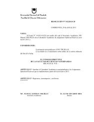
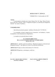
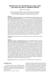
824 Fig. 1 Fig. 3 <strong>REPRODUCTION</strong> Fig. 4 Fig. 2 Fluorescent microscopic images of buffalo embryos at various stages of <strong>de</strong>velopment stained with AO/EB. Images obtained by conventional epifluorescent microscopy. (A)
DISCUSSION <strong>REPRODUCTION</strong> The results of the present study were consistent with other mammalian species that cell proliferation in IVP buffalo embryos is associated with apoptotic cell <strong>de</strong>ath. Apoptosis was first observed at 8- to 16-cell stage in embryos which otherwise had normal morphology The Bcl-2/Bax ratio appears to <strong>de</strong>termine the fate of a cell 5 .RT-PCR analysis of 2- to 12cell human embryos revealed transcripts for both Bax and Bcl-2 throughout these early cleavage stages 6 . The <strong>de</strong>tection of apoptosis related genes in buffalo pre implantation embryos indicates that buffalo embryos are capable of un<strong>de</strong>rgoing programmed cell <strong>de</strong>ath and constitutively express genes that are required to execute the <strong>de</strong>ath program. High expression of cell <strong>de</strong>ath gene (Bax) may confirm that early embryos were inherently biased towards PCD 7 .Though Bax was highly expressed in preimplantation buffalo embryos, the fate (survival or <strong>de</strong>ath) of embryos is finally <strong>de</strong>termined along with other factors such as external stimuli, internal <strong>de</strong>fects and activation of other apoptotic genes .ROS <strong>de</strong>rived oxidative stress during in vitro culture of embryos induce mediate mitochondria <strong>de</strong>pen<strong>de</strong>nt apoptotic response 8 . Addition of antioxidants to the culture media can <strong>de</strong>crease apoptosis in embryos during culture by preventing oxidant-mediated damage 9 . The results of the present study show that supplementation of cysteamine did not have significant effect on the inci<strong>de</strong>nce of apoptosis <strong>de</strong>tected by AO/EB staining. Enhanced <strong>de</strong>velopment of buffalo IVM-IVF embryos following the addition of cysteamine to a maturation and culture medium containing serum has been reported 1,3 but the mechanism through which cysteamine brings about this <strong>de</strong>velopment is not clear. We hypothesized that cysteamine would have promoted <strong>de</strong>velopment of embryos through anti- apoptosis but there was no significant difference between the level of expression of Bax and Bcl XL between control and cysteamine treated groups. Fetal bovine serum supplementation would have masked the effect of cysteamine. Study with serum free medium may give the clear picture of mechanism of cysteamine. In conclusion, the results of the present study suggest that the inci<strong>de</strong>nce of apoptotic morphology was significantly higher in produced buffalo embryos at advanced stages of embryos, the relative abundance of pro-apoptotic Bax transcripts was significantly higher than the anti-apopotic Bcl XL transcripts in all the <strong>de</strong>velopmental stages and supplementation of cysteamine in IVM and IVC media did not have any significant effect on the apoptosis and the relative abundance of Bax and Bcl XL transcripts. Acknowledgement. This work was supported by Niche project on buffalo production and reproduction genomics REFERENCES 1. Anand T., D.Kumar, M.S. Chauhan, R.S. Manik and P.Palta. 2008. Cysteamine supplementation of in vitro maturation medium, in vitro culture medium or both media promotes in vitro <strong>de</strong>velopment of buffalo (Bubalus bubalis) embryo. Rep Fert Dev. 20:253-257 2. <strong>de</strong> Matos, D.G., B. Gasparrini, S.R. Pasqualini and J.G.Thompson. 2002. Effect of glutathione synthesis stimulation during in vitro maturation of ovine oocytes on embryo <strong>de</strong>velopment and intracellular peroxi<strong>de</strong> content. Theriogenology.57: 1443-1451 3. Gasparrini, B., H.Sayoud, G. Neglia, D.G. <strong>de</strong> Matos, I. Donnay and L. Zicarelli. 2003. Glutathione synthesis during in vitro maturation of buffalo (Bubalus bubalis) oocytes: effects of cysteamine on embryo <strong>de</strong>velopment. Theriogenology. 60:943-952. 4. Velez-Pardo, C., Morales, A.T., Del Rio, M.J. and Olivera-Angel, M. 2007. Endogenously generated hydrogen peroxi<strong>de</strong> induces apoptosis via mitochondrial damage in<strong>de</strong>pen<strong>de</strong>nt of NF- B and p53 activation in bovine embryos. Theriogenology, 67: 1285-1296 K 5. Wyllie, AH. Kerr, J.F., Currie, A.R. 1980. Cell <strong>de</strong>ath: the significance of apoptosis. Int Rev Cytol, 68:251-306. 6. Warner, C. M., Cao, W., Exley, G. E., McElhinny, A. S.,Alikani, M., Cohen, J., Scott, R. T. & Brenner, C. A. (1998).Genetic regulation of egg and embryo survival. Hum. Reprod. 13 (Suppl. 3), 178–196. 7. Oltvai ZN,Milliman CL, Korsmeyer SJ. 1993. Bcl-2heterodimerizes in vivo with a conserved homolog, Bax, that accelerates programmed cell <strong>de</strong>ath. Cell 74:609–619. 8. Herrera, B., A.M. Alvarez, A. Sanchez, M. Fernan<strong>de</strong>z, C. Roncero, M. Benito and I.Fabregat. 2001. Reactive oxygen species (ROS) mediates the mitochondrial-<strong>de</strong>pen<strong>de</strong>nt apoptosis induced by transforming growth factor (beta) in fetal hepatocytes. FASEB J. 15:741–751. 9. Uhm, S.J., M.K. Gupta, J.H. Yang, S.H. Lee and H.T. Lee. 2007. Selenium improves the <strong>de</strong>velopmental ability and reduces the apoptosis inporcine parthenotes. Mol Reprod Dev. 74:1386–1394. Buenos Aires, Abril 2010 825