"Removal of Lymphoid Organs". In: Current Protocols in ... - IMBA
"Removal of Lymphoid Organs". In: Current Protocols in ... - IMBA
"Removal of Lymphoid Organs". In: Current Protocols in ... - IMBA

Create successful ePaper yourself
Turn your PDF publications into a flip-book with our unique Google optimized e-Paper software.
SELECTED SURGICAL PROCEDURES<br />
<strong>Removal</strong> <strong>of</strong> <strong>Lymphoid</strong> Organs<br />
The removal <strong>of</strong> lymphoid organs for further culture, isolation <strong>of</strong> cell populations, or direct<br />
test<strong>in</strong>g is one <strong>of</strong> the simplest and most frequently used procedures <strong>in</strong> immunology. Typical<br />
uses <strong>in</strong>clude provid<strong>in</strong>g cells for cytotoxicity or proliferation assays, for analysis <strong>of</strong><br />
cytok<strong>in</strong>e production or response, and for the isolation <strong>of</strong> B, T, and adherent cell subpopulations<br />
for culture or reconstitution <strong>of</strong> other animals. This unit covers the identification<br />
and removal <strong>of</strong> mouse lymphoid organs, but the same protocols are applicable to other<br />
rodents and rabbits with m<strong>in</strong>or modifications.<br />
REMOVAL OF MOUSE LYMPHOID ORGANS<br />
Materials<br />
70% ethanol<br />
Hanks balanced salt solution (HBSS; APPENDIX 2) or tissue culture medium<br />
Iris scissors (straight or angled)<br />
Iris forceps (curved)<br />
Tissue culture plate<br />
Additional reagents and equipment for euthanasia (UNIT 1.8)<br />
Prepare the animal<br />
1. Sacrifice the animal <strong>in</strong> a humane manner. Place it on its back on clean, dry, absorbent<br />
paper.<br />
2. Wet the fur with 70% ethanol to sterilize the area and reduce the possibility <strong>of</strong><br />
contam<strong>in</strong>ation.<br />
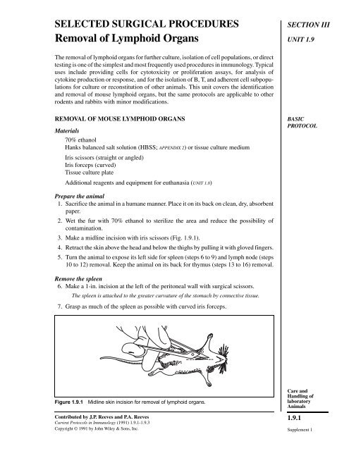
3. Make a midl<strong>in</strong>e <strong>in</strong>cision with iris scissors (Fig. 1.9.1).<br />
4. Retract the sk<strong>in</strong> above the head and below the thighs by pull<strong>in</strong>g it with gloved f<strong>in</strong>gers.<br />
5. Turn the animal to expose its left side for spleen (steps 6 to 9) and lymph node (steps<br />
10 to 12) removal. Keep the animal on its back for thymus (steps 13 to 16) removal.<br />
Remove the spleen<br />
6. Make a 1-<strong>in</strong>. <strong>in</strong>cision at the left <strong>of</strong> the peritoneal wall with surgical scissors.<br />
The spleen is attached to the greater curvature <strong>of</strong> the stomach by connective tissue.<br />
7. Grasp as much <strong>of</strong> the spleen as possible with curved iris forceps.<br />
������������� ������������������������������������������������������<br />
Contributed by J.P. Reeves and P.A. Reeves<br />
<strong>Current</strong> <strong>Protocols</strong> <strong>in</strong> Immunology (1991) 1.9.1-1.9.3<br />
Copyright © 1991 by John Wiley & Sons, <strong>In</strong>c.<br />
SECTION III<br />
UNIT 1.9<br />
BASIC<br />
PROTOCOL<br />
Care and<br />
Handl<strong>in</strong>g <strong>of</strong><br />
laboratory<br />
Animals<br />
1.9.1<br />
Supplement 1
<strong>Removal</strong> <strong>of</strong><br />
<strong>Lymphoid</strong> Organs<br />
1.9.2<br />
8. Gently pull the spleen free <strong>of</strong> the peritoneum, tear<strong>in</strong>g the connective tissue beh<strong>in</strong>d<br />
the spleen.<br />
Alternatively, the top and bottom portions <strong>of</strong> the spleen can be grasped and torn from the<br />
connective tissue one part at a time. It is usually more efficient to remove the spleen all at<br />
once, particularly if large numbers <strong>of</strong> animals will be used.<br />
9. Place the spleen <strong>in</strong> several milliliters <strong>of</strong> HBSS or tissue culture medium <strong>in</strong> a tissue<br />
culture plate.<br />
The choice <strong>of</strong> medium is dependent upon the protocol the cells will be subjected to. See<br />
Chapters 3 & 4.<br />
Remove the lymph nodes<br />
10. Identify the lymph nodes as shown <strong>in</strong> Figure 1.9.2.<br />
The major lymph nodes are found <strong>in</strong> the axillary, cervical, <strong>in</strong>gu<strong>in</strong>al, and mesenteric regions.<br />
<strong>In</strong> normal, nonimmunized animals ma<strong>in</strong>ta<strong>in</strong>ed <strong>in</strong> clean facilities, the lymph nodes are<br />
usually quite small and can easily be overlooked. Axillary nodes can be readily identified<br />
upon dissection <strong>in</strong> this region, and occasionally the adjacent brachial nodes are found <strong>in</strong><br />
the triceps area. Cervical nodes flank the trachea and are beneath the large salivary gland.<br />
<strong>In</strong>gu<strong>in</strong>al nodes are found <strong>in</strong> the gro<strong>in</strong> area <strong>in</strong> a fatpad attached to the sk<strong>in</strong>, but after<br />
retraction <strong>of</strong> the sk<strong>in</strong>, are <strong>of</strong>ten found buried <strong>in</strong> fat at or near the knees. Mesenteric lymph<br />
nodes lie deep <strong>in</strong> the <strong>in</strong>test<strong>in</strong>al mesentery. Both cervical and mesenteric nodes are<br />
frequently overlooked. Typically, 1-5 × 10 7 lymph node cells can be isolated from a s<strong>in</strong>gle<br />
mouse.<br />
11. Grasp the lymph nodes with curved forceps and pull them free <strong>of</strong> attached tissue.<br />
Occasionally, it is necessary to dissect adjacent fat.<br />
12. Place the lymph nodes <strong>in</strong> several milliliters <strong>of</strong> HBSS or tissue culture medium <strong>in</strong> a<br />
tissue culture plate.<br />
������������� ����������������������������������������<br />
Supplement 1 <strong>Current</strong> <strong>Protocols</strong> <strong>in</strong> Immunology
Remove the thymus<br />
13. Make an <strong>in</strong>cision <strong>in</strong> the chest, beg<strong>in</strong>n<strong>in</strong>g at the xiphoid and extend<strong>in</strong>g to the neck<br />
with surgical scissors.<br />
14. Retract the ribs with curved forceps.<br />
It may be necessary to crack the ribs for effective retraction. The thymus is a yellowish-white<br />
bi-lobed organ found just under the ribs, attached above the heart <strong>in</strong> the midl<strong>in</strong>e.<br />
15. Grasp each lobe <strong>of</strong> the thymus with curved forceps and gently pull the thymus away.<br />
Because the thymus is delicate, it is not unusual to tear it dur<strong>in</strong>g removal. Unless maximal<br />
cell recovery is required, the tear<strong>in</strong>g will not be harmful. Thymic size varies with age, be<strong>in</strong>g<br />
maximal at 3 to 5 weeks <strong>in</strong> the mouse and shr<strong>in</strong>k<strong>in</strong>g to a m<strong>in</strong>imum at ∼6 months. The number<br />
<strong>of</strong> cells obta<strong>in</strong>ed is related to the age <strong>of</strong> the animal and the size <strong>of</strong> the thymus.<br />
16. Place the thymus tissue <strong>in</strong> several milliliters <strong>of</strong> balanced salt solution or tissue culture<br />
medium <strong>in</strong> a tissue culture plate.<br />
Key References<br />
Sjod<strong>in</strong>, K., Dalmasso, A.P., Smith, J.M., and Mart<strong>in</strong>ez, C. 1963. Thymectomy <strong>in</strong> newborn and adult mice.<br />
Transplantation 1:521-525.<br />
Van Alten, P.J. 1984. Thymectomy and engraftment <strong>of</strong> thymic tissue. Methods Enzymol. 108:10-19.<br />
Contributed by J.P. Reeves and P.A. Reeves<br />
Food and Drug Adm<strong>in</strong>istration<br />
Bethesda, Maryland<br />
Care and<br />
Handl<strong>in</strong>g <strong>of</strong><br />
laboratory<br />
Animals<br />
1.9.3<br />
<strong>Current</strong> <strong>Protocols</strong> <strong>in</strong> Immunology Supplement 2