Congress Chair - ISTA
Congress Chair - ISTA
Congress Chair - ISTA

You also want an ePaper? Increase the reach of your titles
YUMPU automatically turns print PDFs into web optimized ePapers that Google loves.
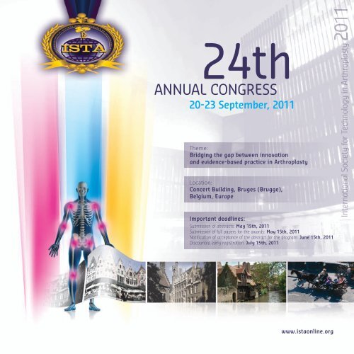
24th<br />
ANNUAL CONGRESS<br />
20-23 September, 2011<br />
Theme:<br />
Bridging the gap between innovation<br />
and evidence-based practice in arthroplasty<br />
Location:<br />
concert Building, Bruges (Brugge),<br />
Belgium, europe<br />
Important deadlines:<br />
Submission of abstracts: May 15th, 2011<br />
Submission of full papers for the awards: May 15th, 2011<br />
Notification of acceptance of the abstract for the program: June 15th, 2011<br />
Discounted early registration: July 15th, 2011<br />
www.istaonline.org<br />
International Society for Technology in Arthroplasty Arthroplasty Arthroplasty Arthroplasty 2011
Invitation<br />
Dear Colleagues,<br />
It is with pleasure that we are inviting you to the 24th annual congress<br />
of the International Society for Technology in Arthroplasty, which is<br />
to be held in Brugge, Belgium at 21-23 September, 2011. <strong>ISTA</strong>’11 is<br />
a meeting for orthopaedic surgeons, scientists, engineers, allied<br />
health and representatives from orthopaedic industry. The goal<br />
of the meeting is to promote transfer of knowledge in the field of<br />
arthroplasty in the broadest thinkable sense.<br />
The theme of this years’ meeting is ‘Bridging the gap between<br />
innovation and evidence based practice in arthroplasty’, which<br />
demonstrates our initiative to address the incompatibilities that are<br />
often present between innovation of orthopaedic implants on the one<br />
hand and evidence based practice on the other. Renowned speakers<br />
are invited to address this issue from their perspective. Furthermore,<br />
key-experts will inform you about the most recent developments<br />
related to technology and clinical performance in arthroplasty.<br />
and we will organize at least two dedicated sessions on Computer<br />
In association with the Belgian and Dutch<br />
Orthopaedic Societies<br />
Assisted Orthopaedic Surgery in conjunction with the International<br />
Society of CAOS. Further scientific high-lights of the meeting are the<br />
presentation of the <strong>ISTA</strong>’s Life Time Achievement Award, the HAP Paul<br />
award and the Biomechanics and Biomaterials Student awards.<br />
The venue is situated in the heart of the beautiful city of Brugge,<br />
which is included in the UNESCO list of the World Heritage. The city<br />
radiates beauty, history and romance and we guarantee that your<br />
presence will be a memorable experience.<br />
The organizing committee is putting together an exciting scientific<br />
as well as a social program and we look forward to receive your<br />
submissions for the award papers, your abstracts for the open<br />
program, and, most of all, look forward to welcome you personally<br />
in Brugge!<br />
Nico Verdonschot PhD<br />
Johan Bellemans PhD MD<br />
Jan Victor PhD MD<br />
Meeting Co-Directors
Scientific program<br />
1. hIp anD Knee arthroplaSty 2. enaBlIng technologIeS<br />
• Knee Mechanics<br />
• Hip Mechanics<br />
• Alternative Bearing Materials in THA<br />
• Ethnic and Gender Issues in Arthroplasty<br />
• Patellar Resurfacing Issue<br />
• Bearing Mobility Issues<br />
• Hip Resurfacing<br />
• High Demand Issues<br />
3. non arthroplaSty<br />
proceDUreS/ISSUeS<br />
• Peri-prosthetic Fracture Management<br />
• Cartilage Repair/Regeneration<br />
• Complication Management<br />
• Hip Disease: Non Arthroplasty Options<br />
• Knee Disease: Non Arthroplasty Options<br />
<strong>Congress</strong> program<br />
tuesday, Sept. 20, 2011<br />
18.30: Welcome reception City Hall, Bruges<br />
Wednesday, Sept. 21, 2011<br />
08.30 - 17.00: Scientific Session<br />
18.00 - 20.00: Posters Session<br />
• Computer Navigation<br />
in Joint Replacement<br />
• Minimally Invasive Surgical<br />
Techniques<br />
• Sensor Technology in Arthroplasty<br />
• Computer simulations<br />
• Musculo-skeletal modeling<br />
• Imaging Technology<br />
• Robotics<br />
• Evidence Based Medicine<br />
• Functional analyses<br />
4. SpIne, ShoUlDer anD SMall<br />
JoInt arthroplaSty<br />
• Spinal Fusion vs. Disc Replacement<br />
• Shoulder Complications and<br />
Arthroplasty Options<br />
• Ankle Complications and<br />
Arthroplasty Options<br />
thursday, Sept. 22, 2011<br />
08.30 - 16.00: Scientific Session<br />
16.00 - 17.00: Award Session<br />
19.30: Award dinner at Provinciaal Hof, Bruges<br />
friday, Sept. 23, 2011<br />
08.00 - 16.00: Scientific Session<br />
ISta<br />
life time<br />
achievement<br />
award<br />
Professor Tony Unsworth<br />
started his career in 1969 at<br />
Leeds University and works<br />
at Durham University since<br />
1976 where he is Director of<br />
the Centre for Biomedical<br />
Engineering and Research<br />
Director for the Faculty of<br />
Science. His research area<br />
is the tribology of human<br />
and artificial joints but in<br />
addition he has published<br />
on aspects of rehabilitation<br />
of the upper limb. He has<br />
been awarded the Tribology<br />
Silver Medal in 1972,<br />
the Donald Julius Groen<br />
Prize, the James Clayton<br />
Prize,1999 (IMechE) and<br />
in 2005 was awarded the<br />
James Alfred Ewing Medal<br />
of the Institution of Civil<br />
Engineers for his research<br />
into the Tribology of human<br />
and artificial human<br />
joints. He has published in<br />
excess of 310 papers and<br />
given circa 100 lectures at<br />
international conferences/<br />
venues.<br />
www.istaonline.org
Invited Speakers (to be adjusted)<br />
Lars Engebretsen, norway<br />
Rick Komistek, USa<br />
Dan Berry, USa<br />
Jerry Engh, USa<br />
Jean-Noel Argenson, france<br />
Philippe Neyret, france<br />
Award Paper Criteria<br />
1. lIfetIMe achIeVeMent aWarD<br />
This award recognizes a life long contribution to advancing the state of the art<br />
in joint reconstruction that has changed the landscape of arthroplasty. This<br />
achievement was acknowledged in the past years to Drs. Burstein, Goodfellows,<br />
Ranawat, Freeman, Yamamuro and Walker. The recipient at <strong>ISTA</strong> 2010 will give<br />
a 20-minute presentation in a plenary session and the award will be presented<br />
during a special ceremony at the Gala Dinner.<br />
The plaque is sponsored by <strong>ISTA</strong>.<br />
3. StUDent BIoMechanIcS<br />
paper aWarD<br />
($1,000 US CASH AWARD)<br />
This award will be presented to a student who<br />
submits the best paper on biomechanics in the field<br />
of arthroplasty. The recipient will give a 10-minute<br />
presentation at a plenary session. The award will be<br />
presented at the Gala Dinner.<br />
applIcatIon for aWarDS 2-4<br />
David Murray, UK<br />
Andrew Amis, UK<br />
?? Lombardi, USa/Italy (??)<br />
Fabio Catani, Italy<br />
Philipp Lobenhoffer, germany<br />
Roland Becker, germany<br />
For the awards No. 2-4, researchers can apply by submitting both an abstract and a full<br />
paper. Authors can submit as many papers as they want for different awards, but only<br />
one paper will be accepted for each presenting author. For students participating for<br />
the awards, a written confirmation of the student-status of the first author should be<br />
provided by the supervisor of the applicant. Applicants are asked to indicate which<br />
award they are applying for in the last section of the abstract submission form.<br />
Please prepare the full paper following the guidelines for the Journal of Arthroplasty<br />
(see http://www.arthroplastyjournal.org/authorinfo) and submit the paper through the<br />
on-line process located on <strong>ISTA</strong>’s website: www.<strong>ISTA</strong>online.org<br />
4. StUDent BIoMaterIalS<br />
paper aWarD<br />
($1,000 US CASH AWARD)<br />
This award will be presented to a student who<br />
submits the best paper on biomaterials in the field<br />
of arthroplasty. The recipient will give a 10-minute<br />
presentation in a plenary session. The award will be<br />
presented at the Gala Dinner.<br />
Christian Gerber, Switzerland<br />
Ate Wymenga, netherlands<br />
Wim Schreurs, netherlands<br />
Jean-Piere Simon, Belgium<br />
Rene Verdonk, Belgium<br />
Arun Mullaji, India<br />
2. “hap” paUl aWarD<br />
($3,000 US CASH AWARD)<br />
Howard A. Paul, DVM, was a tireless researcher advancing the science and technology<br />
of arthroplasty. He was one of the charter members of <strong>ISTA</strong> and an award<br />
has been established to honor his outstanding contribution. A special 20-minute<br />
presentation will be presented in a plenary session for the best paper on new<br />
developments in the field of arthroplasty and the award will be presented during<br />
a special ceremony at the Gala Dinner.<br />
5. poSter aWarDS<br />
($1,000 US CASH AWARD)<br />
Three poster awards will be awarded<br />
– $500, $250 and $250 – to the top three posters<br />
presented at the meeting.<br />
Posters will be judged at the meeting.<br />
The awards will be presented at the<br />
Gala Dinner.<br />
The awards winning papers will be forwarded to the Journal of Arthroplasty for<br />
publication; however, there is no guarantee that the papers will be accepted for<br />
publication.<br />
aBStract SUBMISSIon<br />
Abstract and Award Papers must be submitted via the <strong>ISTA</strong> 2011 official website at<br />
www.<strong>ISTA</strong>online.org. Submissions will open on March 1, 2011 and close on May 15th,<br />
2011. Abstract notifications will be sent on June 15th, 2011. Abstract and Award Papers<br />
will only be considered if they are submitted through the <strong>ISTA</strong> on-line submission<br />
process. For any inquiries, please feel free to contact the congress secretariat using the<br />
information provided on www.<strong>ISTA</strong>online.org.
Registration<br />
regIStratIon feeS for the ISta 2011 are lISteD BeloW.<br />
the profeSSIonal regIStratIon fee InclUDeS<br />
• Final Program and Abstract book (with CD)<br />
• Access to the research electronically<br />
• Reception, lunches and coffee breaks<br />
cMe accreDItatIon<br />
The 24th Annual <strong>Congress</strong> of the International Society for Technology<br />
in Arthroplasty is planned and organized in accordance with the<br />
policies of the European Accreditation Council for Continuing Medical<br />
Education (EACCME). The EACCME is an institution of the European<br />
• Admission to all oral scientific lectures<br />
• Admission to the poster session<br />
• Admission to the exhibit hall<br />
category UntIl JUly 15, 2011 JUly 16 – aUgUSt 15, 2011 froM aUgUSt 16, 2011<br />
Professional € 450 € 500 € 550<br />
<strong>ISTA</strong> member<br />
Fellow/Resident/<br />
€ 400 € 450 € 500<br />
Allied Health € 200 € 250 € 275<br />
Graduate Student € 200 € 250 € 300<br />
Accommodation Airport Transfer<br />
Due to the specific historical charm of the city of Bruges<br />
(Brugge), we made a selection of different hotels close<br />
(walking distance) to the congress location (Concert Hall).<br />
Please consult the website (www.istaonline.org) to find the<br />
different hotel possibilities with the <strong>ISTA</strong> negotiated rates<br />
(reduced rates). You will also find the contact/booking details<br />
of each hotel for direct booking of your accommodation.<br />
Union of Medical Specialists (UEMS), www.uems.be. EACCME<br />
approved credits are recognized by the American Medical Association<br />
for conversion to AMA PRA Category 1 Credit.<br />
A pre-bookable transfer service from Brussels Airport (Zaventem) to<br />
Bruges will be provided at an interesting rate. Information will be<br />
communicated to all registered participants in due time.<br />
www.istaonline.org
Bruges, one of the most beautiful<br />
cities in Europe...<br />
It was a justified motive that prompted UNESCO in 2000 to include the entire historical city centre on the World Heritage list.<br />
Walking along the maze of winding cobbled alleys and romantic canals, you imagine yourself to be in medieval times.<br />
The wealth of museums is a striking image of this city’s stirring history.<br />
Bruges is also home to contemporary culture, such as the new Concert Hall,<br />
which is one of the most prominent music complexes in Flanders.<br />
The restaurants in Bruges which offer gastronomic cuisine<br />
and the exclusive hotels are a true feast for those who enjoy<br />
the good things in life.<br />
Join us to explore this medieval beauty…<br />
<strong>ISTA</strong> supported social events:<br />
Reception Tuesday, September 20, 2011 I City Hall, Bruges.<br />
Award dinner Thursday, September 22, 2011 I The Provincial Court, Bruges.<br />
Participation fee € XX per person I On-line pre-booking only.
Social Partner Program:<br />
Wednesday September 21, 2011 – 18h30 (06.30 pm)<br />
(pre-registration possible through congress registration on-line)<br />
Bruges’ highlight Tour (2 hours) - Price: pp 45 €<br />
This tour will encompass all the high spots that Bruges has to offer. The historic<br />
center is part of the UNESCO World Heritage since the year 2000.<br />
With his loud voice and a bell the Belleman of Bruges announces the tour in front<br />
of the Concert Hall. With our enthusiastic guides you will pass by the well-known<br />
Beguinage, the majestic Palace of Gruuthuse, Our Lady’s Church, the ancient Burg<br />
Social program possibilities:<br />
(bookable on site, upon availability, at the Concert Hall special desk)<br />
Flanders Fields - Day Tour (8hrs) - Price: pp 85 €<br />
A full day tour with private coach or mini-van to Flanders Fields.<br />
Pick-up at the Concert Hall on ‘t Zand. Day tour includes:<br />
• The largest WW1 German cemetery (Student cemetery) in Langemark.<br />
• The world’s largest WW1 cemetery of the Commonwealth:<br />
Tyne cot Cemetery at Passchendale.<br />
• The Menin gate at Ypers, hill 60, hill 62, bunkers during the day.<br />
• Visiting the centre of Ypres and we will have a lunch in Ypres.<br />
• Mount Kemmel cote 160, the Hills of Flanders.<br />
• Different WW1 trenches and also memorials from the Commonwealth.<br />
Brussels - Day Tour (8hrs) - Price: pp 115 €<br />
A full day tour with private coach or mini-van to Brussels.<br />
Pick-up at the Concert Hall on ‘t Zand.<br />
Brussels, the capital of Belgium and Europe is a meeting point of diverse<br />
architectural, literary, historical and cultural currents, set right in the heart of<br />
Belgium. During this tour, you will discover the city of Brussels with the help of our<br />
professional guides, who will describe its historyand social life. Driven by coach you<br />
will discover Laeken, the residential and royal commune with its historical castles,<br />
which forms the private residence of the Royal Family of Belgium. In this area you<br />
will also see the site of the World Exhibition, the Trade Mart, the Planetarium and<br />
Brupark – Mini Europe, all dominated by the impressive Atomium.<br />
During the drive through the European district, at the edge of the Cinquantenaire<br />
Park and its monumental ensemble, built under Leopold II, your guide will outline<br />
the European functions of Brussels. Via the Royal District with its Palace and Park,<br />
the Parliament buildings and the <strong>Congress</strong> Column, we will descend gently to the<br />
lower town and its famous Grand Place.<br />
During the second part of this excursion, you will discover by foot (pedestrian zone)<br />
the Town Hall (Hôtel de Ville) surrounded by Guild Houses, Brussels most famous<br />
citizen Manneken Pis and finally the winding little streets of the Ilôt Sacré.<br />
Incl lunch in Brussels.<br />
square and its gothic City Hall, the Basilica of the Holy Blood and the impressive<br />
market square with the famous Bell Tower.<br />
During the tour we invite you to the secluded inner garden of the Bladelin Court,<br />
the medieval house of the family de Medici, which houses a convent nowadays.<br />
Here a musical duo plays a concert on their self made medieval instruments and<br />
we offer you a little box of tasty chocolates made in Bruges.<br />
At the fish market fish sellers will welcome you in their fish selling scène. They offer<br />
you a tasty fish bite and a real Belgian jenever.<br />
During the tour a private boat trip on the Bruges’ canals is also included!<br />
Bruges Art Route (3 hrs) - Price: pp 52 €<br />
An arty tour where we get an exclusive insight in the lives and work of<br />
4 local artists. We get a look behind the scenes and a demonstration of<br />
their art. For example an airbrush artist, a young letter-cutter in stone,<br />
a glass blower etc. Drinks and little bites are provided during the visits.<br />
Bruges in 5 Senses (3 hrs) - Price: pp 75 €<br />
A sensory walk in 5 steps along Bruges’ hidden spots. We listen to the<br />
sounds of a small concert and taste and smell chocolate at the workshop<br />
of a top chocolatier. In the working place of Peter Quijo we see how a<br />
rough gem is polished to a brilliant diamond. And we finish with a hand<br />
massage. And maybe we can even tempt your sixth sense!<br />
Bruges - Damme by Bike (3,5 hrs) - Price: pp 36 €<br />
Just outside Bruges is a unique natural area that extends to the coast<br />
(North Sea). Jacques Brel called this polders “Le Plat Pays”. Central in this<br />
Polders we find some small mediaeval towns like Damme, Oostkerke and<br />
Hoeke. Little white villages with a medieval past as a link between Bruges<br />
and the sea. Together with our guides we cycle through the beautiful<br />
nature and obviously we do not forget to taste our local specialties such<br />
as beer and waffles.<br />
Tranquil Bruges (3 hrs) - Price: pp 45 €<br />
A private boat awaits us in the historic city center and takes us to the<br />
heart of Saint-Anne, a quiet beautiful part of Bruges that’s unknown to<br />
tourists. Here we find the part of Bruges with its cobblestone streets, alms<br />
houses, windmills, churches and unique views of the canals. During this<br />
walk we visit the well conserved Lace Center and see a demonstration of<br />
this traditional Bruges’ handicraft. And of course we also have a drink in<br />
the oldest pub in town, Café Vlissinghe that dates from 1515.<br />
Golf activities on request.<br />
www.istaonline.org
images: ©JAN DARTHET/TOERISME BRUGGE<br />
Organizing committee<br />
Local Organizers:<br />
Nico Verdonschot<br />
Johan Bellemans<br />
Jan Victor<br />
Dennis Janssen<br />
Wim Schreurs<br />
Pieter Spierings<br />
<strong>ISTA</strong> Board members:<br />
John Hollingdale President<br />
Richard D. Komistek First Past President<br />
Won Yong Shon Second Past President<br />
Robert M. Streicher Secretary General<br />
Hani Haider Program Director<br />
Jeff Taylor Chief Financial Officer<br />
Joseph Fetto Board Member<br />
Claude Rieker Board Member<br />
Mike Tuke Board Member<br />
Shinro Takai Board Member<br />
Scott A. Banks Board Member<br />
InternatIonal SocIety for technology In arthroplaSty<br />
P. O. Box 6564, Auburn, CA 95604<br />
Phone: 916-454-9884 I Fax: 916-454-9882 I E-Mail: ista@pacbell.net
<strong>ISTA</strong> 2011<br />
September 20-‐23, 2011<br />
Concert Hall Bruges, Bruges, Belgium<br />
<strong>Congress</strong> <strong>Chair</strong>:<br />
John Hollingdale, M.
8B : Bearings - hip: #869 September 22nd, 2011, 13:55-14:45<br />
Wear of Ceramic-on-Ceramic Bearings in THRs: Effect of Head Size Under Steep Cup Inclination Angle<br />
and Microseparation and Edge Loading Conditions<br />
*Mazen Al-Hajjar - University of Leeds - Leeds, UK<br />
John Fisher - University of Leeds - Leeds, UK<br />
Joanne Tipper - University of Leeds - Leeds, UK<br />
Sophie Williams<br />
Louise Jennings - University of Leeds - Leeds, UK<br />
*Email: mnmah@leeds.ac.uk<br />
INTRODUCTION<br />
Ceramic-on-ceramic hip replacements have generated great interest in recent years due to substantial improvements<br />
in manufacturing techniques and material properties 1 . Microseparation conditions that could occur due to several<br />
clinical factors such as head offset deficiency, medialised cup combined with laxity of soft tissue resulting in a<br />
translation malalignment, have been shown to cause edge loading, replicate clinically relevant wear mechanisms 2,3<br />
and increase the wear of ceramic-on-ceramic bearings 3,4 . The aim of this study was to investigate the influence of<br />
increasing the femoral head size on the wear of ceramic-on-ceramic bearings under several clinically relevant<br />
simulator conditions.<br />
MATERIALS AND METHODS<br />
The wear of size 28mm and 36mm ceramic-on-ceramic bearings (BIOLOX ® Delta, CeramTec, Germany) was<br />
determined under different in vitro conditions using the Leeds II hip simulator. For each size bearing, two clinical<br />
cup inclination angles were considered, 55 o (n=3) and 65 o (n=3) for the 28mm bearing and 45 o (n=3) and 65 o<br />
(n=3) for the 36mm bearing. The first two (28mm study) or three (36mm study) million cycles ran under standard<br />
gait conditions and a subsequent three million cycles ran under microseparation conditions. A standard gait cycle<br />
included a twin peak load (300N-3000N), extension/flexion (-15 o /+30 o ) and internal/external rotation (±10 o ).<br />
Microseparation 3 was achieved by applying a 0.4-0.5mm medial displacement to the cup relative to the head<br />
during the swing phase of the standard gait cycle resulting in edge loading at heel strike. The lubricant was 25%<br />
(v/v) newborn calf serum, which was changed approximately every 333,000 cycles. The wear volume was<br />
ascertained through gravimetric analysis every million cycles. One-way ANOVA was performed (significance:<br />
p
(LMBRU).<br />
REFERENCES<br />
Masson, International Orthopaedics,2009;33:359-363.<br />
Nevelos etal.,Biomaterials,1999;20(19):1833-40.<br />
Nevelos etal.,Arthroplasty,2000;15(6):793-5.<br />
Stewart etal.,Mater Sci Mater Med,2001;2(10-12):1053-6.<br />
Figures<br />
HYPERLINK "http://app.istaonline.org/figures/1557.jpg" \t "_blank" INCLUDEPICTURE \d "http://<br />
app.istaonline.org/figures/1557.jpg" \* MERGEFORMATINET<br />
Figure 4 HYPERLINK "http://app.istaonline.org/figures/1558.jpg" \t "_blank"<br />
INCLUDEPICTURE \d "http://app.istaonline.org/figures/1558.jpg" \* MERGEFORMATINET<br />
Figure 5 HYPERLINK "http://app.istaonline.org/figures/1559.jpg" \t "_blank"<br />
INCLUDEPICTURE \d "http://app.istaonline.org/figures/1559.jpg" \* MERGEFORMATINET<br />
Figure 6<br />
8B : Bearings - hip: #822 September 22nd, 2011, 13:55-14:45<br />
Wear of Metal-on-Metal Bearings in THRs: Effect of Head Size Under Steep Cup Inclination Angle,<br />
Microseparation and Edge Loading Conditions<br />
*Mazen Al-Hajjar - University of Leeds - Leeds, UK<br />
John Fisher – University of Leeds - Leeds, UK<br />
Sophie Williams<br />
Joanne Tipper - University of Leeds - Leeds, UK<br />
Louise Jennings - University of Leeds - Leeds, UK<br />
*Email: mnmah@leeds.ac.uk<br />
INTRODUCTION<br />
Retrieval and clinical studies of metal-on-metal (MoM) bearings have associated increased wear 1 and elevated<br />
patient ion levels 2 with steep cup inclination angles and edge loading conditions. The University of Leeds have<br />
previously developed a hip simulator method that has been validated against retrievals and shown to replicate<br />
clinically relevant wear rates and wear mechanisms 3,4 . This method involves introducing lateral microseparation<br />
to represent adverse joint laxity and offset deficiency. This study aimed to investigate the effect of<br />
microseparation representing translational malpostion, and increased cup inclination angle, representing rotational<br />
malposition, in isolation and combined on the wear of different sizes (28 and 36mm) MoM bearing in total hip<br />
replacement (THRs).<br />
MATERIALS AND METHODS<br />
The wear of size 28mm and 36mm MoM THRs bearings was determined under different in vitro conditions using<br />
the Leeds II hip simulator. For each size bearing, two clinical cup inclination angles were considered, 45 o (n=3)<br />
and 65 o (n=3). The first three million cycles were run under standard gait conditions and subsequently three<br />
million cycles were run under microseparation conditions. Standard gait cycles included a twin peak load<br />
(300N-3000N), extension/flexion (-15 o /+30 o ) and internal/external rotation (±10 o ). Microseparation 4 was<br />
achieved by applying a 0.4-0.5mm medial displacement to the cup relative to the head during the swing phase of<br />
the standard gait cycle resulting in edge loading at heel strike. The lubricant was 25% (v/v) newborn calf serum.<br />
The wear volume was determined through gravimetric analysis every million cycles. One-way ANOVA was<br />
performed (significance: p
(Figure 1).<br />
DISCUSSION<br />
With the larger size bearings, head-rim contact occurred at a steeper cup inclination angle (>65 o ) providing an<br />
advantage over smaller bearings. Under standard gait conditions, where head-rim contact did not occur, wear was<br />
low, due to mixed lubrication and wear reduction through a protein boundary film. However, edge loading of the<br />
cup, with elevated stress, caused excess damage and wear. This effect was more dominant with microseparation<br />
conditions to that of head-rim contact due to increased cup inclination angle alone.<br />
Under microseparation conditions, there were no significant differences in the wear rates of the 28mm and the<br />
36mm size bearings. However, the wear rates obtained in this study for 28mm and 36mm bearings were<br />
significantly lower than those obtained for size 39mm surface replacement MoM bearings (8.99 mm 3 /million<br />
cycles) tested under the same adverse conditions 5 .<br />
CONCLUSION<br />
This study shows the importance of acetabular cup design and correct surgical positioning of the femoral head and<br />
acetabular cup and restoration of offset and cup centre.<br />
ACKNOWLEDGEMENT<br />
This study was supported by the Furlong Research Charitable Foundation (FRCF) and the National Institute of<br />
Health Research (NIHR) as part of a collaboration with the Leeds Musculoskeletal Biomedical Research Unit<br />
(LMBRU). The components were custom made specifically for this project by Corin Ltd.<br />
REFERENCES<br />
Morlock et al..JBJS-American Volume,2008.90A:p.89-95<br />
De Haan et al.,JBJS Br, 2008.90(10):p.1291-7.<br />
Nevelos et al.,Biomaterials,1999;20(19):1833-40.<br />
Nevelos et al.,J Arthroplasty,2000.15(6):p.793-5.<br />
Leslie et al.,.Clin Orthop Relat Res,2009.467(9):p.2259-2265<br />
Figures<br />
HYPERLINK "http://app.istaonline.org/figures/1560.jpg" \t "_blank" INCLUDEPICTURE \d "http://<br />
app.istaonline.org/figures/1560.jpg" \* MERGEFORMATINET<br />
Figure 2<br />
5B : Spine: #857 September 21st, 2011, 16:20-17:10<br />
The Effect of Varying the Stiffness of Spinal Fusion Devices on the Adjacent Levels Using Multibody<br />
Dynamics Simulation<br />
Pavel Galibarov - AnyBody Technology - Aalborg, Denmark<br />
*Amir Al-Munajjed - AnyBody Technology - Aalborg, Denmark<br />
Sebastian Dendorfer - University of Applied Sciences Regensburg - Regensburg, Germany<br />
Soeren Toerholm - AnyBody Technology - Aalborg, Denmark<br />
John Rasmussen<br />
*Email: aa@anybodytech.com<br />
INTRODUCTION:<br />
Several clinical studies demonstrated long-term adjacent-level effects after implantation of spinal fusion devices[1].<br />
These effects have been reported as adjacent joint degeneration and the development of new symptoms correlating<br />
with adjacent segment degeneration[2] and the trend has therefore gone to motion preservation devices; however,<br />
these effects have not been understood very well and have not been investigated thoroughly[3].<br />
The aim of this study is to investigate the effect of varying the stiffness of spinal fusion devices on the adjacent<br />
vertebral levels. Disc forces, moments and facet joint forces were analyzed.
METHODS:<br />
The AnyBody Modeling System was used to compute the in-vivo muscle and joint reaction forces of a<br />
musculoskeletal model. The full body model used in this study consists of 188 muscle fascicles in the lumbar<br />
spine and more than 1000 individual muscle branches in total. The model has been proposed by de Zee et al.<br />
[3],validated by Rasmussen et al.[4] and by Galibarov et al.[5]. The new model[5] determines the individual<br />
motions between vertebrae based on the equilibrium between forces acting on the vertebrae from muscles and<br />
joints and the passive stiffness in disks and ligaments, figure1a. An adult of 1.75 m and 75 kg with a spinal<br />
implant in L4L5 was modeled. This model was subjected to a flexion-extension motion using different elastic<br />
moduli to analyze and compare to a non-implanted scenario. The analyzed variables were vertebral motion, the disc<br />
reaction forces and moments, as well as facet joint forces in the treated and the adjacent levels: L2L3, L3L4, L4L5<br />
and L5-Sacrum.<br />
RESULTS:<br />
When introducing a spinal fusion device in the L4L5 joint the reaction forces and moments decreased in this joint<br />
with stiffer devices leading to lower joint loads. However, in the adjacent joints, L3L4 and L5Sacrum, an increase<br />
was observed when implanting stiffer devices. Similar trends could be found for the L2L3 joint. The loads in the<br />
facet joints showed the same trends. While introducing a spinal fusion device reduced the facet joint forces in the<br />
treated joint, the loads in the adjacent facet joints were increased according to the stiffness of the implanted device,<br />
figure1b.<br />
DISCUSSION:<br />
While the treated disc joint showed reduced motion and loads, the adjacent levels demonstrated a significant<br />
increase. In particular, the increased facet joint forces in the adjacent levels can lead to adjacent level facet pain or<br />
accelerated facet joint degeneration. Introducing a device resulted in preventing facet contact and therefore facet<br />
joint loads, even using the device with the lowest stiffness.<br />
CONCLUSION:<br />
The presented model shows that clinical complications such as facet joint degeneration in adjacent levels after<br />
implantation of spinal fusion device are consistent with the change in the mechanical-stimulus distribution in the<br />
system.<br />
REFERENCES:<br />
[1] Panjabi, M., Clinical Biomechanics 22(3): 257-265.<br />
[2] Hilbrand and Robbins, M., The Spine Journal 4(6):190-194.<br />
[3] de Zee, M. et al., J Biomech 40(6): 1219-1227.<br />
[4] Rasmussen, J. et al., XXII ISB <strong>Congress</strong>, Cape Town, 2009.<br />
[5] Galibarov, P. et al., ORS Annual Meeting, Long Beach, 2011.<br />
Figures<br />
HYPERLINK "http://app.istaonline.org/figures/1605.jpg" \t "_blank" INCLUDEPICTURE \d "http://<br />
app.istaonline.org/figures/1605.jpg" \* MERGEFORMATINET<br />
Figure 2<br />
9A : Future technologies: #492 September 22nd, 2011, 14:55-15:45<br />
An Alternative Unloading Implant for Medial Knee Oa in the Young and Active Patient<br />
*Fredrik Almqvist - University Hospital Gent - Gent, Belgium<br />
*Email: fredrik.almqvist@uzgent.be<br />
Introduction<br />
Osteoarthritis (OA) represents a leading cause of disability and a growing burden on healthcare budgets. OA is<br />
particularly vexing for young, active patients who have failed less invasive therapies but are not ideal candidates<br />
for HTO or arthroplasty. Often, patients suffering in this wide therapeutic gap face a debilitating spiral of disease<br />
progression, increasing pain, and decreasing activity until they become suitable arthroplasty patients. An
implantable unloading device was evaluated for the treatment of medial knee OA in this patient population.<br />
Joint overload has been cited as a contributor to OA onset or progression. In response, the KineSpring ® System<br />
(Moximed, Inc, USA) has been designed to reduce the load acting on the knee. The unloader is implanted in the<br />
subcutaneous tissue without violating the joint capsule, thus preserving the option of future primary arthroplasty.<br />
The implant may be particularly useful for young, active patients, given the reversibility of the procedure and the<br />
preservation of normal flexibility and range of motion.<br />
Methods and Results<br />
The KineSpring System was implanted in 79 patients with isolated medial knee OA, and the longest duration<br />
exceeds two and a half years. Treated patients were young and obese (mean age: 52 years, range 32 – 75; mean<br />
BMI: > 30 kg/m 2 , range 21 - 45). Acute implant success, adverse events, and clinical outcomes using validated<br />
patient reported outcomes tools were recorded at baseline, post-op, 2 and 6 weeks, and 3, 6, 12 and 24 months<br />
post-op. All centers received ethics committee approvals prior to enrolling patients in the study.<br />
Mean surgical time was 72 min (range 45 – 153 minutes), and all patients were discharged after a few days.<br />
Patients recovered rapidly, achieving full weight bearing within 1 - 2 wks and normal range of motion by 6 weeks.<br />
Most patients experienced significant pain relief and functional improvement by six weeks, with results sustained<br />
beyond the two-year follow-up visit. WOMAC Pain improved from 43 at baseline to 13 at 2 years (p
To test the accuracy of the system a standard primary knee replacement system (Zimmer NexGen) was implanted<br />
on bone replica models, and positioned at 0° to 120° flexion at 30° intervals, simulating a lunge activity. For each<br />
pose, a multi-planar radiography system developed in our lab (Amiri et al., 2011) was used to take a sagittal and a<br />
15° distally rotated radiograph (Figure 2a).<br />
Figure 1 shows the features C, L, and E segmented on the tibia and femur. The 3D reconstruction is performed<br />
based on a number of functions: Functions ‘f’ and ‘g’ reconstruct a 3D point or line based on their 2D projections.<br />
Function ‘h’ finds the plane containing the 3D circular edge based on its two projection ellipses. Function ‘i’ finds<br />
the 3D location of a line based on one projection line, and a known 3D vector normal to the solution 3D line.<br />
Based on these, the coordinate systems of the components were reconstructed (Figure 2b):<br />
Femur_Origin=f(C1A,C1B);<br />
Femur_Anteroposterior=g(L1A,L1B);<br />
Femur_Proximodistal=g(L2A,L2B);<br />
Femur_Mediolateral=i(L,C1A-C1B),{L=L1: if flexion45°};<br />
E_3D=h(E1A,E1B);<br />
Tibia_Origin=f(E1A_Centre,E1B_Centre);<br />
Tibia_Anteroposterior=g(L3A,L3B);<br />
Tibia_Mediolateral=cross(E_3D,Tibia_Anteroposterior);<br />
Tibia_Proximodistal=cross(Tibia_Anteroposterior, Tibia_Mediolateral)<br />
To determine the errors, model-based RSA measures were used as the reference using the reverse-engineered<br />
models of the components in JointTrack software (University of Florida).<br />
Results:<br />
The overall accuracies in terms of bias (the mean error) and precision (standard deviation of the errors) are shown<br />
in Figure 3. The bias was within 0.5-1 mm and 0.9-1.2°, and the calculated precision was in the range of 0.4-0.6<br />
mm and 0.7-1.0°. The overall accuracy was 0.8±0.6 mm and 1±0.7°.<br />
Discussion:<br />
The very good accuracies obtained show the practicality of the methodology. The methodology can be easily<br />
worked out for any type of implant based on the primitive geometric features at the bone-implant interface. This<br />
method can be extremely useful in a large clinical study by eliminating the need for having the 3D models of many<br />
types and sizes of the implant available.<br />
Figures<br />
HYPERLINK "http://app.istaonline.org/figures/567.jpg" \t "_blank" INCLUDEPICTURE \d "http://<br />
app.istaonline.org/figures/567.jpg" \* MERGEFORMATINET<br />
Figure 1 HYPERLINK "http://app.istaonline.org/figures/568.jpg" \t "_blank"<br />
INCLUDEPICTURE \d "http://app.istaonline.org/figures/568.jpg" \* MERGEFORMATINET<br />
Figure 2 HYPERLINK "http://app.istaonline.org/figures/569.jpg" \t "_blank"<br />
INCLUDEPICTURE \d "http://app.istaonline.org/figures/569.jpg" \* MERGEFORMATINET<br />
Figure 3<br />
6A : 3D planning and execution: #548 September 22nd, 2011, 8:30-9:35<br />
ISO-C 3D Imaging of Component Alignment in Total Knee Arthroplasty<br />
*Shahram Amiri - University of British Columbia - Vancouver, Canada<br />
Bassam Masri - University of British Columbia / Department of Orthopaedics - Vancouver, Canada<br />
Andy Vanhouwelingen - Vancouver General Hospital - Vancouver, Canada<br />
David Wilson - University of British Columbia - Vancouver, Canada<br />
Carolyn Anglin - University of Calgary - Calgary, Canada<br />
*Email: shahramiri@gmail.com
Introduction:<br />
Poor clinical outcomes following total knee arthroplasty (TKA) can be related to improper alignment of the<br />
components. The main challenge is the variability in biomechanical references, especially in cases of severe<br />
deformity or dysplasia, and in determining the surgical landmarks intraoperatively. An intraoperative imaging tool<br />
can be very useful to assess the alignments when there is still a chance for correction. We investigated, on<br />
cadaveric specimens, the accuracy of using iso-centric (ISO-C) imaging (that reconstructs 3D from multiple 2D<br />
fluoroscopic images) for this purpose.<br />
Methods:<br />
Six fresh frozen cadaveric knees were implanted with a standard TKA system and imaged using an ISO-C 3D Carm<br />
(Arcadis Orbic ISO-C). Each knee was scanned two times with the Iso-C scanner and with appropriate image<br />
settings to capture the transepicondylar axis (TEA) and the tibial tubercle individually. A CT scan of each<br />
specimen was acquired as the reference for comparison.<br />
The ISO-C 3D reconstructed volumes were analyzed on the C-arm. For the CT images, the 3D data were<br />
processed in Analyze software with the same objective. The surgical and clinical TEA was determined by moving<br />
and rotating an oblique cutting plane (Figure 1a:CT and 1c:ISO-C). This oblique slice was then moved distally to<br />
picture the femoral pegs (Figure 1b:CT and 1d:ISO-C). The angle between these two references (angle α in Figure<br />
1) defined the rotational alignment.<br />
For the tibial component, the first cutting slice was oriented parallel to the component. A second slice was defined<br />
just distal to the component, and then moved distally to find the tibial tubercle in the third slice. The orientation of<br />
the tibial component was determined by fitting a rectangular box to the component boundary (Figure 2a:CT and<br />
2d:ISO-C). The bone orientation was defined by a line connecting the centroid of a polygon drawn over the<br />
boundary of the cortical bone (Figure 2b:CT and 2e:ISO-C) to the medial third of the tibial tubercle (Figure 2c:CT<br />
and 2f:ISO-C). Measurements were repeated five times, the overall accuracies determined in comparison to CT,<br />
and the correlation between the ISO-C and CT determined by the Spearman rank (P
13A : Robotics & navigation: #1099 September 23rd, 2011, 11:15-12:05<br />
No abstract available<br />
Developments in Technology to Improve the Treatment of Osteoarthritis<br />
*Andrew Amis - Imperial College of London - London, UK<br />
*Email: a.amis@imperial.ac.uk<br />
12B : Hip arthroplasty: #1105 September 23rd, 2011, 8:30-9:35<br />
The Usefulness of Computer-Assisted Cup-Positioning in Total Hip Arthroplasty<br />
*Jean-Noel Argenson - Hopital Sainte Marguerite - Marseille, France<br />
*Email: jean-noel.argenson@ap-hm.fr<br />
Background:<br />
Acetabular component malpositioning during hip arthroplasty increases the risk of dislocation, reduces range of<br />
motion and can be responsible for early wear and loosening. There have been numerous reports on the optimal<br />
orientation of the acetabular component in total hip arthroplasty (THA). Lewinnek et al recommended an abduction<br />
angle of 40°±10° and an anteversion of 15°±10° for cup alignment in THA. The purpose of the in vivo study was<br />
to compare computer assisted acetabular component insertion versus free-hand placement. The goal of the<br />
cadaveric study was to compare in vitro a new tool using ultrasound with the standard percutaneous manual<br />
methods for the anterior pelvic plane registration during computer-assisted total hip arthroplasty.<br />
Methods:<br />
A controlled randomized matched prospective study was performed in two groups of 30 patients. In the first<br />
group, cup positioning was assisted by an imageless computer assisted orthopaedics system, based on Bone<br />
Morphingâ (CAOS+ group). In the control group, a free-hand cup placement was performed (CAOS- group). A<br />
same cementless cup has been used in the two groups. All the patients were operated by the same surgeon<br />
through an anterolateral approach. Cup anteversion and abduction angles were measured on three-dimensional CTscan<br />
reconstruction postoperatively for each patient by an independent observer with special cup evaluation<br />
software. In vitro,four clinicians were asked to register ten times in a randomly change order the anterior pelvic<br />
plane landmarks in four different acquisition conditions: a cutaneous acquisition, a draped cutaneous acquisition,<br />
ultrasound acquisition and a direct bone acquisition on two cadavers. The mean and standard deviation of error for<br />
each anterior pelvic plane acquisition method were expressed as rotation and tilt about the relevant reference plane<br />
and compared.<br />
Results:<br />
There were 16 males and 14 females in each group, the mean age was 62 years (24-80) and mean Body Mass<br />
Index was 25. Mean additional time of the CAOS procedure was 12 minutes (8-20). Intraoperative subjective<br />
agreement of the surgeon with the computer guidance system demonstrated a high correlation in 23 cases, weak<br />
correlation in 6 cases and a poor correlation in 1 case. There were no statistical differences between the CAOS+<br />
group and the CAOS- group regarding means of the abduction and anteversion angles but a significant<br />
heterogeneity of variances, with the lowest variations in the CAOS+ group. In vitro, for the draped cutaneous<br />
acquisition method the mean of the rotation and tilt around the reference plane for the two cadavers and the four<br />
operators were respectively 3.8 º±0.21º and 19.25 º±4.1º, for the for the ultrasound acquisition method<br />
respectively 2.8 º±0.21º and 6.2 º± 4.1º, for the cutaneous acquisition method respectively 2 º±0.21º and 16.2 º<br />
±4.1º.<br />
Discussion:<br />
The in vivo study has shown the accuracy of cup positioning using a CT-free navigation system in a prospective<br />
randomized controlled protocol. Based on the number of the cadaveric study, ultrasound acquisition of the anterior<br />
pelvic plane is more accurate, reliable and reproducible in vitro than actual cutaneous digitization.<br />
10A : Navigation: #682 September 22nd, 2011, 16:40-17:30<br />
The Effect of Distal Femoral Cut Height on Coronal Plane Stability in TKA: A Cadaveric Study<br />
Assessing the Effect of Re-Cutting the Distal Femur<br />
*Michael B.Cross - Hospital for Special Surgery - New York, USA<br />
Christopher Plaskos - Hospital for Special Surgery - New York, USA
Aims/Hypothesis:<br />
Denis Nam - Hospital for Special Surgery - New York, USA<br />
Seth Sherman - Hospital for Special Surgery - New York, USA<br />
Stephen Lyman - Hospital for Special Surgery - New York, USA<br />
Andrew Pearle - Hospital for Special Surgery - New York, USA<br />
David J. Mayman - Hospital for Special Surgery - New York, USA<br />
*Email: CrossM@hss.edu<br />
The aims of this study were: 1) to quantitatively analyse the amount of knee extension that is achieved with +2mm<br />
incremental increases in the amount of distal femoral bone that is resected during TKA in the setting of a flexion<br />
contracture, 2) to quantify the amount of coronal plane laxity that occurs with each 2mm increase in the amount of<br />
distal femur resected. In the setting of a soft tissue flexion contracture, we hypothesized that although resecting<br />
more distal femur will reliably improve maximal knee extension, it will ultimately lead to increased varus and/or<br />
valgus laxity throughout mid-flexion.<br />
Methods:<br />
Seven fresh-frozen cadaver legs from hip-to-toe underwent TKA with a posterior stabilized implant using a<br />
measured resection technique with computer navigation system equipped with a robotic cutting-guide, in this IRB<br />
approved, controlled laboratory study. After the initial tibial and femoral resections were performed, the posterior<br />
joint capsule was sutured (imbricated) through the joint space under direct visualization until a 10° flexion<br />
contracture was obtained with the trial components in place, as confirmed by computer navigation. Two distal<br />
femoral recuts of +2mm each where then subsequently made and after the remaining femoral cuts were made, the<br />
trail implants were reinserted. The navigation system was used to measure overall coronal plane laxity by<br />
measuring the mechanical alignment angle at maximum extension, 30°, 60° and 90° of flexion, when applying a<br />
standardized varus/valgus load of 9.8 [Nm] across the knee using a 4kg spring-load located at 25cm distal to the<br />
knee joint line.(Figure 1) Coronal plane laxity was defined as the absolute difference (in °) between the mean<br />
mechanical alignment angle obtained from applying a standardized varus and valgus stress at 0°, 30, 60° and 90°.<br />
Each measurement was performed three separate times and averaged.<br />
The maximal extension angle achieved following each 2mm distal recut was also recorded. Two-tailed student’s ttests<br />
were performed to analyze whether there was difference in the mean laxity at each angle and if there was a<br />
significant improvement in maximal extension with each recut. P-values < 0.05 were considered significant.<br />
Results:<br />
For a 10° flexion contracture, performing the first distal recut of +2mm increased overall coronal-plane instability<br />
by approximately 3° at 30° and 60° of flexion (p < 0.05).(Figure 2) Performing the second recut of +4mm further<br />
increased mid-flexion instability by another 2° (p < 0.01).(Figure 2) Maximum extension increased from 10° of<br />
flexion to 6.4° (±2.5° SD, p < 0.005) and to 1.4° (±1.8° SD, p < 0.001) of flexion with each 2mm recut of the<br />
distal femur.<br />
Conclusions:<br />
Using a reliable, accurate, and reproducible method of measuring coronal plane laxity and maximal knee extension,<br />
we have shown that in the setting of a flexion contracture or tight extension space during TKA, recutting the distal<br />
femur by 2 mm will effectively increase the amount of maximal extension by 4°; however, as a secondary effect,<br />
recutting the distal femur by 2 mm will also lead to 2.5° of increased coronal plane laxity in midflexion.<br />
Figures<br />
HYPERLINK "http://app.istaonline.org/figures/1373.jpg" \t "_blank" INCLUDEPICTURE \d "http://<br />
app.istaonline.org/figures/1373.jpg" \* MERGEFORMATINET<br />
Figure 1 HYPERLINK "http://app.istaonline.org/figures/1374.jpg" \t "_blank"<br />
INCLUDEPICTURE \d "http://app.istaonline.org/figures/1374.jpg" \* MERGEFORMATINET<br />
Figure 2<br />
11A : Knee kinematics: #1107 September 22nd, 2011, 17:40-18:30<br />
Kinematics and Coronal Plane Stability Between a Posterior Stabilized vs Bicruciate Stabilized vs
Purpose<br />
Ultracongruent Total Knee Arthroplasty<br />
*Michael B.Cross - Hospital for Special Surgery - New York, USA<br />
Christopher Plaskos - Hospital for Special Surgery - New York, USA<br />
Denis Nam - Hospital for Special Surgery - New York, USA<br />
Claus Egidy - Hospital for Special Surgery - New York, USA<br />
Joseph Nguyen - Hospital for Special Surgery - New York, USA<br />
Stephen Lyman - Hospital for Special Surgery - New York, USA<br />
Andrew Pearle - Hospital for Special Surgery - New York, USA<br />
David J Mayman - Hospital for Special Surgery - New York, USA<br />
*Email: CrossM@hss.edu<br />
Our aim was to compare the passive kinematics and coronal plane stability throughout flexion in the native and the<br />
replaced knee, using three different TKA designs: posterior stabilized (PS), bi-cruciate substituting (BCS), and<br />
ultracongruent (UC). Our hypotheses were: 1.) a guided motion knee replacement (BCS) offers the closest<br />
replication of native knee kinematics in terms of femoral rollback 2.) the replaced knee will be significantly more<br />
stable in the coronal plane than the native knee; 3.) No difference exists in coronal plane stability between the 3<br />
implants/designs throughout flexion.<br />
Methods<br />
After IRB approval, two cadaveric specimens were used for a pilot study to determine sample size. Five freshfrozen<br />
hip-to-toe cadaveric specimens then underwent TKA using an anatomic measured resection technique with<br />
a computer-navigated robotic femoral cutting-guide. The PS, BCS, and UC TKA designs were implanted in each<br />
knee using the same distal and posterior femoral cuts to standardize the position of the implants. Computer<br />
navigation was then utilized to record the varus/valgus laxity of each implant at 0°, 30°, 60° and 90° of flexion<br />
while applying a standardized 9.8Nm moment.<br />
Passive tibiofemoral kinematics were measured in a continuous passive motion machine from 10º to 110º.<br />
Femoral rollback on the tibia was calculated for the native and replaced knees by measuring the closest point (CP)<br />
on the femoral condyle to a transverse plane perpendicular to the mechanical axis of the tibia at each flexion angle.<br />
Results<br />
Average coronal plane laxity increased with flexion from 0º-90º in the native and replaced knees. All three knee<br />
implant designs had comparable varus/valgus laxity throughout flexion with maximum differences between<br />
designs of
arthroscopy of the native hip of 24 patients.<br />
Results:<br />
The diagnostic yield of hip arthroscopy in symptomatic post-arthroplasty patients was 95.8% (23 / 24) and a<br />
therapeutic arthroscopic intervention resulted in relief of symptoms in 41.6% (10 / 24) of the patients. It led to<br />
revision hip replacement in a further 29.1% (7 / 24). In contrast, hip arthroscopy of the native hip (control group)<br />
had a 100% diagnostic yield and an arthroscopic therapeutic intervention was carried out in all the patients<br />
resulting in symptomatic relief in 87.5% (21 / 24). The mean operative time in the study group (59.7 mins, SD<br />
21.1) was less than the control group (71 mins, SD 17.1, p < 0.05) but the arthroscopic approach was more<br />
difficult.<br />
Conclusion:<br />
The authors suggest the use of hip arthroscopy in well-investigated symptomatic post-arthroplasty patients with an<br />
elusive diagnosis (Fig. 1: Arthroscopic image showing a THR in situ (Furlong, JRI, London, UK) with a ceramic<br />
femoral head (yellow arrow), ceramic acetabular liner (white arrow), florid metallosis (red arrow) and corrosion<br />
on the femoral neck (green arrow) because of impingement against the margin of the acetabular component.) and<br />
also describe the technical modifications necessary with various types of hip arthroplasty.<br />
Figures<br />
HYPERLINK "http://app.istaonline.org/figures/1343.jpg" \t "_blank" INCLUDEPICTURE \d "http://<br />
app.istaonline.org/figures/1343.jpg" \* MERGEFORMATINET<br />
Figure 1<br />
1A : Kinematics and Wear-knee: #915 September 21st, 2011, 8:30-9:35<br />
Rationale, Implant Design and in Vivo Kinematics for a Multi-Compartmental Knee Arthroplasty<br />
System<br />
*Scott Banks - University of Florida - Gainesville, USA<br />
Ali Abbasi - MAKO Surgical Corp. - Fort Lauderdale, USA<br />
Michael Conditt - Mako Surgical - Boerne, USA<br />
Nicholas Dunbar - University of Florida - Gainesville, USA<br />
Jennifer Jones - MAKO Surgical Corp. - Fort Lauderdale, USA<br />
Stefan Kreuzer - Memorial Bone and Joint Research Foundation - Houston, USA<br />
Kevin Leffers - Memorial Bone and Joint Research Foundation - Houston, USA<br />
Jason Otto - MAKO Surgical Corp. - Fort Lauderdale, USA<br />
Toshifumi Watanabe - University of Florida - Gainesville, USA<br />
*Email: banks@ufl.edu<br />
There is great interest to provide repeatable and durable treatments for arthritis localized to one or two<br />
compartments in the cruciate-ligament intact knee. We report a series of efforts to develop and characterize an<br />
implant system for partial knee resurfacing. We studied distal femoral morphology and found that the sagittalplane<br />
relationships between the condylar and trochlear surfaces are highly variable (Figs 1 and 2). In response, we<br />
report the design of a multi-compartmental system of implants intended to anatomically resurface any combination<br />
of compartments (Fig 3). Finally, we report the results of a pilot fluoroscopic study of the in vivo knee kinematics<br />
in patients who received medial, medial plus patellofemoral and bi-condylar knee arthroplasty. The kinematic<br />
results suggest these treatments provide a stable knee with intact cruciate ligament function. This work shows<br />
various partial knee resurfacing treatments have the potential to provide excellent knee mechanics and clinical<br />
outcomes.<br />
Note - A full paper was submitted for consideration of the Hap Paul Award. The figure legends and numbers in<br />
the attached figures correspond to those in the full paper.<br />
Figures<br />
10A : Navigation: #1008 September 22nd, 2011, 16:40-17:30
Introduction<br />
Tactile-Guided Unicompartmental Knee Arthroplasty: Clinical Accuracy<br />
*Michael Conditt - MAKO Surgical Corp - Fort Lauderdale, USA<br />
Nicholas Dunbar - University of Florida - Gainesville, USA<br />
Martin Roche - Holy Cross Hospital - Fort Lauderdale, USA<br />
Brian Park - University of Florida - Gainesville, USA<br />
Sharon Branch - MAKO Surgical Corp - Fort Lauderdale, USA<br />
Scott Banks - University of Florida - Gainesville, USA<br />
*Email: mconditt@makosurgical.com<br />
Unicompartmental knee arthroplasty (UKA) can achieve excellent clinical and functional results for patients<br />
suffering from single compartment osteoarthritis. However, UKA is considered to be more technically challenging<br />
to perform, and malalignment of the implant components has been shown to significantly contribute to UKA<br />
failures. The purpose of this investigation was to determine the clinically realized accuracy of UKA component<br />
placement using surgical navigation and dynamically referenced tactile-robotics.<br />
Methods<br />
Pre-op CT, post-op CT, and surgical plan were available for 22 knees out of the first 45 procedures performed<br />
using a new tactile-guided robotic system. 3D component placement accuracy was assessed by comparing the preoperative<br />
plan with the post-operative implant placement (desired versus actual). Bone and implant models were<br />
obtained from postoperative CT scans taken immediately following the surgery. A 3D to 3D iterative closest point<br />
registration procedure was performed and the measured implant position was directly compared to the preoperative<br />
plan. Errors were assessed as single axis root-mean-square (RMS) entities.<br />
Results<br />
Femoral component RMS placement errors averaged 1.4 mm/2.6º along any single axis. Tibial component RMS<br />
placement errors averaged 1.18 mm/2.14º along any single axis.<br />
Conclusion<br />
Using traditional manual instruments, Cobb et al. found average RMS errors of 2.20mm/5.48º. Using the robotic<br />
approach with bones fixed, Cobb et al. reported RMS errors of 1.11 mm/2.5º, directly comparable to our results<br />
with bones moving freely during surgery. Varus/valgus femoral component alignment and posterior tilt of the<br />
tibial component are within the accepted range to prevent excessive edge loading, leading to tibial plateau collapse<br />
and/or excessive wear. Dynamically-referenced tactile robotics provide a new tool to accurately prepare bone with<br />
minimally invasive approaches. Our results suggest excellent UKA implant placement accuracy can be achieved,<br />
comparable to that demonstrated for statically referenced tactile robotics. The patients were the first group from a<br />
single surgeon using this technique, suggesting good implant alignment is achieved in what normally would be<br />
considered a learning phase. Finally, these patients were treated with the first approved version of this new tool,<br />
suggesting further refinement of this robotic technology will enhance the accuracy and usability of this tool.<br />
2A : CAOS session: #1036 September 21st, 2011, 11:15-12:05<br />
Smart-Instruments for Navigated Freehand Bone Cutting - Hands Free Automatic Laser Bone Marking:<br />
On-Tool Marker (OTM)<br />
O. Andres Barrera - Department of Orthopaedic Surgery and Rehabilitation, University of Nebraska Medical<br />
Center - Omaha, USA<br />
Ibrahim Al-Shawi - University Of Nebraska Medical Center - Omaha, USA<br />
*Hani Haider - UNMC - Omaha, USA<br />
Kevin Garvin - Univ of Nebraska Med Ctr - Ortho Dept. - Omaha, USA<br />
*Email: hhaider@unmc.edu<br />
Introduction:<br />
Navigated freehand cutting (NFC) technology simplifies bone cutting in laboratory trials by directly navigating<br />
implants and power tools [1]. Experiments showed that NFC bone cutting was faster than with conventional jigs.<br />
However, most delays occurred at the start of each cut [2]. Therefore, we further reduced starting times and
gained more accuracy with a NaviPen and a ‘smart’ NaviPrinter [3]. There were used to physically mark a line on<br />
the bone surface indicating where each cut should start. (Fig_1). Further gains are targeted with our introduction<br />
of the On-Tool Marker (OTM); a touch-less laser marking technology as a standalone device or mounted on the<br />
cutting instrument (e.g. on the saw). The OTM points the desired cut by projecting a laser image on the bone.<br />
That image (usually a line or cross) changes dynamically, so that for any given cut the line projection remains<br />
stationary on the bone regardless of the relative location of the device.<br />
Materials & Methods:<br />
The OTM is a standalone wireless module composed of three main parts: a small laser projector, electronics for<br />
control and communication (WiFi), and a tracking frame. It is navigated in real-time with a Polaris tracker.<br />
Software routines on a proprietary NFC system compute its relative position to the target and dynamically recalculate<br />
the image parameters. Such parameters are sent to the OTM for processing, image generation, and<br />
projection (Fig_2). Bandwidth and data integrity were evaluated through bench tests. To assess accuracy of the<br />
projection, a target planar cut was defined on a flat surface (a line drawn on grid paper pasted to a navigated<br />
board), and the NFC system was fed with this geometrical information. The OTM was moved within a volume of<br />
~50cm in diameter (distance to the target plane from 5cm to 50cm), and at various angles up to +/- 80º (in roll,<br />
pitch and yaw). The projected line should coincide with the target line on paper regardless of the relative<br />
positioning of the OTM. Errors (target vs. projected) were measured on the grid paper.<br />
Results:<br />
Well-defined lines were projected at a rate of 17fps. Projected lines remained within +/- 2 mm from the target<br />
(average ~0mm). Errors, largely caused by a lag in the images, were unperceivable after a fraction of a second if<br />
OTM remained still. Among different colors tested, green was the most suitable, based on brightness and<br />
visibility (Fig_3).<br />
Discussion and Conclusion:<br />
A ‘smart’ navigated laser marker was successfully created and tested. The limited refresh speed and lag was not<br />
much of a concern, as common use would not require fast motion. However, further work will focus on<br />
improving these, and devise solutions for projection on non-planar bone models. OTM would speed the surgery<br />
more as it saves the time to use the NaviPen or the NaviPrinter. We estimate this can reach 2-3 minutes based on<br />
some preliminary experiments we conducted and not reported here. Finally, OTM can help reduce the number of<br />
instruments in surgery even further (less inventory, less sterilization, less cost and less worries).<br />
[1] Haider, H., Barrera, O. A., Sekundiak, T. D. and Garvin, K. L.: “Total knee replacement bone cutting<br />
without jigs: Is it time?” - AAOS, Washington, DC, 2005.<br />
[2] Barrera, O. A., Garvin, K. L. and Haider, H.: “Comparing manual dexterity between different ortho<br />
residents” - <strong>ISTA</strong>, Hawaii, 2009.<br />
[3] Barrera, O. A., Menghini, M. J., Garvin, K. L. and Haider, H: “Introduction of a navigated bone ink-jet<br />
marker to improve surgical plan transfer and cutting speed” - <strong>ISTA</strong>, Hawaii, 2009.<br />
Figures<br />
HYPERLINK "http://app.istaonline.org/figures/1415.jpg" \t "_blank" INCLUDEPICTURE \d "http://<br />
app.istaonline.org/figures/1415.jpg" \* MERGEFORMATINET<br />
Figure 1 HYPERLINK "http://app.istaonline.org/figures/1416.jpg" \t "_blank"<br />
INCLUDEPICTURE \d "http://app.istaonline.org/figures/1416.jpg" \* MERGEFORMATINET<br />
Figure 2 HYPERLINK "http://app.istaonline.org/figures/1417.jpg" \t "_blank"<br />
INCLUDEPICTURE \d "http://app.istaonline.org/figures/1417.jpg" \* MERGEFORMATINET<br />
Figure 3<br />
10A : Navigation: #684 September 22nd, 2011, 16:40-17:30<br />
Using Computer Knee Navigation to Measure Genu Recurvatum<br />
*Mary Bayers-Thering - Kaleida Health/ State University of New York at Buffalo - Buffalo, USA<br />
Kenneth Krackow - Kaleida Health/State University of New York at Buffalo - Buffalo, USA<br />
Brian McGrath - State University of New York at Buffalo - Amherst, USA<br />
Matthew Phillips - State University of New York at Buffalo - Amherst, USA
Introduction:<br />
*Email: mbayers-thering@kaleidahealth.org<br />
Genu recurvatum is a deformity rarely seen in patients receiving total knee arthroplasty. This deformity is defined<br />
as hyperextension of the knee greater than 5°. The incidence of recurvatum has been cited in the literature as less<br />
than 1%.<br />
Purpose:<br />
The purpose of this study was to report data on 1510 consecutive total knee replacements (TKR) with navigation<br />
to demonstrate that the incidence of genu recurvatum is higher than what is cited in the literature.<br />
Methods:<br />
This is a retrospective review that was approved by our health science institutional review board. We reviewed<br />
resting, intra-operative alignment of 206 navigated total knee arthroplasty cases with recurvatum. This is data<br />
from 4 surgeons who are lower extremity joint replacement physicians. The range of motion (ROM) is measured<br />
and recorded by the attending physician during routine physical examination of the lower extremity. Demographic<br />
data was used to describe the patient group. The data will include pre-operative, intra-operative and post-operative<br />
ROM. The intra-operative data will be captured by the navigation system, this sytem is accurate to 1° and 1mm.<br />
The post-operative ROM will be obtained from an office visit. We are interested in the post-operative ROM to<br />
demonstrate correction of the recurvatum.<br />
Results:<br />
One thousand five hundred and ten primary TKR were reviewed for this study. Two hundred and six patients<br />
(13.6%) had genu recurvatum as measured by the navigation computer. The range of recurvatum was 0.5 – 30°;<br />
mean 5 degrees (STD 4.3º). Sixty six patients had >5 degrees of recurvatum (4.4%). Only 2 patients had<br />
recurvatum recorded on their pre-operative office visit. These 2 patients did not have extreme recurvatum, 3º and a<br />
few degrees on walking respectively. No patient had recurvatum at the 4 year visit (visit range 3 months – to 4<br />
years). The primary diagnosis for the group was osteoarthritis, 92 %. All cases of recurvatum were treated with<br />
under resection of the femur and correction of the coronal plane. All cases were corrected intraoperatively.<br />
Conclusion:<br />
Etiology of recurvatum can be due to bony insufficiency at the anterior tibia, insufficiency at both femoral<br />
condyles or laxity of the posterior capsule and ligaments. During surgery this can be addressed by under resection<br />
of the femur and undersizing the femoral component to increase the flex space, or soft tissue tightening can be<br />
implemented. Our surgical technique aimed to balance hyperextension with reduction of the distal femoral cut.<br />
Coronal balance is also important in the management of hyperextension. Current total knee designs lack the<br />
extension cam effect and make sagital balancing critical. Recurvatum is difficult to correct after total knee<br />
arthroplasty and this issue is important to address at the time of primary surgery.<br />
The use of a navigation system helped us appreciate a deformity that is not easily detected during routine<br />
examination. This study found that genu recurvatum in patients receiving TKR is significantly higher than what is<br />
reported in the literature. This finding has important implications for the management of a small percentage but<br />
nonetheless significant number of patients. This deformity is not appreciated in the clinical setting during routine<br />
examination.<br />
Keynote Lecture 9 : Ultimate Balancing by Leo Beckers: #968 September 22nd, 2011, 16:25-16:40<br />
Ultimate Balancing<br />
*Leo Beckers - Imeldaziekenhuis - Bonheiden, Belgium<br />
*Email: leobeckers@hotmail.com<br />
IN THE PAST success of TKA has been measured by ROM with maximum flexion as a bench mark, along with<br />
good stability of the knee joint MAINLY IN EXTENSION. Due to changing demographics our TKA population<br />
has shifted to more active and demanding patients which want to return to normal daily living, including<br />
professional and recreational sports activities.<br />
With the patella in place, we define a ligament “balanced resection” technique using the elibra device, and are able<br />
to optimize our results and meet younger, more active patient’s expectations.<br />
Our workflow consists of a flexion gap first technique, maximizing posterior condylar offset, hence maximizing<br />
flexion with optimal ligament balance.
This flexion gap is then transmitted to the extension gap, initially using custom made spacer blocks either neutral<br />
or angled in 1°, 2° or 3° applied to the elibra sensing device and more recently by using a specific designed<br />
extension gap balancer.<br />
The immediate and short term postoperative observations concerning femoral component rotation, patellar<br />
tracking, influence of patella in place versus subluxed on flexion gap balance, varus-valgus alignment and<br />
complete mitigation of ligament releases will be discussed.<br />
3A : Navigation: #399 September 21st, 2011, 13:50-14:40<br />
Joint Line Changes in Cruciate-Retaining Versus Posterior-Stabilized Computer Navigated Total Knee<br />
Arthroplasty<br />
*Hamid Rahmatullah Bin Abd Razak - Singapore General Hospital - Singapore, Singapore<br />
Hee-Nee Pang<br />
Seng Jin Yeo - Singapore General Hospital - Singapore, Singapore<br />
Mann Hong Tan - Singapore General Hospital - Singapore, Singapore<br />
Hwei Chi Chong - Singapore General Hospital - Singapore, Singapore<br />
Ngai Nung Lo - Singapore General Hospital - Singapore, Singapore<br />
*Email: hamidrazak@gmail.com<br />
Purpose<br />
The purpose of this study was to compare joint line changes between posterior-stabilized (PS) and cruciateretaining<br />
(CR) computer navigated total knee arthroplasties (TKA) and to evaluate the impact on functional<br />
outcome.<br />
Background<br />
Restoration of the native joint line has been a common goal in all TKA designs. Computer-navigated TKA in<br />
increasingly being favoured by many surgeons, due to increased precision and lesser complications. Few studies<br />
have reported the effect of computer navigated TKA on joint line restoration. It remains to be seen if the greater<br />
precision offered by computer-navigated TKA in restoration of joint line translates to improvement in functional<br />
outcome.<br />
Methods<br />
This study assessed joint line changes following computer-assisted navigated total knee arthroplasty (TKA). A<br />
total of 195 patients were followed up for a period of 2 years following primary surgery. The change in the joint<br />
line was calculated based on the verified bony resections and the final thickness of the insert. The patients were<br />
stratified into two groups: the CR group and the PS group. The joint line changes of both groups were then<br />
compared using the Student t-test. Multivariate analysis and regression modelling were then utilized to analyze the<br />
functional outcomes of both groups at 6 months and 2 years of follow-up.<br />
Results<br />
A total of 112 CR knees and 83 PS knees were analyzed. PS knees had a significantly greater joint line change as<br />
compared to CR knees with a p-value of 0.04 (Figure 1). Although the knee, function and oxford knee<br />
questionnaire scores were significantly better in the CR group at the 6-month follow-up, this did not translate into<br />
any significant difference in functional scores at the 2-year follow-up. It was also found that the PS group had<br />
significantly better final range of motion.<br />
Conclusion<br />
CR knees are associated with significantly less joint line changes than PS knees in computer navigated TKA. PS<br />
knees have a greater range of motion at 2 years of follow-up. No significant difference in outcome was noted at 2<br />
years follow-up.<br />
Figures<br />
HYPERLINK "http://app.istaonline.org/figures/357.jpg" \t "_blank" INCLUDEPICTURE \d "http://
app.istaonline.org/figures/357.jpg" \* MERGEFORMATINET<br />
Figure 1<br />
12B : Hip arthroplasty: #896 September 23rd, 2011, 8:30-9:35<br />
Background<br />
Analysis of Failed Hemiarthroplasty Hip Prostheses<br />
*Martin Bone - Newcastle University - Newcastle, United Kingdom<br />
James Lord - Newcastle University - Newcastle, United Kingdom<br />
Sunit Patil - Wansbeck General Hospital - Newcastle, United Kingdom<br />
Paul Partington - Wansbeck General Hospital - Ashington, United Kingdom<br />
Thomas Joyce - Newcastle University - Newcastle Upon Tyne, United Kingdom<br />
*Email: martin.bone2@newcastle.ac.uk<br />
Hemiarthroplasty of the hip involves the replacement of the femoral side of the joint with a metal prosthesis,<br />
resulting in metal-on-cartilage articulation. The two most common types of hemiarthroplasty used are the Austin<br />
Moore and the Thomson, both of which are available in either Titanium (Ti) or cobalt chromium (CoCr).<br />
Hemiarthroplasty may be more cost effective in elderly patients who have lower life expectancy and are less active.<br />
Materials and Methods<br />
Three Ti and two CoCr hemiarthroplasty components were obtained following revision surgery. Four had an<br />
articulating diameter of 44mm and the other was 46mm diameter. These five hemiarthroplasties were analysed<br />
using a Mitutoyo LEGEX322 co-ordinate measuring machine (CMM) (manufacturer’s claimed scanning accuracy<br />
of 0.8μm). In each case a wear map was generated and the wear volume from the articulating surface was<br />
calculated using a bespoke MATLAB program.<br />
Results<br />
The two CoCr prostheses had wear volumes of 1.3mm³ and 7.8mm³, while the three Ti prostheses had wear<br />
volumes of 85.4 mm³, 16.3 mm³ and 17.4 mm³.<br />
Figure 1: CoCr Prosthesis with a small wear scar at the rim<br />
Figure 1 shows the location of the wear mark on the bottom edge of the CoCr prosthesis, with a volumetric wear<br />
of 1.3 mm³, and also some scratching at the pole. The maximum wear depth was 0.0082mm.<br />
The second CoCr prosthesis also showed small localised wear marks over the surface and at the pole giving a total<br />
wear volume of 7.8mm³ and a maximum linear wear depth of 0.016mm.<br />
Figure 2: Ti Prosthesis with large wear<br />
Figure 2 shows the Ti prosthesis with the highest wear volume of 85.4 mm³. The worn area (shown in blue)<br />
extended over much of the articulating surface area and the maximum linear depth was 0.080mm.<br />
The second Ti prosthesis (the only sample of 46mm diameter) had a wear volume of 16.3 mm³ and a maximum<br />
linear wear depth of 0.045mm.<br />
Figure 3: Ti Prosthesis with wear mark at the pole<br />
The third Ti prosthesis analysed (shown in Figure 3) had a wear volume of 17.36 mm³. Unlike the others the wear<br />
was concentrated at the pole where the linear wear depth was 0.060mm.<br />
Discussion<br />
To the authors’ best knowledge they are unaware of any other volumetric wear studies on explanted<br />
hemiarthroplasties. The CoCr hemiarthroplasties showed lower wear volumes and smaller linear wear depths than<br />
the Ti prostheses. The location of the wear marks on the articulating surface varied between the retrieved<br />
prostheses.<br />
These results suggest that Ti hemiarthroplasties are wearing more than CoCr prostheses. It is unknown what effect<br />
the two different types of prostheses have upon the cartilage of the acetabulum, or whether there is any difference<br />
in the damage they cause.
Given the results obtained, and the known negative effects of particulate wear debris in the body questions should<br />
be asked regarding the suitability of Ti for use in hemiarthroplasty.<br />
Figures<br />
HYPERLINK "http://app.istaonline.org/figures/1535.jpg" \t "_blank" INCLUDEPICTURE \d "http://<br />
app.istaonline.org/figures/1535.jpg" \* MERGEFORMATINET<br />
Figure 4 HYPERLINK "http://app.istaonline.org/figures/1536.jpg" \t "_blank"<br />
INCLUDEPICTURE \d "http://app.istaonline.org/figures/1536.jpg" \* MERGEFORMATINET<br />
Figure 5 HYPERLINK "http://app.istaonline.org/figures/1537.jpg" \t "_blank"<br />
INCLUDEPICTURE \d "http://app.istaonline.org/figures/1537.jpg" \* MERGEFORMATINET<br />
Figure 6<br />
: #1094 , 0:00-0:00<br />
No abstract available<br />
Patient-Specific Knee Arthroplasty - Component Sizing Affects Outcome<br />
*Michel Bonnin - France<br />
*Email: Bonnin.michel@gmail.com<br />
8A : Knee arthroplasty: #1119 September 22nd, 2011, 13:55-14:45<br />
No abstract available<br />
TKA and Extra-Articular Deformity<br />
*Michel Bonnin - Centre Orthopedique Santy - Lyon, France<br />
*Email: bonnin.michel@gmail.com<br />
6A : 3D planning and execution: #505 September 22nd, 2011, 8:30-9:35<br />
Accuracy of Pre-Operative Planning in Robot-Assisted Unicompartmental Knee Arthroplasty<br />
Sharon H Branch - MAKO Surgical Corp - Fort Lauderdale, USA<br />
Maria Goddard - Wake Forest School of Medicine - Winston-Salem, USA<br />
Jason Lang - Wake Forest University Baptist Medical Center - Winston-Salem, USA<br />
Gary Poehling - Wake Forest University Baptist Medical Center - Winston-Salem, USA<br />
Michael Conditt - Mako Surgical - Boerne, USA<br />
*Riyaz Jinnah - Wake Forest School of Medicine - Winston-Salem, USA<br />
*Email: rjinnah@wfubmc.edu<br />
Introduction<br />
Unicompartmental knee arthroplasty (UKA) was first described over 30 years ago and allows replacement of a<br />
single compartment in patients who have isolated osteoarthritis. 1 However, UKA is more technically challenging<br />
than total knee arthroplasty due to limited exposure as a minimally invasive procedure. In addition to component<br />
alignment and fixation, ligament balancing plays an important role in implant survival. 2 Some failures of early<br />
UKA systems were attributed to a failure to adequately balance the knee. The development of robots to aid in<br />
performing the procedure has lead to renewed interest in this surgical technique.<br />
The use of a robot-assisted system allows the orthopaedic surgeon to verify that balancing sought pre-operatively<br />
correlates with that obtained at surgery. Some studies have shown good post-operative mechanical alignment<br />
utilizing this method. 3 The aim of this study was to examine the variation in pre-operative templated ligament<br />
balance and that obtained during the operation.<br />
Methods<br />
Data were prospectively collected on 52 patients (51 knees) undergoing robot-assisted unicompartmental knee<br />
arthroplasty by a single surgeon. For pre-operative planning, dynamic ligament balancing was obtained of the<br />
operative knee under valgus stress, prior to any bony cuts. Final intra-operative images with the prosthesis in
place were taken without valgus stress. Positive values denoted loose ligamentous balancing while negative values<br />
indicated ligament tightness.<br />
Results<br />
A small variation of less than 1 mm was measured between the pre-operative plan and the final image with the<br />
implant in place. At 0 degrees the mean change was -0.26 mm (range, -4.40 to 2.20 mm), at 30 degrees -0.53 mm<br />
(range, -5.30 to 1.80 mm), at 60 degrees -0.04 mm (range, -3.10 to 2.30 mm) and at 90 degrees 0.16 mm (range,<br />
-2.70 to 2.00 mm). These results show that planned dynamic ligament balancing is accurate to within 0.52 mm.<br />
Conclusion<br />
The technological advancements with robotic feedback in orthopaedic surgery can aid in the success of<br />
unicompartmental knee replacement surgery. Ensuring that pre-operative templated changes match those<br />
performed during surgery is an important predictor of outcome. With proper planning prior to surgery, the use of a<br />
robot in UKA can improve ligament balancing. This can be done at various angles, ensuring excellent ligament<br />
balancing throughout the entire range of motion. Correct component alignment reduces the risk of prosthetic<br />
failure and may increase the length of implant survival. Further fine-tuning of the accuracy of feedback between<br />
the robot and the anatomical points will improve the accuracy of UKA.<br />
References<br />
1. Skolnick MD, Bryan RS, Peterson LFA. Unicompartmental Polycentric Knee Arthroplasty Description<br />
and Preliminary Results. Clinical Orthopaedics and Related Research. 1975;112:208-214.<br />
2. Whiteside LA. Making Your Next Unicompartmental Knee Arthroplasty Last: Three Keys to Success.<br />
The Journal of Arthroplasty. 2005;20(Supplement 2):2-3.<br />
3. Pearle AD, O'Loughlin PF, Kendoff DO. Robot-assisted unicompartmental knee arthroplasty. J<br />
Arthroplasty. Feb 2009;25(2):230-237.<br />
13B : Hip arthroplasty: #1002 September 23rd, 2011, 11:15-12:05<br />
INTRODUCTION<br />
Accuracy of Robotic Arm Assisted Acetabular Cup Implantation<br />
*Michael Conditt - MAKO Surgical Corp - Fort Lauderdale, USA<br />
Douglas E Padgett - New York, USA<br />
Matthew Thompson - MAKO Surgical Corp - Fort Lauderdale, USA<br />
Sharon H Branch - MAKO Surgical Corp - Fort Lauderdale, USA<br />
Jennifer Jones - MAKO Surgical Corp. - Fort Lauderdale, USA<br />
Nicholas Dunbar - University of Florida - Gainesville, USA<br />
Scott Banks - University of Florida - Gainesville, USA<br />
*Email: mconditt@makosurgical.com<br />
Total hip arthroplasty (THA) is regarded as one of the most successful surgeries in medicine. However, recent<br />
studies have revealed that ideal acetabular cup implantation is achieved less frequently than previously thought, as<br />
little as 50% of the time. It is well known that malalignment of the acetabular component in THA may result in<br />
dislocation, reduced range of motion, or accelerated wear. This study reports accuracy of a tactile robotic arm<br />
system to ream the acetabulum and impact an acetabulur cup compared to manual instrumentation.<br />
METHODS<br />
12 fresh frozen cadaveric acetabulae were pre-operatively CT scanned and 3D templating was used to plan the<br />
center of rotation, and anteversion and inclination of the cup. Each specimen received THA, six prepared manually<br />
and six prepared with robotic arm guidance. Tactile, visual, and auditory feedback was provided through robotic<br />
guidance as well as navigated guided reaming and cup impaction. The robotic guidance constrained orientation of<br />
instruments thus constraining anteversion, inclination, and center of rotation for reaming, trialing, and final cup<br />
impaction. Post-operative CT scans were taken of each specimen to determine final cup placement for comparison
to the pre-operative plans.<br />
RESULTS<br />
In all cases, robotic arm guidance resulted in ±4° of anteversion and ±5° of inclination each relative to the preoperative<br />
plan. Absolute RMS errors were 2.16 ± 1.35° for anteversion and 1.91 ± 1.55° for inclination. Cup<br />
placement with robotic guidance was significantly more accurate and precise than placement with manual<br />
instruments. With manual instrumentation the errors were, on average, 4.0 times higher in anteversion and 5.9<br />
times higher in inclination compared to robotic instrumentation.<br />
CONCLUSION<br />
This tactile robotic system substantially improved the accuracy of acetabular reaming and placement of the final<br />
cup compared to traditional manual techniques. With greater knowledge of ideal acetabular cup position, highly<br />
accurate techniques may allow surgeons to decrease the risk of dislocation, promote durability and improve the<br />
ability to restore appropriate leg length and offset. Tactile robotics has proven to be safe and effective in both knee<br />
and hip surgery and provides the potential to redefine the “instrument set” used for orthopedic procedures.<br />
11B : Implant fixation: #718 September 22nd, 2011, 17:40-18:30<br />
Introduction:<br />
Finite Element Study of Augmented Total Knee Replacement<br />
*Gavin Brigstocke - Royal Surrey County Hospital - Guildford, United Kingdom<br />
*Email: brigstocke@hotmail.com<br />
In complex primary and revision total knee replacement (TKR) the operating surgeon may encounter proximal<br />
tibial bone defects. The correct management of such defects is fundamental to both the initial stability and longterm<br />
survival of the prosthesis. Cement or metal augments have been used to address some such type II<br />
unconstrained defects [1].<br />
Aim:<br />
The aim of this finite element (FE) study was to analyse the comparative behaviour of cement and metal based<br />
augments and quantify the stresses within these different augments and underlying cancellous bone.<br />
Materials and methods:<br />
A three-dimensional FE model was constructed from a computer tomography (CT) scan of the proximal tibia<br />
using SIMPLEWARE v3.2 image processing software. The tibial component of a TKR was implanted with either<br />
a block or wedge-shaped augment made of either metal or cement. The model was axially loaded with a force of<br />
3600N and testing was conducted with both evenly and eccentrically distributed loads.<br />
Results:<br />
Upon loading the FE model, the von-Mises stresses in the cancellous bone underneath the augments were found<br />
to be higher with cement based augments in comparison their metal based counterparts. This was evident with<br />
both block and wedge-shaped augments. The FE model demonstrated that compressive stresses within the metal<br />
based augments were greater than those within the cement based augments. This was evident with both block and<br />
wedge designs. Upon even loading the maximum recorded compressive stresses within the metal augments were 5<br />
times less than the endurance limit of the material [3]. However, the maximum recorded compressive stresses<br />
within cement augments were only half the endurance limit of the material [4] and upon eccentric loading<br />
compressive stresses in excess of the endurance limit were recorded.<br />
Discussion:<br />
The FE model has demonstrated that cement based augments undergo a greater deformation when loaded and<br />
therefore transfer greater loads to the underlying cancellous bone. This is a result of the inherent flexibility of the<br />
cement based augment in comparison to the stiffer metal counterparts. The greater transference of load to<br />
cancellous bone with cement based augments may reduce the possibility of stress shielding. However, the<br />
compressive stresses within cement based augments are too close to the endurance limit of the material and with<br />
uneven loading even exceed it. This would imply that cement based augments are more prone to fatigue failure<br />
than their metal counterparts.<br />
Conclusion:
This FE study supports the use of metal based augments over cement based augments in augmented and revision<br />
TKR surgery.<br />
References:<br />
Engh, G.A., Ammeen, D.J. 1999. Bone loss with revision total knee arthroplasty: Defect classification ad<br />
alternatives for reconstruction. Instructional Course Lectures. 48: 167-175.<br />
HYPERLINK "http://www.scopus.com/search/submit/author.url?author=Krichen<br />
+A.&origin=resultslist&authorId=6603019803" \o "Search for all articles by this author" Krichen A.,<br />
HYPERLINK "http://www.scopus.com/search/submit/author.url?author=Ketata<br />
+H.&origin=resultslist&authorId=14039269500" \o "Search for all articles by this author" Ketata H.,<br />
HYPERLINK "http://www.scopus.com/search/submit/author.url?author=Elgasri<br />
+S.&origin=resultslist&authorId=14038630900" \o "Search for all articles by this author" Elgasri S.<br />
HYPERLINK "http://www.scopus.com/record/display.url?eid=2s2.0-33745940673&origin=resultslist&sort=plf-f&cite=2s2.0-33745940673&src=s&nlo=&nlr=&nls=&imp=t&sid=Sc573dD-0dIRbIe9xdh0qAS<br />
%3a30&sot=cite&sdt=a&sl=0" Visualisation of tibiofemoral contact in total knee replacement using optical<br />
device (2006) Knee, 13 (3), pp. 226-230<br />
Donachie, M.J. (2002), "Selection of Titanium Alloys For Design". In Kutz, M.Handbook of Materials Selection,<br />
John Wiley & Sons, Inc., New York, pages 211-215<br />
Lewis G. Properties of acrylic bone cement: State of the art review. J Biomed Mater Res 1997; 38: 155-182.<br />
12A : Knee Mechanics: #719 September 23rd, 2011, 8:30-9:35<br />
Introduction:<br />
Reverse Shear Stresses in Augmented Total Knee Replacement<br />
*Gavin Brigstocke - Royal Surrey County Hospital - Guildford, United Kingdom<br />
*Email: brigstocke@hotmail.com<br />
In complex primary and revision total knee replacement (TKR) the operating surgeon may encounter proximal<br />
tibial bone defects. The correct management of such defects is fundamental to both the initial stability and longterm<br />
survival of the prosthesis. Block or wedge-shaped metal augments are used to address some such type II<br />
unconstrained defects 1 .<br />
Aim:<br />
The aim of this finite element (FE) study was to assess the effects of block and wedge-shaped metal augments<br />
upon the shear stresses in the cement mantle at the bone-implant interface of an augmented TKR.<br />
Materials and methods:<br />
A three-dimensional FE model was constructed from a computer tomography (CT) scan of the proximal tibia<br />
using SIMPLEWARE v3.2 image processing software. The tibial component of a TKR was implanted with either<br />
a block or wedge-shaped metal augment in-situ. The model was axially loaded with a force of 3600N, equating to<br />
four times the body weight of a 90kg patient, and the load evenly distributed between the medial and lateral tibial<br />
plateaux.<br />
Results:<br />
Upon loading of the FE model, shear stresses in the cement-augment interface were found to act towards the<br />
centre of the prosthesis. The maximal magnitudes of these ‘reverse’ shear stresses were 3.6MPa with a blockshaped<br />
augment and 2.6MPa with a wedge-shaped augment. These values are significantly lower than the reported<br />
fatigue limit of cement of 17MPa 2 .<br />
Discussion:<br />
Initially it might appear surprising that the cement shear stresses with the wedge-shaped augment are lower than<br />
those with the block-shaped augment. However, the FE modelling has shown that in both cases the implant and<br />
cement act to prevent the cortical and cancellous bone from expanding when loaded vertically thus inducing a<br />
shear stress at the cement-augment interface acting towards the centre of the prosthesis. The angled loading<br />
induced by a wedge-shaped augment creates shear stress acting away from the centre of the prosthesis thus
educing the overall cement shear stress in the wedge augmented model.<br />
Conclusion:<br />
The FE model has demonstrated reduced cement shear stresses with a wedge-shaped rather than block-shaped<br />
augment. However, both values of maximal recorded shear stresses are below the fatigue limit of cement 2 .<br />
Therefore, either a wedge or block-shaped augments can be used and the choice of augment may be determined by<br />
the shape of the defect and the quality of the underlying bone.<br />
References:<br />
Engh GA, Ammeen DJ. Bone loss with revision total knee arthroplasty: Defect classification ad alternatives for<br />
reconstruction. Instr Course Lect 1999; 48: 167-75.<br />
Lewis G. Properties of acrylic bone cement: State of the art review. J Biomed Mater Res. 1997; 38: 155-82.<br />
8B : Bearings - hip: #993 September 22nd, 2011, 13:55-14:45<br />
Introduction<br />
Wear of Ceramic-on-Carbon Fibre Reinforced PEEK Hip Replacements<br />
*Claire Brockett - University of Leeds - Leeds, UK<br />
Gemma John<br />
Sophie Williams<br />
Graham Isaac - DePuy International Ltd - Leeds, UK<br />
John Fisher - Leeds University<br />
*Email: C.L.Brockett@leeds.ac.uk<br />
Concerns regarding UHMWPE wear particle induced osteolysis in total hip replacement (THR, [1]) have led to<br />
alternative materials to be sought. Carbon-fibre reinforced poly-ether-ether-ketone (CFR-PEEK) has shown<br />
reduced wear in hip and knee configurations compared with conventional polyethylene [2-4]. The aim of this<br />
study was to investigate the wear performance of a ceramic-on-CFR PEEK THR through a simulator study.<br />
Methodology<br />
Five 36mm diameter Biolox Delta heads were paired with extruded CFR-PEEK cups and tested in a hip wear<br />
simulator (Simulator Solutions, UK) for 10 million cycles (Mc). Tests were performed in a Prosim hip simulator,<br />
which applied a twin peak loading cycle, with a peak load of 3kN. Flexion-extension of -15 to +30 degrees was<br />
applied to the head and internal-external rotation of +/- 10 degrees was applied to the cup, components were<br />
mounted anatomically. The lubricant was 25% (v/v) calf serum supplemented with 0.03% (w/v) sodium azide.<br />
Wear was assessed gravimetrically at several intervals adjusted for moisture uptake using loaded and unloaded<br />
soak controls.<br />
Results<br />
The volumetric wear of the CFR-PEEK cups was calculated from weight loss, corrected for fluid uptake by a<br />
mean value of the loaded and unloaded soak controls. The CFR-PEEK bearings exhibited step-like wear<br />
behaviour throughout the study (Figure 1), with periods of ‘higher’ wear rate (between 0.4mm – 1.4mm 3 /Mc) and<br />
periods of a ’lower’ wear rate (less than 0.4mm 3 /Mc), a phenomenon that has previously been observed with this<br />
material [3]]).<br />
Discussion<br />
The wear of a novel ram-extruded CFR-PEEK cup, articulating with a Biolox Delta ceramic head was assessed<br />
through a 10Mc experimental wear study. The mean wear rate over the period of the study was very low, with<br />
less than 1mm 3 /Mc measured. The wear rates observed in this study are comparable with reported wear rates for<br />
36mm diameter hard-on-hard bearings [5], and lower than wear rates measured for a highly cross-linked<br />
polyethylene bearing [6]. The wear rates reported in this study are lower than those previously reported [3],<br />
however, as CFR-PEEK operates within a boundary lubrication regime, the reduced head diameter in the present<br />
study would result in reduced sliding distance and lower surface wear.<br />
References<br />
[1] Ingham and Fisher Biomaterials 2005
[2] Wang et al Tribology Internatioal 1998<br />
[3] Scholes and Unsworth Proc IMechE Part H 2008<br />
[4] Grupp et al Acta Biomateriala 2010<br />
[5] Williams et al CORR 2008<br />
[6] Galvin et al IMechE Part H 2010<br />
Figures<br />
HYPERLINK "http://app.istaonline.org/figures/1347.jpg" \t "_blank" INCLUDEPICTURE \d "http://<br />
app.istaonline.org/figures/1347.jpg" \* MERGEFORMATINET<br />
Figure 1<br />
5B : Spine: #751 September 21st, 2011, 16:20-17:10<br />
Robotic Assisted Surgeries: Six Years Clinical Experience With Spine Assist Platform<br />
*Alexander Bruskin - Lady Davis Carmel Medical Center - Haifa, Israel<br />
Vitaly Alexandrovsky - Lady Davis Carmel Medical Center - Haifa, Israel<br />
Benjamin Berenfeld - Lady Davis Carmel Medical Center - Haifa, Israel<br />
Boris Silberstein - Lady Davis Carmel Medical Center - Haifa, Israel<br />
Yaron Zaulan - Lady Davis Carmel Medical Center - Haifa, Israel<br />
*Email: bruskinal@013.net.il<br />
Robotic assisted spine surgery was a breakthrough in the evolution of spinal surgery, gradually gaining its place<br />
as an alternative technique for conventional spinal procedures. As the general population's life expectancy<br />
increased so does the incidence of spinal pathology and with it emerged an urging need for a safer and more<br />
accurate means of treatment.<br />
In our institute we apply the "Spine Assist" platform for a variety of spinal procedures as Vertebroplasties,<br />
biopsies, Pedicular screws insertion and an inter-vertebral fusion – GOLIF procedures.<br />
This study is designed to analyze the learning curve of each procedure, regarding the amount of fluoro images (FI)<br />
taken, fluoro exposure (FE) time and net operation time.<br />
All spinal procedures using the "Spine Assist" platform were included in this study; all took place from 2006 until<br />
September 2010.<br />
Exclusion criteria were procedures with failed pre-op registration, and robotic assisted procedures that were<br />
converted to conventional fluoroscopic assisted during the operation.<br />
Every single surgery of all types of procedures was analyzed regarding the amount of FI taken, FE time and net<br />
operation time. Pedicular screws insertion was grouped into sets of four, where the same parameters were<br />
evaluated.<br />
Altogether we preformed 106 robotic assisted Vertebroplasty procedures. During this period a distinct learning<br />
curve was observed and analyzed. For the first ten Vertebroplasties an average of 12 FI were taken with a net<br />
operation time of 53.6 min per procedure. Analyzing the first 40 procedures has shown less FI per procedure (5<br />
FI) and a net operation time of 48.6 min/procedure. Data drawn from the 51 following Vertebroplasties has set the<br />
standards of 4 FI with a net operation time of 25.6 min/procedure.<br />
Two Vertebroplasty procedures were not completed due to failure of software registration.<br />
Pedicular screws are a mean for stabilization of vertebral motion units. During a six years period 706 screws were<br />
inserted, out of whom 98 were inserted using percutaneous technique. Comparing the insertion of a set of 4<br />
screws we found a significant improvement regarding the number of FI, FE time and the net operation time
etween the first ten procedures and the rest with a mean of 20 FI /4 FI and net screw insertion time of 82 min/ 25<br />
min respectively. We found no difference in the parameters comparing percutaneous Vs open Pedicular screws<br />
insertion.<br />
The mean accuracy of all procedures was 0.3 mm compared to the pre planned screw trajectory. No false route<br />
was detected in any of the 506 procedures.<br />
This robotic assisted technique is a new and safe approach aiming to shorten the duration of the procedure, thus<br />
reducing the patient and surgeon exposure to radiogenic dose. The essence of robotic assisted surgery is a pre<br />
planned needle/screw trajectory aiming to reduce the possible intra-operative complication, inaccuracies and<br />
possible mishaps emerging during "free hand" procedures.<br />
Gaining more experience using the spine assist platform, as shown in this detailed learning curve, enabled us to<br />
leverage the platform for ultra-accurate procedures as the percutaneous intervertebral fusion – GOLIF,<br />
Vertebroplasty for burst fractures etc.<br />
12B : Hip arthroplasty: #913 September 23rd, 2011, 8:30-9:35<br />
Total Hip Replacement in Obese Patients Enrolled in an Enhanced Recovery Programme<br />
*Pranai Buddhdev - West Hertfordshire Hospitals Trust - Watford, UK<br />
Francois Tudor - West Hertfordshire Hospitals NHS Trust - Watford, UK<br />
Neil Davies - West Hertfordshire Hospitals NHS Trust - Watford, UK<br />
Tim Waters - West Hertfordshire Hospitals NHS Trust - Watford, UK<br />
*Email: pranai2000@hotmail.com<br />
Introduction<br />
Obesity is a direct contributor to degenerative joint disease, and as the prevalence of obesity increases globally it is<br />
likely that more overweight patients will present for hip replacement surgery. There are reports that overweight<br />
patients in the UK’s National Health Service, typically with a Body Mass Index (BMI) over 30 (BMI 30-39<br />
obese, BMI≥40 morbidly obese), are being denied operations on the premise that they are at risk of significant<br />
complications. Enhanced Recovery Programmes (ERP) are designed to enable patients to recover quickly and<br />
return home safely within a few days. The aim of this study was to compare the outcome of hip replacements in<br />
obese and non-obese patients enrolled in our ERP.<br />
Methods<br />
We prospectively studied 350 patients who underwent primary and revision total hip replacements and were<br />
treated through our ERP form March 2010 to January 2011. The mean age was 68 (range 23-92 years). 130<br />
patients (37%) were considered obese with a BMI of >30. 11 patients (3%) were considered morbidly obese with<br />
a BMI >40. They were age & sex-matched with the non-obese patients. Outcomes measured included: Length of<br />
stay, wound complications (including surgical site infections), deep vein thrombosis and blood transfusion<br />
requirements. Data was collected to 42 days following discharge.<br />
Results<br />
There was no significant difference in the length of stay between the obese (BMI >30) and non-obese (BMI
No abstract available<br />
*William Bugbee - Scripps Clinic - La Jolla, USA<br />
*Email: bugbee.william@scrippshealth.org<br />
9A : Future technologies: #686 September 22nd, 2011, 14:55-15:45<br />
Assessment of Bone Ingrowth Potential of E-Beam Produced Surface Topographies With a Biomimetic<br />
Coating<br />
Liesbeth Biemond - Orthopaedic Research laboratory - Nijmegen, Netherlands<br />
Gerjon Hannink - Orthopaedic Research Laboratory - Nijmegen, Netherlands<br />
Nico Verdonschot - Twente - Nijmegen, Netherlands<br />
*Pieter Buma - Orthopaedic Research Laboratory - Nijmegen, Netherlands<br />
*Email: p.buma@orthop.umcn.nl<br />
INTRODUCTION:<br />
Electron beam melting is a promising technique to produce surface structures for cementless implants. Biomimetic<br />
apatite coatings can be used to enhance bone ingrowth. The goal of this study was to evaluate bone ingrowth of an<br />
E-beam produced structure with biomimetic coating and compare this to an uncoated structure and a<br />
conventionally made implant surface.<br />
METHODS:<br />
Implants: The implants (10x4x4mm) were produced with E-beam technology. (Eurocoating). All E-beam<br />
implants had a cubic surface structure (porosity 77%). Two structures were coated (Eurocoating), one with<br />
hydroxyapatite (cubicHA) and one with brushite (cubicBR). One was left uncoated. A control specimen with a<br />
titanium plasma spray coating (TiPS) was also tested. (Figure 1).<br />
Experimental design: Surgery was performed on 12 goats. A double set of specimens was implanted in the iliac<br />
crest. 4 goats were sacrificed 3 weeks after surgery and 8 goats after 15 weeks.<br />
Push out test: The specimens were pushed out the surrounding bone by a Material Testing System (MTS) to<br />
define the mechanical strength of the bone-implant interface.<br />
Histology: Maximum bone ingrowth depth was measured with fluorescence microscopy (5 and 10 weeks) and<br />
light microscopy at HE stained slices (15 weeks).<br />
RESULTS:<br />
The mechanical strength of the bone-implant interface of the cubic structure and the cubicHA were significantly<br />
higher compared to the TiPS control at 15 weeks of implantation. (Figure 2)<br />
The maximum bone ingrowth depth of the cubicHA and cubicBR was significantly greater compared to the<br />
uncoated cubic structure at respectively 5 & 15 and 5, 10 & 15 weeks. (Figure 3)<br />
DISCUSSION & CONCLUSIONS:<br />
The results of this study are promising. The E-beam structure performed better than a clinically successful coating.<br />
Application of a biomimetic CaP based coating on this E-beam surface provided enhanced bone ingrowth. A large<br />
surface area associated with a high porosity (as seen in the cubic structure) is known to allow better bone<br />
ingrowth. However a setback of a high porosity is that it takes more time before full integration is established.<br />
Application of a biomimetic coating appeared to overcome this by providing improved fixation by bone ingrowth<br />
in the early postoperative period.<br />
ACKNOWLEDGEMENTS:<br />
This study is cosponsored by Provincia Autonoma di Trento and Eurocoating SpA, Trento, Italy<br />
Figures<br />
HYPERLINK "http://app.istaonline.org/figures/1602.jpg" \t "_blank" INCLUDEPICTURE \d "http://<br />
app.istaonline.org/figures/1602.jpg" \* MERGEFORMATINET<br />
Figure 4 HYPERLINK "http://app.istaonline.org/figures/1603.jpg" \t "_blank"<br />
INCLUDEPICTURE \d "http://app.istaonline.org/figures/1603.jpg" \* MERGEFORMATINET<br />
Figure 5 HYPERLINK "http://app.istaonline.org/figures/1604.jpg" \t "_blank"<br />
INCLUDEPICTURE \d "http://app.istaonline.org/figures/1604.jpg" \* MERGEFORMATINET
Figure 6<br />
5B : Spine: #852 September 21st, 2011, 16:20-17:10<br />
The Impact of Low Back Pain and Degeneration on the Coupled Out-of-Plane Rotations in the Lumbar<br />
Spine<br />
*Christopher Carr - University of Tennessee - Knoxville, USA<br />
Joseph Cheng - University of Vanderbilt Medical Center - Nashville, USA<br />
Adrija Sharma - University of Tennessee - Knoxville, United States<br />
Mohamed Mahfouz - University of Tennessee - Knoxville, USA<br />
Richard Komistek - The University of Tennessee - Knoxville, USA<br />
*Email: ccarr@cmr.utk.edu<br />
Introduction:<br />
Numerous studies have been conducted to investigate the kinematics of the lumbar spine, and while many have<br />
documented its intricacies, few have analyzed the complex coupled out-of-plane rotations inherent in the low back.<br />
Some studies have suggested a possible relationship between patients having low back pain (LBP) or degenerative<br />
conditions in the lumbar region and various degrees of restricted, excessive, or poorly-controlled lumbar motion.<br />
Conversely, others in the orthopedic community maintain there has been no distinct correlation found between<br />
spinal mobility and clinical symptoms. The objective of this study was to evaluate both the in-plane and coupled<br />
out-of-plane rotational magnitudes about all three motion axes in both symptomatic and asymptomatic patients.<br />
Methods:<br />
Ten healthy, 10 LBP, and 10 degenerative patients were CT scanned and evaluated under fluoroscopic surveillance<br />
while performing flexion/extension of the lumbar spine. Three-dimensional, patient-specific bone models were<br />
created and registered to fluoroscopic images using a 3D-to-2D model fitting algorithm. In vivo kinematics were<br />
derived at specified increments and the overall in-plane flexion/extension and coupled out-of-plane rotations were<br />
analyzed using two techniques. The first method derived the maximal absolute rotational magnitude (MARM) at<br />
each level by subtracting the rotational motion in the increment exhibiting the most negative or least amount of<br />
rotation from the increment having the greatest amount of rotation. The second method was designed to isolate the<br />
path of rotation (POR) of the vertebrae at each level while performing the prescribed flexion/extension activity. By<br />
tracking the rotational path of the cephaled vertebrae as it articulated upon the more caudal vertebrae and summing<br />
the absolute rotation between each increment about each axis the POR was calculated over the entire flexion/<br />
extension activity.<br />
Results:<br />
Using both the MARM and POR methods, the average overall in-plane rotations between L1 and L5 were not<br />
significantly different among any of the groups, although the degenerative group did exhibit less in-plane rangeof-motion<br />
compared to the healthy and LBP patients. At the L4-L5 level, patients in the healthy and LBP groups<br />
achieved 13.1° and 14.4° of rotation, respectively, compared to only 10.7° in the degenerative group. In addition,<br />
both of the symptomatic patient groups experienced less rotation during the extension phase of the activity. The<br />
coupled out-of-plane motions in both the LBP and degenerative subjects were significantly greater than those<br />
observed in healthy subjects (p=0.0199 and p
Introduction:<br />
Mohamed Mahfouz - University of Tennessee - Knoxville, USA<br />
*Email: rtadross@utk.edu<br />
Kinematics tracking is the process by which the motion of the joints is studied. This motion consists of relative<br />
rotation and translation of the joint bones. Joint motion analysis is used in diagnosis of joint pathology, as well as<br />
studying the normal joint function. Currently, fluoroscopy is used in joint kinematics tracking. We are researching<br />
the use of pulse-echo A-mode ultrasound for the bone motion tracking instead of the fluoroscopy to avoid its<br />
radiation. In this work we performed feasibility study using simulation, and concluded that it is feasible to perform<br />
knee motion tracking with accuracy of 2 mm.<br />
Methods:<br />
The idea of the proposed system is to attach a number of single-element ultrasound transducers to a brace as<br />
shown in Figure 1. This brace will have a commercially available optical or electromagnetic tracking system’s<br />
probe attached to it to track the global motion of the brace. The ultrasound transducers will be responsible for<br />
transcutaneously detecting points over the surface of the bone. The bone’s echo extracted from each signal at each<br />
transducer will be registered in the optical or electromagnetic tracker’s coordinate frame to create a set of points<br />
acquired over the surface of the bone. These points represent the bone’s position at that point of time. A 3D model<br />
of the bone is then registered to these points using the iterative closest point method (ICP) to estimate the bone’s<br />
position. At each tracking step, the 3D model will be at a position close to the new position of the points set,<br />
because this process will be repeated at a rate of 100 Hz or more in order to ensure that the change in the bone’s<br />
position between every two successive tracking steps is small enough to guarantee high tracking accuracy. In this<br />
work we simulated the mentioned process using real kinematics data obtained for a patient using fluoroscopy. 3D<br />
models of the proximal tibia and distal femur were segmented from CT scans of the patient’s knee. These models<br />
were then moved using the kinematic data in incremental steps. Simulated points over the surface of the bones<br />
(simulating the points on the bone’s surface to be acquired using ultrasound) were used to track the bones’<br />
simulated motion using another set of the bones 3D models which move only according to the registration with the<br />
simulated points. In other words, the tracking models follow the simulated points’ motion. Simulation was<br />
performed using deep knee bend kinematics data.<br />
Results:<br />
The simulation performed using 24 simulated ultrasound transducers for the femur and 18 for the tibia with the<br />
configuration shown in Figure 2. Accumulated tracking error of 0.02, and o.5 mm was obtained for the femur and<br />
tibia respectively. The tracking step error for the whole cycle is shown in the Figure 3.<br />
Conclusions:<br />
The tracking accuracy obtained from the simulation proves the feasibility of the proposed method for knee<br />
kinematics tracking. This motivated the start of implementation of the system to assess the real accuracy and<br />
performance of the proposed method.<br />
Figures<br />
HYPERLINK "http://app.istaonline.org/figures/1586.jpg" \t "_blank" INCLUDEPICTURE \d "http://<br />
app.istaonline.org/figures/1586.jpg" \* MERGEFORMATINET<br />
Figure 4 HYPERLINK "http://app.istaonline.org/figures/1587.jpg" \t "_blank"<br />
INCLUDEPICTURE \d "http://app.istaonline.org/figures/1587.jpg" \* MERGEFORMATINET<br />
Figure 5 HYPERLINK "http://app.istaonline.org/figures/1588.jpg" \t "_blank"<br />
INCLUDEPICTURE \d "http://app.istaonline.org/figures/1588.jpg" \* MERGEFORMATINET<br />
Figure 6<br />
5A : Knee mechanics: #1095 September 21st, 2011, 16:20-17:10<br />
No abstract available<br />
Principles of 3D Alignment<br />
*Fabio Catani - University of Modena and Reggio Emilia - Modena, Italy<br />
*Email: catani.fabio@policlinico.mo.it<br />
Keynote Lecture 7 : Improving Outcome in Knee Replacement: The Role of Technology by Fabio Catani: #1090 September 22nd, 2011,<br />
13:40-13:55
No abstract available<br />
Improving Outcome in Knee Replacement: The Role of Technology<br />
*Fabio Catani - University of Modena and Reggio Emilia - Modena, Italy<br />
*Email: catani.fabio@policlinico.mo.it<br />
Keynote Lecture 10 : Alternative Bearings, Meaning and Truth: What Patients Understand by Justin Cob: #459 September 22nd, 2011,<br />
16:25-16:40<br />
Introduction:<br />
Alternative Bearings, Meaning and Truth: What Patients Understand<br />
*Justin Cobb - Imperial College - London, UK<br />
*Email: j.cobb@imperial.ac.uk<br />
Are there really 'conventional' bearings, offering more security and less risk than the 'alternative' bearings that<br />
feature in the programme?<br />
Alternative, when used as an adjective has 2 meanings :<br />
1: offering or expressing a choice, as in several alternative plans<br />
2: : different from or functioning outside the usual or conventional:<br />
eg alternative newspaper, alternative rock music, alternative medicine<br />
This paper reviews the elements that make up the bearing couples available today in the developed world, and tests<br />
each bearing against these meanings.<br />
Materials:<br />
What are the alternatives?<br />
the materials available today fall into the following broad families:<br />
Metals: Stainless Steel and Cobalt-Chromium Alloy, are the dominant metals available. There is no<br />
variation in the Steel, but the characterisation of the Co-Cr does vary. Several manufacturers use different carbide<br />
content for the femoral and acetabular components, and different processes. One has been withdrawn from the<br />
market, and others may be at risk of this, although it is not the material itself that seems to be the main issue.<br />
Ceramics include alumina and zirconia ceramics. Alumina has been available unchanged for over 40 years,<br />
although delta ceramic (a zirconia toughened alumina) has only been available unchanged since 2001, making in<br />
available for 10 years.<br />
Polymers: a huge range of polyethylenes are now available, with different individual claims. All claim<br />
superior wear resistance, and oxidation resistance. More than 20 unique products are available in the EU, each<br />
with a proprietary formula giving individual characteristics.<br />
Coatings and surface treatments: these are now available today from many companies, who either ceramicise<br />
the surface of cobalt chrome or titanium with titanium nitride, or use oxinium (a proprietary product from a single<br />
company).<br />
Bearing couples: what are the alternatives<br />
Symmetric and Asymmetric bearings are currently offered. Symmetric bearings are available for Ceramic<br />
on Ceramic and Metal on Metal bearings only. Asymmetric bearings are available with metals, including metal on<br />
poly, and metal on peek. Ceramics can couple with metal or polymers.<br />
Bearing Sizes:<br />
Larger than 32mm should be considered ‘alternative’. The larger metal bearings have seen the start of<br />
crevice corrosion at the taper between titanium and Co-Cr, and even between different Co-Cr alloys. This new
class of complication seems to be unique to metal femoral heads.<br />
Bearing-stem compatibility: Larger metal on metal head bearings have brought an entirely new world of<br />
complications . The choices of trunion are mainly twofold: the 12/14 tapers which differ significantly between<br />
products, and the V40 taper still used by one manufacturer. Neither was designed for use with a larger diameter<br />
head.<br />
Conclusions:<br />
The use of the word ‘alternative’ implies a ‘standard’ or conventional bearing. Ceramic bearings have changed<br />
least, have been immune from the metallosis and crevice corrosion seen with large ball Metal head whether bearing<br />
on metal or polyethylene. They also have reassuring long term results. In 2011 they should be considered the<br />
standard bearing for the young and active patient. The large diameter metal on highly cross-linked poly bearings<br />
should now be considered 'alternative'.<br />
Pleanary session 1 : The future of TKA: #1088 September 21st, 2011, 9:45-10:30<br />
The Future of Knee Arthroplasty: Correcting Patho-Kinematics With the Next Generation of Devices<br />
*Justin Cobb - Imperial College London - London, UK<br />
*Email: j.cobb@imperial.ac.uk<br />
The current generation of knee replacements are based upon assumptions from kinematic studies that preceded<br />
their designs. These implants were further limited by practical restrictions imposed by affordability, materials and<br />
manufacturing, and finally by the methods available to surgeons to prepare the bone and implant them. The early<br />
designs of knee seldom distinguished left from right, as the early kinematic work had not appreciated the very<br />
different functions of the medial and lateral compartments. Trochlea shape and position within devices was also<br />
limited by the published work on the way the knee bends. Surgical insertion has been limited to landmark based<br />
registration, and adjustment of the kinematics by soft tissue releases. However accurately such operations were<br />
performed, they could not restore normal function, as the kinematics of the joint were quite different from the<br />
normal knee.<br />
Recently, we have begun to appreciate three distinct axes of the knee joint: the flexion axis, the extension axis and<br />
the trochlea axis. These can be reliably found from 3d imaging, but cannot be immediately established by eye, or<br />
by conventional jigs, which must rely on unreliable landmarks acquired in surgery.<br />
The current market leaders in knee joint sales do not reflect these three axes in their joint designs, so the<br />
instrumentation used to insert them cannot restore the kinematics of the normal knee. The emerging partial<br />
replacements can be designed to take the axes and their resulting kinematics into account. If they are then inserted<br />
using robotic assistance, or patient specific guides, they can restore joints to these axes reliably. Knee function<br />
following such conservative surgery reflects this improvement in kinematics with higher functional scores and<br />
faster top walking speeds than has ever been possible using conventional devices inserted using the conventional<br />
landmark based surgical techniques.<br />
14B : Revision and difficult cases - Hip: #484 September 23rd, 2011, 13:50-14:40<br />
Abstract<br />
Follow-Up of a Custom Made Triflanged Cup for Severe Acetabular Defects<br />
*Sascha Colen - University Hospital Leuven - Pellenberg, Belgium<br />
Michiel Mulier - Universitaire Ziekenhuizen - Belgium<br />
*Email: sascolen@hotmail.com<br />
Treatment of Paprosky type 3A and 3B defects in revision surgery of a hip arthroplasty is challenging. In previous<br />
cases such acetabular defects were treated with massive structural allograft bone reconstructions using cemented<br />
all-polyethylene cups. In our department we started using custom made triflanged cups to restore the articulation of<br />
the hip.<br />
The triflanged cups were designed on the basis of CT-image analysis. We are using a new type of implant<br />
construction technique with additive technology. This is a production process consisting of ion beam sintering<br />
joining metal powder particles layer upon layer on the basis of a 3D model data. The production technique is<br />
similar to rapid prototyping manufacturing.
7 Patients have been treated with this new technique. The case studies will be presented with their clinical and<br />
radiographic follow-up.<br />
We think that additive technology is a breakthrough in treating this kind of severe acetabular defects.<br />
14A : Patella: #910 September 23rd, 2011, 13:50-14:40<br />
Patellar Kinematics in Different Conditions of Medial Patellofemoral Ligament<br />
*Francesca Colle - Istituto Ortopedico Rizzoli - Bologna, Italy<br />
Simone Bignozzi - Istituto Ortopedico Rizzoli - Bologna, Italy<br />
Nicola Lopomo - Istituto Ortopedico Rizzoli - Bologna, Italy<br />
David Dejour - Centre de Chirurgie Orthopédique et Traumatologique de l'Ouest Lyonnais (COROLYON), -<br />
Lyon, France<br />
Stefano Zaffagnini - Istituto Ortopedico Rizzoli - Bologna, Italy<br />
*Email: f.colle@biomec.ior.it<br />
Introduction<br />
Patellar stability is an important component for a correct kinematic behaviour of the knee that depends on several<br />
factors such as joint geometry, muscles strength and soft tissues actions. Patellofemoral (PF) maltracking can<br />
results in many joint disorders which can cause pain and mobility alterations.The medial patellofemoral ligament<br />
(MPFL) is an important stabilizing structure for the patellofemoral joint. The aim of this study was to analyze<br />
patellofemoral kinematics with particular attention to the contribution of MPFL on patella stability.<br />
Methods<br />
Using a navigation system PF kinematics during passive flexion/extension movements with quadriceps loaded at<br />
60N, was recorded on 6 cadavers in three different anatomical conditions: intact knee, MPFL cut and MPFL<br />
reconstructed with graft. Test on patella was conducted without lateral force and with applied lateral force (25N).<br />
Tilt and lateral shift was evaluated in both cases at 0°. 30°, 60°and 90° of flexion.<br />
Results<br />
Test results without applied force showed that there is no statistical difference between intact knee and MPFL cut<br />
conditions in all ranges of flexion, both for medio-lateral shift and tilt, which have low values.<br />
In test with applied force a significative increase of patellar lateral translation at 30° (16,8 ±<br />
13,4 mm) and 60° (18,6 ± 6,4 mm) was found. MPFL reconstructed knee behaviour was not statistically different<br />
to intact knee both for tilt and medio-lateral shift. Therefore lateral translation was widely reduced with the graft.<br />
Conclusions<br />
Without applied stress intact knees and MPFL cut knees behave in the same way. In applied load conditions<br />
MPFL cut knees show wide lateral translation in respect to intact and reconstructed knees. MPFL reconstructed<br />
knees are similar to intact knee therefore MPFL restraint is significant only in stress conditions.<br />
This may indicate that the MPFL is a aponeurosis, with an active role under stress, but low role during neutral<br />
knee flexion.<br />
Figures<br />
HYPERLINK "http://app.istaonline.org/figures/1716.jpg" \t "_blank" INCLUDEPICTURE \d "http://<br />
app.istaonline.org/figures/1716.jpg" \* MERGEFORMATINET<br />
Figure 4 HYPERLINK "http://app.istaonline.org/figures/1717.jpg" \t "_blank"<br />
INCLUDEPICTURE \d "http://app.istaonline.org/figures/1717.jpg" \* MERGEFORMATINET<br />
Figure 5<br />
10A : Navigation: #1007 September 22nd, 2011, 16:40-17:30<br />
Accuracy of Robotic Assisted Femoral Osteochondroplasty for Treatment of Fai<br />
*Michael Conditt - MAKO Surgical Corp - Fort Lauderdale, USA
INTRODUCTION<br />
Hyosig Kang - MAKO Surgical Corp - Fort Lauderdale, USA<br />
Anil Ranawat - Hospital for Special Surgery - New York, USA<br />
Snehal Kasodekar - MAKO Surgical Corp - Fort Lauderdale, USA<br />
Scott Nortman - MAKO Surgical Corp - Fort Lauderdale, USA<br />
Jennifer Jones - MAKO Surgical Corp. - Fort Lauderdale, USA<br />
*Email: mconditt@makosurgical.com<br />
Symptomatic hip disorders associated with cam deformities are routinely treated with surgery, during which the<br />
deformity is resected in an effort to restore joint range of motion, reduce pain, and protect the joint from further<br />
degeneration. This is a technically demanding procedure and the amount of correction is potentially critical to the<br />
success of the procedure: under-resection could lead to continued progression of the OA disease process in the<br />
joint, while over-resection puts the joint at risk for fracture. This study compares the accuracy of a new robotically<br />
assisted technique to a standard open technique.<br />
METHODS<br />
Sixteen identical Sawbones models with a cam type impingement deformity were resected by a single surgeon<br />
simulating an open procedure. An ideal final resected shape was the surgical goal in all cases. 8 procedures were<br />
performed manually using a free-hand technique and 8 were performed using robotic assistance that created a 3dimensional<br />
haptic volume defined by the desired post-operative morphology. All of 16 sawbones, including<br />
uncut one as well, were scanned by Roland LPX-600 Laser scanner with 1mm plane scanning pitch and 0.9<br />
degree of rotary scanning. Post-resection measurements included arc of resection, volume of bone removed and<br />
resection depth and were compared to the pre-operative plan.<br />
RESULTS<br />
The desired arc of resection was 117.7° starting at -1.8° and ending at 115.9°. Manual resection resulted in an<br />
average arc of resection error of 42.0 ± 8.5° with an average start error of -18.1 ± 5.6° and end error of 23.9 ±<br />
9.9° compared to a robotic arc of resection error of 1.2 ± 0.7° (p
Achieving a wide surface area of contact between host and allograft bone is one of the most important factors to<br />
help reduce the non-union rate. We developed a novel technique of haptic robot-assisted surgery to reconstruct<br />
bone defects left after primary bone sarcoma resection with structural allograft.<br />
METHODS<br />
Using a sawbone distal femur joint-sparing hemimetaphyseal resection/reconstruction model, an identical bone<br />
defect was created in six sawbone distal femur specimens. A tumor-fellowship trained orthopedic surgeon<br />
reconstructed the defect using a simulated sawbone allograft femur. First, a standard, ‘all-manual’ technique was<br />
used to cut and prepare the allograft to best fit the defect. Then, using an identical sawbone copy of the allograft,<br />
the novel haptic-robot technique was used to prepare the allograft to best fit the defect. All specimens were<br />
scanned via CT. Using a separately validated technique, the surface area of contact between host and allograft was<br />
measured for both (1) the all-manual reconstruction and (2) the robot-assisted reconstruction. All contact surface<br />
areas were normalized by dividing absolute contact area by the available surface area on the exposed cut surface of<br />
host bone.<br />
RESULTS<br />
The mean area of contact between host and allograft bone was 24% (of the available host surface area) for the allmanual<br />
group and 76% for the haptic robot-assisted group (p=0.004).<br />
CONCLUSIONS<br />
This is the first report to our knowledge of using haptic robot technology to assist in structural bone allograft<br />
reconstruction of defects left after primary bone tumor resection. The findings strongly indicate that this<br />
technology has the potential to be of substantial clinical benefit. Further studies are warranted.<br />
FIGURES<br />
Figure 1. Representative coronal CT image of the final host-allograft reconstruction for a manually reconstructed<br />
specimen. This figure demonstrates how the manual reconstruction technique fails to achieve substantial contact on<br />
all sides of the host-allograft junction.<br />
Figure 2. Representative coronal CT image of the final host-allograft reconstruction for a robotically reconstructed<br />
specimen. This figure demonstrates how the robotic reconstruction technique achieves substantial contact on all<br />
sides of the host-allograft junction.<br />
Figures<br />
HYPERLINK "http://app.istaonline.org/figures/1375.jpg" \t "_blank" INCLUDEPICTURE \d "http://<br />
app.istaonline.org/figures/1375.jpg" \* MERGEFORMATINET<br />
Figure 1 HYPERLINK "http://app.istaonline.org/figures/1376.jpg" \t "_blank"<br />
INCLUDEPICTURE \d "http://app.istaonline.org/figures/1376.jpg" \* MERGEFORMATINET<br />
Figure 2<br />
10A : Navigation: #735 September 22nd, 2011, 16:40-17:30<br />
Bowing of the Femur: A New Reference Plane for Axial Alignment of Total Knee Replacements<br />
*Kristoff Corten - UZ Pellenberg - Leuven, Belgium<br />
Filip Jonkergouw - Catholic University Leuven - Leuven, Belgium<br />
Ward Bartels - Catholic University Leuven - Leuven, Belgium<br />
Harry Van Lenthe - Catholic University Leuven - Leuven, Belgium<br />
Johan Bellemans - UZ LEUVEN - Pellenberg, Belgium<br />
Jean-Pierre Simon - University Hospital Pellenberg - Pellenberg, Belgium<br />
Jos Vander Sloten - Katholieke Universiteit Leuven - Heverlee, Belgium<br />
*Email: kristoffcorten@me.com<br />
Summary sentence<br />
The bowing of the femur defines a curvature plane to which the proximal and distal femoral anatomic landmarks<br />
have a predictable interrelationship. This plane can be a helpful adjunct for computer navigation to define the pre-
operative, non-diseased anatomy of the femur and more particularly the rotational alignment of the femoral<br />
component in total knee arthroplasty (TKA).<br />
Background and aims<br />
There is very limited knowledge with regards to the sagittal curvature -or bowing- of the femur. It was our aim (1)<br />
to determine the most accurate assessment technique to define the femoral bowing, (2) to define the relationships<br />
of the curvature plane relative to proximal and distal anatomic landmarks and (3) to assess the position of femoral<br />
components of a TKA relative to the femoral bowing.<br />
Materials and methods<br />
Four independent algorithms were developed and tested on 3D models of 18 cadaveric femora. A sensitivity study<br />
showed that a bisector-based method supplied the most stable results. In order to verify if the curvature plane can<br />
be used for TKA alignment, the anteversion angle was determined relative to this plane and compared<br />
with anteversion angles defined using the coronal plane.<br />
Results<br />
The average curvature of the cadaveric femora was 895.85 mm (SD = 184.53 mm).<br />
The mean anteversion angle calculated along the projected mechanical or anatomical axis in the coronal plane were<br />
8.2+/-5.2° and 7.6+/- 4.8°. These angles calculated along the projected mechanical or anatomical axis in the<br />
curvature plane were 8.2+/-5.2° and 5.2+/-4.8° respectively (p>0.05).<br />
Assessment of the component placement relative to the mechanical axis showed that in the coronal plane, an<br />
average deviation of 1.84° was measured. In the sagital plane, the average deviation from the mechanical axis was<br />
2.01°. The components were placed in 1 to 2° of extension relative to the femoral bowing.<br />
Discussion<br />
A new and stable algorithm was successfully developed to determine the curvature of the femoral shaft. This<br />
curvature was comparable to 2 previously reported curvatures.<br />
Our study also demonstrates a predictable interrelationship between the femoral shaft curvature on one side and<br />
the rotation of the distal femur on the other side. This finding is of great interest in view of a recent trend amongst<br />
knee surgeons to aim at anatomical restoration of the patient’s original anatomy. Patient matched cutting blocks as<br />
well as patient specific implants are today increasingly considered in daily practice in an attempt to restore the<br />
patient’s natural anatomy and biomechanics. Computational methods to reverse engineer the pre-diseased status of<br />
the knee joint regarding its anatomy and orientation are therefore of great importance. The findings from our study<br />
suggest that the femoral shaft’s curvature is a helpful adjunct to this. Furthermore, abnormal rotational alignment<br />
of an axially malaligned component can be assessed accurately with this new reference plane. However, further<br />
research on implementing this algorithm and this plane into clinical practice is mandatory.However, further<br />
research on implementing this into clinical practice is required.<br />
11B : Implant fixation: #739 September 22nd, 2011, 17:40-18:30<br />
Is There a Place for Conservative Treatment of a Vancouver Type B2 Fracture Around a Cemented<br />
Exeter Stem?<br />
*Kristoff Corten - UZ Pellenberg - Leuven, Belgium<br />
Jean-Pierre Simon - University Hospital Pellenberg - Pellenberg, Belgium<br />
*Email: kristoffcorten@me.com<br />
The Exeter stem is a polished cemented stem that has been associated with an excellent survivorship. However,<br />
this wedge shaped stem has also been associated with a relative higher risk for a peri-periprosthetic fracture due to<br />
the wedge-shaped configuration that can lead to a Vancouver type B2 fracture when the stem is being driven<br />
downwards inside the femoral canal by a traumatic blast. Traditionally, these fractures should be treated with a<br />
revision stem because the stem has become loosened in the fractured cement mantle. We present a case series of 5<br />
cases where our treatment algorithm was to first let the non-displaced fracture to consolidate by 6 weeks of limited<br />
weight bearing as tolerated in order to conduct a second stage in-cement revision. This would simplify the revision<br />
procedure dramatically. However, all patients are currently pain free and do not require revision surgery although<br />
they are being monitored very closely.<br />
We conclude that non-displaced Vancouver type B2 fractures can be approached by a 2 stage treatment algorithm<br />
where the initial step is to let the fracture consolidate with limited weight bearing.
12A : Knee Mechanics: #949 September 23rd, 2011, 8:30-9:35<br />
The Relationship of Joint Line and Flexion/Extension Axes of the Knee to the Mechanical Axis in the<br />
Coronal Planes<br />
David Jacofsky - The CORE Institute - Phoenix, USA<br />
Jerry D'Alessio - Stryker - Mahwah, USA<br />
*Amisha Patel - Stryker Orthopaedics - Mahwah, USA<br />
Mark Kester - Stryker Orthopaedics - Mahwah, USA<br />
*Email: amisha.patel@stryker.com<br />
INTRODUCTION<br />
Recent studies indicated that the knee has a single flexion/extension axis but debated the location of this axis. The<br />
relationship of the flexion/extension axis in the coronal plane to the mechanical axis has received little attention.<br />
The purpose of this study was to investigate the relationship of the various axes and references with respect to the<br />
mechanical axis in the coronal plane<br />
MATERIALS AND METHODS<br />
Subjects were prospectively scanned into a Virtual Bone Database (Stryker Orthopaedics, Mahwah, NJ).<br />
Database is a collection of body CT scans from subjects collected globally. Only CT Scans that met the following<br />
qualifications were accepted: ≤1 mm voxels and had slice thickness that was equal to the spacing between the<br />
slices (≤ 1.0mm),<br />
For each CT Scan, a frontal plane was created through the 2 most posterior points of the medial/lateral condyles<br />
and the most posterior point of the trochanter. Then, a transverse plane was created perpendicular to the frontal<br />
plane and bisects the 2 most distal points on the medial/lateral condyles. Finally, a saggital plane was created that<br />
was perpendicular to the frontal and transversal planes.<br />
The following axes were identified: Mechanical Axis of the Femur (MAF) (line between the center of the femoral<br />
head and the center of the knee sulcus); Transepicondylar Axis Posterior Cylindrical Axis (PCA) (line between<br />
the Medial/Lateral Condylar Circle – best fit circle to three points identified on surface).<br />
Measurements made: Angle of MAF and the Joint-Line (Femoral Joint Angle), Angle of the MAF and the<br />
Transepicondylar Axis (Femoral TE Angle), and Angle of the MAF and the Posterior Cylindrical Axis (Femoral<br />
PC angle). Angles measuring 90° were neutral or perpendicular to the MAF. Angles measured 90° were varus.<br />
RESULTS<br />
CT Scans from 519 knees were studied. The mean femoral joint angle was 86.1°±2.0°(Range:80.2°-92.2°). The<br />
mean TE angle was 88.8°±2.5°(Range:81.7°-98.4°). The mean Femoral PC angle was 87.9°±2.2°(Range:<br />
81.8°-94.0°). The average deviations from a neutral resection were 3.8°, 1.2° and 2.1° for the Femoral Joint<br />
Angle, Femoral TE Angle respectively. The mean Femoral Joint angle had the lowest variability, while the mean<br />
Femoral TE angle showed the largest.<br />
CONCLUSION<br />
On average, the transepicondylar axis and the posterior cylindrical axis were approximately perpendicular to the<br />
mechanical axis in the coronal plane. Although surgeons do not align components in the coronal plane specifically<br />
to either axis, this data suggests that the average value is within the accepted ±3° range reported. The PCA values<br />
are closer to the values of the femoral joint line when compared to the TEA. The PCA may be a more reproducible<br />
landmark as it may be determined by either preoperative imaging or intraoperatively from instrumentation that<br />
references the distal/posterior surfaces. Further research is warranted.<br />
Figures<br />
8A : Knee arthroplasty: #827 September 22nd, 2011, 13:55-14:45<br />
Bicompartmental Knee Arthroplasty May Restore Normal Knee Kinematics<br />
Shantanu Patil - Scripps - La Jolla, USA<br />
Michael Manning - Scripps Clinic - La Jolla, USA
Introduction:<br />
Hideki Mizu-uchi - Kyushu University - Fukuoka, Japan<br />
Kace Ezzet<br />
*Darryl D'Lima - Scripps - La Jolla, USA<br />
*Email: ddlima@gmail.com<br />
It is well known that total knee arthroplasty (TKA) does not preserve normal knee kinematics. This outcome has<br />
been attributed to alteration of soft-tissue balance and differences between the geometry of the implant design and<br />
the normal articular surfaces. Bicompartmental knee arthroplasty (BKA) has been developed to replace the medial<br />
and anterior compartments, while preserving the lateral compartment, the anterior cruciate ligament (ACL), and the<br />
posterior cruciate ligament (PCL). In a previous study, we reported that unicompartmental knee arthroplasty did<br />
not significantly change knee kinematics and attributed that finding to a combination of preservation of soft-tissue<br />
balance and minimal alteration of joint articular geometry (Patil, JBJS, 2007). In the present study, we analyzed<br />
the effect of replacing trochlear surface in addition to the medial compartment by implanting cadaver knees with a<br />
bicompartmental arthroplasty design. Our hypothesis was that kinematics after BCKA will more closely replicate<br />
normal kinematics than kinematics after TKA.<br />
Methods:<br />
Eight human cadaveric knees underwent kinematic analysis with a surgical navigation system. Each knee was<br />
evaluated in its normal intact state, then after BKA with the Deuce design (Smith & Nephew, Memphis, TN), then<br />
after ACL sacrifice, and finally after implanting a PCL-retaining TKA (Legion, Smith & Nephew). Knees were<br />
tested on the Oxford knee rig, which simulates a quadriceps-driven dynamic deep knee bend. Tibiofemoral<br />
rollback and rotation and patellofemoral shift and tilt were recorded for each condition and compared using<br />
repeated measures ANOVA for significance.<br />
Results:<br />
Statistically significant differences were noted in femoral rollback between TKA and Intact conditions but not<br />
between Intact and BKA or between Intact and BKA without ACL. Statistically significant differences were<br />
noted in tibiofemoral rotation between TKA and Intact conditions but not between Intact and BKA or between<br />
Intact and BKA without ACL. No significant differences in patellar lateral shift or lateral tilt were found among<br />
the four conditions tested.<br />
Discussion & Conclusion:<br />
BKA prostheses that preserve the ACL and PCL allow for more normal knee kinematics than does conventional<br />
TKA. Our results supported our primary hypothesis that a bicompartmental approach would not significantly alter<br />
knee kinematics. These results also imply that replacement of the medial compartment and trochlear surface are<br />
not major factors contributing to altered knee function. The results that we observed may not necessarily apply to<br />
other BKA designs and should therefore not be extrapolated beyond the prosthesis designs in this study.<br />
Additionally, the current study was designed to only evaluate kinematics, and we can not make conclusions<br />
regarding implant wear, fixation, durability, ideal patient selection, and reproducibility of successful clinical<br />
outcomes. Lastly, the current study was undertaken using relatively normal cadaveric knees whereas in vivo<br />
arthroplasty is typically reserved for arthritic knees that are often affected by contracture and/or deformity. We<br />
therefore believe that clinical studies with well-defined measures of success need to be conducted before farreaching<br />
conclusions can be drawn regarding the utility of these implants in clinical practice.<br />
12A : Knee Mechanics: #1073 September 23rd, 2011, 8:30-9:35<br />
Knee Forces During ADL and Sports Activities in TKA Patients<br />
*Darryl D'Lima - Not Known - Not Known, USA<br />
*Email: DLima.Darryl@scrippshealth.org<br />
Knee mechanics - Knee forces during ADL and sports activities in TKA patients<br />
Darryl D'Lima, MD, PhD<br />
Background:<br />
Tibiofemoral forces are important in the design and clinical outcomes of TKA. Knee forces and kinematics have<br />
been estimated using computer models or traditionally have been measured under laboratory conditions. Although<br />
this approach is useful for quantitative measurements and experimental studies, the extrapolation of results to<br />
clinical conditions may not always be valid. We therefore developed a tibial tray combining force transducers and
a telemetry system to directly measure tibiofemoral compressive forces in vivo.<br />
Methods:<br />
Tibial forces were measured for activities of daily living, athletic and recreational activities, and with orthotics and<br />
braces, for 4 years postoperatively. Additional measurements included video motion analysis, EMG, fluoroscopic<br />
kinematic analysis, and ground reaction force measurement. A third-generation system was developed for<br />
continuous monitoring of knee forces and kinematics and for classifying and identifying unsupervised activities<br />
outside the laboratory using a wearable data acquisition hardware.<br />
Results:<br />
Peak forces measured for the following activities were: walking (2.6±0.2xBW); jogging (4.2±0.2)xBW;<br />
stationary bicycling (1.3±0.15)xBW; golfing (4.4±0.1)xBW; tennis (4.3±0.4)xBW; skiing (4.3±0.1)xBW;<br />
hiking(3.2±0.3)xBW; StairMaster exercise (3.3±0.3)xBW; Elliptical machine exercise (2.3±0.2)xBW; leg press<br />
machine (2.8±0.1)xBW; knee extension machine (1.5±0.03)xBW, rowing machine (0.9±0.1)xBW.<br />
Conclusions:<br />
In vivo measured knee forces can be used to enhance existing in vitro models and wear simulators and to improve<br />
prosthetic designs and biomaterials as well as guide physicians in their recommendations to patients of “safe”<br />
activities following TKA.<br />
15A : Alternative treatments: #627 September 23rd, 2011, 14:50-15:40<br />
Vibroarthrography as a Potential Non-Invasive Diagnostic Tool: Application to Articular Cartilage<br />
Condition Assessment.<br />
*Filip Leszko - University of Tennessee - Knoxville, USA<br />
Sumesh Zingde - U of Tennessee - Knoxville, USA<br />
Jean-Noel Argenson - Hopital Sainte Marguerite - Marseille, France<br />
Douglas Dennis - Colorado Joint Replacement - Denver, USA<br />
Ray Wasielewski - M.I.O. - Columbus, USA<br />
Mohamed Mahfouz - University of Tennessee - Knoxville, USA<br />
Richard Komistek - The University of Tennessee - Knoxville, USA<br />
Thibaut De Bock - The University of Tennessee - Knoxville, USA<br />
*Email: fleszko@cmr.utk.edu<br />
Anterior knee pain is one of the most frequently reported musculoskeletal complaints in all age groups. However,<br />
patient's complaints are often nonspecific, leading to difficulty in properly diagnosising the condition. One of the<br />
causes of pain is the degeneration of the articular cartilage. As the cartilage deteriorates, its ability to distribute the<br />
joint reaction forces decreases and the stresses may exceed the pain threshold. Unfortunately, the assessment of<br />
the cartilage condition is often limited to a detailed interview with the patient, careful physical examination and xray<br />
imaging. The X-ray screening may reveal bone degeneration, but does not carry sufficient information of the<br />
soft tissues' conditions. More advanced imaging tools such as MRI or CT are available, but these are expensive,<br />
time consuming and are only suitable for detection of advanced arthritis. Arthroscopic surgery is often the only<br />
reliable option, however due to its semi-invasive nature, it cannot be considered as a practical diagnostic tool.<br />
However, as the articular cartilage degenerates, the surfaces become rougher, they produce higher vibrations than<br />
smooth surfaces due to higher friction during the interaction. Therefore, it was proposed to detect vibrations noninvasively<br />
using accelerometers, and evaluate the signals for their potential diagnostic applications.<br />
Vibration data was collected for 75 subjects; 23 healthy and 52 subjects suffering from knee arthritis. The study<br />
was approved by the IRB and an Informed Consent was obtained prior to data collection. Five accelerometers<br />
were attached to skin around the knee joint (at the patella, medial and lateral femoral condyles, tibial tuberosity and<br />
medial tibial plateau). Each subject performed 5 activities; (1) flexion-extension, (2) deep knee bend, (3) chair<br />
rising, (4) stair climbing and (5) stair descent. The vibration and motion components of the signals were separated<br />
by a high pass filter. Next, 33 parameters of the signals were calculated and evaluated for their discrimination<br />
effectiveness (Figure 1). Finally the pattern recognition method based on Baysian classification theorem was used<br />
for classify each signal to either healthy or arthritic group, assuming equal prior probabilities.<br />
The variance and mean of the vibration signals were significantly higher in the arthritic group (p=2.8e-7 and<br />
p=3.7e-14, respectively), which confirms the general hypothesis that the vibration magnitudes increase as the
cartilage degenerates. Other signal features providing good discrimination included the 99 th quantile, the integral<br />
of the vibration signal envelope, and the product of the signal envelope and the activity duration. The pattern<br />
classification yielded excellent results with the success rate of up to 92.2% using only 2 features, up to 94.8%<br />
using 3 (Figure 2), and 96.1% using 4 features.<br />
The current study proved that the vibrations can be studied non-invasively using a low-cost technology. The<br />
results confirmed the hypothesis that the degeneration of the cartilage increases the vibration of the articulating<br />
bones. The classification rate obtained in the study is very encouraging, providing over 96% accuracy. The<br />
presented technology has certainly a potential of being used as an additional screening methodology enhancing the<br />
assessment of the articular cartilage condition.<br />
2A : CAOS session: #823 September 21st, 2011, 11:15-12:05<br />
Automatic Real-Time Reconstruction of Patient-Specific 3D Knee Model Using Ultrasound RF Data<br />
Thibaut De Bock - The University of Tennessee - Knoxville, USA<br />
*Rimon Tadross - University of Tennessee - Knoxville, United States<br />
Mohamed Mahfouz - University of Tennessee - Knoxville, USA<br />
Ray Wasielewski - Grant Medical Center - Columbus, United States<br />
*Email: rtadross@utk.edu<br />
Introduction<br />
In this work, we present the first real-time fully automatic system for reconstruction of patient-specific 3D knee<br />
bones models using ultrasound raw RF data. The system was experimented on two cadaveric knees, and<br />
reconstruction accuracy of 2 mm was achieved.<br />
Methods<br />
To use the highest available contrast and spatial resolution in the ultrasound data, the raw RF signals were used<br />
directly to automatically extract the bone contours from the ultrasound scans. Figure 1 shows a sample ultrasound<br />
B-mode image for cadaver’s distal femur, showing some of the scan lines raw RF signals as well as the final<br />
extracted contour using our method.<br />
An ultrasound machine (SonixRP, Ultrasonix Inc) was used to scan the knee joint and the RF data of the scans are<br />
acquired by custom-built (using Visual C++) software running on the ultrasound machine. An optical tracker<br />
(Polaris Spectra, Northern Digital Inc) was attached to the ultrasound probe to track its motion while being used in<br />
scanning.<br />
The scanning of the knee was performed at two flexion angles (full extension, and deep knee bend). At each<br />
position, the knee was fixed in order to collect scans that represent a partial surface of the bone (which will be later<br />
mutually registered to represent the whole bone’s surface). Figure 4 shows fluoroscopy images of a patient’s<br />
knee, showing the different articulating surfaces of the knee bones visible to the ultrasound at different flexion<br />
angles. Figure 5 shows a dissected cadaver’s knee showing the articulating surfaces visible to ultrasound at 90<br />
degrees flexion.<br />
The custom-built software collects the RF data synchronized with the probe tracking data for each ultrasound<br />
frame. Each frame of the RF data is then processed to extract the bone contour. The bone contours are<br />
automatically extracted from the RF data frame with frame rate of 25 frames per second. Figure 2 shows a<br />
flowchart for the contour extraction process.<br />
The extracted bone contours were then used by the our software, along with the ultrasound probe’s tracking data,<br />
to reconstruct point clouds representing the bones’ surfaces. These point clouds were then aligned to the mean<br />
model of the bone’s atlas using ICP and integrated together to form 3D point cloud of the bone’s surface. A 3D<br />
model of the bone is then reconstructed by morphing the mean model to match the point cloud. Figure 3 shows a<br />
flowchart for the point cloud and 3D model reconstruction process.<br />
Results<br />
The developed system was tested on two cadavers’ knees. The cadavers’ knees were CT-scanned and manually<br />
segmented. The reconstructed models using ultrasound were then compared to the segmented models. An average<br />
error of 2 mm was achieved. Figure 6 shows sample ultrasound RF signals, and their processed version and the<br />
extracted bone echoes. Figure 7 shows sample ultrasound frames and the extracted bone contours from them.<br />
Figure 8 shows the reconstructed point clouds and 3D models for two distal femurs and a proximal tibia.
Figures<br />
HYPERLINK "http://app.istaonline.org/figures/1589.jpg" \t "_blank" INCLUDEPICTURE \d "http://<br />
app.istaonline.org/figures/1589.jpg" \* MERGEFORMATINET<br />
Figure 2<br />
11B : Implant fixation: #851 September 22nd, 2011, 17:40-18:30<br />
Impact of Implant Size on the Cement Mantle Thickness and Cement-Filling Index in Hip Resurfacing.<br />
*Roel De Haan - University Hospital Brussel - Brussels, Belgium<br />
Thierry Scheerlinck - Universitair Ziekenhuis Brussel - Brussels, Belgium<br />
*Email: r.de.haan@telenet.be<br />
INTRODUCTION<br />
Thermal necrosis of the femoral head, due to heat generation during cement polymerization, is a concern in hip<br />
resurfacing. Bone necrosis could cause fractures and/or implant loosening. Some authors 1 found an inverse<br />
relationship between the size of the femoral component and the risk of revision after hip resurfacing. We postulate<br />
that smaller implants contain proportionally more cement than larger ones and that this could explain the effect of<br />
implant size on revision rate. As such, we investigated the relation between implant size and both, the average<br />
cement mantle thickness and the cement-filling index (fraction of cement volume and total volume within the<br />
implant).<br />
MATERIALS AND METHODS<br />
Nineteen human femoral heads, collected during total hip arthroplasty, were machined for hip resurfacing with<br />
original ReCap (Biomet) instruments. The head sizes were chosen so we could implant two resurfacing heads for<br />
each even size between 40 and 56 mm, and one for size 58 mm. Each reamed head was provided with a number of<br />
anchoring holes proportional to the head size and was kept at 37°C. After pressure-lavage with water at 20°C,<br />
polymeric replicas of the original Recap implants were cemented according to a strict protocol. The exact amount<br />
of Refobacin Bone Cement LV (Biomet) needed to fill half the volume of the implant was pored into the<br />
resurfacing head and 2.5 minutes after starting cement mixing, the implant was manually impacted on the reamed<br />
femoral head.<br />
Specimens were scanned with computer tomography from the distal border of the resurfacing head to the top of<br />
the dome and CT-images were analyzed with an adapted version of validated segmentation software 2 . Based on<br />
gray values we identified four different elements: the polymeric stem and the outer shell of the implant, the cementfree<br />
cancellous bone and the cement mantle. Both, the average cement mantle thickness and the cement-filling<br />
index were calculated as described previously 3 .<br />
RESULTS<br />
The average cement mantle thickness was 2.63 mm (SD: 0.86; 1.65-4.60), the average cement-filling index was<br />
36.65% (SD: 10.81; 21.52-57.60). Cement mantle thickness was poorly correlated with implant size (Pearson’s<br />
correlation coefficient: -0.12; p=0.628; fig. 1), whereas the cement-filling index had a moderate to good correlation<br />
(Pearson’s correlation coefficient: -0.51; p=0.026 ; fig. 2)<br />
CONCLUSION<br />
Our results show that the cement mantle thickness is not related to implant size, but that smaller femoral<br />
resurfacing heads are easier to fill-up with cement than larger once. As such, we expect more thermal bone<br />
necrosis associated to the higher cement-filling index of smaller implants. This could explain their higher early<br />
revision rate.<br />
Figure 1: Cement thickness versus implant size of the femoral component.<br />
Figure 2: Cement-filling index versus implant size of the femoral component.<br />
REFERENCES<br />
1. Corten et al. Clin Orthop Relat Res. 2010; 468(2): 351-7<br />
2. Scheerlinck et al. J Orthop Res. 2005 Jul; 23(4): 698-704.
3. Scheerlinck et al. J Bone Joint Surg Am. 2010; 92: 375-87.<br />
Figures<br />
HYPERLINK "http://app.istaonline.org/figures/1611.jpg" \t "_blank" INCLUDEPICTURE \d "http://<br />
app.istaonline.org/figures/1611.jpg" \* MERGEFORMATINET<br />
Figure 3 HYPERLINK "http://app.istaonline.org/figures/1612.jpg" \t "_blank"<br />
INCLUDEPICTURE \d "http://app.istaonline.org/figures/1612.jpg" \* MERGEFORMATINET<br />
Figure 4<br />
6B : Survival / Outcome: #1121 September 22nd, 2011, 8:30-9:35<br />
Clinical Outcome of Hip Resurfacings Depends on Design<br />
*Koen De Smet - Anca Medical Center - Gent, Belgium<br />
*Email: koen.desmet@skynet.be<br />
Surface arthroplasty or resurfacing represents a significant development in the evolution of hip replacement. A hip<br />
resurfacing arthroplasty (HRA) is a bone conserving alternative to total hip arthroplasty (THA) that restores<br />
normal joint biomechanics and load transfer and ensures joint stability. Metal-on-metal (MoM) bearings have been<br />
preferred for these large diameter articulations because of their lower volumetric wear and smaller particulate<br />
debris compared to metal-on-poly-ethylene bearings. Of the many engineering factors which have contributed to<br />
the success of the MoM bearing, the metallurgy, diametral clearance, sphericity and surface finish were thought to<br />
be most important. More recently, adverse reactions to metal particles and ions generated by wear and corrosion of<br />
the metal surfaces have focused the attention on the importance of coverage angle and cup positioning. Currently,<br />
the scientific consensus is that cup coverage angle, diametral clearance and metallurgy have their importance in that<br />
order. Precise understanding of manufacturing variables is imperative in obtaining clinical consistency and safety<br />
in the patient. It is important to examine femoral fixation, bone remodeling, and wear of MoM implants. For the<br />
second and third generation MoM HRA various designs and biomaterials have been used. We have conducted a<br />
randomised, controlled trial comparing 9 different hip resurfacing prostheses. Clinical and radiographic outcome<br />
and whole blood, serum and urine metal ion levels are evaluated at 6 months, 1 year and 2 years in 180 patients<br />
with 9 different HRA designs and the differences are analyzed. Besides, the design quality of the 9 different metalon-metal<br />
prostheses and their accessory instruments have been judged during the operation. The Durom with its<br />
Metasul history may claim a metallurgic advantage, and in combination with the highest coverage angle of all<br />
cups, it may be the best wear couple, as suggested by low ion measurements. However, as discussed above, an<br />
optimal bearing alone is not sufficient to achieve a successful hip resurfacing.<br />
Plenary session 3 : The future of TKA: #1122 September 23rd, 2011, 9:45-10:30<br />
Why I Choose for Surface Replacement in Young THA Patients: Results and Outcome of Revisions<br />
*Koen De Smet - Anca Medical Center - Gent, Belgium<br />
*Email: koen.desmet@skynet.be<br />
The surgical treatment of young adults with end-stage hip disease has been a challenge. Inferior THA survival in<br />
the young, perceived advantages of hip resurfacing versus THA and advancements in tribology, led to the<br />
introduction of 3rd generation Metal-on-Metal-Hip-Resurfacing-Arthroplasty (MoMHRA). To-date, thousands of<br />
such prostheses have been implanted worldwide in younger patients, yet little is known regarding long-term<br />
outcome. The only studies reporting greater than 10 year outcome come from designer centres with survivorship<br />
varying between 88.5 – 96% at 12 years. Arthroplasty Registries (AR) have reported less favourable<br />
survivorships with female gender and size having a negative effect on survival. In our independent hip<br />
resurfacing centre in Ghent, Belgium, a single surgeon has implanted more than 3500 HRA over more than 12<br />
years. A cohort of 149 patients who received a Birmingham Hip Resurfacing (BHR) at a mean age of 50 years at<br />
surgery have now reached a minimum 10 years follow-up. The overall 12-year survival in these young adults is<br />
93.1% (95% CI: 88.3 -98.0), 99% in males and 87.3% in females. These survivorship data are superior to<br />
registry reported figures of THA amongst young patients and correspond well with previous reports from<br />
designer centres. The long-term survivorship and clinical outcome of the BHR are excellent in men, uninfluenced<br />
by preoperative diagnosis or age. However, survivorship in women is inferior and usually related to increased<br />
wear and reactions to metal debris.<br />
Malpositioning of components with associated wear-induced soft tissue fluid collections is the most frequent
factor leading to failure of a HRA. In our experience, mid-term outcome following revision is good and<br />
complication and re-revision rates can be low. Surgical experience, early intervention in cases of mal-positioned<br />
implants, clinical use of ion levels, implantation of larger ceramic-on-ceramic THA femoral heads and patient<br />
education are factors in improving outcome and reducing complication and re-revisions following HRA revision.<br />
7B : Shoulder: #1081 September 22nd, 2011, 11:15-12:05<br />
Three-Dimensional Glenohumeral Relationship of Different Surgical Glenoid Planes: A Three-<br />
Dimensional CT-Scan Study.<br />
*Lieven De Wilde - UZ Gent - Gent, Belgium<br />
*Email: Lieven.dewilde@ugent.be<br />
Three-dimensional Glenohumeral Relationship of Different Surgical Glenoid Planes: a Three-dimensional CTscan<br />
Study<br />
By Prof. Dr. Lieven De Wilde<br />
Abstract:<br />
Background: There is no consensus on which glenoid plane should be used in total shoulder arthroplasty.<br />
Nevertheless, anatomical reconstruction of this plane is imperative for the success of a total shoulder arthroplasty.<br />
Methods:<br />
Three-dimensional reconstruction CT-scans were performed on 152 healthy shoulders. Four different glenoid<br />
planes, each determined by three surgical accessible bony reference points, are determined. The first two are<br />
triangular planes, defined by the most anterior and posterior point of the glenoid and respectively the most inferior<br />
point for the Saller’s Inferior plane and the most superior point for the Saller’s Superior plane. The third plane is<br />
formed by the best fitting circle of the superior tubercle and the most anterior and posterior point at the distal third<br />
of the glenoid (Circular Max). The fourth plane is formed by the best fitting circle of three points at the rim of the<br />
inferior quadrants of the glenoid (Circular Inferior). We hypothesized that the plane with normally distributed<br />
parameters, narrowest variability and best reproducibility would be the most suitable surgical glenoid plane.<br />
Results:<br />
No difference in position of the mean humeral center of rotation is found between the Circular Max and Circular<br />
Inferior plane (X=91.71degrees/X=91.66degrees p=0.907 and Y=90.83degrees /Y=91.7degrees p=0.054<br />
respectively), while clear deviations are found for the Saller’s Inferior and Saller’s Superior plane (p < 0.001). The<br />
Circular Inferior plane has the lowest variability to the coronal scapular plane (p
Kinematic Alignment Versus Standard Mechanical Alignment in Cadaveric Knee Specimen<br />
*William Hozack - Rothman Institute - Philadelphia, USA<br />
Michael Nogler - Medical University of Innsbruck - Innsbruck, Austria<br />
Dermot Callopy - Medical institute - Innsbruck, Austria<br />
Eckart Mayr - Medical University of Innsbruck - Innsbruck, Austria<br />
Gregory Deirmengian - Rothman Institute - Philadelphia, USA<br />
Kathrin Sekyra - Medical Institute - Innsbruck, Austria<br />
*Email: research@rothmaninstitute.com<br />
INTRODUCTION<br />
While standard instrumentation tries to reproduce mechanical axes based on mechanical alignment guides, a new<br />
“shape matching” system derives its plan from kinematic measurements using pre-operative MRIs. The current<br />
study aimed to compare the resultant alignment in a matched pair cadaveric study between the Shape Match and a<br />
standard mechanical system.<br />
METHODS<br />
A prospective series of Twelve (12) eviscerated torso’s were acquired for a total of twenty four (24) limb<br />
specimens that included intact pelvises, femoral heads, knees, and ankles. The<br />
cadavers received MRI-scans, which were used to manufacture the Shape Match cutting guides. Additionally all<br />
specimen received “pre-operative” CT-scans to determine leg axes.Two (2)<br />
investigating surgeons performed total knee arthroplasties on randomly chosen sides by following the surgical<br />
technique using conventional instruments. On the contralateral sides, implantation of the same prosthesis was done<br />
using the Kinematic Shape Match Cutting Guides. A navigation system was used to check for leg alignement.<br />
Implant alignement was determined using post-operative CT-scans. For statistical analysis SPSS was used.<br />
RESULTS<br />
In measurements using the navigation system, the overall alignment of the leg showed no significant differences<br />
between the two tested systems. This was also found in the CT-Measurements. In the Shape Match group the<br />
difference between the planned and<br />
the final implantation regarding overall limb alignment ranged between -0,5° (valgus) and 6° varus (p=0,518; CI<br />
-1,97°/1,05°). The leg alignement in the conventional group ranged between -2,5° and 13° varus (p=0,176; CI<br />
-4,93°/1,02).<br />
DISCUSSION AND CONCLUSION<br />
As expected, the two compared system employ different alignment strategies, which reflected in variations of the<br />
combinations of the three-dimensional component position on the femur and the tibia. These different strategies<br />
result in overall leg alignment that compares well between the two different methods, with fewer outliers in the<br />
Shape Match group.<br />
9A : Future technologies: #536 September 22nd, 2011, 14:55-15:45<br />
Paprosky Type IIIb Pelvic Defect Reconstruction by Patient-Specific Technology<br />
*Hendrik Delport - Ortho-Expert - Sint-Niklaas, Belgium<br />
Michiel Mulier - Universitaire ziekenhuizen - Belgium<br />
*Email: hendrik.delport@telenet.be<br />
Introduction<br />
As population grows older, and patients receive primary joint replacements at younger age, more and more<br />
patients receive a total hip prosthesis nowadays. Ten-year failure rates of revision hip replacements are estimated at<br />
25.6%. The acetabular component is involved in over 58% of those failures. From the second revision on, the<br />
pelvic bone stock is significantly reduced and any standard device proves inadequate in the long term [Villanueva<br />
et al. 2008]. To deal with these challenges, a custom approach could prove valuable [Deboer et al. 2007].<br />
Materials and methods
A new and innovative CT-based methodology allows creating a biomechanically justified and defect-filling<br />
personalized implant for acetabular revision surgery [Figure 1].<br />
Bone defects are filled with patient-specific porous structures, while thin porous layers at the implant-bone<br />
interface facilitate long-term fixation. Pre-operative planning of screw positions and lengths according to patientspecific<br />
bone quality allow for optimal fixation and accurate transfer to surgery using jigs.<br />
Implant cup orientation is anatomically analyzed for required inclination and anteversion angles. The implant is<br />
patient-specifically analyzed for mechanical integrity and interaction with the bone based upon fully individualized<br />
muscle modeling and finite element simulation.<br />
Results<br />
Ten clinical severe pelvic bone defects, classified Parosky IIIb, have benefited from this methodology so far.<br />
Implant outline, thickness, fixation and cup orientation was adapted to the anatomical situation. Stress shielding of<br />
the bone was eliminated by taking into account personalized muscle anatomy, bone quality and patient weight<br />
while evaluating the design performance. All implants were applied smoothly intra-operatively because of<br />
personalized case documentation provided and jig technology for accurate pre-drilling of screw holes.<br />
Postoperative imaging showed excellent results. Short term follow-up indicates good outcome for the patients<br />
treated, restoring mobility and functionality, and showing solid anchorage to the bone.<br />
Conclusion<br />
Complex acetabular bone defects can be treated adequately and efficiently, using a personalized solution which<br />
addresses both patient’s and surgeon’s needs.<br />
Figures<br />
HYPERLINK "http://app.istaonline.org/figures/554.jpg" \t "_blank" INCLUDEPICTURE \d "http://<br />
app.istaonline.org/figures/554.jpg" \* MERGEFORMATINET<br />
Figure 1<br />
4B : Hip resurfacing: #740 September 21st, 2011, 14:50-15:40<br />
Impact of Cementing Technique on the Cement Mantle in Hip Resurfacing.<br />
*Hendrik Delport - Ortho-Expert - Sint Niklaas, Belgium<br />
*Email: hendrik.delport@telenet.be<br />
Impact of Cementing Technique on the Cement Mantle in Hip Resurfacing.<br />
An In Vitro CT-Scan Analysis<br />
HYPERLINK "mailto:hendrik.delport@telenet.be" H. Delport 1 HYPERLINK<br />
"mailto:Thierry.Scheerlinck@uzbrussel.be" T. Scheerlinck 2<br />
1 Translational Research St Niklaas, Belgium, 2 UZ Brussel, Belgium,<br />
INTRODUCTION:<br />
The cement quantity and distribution within femoral hip resurfacings are important for implant survival. Too much<br />
cement could cause thermal bone necrosis during polymerisation. Insufficient cement and cement-implant<br />
interfacial gaps might favour mechanical loosening. Exposed cancellous bone within the implant, might facilitate<br />
debris-induced osteolysis. This study assessed the impact of the cementing technique on the cement mantle quality<br />
in hip resurfacing.<br />
METHODS:<br />
We prepared 60 bovine condyles for a 46 mm ReCap (Biomet) resurfacing and cemented polymeric replicas of the<br />
original implant using five different techniques: low-viscosity cement filling half the implant with and without<br />
suction (LVF+/-S), medium-viscosity cement spread inside the implant (MVF), medium-viscosity cement packed<br />
on bone (Packing) and a combination of both last techniques (Comb.). Half the specimens had six anchoring<br />
holes. Specimens were CT-scanned and analyzed with validated segmentation software [1].<br />
We assessed, with an analysis of covariance, the effect of the cementing technique (fixed factor), the presence of
anchoring holes (fixed factor) and the bone density (covariate) on the cement mantle quality.<br />
RESULTS:<br />
In contrast to both fixed factors, bone density had no significant effect on the cement mantle quality. Both LVF<br />
techniques, created a heterogeneous cement mantle with large quantities of cement especially in the dome of the<br />
implant (Fig.1 & 2). Large areas of uncovered cancellous bone were found at the base (Fig. 2). Suction had no<br />
major effect. The MVF technique allowed a better control of the cement quantity (Fig. 1) but cement mantle<br />
heterogeneity and exposed cancellous bone distally persisted. With the combined technique, large cement<br />
quantities were found within the implant (Fig. 1), the cement mantle remained heterogeneous but the amount of<br />
uncovered bone distally decreased. Cement packing controlled the cement quantity and distribution within the<br />
implant best (Fig. 1 & 2). However, interfacial gaps [2] covered 10% of the proximal cement-implant interface and<br />
exposed bone distally could not be prevented (Fig. 2).<br />
When large quantities of cement were available (LVF+/-S and Comb.), anchoring holes allowed even more cement<br />
to be pressurised into the cancellous bone (Fig. 1).<br />
DISCUSSION & CONCLUSIONS:<br />
During implantation with a filling technique (LVF+/-S, MVF & Comb.), cement inside the implant was scraped<br />
along the reamed head and forced to accumulate proximally. This overfilled the dome and left bone exposed at the<br />
base. During cement packing, the air-filled implant scraped excessive cement from the reamed head. This resulted<br />
in the thinnest, most homogeneous cement mantle and avoided overfilling. However, air got trapped below the<br />
implant and formed interfacial gaps. Anchoring holes in cancellous bone of the reamed head should be avoided to<br />
prevent overfilling the reamed head with cement.<br />
REFERENCES:<br />
1 Scheerlinck et al., J. Orthop Res. 2005;23:698-703. 2 Scheerlinck et al., J Bone Joint Surg Br. 2008;90:107-13.<br />
Fig. 1: Relation between cementing technique and % of the head filled-up with cement. Means and standard<br />
errors adjusted assuming all specimens had a density of â445.56 HU.<br />
Fig. 2: CT reconstruction of a resurfacing. (A) LVF+S & holes (heterogeneous and overfilled). (B) Cement<br />
packing & holes (homogenous, less filled but with interfacial gaps [arrowheads]).<br />
Figures<br />
HYPERLINK "http://app.istaonline.org/figures/1643.jpg" \t "_blank" INCLUDEPICTURE \d "http://<br />
app.istaonline.org/figures/1643.jpg" \* MERGEFORMATINET<br />
Figure 3 HYPERLINK "http://app.istaonline.org/figures/1644.jpg" \t "_blank"<br />
INCLUDEPICTURE \d "http://app.istaonline.org/figures/1644.jpg" \* MERGEFORMATINET<br />
Figure 4<br />
4B : Hip resurfacing: #959 September 21st, 2011, 14:50-15:40<br />
Impact Loads and Stresses in Hip Resurfacing<br />
*Alexander Dickinson - University of Southampton - Southampton, United Kingdom<br />
Andrew Taylor - Aurora Medical Ltd - Chilworth, UK<br />
Martin Browne - Bioengineering Group, SES - Southampton, UK<br />
*Email: alex.dickinson@soton.ac.uk<br />
INTRODUCTION<br />
Resurfacing prostheses are implanted by impaction onto the prepared femoral head. Ceramic resurfacings can be<br />
proposed as an alternative to metal implants, combining bone conservation with mitigation of sensitivity reaction<br />
risks. With low wall-thickness required for bone conservation, their strength must be verified. This study aimed to<br />
assess a ceramic resurfacing prosthesis' strength under surgical loads using a computational model, tuned and<br />
verified with physical tests.<br />
METHODS<br />
1) Tests were conducted to obtain baseline impact data (Fig1 left). Ø58mm DeltaSurf prostheses (Finsbury<br />
Development Ltd., UK), made from BIOLOX Delta (CeramTec AG, Germany) ceramic were cemented onto<br />
40pcf polyurethane foam stubs (Sawbone AG, Sweden) attached to a load cell (Instron 8874, Instron Corp.,
USA). Ten repeatable 2ms -1 slide hammer impacts were applied with a 745g mass. The reaction force at the bone<br />
stub base was recorded, and the cumulative impulse was calculated by integrating reaction force over time.<br />
2) A half-plane symmetry model was developed using LS-DYNA (ANSYS Inc., USA) explicit dynamic FE<br />
analysis software (Fig1, right). The bone stub was constrained, and the mallet was given an initial velocity of<br />
2.0m/s. Outputs were the impact reaction force at the bone stub base, the impact duration and the peak tensile<br />
prosthesis stress.<br />
First, the model was solved representing the experimental setup, to fit damping parameters. Then the damped<br />
model was used to predict the peak prosthesis stresses under more clinically representative loads from a 990g<br />
mallet. The smallest (Ø40mm) and largest (Ø58mm) prosthesis heads in the size range were analysed, with two<br />
impact directions: along the prosthesis axis, and with the impactor inclined at 10°.<br />
RESULTS AND DISCUSSION<br />
The experimental tests gave a mean peak impact force of 4.70kN (S.D.0.11kN), an impact duration of 1.1ms (S.D.<br />
0.06ms) and a total impulse of 2.88Ns (S.D. 0.017Ns). The damped basic FE model gave a peak impact force of<br />
7.0kN, an impact duration of 0.70ms and a total impulse of 2.88Ns. The model overestimated the measured peak<br />
impact force by 49% and underestimated the impact duration by 36% (Fig.2), but was in close agreement with the<br />
measured cumulative impulse (Fig3).<br />
The peak force and impulse results were consistent with surgical and cadaveric test measurements [1,2].<br />
Comparison with a similar computational analysis [3] suggest that this study’s stiff polyurethane foam stub<br />
represents a worse case than bone, with a similar overall impulse but a higher peak force and lower impact<br />
duration. The model therefore represents a conservative case, which is beneficial in pre-clinical analysis.<br />
With the surgical impact model, peak prosthesis stresses of 42.5MPa and 68.7MPa were predicted for the Ø40mm<br />
head with axial and inclined impaction respectively. For the Ø58mm head, the peak stresses were 20.0MPa (axial)<br />
and 27.9MPa (inclined). Stresses were highest with inclined loading, which stressed the prosthesis stem root. The<br />
maximum stress predicted was 6% of the 1150MPa material strength [4], indicating that the prosthesis strength<br />
should be sufficient to sustain surgical impaction loads.<br />
REFERENCES<br />
1. Kohan, TransORS 2007.<br />
2. Schlegel, JArthroplasty 2011.<br />
3. Hogg, TransORS 2008.<br />
4. Willmann, AdvEngMat 2001.<br />
Figures<br />
HYPERLINK "http://app.istaonline.org/figures/1651.jpg" \t "_blank" INCLUDEPICTURE \d "http://<br />
app.istaonline.org/figures/1651.jpg" \* MERGEFORMATINET<br />
Figure 3 HYPERLINK "http://app.istaonline.org/figures/1652.jpg" \t "_blank"<br />
INCLUDEPICTURE \d "http://app.istaonline.org/figures/1652.jpg" \* MERGEFORMATINET<br />
Figure 4<br />
10B : Bearings - hip: #798 September 22nd, 2011, 16:40-17:30<br />
Introduction<br />
Quantification of Vitamin E in UHMWPE<br />
*Till Diesing - Zimmer GmbH - Winterthur, Switzerland<br />
Norman Stark - Zimmer GmbH - Winterthur, Switzerland<br />
*Email: till.diesing@zimmer.com<br />
Increased oxidative stability of orthopedic implants can be achieved by adding an antioxidant, such as Vitamin E<br />
(VE) to UHMWPE HYPERLINK "" \l "_edn1" [1]. The effect of shelf live and accelaterated aging in<br />
combination with shelf live on antioxidative effectiveness of VE needs to be investigated to better understand the<br />
long-time behavior of VE-blended UHMWPE in an oxidative environment. Currently, IR techniques provide
detection limits as low as 0.05 %w/w HYPERLINK "" \l "_edn2" [2], also it is known that thermo analytical<br />
techniques can push the limit of detection down to 10 ppm HYPERLINK "" \l "_edn3" [3]. The goal of this study<br />
was to quantify VE in UHMWPE powder and compression molded UHMWPE with 11 different VE<br />
concentrations using FTIR and DSC techniques and to establish respective regression curves.<br />
Methods & Materials<br />
GUR 1050 UHMWPE resin (Ticona, Germany) was blended with VE (DSM, Netherlands) to the following<br />
target concentrations (%w/w): 0.01, 0.02, 0.05, 0.1, 0.25, 0.35. Concentrations of 0.001, 0.002, 0.003, 0.004,<br />
0.005 were obtained by mixing of the 0.1 powder with virgin GUR 1050 powder. VE-free GUR 1050 was used<br />
as reference. Samples of blended resin were compression molded in a low oxygen environment atmosphere at<br />
Zimmer Inc. (Warsaw, IN). Subsequently the blocks were microtomed, creating films with a thickness of 200µm<br />
for each concentration. The VE concentration was measured using infrared (IR) spectroscopy (BioRad FT6000).<br />
The ratios were calculated by normalizing the integrated ether C-O(R) signal (1232cm -1 to 1275cm -1 ) and the<br />
integrated hydroxyl C-O(H) signal (1190cm -1 to 1228cm -1 ) using the twisting CH bond (1980cm â1 to<br />
2100cm -1 ) as reference peak area. Oxidative induction time (OIT) at 200°C was measured according to ASTM D<br />
3895-98 using blended and mixed powder samples.<br />
Results<br />
Infrared spectroscopy: A strong positive linear correlation between the normalized peak areas and the effectively<br />
blended VE content was detected for both ether (Figure 1) and hydroxyl ratios.<br />
Figure 1: Normalized ether signal plotted over blended VE sample (928 IR spectra / 7 VE concentrations).<br />
Samples below 0.01 %w/w were not FTIR tested as they fell below the detection limits of 0.0074 and 0.0092<br />
%w/w HYPERLINK "" \l "_edn4" [4] based on ether and hydroxyl ratios.<br />
Oxidative-induction time: A logarithmic correlation between oxidation time and the effectively blended VE content<br />
was detected (Figure 2).<br />
Figure 2: OIT over effectively blended VE concentration (36 data points / 12 VE concentrations)<br />
Using OIT, VE concentrations down to 0.001 and 0.002 %w/w VE were quantified with D absolute of below<br />
0.0002 %w/w and D relative of below 20% to the regression (Figure 3).<br />
Figure 3: Relative differences of quantification of VE over blended VE to regression via OIT and IR<br />
Discussion<br />
This study proves detectability of VE concentrations of 0.01 %w/w via calibrated IR absorbance and 0.001 %w/w<br />
using a calibrated OIT method at 200°C. Thus, mapping of VE chemical moities within UHMWPE samples<br />
below 0.05 %w/w VE by IR and detection of antioxidative stabilization in UHMWPE samples containing less<br />
than 0.002 %w/w VE can be achieved. Based on these findings, highly accurate VE measurements for aged,<br />
unaged, retrieved and differently processed materials shall be enabled.<br />
References<br />
HYPERLINK "" \l "_ednref1" [1] S. M. Kurtz, UHMWPE Biomaterials Handbook, 2nd Edition, Chapter 16<br />
HYPERLINK "" \l "_ednref2" [2] Ebru Oral et al, Biomaterials 31 (2010) 7051-7060<br />
HYPERLINK "" \l "_ednref3" [3] H. E. Bair, Polym. Eng. Sci. 13 (1973) 435-439<br />
HYPERLINK "" \l "_ednref4" [4] calculated in accordance to DIN 32645:2008-11<br />
Figures<br />
HYPERLINK "http://app.istaonline.org/figures/1655.jpg" \t "_blank" INCLUDEPICTURE \d "http://<br />
app.istaonline.org/figures/1655.jpg" \* MERGEFORMATINET
Figure 4 HYPERLINK "http://app.istaonline.org/figures/1656.jpg" \t "_blank"<br />
INCLUDEPICTURE \d "http://app.istaonline.org/figures/1656.jpg" \* MERGEFORMATINET<br />
Figure 5 HYPERLINK "http://app.istaonline.org/figures/1657.jpg" \t "_blank"<br />
INCLUDEPICTURE \d "http://app.istaonline.org/figures/1657.jpg" \* MERGEFORMATINET<br />
Figure 6<br />
13B : Hip arthroplasty: #666 September 23rd, 2011, 11:15-12:05<br />
Dislocation Potential in Conventional and Dual Mobility Hip Joint Couples<br />
*Nick Dong - Stryker Orthopaedics - Mahwah, USA<br />
Jim Nevelos - Stryker Orthopaedics - Mahwah, USA<br />
Mayur Thakore - Stryker - Mahwah, USA<br />
Aiguo Wang - Stryker Orthopaedics - Mahwah, USA<br />
Michael Manley - Stryker - Franklin Lakes, USA<br />
Hugh Morris - Jewett Orthopaedic Clinic - Witer Park, USA<br />
*Email: nick.dong@stryker.com<br />
Studies have indicated that the shallow Ultra High Molecular Weight Polyethylene (UHMWPE) acetabular socket<br />
or the socket with no head center inset can significantly increase the risk of hip joint dislocation. A previous<br />
study suggested the rim loading model in UHMWPE socket and metal femoral head can generate an intrinsic<br />
dislocating force component pushing head out of socket. Recently there has been renewed interest in dual mobility<br />
articulations due to the excellent stability. The outer bearing couple of the dual mobility articulations are comprised<br />
of the UHMWPE femoral head and metal acetabular socket while inner bearing is the locked conventional metalpoly<br />
construct. The acetabular socket is also featured by an anatomically shaped head inset wall. The purpose of<br />
this study was to theoretically compare the intrinsic dislocating force between conventional metal head on<br />
UHMWPE socket articulations and the poly head on metal socket articulations used in the dual mobility cup under<br />
direct loading.<br />
The 3-D finite element analysis (FEA) models were same as previous study but with different material<br />
combinations. Sixty FEA model assemblies were consisted of CoCr or UHMWPE femoral heads and their<br />
corresponding 10mm thick generic UHMWPE or CoCr acetabular sockets. There were five different head center<br />
insets of 0, 0.5, 1, 1.5 and 2mm for each of six bearing diameters of 22, 28, 32, 36, 40 and 44mm for either<br />
sockets. The joint load of 2,446N was applied through the femoral head center as the same fashion as previous<br />
study. The dislocating force generated by the joint loading force intrinsically pushed femoral head out of socket.<br />
FEA results were verified with two data points of physical testing of actual UHMWPE 28mm ID liners with 0<br />
and 1.5mm head center insets.<br />
The highest dislocating force was 1,269N per 2,446N of rim loading force for the 0mm head center inset in poly<br />
cup with 22mm CoCr femoral head or the case of easiest to dislocate. The lowest dislocating force was 17.7N per<br />
2,446N force for the 2mm inset in CoCr socket with 44mm poly head which therefore was the least likely to<br />
dislocate. The average dislocating force decreased by 78% from metal head- poly cup couple to poly head - metal<br />
cup couple. The dislocating force decreased as the head center inset and head size increased in all material cases.<br />
The study suggests that not only the head center inset and head size but also the bearing material combinations can<br />
affect the intrinsic dislocating force component. The dual mobility poly head and metal socket couple generates<br />
less intrinsic dislocating force in all comparable conditions for conventional metal head and poly socket couple.<br />
During the hip separation and vertical placement of the cup, all variables found in this study may play the<br />
important rules to maintain joint stability. The stiffened cup rim reduces the deformation and thus reduces the<br />
potential cup wedge effect to generate dislocating force. The result of this study should provide the guidance to<br />
improve acetabular cup design for better joint stability<br />
Figures<br />
HYPERLINK "http://app.istaonline.org/figures/1556.jpg" \t "_blank" INCLUDEPICTURE \d "http://<br />
app.istaonline.org/figures/1556.jpg" \* MERGEFORMATINET<br />
Figure 2<br />
Plenary session 2 : Award Session: #1124 September 22nd, 2011, 9:45-10:30
How to Innovate Safely; Conducting Better-Designed Clincial Studies to Prove Efficacy of New Implants<br />
and Treatments<br />
*Lars Engebretsen - Norwegian School of Sport Sciences - Oslo, Norway<br />
*Email: lars.engebretsen@ioks.uio.no<br />
This talk will highlight the current situation for evidence based medicine in orthopaedic sports traumatology. By<br />
using examples from my own research I will tell you why I became interested in this field. Then we will go<br />
through new studies and current status in the fields of cartilage treatment, ligament reconstruction with a specific<br />
eye towards non surgical and surgical treatment and the relatively new field of prevention of sports injuries. These<br />
slides will highlight our current short comings and suggest improvement. The talk will be summarised with an<br />
example of perfect design, implementation and publication in hip surgery and with the rules of randomized<br />
controlled studies.<br />
1. Studies should be prospective with a clearly defined hypothesis and one clearly defined primary end point. They<br />
should be randomized controlled trials with an adequate randomization procedure and power analysis for the<br />
primary end point. Secondary end points should only be used as supportive evidence to the primary hypothesis.<br />
2. Patient inclusion and exclusion criteria should be clearly established and reported. The recruitment rate should<br />
be reported, and attempts should be made to account for eligible patients who are not included and those who are<br />
lost follow-up.<br />
3. The outcome measure should be validated for use on patients with the injury being researched.<br />
4. Outcome assessment should be made by an independent investigator. The assessment should be in a written<br />
form and ideally be completed by the patient without investigator assistance.<br />
5. The timing of the outcome assessment should be clearly stated. Results from various time-points after surgery<br />
should not be reported as one outcome. Assessments should be both clinical and functional. The minimum<br />
duration of follow-up should be more than twenty-four months.<br />
6. Detailed rehabilitation protocols should be established and reported. Attempts should be made to monitor<br />
compliance. The protocols should be applied in a standardized manner to both patient cohorts.<br />
6B : Survival / Outcome: #1123 September 22nd, 2011, 8:30-9:35<br />
Significant Improvements in Clinical Practice Due to the Knee Arthroplasty Registries<br />
*Lars Engebretsen - Norwegian School of Sport Sciences - Oslo, Norway<br />
*Email: lars.engebretsen@ioks.uio.no<br />
This talk will present the practical use, results and current conclusions from the Norwegian Joint Registry. The<br />
following will be highlighted.<br />
Fixation<br />
Patella resurfacing?<br />
Implant selection and mobile bearing<br />
Computer navigation<br />
UKA and PROM in UKA and TKA<br />
Volume of surgery<br />
Tenders<br />
Improvement in survival<br />
International collaboration<br />
Hospital reports<br />
9B : Short stems: #494 September 22nd, 2011, 14:55-15:45
A New Bone Preserving Hip Arthroplasty (BMHR) for Young and Active Patients<br />
*Michael Faensen - DRK-Kliniken Berlin - Berlin, Germany<br />
Olaf Meyer - DRK-Kliniken Berlin - Berlin, Germany<br />
*Email: M.faensen@drk-Kliniken-berlin.de<br />
Bone preserving hip arthroplasty devices are appealing for use in young patients because their high-demand<br />
activities and extended lifetimes makes the prospect of multiple revisions a reality. Therefore prostheses which<br />
ensure a straightforward revision with a low complication rate and good clinical outcome are favourable for young<br />
and active patients.<br />
Modern hip resurfacing serves these conditions and shows very good mid-term and now longer term (10 and 13<br />
years) results especially in osteoarthritis. With other diagnoses like avascular necrosis, deformities of the femoral<br />
head in m. Perthes or slipped femoral epiphysis (SUFE), or in large bone cysts and erosive arthritis the bone stock<br />
of the femoral head gives insufficient support to the femoral component. In these conditions the alternative to a<br />
resurfacing procedure had been a stemmed total hip arthroplasty (THA).<br />
The Birmingham Mid Head Resection device (BMHR; Smith&Nephew Orthopaedics) is an alternative to<br />
resurfacing and to a stemmed THA. The BMHR device consists of an uncemented short stem made of titanium<br />
alloy and a large diameter cobalt-chrome head. The stem does not enter the femoral canal thus facilitating future<br />
revisions. The metal-on-metal bearing is the same as in resurfacing. The instrumentation allows switching from a<br />
planned BHR to the BMHR. The BMHR uses the unique anatomy of the head neck junction to prepare internally<br />
a cone that matches the frustoconical section of the BMHR stem. Thus a cement free press fit can be achieved.<br />
This maintains anatomical load transmission and avoids osteopenia of the proximal fenur.<br />
Since 2006 we have performed 662 BMHR implantations. The indications were osteoarthritis in about 70%,<br />
dysplasia in 20%, AVN 5,5%, posttraumatic OA in 3%, SUFE and m. Perthes in 1%. Complications occurred in<br />
3,2%. Fractures of the femoral neck occurred in 8 patients, 4 of them caused by technical errors in the beginning, 3<br />
because of higher risk indication. All revisions were performed successfully and the cup was retained. Low grade<br />
infections in 2 cases with one stage revision and 3 unstable cups needed to be reinserted. All revisions were<br />
successful. One early dislocation was treated by closed reduction, another remained unstable and was treated by<br />
THA. In conclusion we continue to use the BMHR to bridge the gap between resurfacing and stemmed THA<br />
because the complications we experienced are not inevitable and had become very rare with our growing<br />
experience.<br />
9B : Short stems: #416 September 22nd, 2011, 14:55-15:45<br />
Stability of Short Stem Femoral Implants: A Multi-Centered Clinical Report on the First 150 Lateral<br />
Flare Stems<br />
*Joseph Fetto - NYU/Hospital for Joint Disease - New York, USA<br />
*Email: joseph.fetto@nyumc.org<br />
Short Stems: Are They Safe?<br />
Joseph F. Fetto MD<br />
Recent trends in surgical techniques for THR, i.e. MIS and anterior approaches, have spawned an interest<br />
in and possible need for shorter femoral prostheses. Although, early clinical investigations with custom short<br />
stems have reported very encouraging results, the transition to off-the-shelf (OTS) versions of shorter length<br />
prostheses has not met with the same degree of success. Early reports with OTS devices have documented<br />
unacceptably high and significant incidences of implant instability, migration, mechanical/aseptic failure, and<br />
technical difficulty in achieving reproducible implantation outcomes. They have highlighted the absolute need for a<br />
better understanding of the consequences of changes in implant design as well as for improvements in<br />
instrumentation and surgeon training.<br />
Two basic questions must be addressed. First, what is the purpose of a stem? And second, can stem length<br />
be reduced and if so by how much can this be safely done. What are the effects of stem shortening and are there<br />
other design criteria which must take on greater importance in the absence of a stem to protect against implant<br />
failure.<br />
To examine these questions a testing rig was constructed which attempts to simulate the in vivo loading
situation of a hip, fig.1. Fresh cadaveric femora were tested with the femora intact and then with femoral<br />
components of varying stem length implanted to examine the distribution of stresses within the femur under<br />
increasing loads as a function of stem length.<br />
Our studies indicated that a stem is not an absolute requirement in order to achieve a well functioning,<br />
stable implant. However in order to reduce the possibility of mechanical failure a reduced stem or stemless implant<br />
absolutely must have three important characteristics to its design. First, it must have sufficient medial/lateral<br />
dimension to provide stability against subsidence and varus stress; second it must have a flat posterior surface,<br />
parallel and in contact with the posterior endosteal surface of the proximal femur with which to maximize A/P<br />
stability against flexion/extension forces (As a consequence of this design feature, appropriate anteversion must be<br />
achieved in the neck region of the prosthesis and not by rotation of the implant within the proximal metaphyseal<br />
cavity of the femur); and third, the implant must also have a cross-sectional geometry that will stabilize against<br />
torsional loading about the long axis of the femur.<br />
Therefore, simply reducing the length of an existing implant to accommodate changes in surgical techniques may<br />
not be a reasonable or safe design change. Such shortened versions of existing stem designs must be rigorously<br />
tested before being released for general use. The required design parameters outlined above have been clinically<br />
validated in custom fabricated implants. They have been shown to reduce aseptic loosening and migration of a<br />
short stem femoral implant. This report will provide the clinical review of a multi-center experience with the first<br />
150 off-the-shelf “Lateral Flare” short stem implants.<br />
6B : Survival / Outcome: #476 September 22nd, 2011, 8:30-9:35<br />
Leg Length Change in Total Hip Arthroplasty With Subtrochanteric Femoral Shortening Osteotomy for<br />
Crowe Type IV Developmental Hip Dysplasia<br />
*Takaaki Fujishiro - Kobe University Graduate School of Medicine - Kobe, Japan<br />
Takayuki Nishiyama - Kobe University Graduate School of Medicine - Kobe, Japan<br />
Shinya Hayashi - Kobe University Graduate School of Medicine - Kobe, Japan<br />
Noriyuki Kanzaki - Kobe University Graduate School of Medicine - Kobe, Japan<br />
Ken Takebe - Kobe University Graduate School of Medicine - Kobe, Japan<br />
Masahiro Kurosaka - Kobe University Graduate School of Medicine - Kobe, Japan<br />
*Email: taka2446@yg8.so-net.ne.jp<br />
Background:<br />
Total hip arthroplasty for Crowe type IV developmental dysplasia of the hip is a technically demanding procedure.<br />
Restoration of the anatomical hip center frequently requires limb lengthening in excess of 4 cm and increases the<br />
risk of neurologic traction injury. However, it can be difficult to predict potential leg length change, especially in<br />
total hip arthroplasty for Crowe type IV developmental hip dysplasia. The purpose of the present study was to<br />
better define features that might aid in the preoperative prediction of leg length change in THAs with<br />
subtrochanteric femoral shortening osteotomy for Crowe type IV developmental dysplasia of the hip.<br />
Patients and Methods:<br />
Primary total hip arthroplasties with subtrochanteric femoral shortening osteotomy were performed in 70 hips for<br />
the treatment of Crowe type IV developmental hip dysplasia. The patients were subdivided into two groups with<br />
or without iliofemoral osteoarthritis. Leg length change after surgery was measured radiographically by<br />
subtracting the amount of resection of the femur from the amount of distraction of the greater trochanter.<br />
Preoperative passive hip motion was retrospectively reviewed from medical records and defined as either higher or<br />
lower motion groups.<br />
Results:<br />
The preoperative flexion of patients without iliofemoral osteoarthritis was significantly higher than for patients<br />
with iliofemoral osteoarthritis. All hips without iliofemoral OA had higher motion. The preoperative flexion in the<br />
higher motion group both with and without iliofemoral OA was significantly greater than in the lower group with<br />
iliofemoral OA (Figure 1). Leg length change in patients without iliofemoral osteoarthritis was significantly<br />
greater than with iliofemoral osteoarthritis (Figure 2), and the higher hip motion group had greater leg length<br />
change in THA than the lower motion group. No clinical evidence of postoperative neurologic injury was<br />
observed in patients with iliofemoral OA. Postoperative transient calf numbness in the distribution of the sciatic<br />
nerve was observed in 2 of 25 hips without iliofemoral OA (8.0%), however, no sensory and motor nerve deficit
was observed.<br />
Discussion:<br />
The authors hypothesized that preoperative hip motion could affect soft tissue contractures, and our findings<br />
suggest that the soft tissues surrounding the hip joint with iliofemoral OA should be more contracted than the hip<br />
without OA. We also found leg length change in the higher motion group was greater than in the lower motion<br />
group. Previous studies reported limb lengthening in excess of 4 cm could increase the risk of nerve palsy.<br />
Transient calf numbness in the distribution of the sciatic nerve was observed in 2 hips without iliofemoral OA and<br />
their leg length change was not greater than 4 cm. Our findings suggest that hips without iliofemoral OA should<br />
be paid attention to protect the nerves from excessive elongation. The current study identifies several features that<br />
might help predict leg length change during the preoperative planning of total hip arthroplasty for Crowe type IV<br />
developmental hip dysplasia.<br />
Figures<br />
HYPERLINK "http://app.istaonline.org/figures/435.jpg" \t "_blank" INCLUDEPICTURE \d "http://<br />
app.istaonline.org/figures/435.jpg" \* MERGEFORMATINET<br />
Figure 1 HYPERLINK "http://app.istaonline.org/figures/436.jpg" \t "_blank"<br />
INCLUDEPICTURE \d "http://app.istaonline.org/figures/436.jpg" \* MERGEFORMATINET<br />
Figure 2<br />
7B : Shoulder: #1126 September 22nd, 2011, 11:15-12:05<br />
Trabecular Metal Technology in Shoulder Arthroplasty<br />
*Leesa Galatz - Washington University Orthopedics - St. Louis, USA<br />
*Email: galatzl@wustl.edu<br />
Shoulder arthroplasty has experienced exponential growth in the past 10-15 years, largely due to improvements in<br />
anatomical design, increased application of technology to address various clinical pathology, and improved access<br />
to experienced shoulder surgeons. Glenohumeral arthritis has historically been the most common indication for a<br />
shoulder replacement, and glenoid wear has been the main concern with regards to longevity of the prosthesis.<br />
Attempts to improve glenoid components involve alterations in peg or keel configuration, as well as the<br />
introduction of metal backed constructs. Early experience with metal backed components led to very poor results<br />
with often catastrophic loosening and destruction of glenoid bone. Proximal humerus fractures are another<br />
common indication for a shoulder arthroplasty, and in these cases, tuberosity fixation and healing are the challenge<br />
precluding a consistently successful result. More recently, base plate fixation in the setting of a reverse shoulder<br />
arthroplasty has come to the forefront as a significant factor.<br />
Trabecular metal technology has emerged as a compelling method of enabling powerful bone ingrowth to the<br />
surfaces of arthroplasty components. Trabecular metal is composed of tantalum. It is used to form a carbon<br />
scaffold which has a modulus between that of cancellous and cortical bone, thus has some flexibility when made<br />
into an independent construct. Vapor deposition onto arthroplasty surfaces provides a bone ingrowth surface.<br />
There is interest in utilizing trabecular metal for glenoid and tuberosity fixation in particular.<br />
Trabecular metal proximal coated stems provide an ingrowth surface for tuberosity fixation in the setting of<br />
proximal humerus fractures. Long term results are still pending. Because the metal is much less stiff then other<br />
metals, trabecular metal has recently been used along the back of polyethylene glenoids. The original design had a<br />
problem with fracture at the base of the pegs. A redesigned component instituting a cruciate design was<br />
implemented, and is currently available on a limited release basis with promising early results. The use of<br />
trabecular metal on the deep surface of the reverse arthroplasty baseplate and the proximal aspect of the reverse<br />
stem has led to successful fixation, allowing cementless fixation of both the humeral and glenoid components.<br />
Learning objectives of this presentation include:<br />
Understand the mechanical characteristics of trabecular metal and its bone ingrowth characteristics.<br />
Familiarize with currently available prosthetics incorporating trabecular metal technology.<br />
Case presentations utilizing trabecular metal coated components.<br />
Plenary session 3 : The future of TKA: #1127 September 23rd, 2011, 9:45-10:30
Cementless Total Hip Arthroplasty in Young Patients<br />
*Rudolph Geesink - University of Maastricht - Maastricht, Netherlands<br />
*Email: rgeesink@me.com<br />
Cementless arthroplasty has progressed substantially in the recent decades from pressfit implantation to porouscoated<br />
and later HA-coated implant fixation as its ultimate current state-of-the-art incarnation. As a consequence<br />
ever younger and older patients have received the benefits of hip and other arthroplasty although attention to agerelated<br />
factors is key to success. Key factors for success, from the implant perspective, are adequate primary<br />
stability of the device in the bone supported by design and surface structure variables that together with optimal<br />
implant biocompatibility result in durable osseo-integration of the device. The high activity levels of younger<br />
patients but similarly the generally inferior muscular condition of elderly patients require special attention for the<br />
stability of the hip joint with avoidance of impingement. Also bone quality may be a topic for consideration to<br />
avoid problems. Excellent survival rates past twenty years are documented in both literature and registries with<br />
quantitative studies confirming the excellent implant stability and bone quality. With an optimal consideration for<br />
patient related factors as well as anatomic reconstruction of the arthroplasty, modern cementless arthroplasty<br />
provides every patient an outlook on both excellent long term functionality and survival.<br />
Keynote Lecture 6 : Modern design concepts in shoulder arthroplasty by Christian Gerber: #1128 September 22nd, 2011,<br />
11:00-11:15<br />
No abstract available<br />
Modern Design Concepts in Shoulder Arthroplasty<br />
*Christian Gerber - Balgrist University Hospital - Zurich, Switzerland<br />
*Email: christian.gerber@balgrist.ch<br />
2A : CAOS session: #450 September 21st, 2011, 11:15-12:05<br />
Pre-Operative Navigation in Performance of TKA: Assessment of Patient-Specific (MyKnee) Cutting<br />
Blocks in Performance of TKA<br />
*Tyler Goldberg - Texas Orthopedics Sports & Rehabilitation Associates - Austin, USA<br />
W. Todd Curry - Hill Country Clinical Research, Inc. - Austin, USA<br />
Qing Qin - University of Texas at Austin - Austin, USA<br />
*Email: tgoldberg@txortho.com<br />
Introduction:<br />
Multiple techniques exist for performance of Total Knee Arthroplasty (TKA). In April 2010, MyKnee® Patient-<br />
Specific Instrumentation (Medacta International, SA, Castel San Pietro, Switzerland) was approved for use in<br />
TKA in the United States. The present retrospective study seeks to evaluate early results of this technique. 29<br />
consecutive patients (30 consecutive TKA operations) underwent TKA using the MyKnee cutting-blocks. These<br />
results were compared to 30 consecutive patients utilizing Standard TKA method. The findings represent the<br />
author’s first MyKnee patients, and thus early learning curve for this technique. IRB approval for retrospective<br />
research was obtained prior to the evaluation of the data.<br />
Methods:<br />
30 consecutive patients (14 males, 16 females) underwent TKA using the MyKnee technique. Pre-operative longstanding<br />
radiographs were taken and compared to 6-week post-operative radiographs. Intraoperative data includes<br />
the femoral and tibial resections thickness: Distal Medial femoral, Distal Lateral femoral, Posterior Medial<br />
Femoral, Posterior Lateral femoral, Medial Tibia, and Lateral Tibia. These were compared to the Planned vs.<br />
Actual resections. Tourniquet time was recorded as a measure of speed of surgery. These were compared to 30<br />
consecutive patients using Conventional TKA technique. Intraoperative complications were also recorded.<br />
Results:<br />
For the MyKnee group, 21 patients had pre-operative varus deformities with a mechanical alignment of 7.8°<br />
(range 1.2°-15.2°). 7 patients had Pre-operative valgus deformities averaging 6.9° (range 1.3°-14.5°). 2 patients<br />
were neutral. Post-operative alignment for all patients (n=23) was varus 1.92° (range 0°-5.8°). 78% of patients<br />
were within 3° and 97% of patients were within 3.6°. Only 1 patient was outside 3.6°, measuring 5.4° valgus<br />
(Figure 3).
In comparison, the Standard TKA group had 21 patients with pre-operative varus deformities averaging 7.3°<br />
(range 0°-16.5°) while 7 knees were valgus 6.3° (range 1.2°-10.6°) and one was neutral. Post-operative<br />
alignment for these patients measured varus 1.85°. 79% of patients were within 3°; however the outliers were<br />
much more dramatic ranging 3.5°-9.2°.<br />
30 Femora and 21 Tibial resections were available for review using the MyKnee technique. The Actual vs.<br />
Planned resections for the Distal Medial Femoral resection was 9.5 vs. 9.1mm respectively. Further Actual vs.<br />
Planned Femoral resections include Distal Lateral Femoral 8.4 vs. 6.3mm; Posterior Medial Femoral 9.3 vs.<br />
9.5mm; and Posterior Lateral Femoral 8.6 vs. 7.0mm. The Actual vs. Planned Tibial resections recorded include<br />
Medial 6.07 vs. 6.29mm and Lateral 9.36 vs. 8.19mm.<br />
Statistically, there is no significant difference in post-op degree (1.85° vs. 1.92°). Tourniquet time (TT) averaged<br />
32.97 minutes in the Standard TKA group vs. 37.03 minutes in the MyKnee group, which isn’t significantly<br />
different. However, the final 15 MyKnee patients had an average time of 33.46 minutes. EBL was minimal each<br />
cohort. No intraoperative complications were recorded in either group.<br />
Discussion:<br />
Many techniques exist for performance of TKA. The present study shows definitively that Intraoperative<br />
resections and Post-operative alignments can be accurately achieved with pre-operative CT planning and using<br />
Patient-Specific Instrumentation. In conclusion, using Patient-Specific Instrumentation is safe, quick, and accurate<br />
in performance of TKA.<br />
Figure 1: MyKnee Patient-specific Instrumentation<br />
Figure 2: Pre-op vs. Post-op Mechanical Axis (HKA)<br />
Figures<br />
HYPERLINK "http://app.istaonline.org/figures/408.jpg" \t "_blank" INCLUDEPICTURE \d "http://<br />
app.istaonline.org/figures/408.jpg" \* MERGEFORMATINET<br />
Figure 1 HYPERLINK "http://app.istaonline.org/figures/409.jpg" \t "_blank"<br />
INCLUDEPICTURE \d "http://app.istaonline.org/figures/409.jpg" \* MERGEFORMATINET<br />
Figure 2<br />
3B : Hip mechanics: #922 September 21st, 2011, 13:50-14:40<br />
Abstract<br />
Background:<br />
Medulloscopy to Evaluate Canal Preparation in Primary THA<br />
*Kris Govaers - St Blasius Hospital - Dendermonde, Belgium<br />
Geert Meermans - sint blasius hospital - Dendermonde, Belgium<br />
*Email: kgovaers@skynet.be<br />
Cemented femoral stems have an excellent long-term outcome. Modern cement techniques should be used to<br />
optimize femoral stem fixation. Bleeding from the bone surface during cemented hip arthroplasty compromises the<br />
bone-cement interface. However, no studies have examined this bleeding in vivo nor the effect the different<br />
cleaning methods used. In the present study we evaluated bleeding patterns and efficacy of cleaning methods used<br />
in third generation cementing techniques.<br />
Methods:<br />
We prospectively performed a medulloscopy with a 10 mm laparoscope in 200 primary hip arthroplasties.<br />
Intramedullary bleeding was evaluated after femoral canal preparation and use of the different cleaning methods.<br />
The femoral canal was divided into three areas to facilitate comparison. The intramedullary bleeding was<br />
standardized on a four point scale. A non-parametric repeated measures ANOVA was used for statistical analysis.<br />
Results:<br />
Cotton swabs and brushes did not reduce the intramedullary bleeding significantly after broaching of the canal.<br />
Compared to these standard cleaning methods, pulsed lavage and the addition of brushing provided better blood<br />
removal (p
Conclusion:<br />
Most standard preparation techniques are insufficient to prepare the femoral canal before cement insertion. In case<br />
of severe intramedullary bleeding, an arterial bleeding should be ruled out and if necessary treated with the aid of<br />
diathermy tools. We recommend pulsed lavage combined with a brush and a canal filling tampon for femoral canal<br />
preparation in cemented primary hip arthroplasty for optimal reduction of intramedullary bleeding.<br />
15B : Bearing materials: #904 September 23rd, 2011, 14:50-15:40<br />
Squeaking in Ceramic-on-Ceramic Hips Can Be Related to Periprosthetic Bone.<br />
*Bernd Grimm - AHORSE Foundation, Atrium Medical Center, Dept Orthopaedics - Heerlen, Netherlands<br />
Ide Heyligers - AHORSE Foundation, Atrium Medical Center, Dept Orthopaedics - Heerlen, Netherlands<br />
*Email: b.grimm@atriummc.nl<br />
Introduction:<br />
Increasing numbers and incidence rates of noisy (squeaking, scratching or clicking) ceramic-on-ceramic (CoC)<br />
total hip arthroplasties (THA) are being reported. The etiology seems to always involve stripe wear producing a<br />
stick-slip effect in the bearing which excites vibrations. As stripe wear is also found in silent CoC bearings, a<br />
theory has been developed that the vibrations become audible only via amplification through the vibrating stem.<br />
This was supported by showing that the excitation frequency and the resonance frequency of the plain stem are<br />
similar [1]. However, stem resonance in-vivo would be influenced by the periprosthetic bone damping and<br />
transmitting stem vibrations. Thus, if stem resonance is conditional for noisy COC hips, these should show<br />
periprosthetic bone different to silent hips.<br />
This study compares stem fit&fill and periprosthetic bone between noisy and silent CoC hips.<br />
Methods:<br />
In a consecutive series of 186 primary CoC hips with identical stems, cups (Stryker ABG-II) and femoral heads<br />
(Alumina V40, 28mm) a dedicated patient questionnaire [2] identified 38 noisy hips (incidence rate: 20.4%,<br />
squeakers: n=23). Stem fit&fill and cortical wall thickness (CWT, medial and lateral) were measured on post-op<br />
AP x-rays according to an established method [3, Fig 1]. Measurements were repeated by a single blinded<br />
observer in a control group of silent hips matched for gender, age, stem size and follow-up time (4.6yrs). Fit&fill<br />
and CWT were compared between the noisy and silent group at proximal, mid-stem and distal level and on the<br />
medial and lateral side.<br />
Results:<br />
The endosteal canal width was equal in noisy (N) and silent hips (S) at all levels (e.g. proximal: N=39.7±5.5mm,<br />
S=41.3±5.7mm, Fig 2). On the lateral side also cortical wall thickness (CWT) was the same at all levels (e.g.<br />
proximal: N=2.0±0.8mm, S=1.9±0.9mm). However, on the medial side, noisy hips had higher CWT at proximal<br />
(N=4.9±2.8mm, S=3.0±2.1mm, p
2 Grimm et al. Paper F13, EFORT 2008<br />
3 Kim and Kim JBJS-B 1993<br />
Figures<br />
HYPERLINK "http://app.istaonline.org/figures/1682.jpg" \t "_blank" INCLUDEPICTURE \d "http://<br />
app.istaonline.org/figures/1682.jpg" \* MERGEFORMATINET<br />
Figure 4 HYPERLINK "http://app.istaonline.org/figures/1683.jpg" \t "_blank"<br />
INCLUDEPICTURE \d "http://app.istaonline.org/figures/1683.jpg" \* MERGEFORMATINET<br />
Figure 5 HYPERLINK "http://app.istaonline.org/figures/1684.jpg" \t "_blank"<br />
INCLUDEPICTURE \d "http://app.istaonline.org/figures/1684.jpg" \* MERGEFORMATINET<br />
Figure 6<br />
6B : Survival / Outcome: #871 September 22nd, 2011, 8:30-9:35<br />
Evaluating Different Functional Outcome Measures: Clinician-Administered Scales vs. Patient-<br />
Administered Outcome Measures vs. Objective Functional Tests.<br />
*R. Senden - AHORSE Research Foundation, Atrium Medical Center Parkstad - Heerlen, Netherlands<br />
I.C. Heyligers - AHORSE Research Foundation, Atrium Medical Center Parkstad - Heerlen, Netherlands<br />
B. Grimm - AHORSE Research Foundation, Atrium Medical Center Parkstad - Heerlen, Netherlands<br />
*Email: rach_senden@yahoo.com<br />
Introduction:<br />
Patient satisfaction becomes an important aspect in clinical practice causing a shift from clinician-administered<br />
scales (CAS) towards patient-administered measurement outcomes (PROMs). Besides, clinical outcome can<br />
objectively be evaluated using inertia-based motion analysis (IMA). This study evaluates different outcome<br />
measures by investigating the 1) effect of replacing CAS by PROMS on outcome assessment, 2) redundancy<br />
between scales, 3) additional value of IMA in outcome scoring.<br />
Methods:<br />
This cross-sectional study included 27 primary unilateral total knee arthroplasty patients (m/f=12/19; age=66.2<br />
yrs), 6 weeks (n=12) and 6 months (n=15) postoperative, who covered a wide range of the scores. One CAS<br />
(Knee Society Score (KSS; knee and function subscore), two PROMs (Knee Injury and Osteoarthritis Outcome<br />
Score Physical Shortform (KOOS-PS), Visual Analogue Scale satisfaction (VAS)) and a functional test (IMA<br />
block step test) were completed. For IMA, patients stepped up and down a 20cm block starting with the affected<br />
and followed by the non-affected leg, while wearing an inertia-sensor (3D accelero- and gyrometer) at the lower<br />
back (fig.1). IMA-parameters like performance time (s), bending angle (°), pelvic-obliquity angle (°), were<br />
calculated using self-designed algorithms. Differences between legs were determined by ratios (affected/nonaffected<br />
leg). Pearson’s correlations were done, considering r
PS lacks objective aspects of function as was shown by the limited amount of correlations with IMA. This may be<br />
due to the lack of stair climbing assessment in the KOOS-PS in contrary to KSS-function.<br />
Conclusions:<br />
The shift from CAS to PROMs may result in a loss of (objective) information, but will add a satisfaction aspect.<br />
Improvements in PROMS (e.g. add moderate-demanding activities like stair climbing) are therefore recommended.<br />
The use of IMA may be an alternative as it provides an objective assessment capturing satisfaction and PROMslike<br />
(KSS-function) aspects. Thereby, CAS will be improved (e.g. new KSS) which may be promising as well.<br />
Figures<br />
HYPERLINK "http://app.istaonline.org/figures/1692.jpg" \t "_blank" INCLUDEPICTURE \d "http://<br />
app.istaonline.org/figures/1692.jpg" \* MERGEFORMATINET<br />
Figure 3 HYPERLINK "http://app.istaonline.org/figures/1693.jpg" \t "_blank"<br />
INCLUDEPICTURE \d "http://app.istaonline.org/figures/1693.jpg" \* MERGEFORMATINET<br />
Figure 4<br />
2A : CAOS session: #1039 September 21st, 2011, 11:15-12:05<br />
Can the Future Bring TKR Without Implant Specific Instruments?<br />
*Hani Haider - UNMC - Omaha, USA<br />
O. Andres Barrera - Department of Orthopaedic Surgery and Rehabilitation, University of Nebraska Medical<br />
Center - Omaha, USA<br />
Curtis Hartman - UNMC<br />
Kevin Garvin - Univ of Nebraska Med Ctr - Ortho Dept. - Omaha, USA<br />
*Email: hhaider@unmc.edu<br />
Introduction:<br />
Computer aided surgery aims to improve surgical outcomes with image-based guidance. Navigated Freehand bone<br />
Cutting (NFC) takes this further by eliminating the need for cumbersome mechanical jigs. Multiple previous<br />
experiments on plastic and porcine bones, performed by surgeons with different level of expertise, suggested that<br />
the NFC technique was feasible. This study pushes NFC further by using the technique to perform complete total<br />
knee replacement (TKR) surgeries on cadavers (including implant cementing of tibia and femur).<br />
Materials and Methods:<br />
A single surgeon performed a series of TKR surgeries on full cadaveric legs. Cruciate sacrificing implants were<br />
selected because these were considered more challenging for a freehand cutting approach due to the extra number<br />
and complexity of the cuts needed around a posterior stabilizing post recess when present.<br />
A proprietary NFC prototype system was used, with real time graphics to indicate where/how to cut the bone<br />
without jigs. The system comprised a navigated smart oscillating saw, reciprocating saw and drill without any of<br />
the conventional jigs typically used in TKR.<br />
The tasks performed included (and were grouped) to include pre-surgical planning, incision, placement of<br />
navigation pins & markers on tibia and femur, bone registration, marking and cutting, cut surface digitization (for<br />
quality assessment), implant placement and cementing, assessment of implant fit and location, and pin removal and<br />
wound closing.<br />
Results:<br />
Experiments ran smoothly without software, hardware, or workflow (logic) failures. One tibia required reregistration<br />
after failing the registration validation process, and one reflective ball on the smart saw had to be<br />
replaced during surgery due to a defective fastening. Overall average surgery time was 1 hour and 20 minutes.<br />
The cutting process took the most time (31% of total time) followed by cementing and bone registration (14% and<br />
12%, respectively). Surface smoothness of the bone cuts on human cadavers was better than what was previously<br />
obtained for synthetic bone.<br />
Discussion and Conclusion:<br />
The results indicate that Navigated Freehand Cutting technology could eventually be used on patients, as surgical<br />
time, implant alignment, cut quality, and other metrics are consistent or better than those of conventional<br />
approaches, even with this prototype system. New computer-human interfaces under development are expected to
educed cutting, registration, digitization times, promising a faster overall surgery. We speculate that Navigated<br />
Freehand Cutting (NFC) is no longer a dream, no longer just feasible, but on the way to clinical trials not too far in<br />
the future.<br />
Figures<br />
HYPERLINK "http://app.istaonline.org/figures/1418.jpg" \t "_blank" INCLUDEPICTURE \d "http://<br />
app.istaonline.org/figures/1418.jpg" \* MERGEFORMATINET<br />
Figure 1<br />
14B : Revision and difficult cases - Hip: #575 September 23rd, 2011, 13:50-14:40<br />
Introduction:<br />
Mobilization by Means of Uncemented Hip Arthroplasty for Arthrodesed or Ankylotic Hips<br />
Naoyuki Hakuta - Showa University Fujigaoka Hospital - Yokohama, Japan<br />
Masashi Tsuchida - Ebina General Hospital - Yokohama, Japan<br />
Keita Yamaoka - Showa University Fujigaoka Hospital - Yokohama, Japan<br />
Hiroshi Sunami - Ebina General Hospital - Ebina, Japan<br />
*Atsushi Kusaba - Ebina General Hospital - Ebina, Japan<br />
Saiji Kondo - Ebina General Hospital - Ebina, Japan<br />
Yoshikatsu Kuroki - Ebina General Hospital - Ebina, Japan<br />
*Email: weardebris@aol.com<br />
Conversion of immovable hip to a total hip arthroplasty provides a solution, improving function, reducing back<br />
and knee pain, and slowing degeneration of neighboring joints associated with hip dysfunction while the<br />
mobilization by total hip arthroplasty is rather uncommon and challenging surgery.<br />
Materials and methods:<br />
Since 1998 we have performed 28 uncemented total hip arthroplasties for arthrodesed or ankylotic Hip. Among<br />
them 25 hips in 24 patients (four males and 20 females) with minimum of six months follow-up were evaluated.<br />
Thirteen hips were arthrodesed and twelve hips were ankylotic. One patient had arthrodesed hip in one side and<br />
ankylotic one in the other side. The mean age at the surgery was 63 (42 to 80). Two patients were Jehovah’s<br />
witnesses. All 13 arthrodeses had been performed at other hospitals due to developmental dysplasia (11 hips),<br />
tuberculous coxitis (one hip), and infection after osteotomy (one hip). The underlying disease for the ankylosis<br />
was tuberculous coxitis for one hip and dysplastic osteoarthritis for 12 hips. Spongiosa Metal Cup (GHE, ESKA<br />
Orthodynamics AG, Lübeck, Germany) was used for 21 hips (screw fixation was added for two hips),<br />
Alloclassic Cup (Zimmer GmbH, Winterthur, Switherland) for one hip, Bicon Plus Cup (Smith & Nephew AG,<br />
Rotkreuz, Switherland) for one hip, and Müller’s Reinforcement Ring (Zimmer GmbH, Winterthur, Switherland)<br />
for two hips. The bearing couple was ceramic on ceramic (Biolox forte, Ceramtec AG, Prochingen, Germany) for<br />
14 hips, ceramic on polyethylene for eight hips, and metal on metal for three hips. Spongiosa Metal Stem (GHE,<br />
ESKA Orthodynamics AG, Lübeck, Germany) was used for 15 hips, SL Plus Stems (Smith & Nephew AG,<br />
Rotkreuz, Switherland) for nine hips, and Alloclassic Stem (Zimmer GmbH, Winterthur, Switherland) for one hip.<br />
All surgeries were carried out through an anterolateral approach. Twelve hips required the adductor tenotomy<br />
against the stiffness. The average follow-up period was 3.7 (0.5 to 10.6) years.<br />
Result:<br />
The average total blood loss during total hip arthroplasty was 685 (150 to 2042) milliliters and the average<br />
operative time was 102 (64 to 178) minutes. A perforation occurred in one femur. In this patient a plate (used for<br />
the previous arthrodesis) was buried in the femoral cortex. Trochanteric fracture occurred in another hip. The<br />
average post-operative range of motion was 65 (35 to 100) degrees in flexion, 2 (-10 to 15) in extension, 18 (5 to<br />
30) in abduction, 10 (5 to 20) in adduction, 25 (10 to 45) in external rotation, and 14 (-5 to 30) in internal rotation.<br />
We had no postoperative dislocation. One patient required one-stage revision because of the recurrent infection at<br />
three years after the primary total hip arthroplasty. In all other patients the implants were stable at the final followup.<br />
Conclusions:<br />
An immovable hip brings about a lot of inconveniences. Though the surgery involved technical difficulties, it
provided a better quality of life for the patients. Mobilization by means of uncemented total hip arthroplasty can be<br />
carried out successfully for immovable hips.<br />
1A : Kinematics and Wear-knee: #708 September 21st, 2011, 8:30-9:35<br />
Results From a New Method for Obtaining in Vivo Fluoroscopic Arthoplasty Evaulations With Normal<br />
Patient Movement<br />
*William Hamel - University of Tennessee - Knoxville, USA<br />
Richard Komistek - The University of Tennessee - Knoxville, USA<br />
Douglas Dennis - Colorado Joint Replacement - Denver, USA<br />
*Email: whamel@utk.edu<br />
Commercial C-arm fluoroscopes are routinely used to analyze human skeletal joints during motions such as deep<br />
knee bends, or chair rises. Such diagnostics are used to characterize pre and post operative arthoplasty results,<br />
particularly in association with total joint replacement procedures. Stationary fluoroscopes restrict the patient<br />
motion and load conditions, thus diminishing the diagnostic utility of the results. A new class of fluoroscopy has<br />
been developed in which a robotic mechanization is used to allow selected joints to be x-rayed while the human<br />
subjects perform natural motions such as walking. The tracking fluoroscope system (TFS) is a mobile robot that<br />
acquires real-time x-ray records of hip, knee, or ankle joint motion while the patient walks normally. Because the<br />
fluoroscope line of sight dynamically tracks the joint of interest, the TFS provides clearer and contained joint<br />
images.<br />
The technical features of the TFS will be reviewed, recent development testing summarized, and the results<br />
of preliminary patient trials presented.<br />
7B : Shoulder: #840 September 22nd, 2011, 11:15-12:05<br />
Shoulder Muscle Coordination and Activation During a Rotational Task Based on an Activity of Daily<br />
Living: An Electromyographic Study<br />
*David Hawkes - University of Liverpool - Liverpool, UK<br />
Omid Alizadehkhaiyat - University of Liverpool - Liverpool, UK<br />
Anthony Fisher - University of Liverpool - Liverpool, UK<br />
Graham Kemp - University of Liverpool - Liverpool, UK<br />
Margaret Roebuck - University of Liverpool - Liverpool, UK<br />
Simon Frostick - Royal Liverpool and Broadgreen University Hospitals - Liverpool, UK<br />
*Email: daveh_11@yahoo.co.uk<br />
Introduction:<br />
Shoulder motion results from a complex interaction between the interconnected segments of the shoulder girdle.<br />
Coordination is necessary for normal shoulder function and is achieved by synchronous and coordinated muscle<br />
activity. During rotational movements, the humeral head translates on the glenoid fossa in the anterior-posterior<br />
plane. Tension developed by the rotator cuff muscles compresses the humeral head into the glenoid fossa. This<br />
acts to limit the degree of humeral head translation and establishes a stable GH fulcrum about which the arm can<br />
be moved. Previous studies have been limited by the use of contrived movement protocols and muscular<br />
coordination has not been previously considered with regard to shoulder rotation movements. This study reports<br />
the activation profile and coordination of 13 muscles and 4 muscle groups during a dynamic rotational movement<br />
task based on activities of daily living.<br />
Methods:<br />
Eleven healthy male volunteers were included in the study. Electromyography (EMG) was recorded from 13<br />
muscles (10 surface and 3 fine-wire intramuscular electrodes) using a wireless EMG system. EMG was recorded<br />
during a movement task in which the shoulder was consecutively rotated internally (phase 1) and externally (phase<br />
2) with a weight in the hand. Muscle group data was calculated by ensemble averaging the activity of the<br />
individual component muscles. Mean signal amplitude and Pearson correlation coefficient (PCC) analysed muscle<br />
activation and coordination, respectively.<br />
Results:<br />
The mean length of phase 1 (internal rotation) and phase 2 (external rotation) was 1.1s (SD+0.15) and 1.09s (SD
+0.18), respectively with no significant difference between them. Mean signal amplitude was significantly higher<br />
during external rotation for the anterior, middle and posterior deltoid, teres major and the rotator cuff muscles<br />
(Table 1). Significant positive correlations were identified between the activation patterns of the deltoid and rotator<br />
cuff groups (PCC=0.95, p=
than the spacer-guided system. Clinically, there were no differences between the groups. The valgus laxity found<br />
with the spacer-guided system better approximates the valgus laxity values of healthy elderly.<br />
8A : Knee arthroplasty: #817 September 22nd, 2011, 13:55-14:45<br />
Kinematics of an Anatomically Designed Cruciate-Retaining Total Knee Arthroplasty Implanted Using a<br />
Spacer-Guided PCL Balancing Technique.<br />
*Petra Heesterbeek - Sint Maartenskliniek, RD&E - Nijmegen, The Netherlands<br />
Luc Labey - Smith and Nephew - Leuven, Belgium<br />
Pius Wong - European Centre for Knee Research, Smith & Nephew - Leuven, Belgium<br />
Bernardo Innocenti - European Centre for Knee Research - Leuven, Belgium<br />
Ate Wyemnga - St Maartenskliniek - Nijmegen, The Netherlands<br />
*Email: p.heesterbeek@maartenskliniek.nl<br />
Introduction<br />
After total knee arthroplasty (TKA) with a PCL-retaining implant the location of the tibiofemoral contact point<br />
should be restored in order to obtain normal kinematics. The difficulty during surgery is to control this location<br />
since the position of the femur on the tibia cannot easily be measured from the back of the joint. Therefore, we<br />
developed a simple “spacer technique” to check the contact point indirectly in 90° flexion after all bone cuts are<br />
made by measuring the step-off between the distal cut of the femur and the anterior edge of the tibia with a spacer<br />
in place. The goal of this experiment was to investigate whether this new PCL balancing approach with the spacer<br />
technique created the correct contact point location.<br />
Methods<br />
Nine fresh-frozen full leg cadaver specimens were used. After native testing, prototype components of a new<br />
PCL-retaining implant were implanted using navigation and a bone-referenced technique. After finishing the bone<br />
cuts of tibia and femur, the spacer was inserted in flexion and positioned on the anterior edge of the bony surface<br />
to measure the step-off. If necessary, an extra cut was made to balance the PCL.<br />
The specimen was mounted on the knee kinematics rig and a squat with constant vertical ankle force (130N) and<br />
constant medial and lateral hamstrings forces (50N) was performed between 30° and 130° of knee flexion. The<br />
trajectories of the reflective tibial and femoral markers were continuously recorded using six infrared cameras. The<br />
projections of the femoral condylar centers on the horizontal plane of the tibia were calculated and compared.<br />
Results<br />
Of the 9 specimens, the calculated step-off was correct in 7 after finishing the bone cuts and in 2 specimens an<br />
additional tibia cut with 2-3 degrees more slope was sufficient to achieve the correct step-off. No lift-off of the<br />
tibial tray occurred during the tests. The patterns of the kinematics of the native and replaced knee showed a<br />
considerable similarity (fig 1). The projected medial femoral condylar center of the knee implant is at the same<br />
position as the projected medial femoral condylar center of the native knee. No paradoxical roll forward is seen in<br />
the knee implants, showing that the PCL balancing apparently seems to work quite well. The projected lateral<br />
femoral condylar center of the knee has a similar kinematic pattern in flexion before and after TKA. The knee<br />
implant shows a slightly more anterior location near extension but this is only marginal.<br />
Discussion and conclusion<br />
The kinematics of the PCL-retaining implant are on average comparable to the kinematic pattern of the native knee.<br />
Apparently, the joint surfaces of the anatomic knee designed with a dished medial insert surface and a convex<br />
lateral insert surface and a 3 degrees varus of the joint line is guiding the motion towards that of a normal knee<br />
joint. We feel that correct balancing of the PCL during implantation is of major importance in achieving these<br />
results. The spacer technique to balance the PCL seems to work well in this experiment.<br />
Figures<br />
HYPERLINK "http://app.istaonline.org/figures/1674.jpg" \t "_blank" INCLUDEPICTURE \d "http://<br />
app.istaonline.org/figures/1674.jpg" \* MERGEFORMATINET<br />
Figure 2<br />
1B : MIS - Hip: #456 September 21st, 2011, 8:30-9:35
Evaluation of Complications of Mini Antero-Lateral Approach of Primary Total Hip Arthroplasty for<br />
Patients With DDH<br />
*Kazuo Hirakawa - Shonan Kamakura Joint Reconstruction Center - Kamakura, JAPAN<br />
Koji Tsuji - Shonan Kmakura Joint Reconstruction Center - Kamakura, JAPAN<br />
Riichiro Tsukamoto - Shonan Kamakura Joint Reconstruction Center<br />
*Email: osteolysis@aol.com<br />
Purpose:<br />
Dysplastic acetabulum (DDH) have some difficulty even if with conventional approach of THA. Indication or<br />
contraindication is not clear with MIS THA. The purpose of this study was to evaluate complications with mini<br />
antero-lateral approach for DDH patients.<br />
Materials & Methods:<br />
1523 DDH hips were evaluated. Follow-up periods were averaged 36 mos. (24-74). 612 were Crowe I, 628 of II,<br />
and 283 of III. Crowe IV hips were contra-indicated. Leg length discrepancy (LLD) before and after surgery, OR<br />
time, complications during and after surgery, and length of hospital stay were evaluated.<br />
Results:<br />
Average LLD before surgery was 1.3cm in Crowe I, 1.8cm in II, 3.3cm in III, but recovered within 5 mm after<br />
surgery. Average OR time was 56 minutes, 68, and 96 (p75 min) compared to no contractured hips (
women averaged age was 60 (34 to 75). Harris hip score improved from 45 to 92 before and latest follow-up. All<br />
patients did not need t-cane 2 years after surgery. Center of rotation was averaged 6.8 (5.2 to 8.1) cm reducted to<br />
anatomical position. Femoral shortening averaged 3.3 (1.7 to 4.5) cm, so total correction was 3.5cm (3.1-4.2). No<br />
nerve palsy and had good muscle recovery without T-cane. Patients with normal contra lateral side had averaged 2<br />
cm (1.5~3) heel up orthosis after surgery, but had no complaint about heel up situation in activity of daily living.<br />
Discussion & Conclusion:<br />
Amount of correction with leg length discrepancy during THA had controversial issues especially in cases of<br />
completed dislocated hip joint. Several authors demonstrated that muscle-evoked potentials elicited by brain<br />
electric stimulation monitoring, but problems occurred during epidural or spinal anesthesia. Averaged 3.5cm Total<br />
correction of leg length was very safe and satisfactory results for all Crowe IV patients.<br />
12A : Knee Mechanics: #453 September 23rd, 2011, 8:30-9:35<br />
Calculation of the Forces Acting on the Knee Joint When Rising From a Kneeling Position (Effects of the<br />
Leg Alignment and Using Arms on the Knee Joint Force)<br />
*Shunji Hirokawa - Saga University - Saga, Japan<br />
Michihiko Fukunaga - Ariake National College of Technology - Omuta, Japan<br />
Masatsugu Tsukamoto - Saga University - Saga, Japan<br />
Takayuki Akiyama - Saga University - Saga, Japan<br />
Etsuo Horikawa - Saga University - Saga, Japan<br />
Masaaki Mawatari - Saga University - Saga, Japan<br />
*Email: hiroka@cc.saga-u.ac.jp<br />
The objective of this study is to determine the knee joint forces when rising from a kneeling position. We have<br />
developed a new type of knee prosthesis which is capable of attaining Japanese style sitting. To run the<br />
simulations and experiments needed to assess the performance of this prosthesis, it is necessary to know what<br />
forces act on the knee during deep flexion. Because these data are lacking, we created a 2D mathematical model of<br />
the lower leg to help determine knee joint forces during deep flexion. Healthy subjects of ten males (age of<br />
25±4years, height of 170.3±9.1cm, and weight of 67.0±22.2kg) and five females (25±3years, 161±7.1cm,<br />
47.7±6.2kg) participated in the experiment. Ground reaction force and joints angles were measured using a force<br />
plate and a motion recording system respectively. The collected data were entered into our mathematical model,<br />
and the muscle forces and the knee joint forces were calculated. To verify our model, we first used it to run<br />
simulation of middle and high flexions of the knee joint. In vivo data for these actions are available in the literature,<br />
and the results from our simulation were in good agreement with these data. We then collected the data and run<br />
simulation when rising from a kneeling position under the conditions shown in Fig.1. They were a) double leg<br />
rising (both legs are aligned) without using the arms, b) ditto but using the arms, c) single leg rising (legs are in<br />
the front and the rear respectively) without using the arms, and d) ditto but using the arms. We obtained the<br />
following results. The statistics of the maximum values on the single knee joint for each condition were; a)<br />
F max =5.1±0.4 [BW: (force on the knee joint)/(body weight)] at knee flexion angle of Q=140±8°, b)<br />
F max =3.2±0.9[BW] at Q=90±10°, c) F max-d =5.4±0.5[BW] at Q d =62±20° for the dominant leg and F maxs<br />
=3.0±0.5[BW] at Q s =138±6° for the supporting leg respectively, and d) F max-d =3.9±1.5[BW] at Q d =70±17°<br />
for the dominant, and F max-s =2.1±0.5 [BW] at Q s =130±11° for the supporting. We may conclude that the single<br />
leg rising should be recommended since the maximum knee joint force did not become large as long as the knee<br />
was at deep flexion. The values introduced in this study could be used to assess the strength of the knee prosthesis<br />
at deep flexion. To obtain more realistic values of the joint forces, it is necessary to determine the ratio of the<br />
forces exerted by the mono-articular and the bi-articular joint muscles.<br />
Figures<br />
HYPERLINK "http://app.istaonline.org/figures/410.jpg" \t "_blank" INCLUDEPICTURE \d "http://<br />
app.istaonline.org/figures/410.jpg" \* MERGEFORMATINET<br />
Figure 1<br />
15A : Alternative treatments: #820 September 23rd, 2011, 14:50-15:40<br />
Characterisation of TRUFIT® Hydrogel Materials for Knee Repair Purposes
Yu-Hsiu Hsu - University of Portsmouth - Portsmouth, UK<br />
Kamel Madi - University of Portsmouth - Portsmouth, UK<br />
*Jie Tong - University of Portsmouth - Portsmouth, UK<br />
Andrew Cossey - Queen Alexandra Hospital, University of Portsmouth - Portsmouth, UK<br />
Adu Au - Smith & Nephew - Andover, MA, USA<br />
*Email: jie.tong@port.ac.uk<br />
Introduction<br />
Novel hydrogel implants, TRUFIT ® bone plugs, have been developed by Smith & Nephew to replace worn-out<br />
cartilage surfaces, restoring mobility and relieving joint pain. There is limited information, however, on the<br />
biomechanical properties of the implants. Therefore, appropriate mechanical testing and modelling must be carried<br />
out to assess their mechanical properties for load bearing applications.<br />
In this study, compressive properties of TRUFIT ® bone and dual layer implants were examined under selected<br />
physiological loading conditions. The bone layer of the implant was also modelled using a biphasic<br />
poroviscoelastic (BPVE) material constitutive law and the results from the model are compared with those from<br />
the experiments.<br />
Materials and Methods<br />
TRUFIT ® CB plugs, with diameters of 11 and 5mm, were sectioned to obtain single layer bone and dual layer<br />
samples, with an aspect ratio of 0.86. Specimens were tested in confined and unconfined compressions at two<br />
constant strain rates of 0.002/sec (walking) and 0.1/sec (impact) [1-3] on a MTS servo-hydraulic test machine<br />
equipped with a bionix envirobath. All samples were tested in phosphate buffered saline (PBS) solution at 37 o C.<br />
A preload of 0.1 MPa was applied and preconditioning (10 cycles of 0.008 strain) at a constant strain rate of 0.005<br />
sec -1 [4] was used. The compressive modulus was calculated from the slope of the linear part of the stress-strain<br />
curve. In addition, whilst stress relaxation tests were performed on the bone samples in unconfined compression<br />
up to 5% strain, at a strain rate of 0.01/s (running) [1-2].<br />
Biphasic Modelling<br />
The bone implant was modelled as a biphasic poroviscoelastic (BPVE) material assuming constant permeability<br />
and linear viscoelasticty. An axisymmetric finite element model of the implant in unconfined compression was<br />
built using FEBio [5], with 8-node tri-linear displacement and pore pressure elements. The governing equations<br />
for linear BPVE theory are summarized in [6]. Six material coefficients were obtained to describe the model, as<br />
shown in Table 1. E and µ are the Young’s modulus and Poisson coefficient of the solid matrix; k is the hydraulic<br />
permeability; G*, t 1 and t 2 represent the discrete relaxation spectrum magnitude and time relaxation constants used<br />
to describe the intrinsic viscoelastic nature of the solid matrix. The Young’s modulus of the solid matrix was<br />
calculated from the equilibrium stress versus strain in the linear range. The Poisson coefficient of the porous solid<br />
matrix was determined also from 3D in situ step-wise compressive tests using Digital Volume Correlation.<br />
Permeability measurements were performed, where steady state flow rate versus pressure gradient was measured<br />
and the hydraulic permeability was calculated using the Darcy’s law. An inverse iterative FE technique was used<br />
to identify the remaining coefficients from the stress relaxation experiments.<br />
Results & discussion<br />
The compressive moduli are summarized in Fig 1. The preliminary results seem to suggest that strain rate seems to<br />
have a dominant effect on compressive modulus. Higher strain rate would always result in higher modulus. On the<br />
other hand, the influence of confinement seems to be small. Higher moduli were observed for bones. Smaller sized<br />
(5mm) bone samples seem to have a higher modulus at both strain rates. For plugs, significantly higher modulus<br />
was found for 5mm samples in walking but similar results were obtained in impact.<br />
Fig. 2 shows a typical curve fitting exercise of the BPVE model using the experimentally determined stress<br />
relaxation curve (R 2 =0.95), from which model parameters were obtained. The BPVE model is able to account for<br />
the initial, transient and stationary regime of stress relaxation. Moreover, the model is able to reproduce the<br />
monotonic unconfined compressive responses at two strain rates (walking and impact), as illustrated in Fig. 3.<br />
Acknowledgements<br />
The authors would like to thank Smith & Nephew for providing the samples.<br />
References
1. Carter, D.R. and W.C. Hayes, J Bone Joint Surg (Am), 1977. 59(7): p. 954-962.<br />
2. Lanyon, l.E., et al., Acta Orthopaedica Scandinavica, 1975. 46: p. 256-268.<br />
3. Burstein, A.H. and V.H. Frankel, Ann. New York Acad. Sciences, 1968. 146: p. 158-165.<br />
4. Keaveny, T.M., et al., J Orth Res, 1997. 15(1): p. 101-110.<br />
5. FEBIO, Finite Element For Biomechanics, http://mrl.sci.utah.edu/software<br />
6. HYPERLINK "http://scitation.aip.org/vsearch/servlet/VerityServlet?<br />
KEY=ASMEDL&possible1=DiSilvestro%2C+Mark<br />
+R.&possible1zone=author&maxdisp=25&smode=strresults&pjournals=AMREAD%2CJAMCAV<br />
%2CJBENDY%2CJCNDDM%2CJCISB6%2CJDSMAA%2CJEPAE4%2CJERTD2%2CJETPEZ<br />
%2CJEMTA8%2CJFEGA4%2CJFCSAU%2CJHTRAO%2CJMSEFK%2CJMDEDB<br />
%2CJMDOA4%2CJMOEEX%2CJPVTAS%2CJSEEDO%2CJOTRE9%2CJOTUEI%2CJVACEK<br />
%2CJTSEBV&aqs=true" DiSilvestro M.R., HYPERLINK "http://scitation.aip.org/vsearch/servlet/VerityServlet?<br />
KEY=ASMEDL&possible1=Zhu%2C<br />
+Qiliang&possible1zone=author&maxdisp=25&smode=strresults&pjournals=AMREAD%2CJAMCAV<br />
%2CJBENDY%2CJCNDDM%2CJCISB6%2CJDSMAA%2CJEPAE4%2CJERTD2%2CJETPEZ<br />
%2CJEMTA8%2CJFEGA4%2CJFCSAU%2CJHTRAO%2CJMSEFK%2CJMDEDB<br />
%2CJMDOA4%2CJMOEEX%2CJPVTAS%2CJSEEDO%2CJOTRE9%2CJOTUEI%2CJVACEK<br />
%2CJTSEBV&aqs=true" Zhu Q., and HYPERLINK "http://scitation.aip.org/vsearch/servlet/VerityServlet?<br />
KEY=ASMEDL&possible1=Suh%2C+Jun-Kyo<br />
+Francis&possible1zone=author&maxdisp=25&smode=strresults&pjournals=AMREAD%2CJAMCAV<br />
%2CJBENDY%2CJCNDDM%2CJCISB6%2CJDSMAA%2CJEPAE4%2CJERTD2%2CJETPEZ<br />
%2CJEMTA8%2CJFEGA4%2CJFCSAU%2CJHTRAO%2CJMSEFK%2CJMDEDB<br />
%2CJMDOA4%2CJMOEEX%2CJPVTAS%2CJSEEDO%2CJOTRE9%2CJOTUEI%2CJVACEK<br />
%2CJTSEBV&aqs=true" Francis Suh J.K., J Biomech Eng, 123(2), 191-197, 2001.<br />
Fig 1 Compressive modulus of TRUFIT bones and bone/cartilage plugs in selected testing conditions.<br />
Fig 2 Typical experimental stress relaxation response of the implant and BPVE model simulation.<br />
Fig 3 Experimental and simulated stress vs. strain responses of the bone implant, unconfined compression.<br />
Table1: BPVE coefficients of the scaffold implant.<br />
Figures<br />
HYPERLINK "http://app.istaonline.org/figures/1566.jpg" \t "_blank" INCLUDEPICTURE \d "http://<br />
app.istaonline.org/figures/1566.jpg" \* MERGEFORMATINET<br />
Figure 5 HYPERLINK "http://app.istaonline.org/figures/1567.jpg" \t "_blank"<br />
INCLUDEPICTURE \d "http://app.istaonline.org/figures/1567.jpg" \* MERGEFORMATINET<br />
Figure 6 HYPERLINK "http://app.istaonline.org/figures/1568.jpg" \t "_blank"<br />
INCLUDEPICTURE \d "http://app.istaonline.org/figures/1568.jpg" \* MERGEFORMATINET<br />
Figure 7 HYPERLINK "http://app.istaonline.org/figures/1569.jpg" \t "_blank"<br />
INCLUDEPICTURE \d "http://app.istaonline.org/figures/1569.jpg" \* MERGEFORMATINET<br />
Figure 8<br />
10B : Bearings - hip: #525 September 22nd, 2011, 16:40-17:30<br />
In Vitro Metal-on-Metal Subluxation Test<br />
*Azad Hussain - Smith & Nephew Orthopaedics Ltd - Leamington Spa, UK<br />
Amir Kamali - Smith & Nephew - Leamington Spa, England<br />
Chenxi Li - Smith & Nephew Orthopaedics Ltd - Leamington Spa, England<br />
Joseph Pamu - Smith & Nephew - Leamington Spa, England<br />
*Email: azad.hussain@smith-nephew.com
Metal-on-Metal devices generate significantly lower volumetric wear than conventional total hip replacements.<br />
However, clinically some patients may suffer some form of laxity in their joints leading to subluxation of the joint,<br />
which in turn may cause edge loading of an implant thereby increasing the chances of failure due to higher than<br />
expected wear.<br />
In this study, the effect of subluxation on MoM implant wear was investigated on a hip joint simulator.<br />
Materials & Methods<br />
Two groups of 44 mm MoM devices were tested, n=3 in each group. The devices were subjected to 1 and 2 mm<br />
of subluxation. The flexion/extension was 30° and 15° respectively, internal/external rotation was ±10°, and cup<br />
inclination was 35°. The force was Paul type stance phase loading with a maximum load of 3 kN, with ISO<br />
swing phase load of 0.3 kN, run at 1 Hz .<br />
The test was carried out on a ProSim deep flexion & subluxation hip wear simulator (SimSol, UK). Rather than<br />
separating the head and the cup (microseparation), or reducing the swing phase load, this simulator is equipped<br />
with a novel mechanism to achieve translation of the head, while subjecting the devices to subluxation. During the<br />
swing phase, a controlled lateral force necessary for the translation of the head is applied by a cam mechanism,<br />
head retraction will then take place on heel strike.<br />
The lubricant used was new born calf serum with 0.2 wt. % sodium azide concentration diluted with de-ionised<br />
water to achieve average protein concentration of 20 g/l. Lubricant was changed every 250k cycles. Gravimetric<br />
wear measurements have been taken at 0.25 & 0.5 Mc stages.<br />
Results<br />
Tests conducted with 1mm (Group 1) and 2mm (Group 2) subluxation significantly increased volumetric wear<br />
compared to standard hip simulator tests [1]. At 0.5 million cycles, group 1 and 2 produced an average volume<br />
loss of 4.38±0.98 mm 3 (95% CL) and 7.07±1.64 mm 3 (95% CL) respectively.<br />
Discussion/conclusion<br />
Well positioned and well-fixed hip implants perform well in vitro and in vivo; however optimal performance a<br />
device can be affected by a number of factors from design, technical factors, patient factors, surgical technique to<br />
position of the device in vivo.<br />
The study presents test results of a hip joint simulator with a subluxation mechanism to simulate clinically relevant<br />
subluxation during the swing phase of a gait cycle under the ISO swing phase load of 0.3kN, with differing levels<br />
of luxation. Increasing the level of subluxation in turn increased volumetric wear due to greater head contact at the<br />
superior rim of the cup. Further tests will be conducted with high cup inclination angles (>45°) and subluxation to<br />
determine the effect upon wear. Tests which can simulate the (ideal and adverse) conditions clinically can help to<br />
improve the design and understanding of implant behaviour in vivo.<br />
Reference:[1] Hussain et al, Edge Loading of Retrieved MoM Resurfacing Acetabular Cups, ORS 2011<br />
Figures<br />
HYPERLINK "http://app.istaonline.org/figures/534.jpg" \t "_blank" INCLUDEPICTURE \d "http://<br />
app.istaonline.org/figures/534.jpg" \* MERGEFORMATINET<br />
Figure 1<br />
Keynote Lecture 5 : Fast-Track Hip and Knee Arthroplasty: Current Status and Future Challengesby Hen: #1080 September 22nd, 2011,<br />
11:00-11:15<br />
Fast-Track Hip and Knee Arthroplasty: Current Status and Future Challenges<br />
*Henrik Husted - Hvidovre University Hospital - Hvidovre, Denmark<br />
*Email: henrik.husted@hvh.regionh.dk<br />
Abstract, <strong>ISTA</strong>, Friday 22.nd of September 11.00-11.20, Fast track THA and TKA<br />
Abstract title: "Fast-track hip and knee arthroplasty: current status and future challenges".
Author: Henrik Husted, MD, Denmark<br />
Abstract: Fast-track THA and TKA is a dynamic process combining clinical and logistical enhancements to ensure<br />
the best outcome for all patients regarding faster early functional recovery and reduced morbidity. Focus is on<br />
reducing convalescence by ensuring a smooth pathway with the best available clinical treatment from admission to<br />
discharge – and beyond. Main focus areas include pain treatment, mobilization, organizational aspects, traditions,<br />
and care principles. Outcome is typically evaluated as: a) length of stay in hospital (LOS), patient satisfaction, and<br />
reduced convalescence in the form of earlier achievement of functional milestones; b) safety aspects (reduced<br />
morbidity and mortality in the form of complications and readmissions in general and dislocations/manipulations<br />
in specific); c) feasibility (can the track be applied to other subgroups of patients, i.e. bilaterals or revisions?); and<br />
d) economic savings. Favorable outcomes regarding all these parameters have been documented for fast-track<br />
THA and TKA which has also resulted in the development of a Rapid Recovery Programme (Biomet).<br />
This presentation will highlight the current status of fast-track THA and TKA with a kaleidoscopic overview of<br />
the documented best available treatment on the main focus areas as well as address future challenges for improving<br />
even further – which includes a revision of traditions and answering of the question: “why is the patient in hospital<br />
today?”<br />
LOS is now 1-2 days for unselected patients in leading departments with few readmissions, high patient<br />
satisfaction and economic savings. In Denmark, the nationwide median LOS is now 4 days and improved logistic<br />
features include homogeneous entities, regular staff, high level of continuity, preoperative information including<br />
intended LOS, admission on the day of surgery and functional discharge criteria. The improved clinical features<br />
include both intraoperative (spinal anesthesia, local infiltration analgesia (LIA), plans for fluid therapy, small<br />
standard incisions, no drains, compression bandages and cooling) and postoperative (deep venous thrombosis<br />
prophylaxis starting 6-8 hours postoperatively, multimodal opioid-sparing analgesia, early mobilization and<br />
discharge when functional criteria are met) facilitating early rehabilitation and discharge.<br />
Future challenges include identification of high-pain responders to improve multimodal pain treatment;<br />
identification of high-risk patients regarding complications in fast-track set-ups; how to reduce postoperative<br />
cognitive dysfunction; how to reduce orthostatic intolerance; and when how and to whom to initiate and give<br />
rehabilitation.<br />
References:<br />
Husted H, Kristensen BB, Otte KS, Ørsnes T, Wong C, Kehlet H. Low incidence of thromboembolic<br />
complications in a fast-track set-up with hip and knee arthroplasty. Acta Orthop 2010;81:599-605<br />
Husted H, Otte KS, Kristensen BB, Ørsnes T, Kehlet H. Readmissions after fast-track hip and knee arthroplasty.<br />
Arch Orthop Trauma Surg 2010;130:1185-91.<br />
Husted H, Holm G, Jacobsen S. Predictors of length of stay and patient satisfaction after hip and knee<br />
replacement surgery: fast-track experience in 712 patients. Acta Orthop 2008;79:168-73.<br />
Husted H, Hansen HC, Holm G, Bach-Dal C, Rud K, Andersen KL, Kehlet H. What determines length of stay<br />
after total hip and knee arthroplasty? A nationwide study in Denmark.<br />
Arch Orthop Trauma Surg. 2010;130:263-8.<br />
Husted H, Troelsen A, Otte KS, Kristensen BB, Kehlet H. Fast-track bilateral total knee arthroplasty: excellent<br />
outcome. J Bone Joint Surg (Br) 2011;93:351-6.<br />
Husted H, Otte KS, Kristensen BB, Kehlet H. Fast-track revision knee arthroplasty – a feasibility study. Acta<br />
Orthop 2011 May; E-pub.<br />
Husted H, Solgaard S, Hansen TB, Søballe K, Kehlet H. Care principles of 4 fast track arthroplasty departments<br />
in Denmark. Dan Med Bull 2010;57:A4166.<br />
Otte KS, Husted H, Andersen LØ, Kristensen BB, Kehlet H. Local infiltration analgesia in total knee arthroplasty<br />
and hip resurfacing: a methodological study. Acute Pain 2008;10:111–6.<br />
Andersen LØ, Husted H, Otte KS, Kristensen BB, Kehlet H. High-volume infiltration analgesia in total knee<br />
arthroplasty. A randomized, double-blind, placebo-controlled trial. Acta Anaesthesiol Scand.
2008;52:1331-5.<br />
Andersen LØ, Husted H, Otte KS, Kristensen BB, Kehlet H. A compression bandage prolongs duration of local<br />
infiltration analgesia in total knee arthroplasty. Acta Orthop 2008;79:800-5.<br />
Kehlet H. Fast-track surgery – an update on physiological care principles to enhance recovery. Langenbecks Arch<br />
Surg. 2011 Jun;396(5):585-90.<br />
13A : Robotics & navigation: #554 September 23rd, 2011, 11:15-12:05<br />
May I Believe Neck Anteversion Observed Intra-Operatively? -Evaluation of in Vivo Position of the Stem<br />
Using 3D CAD-<br />
*Hirotaka Iguchi - Nagoya City University Graduate School of Medical Siences - Nagoya, Japan<br />
Nobuyuki Watanabe - Nagoya City University - Nagoya city, Japan<br />
Kaneaki Tawada - Department of Arthroplastic Medicine, Nagoya City University - Nagoya, Japan<br />
Shinichi Hasegawa - Nagoya City University - Nagoya, Japan<br />
Gen Kuroyanagi - Nagoya East Medical Center, Nagoya City University, Graduate School of Medical Sciences -<br />
Nagoya, Japan<br />
Atsunori Murase - Naogya East Medical Center - Nagoya, Japan<br />
Satona Murakami - Nagoya City University - Nagoya, Japan<br />
Takahiro Fukui - Toyohashi Medical Centre - Toyohashi, Japan<br />
Masaaki Kobayashi - Nagoya City University Medical School - Nagoya City, Japan<br />
Joseph Fetto - NYU/Hospital for Joint Disease - New York, USA<br />
*Email: iguchi@med.nagoya-cu.ac.jp<br />
Introduction<br />
To obtain a better range of motion and to reduce the risk of dislocation, neck and cup anteversion are considered<br />
very important. Especially for the reduction of the risk of dislocation, the mutual alignment between neck and cup<br />
anteversion (combined anteversion) is often discussed. A surgeon would compare the neck direction to the calf<br />
direction with the knee in 90 degrees flexion. When an excessive anteversion was observed, the neck anteversion<br />
would be reduced using modular neck system or setting the stem a little twisted inside the canal with the tradeoff<br />
of the stem stability. Another choice would be the adjustment of cup alignment. Combined anteversion is defined<br />
the summation of cup anteversion in axial plane and stem anteversion in axial plane. But in realty the impingement<br />
occurs with 3 dimensional relationships between neck and cup with very complicated geometries. In that meaning,<br />
the definition of the angles could be said ambiguous too. The bowing of the femur also makes the relationships<br />
more complicated. Upon those backgrounds, we have been performing 3D preoperative planning for total hip<br />
arthroplasty on every case. In the present study, in vivo position of the stem in each case was determined then the<br />
anteversion observed on surgical view and anteversion around femoral mechanical axis are compared using 3D<br />
CAD software.<br />
Materials and Methods<br />
Ten recent cases from our hip arthroplasty with 3D preoperative planning were reviewed for this purpose. The<br />
bone geometries were obtained from CAT scans with very low X-ray dose using Mimics® (Materialize,<br />
Belgium). Preoperative planning for Revelation stem® (DJO, USA) was performed using Mimics® (Materialize,<br />
Belgium). Femoral mechanical axis was defined as a line between center of femoral head and the middle point of<br />
medial and lateral epicondyle of the femur. Then mechanical anteversion is assessed from posterior condylar line.<br />
On the other hand, the calf was rotated 90 degrees around epiconlylar axis of each femur, and in vivo stem<br />
position was estimated then, stem axis was aligned perpendicular to the view. The anteversion in the surgical view<br />
was assessed from that view as the angle toward the calf. (Fig. 1) Using in vivo stem alignment, the impingement<br />
angle was also assessed.<br />
Results
Anteversion was in average 10 degree overestimated in the surgical view. Only one case was considered to have<br />
impingement risk and reduction of the anteversion was performed using custom stem.<br />
Discussion<br />
In real surgical view, the anteversions are often observed to be more. In the present study instability of the knee<br />
was not considered. If the surgeon has performed inappropriate modification of the stem and cup anteversion, it<br />
can increase the risk of the dislocation and worse mechanical conditions. The in vivo prosthesis alignment should<br />
not be discussed with the angles from surgical view, but should be well planed 3 dimensionally preoperatively.<br />
Figures<br />
HYPERLINK "http://app.istaonline.org/figures/574.jpg" \t "_blank" INCLUDEPICTURE \d "http://<br />
app.istaonline.org/figures/574.jpg" \* MERGEFORMATINET<br />
Figure 1<br />
14A : Patella: #796 September 23rd, 2011, 13:50-14:40<br />
Influence of Design on Potential Periprosthetic Stress Shielding: A Finite Element Analysis<br />
*Bernardo Innocenti - European Centre for Knee Research - Leuven, Belgium<br />
Hans-Peter van Jonbergen - Department of Orthopedic Surgery, Deventer Hospital, - Deventer, The Netherlands<br />
Luc Labey - Smith and Nephew - Leuven, Belgium<br />
Nico Verdonschot - Twente - Nijmegen, Netherlands<br />
*Email: bernardo.innocenti@smith-nephew.com<br />
INTRODUCTION:<br />
Patellofemoral joint (PFJ) replacement is a successful treatment option for isolated patellofemoral osteoarthritis.<br />
With this approach only the involved joint compartment is replaced and the femoro-tibial joint remains intact.<br />
Minimizing periprosthetic bone loss, which may occur due to the stress shielding effect of the femoral component,<br />
is important to insure long-term outcomes. The objective of this study was to investigate, using finite element<br />
analyses, the effects of patellofemoral replacement on the expected stress distribution of the distal femur eventually<br />
leading to changes in bone density.<br />
METHODS:<br />
MRI images of a healthy knee were acquired, segmented and reconstructed into a 3D physiological model of the<br />
bony and cartilaginous geometries of distal femur and patella with patellar tendon and insertion of the quadriceps<br />
tendon. This model was modified to include PFJ replacements with either a Journey PFJ or a Richards II PFJ<br />
prosthesis, and a Genesis II TKA (Smith&Nephew, Memphis, TN). The prosthetic components were<br />
incorporated in the intact model based on the manufacturer’s instructions or previously described surgical<br />
techniques (Figure 1).<br />
Cortical bone was modeled with orthotropic properties, while homogeneous linear isotropic elasticity was<br />
assumed for trabecular bone, cartilage, cement and femoral components materials. The patellar tendon was given<br />
Neo-Hookean behavior. UHMWPE patellar buttons for all designs were assigned non-linear elasto-plastic<br />
material.<br />
The simulated motion consisted of a 10 second loaded squat, starting from 0° until a flexion angle of 120°<br />
matching experimental kinematics tests performed in previous in-vitro analysis on physiological cadaveric legs<br />
[1-2]. The patella model was constrained fixing the distal part of the patellar ligament and applying a quadriceps<br />
force distributed on the quadriceps insertion on the proximal surface of the patella.<br />
During the dynamic simulation the average Von Mises stress was calculated in two regions of interest (ROI)<br />
defined in the femoral bone: one anterior and one proximal. The location of the ROIs was defined to fit the same<br />
regions as used in a previous bone mineral density analysis following patellofemoral arthroplasty (height 1cm,<br />
length 1cm).<br />
RESULTS AND DISCUSSION:<br />
Overall, the average bone stresses in both ROIs increased with flexion angle. Maximal stresses during squat were<br />
reached at 90° flexion angle, (2.8-3.8 MPa for the anterior ROI and 1.4-1.6 MPa for the proximal ROI). Mean<br />
stresses in the proximal ROI were similar for both PFJ designs and the physiological model, and slightly lower
for the TKA. Between 80° and 120°, anterior ROI bone stresses for Journey PFJ design were comparable to the<br />
physiological knee, while reduced by almost 25% for the other designs (Figure 1).<br />
These results suggest a different stress-shielding behavior depending on design geometry and material properties.<br />
CONCLUSIONS:<br />
This study evaluated periprosthetic bone stress distributions of different patellofemoral replacements. The<br />
numerical analyses of physiological and replaced knee models predicted a decrease in stress behind the anterior<br />
flange of the femoral component for some designs. This reduction was dependent on prosthesis design geometry<br />
and materials properties.<br />
Reference<br />
1) Victor et al., AJSM 2009.<br />
2) Victor et al., JOR 2010.<br />
Figures<br />
HYPERLINK "http://app.istaonline.org/figures/1671.jpg" \t "_blank" INCLUDEPICTURE \d "http://<br />
app.istaonline.org/figures/1671.jpg" \* MERGEFORMATINET<br />
Figure 2<br />
12B : Hip arthroplasty: #522 September 23rd, 2011, 8:30-9:35<br />
Intraoperative Radiography to Avoid Incomplete Seating of Trident Acetabular System Ceramic Liners<br />
*Shintaro Iwai - Kanazawa University - Kanazawa, Japan<br />
Tamon Kabata - Kanazawa University, Graduate School of Medical Science - Japan<br />
Toru Maeda - Kanazawa Redcross Hospital - Kanazawa, Japan<br />
Yoshitomo Kajino - Kanazawa University - Kanazawa, Japan<br />
Kyo-ichi Ogawa - Kanazawa University - Kanazawa, Japan<br />
Kazunari Kuroda - Kanazawa University - Kanazawa, Japan<br />
Hiroyuki Tsuchiya - Kanazawa University - Kanazawa, Japan<br />
*Email: shntr.iwai@gmail.com<br />
Introduction<br />
Alumina-on-alumina bearings exhibit low wear rates in vitro and one commonly used ceramic implant is the<br />
Trident system (Stryker, Mahwah, NJ). There are some reports of incomplete seating of the ceramic liner in the<br />
Trident acetabular shell. However, it is often difficult to detect incomplete seating intraoperatively. We sought to<br />
prevent incomplete seating using intraoperative radiography.<br />
Materials and Methods<br />
We retrospectively reviewed 19 hips in 17 patients who had undergone primary total hip arthroplasty using a<br />
Trident shell with a metal-backed alumina liner between 2007 and 2010. There were 16 women and 1 man, with<br />
an average age of 45.7 years. Preoperative diagnosis revealed 14 cases of osteoarthritis and 5 cases of<br />
osteonecrosis. All procedures were performed using a posterolateral approach with PSL cups. The minimum<br />
follow-up time was 12 months (average 28 months). All procedures included an intraoperative anteroposterior<br />
view radiograph to evaluate cup seating. If incomplete seating was recognized we reinserted the liner.<br />
Postoperatively, radiographs (supine anteroposterior and cross table lateral views) and computed tomography<br />
were performed in all cases in order to assess any residual incomplete seating. We investigated whether it was<br />
possible to avoid incomplete seating using intraoperative radiography.<br />
Results<br />
Six (32%)of 19 hips had evidence of incomplete seating. Of these, 3 revealed incomplete seating on intraoperative<br />
radiography, 2 were reinserted adequately, and the liner was replaced with a polyethylene liner in one case.<br />
Postoperative radiography revealed incomplete seating in 3 cases. One hip had become correctly seated as shown<br />
by follow-up radiography at 3 months and the other hips remained incompletely seated for the follow up period.<br />
The location of the gap between the socket and liner caused by incomplete seating was inferomedial in all cases, as<br />
seen on the intraoperative anteroposterior view radiographs. We were able to avoid incomplete seating in all of
these cases except for one, which was missing the gap. Cases in which the location of the gap was anterior could<br />
not be diagnosed by intraoperative radiographs, and were diagnosed postoperatively. Incomplete seating was seen<br />
in 3 of 9 cases that used a 2.8 mm shell thickness, and in 3 of 10 cases that used a 3.8 mm thickness. No case had<br />
complete dislocation or failure of the ceramic liner. There were no revision surgeries.<br />
Discussions<br />
Although there have been no published case reports regarding complete dislocation or failure of the ceramic liner<br />
caused by incomplete seating, adverse influences that are caused by incomplete seating remain uncertain. Some<br />
reports describe that incomplete seating was potentially attributed to poor exposure, bony and soft tissue<br />
impingement, and cup deformity. The attempt to avoid incomplete seating using intraoperative radiography was<br />
effective in cases where medial or lateral gaps were seen. However, it was ineffective in cases where gaps were<br />
anterior or posterior. Trident system ceramic liners need to be used with care to avoid incomplete seating.<br />
7A : MIS - UNI knee: #950 September 22nd, 2011, 11:15-12:05<br />
Surgical Approach Affects Long Term Functional Outcomes in Gait After Receiving a Total Knee<br />
Arthroplasty<br />
Brian Onstot - Banner Research Institute - Sun City West, USA<br />
Bethany Larsen - Banner Research Institute - Sun City West, USA<br />
*Marc Jacofsky - The CORE Institute - Phoenix, USA<br />
David Jacofsky - The CORE Institute - Phoenix, USA<br />
*Email: marc.jacofsky@thecoreinstitute.com<br />
Introduction<br />
Minimally invasive, computer navigated techniques are gaining popularity for total knee replacement (TKA).<br />
While these techniques may have the potential to provide improved functional outcomes with more rapid recovery,<br />
little quantitative data exists comparing long-term gait function following surgery with different exposure<br />
approaches. This study compares functional gait differences between surgical approach groups two year<br />
following TKA. Kinetics, kinematics, and temporospatial parameters were assessed to determine if differences<br />
exist between groups in long term follow-up.<br />
Methods<br />
This study was approved by the Banner IRB (Sun Health Panel). 95 subjects volunteered to participate in the<br />
study and signed informed consent prior to testing. The subjects were prospectively randomized to one of four<br />
surgical approach groups, mini-midvastus (MV), mini-subvastus (SV), mini-parapatellar (MP), and standard<br />
parapatellar (SP). These subjects were also compared to 45 age-matched, asymptomatic controls. Surgery was<br />
performed by one of two fellowship trained orthopedic surgeons specializing in adult reconstruction. Subjects<br />
were assessed in the gait laboratory two years after receiving surgery. Three dimensional kinetic and kinematic<br />
data were captured using a ten-camera passive marker system, a modified Helen Hayes marker set (Eagle-4,<br />
Motion Analysis, Santa Rosa, CA), and four floor embedded force platforms (AMTI Inc., Watertown, MA).<br />
Subjects were instructed to walk at a self selected speed down an 8 meter walkway. Kinetic and kinematic data<br />
were post processed using EVaRT and OrthoTrak 6.23 biomechanical software (Motion Analysis, Santa Rosa,<br />
CA). Statistical analyses were performed using SPSS (v14.0, SPSS Inc, Chicago, IL) and included a one-way<br />
ANOVA and post hoc testing.<br />
Results<br />
50 subjects returned for a two year gait analysis. Selected results are provided in Table 1. All approach groups<br />
regained near normal knee function compared to age matched controls. Motion analysis provided specific<br />
statistical differences between parameters about the knee and hip. The MV approach group maintained greater<br />
flexion than other groups at the knee and hip throughout the gait cycle. The MP group maintained the most<br />
extended knee postures throughout the task with significant differences from controls being noted during peak<br />
flexion in swing (p = 0.039) and at foot strike (p = 0.034). They also had reduced external knee rotation angles (p<br />
= 0.010) and a larger pelvic rotation range of motion (p = 0.020). Although not significant, the MP group had a<br />
concurrent increase in pelvic obliquity on the operative limb during weight acceptance. The MP group also had the<br />
highest velocity, cadence, stride length, and the earliest toe off when compared to other groups.<br />
Discussion<br />
The results indicate that there are subtle differences in gait strategy between approach groups at the two year time<br />
point. The MV group maintains increased flexion angles at the hip and knee throughout the gait cycle which could
e characterized as a “bent-hip bent-knee” gait. This could be due to differences in capsular and muscle scarring<br />
between the different surgical approach groups. The MP approach group maintained more extended knee postures<br />
with improved velocity, cadence, and stride length. No differences in pain were detected in clinical scores.<br />
Figures<br />
2B : Patient management: #952 September 21st, 2011, 11:15-12:05<br />
New Rehabilitation Device Increases Patient Ambulation Following Total Knee Replacement<br />
David Jacofsky - The CORE Institute - Phoenix, USA<br />
Sarah Kocisky - The CORE Institute - Phoenix, USA<br />
*Marc Jacofsky - The CORE Institute - Phoenix, USA<br />
*Email: marc.jacofsky@thecoreinstitute.com<br />
Introduction<br />
The current standard of care for postoperative support during ambulation is a walker and accompanying gait belt.<br />
The use of a walker necessitates awkward body positioning, adequate upper body strength, and prohibits natural<br />
foot over foot progression during gait. Additionally, use of a gait belt necessitates that the therapist remains<br />
immediately beside or behind the patient, limiting the view of the patient's gait pattern and placing the therapist and<br />
patient at risk should the patient fall. The Secure Tracks (Fig 1) is a patient support device which runs in an<br />
overhead track and supports the patient in the periaxillary region, providing a more natural body position and foot<br />
progression while limiting the risk of falls. This prospective randomized study compares the rate of ambulation<br />
and other clinical outcomes measures in a population of total knee replacement recipients postoperatively.<br />
Methods<br />
A total of 31 unilateral total knee recipients were enrolled in this prospective randomized comparison between the<br />
standard of care gait training and the Secure Tracks device. IRB approval was obtained from the relevant<br />
oversight board. Patients were permitted to weight-bear as tolerated starting the evening of their surgical<br />
procedure. Patients were instructed to walk until they felt fatigued or unsafe and were not encouraged or<br />
discouraged to stop. The therapists tracked the distance each patient walked during each of their ambulation<br />
sessions and also recorded any incidence of falls or other adverse events. A timed up and go test (TUG) and<br />
Visual Analogue Scale for pain (VAS) were also administered at the time of consent, at discharge from the<br />
hospital, and at the 2 week clinic followup appointment.<br />
Results<br />
The mean distance walked by each rehabilitation group at all time points can be found In Table 1. On average,<br />
Secure Tracks patients walk between 52% and 152% further in each ambulation session. This amounted to a<br />
statistically significant increase on the day of surgery (p=0.021) and the second evening postop (p=0.018). The<br />
total distance walked while in the hospital was 96% greater in the Secure Tracks group (2,174 ft), compared to the<br />
standard rehabilitation group (1,170 ft) p = 0.035.<br />
The results of the timed up and go test are contained In Table 2. Patients in the Secure Tracks group experienced<br />
significantly less pain during the TUG test at 2 weeks postoperatively (p = 0.049) and showed a trend to complete<br />
the task 3 seconds faster (p = 0.11). There was no statistically significant difference between the time required to<br />
complete the task, or the pain level experienced during the task, preoperatively or at discharge.<br />
Discussion and Conclusion<br />
The Secure Tracks proved to be a safe and effective patient support device that significantly increased the distance<br />
that patients walked during the postoperative period. The increased ambulation immediately following surgery<br />
likely contributed to the increased speed and decreased pain during the timed up and go test two weeks<br />
postoperatively.<br />
Figures<br />
7A : MIS - UNI knee: #772 September 22nd, 2011, 11:15-12:05
Introduction<br />
Femur Preparation for UKR Using Precision Freehand Sculpting<br />
*Branislav Jaramaz - Blue Belt Technology, Inc. - Pittsburgh, USA<br />
Constantinos Nikou - Blue Belt Technology, Inc. - Pittsburgh, USA<br />
*Email: branko@bluebelttech.com<br />
Precision Freehand Sculpting(PFS), is a hand-held semi-active robotic technology for bone shaping that works<br />
within the surgical navigation framework. PFS can alternate between two control modes – one based on control of<br />
exposure of the cutting bur (“Exposure Control”) and another based on the control of the speed of the cutting bur<br />
(“Speed Control”). In this study we evaluate the performance of PFS in preparing the femoral bone surface for<br />
unicondylar knee replacement (UKR).<br />
Methods<br />
The experiment is designed to prepare a synthetic bone for unicondylar knee replacement (UKR). The implant plan<br />
is mapped to individual specimen using a jig that fit in a unique and repeatable way to all specimens. During bone<br />
preparation, the PFS handpiece and the specimen are both tracked with the Polaris Spectra (Northern Digital Inc.)<br />
using passive reflective markers. The cutting plan is specified so that the specimens can receive a specially<br />
designed implant after the cut is finished. The implant is a modified commercial design with three planar back<br />
faces and two pegs. In addition there are 10 conical divots on the implant surface that can be used to register the<br />
implant after it is placed on the prepared bone surface. The distal and distal-anterior facets were cut with a 5 mm<br />
cylindrical bur using Extension Control. The posterior facet and the post holes were cut using 6 mm spherical bur<br />
using Speed Control.<br />
Three subjects cut 5 specimens each. One subject was an experienced PFS user. The second user was somewhat<br />
less experienced, and the third user was completely inexperienced with the use of PFS. The performance was<br />
evaluated in terms of the implant fit and the performance time. The final implant fit was characterized using a<br />
MicroScribe MX desktop coordinate measuring arm.<br />
Results<br />
The average cut times for the first two cuts combined were 4:45 min, and for the posterior cut 3:26 min. The<br />
average distances/st.dev. from the planned implant position were 0.54 /0.23 mm and the angular differences were<br />
average/st.dev. of 1.08/ 0.53 degrees.<br />
Conclusions<br />
All specimens were cut accurately, and with clinically acceptable surface finish. No implants were significantly<br />
malpositioned, nor were any unable to be positioned due to poor fit or planar malalignment.<br />
For both experienced users, the procedure times were short, averaging below 8 min, whereas the inexperienced<br />
user demonstrated rapid improvement in performance time.<br />
9B : Short stems: #418 September 22nd, 2011, 14:55-15:45<br />
Stability and Strain Distribution for a New Conservative Hip Prosthesis: A Bone Preserving Option for<br />
the Younger Patient<br />
*Ivor Vanhegan - University College London - Stanmore, UK<br />
Shivan Jassim - Norfolk & Norwich University Hospital - Norwich, UK<br />
Sabastian Sturridge - UCL - London, UK<br />
Sunita Ahir - UCL - London, UK<br />
Jia Hua - University College London - Stanmore, UK<br />
Johan Witt - UCL - London, UK<br />
Paul Nielsen - UCL - London, UK<br />
Gordon Blunn - Royal National Orthopaedic Hospital - London, UK<br />
*Email: ivanhegan@nhs.net<br />
Introduction.<br />
A new conservative hip stem has been designed to address the complex problem of total hip arthroplasty in the<br />
younger population.
Objectives.<br />
To assess the stability and strain distribution of a new conservative hip stem.<br />
Materials and Methods.<br />
The prosthesis is tapered and collared and made from titanium (Ti6Al4V) with a titanium porous plasma spray to<br />
encourage bony ingrowth (Figure.1). It is circular-trapezoidal in cross-section to provide optimal ‘fit and fill’ in<br />
the femoral neck.<br />
(i) Finite Element Analysis (FEA)<br />
Computed tomography scans of an intact femur were modelled using MARC software and consisted of 161390<br />
elements and 174881 nodes. The implant was modelled (Unigraphics) as a titanium alloy stem with a cobaltchrome<br />
alloy head and consisted of 93440 hexahedral elements and 101133 nodes.<br />
This study compared the strains in the femoral calcar of an intact femur with a stem ‘implanted’ in neck shaft<br />
angles of 125°, 135°, and 145°. The head of all models received a load of 2.3KN at 7 degrees medially.<br />
(ii) Photoelastic Coating<br />
A photoelastic coating was moulded around the medial cortices of ten third generation femora Sawbones. Strain<br />
before and after prosthesis insertion was measured at one-centimetre intervals down the medial cortex of the bones<br />
using a polariscope. The bones were positioned in a simplified single leg stance (7° physiological alignment), and<br />
loaded at 2.3 KN with strain recorded.<br />
(iii) Linear Variable Differential Transducers (LVDT’s)<br />
Micromotion and migration of the prosthesis was measured using LVDT’s. The femoral heads were cyclically<br />
loaded with 2.3KN at 1Hz for 2,500 cycles and held in a single leg stance. The bones were then repositioned at<br />
70° of flexion to produce torsional (stair climbing) forces and loaded with 0.5KN for 2,500 cycles.<br />
Statistical analysis of non-parametric data was performed using a two-tailed Wilcox signed rank test (p
Surgical Navigation Strikes Back<br />
*Jean-Yves Jenny - Not Known - Strasbourg, France<br />
*Email: Jean-Yves.JENNY@chru-strasbourg.fr<br />
Surgical navigation in joint replacement has been developed for more than 10 years. After the initial enthusiastic<br />
period, it appears that few surgeons have included this technology into their routine practice. The reasons for this<br />
backflow are lack of evidence of any clinical superiority for navigation implanted prostheses, higher costs and<br />
longer operative time. However, navigation systems have evolved, and might still belong to the future of joint<br />
replacement.<br />
Although most studies did not observe clinically relevant differences between navigated and conventional joint<br />
replacement, some registry studies identified significant advantages in favor of navigation: less blood loss, less<br />
early revision, subtle but relevant functional improvement… If TKR may be more forgiving, there is a trend to<br />
use less invasive implants (UKR), which are technically more demanding and may benefit from navigation.<br />
Ligamentous balancing may be more accurate and more reproducible with the help of navigation, and in that way<br />
patient specific templates may benefit from navigation. New techniques (short stem hip implants, hip resurfacing)<br />
have a relevant learning curve which may be fastened with navigation support.<br />
Another key point may be the individual joint reconstruction: anatomy is different from one patient to the other,<br />
and navigation may help detecting these subtle differences to adapt a more physiological joint reconstruction,<br />
instead reconstructing all joints on the same model.<br />
New navigation systems now available are designed in a more user-friendly style, with more straightforward<br />
workflow, and may be adapted to every surgeon’s need.<br />
Finally, navigation system may act as documentation and quality control system for health care providers, as well<br />
as a very powerful research tool for scientists and manufacturers.<br />
7A : MIS - UNI knee: #1006 September 22nd, 2011, 11:15-12:05<br />
Robot Assisted Unicompartmenal Knee Arthroplasty: Outcomes of 500 Consecutive Procedures<br />
*Michael Conditt - MAKO Surgical Corp - Fort Lauderdale, USA<br />
Maria Goddard - Wake Forest School of Medicine - Winston-Salem, USA<br />
Jason Lang - Wake forest university Baptist medical center - Winston-Salem, USA<br />
Samuel Bircher - Wake Forest School of Medicine - Winston-Salem, USA<br />
Bo Lu - Wake Forest University Baptist Medical Center - Winston-Salem, USA<br />
Gary Poehling - Wake forest university Baptist medical center - Winston-Salem, USA<br />
Riyaz Jinnah - Wake Forest School of Medicine - Winston-Salem, USA<br />
*Email: mconditt@makosurgical.com<br />
INTRODUCTION<br />
Unicompartmental knee arthroplasty (UKA) allows replacement of a single compartment in patients who have<br />
isolated osteoarthritis as a minimally invasive procedure. However, limited visualization of the surgical site<br />
provides challenges in ensuring accurate alignment and placement of the prosthesis.<br />
With robot-assisted surgery, correct implant positioning and ligament balancing are obtainable with increased<br />
accuracy. To date, there has not been a large series reported in the literature of UKAs performed with robotic<br />
assistance. The aim of this study was to examine the clinical outcomes of robot-assisted UKA patients.<br />
METHODS<br />
510 patients who underwent robotic-assisted UKA between July 2008 and June 2010 were identified (average age<br />
63.7 years, range: 22 to 28 years). Clinical outcomes were evaluated using the Oxford Knee Score (OKS) and<br />
patients without recent follow-up were phoned. Revision rate and time to revision were also examined.<br />
RESULTS<br />
Average length of stay was 1.4 days (range: 1 to 7 days). There was minimal blood loss with most procedures.<br />
There were two intra-operative complications, both in early patients in the series. The first intra-operative<br />
complication was broken alignment pins in both the femur and tibia. In the second complication, preparation was<br />
finished manually with a burr due to registration problems with the software. Both patients were doing well at
most recent follow up and neither experienced further complications.<br />
At latest clinical follow-up, patients reported a mean OKS of 36.1 + 9.92. The revision rate was 2.5% with 13<br />
patients either converted from an inlay to onlay prosthesis or conversion to TKA. The most common indication for<br />
revision was tibial component loosening, followed by progression of arthritis. One patient was revised due to<br />
infection. Mean time to revision was 9.55 + 5.48 months (range: 1 to 19 months).<br />
CONCLUSION<br />
UKA with a robotic system provides good pain relief and functional outcomes at short-term follow-up. Ensuring<br />
correct component alignment and ligament balancing increases the probability of a favorable outcome. Proper<br />
patient selection for appropriate UKA candidates remains an important factor for successful outcomes. In<br />
combination with robotic assistance there can be a reduction in many of the failures seen with early systems.<br />
Pleanary session 1 : The future of TKA: #1140 September 21st, 2011, 9:45-10:30<br />
Abstract not available<br />
Robotics: When the Surgeon Will Be Obsolete<br />
*Riyaz Jinnah - Wake Forest School of Medicine - Winston-Salem, USA<br />
*Email: rjinnah@wfubmc.edu<br />
8B : Bearings - hip: #883 September 22nd, 2011, 13:55-14:45<br />
Early in Vivo Failure of 36mm Ceramic-on-Metal Hip Prostheses<br />
*Thomas Joyce - Newcastle University - Newcastle upon Tyne, United Kingdom<br />
James Lord - Newcastle University - Newcastle Upon Tyne, United Kingdom<br />
Anthony Nargol - University Hospital Of North Tees - Stockton on Tees, United Kingdom<br />
David Langton - Newcastle University - Newcastle upon Tyne, United Kingdom<br />
*Email: t.j.joyce@ncl.ac.uk<br />
Introduction<br />
Total hip prostheses which use a ceramic head within a metal liner are a relatively recent innovation. As such,<br />
survivorship rates from independent centres alongside explant analysis are rare. The early clinical experience with<br />
this novel ceramic-on-metal (CoM) bearing couple is reported alongside explant analysis of failed devices.<br />
Methods and materials<br />
All CoM hips implanted between 2008 and 2009 at a single hospital by a single surgeon were reviewed.<br />
Radiographs were analysed using EBRA software to determine acetabular cup inclination and anteversion angles.<br />
Blood metal ion concentrations were measured using inductively coupled plasma mass spectroscopy (ICPMS).<br />
Explants were measured for bearing surface and taper wear using a high precision co-ordinate measuring machine<br />
(Mitutoyo Legex 322, manufacturer’s claimed accuracy 0.8µm). The roughness of the articulating surfaces of<br />
heads and liners was measured with a non-contact profilometer (ZYGO NewView 5000, 1nm resolution).<br />
Results<br />
In 56 patients 56 CoM hips were implanted. Mean (range) age was 64 years (34-87). There were 41 females and<br />
15 males. Patients were followed-up for a mean of 1.5 years. Three hips were revised at mean of 1.2 years (2<br />
female, 1 male) with a further 3 listed for revision under 1.5 years giving an overall failure rate of 10.7%. All<br />
these patients reported with pain. X-rays of failed devices showed a characteristic pattern of femoral stem<br />
loosening. Serum cobalt and chromium were less than 2 micrograms/L. Explant analysis of the three revised hips<br />
showed wear at the liner rim in each case. In two of these cases the wear extended completely around the<br />
circumference. The wear volumes were 4.1, 2.0 and 2.3mm 3 respectively. The ceramic heads were unworn but<br />
some transfer of metal could be seen visually. There was no significant wear or deformation at the taper<br />
junctions. Typical ceramic head roughness values were 3nm Ra and so most of the surface area of the heads<br />
remained in a pristine condition.<br />
Discussion<br />
The high early failure rate using a COM articulation is concerning. Explant analysis suggests equatorial contacts<br />
with propagation of high frictional forces distally. These forces may have caused early loosening of the femoral
stems. Orthopaedic surgeons and bioengineers need to be aware of this new mechanism of failure in this novel<br />
biomaterial coupling which is associated with low metal ions.<br />
14B : Revision and difficult cases - Hip: #676 September 23rd, 2011, 13:50-14:40<br />
Minimum 5 Year Results of Cementation of a Metal-Inlay Polyethylene Liner Into a Stable Metal Shell in<br />
Revision Total Hip Arthroplasty<br />
WooBin Jung - Center for Joint Disease, Chonnam National University Hwasun Hospital - Hwasun-Gun,<br />
Republic of Korea<br />
*Taek Rim Yoon - Center for Joint Disease, - Hwasun-Gun, Republic of Korea<br />
Kyung Soon Park - Center for Joint Disease, Chonnam National University Hwasun Hospital - Hwasun-Gun,<br />
Republic of Korea<br />
Gi-Heon Park - Center for Joint Disease, Chonnam National University Hwasun Hospital - Hwasun-Gun,<br />
Republic of Korea<br />
Young Hoon Park - Center for Joint Disease, Chonnam National University Hwasun Hospital - Hwasun-Gun,<br />
Republic of Korea<br />
*Email: tryoon@jnu.ac.kr<br />
Introduction:<br />
This study was performed to evaluate the minimum 5-year clinical and radiological results of liner cementation into<br />
a stable acetabular shell using a metal-inlay, polyethylene liner during revision total hip arthroplasty (THA).<br />
Methods:<br />
Sixty-six hips (63 patients) that underwent revision THA using a metal-inlay polyethylene liner cementation were<br />
included. The causes of revision were; polyethylene wear in 37 cases, femoral stem loosening in 20 cases, ceramic<br />
head fracture in 4 cases, and recurrent dislocation in 5 cases. Clinical results were graded at final follow-up using<br />
Harris hip scores, and radiographs were evaluated to determine acetabular component inclination, the stabilities of<br />
acetabular and femoral components, correction of hip centers, and the progression of osteolysis.<br />
Results:<br />
The average follow-up was 87.3 months (range 60.1~134.3). Mean Harris hip scores improved from 64<br />
preoperatively to 87.6 at final follow-up. Seven cases (10.6%) of dislocations occurred after revision surgery and<br />
2 cases (3.0%) underwent acetabular revision or soft tissue augmentation. One cemented liner (1.5%) was<br />
dislodged and acetabular revision was performed using an acetabular reinforcement ring and a morselized bone<br />
graft. Two cases (3.0%) developed an infection and both underwent debridement and prosthesis with antibioticloaded<br />
acrylic cement (PROSTALAC) and intravenous antibiotics. Radiographic evaluations revealed osteolytic<br />
progression in the acetabular cup in 3 cases and osteolytic progression at the femoral stem in 7 cases, but none of<br />
these 10 cases underwent revision of the acetabular or femoral component. No cases of metallosis, metallic<br />
hypersensitivity, or cancer were encountered.<br />
Conclusion:<br />
This study shows that liner cementation into a stable metal shell provides relatively good clinical results. This<br />
technique offers lower surgical morbidity, a short operation time, and rapid patient recovery.<br />
Summary:<br />
Good clinical and radiologic outcomes were obtained at more than 5-years after liner cementation into a stable<br />
acetabular shell using a metal-inlay polyethylene liner during revision THA.<br />
13B : Hip arthroplasty: #527 September 23rd, 2011, 11:15-12:05<br />
Introduction:<br />
Metal Ion Release at the Taper Junction in Metal on Metal (MoM) Devices<br />
*Joseph Pamu - Smith & Nephew - Leamington Spa, England<br />
Azad Hussain - Smith & Nephew Orthopaedics Ltd - Leamington Spa, UK<br />
Jasper Daniel - University of Cambridge - Cambridge, United Kingdom<br />
Amir Kamali - Smith & Nephew - Leamington Spa, England<br />
Chenxi Li - Smith & Nephew Orthopaedics Ltd - Leamington Spa, England<br />
*Email: joseph.pamu@smith-nephew.com<br />
Ion analysis has been used as one of the key indicators to assess the performance of MoM devices in patients.
Modular devices, in particular having larger overall surface area (the stem and sleeve), and locking interfaces (head<br />
– bore, sleeve- taper and sleeve-bore, stem-taper surfaces) than other MoM devices are expected to release greater<br />
number of ions. Concerns have been expressed that the ion release at the taper junction might be a potential cause<br />
leading to the failure of the implant [Garbuz et al, 2010].<br />
The aim of this study was to look into the wear and the associated ion release from the taper junction and the<br />
articulating surface of modular devices.<br />
Method:<br />
For the first time a novel design has been used to isolate the taper junction on modular devices on the hip<br />
simulators in order to compare the wear at the taper junction and articulating surface. The taper junction has been<br />
isolated in a small gaiter, while the head and cup were contained in a large gaiter. CoCrMo sleeves having an<br />
offset of +8 mm have been used on 50 mm modular heads along with Ti6Al4V stems. The acetabular components<br />
were standard BHR cups. Three devices (Smith & Nephew, UK) have been tested with newborn calf serum as a<br />
lubricant (in the large gaiter) and also as the medium containing the taper junction (in the small gaiter). The serum<br />
samples from the articulating surface and taper junction were analysed using HR-ICPMS. The locking interfaces<br />
at the taper junction have been left intact throughout the duration of the test. Both the head and the cup have been<br />
tested under anatomical conditions using the standard implant development centre’s (IDC) profile for 2 million<br />
cycles (Mc). The lubricant was newborn calf serum with 0.2% sodium azide diluted with de-ionised water to<br />
achieve protein concentration of 20 mg/ml. The flexion/extension was 30°/15° and the internal/external rotation<br />
was ±10°. The force was Paul-type stance phase loading with a maximum load of 3 kN and a standard ISO swing<br />
phase load of 0.3 kN. The frequency was 1 Hz, with an 8 hour stop after every 16 hours of testing.<br />
Results and discussion:<br />
The ion analysis results from the articulating surface expressed in cumulative volume showed a biphasic wear<br />
trend at 0.96 ± 0.15 mm 3 (0.5 Mc) and 1.05 ± 0.13 mm 3 (2 Mc). The ion analysis results obtained for the<br />
samples from the small gaiter are (1.83 ± 0.35) x 10 -3 mm 3 at 0.5 Mc and (4.52 ±0.83) x 10 -3 mm 3 (2 Mc). On<br />
comparison, the ion release from the taper junction is 100 fold lower than that from the articulating surface. It<br />
should be noted that the ion release from the taper junction can be influenced by the material, tolerance, surface<br />
finish and design of the taper joint.<br />
Conclusion:<br />
The ion release associated with the taper junction is extremely low when compared with the ion release from the<br />
articulating surface.<br />
Reference:<br />
Garbuz et al, CORR (2010) 468:318–325<br />
1B : MIS - Hip: #472 September 21st, 2011, 8:30-9:35<br />
Efficiency of the "Magic Tower" Device in Total Hip Arthroplasty Using the Direct Anterior Approach<br />
*Koichi Kanekasu - Saiseikai Takaoka Hospital - Takaoka, Japan<br />
Hiroshi Hisakado - Saiseikai Takaoka Hospital - Takaoka, JAPAN<br />
*Email: knks@p1.tcnet.ne.jp<br />
Introduction:<br />
Total hip arthroplasty (THA) using the direct anterior approach (DAA) in a supine position is a minimally<br />
invasive surgery that reduces postoperative dislocation. Excellent exposure of both the acetabulum and proximal<br />
femoral part is important to reduce intraoperative complications. Generally, two surgical assistants need to hold<br />
four retractors to maintain excellent exposure of the acetabulum. We examined intra- and postoperative<br />
complications as indicators of the efficiency of using the “Magic Tower” (MT) device compared with a non-MT<br />
group.<br />
Material and Method:<br />
Twenty consecutive DAA THAs using MT were analyzed, and 20 DAA THAs not using MT were also<br />
analyzed. MT is a retractor-holding device, and has an arm structure that can be moved in a wide variety of<br />
directions. This device holds a retractor stably, and each movement of the arm can be locked by one click.<br />
Operating time, blood loss, length of skin incision, intraoperative complications, and number of assistants were
ecorded. Postoperative radiographs were obtained to evaluate implant position.<br />
Results:<br />
Mean operating time was 105 min in the MT group and 118 min in the non-MT group. Mean blood loss was 232<br />
g in the MT group and 233 g in the non-MT group. Mean length of skin incision was 80 mm in the MT group and<br />
85 mm in the non-MT group. Mean cup inclination was 45.8° in the MT group and 47.3° in the non-MT group.<br />
Postoperative implant position was also excellent in both groups. In all comparisons, no significant differences<br />
were seen between groups. No intraoperative complications were encountered. Two assistants were required in the<br />
non-MT group, and one in the MT group.<br />
Discussion:<br />
A majority of the complications reported with THA can be attributed to access issues, i.e., difficulties in exposure<br />
and accurate component implantation. To achieve excellent exposure at the acetabulum, four retractors (anterior,<br />
posterior, cranial, and caudal) are desirable. In such procedures, two surgical assistants are needed to hold<br />
retractors. One of these assistants needs to hold the anterior retractor and cranial/caudal retractor from the opposite<br />
side of the surgery beyond the abdomen of the patient. However, the assistant on the opposite side cannot achieve<br />
good exposure, as strong retraction of the anterior part of the acetabulum may cause complications of femoral<br />
nerve palsy. The MT is able to hold a retractor firmly by applying pressure toward the acetabulum instead of<br />
traction, and also reduces the number of surgical assistants required. While preparing the femur, exposure of the<br />
femoral canal was also better than in the non-MT group.<br />
Conclusion:<br />
In primary DAA THA, no significant differences between groups were identified. However, the MT is clearly a<br />
useful device that allows maintenance of excellent exposure, reducing the number of surgical assistants required.<br />
Figures<br />
HYPERLINK "http://app.istaonline.org/figures/425.jpg" \t "_blank" INCLUDEPICTURE \d "http://<br />
app.istaonline.org/figures/425.jpg" \* MERGEFORMATINET<br />
Figure 1 HYPERLINK "http://app.istaonline.org/figures/426.jpg" \t "_blank"<br />
INCLUDEPICTURE \d "http://app.istaonline.org/figures/426.jpg" \* MERGEFORMATINET<br />
Figure 2 HYPERLINK "http://app.istaonline.org/figures/427.jpg" \t "_blank"<br />
INCLUDEPICTURE \d "http://app.istaonline.org/figures/427.jpg" \* MERGEFORMATINET<br />
Figure 3<br />
13A : Robotics & navigation: #444 September 23rd, 2011, 11:15-12:05<br />
Efficiency of the Complete Compass System for Identifying the Femoral Head Center in Total Knee<br />
Arthroplasty<br />
*Koichi Kanekasu - Saiseikai Takaoka Hospital - Takaoka, Japan<br />
Hiroshi Hisakado - Saiseikai Takaoka Hospital - Takaoka, Japan<br />
*Email: knks@p1.tcnet.ne.jp<br />
Introduction<br />
Alignment and positioning of implants is important in total knee arthroplasty (TKA). Identifying the femoral hip<br />
center (FHC) without fluoroscopy or computer navigation is considered difficult. The Complete Compass system<br />
(CoCo) is a femoral extramedullary guidance system designed to identify the FHC. This apparatus provides an<br />
accurate representation of the femoral functional axis in the coronal plane without a computer navigation system.<br />
We compared postoperative implant alignment of patients undergoing total knee arthroplasty between CoCo and<br />
intraoperative computer navigation.<br />
Materials and Methods<br />
Twenty-five consecutive TKAs using CoCo were analyzed. CoCo has a pivotal arm with a pivotal shaft arranged<br />
to extend perpendicular to the coronal plane. A marker is attached to the pivotal arm to depict a circular arc on the<br />
marking plate with rotation of the pivotal arm. The pivotal shaft is placed at the intercondylar notch of the femur.<br />
The distance from the pivotal shaft to the marker is equal to the distance from the intercondylar notch of the femur<br />
to the FHC of the patient based on preoperative measurements in the coronal plane. This apparatus has a level of<br />
the horizontal plane and the condition of the pivotal shaft is able to match neutral positions in the sagittal and axial<br />
planes. The intersection of two arcs drawn by using CoCo with the hip joint in abduction and adduction indicates
the FHC position. Postoperative coronal and sagittal views radiographs were obtained. Twenty-five TKAs<br />
implanted using computer navigation were also analyzed for postoperative alignment. For two groups, targeted<br />
implant position was 90° in both planes for the femoral functional axis.<br />
Results<br />
In the CoCo group, mean absolute difference between planned and actual femoral placement was 0.5º (standard<br />
deviation (SD), 0.7) in the coronal plane and 2.8º (SD, 1.3) in the sagittal plane. For the computer navigation<br />
group, mean difference from ideal placement for the femur was 0.6º (SD, 0.7) in the coronal plane and 2.2º (SD,<br />
1.4) in the sagittal plane. In all comparisons, no significant differences were seen between CoCo and computer<br />
navigation.<br />
Conclusion<br />
In this study, the Coco group achieved accurate alignment and implant positioning without computer navigation,<br />
and results were comparable to computer navigation TKA. CoCo is a simple system, and has the possibility to<br />
take the place of expensive computer navigation systems.<br />
Figures<br />
HYPERLINK "http://app.istaonline.org/figures/428.jpg" \t "_blank" INCLUDEPICTURE \d "http://<br />
app.istaonline.org/figures/428.jpg" \* MERGEFORMATINET<br />
Figure 1<br />
8A : Knee arthroplasty: #440 September 22nd, 2011, 13:55-14:45<br />
Criteria for Cruciate Retaining or Posterior Substitute and Combination of Measured Resection<br />
Technique and Gap Technique in Total Knee Arthroplasty<br />
*Ryutaku Kaneyama - Funabashi Orthopedic Hospital - Funabashi, Japan<br />
Hideaki Shiratsuchi - Funabashi Orthopedic Hospital - Funabashi-Shi, Japan<br />
Kazuhiro Oinuma - Funabashi Orthopedic Hospital - Funabashi-Shi, Japan<br />
Makoto Otsuka - Kimitsu Chuo Hospital - Kisarazu, Japan<br />
*Email: ryutaku727@gmail.com<br />
Introduction: Some authors have reported that if PCL is resected, flexion gap(FG) will become wider than<br />
extension gap(EG). Sacrifice or sparing of PCL influences the equality of EG and FG. Meanwhile, measured<br />
resection technique(MRT) and gap technique(GT) has different system to adjust gap and balance. There are no<br />
criteria for choosing between CR or PS component and MRT or GT nevertheless its influences on gap and<br />
balance in TKA.<br />
Materials and Methods: EG and FG were measured intra-operatively with PCL intact to assess the characteristics<br />
of EG and FG. EG was created ordinarily. To measure FG before the final femoral cutting with PCL intact, small<br />
temporary FG was created by a pre-cut of the femoral posterior condyle with a 4-in-1 femoral cutting guide bigger<br />
than the measured size. After removal of all osteophytes, the gaps were measured by a tension device. To compare<br />
both gaps, FG was corrected by the amount of the pre-cut. According to EG and corrected FG, a component type<br />
was selected. If there was enough FG with PCL intact, CR component was implanted and if not, PS component<br />
was selected. If necessary, soft tissue was released. Finally, the optimal size of the femoral component for<br />
adequate EG and FG was estimated and rotation of the femoral component was decided. One hundred and fifty<br />
three knees with osteoarthritis were investigated.<br />
Results: EG ranged from 8 to 29 (17.5±3.4) mm and corrected FG ranged from 10 to 31 (20.2±3.9) mm. The<br />
range of the difference between the two gaps was -4 to 12 (2.7±3.2) mm, and FG was significantly larger than<br />
EG. Based on the measured gaps, CR component was used in 118 knees and PS in only 35 knees. The gap<br />
increase by PCL resection ranged from 0 to 3 (0.5±0.7) mm in EG and from 0 to 7 (2.5±2.0) mm in FG. FG<br />
increase was significantly larger than EG increase. Gap balance in EG and FG were estimated in 131 knees before<br />
the final femoral cutting. Extension balance was 1.6±2.0 degree varus and flexion balance was 0.4±3.2<br />
degree valgus on average. Finally, 114 knees were implanted without change of the femoral component rotation as<br />
MRT and the rotation was changed in 17 knees. Parallel cut to the tibial surface as GT was performed in 5 knees<br />
and the rotation was positioned between MRT and GT in 12 knees.<br />
Discussion: Our results indicate that the selection of PS component in all cases would have resulted in a much<br />
larger FG in many cases. Given the wide variations in EG, FG, and FG increase, it would be difficult to use only
one component, CR or PS, in every case. To attain adequate gaps, better results are achieved by deciding which<br />
component to use, CR or PS, based on intra-operative gap measurement. With this technique, MRT and GT could<br />
be combined and the femoral component rotation could be decided freely at the final step of the surgery. There is<br />
no longer a necessity to distinguish MRT from GT.<br />
: #908 , 0:00-0:00<br />
Total Knee Arthroplasty Using the All-Polyethylene Tibial Component - Early Outcomes<br />
*Jatinder Kang - Harrow, UK<br />
Aklaq Wazir - University Hospitals of Leicester - Leicester, United Kingdom<br />
Angus Fong - University Hospitals of Leicester - Leicester, United Kingdom<br />
Suriti Joshi - King's College London - London, United Kingdom<br />
Tom Marjoram - University Hospitals of Leicester - Leicester, United Kingdom<br />
Aadin Hussein - University Hospitals of Leicester - Leicester, United Kingdom<br />
Ram Reddy - University Hospitals of Leicester - Leicester, United Kingdom<br />
Robert U. Ashford - University Hospitals of Leicester - Leicester, United Kingdom<br />
Steven Godsiff - University Hospitals of Leicester - Leicester, United Kingdom<br />
*Email: js@kang.me.uk<br />
Objectives<br />
All Polyethylene Tibial components in Total Knee Arthroplasty have been in use for some years, studies showing<br />
equivalent results to Total Knee Arthroplasty (TKA) with metal-backed Tibial components at 10 years have<br />
shown no significant difference between the two on radiostereometric analysis and revision rates[1].<br />
Post operative patient outcome data using standard metal-backed Tibial components is widely reported in the<br />
literature. This study is looking at patient outcomes following All-polyethylene tibial component TKA. We<br />
hypothesize that using standard patient outcome measures, an improvement comparable with that expected for<br />
metal-backed tibial component TKA will be shown with All-polyethylene tibial component TKA.<br />
Methods<br />
Between August 2006 and August 2008, 229 all-polyethylene tibial component TKA were implanted at the<br />
elective orthopedic unit. The choice of implant was entirely dependent on surgeon's preference.<br />
Of the 229 patient's, 225 details were available for review, 27 did not wish to take part in the study and 1 patient<br />
died a year following surgery of an unrelated illness. The remaining 197 patients agreed to take part in the study.<br />
The patient's were contacted either in person or over the telephone and asked to completed questionnaires for<br />
standard knee scoring.<br />
These included: the Oxford Knee Score (OKS), the WOMAC Score and the SF-12 Score, both pre-operatively<br />
and post operatively.<br />
Results<br />
All three Scoring systems used showed an overall improvement post-operatively, as would be reasonably be<br />
expected.<br />
The results for the OKS, WOMAC and SF-12 all showed an improvement comparable with that expected for<br />
metal-backed tibial component TKA.<br />
Conclusions<br />
Total Knee Arthroplasty using an all-polyethylene tibial component has been shown with these early results to be<br />
a reasonable alternative to metal-backed options offering some advantages. [1,2]<br />
References<br />
Bettinson KA, Pinder IM, Moran CG, Weir DJ, Lingard EA: JBJS(Am) 2009;91:1587-1594.<br />
Gioe TJ, Maheshwari AV: JBJS(Am) 2010;92:478-487.<br />
1B : MIS - Hip: #847 September 21st, 2011, 8:30-9:35
Predicting Difficulty of Femoral Preparation Through Radiographic Analysis in Minimally Invasive<br />
Approach for Total Hip Arthroplasty.<br />
*Azim Karim - Memorial Bone and Joint Clinic - Houston, USA<br />
Kevin Leffers - Memorial Bone and Joint Research Foundation - Houston, USA<br />
Stefan Kreuzer - Memorial Bone and Joint Research Foundation - Houston, USA<br />
*Email: akarim@mbjc.net<br />
Introduction:<br />
The advantages of the direct anterior approach (DAA) for total hip arthroplasty include the preservation of external<br />
rotators and hip abductors thus leading to quicker recovery times. To our knowledge, there is no objective method<br />
in the literature to predict the level of difficulty for femoral exposure through the DAA. It would be beneficial to<br />
the surgeon learning the DAA to assess difficulty pre-operatively to avoid prolonged operative times.. The<br />
purpose of this study was to develop a predictive model of femoral exposure difficulty in the DAA using a<br />
combination of demographic data and radiographic measurements.<br />
Methods:<br />
305 post-operative radiographs of consecutive THA’s in patients (184 female, 120 male) with primary or<br />
secondary osteoarthritis, mean age 64.6 (range 26-91, SD=11.43) performed through the DAA by one of the coinvestigators<br />
from 12/2005 to 12/2009 were retrospectively reviewed by two separate observers. The observers<br />
were blinded to the difficulty level of femoral exposure. Standard post-operative AP pelvis films were assessed<br />
with TraumaCad software (TraumaCad 2.2, Voyant Health, Columbia, MD) to make radiographic measurements<br />
as shown in Figure 1-2. Each radiograph was calibrated using the size of the femoral head implant. Exclusion<br />
criteria included films that had inadequate coverage of the entire pelvis, mal-rotation, or poor exposure. Statistical<br />
analysis was performed using STAT 9.1 (StatCorp; College Station, Texas, USA). A two-sided Kruskal–Wallis<br />
test was utilized for non-parametric data. Chi-squared tests and Fisher’s Exact Test were used to compare<br />
proportions. Statistically significant associations were then added to a multivariate model predicting an outcome of<br />
difficult exposure.<br />
Results:<br />
The difficult exposures were equally distributed throughout the study period. The side of the THA was not<br />
associated with a difficult exposure (χ 2 =0.5516, p=0..968) whereas 66% of difficult cases were male<br />
(χ 2 =38.5323, p=0.0001 ). Height, weight, BMI, and age were all independent predictors of a difficult exposure,<br />
with taller (>175cm) more difficult than shorter (p=.0001), heavier (>100kg) more difficult than lighter (p=.0001),<br />
higher BMI (>32) being more difficult than lower BMI (p=.0001), and younger age (306mm) between the GT (p=.007). The distance between each ASIS<br />
(p=0.375), ASIS to GT (p=.191), and ASIS to SLA (p=.191) were not predictive of difficult femoral<br />
preparations. From this, we determined a simple pre-operative scoring tool which allows the surgeon to predict<br />
difficult femoral preparations with an 87% sensitivity and easy preparations with >95% specificity.<br />
Conclusion:<br />
The DAA approach has proven difficult to learn for many surgeons. Careful patient selection can facilitate the<br />
learning curve and improve patient outcomes. We describe a simple to implement preoperative rating scale, which<br />
gives the surgeon learning DAA an algorithm for appropriate patient selection. Selecting the appropriate patient<br />
can reduce the risks to the patient and minimize the cost to society of integrating new surgical techniques.<br />
Figures<br />
15B : Bearing materials: #582 September 23rd, 2011, 14:50-15:40<br />
Annealing Stabilizes Vitamin E Radicals and Accelerates Crosslinking Reaction in Electron-Beam-<br />
Irradiated Dl-Alpha-Tocopherol-Blended Ultra High Molecular Weight Polyethylene<br />
*Toru Kawasaki - Kyoto University - Kyoto, Japan<br />
Daisuke Hamada - SIMADZU Co., Ltd. - Kyoto, Japan<br />
Kunihiko Tajima - Kyoto Institute of Technology - Kyoto, Japan<br />
Yasuhiro Sakurai - Kyoto Institute of Technology - Kyoto, Japan<br />
Keita Uetsuki - Nakashima Medical Co., Ltd. - Okayama, Japan<br />
Naohide Tomita - Kyoto University
INTRODUCTION:<br />
*Email: kawachaki_stojkovic@hotmail.com<br />
Electron-beam-irradiated dl-α-Tocopherol (Vitamin E)-blended UHMWPE is now being considered as a potential<br />
new bearing surface material for hip prosthesis [1]. However, Vitamin E stabilizes some of the primary freeradicals<br />
required for crosslinking, thereby reducing the material’s crosslink density [2]. Additionally, some<br />
biological-stabilization effects of Vitamin E may also be reduced by oxidation. In this study, Vitamin E radicals in<br />
electron-beam-irradiated UHMWPE were measured and identified using Electron Spin Resonance (ESR), and the<br />
effects of annealing on radical stabilization and crosslink density were examined.<br />
MATERIALS & METHODS:<br />
Both pure UHMWPE and Vitamin E added (0.3% w/w) resin was used to produce bulk specimens via vacuum<br />
direct compression molding at 220°C under 25 MPa for 30 min. Cylindrical pins (3.5 mm diameter, 40 mm<br />
length) for ESR measurement were then machined and placed in vacuum packaging. The pins were irradiated at<br />
300 kGy, with half of each test group annealed at 80°C for 24 hours. Free radical measurements were made using<br />
a high-sensitive X-band ESR operating at 9.44 GHz. Detection of Vitamin E radicals was performed by<br />
comparing the characteristic symmetrical spectrum of oxidized Vitamin E to the spectra observed for the pins<br />
using both g-value and linewidth as references. Crosslink density was measured via gel fraction analysis and was<br />
performed in accordance with ASTM D2765. Thin sections (20 × 40 mm 2 , 200 μm) were machined from the<br />
bulk specimens, which were then placed in vacuum packaging, irradiated and annealed at the same conditions as<br />
those for the ESR measurements. Two of these thin sections were then placed in a stainless-steel cage (200 µm<br />
pore diameter) and were immersed in decahydronaphtalene at 200°C for 24 hours. These specimens were then<br />
extracted using soxhlet extractor at 100°C for 24 hours and dried in vacuum at 150°C for 12 hours.<br />
RESULTS:<br />
The characteristic symmetrical spectrum of oxidized Vitamin E was measured and identified in the electron-beamirradiated<br />
Vitamin E-blended specimens [Fig. 1]. For the annealed samples, this spectrum was reduced [Fig. 2].<br />
The annealing treatments increased gel fraction [Fig. 3] and decreased the total amount of primary free-radicals<br />
[Fig. 4] in the electron-beam-irradiated Vitamin E-blended specimens at the same rate as that for the Virgin<br />
specimens.<br />
DISCUSSION & CONCLUSIONS:<br />
Electron-beam-irradiated Vitamin E-blended specimens showed the same characteristic symmetrical spectrum as<br />
that of oxidized Vitamin E. Thus, measurement and identification of Vitamin E radicals in electron-beam-irradiated<br />
UHMWPE was confirmed. Also, annealing treatment at 80°C for 24 hours was effective in stabilizing Vitamin E<br />
radicals. The results showed in [Fig. 3] and [Fig. 4] suggest that the treatments increased gel fraction by<br />
accelerating the reaction between primary free-radicals. The results also suggest that Vitamin E radical stabilization<br />
was achieved through the interaction of Vitamin E radicals amongst themselves, and not through the interchange<br />
between Vitamin E radicals and primary free-radicals.<br />
REFERENCES:<br />
1 P.Bracco et al, Polymer Degradation and Stability, 92 (2007), 2155-2162. 2 E.Oral et al, Biomaterials, 26<br />
(2005), 6657-6663.<br />
ACKNOWLEDGEMENTS:<br />
The authors wish to acknowledge K.Tajima at Kyoto Institute of Technology for technical support.<br />
Figures<br />
HYPERLINK "http://app.istaonline.org/figures/518.jpg" \t "_blank" INCLUDEPICTURE \d "http://<br />
app.istaonline.org/figures/518.jpg" \* MERGEFORMATINET<br />
Figure 1 HYPERLINK "http://app.istaonline.org/figures/519.jpg" \t "_blank"<br />
INCLUDEPICTURE \d "http://app.istaonline.org/figures/519.jpg" \* MERGEFORMATINET<br />
Figure 2 HYPERLINK "http://app.istaonline.org/figures/520.jpg" \t "_blank"<br />
INCLUDEPICTURE \d "http://app.istaonline.org/figures/520.jpg" \* MERGEFORMATINET<br />
Figure 3 HYPERLINK "http://app.istaonline.org/figures/521.jpg" \t "_blank"<br />
INCLUDEPICTURE \d "http://app.istaonline.org/figures/521.jpg" \* MERGEFORMATINET<br />
Figure 4
1A : Kinematics and Wear-knee: #931 September 21st, 2011, 8:30-9:35<br />
Wear Performance of Modular and Metal-Backed Monoblock Components With Sequentially<br />
Crosslinked UHMWPE<br />
Laryssa Korduba - Stryker - Mahwah, USA<br />
Kim-Phoung Le - Stryker - Mahwah, USA<br />
Lizeth Herrera - Stryker - Mahwah, USA<br />
Aaron Essner - Stryker - Mahwah, USA<br />
*Amisha Patel - Stryker Orthopaedics - Mahwah, USA<br />
Mark Kester - Stryker Orthopaedics - Mahwah, USA<br />
James Hept - Stryker - Mahwah, USA<br />
*Email: amisha.patel@stryker.com<br />
INTRODUCTION<br />
For cementless TKA, highly crosslinked UHWMPE is traditionally used with modular components because of<br />
manufacturing and sterilization complexities of monoblock metal-backed components. However, it would be very<br />
useful to have a highly crosslinked UHMWPE monoblock metal-backed cementless component to address<br />
historical clinical issues. The purpose of this study was to evaluate the wear properties of a unique process for<br />
achieving a monoblock metal-backed cementless component featuring highly crosslinked polyethylene to standard<br />
highly crosslinked UHWMPE.<br />
MATERIALS AND METHODS<br />
The knee system used for testing consisted of cobalt chrome femoral components and tibial trays (Triathlon ® ,<br />
Stryker Orthopaedics, Mahwah, NJ). Modular tibial inserts were machined from GUR 1020 polyethylene that<br />
was irradiated to 30 kGy and annealed three times (Modular, n=5) (X3, Stryker Orthopaedics, Mahwah, NJ).<br />
Monoblock tibias were direct compression molded to a metal substrate and then irradiated to 30 kGy and annealed<br />
three times. For the purposes of this test, the polyethylene was removed from the monoblock component and<br />
machined into a standard tibial insert (Monoblock, n=5).<br />
A 6-station knee simulator was utilized for testing (MTS, Eden Prairie, MN). All motion and loading was<br />
computer controlled and waveforms followed ISO 14243-3 [1]. Testing was conducted at a frequency of 1 Hz for<br />
3 million cycles. The lubricant used was Alpha Calf Fraction serum (Hyclone Labs, Logan, UT) diluted to 50%<br />
with a pH-balanced 20-mMole solution of deionized water and EDTA [2]. The serum solution was replaced and<br />
inserts were weighed for gravimetric wear at least every 0.5 million cycles. Standard test protocols were used for<br />
cleaning, weighing and assessing the wear loss of the tibial inserts [3]. Soak control specimens were used to<br />
correct for fluid absorption with weight loss data converted to volumetric data (by material density). Statistical<br />
analysis was performed using the Student’s t-test with significance determined at the 95% confidence level (p <<br />
0.05).<br />
RESULTS<br />
The average volume loss and volumetric wear rates at 3 million cycles are represented in Figure 1 and Figure 2.<br />
Group 1: Highly Crosslinked UHMWPE had a wear rate of 2.8±0.6 mm 3 /mc, where the Group 2: Monoblock<br />
Highly Crosslinked UHMWPE was 2.8±0.2 mm 3 /mc. There was no statistical difference (p = 0.95).<br />
CONCLUSION:<br />
The results of testing show that the Monoblock Highly Crosslinked UHMWPE material is the same in wear<br />
performance relative to Modular Highly Crosslinked UHMWPE material. These results suggest that this new<br />
material may have applications in monoblock implant design applications but further testing for specific<br />
applications is needed.<br />
References:<br />
ISO/DIS 14243-3. – Implants for surgery –Wear of total knee joint prostheses – Part 3: Loading and displacement<br />
parameters for wear testing machines with displacement control and corresponding environmental conditions for<br />
test.<br />
Wang, A., Essner, A., Schmidig, G., “The effects of lubricant Composition on In Vitro wear testing of polymeric<br />
acetabular components”. J. Biomed. Mater. Res. Part B: Appl Biomater 68B: 45-52, 2004.
ASTM F2025 - Standard Practice for Gravimetric Measurement of Polymeric Components for Wear Assessment.<br />
Figures<br />
14A : Patella: #921 September 23rd, 2011, 13:50-14:40<br />
The Effect of Mal-Aligned Conditions on Shear Forces on Patellar Components<br />
Laryssa Korduba - Stryker - Mahwah, USA<br />
Rob Klein - Stryker - Mahwah, USA<br />
*Aaron Essner - Stryker - Mahwah, USA<br />
Mark Kester - Stryker Orthopaedics - Mahwah, USA<br />
*Email: aaron.essner@stryker.com<br />
INTRODUCTION<br />
Wear and fracture of patellar components has been frequently reported as a failure mode for cemented and press-fit<br />
patellar components. Malalignment of the patellar components may cause higher contact stresses, which may lead<br />
to excessive wear, delamination, and/or component fracture. In vitro testing of the patella in a clinically relevant<br />
malaligned condition is necessary to demonstrate adequate performance of the patellar component and assess the<br />
endurance of its fixation features under severe loading conditions. The purpose of this study was to test in vitro<br />
the patellar components under malaligned conditions using a knee joint simulator.<br />
MATERIALS AND METHODS<br />
A 6 station MTS (Eden Prairie, MN) knee joint wear simulator and Alpha Calf Fraction serum (Hyclone Labs,<br />
Logan, UT) diluted to 50% with a pH-balanced 20-mMole solution of deionized water and EDTA was used<br />
(protein level = 20 g/l) for testing. Asymmetric, all-polyethylene, patellar components with an overall construct<br />
thickness of 11 mm (Duracon ® , Stryker Orthopaedics, Mahwah, NJ) were used. Appropriately sized cobaltchrome<br />
femoral components articulated against the patellae.<br />
The patellae were cemented (Simplex, Stryker Orthopaedics, Mahwah, NJ) to delrin fixtures, which placed the<br />
patella in 10° of lateral tilt (Figure 1). This angle was chosen based off the work of Huang et al, which was one of<br />
the larger average tilt angles reported in vivo. Replicating this scenario in vitro allows for observation of the<br />
potential scenario that may occur as the femoral component maintains contact strictly on the thinner lateral edge of<br />
the patella, concentrating both the axial and shear loads on a small area of polyethylene.<br />
The loading and kinematic profiles used for testing were published previously (maximum axial load: 2450N and<br />
maximum patellofemoral angle: 54°. Variations of the loading profile were studied by evaluating the effects of<br />
heavier patients, which increased the maximum axial load to 3100N(250lb patient) and 3750N(300lb patient)<br />
(Figure 2). Lateral offset was tested to evaluate the effect of malalignment. Increments of 1mm were analyzed<br />
starting from the neutral position, eventually reaching a maximum lateral offset of 5mm.<br />
A 6-dof load cell was placed beneath the patella fixturing to capture dynamic loads (ATI, Apex, NC). The axial<br />
and medial/lateral shear loads where used to calculate the resultant medial/lateral shear force being applied to the<br />
patellar pegs.<br />
RESULTS<br />
The results of using a heavier loading profile and increasing lateral offset are shown in Figure 3. At neutral<br />
alignment, the effect of increasing the axial load caused an increase of 10% in resultant shear force. At 5 mm of<br />
lateral offset, the increase in loading caused the shear force to increase by 16%. With each loading profile,<br />
increasing the lateral offset from 0 to 5 mm caused the resultant shear force to increase two-fold.<br />
DISCUSSION<br />
This test model allows for an aggressive method of testing patellar implants and it includes variables to adjust for<br />
severity (lateral offset and joint reaction force). Although increasing the amount of lateral patellar offset increases<br />
the resultant shear forces, the patellar wear rates remained minimal and constant. Hence, a femoral component that<br />
has a forgiving patellar tracking may demonstrate minimal wear, even when evaluated in extremely aggressive test<br />
conditions. Note: These results are specific to the device used since the results will be dependent on the function<br />
and design of the patellar implant and patella/femur track.
Figures<br />
10B : Bearings - hip: #620 September 22nd, 2011, 16:40-17:30<br />
Long-Term Survivorship and Wear Rates of Ceramic and Metal Femoral Heads on Conventional<br />
Polyethylene in Young Patients: A Matched Pair Analysis<br />
*Gregory Klingenstein - Hospital for Special Surgery - New York, USA<br />
Morteza Meftah - Hospital For Special Surgery - New York, USA<br />
Amar Ranawat - Lenox Hill Hospital - New York, USA<br />
Chitranjan Ranawat - Ranawat Orthopaedics. PLLC - New York, USA<br />
*Email: skidoc@gmail.com<br />
Introduction:<br />
Ceramic femoral heads have proven to be more scratch resistant with better wettability and improved wear<br />
characteristics compared to metal heads in the laboratory setting. The objective of this study was to compare longterm<br />
survivorship and in vivo wear rates of ceramic and metal femoral heads against conventional polyethylene<br />
articulation in young patients.<br />
Materials and Methods:<br />
Thirty-one matched pair of alumina and metal femoral heads against conventional polyethylene in young patients<br />
(≤ 65 years) were analyzed for wear and failures for mechanical reasons. The match was based on gender and age<br />
at the time of surgery. All procedures were performed between June 1989 and May 1992 by a single surgeon via<br />
posterolateral approach, using non-cemented RB (Ranawat-Bernstein) stems, HG II (Harris-Galante) cups, 4150<br />
conventional polyethylene and 28mm femoral heads.<br />
Hospital for Special Surgery (HSS) hip score was used for clinical analysis. Wear measurements were performed<br />
between the initial anteroposterior standing pelvis radiographs, at a minimum of one year after the index procedure<br />
to eliminate the effect of bedding-in period, and the latest follow-up. Two independent observers analyzed<br />
polyethylene wear rates using the computer-assisted Roman 1.70 software. In revision cases, the wear rates were<br />
calculated from radiographs prior to revision surgery. A pair student t test was performed to analyze the statistical<br />
difference. Two-tailed ρ values less than 0.05 were considered statistically significant.<br />
Results:<br />
The mean age was 54.5 ± 8.5 at the time of surgery (range 23.3 – 65). Average clinical and radiographic followup<br />
were 17 ± 2.1 (range 12.8 – 20) and 14.1 ± 2.6 years (range 10 – 19.1) respectively. The mean HSS score in<br />
ceramic and metal groups were 30.4 ± 8 (range 24 - 56) and 36.6 ± 4.7 (range 20 - 40) respectively. The mean<br />
wear rate for the ceramic group and the metal group were 0.086 ± 0.046 mm/year and 0.137 ± 0.052 mm/year,<br />
respectively which was statistically significant (p < 0.001). There were no revisions in the ceramic group for<br />
osteolysis or loosening, however one patient required a strut graft for femoral osteolysis that was distal to the tip<br />
of the implant due to non-circumferential porous coating of the stem. There were 3 cup revisions in the metal<br />
group, all for acetabular osteolysis, and no stem lysis or loosening. The Kaplan-Meier survivorship for revision<br />
for mechanical failure in the ceramic and metal group was 100% and 90.3% respectively.<br />
Discussion:<br />
The low mean wear rate of ceramic compared to metal in this study is consistent with previously published<br />
laboratory reports. There was no revision for loosening or osteolysis in the ceramic group, with 100%<br />
survivorship for revision due to mechanical failures, which demonstrates superior durability of this material<br />
compared to metal femoral heads. The strength of this study is that this the first long-term report comparing<br />
ceramic and metal femoral heads against conventional polyethylene using a matched pair analysis in young<br />
patients.<br />
12B : Hip arthroplasty: #616 September 23rd, 2011, 8:30-9:35<br />
Change in Pelvic Tilt After Total Hip Arthoplasty<br />
William Murphy - New England Baptist Hospital - Boston, USA<br />
Greg Klingenstein - New England Baptist Hospital - Boston, USA<br />
*Stephen Murphy - New England Baptist Hospital - Boston, USA<br />
Guoyan Zheng - University of Bern - CH-3014 Bern, Switzerland<br />
*Email: stephenbmurphymd@gmail.com
Introduction:<br />
The optimal goal for cup positioning in hip arthroplasty in individual patients is affected by many factors including<br />
surgical exposure, femoral anteversion, and pelvic tilt. Some navigation systems ignore pelvic tilt and are based<br />
strictly on the anterior pelvic plane while others incorporate pelvic tilt, as measured in the supine position on the<br />
operating table. Neither approach incorporates knowledge of preoperative spino-pelvic flexibility or predictions of<br />
the change in spino-pelvic attitude or flexibility following surgery. While prior studies have shown little change in<br />
pelvic tilt postoperatively, one recent study based on gait analysis, suggested that changes in pelvic tilt are not<br />
predictable. The current study aims to assess changes in pelvic tilt following surgery.<br />
Methods:<br />
24 patients, 12 male and 12 female, underwent THA using CT-based navigation. Each patient had supine and<br />
standing AP pelvis radiographs both pre-operatively and at a minimum of 1 year post-operatively. Pelvic tilt on<br />
each radiograph was measured using a noncommercial two-dimensional/three-dimensional matching application.<br />
(HipMatch; Institut for Surgical Technology and Biomechanics, Bern, Switzerland). This software application<br />
uses a fully auto- mated registration procedure that can match the three- dimensional model of the preoperative CT<br />
with the projected pelvis on a postoperative radiograph. This method has been validated and for measurement of<br />
cup position for example showed a mean accuracy of 1.7° +/- 1.7° (rang-4.6° to 5.5°) in the coronal plane and<br />
0.9° +/- 2.8° (rang-5.2° to 5.7°) in the sagittal plane compared with postoperative CT measurements. The software<br />
showed a good consistency with an intraclass correlation coefficient (ICC) for inclination of 0.96 (95% confidence<br />
interval [CI]: 0.93 to 0.98) and for anteversion of 0.95 (95% CI: 0.91 to 0.98). A good reproducibility and<br />
reliability for both inclination and anteversion was found with an ICC ranging from 0.95 to 0.99. No systematic<br />
errors in accuracy were detected with the Bland- Altman analysis. Using the HipMatch 2D/3D application,<br />
changes in pelvic tilt before and after surgery were assess in both the supine and standing positions.<br />
Results:<br />
Preoperatively, the mean standing pelvic tilt was .9 degrees (range 10.9 to -9.2) and the mean supine pelvic tilt was<br />
3.7 degrees (range 11.8 to -7.7). Postoperatively, the mean standing pelvic tilt was 1.1 degrees (range 13.8 to<br />
-12.3) and the mean supine pelvic tilt was 5.9 degrees (-4.0 to 16.5). The maximum change following surgery in<br />
individual patients was -4.9 degrees standing and -8.5 degrees supine. Pre-operative supine pelvic tilt predicts<br />
post-operative supine pelvic tilt with an r 2 of .67. Pre-operative standing pelvic tilt predicts post-operative standing<br />
pelvic tilt with an r 2 of 0.91.<br />
Discussion:<br />
Overall, in both the standing and supine positions, pelvic tilt changed very little as a result of total hip arthroplasty<br />
in this sample of patients and pre-operative pelvic tilt is clearly predictive of post-operative pelvic tilt. Preoperative<br />
assessment of pelvic tilt, as measured either in the supine or standing position, may be useful information when<br />
determining optimal cup positioning goals for total hip arthroplasty. We recommend that both preoperative<br />
assessment of pelvic tilt and preoperative or intraoperative assessment of femoral anteversion should be<br />
considered when determining optimal acetabular component positioning.<br />
13A : Robotics & navigation: #987 September 23rd, 2011, 11:15-12:05<br />
Total Knee Arthroplasty With a Robotic Cutting Guide - a Retrospective Study Assessing Accuracy and<br />
Surgical Time.<br />
Jan Koenig - Mercy Medical Center - Rockville Centre, US<br />
Eduardo Suero - Hospital for Special Surgery - New York, US<br />
*Christopher Plaskos - Hospital for Special Surgery - New York, USA<br />
*Email: christopher.plaskos@praxim.com<br />
Introduction:<br />
Robotic-guided arthroplasty procedures are becoming increasingly common, though to our knowledge there are no<br />
published studies on robotic cutting guides in TKA. We introduced a new computer-navigated TKA system with a
obotic cutting-guide into a community-based hospital and characterized the accuracy and efficiency of the<br />
technique with respect to bone cutting, component alignment and final limb alignment, and tourniquet time.<br />
Methods:<br />
The first 100 cases from a single-surgeon were retrospectively reviewed following IRB approval. Intra-operative<br />
bone-cut accuracy and overall limb alignment as measured by the computer were collected and divided into<br />
consecutive quartiles: Group I, cases 1-25; Group II, cases 26-50; Group III, cases 51-74; Group IV, cases<br />
75-100. All resections were planned neutral to the mechanical axis. Postoperative component alignment and the<br />
overall mechanical axis limb alignment in the coronal plane were also measured on standing long-leg AP<br />
radiographs by two independent observers at a minimum six weeks post-op. This mechanical radiographic<br />
alignment was available for 62 cases. Tourniquet time (the time prior to incision until after cementation) and<br />
robotic cutting guide use time were also analyzed.<br />
Results:<br />
Intra-operative Computer Data: Bone-cut accuracy was a mean 0.1° valgus, SD±0.8° for both the femur and tibia<br />
(range, femur: 2.0° valgus to 1.5° varus; range, tibia: 3.5° valgus to 1.5° varus). Final limb alignment was within<br />
3° of neutral for 98% (96/98) of cases (range: 2.0° valgus to 3.5° varus).<br />
Radiographic Alignment Data: Pre-operative mechanical alignment ranged from -14.5° valgus to 21.5° varus.<br />
Radiographic femoral and tibial component alignment was within 3° of neutral in 98.4% of cases (61/62). Final<br />
limb alignment was within 3° of neutral for 87.1% (54/62) of cases (range: 4.5° varus to 4.5° valgus).<br />
Learning curve: Mean tourniquet time was 10 minutes longer for Group I (60 minutes ± 9.9SD, range 46-79)<br />
than for groups II, III, and IV (average mean 49.5min, range 35-68), p=0.0001. Within Group I, mean tourniquet<br />
time for the first ten and second ten procedures was 65 ± 10.6 min and 55 ± 8.3 min, respectively, p=0.034.<br />
Robotic-guide use time was also longer for the first quartile (7.8 ± 1.9 minutes, range 4-12), than for Groups II,<br />
III, and IV (average 5.2 minutes, range, 3-8), p0.05).<br />
Conclusion:<br />
Imageless computer-navigated TKA with a robotic cutting guide allowed one surgeon to make bone resections<br />
within 3° of neutral in 98% of cases. Radiographic limb alignment was less precise, which is consistent with the<br />
known limitations inherent to this measurement technique. During the learning curve phase, surgeons can expect<br />
the procedure to take an average of 15 extra minutes during the first ten cases and 5 extra minutes during the<br />
second ten without compromising accuracy.<br />
Pleanary session 1 : The future of TKA: #1079 September 21st, 2011, 9:45-10:30<br />
TKA Mechanics: What Does the Future Hold ?<br />
TKA Mechanics: What Does the Future Hold ?<br />
*Richard Komistek - University of Tennessee - Knoxville, USA<br />
*Email: rkomistek@aol.com<br />
1A : Kinematics and Wear-knee: #1102 September 21st, 2011, 8:30-9:35<br />
IN VIVO MECHANICS of TKA: Kinematics, Kinetics and Sound<br />
*Richard Komistek - The University of Tennessee - Knoxville, USA<br />
*Email: rkomistek@aol.com<br />
INTRODUCTION<br />
Multiple video fluoroscopic analyses have been performed to determine the in vivo kinematic patterns of total knee<br />
arthroplasty (TKA) and non implanted knees. Unfortunately, many of these studies were not correlated with<br />
bearing surface forces and possible failure modes that could be detected with a sound sensor. Therefore, the<br />
objective of the present study was to conduct a comparative analysis of the kinematic data derived for all subjects<br />
having a TKA who were analyzed over the past seventeen years at our laboratory and to determine how these<br />
patterns correlate with bearing surface forces and joint sound.<br />
METHODS
Initially, femorotibial contact positions and axial rotation magnitudes were derived for subjects having either a non<br />
implanted or implanted knee. Non implanted knees consisted of normal and anterior cruciate ligament (ACL)<br />
deficient knees (ACLD). Implanted knees consisted of posterior stabilized (PS) fixed (PSF) and mobile (PSM)<br />
bearing, posterior cruciate ligament retaining (PCR) fixed (PCRF) and mobile (PCRM) bearing, posterior cruciate<br />
sacrificing mobile (PCSM) bearing and ACL retaining fixed (ACRF) bearing TKA. Each subject, while under<br />
fluoroscopic surveillance, performed a weight-bearing deep knee bend and/or normal gait. Using a threedimensional<br />
(3D) model fitting approach, the relative pose of knee implant components were determined in 3D<br />
from a single-perspective fluoroscopic image by manipulating a CAD model in three-dimensional space. Anterior/<br />
posterior (A/P) contact positions for both the medial and lateral condyles and axial rotation of the femoral<br />
component relative to the tibial component were assessed. Then, a subset of these subjects were further analyzed<br />
to determine their in vivo bearing surface forces and joint sound using a more recently derived protocol for<br />
analyzing audible signals.<br />
RESULTS<br />
During gait, subjects having a ACRF TKA experienced the most normal-like kinematic patterns, where the<br />
femoral condyles were more anterior than other TKA designs. Subjects having a PCSM TKA experienced the<br />
least amount of femoral head sliding during gait, whereas the medial and lateral condyles remained near the<br />
midline of the tibia in the sagittal plane. Subjects having a mobile bearing TKA did seem to experience axial<br />
rotation during this activity. Subjects having a normal knee experienced posterior motion of the lateral condyle<br />
from full extension to maximum knee flexion (maximum = 27 mm), while their medial condyle experienced less<br />
motion (maximum 12 mm). Similar to the subjects with a normal knee, all subjects having an ACRF TKA<br />
experienced posterior motion of their lateral condyle, albeit less than the normal knee, and less medial condyle<br />
motion. Subjects having a PCRF or PCRM TKA experienced highly variable kinematic patterns. Many of these<br />
subjects experienced an anterior sliding motion with increasing knee flexion. Subjects having either a PSF or PSM<br />
TKA experienced a higher incidence and magnitude of posterior condylar motion, but less in magnitude when<br />
compared with the normal knee. Not all PS TKA knees produced similar results and some experienced poor<br />
weight-bearing range-of-motion and more erratic kinematic patterns. Subjects having a PCSM TKA experienced a<br />
contact position that remained centralized during the deep knee bend activity, often leading to posterior<br />
impingement and less weight-bearing range-of-motion. Subjects having a mobile bearing TKA experienced greater<br />
contact area and less contact stress than subjects having a fixed bearing TKA and subjects having a high flexion<br />
TKA experienced less contact stress in deep flexion than subjects having a more traditional-type TKA design. The<br />
sound sensor revealed various sounds related to cam/post mechanism engagement, condylar lift-off, patellofemoral<br />
interaction and other sounds that at present, having not yet been correlated to identifiable mechanics parameters.<br />
DISCUSSION<br />
The magnitudes of posterior femoral rollback during deep flexion in all TKA designs tested were less than in the<br />
normal knee. This may explain, at least in part, why knee flexion following TKA is reduced when compared to the<br />
normal knee. During gait, PSF and PSM TKA designs experienced similar kinematic patterns as those designs<br />
that lacked a cam and post mechanism. Subjects having a TKA experienced significantly less axial rotation when<br />
compared to the normal knee (p
Introduction:<br />
Failure of total knee arthroplasty (TKA) is mainly caused by biological reactions against wear particles generated<br />
at the implant. So far, wear has been mainly attributed to polyethylene (PE) and much effort has been put into<br />
understanding and optimizing the wear mechanism of PE in recent years. However, evaluation of metal wear<br />
particles and ion release in TKR has been neglected so far although the implants present large metal surface areas.<br />
In the present study we aimed to analyse the wear performance of TKA and to study the kinetics of metal ion and<br />
particle release. We hypnotized that due to abrasion and corrosion TKA will release relevant levels of Cobalt (Co),<br />
Chromium (Cr), Molybdenum (Mo) and Titanium (Ti).<br />
Methods:<br />
Implants were subjected to an in-vitro simulation applying physiological loadings and motions for 5 million<br />
walking cycles. Wear processes were determined gravimetrically and by measuring the release of Co, Cr, Mo and<br />
Ti ions using HR-ICP-MS. Surface alterations were determined through surface roughness measurements.<br />
Results:<br />
An average PE wear rate of 7.28 mg/10 6 cycles (R=0.995;p≤0.001) was determined<br />
After 5 million cycles the cumulative release of metals measured 1.63 ± 0.28mg for Co, 0.47 ± 0.06mg for Cr,<br />
0.42 ± 0.06 mg for Mo and 1.28 ± 0.14mg for Ti. The metal release progressed linearly and the rate of sole<br />
surface corrosion was 0.06mg/10 6 cycles (R=0.993;p≤0.001), whereas the rate of articulation induced metal<br />
release was found to be 0.80mg/10 6 cycles (R=0.996;p≤0.001), (Fig. 1).<br />
Discussion:<br />
For other metallic implant devices it is well known that metal wear products are able to interact with the immune<br />
system potentially leading to immunotoxic effects like hypersersensitivity or the formation of pseudotumors. To<br />
our knowledge, this is the first study that analysed the release of metallic wear products in TKA in vitro. We<br />
found that approx. 10% of the whole wear products are metallic and we believe that these particles and ions are<br />
relevant. Their effect regarding the clinical outcome of TKR will be analysed in further studies.<br />
Figures<br />
HYPERLINK "http://app.istaonline.org/figures/430.jpg" \t "_blank" INCLUDEPICTURE \d "http://<br />
app.istaonline.org/figures/430.jpg" \* MERGEFORMATINET<br />
Figure 1<br />
1B : MIS - Hip: #936 September 21st, 2011, 8:30-9:35<br />
Major and Minor Complications Following Minimally Invasive Anterior Approach With Fracture Table<br />
for Total Hip Arthroplasty<br />
Stefan Kreuzer - Memorial Bone and Joint Research Foundation - Houston, USA<br />
*Azim Karim - Memorial Bone and Joint Clinic - Houston, USA<br />
David Balderee - Memorial Bone and Joint Research Foundation - Houston, USA<br />
*Email: akarim@mbjc.net<br />
Introduction:<br />
The anterior approach to primary total hip arthroplasty is an unfamiliar approach to most surgeons that is<br />
considered to be minimally invasive based on the premise that there is less soft tissue damage and quicker postoperative<br />
recovery time. We present our experience of using the anterior approach exclusively by a single surgeon<br />
at multiple surgical centers for a period of 3.5 years.<br />
Method:<br />
709 consecutive patients undergoing primary hip arthroplasties from 8/2007 to 12/2010 through a direct anterior<br />
approach were performed by single surgeon with extensive training in the approach. The procedure was<br />
performed with the patient supine on a fracture table (Trumph arch table extension) through an anterior approach<br />
as described by Dr. Joel Matta through a Smith-Peterson interval. Intra-operative data and complications were<br />
collected prospectively and to avoid missing any complications, electronic medical records (Alteer) were<br />
retrospectively reviewed.
Results:<br />
The demographic characteristics of patients are listed in Table 1 and intra-operative data collected presented in<br />
Table 2. The overall major complication rate was 2.81% (19/709). Overall revision rate due to any cause was<br />
1.83% (13/709). Wound related complications were 6.67% which included any type of drainage noted during post<br />
op clinic visits, wound dehiscence, stitch abscesses, or superficial infections requiring irrigation and debridement.<br />
Female Male<br />
Sample (709) 385 324<br />
Mean age (years) 65.41 ± 11.41 (range, 29-91) 63 ± 11.8 (24-93)<br />
Mean BMI (kg/m 2 )<br />
28.1 ± 5.6 (range 16-53) 29.9 ± 4.9 (19-47)<br />
Mean follow-up time (months) 22 ± 7.5 19 ± 8.2<br />
Table 1. Demographic data presented as mean values ± standard deviation.<br />
Average surgical time (minutes) 59.4 ± 14.2 (range, 35-165)<br />
Average estimated blood loss (mL) 293 ± 166.7 (range, 10-1200)<br />
Femoral head diameter<br />
28 mm 17<br />
32 or 36 mm 640<br />
40 mm 54<br />
Table 2. Surgical data with mean values ± standard deviation.<br />
Number of cases 709<br />
Average length of hospital stay<br />
(days)<br />
Minor complications<br />
2.4 ± 1.3 (range, 1-8)<br />
Iliopsoas tendonitis 13 1.83%<br />
Rectus tendonitis 8 1.13%<br />
Greater trochanter bursitis 62 8.75%<br />
Iliotibial band pain 31 4.4%<br />
Stitch abscess 11 1.55%<br />
Wound complications 24 3.38%<br />
Major complications<br />
Infection<br />
Superficial 5 0.71%<br />
Deep 7* 0.98%<br />
Failed THA 4* 0.70%<br />
Peri-prosthetic fracture 1* 0.14%<br />
Dislocation 2 (1*) 0.28%<br />
Table 3. Post-operative data with incidences of major and minor complications. * indicates requiring revision<br />
surgery.<br />
Discussion:<br />
The anterior approach through a modified Smith-Petersen approach provides preservation of the major hip<br />
muscles to facilitate recovery. However, the lack of familiarity with the approach has prevented widespread<br />
adoption of the method. Our overall major complication rate was in the lower end of the range of published
complication rates (range, 1.36%-15.79%). Although the high incidence of wound complications is of concern, it<br />
can be explained by the location of the incision being in an area where large skin folds or moist skin make healing<br />
difficult. We have since implemented a preoperative protocol to sterilize the area near the inguinal area and<br />
included silver impregnated dressing to help decrease our wound related complications.<br />
12A : Knee Mechanics: #879 September 23rd, 2011, 8:30-9:35<br />
The Influence of Malrotation and Material of the Femoral Component on Patellofemoral Contact<br />
Mechanics and Wear During Gait<br />
*Jean-Philippe Luyckx - Smith & Nephew - Leuven, Belgium<br />
Christophe Verlinden - UZ Leuven - Leuven, Belgium<br />
Jens Vanbiervliet - UZ Leuven - Leuven, Belgium<br />
Luc Labey - Smith and Nephew - Leuven, Belgium<br />
Bernardo Innocenti - European Centre for Knee Research - Leuven, Belgium<br />
Johan Bellemans - UZ Leuven - Pellenberg, Belgium<br />
Hilde Vandenneucker - UZ Pellenberg - Belgium<br />
*Email: Jean-Philippe.Luyckx@smith-nephew.com<br />
Introduction<br />
Malrotation of the femoral component is a cause of patellofemoral maltracking after TKA. Its precise effect on the<br />
patellofemoral (PF) mechanics has not been well quantified. The aim of this study was to investigate the effect of<br />
malrotation of the femoral component on PF initial contact area, initial contact pressure and wear after 4 million<br />
full gait cycles in TKA using a knee simulator. Moreover, the influence of the counterface material (CoCr or<br />
OxZr) on PF wear was also investigated.<br />
Materials & Methods<br />
Femoral components (FCs) were cemented onto specially designed fixtures, allowing positioning of the FC in<br />
different angles of axial rotation. Patellar buttons and FCs were then mounted in a Prosim knee simulator.<br />
Patellofemoral contact mechanics<br />
Seven axial rotation configurations were tested: neutral (FC parallel to the epicondylar axis), 2.5° endo- and<br />
exorotation, 5° endo- and exorotation and 7.5° endo- and exorotation.<br />
Patellar contact location, contact area and contact pressure were measured dynamically during 20 gait cycles with a<br />
Tekscan sensor covering the patella collecting data at a rate of 100 frames per second.<br />
Patellofemoral wear<br />
For three alignments (neutral, 5° endo- and exorotation), a PF wear test of 4 million cycles in bovine serum<br />
(diluted to 40%) was done with three CoCr and three OxZr components on conventional ultra-high molecular<br />
weight polyethylene (UHMWPE, density: 0.93mg/mm³). Every 0.5 million cycles the test lubricant was replaced,<br />
the patellar samples were cleaned and dried and polyethylene wear was measured gravimetrically. A linear<br />
regression model was used to calculate the wear rate of each patellar sample. Aggregate wear rates were<br />
determined for each test condition by pooling the measurements of all three patellar samples.<br />
Results<br />
For all six endorotation and exorotation configurations, the contact area was significantly lower and the contact<br />
pressure significantly higher than the neutral position (p < 0.001, Figs 1 and 2). In the patellofemoral wear test, the<br />
highest average wear rate was found in the group of endorotated CoCr femoral components (0.54 mm³/Mcycle),<br />
but this is still only 11% of a typical tibiofemoral wear rate with the same CoCr component (5 mm³/Mcycle). The<br />
following trends in the average wear rates could be observed: the average wear rate for CoCr (0.34 mm³/Mcycle)<br />
was higher than for OxZr (0.19 mm³/Mcycle) and the average wear rate for 5° endorotation (0.35 mm³/Mcycle)<br />
was higher than for 5° exorotation (0.21 mm³/Mcycle) and neutral alignment (0.23 mm³/Mcycle) (Figs 3 and 4).<br />
None of these differences reached statistical significance (p=0.05), though.<br />
Discussion<br />
Our results indicate that both internally and externally malrotated femoral components significantly decrease<br />
contact areas and significantly increase contact pressures in the patellofemoral joint. These significant changes in<br />
contact pressure didn’t translate in significant changes in wear, however. Overall, patellofemoral wear is very<br />
small compared to tibiofemoral wear, in all the configurations that we investigated.
Based on our results, we can conclude that clinical problems with patellar maltracking after femoral component<br />
malrotation seem not to be related to increased wear, but rather to pain and patellar instability.<br />
Figures<br />
HYPERLINK "http://app.istaonline.org/figures/1636.jpg" \t "_blank" INCLUDEPICTURE \d "http://<br />
app.istaonline.org/figures/1636.jpg" \* MERGEFORMATINET<br />
Figure 4 HYPERLINK "http://app.istaonline.org/figures/1637.jpg" \t "_blank"<br />
INCLUDEPICTURE \d "http://app.istaonline.org/figures/1637.jpg" \* MERGEFORMATINET<br />
Figure 5 HYPERLINK "http://app.istaonline.org/figures/1638.jpg" \t "_blank"<br />
INCLUDEPICTURE \d "http://app.istaonline.org/figures/1638.jpg" \* MERGEFORMATINET<br />
Figure 6 HYPERLINK "http://app.istaonline.org/figures/1639.jpg" \t "_blank"<br />
INCLUDEPICTURE \d "http://app.istaonline.org/figures/1639.jpg" \* MERGEFORMATINET<br />
Figure 7<br />
12A : Knee Mechanics: #807 September 23rd, 2011, 8:30-9:35<br />
Influence of Joint Line Elevation on Kinematics and Collateral Ligament Strains in Revision Total Knee<br />
Arthroplasty<br />
*Luc Labey - Smith and Nephew - Leuven, Belgium<br />
Yan Chevalier - European Center for Knee Research, Smith&Nephew - Leuven, Belgium<br />
Shingo Fukagawa - Karatsu, Japan<br />
Bernardo Innocenti - European Centre for Knee Research - Leuven, Belgium<br />
Tomasz Okon - Postgraduate Medical Centre - Otwock, Poland<br />
Johan Bellemans - UZ LEUVEN - Pellenberg, Belgium<br />
Jacek Kowalczewski - Postgraduate Medical Centre - Otwock, Poland<br />
*Email: luc.labey@smith-nephew.com<br />
Introduction<br />
Optimal knee joint function obviously requires a delicate balance between the osseous anatomy and the<br />
surrounding soft tissues, which is distorted in the case of joint line elevation (JLE). Although several studies have<br />
found no correlation between JLE and outcome, others have linked JLE to inferior results. The purpose of this in<br />
vitro investigation was to evaluate the effect of JLE on tibiofemoral kinematics and collateral ligament strains.<br />
Materials and Methods<br />
Six cadaver knees were equipped with reflective markers on femur and tibia and CT scans were made. A total<br />
knee arthroplasty (TKA) was performed preserving the native joint level. The knees were then tested in passive<br />
flexion-extension and squatting in a knee kinematics simulator while marker positions were recorded with an<br />
optical system. During squatting quadriceps forces were measured as well as tibio-femoral contact pressures.<br />
Finally, a revision TKA was performed with JLE by 4 mm. The femoral component was downsized and a thicker<br />
insert was used. The knees were again tested as before.<br />
Based on the bony landmarks identified in the CT scans and the measured trajectories of the markers, relative<br />
tibiofemoral kinematics could be calculated as well as distance changes between insertions of the collateral<br />
ligaments.<br />
Statistical tests were carried out to detect significant differences in kinematic patterns, ligaments elongation,<br />
tibiofemoral contact pressures and quadriceps forces between the primary TKA and after JLE.<br />
Results<br />
Tibiofemoral kinematics are shown in Figure 1. For both passive flexion and squatting, tibial external rotation and<br />
adduction were similar before and after JLE.<br />
In passive flexion, JLE decreased the posterior translation of the femoral medial and lateral condyle centres,<br />
especially beyond 40 degrees of flexion.<br />
A slight 5% anterior shift of both centres was noted after JLE during squatting, but this was not significant.<br />
Strains in the collateral ligaments are shown in Figure 2. The collateral ligament lengths remained constant during<br />
passive flexion and were unaffected by elevation of the joint line. During squatting, the sMCL stretched with
flexion after primary TKA and this behaviour stayed constant when the joint line was elevated. The LCL showed a<br />
similar loosening trend in both TKA configurations.<br />
Also tibiofemoral joint kinetics were not affected by JLE: quadriceps force and contact pressures all remained<br />
essentially unchanged during squatting before and after JLE.<br />
Discussion and conclusion<br />
Although clinical observations have indicated that JLE is associated with inferior clinical results, the effects of JLE<br />
on knee biomechanics which might explain these outcomes remain relatively unknown. In this study, we<br />
specifically evaluated those effects on tibiofemoral kinematics and kinetics, as well as elongation of the collateral<br />
ligaments.<br />
As our current study did not detect any effect of JLE in tibiofemoral kinematics, kinetics, and strains of collateral<br />
ligaments in revision TKA, it is possible that these effects may be limited to or triggered at the patello-femoral<br />
joint, and more significant with higher joint line elevations than the 4-mm level tested in the current study. This<br />
hypothesis needs to be further investigated in future in-vitro and in-vivo studies.<br />
Figures<br />
HYPERLINK "http://app.istaonline.org/figures/1668.jpg" \t "_blank" INCLUDEPICTURE \d "http://<br />
app.istaonline.org/figures/1668.jpg" \* MERGEFORMATINET<br />
Figure 3 HYPERLINK "http://app.istaonline.org/figures/1669.jpg" \t "_blank"<br />
INCLUDEPICTURE \d "http://app.istaonline.org/figures/1669.jpg" \* MERGEFORMATINET<br />
Figure 4<br />
7B : Shoulder: #543 September 22nd, 2011, 11:15-12:05<br />
The Suboptimal Assembly of the Glenoidal Component in Reverse Shoulder Arthroplasty: The<br />
Mechanical Influence of Soft Tissue Interpositioning and Glenosphere Misalignment<br />
*Steven Leuridan - Katholieke Universiteit Leuven - Heverlee, Belgium<br />
Jos Vander Sloten - Katholieke Universiteit Leuven - Heverlee, Belgium<br />
Wim Desmet - Katholieke Universiteit Leuven - Heverlee, Belgium<br />
Lieven De Wilde - Universitair Ziekenhuis Gent - Gent, Belgium<br />
Philippe Debeer - Universitair Ziekenhuis Pellenberg - Pellenberg, Belgium<br />
*Email: steven.leuridan@mech.kuleuven.be<br />
INTRODUCTION<br />
Glenosphere disengagement can be a potential serious default in reverse shoulder arthroplasty [1]. To ensure a<br />
good clinical outcome, it is important for the surgeon to obtain an optimal assembly of the glenosphere - base plate<br />
system during surgery. However interpositioning of material particles (bone, soft tissue) between the contact<br />
surface of the glenosphere and the base plate and/or a misalignment of the glenosphere relative to the base plate<br />
can result in a suboptimal assembly of the glenosphere – base plate system [2]. This misalignment is typically<br />
caused by unwanted contact between the glenosphere and the scapula due to inadequate reaming. Both defects<br />
prevent the Morse taper from fully engaging, leading to a system configuration for which the assembly was not<br />
designed to be loaded in vivo. This study quantifies the influence these defects have on the relative movement<br />
between the glenosphere and metaglene.<br />
MATERIALS AND METHODS<br />
A biaxial test setup [Fig.1] was developed to mechanically load the glenoidal assembly (base plate + glenosphere)<br />
of 5 Depuy ® Delta Xtend 38 prostheses. The setup allows applying a cyclic loading pattern to the glenoidal<br />
component with a constant actuator load of 750 N. Each of the 5 samples was tested for 5000 cycles on 3 defects:<br />
an interpositioning of 150 µm thick (0.48 mm 3 ) and two local underreaming defects, pushing one side of the<br />
glenosphere up 0.5 mm and 1 mm respectively, hence causing a misalignment. The relative movement was<br />
recorded using 4 Linear Variable Differential Transducers (LVDTs). The cycling frequency is 1 Hz.<br />
RESULTS<br />
A mean increase in relative movement of 26.84% (standard deviation: +- 18.2 %) and 38.04% (standard deviation
+- 28.73%) was measured for respectively the 0.5 and 1 mm misalignment defect. The interpositioning of material<br />
with a thickness of 150 µm thick caused the relative movement between glenosphere and metaglene to increase by<br />
38.5 % (standard deviation +-26.56 %). For each sample and each defect the changes in relative movement<br />
between an optimal assembly and the suboptimal assemblies were significant at the 1% level.<br />
DISCUSSION AND CONCLUSIONS<br />
Relative cyclic movement between two components is an important wear and fatigue parameter. An increase in this<br />
parameter might lead to increased wear and fatigue problems. The results show how interpositioning and<br />
misalignment defects are linked to an important increase in relative cyclic movement between the glenosphere and<br />
metaglene and thus underline the importance of avoiding both defects leading to a suboptimal assembly during<br />
surgery.<br />
[1] Molé, D. et al., Montpellier: Sauramps Médical ; 289-301, 2006<br />
[2] Middernacht, B. et al., Clinical Orthopaedics and Related Research, Volume 466, Number 4, April 2008 : 892<br />
– 898. Erratum in CORR 2008 Dec; 466(12) : 3163, 2008<br />
Figures<br />
HYPERLINK "http://app.istaonline.org/figures/566.jpg" \t "_blank" INCLUDEPICTURE \d "http://<br />
app.istaonline.org/figures/566.jpg" \* MERGEFORMATINET<br />
Figure 1 HYPERLINK "http://app.istaonline.org/figures/1678.jpg" \t "_blank"<br />
INCLUDEPICTURE \d "http://app.istaonline.org/figures/1678.jpg" \* MERGEFORMATINET<br />
Figure 2<br />
6A : 3D planning and execution: #888 September 22nd, 2011, 8:30-9:35<br />
Femoral Anterior Bow and Its Influence on TKA Femoral Placement and Size<br />
*Evan Leibowitz - Stryker Orthopaedics - Mahwah, USA<br />
Scott Logan - Stryker Orthopaedics - Mahwah, USA<br />
Gregg Schmidig - Stryker Orthopaedics - Mahwah, USA<br />
Jillian Schinski - Stryker - Mahwah, USA<br />
Mark Kester - Stryker Orthopaedics - Mahwah, USA<br />
Amisha Patel - Stryker Orthopaedics - Mahwah, USA<br />
*Email: evan.leibowitz@stryker.com<br />
Traditional instrumentation relies on rigid IM rods to determine the distal femoral resection which influences size<br />
and orientation of the femoral component. Anterior femoral bowing may unexpectedly affect implant sizing. The<br />
purpose of this study was to determine the sensitivity of a flexible rod to the femoral anterior bow versus a<br />
traditional rod.<br />
A database of 93 Asian bone models from CT images was utilized. The bones were subdivided into those having<br />
proximal third, distal third, or overall femoral bows. Only the latter group was selected for further analysis, which<br />
consisted of 54 with an average bow of 98cm (±20cm). The rigid and flexible rods were placed iteratively so that<br />
the proximal portion of the rod touched the anterior cortical-cancellous boundary and no portion of the rod<br />
protruded through that boundary. The flexible rod was allowed to flex, as a substantially thin central portion flexes<br />
exclusively in the sagittal plane. The relative angle difference between the position of the flexible and rigid rod<br />
were calculated.<br />
Three femura were chosen from the subset with bows of 123cm, 100cm and 78cm. The femura showed<br />
differences between the rigid and flexible rod of 7.5 o , 4.5 o while no significant angle measured for the smallest<br />
bow. Implants were virtually assembled onto the bones and the greatest bowed femur’s component reduced one<br />
size from the rigid to the flexible rod orientation.<br />
The results of this study show that higher bowed femura yielded larger angular deviations between rigid and<br />
flexible rods. For higher bowed femura, the flexible rod allows smaller components to be implanted. The flexible<br />
rod serves the same purpose as a conventional rod by defining the distal valgus orientation but allows component<br />
orientation in the sagittal plane closer to the femoral bow.
3A : Navigation: #806 September 21st, 2011, 13:50-14:40<br />
Comparison Between the Functional Flexion Axis of the Knee and the Transepicondylar Axis in Arthritic<br />
Knees<br />
*Francesca Colle - Istituto Ortopedico Rizzoli - Bologna, Italy<br />
Simone Bignozzi - Istituto Ortopedico Rizzoli - Bologna, Italy<br />
Nicola Lopomo - Istituto Ortopedico Rizzoli - Bologna, Italy<br />
Stefano Zaffagnini - Istituto Ortopedico Rizzoli - Bologna, Italy<br />
Maurilio Marcacci<br />
*Email: f.colle@biomec.ior.it<br />
Introduction<br />
Several in vitro and in vivo studies have found correspondence between transepicondylar axis (TEA) and<br />
functional flexion axis (FFA) in healthy subjects. In addition some studies suggest that the use of FFA for<br />
rotational alignment of femoral implant may be more accurate than TEA. Ostheoarthritis (OA) may modify limb<br />
alignment and therefore flexion axis, introducing a bias at different flexion ranges during kinematic acquisition. In<br />
this study we want to understand whether OA affects somehow the FFA evaluation compared to TEA and<br />
whether the FFA could be considered a usable reference for implant positioning for osteoarthritic knees<br />
Methods<br />
We included a group of 111 patients undergoing TKA. With a navigation system, we recorded intraoperative<br />
kinematic data in three different ranges of motion (0°-120°; 35°-80°; 35°-120°). We compared the difference in<br />
orientation of FFA (computed with the mean helical axis method) in the three ranges as also the difference with the<br />
TEA on frontal and axial planes. The correlation of preoperative limb deformity with FFA and TEA was also<br />
performed.<br />
Results<br />
In OA patients an average difference of -2.8° ± 5.0° between TEA and FFA was found on frontal plane, while on<br />
axial plane results showed a difference of 0.6° ± 4.7°. No statistical difference was found among the three ranges<br />
in axial view whereas some difference was found in frontal view (p
*Email: t.j.joyce@ncl.ac.uk<br />
Metal-on-metal hip resurfacing prostheses are a relatively recent intervention for relieving the symptoms of<br />
common musculoskeletal diseases such as osteoarthritis. While some short term clinical studies have offered<br />
positive results, in a minority of cases there is a recognised issue of femoral fracture, which commonly occurs in<br />
the first few months following the operation. This problem has been explained by a surgeon’s learning curve and<br />
notching of the femur but, to date, studies of explanted early fracture components have been limited.<br />
Tribological analysis was carried out on fourteen retrieved femoral components of which twelve were revised after<br />
femoral fracture and two for avascular necrosis (AVN). Eight samples were Durom (Zimmer, Indiana, USA)<br />
devices and six were Articular Surface Replacements (ASR, DePuy, Leeds, United Kingdom). One AVN<br />
retrieval was a Durom, the other an ASR. The mean time to fracture was 3.4 months. The AVNs were retrieved<br />
after 16 months (Durom) and 38 months (ASR).<br />
Volumetric wear rates were determined using a Mitutoyo Legex 322 co-ordinate measuring machine (scanning<br />
accuracy within 1 micron) and a bespoke computer program. The method was validated against gravimetric<br />
calculations for volumetric wear using a sample femoral head that was artificially worn in vitro. At 5mm 3 ,<br />
10mm 3 , and 15mm 3 of material removal, the method was accurate to within 0.5mm 3 . Surface roughness data was<br />
collected using a Zygo NewView500 interferometer (resolution 1nm).<br />
Mean wear rates of 17.74mm 3 /year were measured from the fracture components. Wear rates for the AVN<br />
retrievals were 0.43mm 3 /year and 3.45mm 3 /year. Mean roughness values of the fracture retrievals (PV = 0.754,<br />
RMS = 0.027) were similar to the AVNs (PV = 0.621, RMS = 0.030), though the AVNs had been in vivo for<br />
significantly longer.<br />
Theoretical lubrication calculations were carried out which found that in both AVN retrievals and in seven of the<br />
twelve cases of femoral fracture the roughening was sufficient to change the lubrication regime from fluid film to<br />
mixed. Three of these surfaces were bordering on the boundary lubrication regime. The results show that even<br />
before the femoral fracture, wear rates and roughness values were high and the implants were performing poorly.<br />
13A : Robotics & navigation: #429 September 23rd, 2011, 11:15-12:05<br />
Computer Assisted Total Knee Arthroplasty Following Prior Femural Fractures With Retention of<br />
Hardware<br />
*Alfonso Manzotti - CTO Hospital - Milan, Italy<br />
Norberto Confalonieri - CTO Hospital - Milan, Italy<br />
Norberto Confalonieri<br />
*Email: alf.manzotti@libero.it<br />
Introduction:<br />
Post traumatic arthritis of the knee can be a consequence of distal femur fracture and retained hardware can<br />
complicate any further surgical option including arthroplasty. Both staged surgical procedures to remove before<br />
the hardware or simultaneous procedure of arthroplasty and removal of hardware have been indicated with an<br />
increased risk of complications. Aim of this study is to present a consecutive series of TKA following distal femur<br />
fracture using a computer assisted technique without the removal of retained hardware assessing both the efficacy<br />
of navigation in managing these complex cases as “routinary” primary arthroplasties.<br />
Material and Methods:<br />
A consecutive series of 16 patients treated with a computer assisted TKR following femoral fracture and with<br />
retained hardware were included in the study (group A). The interval between the fracture and operation averaged<br />
5.8 years (range 1-12 years), the retained hardware was an intramedullary nail in 6 cases, distal lateral plates in 7<br />
cases and screws in 4 cases. All patients in group A were matched with a patient who had undergone to a<br />
computer assisted TKR using the same implant and software because of atraumatic knee arthritis in the same<br />
period (group B). Patients were matched in terms of age, gender, pre-operative range of motion, pre-operative<br />
arthritis severity according to Albaack classification, type and grade of deformity and implant features (cruciate<br />
retaining or sacrificing).There were 10 male and 6 female for each group, the mean pre-operative age was 64.3<br />
years (range: 54 -72) for the group A and 65.4 years (range: 53-74) for the group B. The mean pre-operative<br />
flexion was 85.5 degrees (range: 65-115) and 88.1 degrees (range: 70-115) for the post traumatic group and the<br />
matched group respectively.
Results:<br />
There were no statistical significant differences in surgical time, hospital staying, intra/post operative<br />
complications. Likewise at a mean follow-up of 47 months no statistically significant difference was seen for the<br />
Knee Society, Functional, GIUM and WOMAC scores between the 2 groups. Implant alignment was similar<br />
between the 2 groups with similar radiological parameters.<br />
Conclusions:<br />
The results of this study demonstrated that knee arthritis following distal femoral fracture can be safely managed<br />
using computer assisted TKA without any need of hardware removal and obvious costs savings. The Authors<br />
achieved both same results and same complication rate of similar uncomplicated primary TKR.<br />
6B : Survival / Outcome: #664 September 22nd, 2011, 8:30-9:35<br />
Predictors of Readmission After Total Hip and Total Knee Arthroplasty<br />
Geoff Marecek - Northwestern University Feinberg School of Medicine - Chicago, USA<br />
*James Saucedo - Northwestern University Feinberg School of Medicine - Chicago, USA<br />
S. David Stulberg - Northwestern Memorial Hospital - Chicago, USA<br />
Lalit Puri - Northwestern University-Ortho Dept. - Glenview, USA<br />
*Email: saucedojames@gmail.com<br />
Introduction:<br />
Readmission after Total Hip Arthroplasty (THA) or Total Knee Arthroplasty (TKA) places a great burden on the<br />
health care system. As reimbursement systems place increased emphasis on quality measures such as readmission<br />
rates, identifying and understanding the most common drivers for readmission becomes increasingly important.<br />
Methods:<br />
We queried an electronic database for all patients who underwent THA or TKA at our institution from 2006<br />
through 2010. We identified those who were readmitted within 90 days of discharge from the initial admission and<br />
set this as our outcome variable. We then reviewed demographic and clinical data such as age, index procedure,<br />
length of stay (LOS), readmission diagnosis, co-morbidities and payer group and set these as our variables of<br />
interest. We used chi-square tests to characterize and summarize the patient data and logistic regression analyses<br />
to predict the relative likelihood of patient readmission based on our control variables. Statistical significance was<br />
defined as p
exposed by transtrochanteric approach. Medial gluteus muscle is retracted to expose the ilium without detachment<br />
from iliac crest. Similarly the rectus femoris muscle tendon was retracted, not excised, from the anterior inferior<br />
iliac spine. The lateral part of the osteotomized ilium is cut in lunate and trapezoid shape to form the bone graft<br />
instead of the outer cortical bone of the ilium.<br />
PATIENTS:<br />
We performed RAO on 66 patients (75 hips) using this modified procedure between 2000 and 2009. Follow-up<br />
rate was 95% (71/75 hips). Of 71 hips, 28 had early-stage, and 43 had advanced-stage osteoarthritis. Mean patient<br />
age was 39.7 years at time of surgery. Mean length of follow-up was 5.3 years. Clinical assessment was<br />
performed using the Merle d’Aubigne & Postel scores. Radiographically, the lateral center-edge (CE) angle, the<br />
Sharp angle and acetabular head index (AHI) were evaluated pre- and post-operatively.<br />
RESULTS:<br />
Mean CE angle, Sharp angle and AHI improved pre- to post-operatively from -1.3 degrees to 36.5 degrees<br />
(p
horizontal translation from the most anterior to most posterior contact point (AP contact range). The SI and AP<br />
contact ranges for all shoulder categories were compared using paired t-tests for each exercise.<br />
Results:<br />
Abduction - According to preliminary results, the averages range of abduction for healthy, OA, and post-TSA<br />
shoulders was 51.5 °, 19.4°, and 56.7°, respectively. The average SI contact range of abduction for healthy, OA,<br />
and post-TSA shoulders was 14.1 mm, 16.4 mm, and 14.1 mm, respectively while the AP contact range was 10.0,<br />
14., and 14.3, respectively. The ranges of abduction between healthy and OA and between OA and post-TSA<br />
shoulders, and the AP contact range for healthy and OA shoulders displayed statistically significant differences at<br />
the α=0.05 level.<br />
External Rotation- The averages range of External Rotation for healthy, OA, and post-TSA shoulders was 63.6°,<br />
31.1°, and 44.5°, respectively. The averages SI contact range of External Rotation for healthy, OA, and post-TSA<br />
shoulders was 20.7, 12.7, and 15.9 mm, respectively while the averages AP contact range was 8.5,12.9 mm, and<br />
13.8 mm, respectively. The ranges of abduction for healthy and OA as well as AP contact range for healthy and<br />
OA shoulders were statistically different at the α=0.05 level.<br />
Conclusions:<br />
This study’s preliminary results indicate that healthy, OA, and post-TSA shoulders show statistically significant<br />
difference in kinematic trends including ROM and contact point translation. These differences may result from the<br />
varying geometries of each condition or from subjects altering kinematic trends to reduce pain in OA shoulders. In<br />
addition, this study may provide a reference for future studies analyzing the kinematics of post TSA shoulders.<br />
12A : Knee Mechanics: #629 September 23rd, 2011, 8:30-9:35<br />
The Development of a Predictive Physiological Rigid Body Model of the Knee<br />
*John Kyle Mueller - University of Tennessee - Knoxville, USA<br />
Adrija Sharma - University of Tennessee - Knoxville, USA<br />
Richard Komistek - University of Tennessee - Knoxville, USA<br />
Bradley Meccia - University of Tennessee - Knoxville, USA<br />
*Email: jmueller@cmr.utk.edu<br />
Orthopaedic companies spend years and millions of dollars developing and verifying new total knee arthroplasty<br />
(TKA) designs. Recently, computational models have been used in the hopes of increasing the efficiency of the<br />
design process. The most popular predictive models simulate a cadaveric rig. Simulations of these rigs, although<br />
useful, do not predict in vivo behavior. Therefore, in this current study, the development of a physiological<br />
forward solution, or predictive, rigid body model of the knee is described.<br />
The models simulate a non-weight bearing extension activity or a weight-bearing deep knee bend (DKB) activity.<br />
They solve for both joint forces and kinematics simultaneously and were developed from the ground up. The<br />
models are rigid body and use Kane’s dynamical equations. The model began with a simple two dimensional nonweight<br />
bearing extension activity model of the tibiofemoral joint. Step by step the model was expanded.<br />
Quadriceps and hamstring muscles were added to drive the motion. Ligaments were added represented by multiple<br />
non-linear spring elements. The model was expanded to three-dimensions (3D) allowing out of plane motions<br />
and calculation of medial and lateral condylar forces. The patella was added as its own body allowing for<br />
simulation of the patellofemoral joint. The model was then converted to a weight bearing deep knee bend activity.<br />
A pelvis and trunk were added and muscles were given physiological origin and insertion points. A modified<br />
proportional-integral-derivative (PID) controller was implemented to control the rate of flexion and also to assist in<br />
joint stability by adjusting the force in individual quadriceps muscles. A method for representing articulating<br />
geometry was developed. Once the deep knee bend model was fully developed (Figure 1) it was converted back to<br />
a non-weight bearing extension model (Figure 2) resulting in simulations of a normal knee performing a weight<br />
bearing and non-weight bearing activity. The tibiofemoral kinematic results were compared to in vivo kinematics<br />
obtained from a fluoroscopy study of five normal subjects. Parameters from the CT models of one of these<br />
subjects (Subject 3) were used in the model.<br />
The model kinematics behave as the normal knee does in vivo. The kinetic results were within reasonable ranges<br />
with a maximum total quadriceps force of 0.86 BW and 4.73 BW for extension and DKB simulations,<br />
respectively (Figure 3 and Figure 4). The maximum total tibiofemoral forces were 1.26 BW and 3.70 BW for<br />
extension and DKB, respectively. The relationship between the quadriceps force, patella ligament force and<br />
patellofemoral forces are consistent with how the extensor mechanism behaves (Figure 3 and Figure 4). The
patellofemoral forces are low between 0 and 20 degrees flexion and the patella ligament and quadriceps forces are<br />
close in magnitude from 0 to around 70 degrees flexion when the patellofemoral forces increase and the<br />
quadriceps forces increase relative to the patella ligament force. The model allows for virtual implantation of TKA<br />
geometry and after kinematic and kinetic validation from in vivo TKA data can be used to predict the behavior of<br />
TKA in vivo.<br />
Figures<br />
Keynote Lecture 8 : Future expectation of TKA by Yukihide Minoda: #1085 September 22nd, 2011, 13:40-13:55<br />
Future Expectation of TKA<br />
*Yukihido Minoda - Osaka City University Graduate School of Medicine - Osaka, Japan<br />
Yoshinori Kadoya - Osaka, Japan<br />
Akio Kobayashi - Shiraniwa Hospital - Nara, Japan<br />
Hiroyoshi Iwaki - Osaka City University Medical School - Osaka City, Japan<br />
Kentarou Iwakiri - Shiraniwa Hospital - Nara, Japan<br />
Takahiro Iida - Osaka City Univ. Medical School - Osaka City, Japan<br />
Yoshio Matsui<br />
Mitsuhiko Ikebuchi - Osaka City University Medical School - Osaka, Japan<br />
Taku Yoshida - Osaka City University Medical School - Osaka, Japan<br />
Hiroaki Nakamura - Osaka City University Medical School - Osaka, Japan<br />
*Email: yminoda@msic.med.osaka-cu.ac.jp<br />
September 23 13.40 - 14.00 Keynote 10<br />
Future expectation of TKA<br />
Yukihide Minoda 1 , Yoshinori Kadoya 2 , Akio Kobayashi 3 , Hiroyoshi Iwaki 1 , Kentarou Iwakiri 3 , Takahiro<br />
Iida 1 , Yoshio Matsui 5 , Mitsuhiko Ikebuchi 1 , Taku Yoshida 1 , Shigeru Nakagawa 5 , Hiroaki Nakamura 1<br />
Department of Orthopaedic Surgery, Osaka City University Graduate School of Medicine, Osaka, Japan<br />
Department of Orthopaedic Surgery, Hanwa Joint Reconstruction Center, Osaka, Japan<br />
Department of Orthopaedic Surgery, Shiraniwa Hospital, Nara, Japan<br />
Department of Orthopaedic Surgery, Saiseikai Nakatsu Hpostial, Osaka, Japan<br />
Department of Orthopaedic Surgery, Osaka Rosai Hospital, Osaka, Japan<br />
Abstract<br />
Over the past decade, there has been an increase in the number of total knee arthropalsty (TKA). Demand of TKA<br />
for the young patients who often have high physical demands is also increasing. However, the revision rate in<br />
such young patients is much higher due to polyethylene (PE) wear and instability (Julin J, Acta Orthop 2010).<br />
Therefore, next generation total knee prostheses are expected to decrease PE wear and to provide stability.<br />
Although in vitro study such as wear simulator test provides important information about PE wear, we have often<br />
encountered the discrepancy between the in vitro results and in vivo results. Thus we have performed in vivo PE<br />
wear particle analysis, and showed that in vivo PE wear was affected by the design of articulating surface and the<br />
materials of femoral component and PE insert (Minoda Y, JBJS Am 2009). Medial pivot design, ceramic femoral<br />
component, and highly cross-linked PE decreased in vivo PE wear particle generation.<br />
Patients who underwent bilateral staged TKAs were more likely to prefer medial pivot prosthesis or ACL-PCL<br />
retaining prosthesis than the other types of prostheses, because they feels “more stable overall” (Pritchett JW, J<br />
Arthroplasty 2011). In vivo fluoroscopic 3D analysis showed that medical pivot and bi-cruciate substituting<br />
designs restored physiological knee motion and provided higher reproducibility (Mueller J. Komistek RD, Trans<br />
ORS 2009, Iwakiri K, Trans ORS 2007).<br />
The excellent mid-term clinical results of those newly introduced total knee prosthesis, such as alumina medial<br />
pivot TKA (Iida T, ORS 2008), medial pivot TKA (Mannan K, JBJS Br 2009, Kakachalions T, Knee 2009),<br />
ACL-PCL retaining TKA (Clouter JM, JBJS Am 1999), and highly cross-linked PE (Hodrick JT, CORR 2008),
have been reported.<br />
From the point of view of in vivo PE wear, in vivo stability, and the mid-term clinical results, we suspect that<br />
medial pivot prosthesis is one of the prostheses which meet the demand in future especially for young active<br />
patients.<br />
5B : Spine: #782 September 21st, 2011, 16:20-17:10<br />
Introduction:<br />
Effects of Anterior Cervical Decompression and Fusion on Adjacent Level Kinematics<br />
*Junichi Mizuno - Southern Tohoku General Hospital - Iwanuma, Japan<br />
Nozomu Inoue - RUSH Medical Center - Chicago, USA<br />
Alejandro A Espinosa Orias - RUSH Medical Center - Chicago, USA<br />
Sadayoshi Watanabe - Tokyo Hospital - Tokyo, Japan<br />
Yoshitaka Hirano - Southern Tohoku General Hospital - Uwanuma, Japan<br />
Tomonori Yamaguchi - Doshisha University - Kyoto, Japan<br />
Yasuharu Mizuno - Ammutec Inc. Ltd. - Tokyo, Japan<br />
*Email: mizuno@shibire.com<br />
Anterior cervical decompression and fusion (ACDF) is considered a standard surgical treatment to degenerative<br />
discogenic diseases. Lately, the question arises whether or not ACDF significantly influences the progression of<br />
adjacent disc degeneration (ADD). The etiology of ADD is obscure and it has not been fully understood whether<br />
ADD is a consequence of fusion or it represents the aging pathway of the degenerative cervical process, thus<br />
making it a controversial topic [1-3]. There have been several discussions about the possibility of ACDF altering<br />
biomechanical conditions at adjacent segments, therefore resulting in increased load and excessive motion [3,4].<br />
The purpose of this study was to compare the cervical segmental motion pre- and post-ACDF using novel 3D<br />
analytical techniques.<br />
Methods:<br />
Nine patients (2F/7M, mean age: 54.1 years, range 36-76 y.o.) underwent ACDF due to symptomatic cervical<br />
degenerative discogenic disease. One-level ACDF was performed in 4 patients, whereas 2-level ACDF was done<br />
in five, using cylindrical titanium porous cage implants. Pre- and post (postoperative periods ranged from 11months,<br />
25 days to 12-months, 22 days, mean postoperative period: 12.09 months) surgery, dynamic-CT<br />
examinations were conducted in neutral, flexion and extension positions. Subject-based 3D CT models were<br />
created for segmental motion analysis (Fig. 1). Six-degrees-of-freedom 3D segmental movements were analyzed<br />
using a validated Volume-Merge methods (accuracy: 0.1 mm in translation, 0.2°in rotation) [5]. The segmental<br />
translation was evaluated by the segmental translations of gravity centers of endplates (Fig. 2). Disc-height<br />
distribution was measured using a custom-written Visual C++ routine implementing a lease-distance calculation<br />
algorithm. The mean translation distance was calculated for the each adjacent level (Fig. 2). Differences of<br />
segmental motions and mean disc height between pre- and post-surgery at each level were compared by the<br />
Wilcoxon signed rank test. Results were presented mean±SEM.<br />
Results:<br />
Regarding the fusion level, the data shows decreases in both the flexion/extension (F/E) angular range of motion<br />
(ROM) (7.46±1.17°preoperatively vs. 3.14±0.56°post-operatively, p
The use of a high-accuracy in vivo 3D kinematic analysis method enabled the detection of subtle changes in<br />
segmental movement between pre- and post-ACDF conditions. The result of the current study showed increased<br />
segmental movements in F/E angles at the adjacent level. These results are consistent with the some previous<br />
studies in the literature [4,6-11]. The magnitude of the increased movement, however, was only 1.74°from fullfull-flexion<br />
to full-extension and no increase was found in AP translation. No disc height loss associated with disc<br />
degeneration was observed during a 1-year period after ACDF. Longer follow-up studies with larger patient<br />
cohorts will be required to investigate whether the increased F/E angle at the adjacent level effectively causes<br />
symptomatic ADD<br />
References:<br />
[1] Hilibrand AS, Proc. AAOS Annual Meeting 2010, New Orleans, LA<br />
[2] Anderson PA, Proc. AAOS Annual Meeting 2010, New Orleans, LA<br />
[3] Hillibrand AS and Robbins M, Spine J 4(Supple):190S-194S, 2004<br />
[4] Seo M and Choi D, Br J Neurosurg 22:195-199, 2008<br />
[5] Ochia RS, et al:Spine 32:1394-1399, 2007<br />
[6] Ishihara H, et al, Spine J 4:624-628, 2004<br />
[7] Rihn JA, et al, Instr Course Lect 58:747-756, 2009<br />
[8] Nabban A, et al, J Long Term Eff Med Implants 17:229-236, 2007<br />
[9] Hillibrand AS, et al, Spine 31:1688-1692, 2006<br />
[10] Baba H, et al, Spine 18: 2167-2173, 1993<br />
[11] Cunningham BW, et al, Spine J 10:341-349, 2010<br />
Figure legends:<br />
Fig. 1. Representative patient-based 3D CT computer models in flexion, neutral and extension position. Fusion<br />
level: C4/5.<br />
Fig. 2. Local coordinate system and gravity center of the endplates of a motion segment used in segmental<br />
translation analysis and quantification of its corresponding disc height distribution. Color scale bar is in mm,<br />
ranges; 1 to 4 mm.<br />
Fig. 3. Comparison of the pre- and postoperative conditions for motion parameters at the fusion level and the<br />
adjacent levels. Left: Angular range of motion (ROM) for the Flexion/Extension motion in degrees. Right:<br />
Anterior-posterior segmental translation in mm. Error represents the standard error of the mean.<br />
2B : Patient management: #810 September 21st, 2011, 11:15-12:05<br />
IN VITRO ANTIBIOTIC SUSCEPTIBILITY of Staphylococcus Aureus and Staphylococcus<br />
Epidermidis ISOLATED FROM RETRIEVED PROSTHETIC IMPLANTS<br />
*Diana Molina-Manso - IIS-Fundacion Jimenez Diaz - Madrid, Spain<br />
Gema del Prado - IIS-Fundación Jiménez Díaz - Madrid, Spain<br />
Miguel Manrubia-Cobo - IIS-Fundación Jiménez Díaz - Madrid, Spain<br />
Alberto Ortiz-Pérez - IIS-Fundación Jiménez Díaz - Madrid, Spain<br />
José Cordero-Ampuero - Hospital Universitario La Princesa - Madrid, Spain<br />
Enrique Gómez-Barrena - Hospital Universitario La Paz, Universidad Autónoma de Madrid - Madrid, Spain<br />
*Email: dmolina@fjd.es<br />
INTRODUCTION:<br />
Prosthetic joint related-infections (PJRI) are severe complications in orthopaedic surgery. Staphylococcus aureus
and Staphylococcus epidermidis are the most commonly isolated pathogens from implants (1). The variable<br />
antimicrobial susceptibility found in these microorganisms, makes it necessary to perform individual susceptibility<br />
studies in order to select the best antibiotic combination for clinical management (2).<br />
MATERIAL AND METHODS:<br />
35 staphylococcal strains (17 S. aureus, 18 S. epidermidis) were isolated from PJRI using a previously described<br />
sonication protocol (3). Biofilm-producing collection strains S. aureus 15981 (4) and S. epidermidis ATCC 35984<br />
were also included in the study. In vitro susceptibility was evaluated against 7 antimicrobial agents: rifampin,<br />
vancomycin, ciprofloxacin, cotrimoxazole, cloxacillin, clindamycin, and daptomycin. Minimal Inhibitory<br />
Concentration (MIC) assays were determined according to EUCAST recommendations and breakpoints (5).<br />
Minimal Bactericidal Concentration (MBC) was also calculated by colony counting after plating the well contents.<br />
RESULTS:<br />
Antibiotic susceptibility assay results are shown in tables 1 and 2. It is especially remarkable the high number of<br />
meticillin-resistant S. aureus (MRSA) strains. Cotrimoxazole and clindamycin showed better results for S. aureus.<br />
Rifampin, vancomycin, and daptomycin showed a very good activity, although some resistant strains were<br />
detected for the first two. MBC values showed a strain-dependant activity of rifampin and vancomycin. Only<br />
daptomycin showed bactericidal activity against all the tested strains.<br />
DISCUSSION & CONCLUSIONS:<br />
The treatment of PJRI is still a challenge due to the variable antibiotic susceptibility and the growing number of<br />
multidrug-resistant strains. The high number of MRSA detected in our study, makes it necessary to search other<br />
antibiotics as an alternative to vancomycin, the traditional elective treatment. The high in vitro activity of<br />
daptomycin against the tested strains suggests that it could be an important alternative according to other promising<br />
results (6). Rifampin associated with other antimicrobials and cotrimoxazole, with a good in vitro activity against<br />
most MRSA strains, could be other potential strategies (7, 8).<br />
REFERENCES:<br />
[1] HYPERLINK "http://www.ncbi.nlm.nih.gov/pubmed?term=%22Del%20Pozo%20JL%22%5BAuthor%5D"<br />
Del Pozo JL et al. HYPERLINK "http://www.ncbi.nlm.nih.gov/pubmed/19692690" \l "#" \o "The New England<br />
journal of medicine." N Engl J Med (2009) 361 787-94.<br />
[2] Esteban et al. Expert Opin Pharmacother (2011) 12 899-912.<br />
[3] Esteban et al. 7 th Combined Meeting of the Ortopaedic Research Societies. Kyoto, 2010. Paper No. 121<br />
[4] Valle et al. Mol Microbiol (2003) 48 1075-87.<br />
[5] EUCAST Clinical Breakpoint Table v. 1.3 2011-01-05.<br />
[6] Rice et al. Arch Orthop Trauma Surg (2009) 129 1495–150.4<br />
[7] Drancourt et al. Antimicrob Agents Chemother (1993) 37 1214-1218.<br />
[8] Stein et al. Antimicrob Agents Chemother (1998) 42 3086–3091.<br />
ACKNOWLEDGEMENTS:<br />
This study was funded by grants from Spanish Ministry of Science (FUNCOAT-CSD2008-00023) and<br />
Comunidad de Madrid (S2009/MAT-1472-1). DMM was granted by Fundación Conchita Rábago de Jiménez<br />
Díaz. AOP and GDP were funded by the Comunidad de Madrid.<br />
Figures<br />
HYPERLINK "http://app.istaonline.org/figures/1607.jpg" \t "_blank" INCLUDEPICTURE \d "http://<br />
app.istaonline.org/figures/1607.jpg" \* MERGEFORMATINET<br />
Figure 3 HYPERLINK "http://app.istaonline.org/figures/1608.jpg" \t "_blank"<br />
INCLUDEPICTURE \d "http://app.istaonline.org/figures/1608.jpg" \* MERGEFORMATINET<br />
Figure 4
10A : Navigation: #887 September 22nd, 2011, 16:40-17:30<br />
The Effect of Traditional vs Single Use Instrumentation on or Efficiency in Navigated Primary TKA<br />
*Michael Mont - Sinai Hospital of Baltimore - Baltimore, USA<br />
Daniel Dethmers - Bonutti Clinic - Effingham, USA<br />
Mark McElroy - Sinai Hospital of Baltimore - Baltimore, USA<br />
Aaron Johnson - Sinai Hospital of Baltimore - Baltimore, USA<br />
Amisha Patel - Stryker Orthopaedics - Mahwah, USA<br />
Mark Kester - Stryker Orthopaedics - Mahwah, USA<br />
*Email: mmont@lifebridgehealth.org<br />
Summary:<br />
Single use instrumentation had a significant reduction on OR Turnover time and instrument setup/clean up time<br />
compared to traditional instrumentation.<br />
Introduction:<br />
Recently, focus has shifted to improving OR efficiency by surgeons and hospital admin. The purpose of this study<br />
was to determine the effect of traditional instrumentation vs. single use instrumentation (SUI) on OR efficiency in<br />
navigated primary TKA.<br />
Methods:<br />
This prospective randomized study was conducted at a single center, community hospital by a single surgeon.<br />
Patients were split into two groups: Group 1 (Navigated Traditional (n=23)) and Group 2 (Navigated SUI<br />
(n=26)). Efficiency was examined by measuring specific time intervals based on milestones in the OR preparation,<br />
surgical procedure, and OR cleanup.<br />
Results:<br />
Instrument set up time was reduced by an average of 8.5 minutes (min) in Group2 vs. Group1 (p=0.00). Patient<br />
set up took slightly longer in Group2 (1.47 min) but not statistically significant (p=0.50). Both groups had similar<br />
procedure time (p= 0.21) and patient OR Time (120 min for both groups p=0.93). Instrument clean up time was<br />
significantly shorter in Group 2 (difference 3.44 min (p=0.01). Overall surgical episode was significantly faster in<br />
the Group 2 by 9.57 min (p=0.02). Tourniquet time significantly reduced in the Group 2 by 4.92 min (p=0.02).<br />
Overall Instrument set up and clean up was faster by 11.94 min in Group 2 vs. Group 1. Notably, the OR<br />
turnover time was reduced by 23.52 min in the Group 2 vs. Group 1.<br />
Conclusion:<br />
Single-use instruments had a significant reduction on OR Turnover time and instrument setup/cleanup time<br />
compared to traditional instrumentation. A larger prospective trial is warranted.<br />
2A : CAOS session: #709 September 21st, 2011, 11:15-12:05<br />
Comparison of Robot-Assisted and Conventional Total Knee Arthroplasty: a Controlled Cadaver Study<br />
Using Multiparameter Quantitative Three-Dimensional CT Assessment of Alignment<br />
*Young-Wan Moon - Department of Orthopedic Surgery, Samsung Medical Center, Sungkyunkwan University<br />
School of Medicine - Seoul, Korea<br />
Youn-Soo Park<br />
Chul-Won Ha<br />
Jeong-Hoon Han - Samsung medical center, Sungkyunkwan University - Seoul, Korea<br />
*Email: yw1moon@gmail.com<br />
A functional total knee replacement has to be well aligned, which implies that it should lie along the mechanical<br />
axis and in the correct axial and rotational planes. Incorrect alignment will lead to abnormal wear, early mechanical<br />
loosening, and patellofemoral problems. There has been increased interest of late in total knee arthroplasty with<br />
robot assistance. This study was conducted to determine if robot-assisted total knee arthroplasty is superior to the<br />
conventional surgical method with regard to the precision of implant positioning.<br />
Twenty knee replacements of ten robot-assisted and another ten conventional operations were performed on ten<br />
cadavers. Two experienced surgeons performed the surgery. Both procedures were undertaken by one surgeon on
each cadaver. The choice of which was to be done first was randomized. After the implantation of the prosthesis,<br />
the mechanical-axis deviation, femoral coronal angle, tibial coronal angle, femoral sagittal angle, tibial sagittal<br />
angle, and femoral rotational alignment were measured via three-dimensional CT scanning. These variants were<br />
then compared with the preoperative planned values.<br />
In the robot-assisted surgery, the mechanical-axis deviation ranged from -1.94 to 2.13° (mean: -0.21°), the femoral<br />
coronal angle ranged from 88.08 to 90.99° (mean: 89.81°), the tibial coronal angle ranged from 89.01 to 92.36°<br />
(mean: 90.42°), the tibial sagittal angle ranged from 81.72 to 86.24° (mean: 83.20°), and the femoral rotational<br />
alignment ranged from 0.02 to 1.15° (mean: 0.52°) in relation to the transepicondylar axis. In the conventional<br />
surgery, the mechanical-axis deviation ranged from -3.19 to 3.84°(mean: -0.48°), the femoral coronal angle ranged<br />
from 88.36 to 92.29° (mean: 90.50°), the tibial coronal angle ranged from 88.15 to 91.51° (mean: 89.83°), the<br />
tibial sagittal angle ranged from 80.06 to 87.34° (mean: 84.50°), and the femoral rotational alignment ranged from<br />
0.32 to 4.13° (mean: 2.76°) in relation to the transepicondylar axis. In the conventional surgery, there were two<br />
cases of outlier outside the range of 3° varus or valgus of the mechanical-axis deviation. The robot-assisted<br />
surgery showed significantly superior femoral-rotational-alignment results compared with the conventional<br />
surgery (p=0.006). There was no statistically significant difference between robot-assisted and conventional total<br />
knee arthroplasty in the other variants. All the variants were measured with high intraobserver and interobserver<br />
reliability.<br />
In conclusion, Robot-assisted total knee arthroplasty showed excellent precision in the sagittal and coronal planes<br />
of the three-dimensional CT. Especially, better accuracy in femoral rotational alignment was shown in the robotassisted<br />
surgery than in the conventional surgery despite the fact that the surgeons who performed the operation<br />
were more experienced and familiar with the conventional surgery than with robot-assisted surgery. It can thus be<br />
concluded that robot-assisted total knee arthroplasty is superior to the conventional total knee arthroplasty.<br />
Keynote Lecture 14 : Is Bigger Really Better? by Michael Morlock: #428 September 23rd, 2011, 13:35-13:50<br />
Is Bigger Really Better?<br />
*Michael Morlock - TUHH Hamburg University of Technology - Hamburg, Germany<br />
Nicolas Bishop - TUHH Hamburg University of Technology - Hamburg, Germany<br />
Carsten Perka - Charite University Hospital - Berlin, Germany<br />
*Email: morlock@tuhh.de<br />
Head sizes used in total hip arthroplasty (THA) has increased drastically from the original 22mm used by<br />
Charnley. This is due to two factors: the use of hard-on-hard materials for the bearing articulation and the<br />
increasing problem of dislocation.<br />
The tribological aspect: Hard-on-hard materials enable mixed or fluid film lubrication due to their good<br />
wettability. The development of a fluid film layer is encouraged by smaller surface pressures (larger area) and<br />
higher velocity at the articulating interface (larger radius), suggesting that larger diameters exhibit better lubrication<br />
and such less wear. This was effectively proven in pre-clinical simulator studies and used as argument to increase<br />
the diameters of metal-on-metall and ceramic-on-ceramic bearings. Clinically the tribological advantage of larger<br />
diameters has not yet been shown. For hard-on-soft bearings the situation is different. Due to the bad wettability<br />
of Polyethylene (PE), the abrasive wear regime is dominant. This means that the longer wear path of a larger<br />
diameter will inevitably carry a larger amount of wear debris. Despite this relation, the heads used in combination<br />
with PE were also increased up to 40mm diameter, justified by the overall greatly reduced wear amount of the new<br />
generation(s) of cross-linked PE and favourable simulator results. First in-vivo studies have shown that larger<br />
heads carry larger amounts of wear particles. Whether this increase is relevant with respect to osteolysis is still<br />
unclear and will have to be shown in longer term studies.<br />
The biomechanical aspect: Larger heads require a larger "jumping" distance until they dislocate. Consequently<br />
the use of larger heads reduces dislocation rates, which was shown in multiple clinical studies. However, the<br />
reduction in dislocation rate achieved by increasing diameters varies greatly. Some centres achieve dislocation rates<br />
below 1% with 28mm heads, other centres require 36mm heads to achieve the same result. No study shows any<br />
further advantage with head diameters larger than 36mm. Despite their obvious biomechanical advantage with<br />
regard to stability, larger heads also have large disadvantages. Larger heads carry inevitably larger friction<br />
moments, requiring better anchoring of the components. In unfavourable conditions (start-up, break-down of<br />
lubrication film), friction moments of hard-on-hard bearings can get very high and reach or even exceed the<br />
loosening torque of the head on the taper. Depending on the head impaction force during assembly, the loosening<br />
torques amount to 8 to 17Nm. Movement at the head-taper connection possibly causes wear and increased
corrosion at this interface. Larger head diameters also require thinner shells and/or liners, leading to problems with<br />
liner chipping or incomplete seating. Large head diameters have also lead to the use of sub-hemispherical cups<br />
with reduced covering surface, increasing the risk of fluid film break down due to edge loading if not well<br />
positioned. Finally, larger heads might give the surgeon a wrong feeling of security regarding a sub-optimal<br />
positioned cup.<br />
The question regarding "the optimal" head diameter is open for discussion and needs to consider the bearing<br />
material used. Head size should be limited to a reasonable compromise, which based on the information currently<br />
available, could be 36mm. Join the "36 and under" club.<br />
2A : CAOS session: #558 September 21st, 2011, 11:15-12:05<br />
VALIDATION of Ipod BASED NAVIGATION SYSTEM for TOTAL KNEE ARTHROPLASTY<br />
*Arun Mullaji - The Arthritis Clinic - Mumbai, India<br />
Gautam Shetty - BREACH CANDY HOSPITAL, MUMBAI - Mumbai, INDIA<br />
*Email: arunmullaji@hotmail.com<br />
Navigation has shown to improve limb and component alignment during total knee arthroplasty (TKA). However,<br />
most navigation systems involve bulky and expensive hardware, increased operative time and cost. A novel handheld<br />
image-free navigation device has been recently approved with the aim to reduce inventory, cost and surgical<br />
time with its small size and quick registration features. We aimed to determine limb and component alignment and<br />
validate the accuracy of the iPod based navigation system.<br />
Intraoperative navigation data for proximal tibia and distal femur cuts, femoral component rotation, limb alignment<br />
and component alignment in routine imageless navigation system (Ci Navigation, Brainlab) was compared with<br />
the novel iPod-based hand-held navigation system (DASH, Smith & Nephew) in 20 TKAs. Postoperative fulllength<br />
hip-to-ankle and lateral radiographs were evaluated to measure the hip-knee-ankle (HKA) angle and coronal<br />
and sagittal alignment of the femoral and tibial component.<br />
The iPod-based navigation system showed good agreement with the Ci Navigation system for limb alignment,<br />
tibial and distal femoral cuts in the majority of the limbs despite its quick registration feature. The iPod-based<br />
system is hence a positive step towards making navigation systems for TKA more compact, user-friendly, time<br />
and cost-effective.<br />
Keynote Lecture 3 : Benifits of navigation in TKA by Arun Mullaji: #1129 September 21st, 2011, 13:35-13:50<br />
Benefits of Navigation in TKA<br />
*Arun Mullaji - The Arthritis Clinic - Mumbai, India<br />
*Email: arunmullaji@hotmail.com<br />
There is ample data to confirm that Computer-assisted total knee replacement improves alignment of the limb when<br />
compared with the conventional technique. There is also published evidence that optimum alignment correlates<br />
with longevity of implants. CAS enables accurate component alignment of both femoral and tibial components. It<br />
enables accurate restoration of the posterior tibial slope which has important consequences for flexion range and<br />
stability of the component in flexion especially if mobile bearing implants are considered. CAS also aids in<br />
correctly orienting rotation of the femoral component; this has value in minimizing patellar maltracking. We will<br />
present our data showing accurate restoration of joint line and posterior femoral offset. As CAS ensures<br />
alignment, rotation, sizing and positioning of components, the surgeon is free to devote his efforts to ensuring<br />
soft-tissue balance and stability, since TKA is really a ‘soft-tissue’ operation.<br />
How CAS is of immense value in deformity correction and soft-tissue balancing will be illustrated with examples.<br />
It helps in better understanding and quantification of the effects of soft-tissue release on flexion-extension gaps<br />
and this is of great value not only for minimal deformities (to minimize releases) but also for severe deformities (to<br />
ensure complete correction by adequate release). CAS is invaluable in helping equalize flexion-extension gaps;<br />
how it can help balance the flexion gap to the extension gap by ‘virtual surgery’ will be depicted with examples. It<br />
is particularly useful in presence of hardware in the femur or tibia and for concomitant extra-articular deformity.<br />
We have also found a consistent improvement in recovery of functional milestones with CAS with similar results<br />
for both unilateral and bilateral TKAs. Furthermore, there is evidence to support that ensuring alignment has<br />
important benefits in improving functional and quality of life scores. In addition, those with alignment of<br />
mechanical axis within 3 degrees of normal have been shown to have a shorter stay in hospital by 2 days. Studies
have shown reduced blood loss and incidence of emboli after CAS TKA.<br />
Using CAS routinely for all cases, the author is ‘time neutral’. While there is always room for improvement with<br />
evolving technologies and CAS is no exception, it already has enormous benefits in the performance and outcome<br />
of TKA, and is an important part of the surgical armamentarium for a successful knee arthroplasty.<br />
REFERENCES<br />
Shetty G, Mullaji A. Simultaneous bilateral versus unilateral computer-assisted total knee arthroplasty: A<br />
prospective comparison of early postoperative pain and functional recovery The Knee; 2010 (17): 191-5<br />
Mullaji A, Sharma A, Marawar S, Kanna R. Quantification of Effect of Sequential Posteromedial Release on<br />
Flexion and Extension Gaps A Computer-Assisted Study in Cadaveric Knees. J Arthroplasty 2009; 24(5):<br />
795-805<br />
Mullaji A, Kanna R, Marawar S, Kohli A. Comparison of limb and component alignment using computerassisted<br />
navigation versus image intensifier-guided conventional total knee replacement: A prospective<br />
randomised single-surgeon study of 467 knees. J Arthroplasty 2007; 22(7): 953-959.<br />
Mullaji A, Lingaraju AP, Shetty GM. Alignment of Computer-Assisted Total Knee Arthroplasty in Patients<br />
With Altered Hip Center J Arthroplasty (in press)<br />
11A : Knee kinematics: #503 September 22nd, 2011, 17:40-18:30<br />
Introduction:<br />
In Vivo Kinematics of Bi-Surface Total Knee System<br />
*Shinichiro Nakamura - Kyoto University - Kyoto, Japan<br />
Takashi Nakamura - Kyoto University, Graduate School of Medicine - Kyoto, Japan<br />
Masahiko Kobayashi - Kyoto University - Kyoto, Japan<br />
Hiromu Ito - Kyoto University - Kyoto, Japan<br />
Noboru Ikeda - Tamatsukuri Kosei-nenkin Hospital - Matsue, Japan<br />
Kenji Nakamura - Tamatsukuri Kosei-nenkin Hospital - Matsue, Japan<br />
Richard Komistek - The University of Tennessee - Knoxville, USA<br />
*Email: shnk@kuhp.kyoto-u.ac.jp<br />
Achieving high flexion after total knee arthroplasty is very important for patients in Asian countries where deep<br />
flexion activities are an important part of daily life. The Bi-Surface Total Knee System (Japan Medical Material,<br />
Kyoto, Japan), which has a unique ball-and-socket mechanism in the mid-posterior portion of the femoral and<br />
tibial components, was designed to improve deep knee flexion and long-term durability after total knee<br />
arthroplasty (Figure 1). The purpose of this study was to determine the in vivo three-dimensional kinematics of<br />
Bi-Surface Total Knee System in order to evaluate and analyze the performance of this system with other<br />
conventional TKA designs currently available in the market today.<br />
Materials and Methods:<br />
Three-dimensional kinematics were evaluated during a weight-bearing deep knee bend activity using fluoroscopy<br />
and a 2D-to-3D registration technique for 66 TKA. Each knee was analyzed to determine femorotibial kinematics,<br />
including weight-bearing range of motion, anterior/posterior contact position, and tibio-femoral rotation.<br />
Results:<br />
The average weight-bearing range of motion for the entire group was 125.5Ë. Forty three of sixty six knees had<br />
greater range of motion than 120Ë. At full extension, the average contact positions were -0.5mm (range, from<br />
-12.2mm to 6.8mm; standard deviation 3.5mm), and -3.8mm (range, from -14.1mm to 6.0mm; standard deviation<br />
4.9mm) for the medial compartment and the lateral compartment, respectively. At maximum flexion, the average<br />
contact position was -9.2mm (range, from -17.8mm to 2.4mm; standard deviation 3.7mm), and -14.8mm (range,<br />
from -20.1mm to 5.7mm; standard deviation 2.7mm) for the medial compartment and the lateral compartment,<br />
respectively (Figure 2). From full extension to maximum flexion, the average posterior femoral rollback observed<br />
was -8.7mm (range, from -22.1mm to 1.0mm; standard deviation 4.3mm) for the medial compartment, and
-11.0mm (range, from -21.9mm to 6.6mm; standard deviation 5.4mm) for the lateral compartment. At full<br />
extension, the average axial orientation was 3.8Ë (range, from -5.3Ë to 26.4Ë; standard deviation 5.2Ë)<br />
of external femoral rotation. At maximum flexion, the average axial orientation was 9.5Ë (range, from -5.9Ë<br />
to 27.7Ë; standard deviation 6.7Ë). Therefore, from full extension to maximum flexion, the average amount<br />
of axial rotation was 5.7Ë (range, from -15.1Ë to 22.2Ë; standard deviation 6.4Ë).<br />
Discussion:<br />
The Bi-Surface Knee System was designed to accommodate the life style led by Asian populations, by aiming to<br />
improve both, knee flexion and long-term durability. Though durability of the device is beyond the scope of this<br />
study, subjects in this study did achieve high weight-bearing flexion, excellent posterior femoral rollback of both<br />
condyles and a normal axial rotation patterns, albeit, less than the normal knee. Also, this is the first in vivo study<br />
conducted to understand the kinematic patterns generated for subject implanted with this device. The amount of<br />
posterior rollback and axial rotation were found to be similar in nature to the normal knee as well as other<br />
established TKA devices available for implantation today, but again less than the normal knee.<br />
Figures<br />
HYPERLINK "http://app.istaonline.org/figures/498.jpg" \t "_blank" INCLUDEPICTURE \d "http://<br />
app.istaonline.org/figures/498.jpg" \* MERGEFORMATINET<br />
Figure 1 HYPERLINK "http://app.istaonline.org/figures/499.jpg" \t "_blank"<br />
INCLUDEPICTURE \d "http://app.istaonline.org/figures/499.jpg" \* MERGEFORMATINET<br />
Figure 2<br />
Keynote Lecture 12 : Bioactive Titanium applied for implant and bone substitute by Takashi Nakamura: #1074 September 23rd, 2011,<br />
11:00-11:15<br />
Bioactive Titanium Applied for Implant and Bone Substitute<br />
*Takashi Nakamura - Kyoto Medical Center - Kyoto, Japan<br />
*Email: ntaka@kuhp.kyoto-u.ac.jp<br />
Bioactive Titanium Applied for Implant and Bone Substitute<br />
Takashi Nakamura, M.D., Ph. D.<br />
Director of Kyoto Medical Center<br />
Professor Emeritus, Kyoto University<br />
It is very important to fix implant to bone. Bioactive materials as hydroxyapatite or glass-ceramics have bonebonding<br />
ability. Hydroxyapatite-coating is applied to cementless THA or TKA. I and coworkers investigated<br />
bone-bonding mechanism of bioactive material and found that bone-like apatite formation play key role for<br />
bonding. If the surface of metal is changed to form apatite on it in body, the inert metal changes into bone-bonding<br />
material. We developed alkaline and heat treatment of titanium to change titanium to bone –bonding material as<br />
follows. At first, titanium is dipped in 5N NaOH solution for 24 hours, at second the metal is washed in pure<br />
water and finally it is sintered in 500 degree C for 2 hours. The treated surface has bioactivity, bone bonding<br />
ability like hydroxyapatite. The advantage of this treatment over hydroxyapatite-coating procedure is to treat the<br />
porous surface without any change of pore figures. As to hydroxyapatite-coating procedure, pore of the small<br />
diameter is filled with hydroxyapatite and pore figures are change. We applied this alkaline and heat treatment to<br />
cementless THA and its good results of more than ten years was reported.<br />
Porous titanium can be changed to bioactive material by alkaline and heat treatment. This bioactive porous titanium<br />
was found to have a property of material-induced osteoinduction, that is, the bone formation in pore of porous<br />
titanium implanted in canine back muscle. They can be used for bone substitute for big bone defect. We used two<br />
procedures to make porous titanium, sintering of titanium powder with spacer particle of ammonium sulfate and<br />
selective lazar melting. The latter procedure can produce any type of pore structure of titanium. Selective laser<br />
melting was employed to fabricate porous Ti implants (diameter 3.3 mm, length 15 mm) with a channel structure<br />
comprising four longitudinal square channels, representing pores, of different diagonal widths, 500, 600, 900, and<br />
1200 micrometer. These were then subjected to chemical and heat treatments to induce bioactivity. Significant
osteoinduction was observed in widths 500 and 600 micrometer, with the highest observed osteoinduction<br />
occurring at 5 mm from the end of the implants. A distance of 5 mm probably provides a favorable balance<br />
between blood circulation and fluid movement.<br />
New bioactive bone cement is another topic of the application of bioactive titanium in this lecture. The bone cement<br />
contains barium sulphate for radiocontrast. We developed a procedure to replace barium sulphate with bioactive<br />
titanium powder. This new bone cement has not only better biocompatibility than conventional cement but also<br />
bone bonding ability. It is potent material for the fixation of implant to bone. I will speak the evaluation of this<br />
cement using canine model of THA.<br />
9A : Future technologies: #808 September 22nd, 2011, 14:55-15:45<br />
Effect of Fiber Morphology on Short-Fiber Reinforcement of Calcium Phosphate Cement<br />
*Tadahiro Natsume - Nagoya University - Nagoya, Japan<br />
Takanobu Nishizuka - Nagoya University - Nagoya, Japan<br />
Michiro Yamamoto - Nagoya University - Nagoya, Japan<br />
Takaaki Shinohara - Nagoya University - Nagoya, Japan<br />
Hitoshi Hirata - Nagoya University - Nagoya, Japan<br />
*Email: tadahiro_natsume@kuc.biglobe.ne.jp<br />
Background:<br />
Calcium phosphate cement (CPC) is a promising biomaterial which can be used in numerous medical procedures<br />
for bone tissue repairing because of its excellent osteoconductivity. An injectable preparation and relatively short<br />
consolidation time are particularly useful characteristics of CPC. However, the low strength of CPC and its<br />
brittleness restrict its use. One method for toughening brittle CPC is to incorporate fibrous materials into its matrix<br />
to create a composite structure. Fibers are widely used to reinforce matrix materials in a variety of areas.<br />
Objective:<br />
We hypothesized that there must be an optimal fiber length and structure which can balance these conflicting<br />
aspects of fiber reinforcement. The purpose of this study is to prove our conjectures that adding a small amount of<br />
short fibers significantly improves the hardness and the toughness of CPC while maintaining its injectability with<br />
a syringe and that fiber morphologies that have crimps and surface roughness are favorable for reinforcing.<br />
Material and Methods:<br />
We used 3 types of short fibers of approximately 20-50 micrometer in diameter and 2 mm in length in this study:<br />
crimpy wool, crimpy polyethylene and straight polyethylene fibers. All of the materials were prepared by mixing a<br />
solvent with CPC powder with or without fiber. We grouped as follow, the control group, the wool group, the<br />
crimpy polyethylene group, the straight polyethylene group. After soaking in 37 degrees Celsius Simulated Body<br />
Fluid� for 1, 3, or 7 days, they were tested for each period. Impact strength test by the falling weight and<br />
compression test were performed.<br />
Result:<br />
In the impact strength test, after soaking for 1 day, impact resistance in the wool group was approximately 180<br />
times greater than in the control group. When soaking for 3 days or more, impact resistance of wool group<br />
improve better than control group. The impact resistance of the wool group was the greatest among the four<br />
groups in soaking for 3 days. In the compression test, the yield strength and ultimate strength of the wool group<br />
were significantly higher than ultimate strength of the control group. The wool group has stress–strain curves that<br />
are typical of those of ductile materials, whereas the stress–strain curves of the control group resemble those of<br />
brittle materials. This indicates that fiber reinforcement drastically alters the physical properties of CPC converting<br />
it from brittle to ductile.<br />
Conclusion:<br />
In the present study, we sought to develop a method for producing injectable fiber-reinforced CPC. We focused<br />
on morphology and surface roughness of fiber in the reinforcement of CPC. This study clearly showed that CPC<br />
was substantially strengthened and toughened by crimpy short fiber reinforcement. CPC reinforced with short<br />
fibers which have morphology similar to wool should be a promising tool for orthopedic surgeons.<br />
15B : Bearing materials: #553 September 23rd, 2011, 14:50-15:40
Purpose:<br />
Ultrasound Screening of Periarticular Soft-Tissue Abnormalities Around Metal, Ceramic, and<br />
Polyethylene Bearings<br />
*Takashi Nishii - Osaka University Graduate School of Medicine - Osaka, Japan<br />
Takashi Sakai - Osaka University Graduate School of Medicine - Suita, Japan<br />
Masaki Takao - Osaka University Graduate School of Medicine - Suita, Japan<br />
Hideki Yoshikawa - Osaka University Graduate School of Medicine - Osaka, Japan<br />
Nobuhiko Sugano - Osaka University Graduate School of Medicine - Suita, Japan<br />
*Email: nishii@rf7.so-net.ne.jp<br />
There are concerns of soft-tissue reactions such as metal hypersensitivity or pseudotumors for metal-on-metal<br />
(MoM) bearings in hip arthroplasty, however, such reactions around ceramic or polyethylene bearings are<br />
incompletely understood. The present study was conducted to examine the capabilities of ultrasound screening and<br />
to compare the prevalence of periarticular soft-tissue lesions among various types of bearings.<br />
Methods:<br />
Ultrasound examinations were conducted in 163 hips (153 patients) with arthroplasty after mean a follow-up of<br />
8.1 years (range, 1–22 years). This included 39 MoM hip resurfacings (M-HR) including 30 Birmingham hip<br />
resurfacings (BHR) and 9 ADEPT resurfacings; 36 MoM total hip arthroplasties (M-THA) with a large femoral<br />
head including 26 BHR and 10 ADEPT bearings; 21 ceramic-on-ceramic THAs (C-THA) of Biolox forte alumina<br />
bearings; 24 THAs with a conventional polyethylene liner (cPE-THA) including 19 Lubeck and 5 Omnifit<br />
systems; and 43 THAs with a highly cross-linked polyethylene liner (hxPE-THA) including 28 Crossfire and 15<br />
Longevity liners. All procedures were performed in the lateral position through the posterior approach without<br />
trochanteric osteotomy. The M-HR group had a significantly higher frequency of male patients than the C-THA,<br />
cPE-THA, and hxPE-THA groups, and the patients in the M-HR group were younger than those in the other four<br />
groups. Ultrasound images were acquired as a still picture and in video format as the hip moved in flexion and<br />
rotation, and 4 qualitative classifications for periarticular soft-tissue reactions were determined as normal pattern,<br />
joint-expansion pattern (marked hypoechoic space between the anterior capsule and the anterior surface of the<br />
femoral component), cystic pattern (irregularly shaped hypoechoic lesions), and mass pattern (a large mass<br />
extending anterior to the femoral component). Magnetic resonance imaging (MRI) was subsequently performed in<br />
45 hips with high-frequency encoding bandwidths. For the reliability of ultrasound screening, positive predictive<br />
value, negative predictive value, and the accuracy of the presence of abnormal patterns on ultrasound were<br />
calculated using the abnormal lesions on MRI as a reference.<br />
Results:<br />
Among the 45 hips that underwent MRI, periarticular abnormal lesions were detected in 26 hips (58%). Using<br />
MRI findings as reference, positive predictive value, negative predictive value, and the accuracy of ultrasound<br />
examination for the detection of soft-tissue lesions were 83%, 71%, and 78%, respectively. Abnormal ultrasound<br />
lesions with joint expansion, cystic, or mass patterns were most frequently observed in the cPE-THA group<br />
(50%), followed by the M-THA (25%), hxPE-THA (23%), M-HR (18%), and C-THA groups (14%). Compared<br />
to the hxPE-THA group, the frequency of abnormal patterns did not differ significantly in the two MoM groups.<br />
A mass pattern was detected in 3 hips of the M-THA group and 1 hip of the C-THA group (Figure 1). Abnormal<br />
ultrasound lesions were significantly associated with the presence of symptoms.<br />
Conclusion:<br />
Various soft-tissue reactions could be observed other than those for MoM bearings, and pseudotumors may not<br />
be a specific feature of MoM bearings. Ultrasound examination may be a suitable screening tool for further large<br />
prospective investigations of soft-tissue reactions around various types of bearings.<br />
Figures<br />
HYPERLINK "http://app.istaonline.org/figures/573.jpg" \t "_blank" INCLUDEPICTURE \d "http://<br />
app.istaonline.org/figures/573.jpg" \* MERGEFORMATINET<br />
Figure 1<br />
8B : Bearings - hip: #596 September 22nd, 2011, 13:55-14:45
Ten-Year-Follow-up Study of Ceramic-on-Ceramic Total Hip Arthroplasty With Modular Necks<br />
*Takashi Nishii - Osaka University Graduate School of Medicine - Osaka, Japan<br />
Takashi Sakai - Osaka University Graduate School of Medicine - Suita, Japan<br />
Masaki Takao - Osaka University Graduate School of Medicine - Suita, Japan<br />
Hideki Yoshikawa - Osaka University Graduate School of Medicine - Osaka, Japan<br />
Nobuhiko Sugano - Osaka University Graduate School of Medicine - Suita, Japan<br />
*Email: nishii@rf7.so-net.ne.jp<br />
Purpose:<br />
Ceramic-on-ceramic bearings in total hip arthroplasty (CoC THA) have theoretical advantages of wear resistance<br />
and favorable biocompatibility of ceramic particles to the surrounding bony and soft tissue. Long-time durability<br />
of CoC THA has been expected, however, clinical results over 10 years after operation were scarcely reported. In<br />
the present study, clinical results at follow of 10 years were examined for CoC THAs with a changeable femoral<br />
neck which allowed correction of anteversion of the femoral component in cases with abnormal femoral<br />
anteversion in dysplastic hips.<br />
Methods:<br />
During 1997 and 2000, 203 cementless CoC THAs in 158 patients were conducted in our hospital. Six patients<br />
died because of unrelated causes and 5 patients were lost to followup, and the remaining 188 hips in 147 patients<br />
were analyzed at the mean followup period of 10.8 years (3.7 to 13.5). There were 24 men and 123 women, and<br />
the average age at operation was 54 years (26 to 73). The hip diseases for operation were osteoarthritis in 165<br />
hips, osteonecrosis of the femoral head in 21 hips and failure of hemiarthroplasty in 2 hips. The operation was<br />
performed in the lateral position through the posterior approach without trochanteric osteotomy. The articulation<br />
was composed of Biolox forte alumina liner fitted into beads-coated hiemispherical titanium shell, and a 28-mm<br />
Biolox forte alumina femoral head (Cremascoli). The femoral component was either AnCA stem or customdesigned<br />
stem, coupled with a modular neck allowing selection of 5 variable offsets and anteversions<br />
(Cremascoli). Clinical and radiological findings, and complications during the followup period were analyzed.<br />
Results:<br />
During the follow-up, 8 hips were revised, due to repeated dislocation (2 hips), periprosthetic fracture (1 hips),<br />
cup loosening (1 hip), fracture of ceramic liner rim (2 hips), and cup loosening along with ceramic liner rim<br />
fracture (2 hips). Cup abduction angle of the 4 cases with ceramic liner rim fracture were high at 49 degrees or<br />
more, as compared with the mean cup abduction angle of 41 degrees in the other hips without ceramic fracture.<br />
The survival rate at 10 years was 96.0 % when failure was revision. In the hips other than the revised cases, 6 hips<br />
showed cup loosening and no hip showed stem loosening on radiographs. Osteolysis was seen in 3 hips around<br />
the cup and 4 hips around the stem. Nineteen patients (10%) complained of abnormal noises around the hip, but<br />
most of the noises were click sound.<br />
Discussion:<br />
The 10-year results of cementless CoC THAs with a modular neck were favorable with few cases of osteolysis.<br />
However, relatively high frequency of cup loosening may be a concern, since increased cup abduction angle<br />
associated with cup loosening may cause ceramic liner fracture.<br />
6B : Survival / Outcome: #830 September 22nd, 2011, 8:30-9:35<br />
The Key to Satisfaction Following TKA: Results From the New Knee Society Scoring System<br />
*Philip Noble - Institute of Orthopedic Research and Education - Houston, USA<br />
Adam Brekke - Institute of Orthopedic Research and Education - Houston, USA<br />
Dan Daylamani - Institute of Orthopedic Research and Education - Houston, USA<br />
Robert Bourne - London Health Sciences Centre - London, Canada<br />
Giles Scuderi<br />
*Email: pnoble@bcm.edu<br />
Introduction:<br />
The new Knee Society Score has been developed and validated, in part, to characterize better the expectations,<br />
components of satisfaction, and the physical activities of the younger, more diverse modern population of TKA<br />
patients. This study aims to reveal patients’ activity levels’ post-TKA and to determine how it contributes to their
subjective evaluation of the surgery.<br />
Methods:<br />
As part of a multi-centered and regionally diverse study sponsored by the Knee Society, the new Knee Society<br />
Score (KSS) was administered 243 patients (44% male; avg 66.4years; 56% female, avg 67.7years) following<br />
primary TKA (follow up > 1year, avg. 25mos). The new, validated KSS questionnaire consists of a traditional<br />
objective component, as well as subjective components inquiring into patient symptoms, satisfaction, expectations<br />
and activity levels as well as a survey of three physical activities that are viewed as important to the patients.<br />
Responses were analyzed as a whole group and as subgroups of male and female and as younger (65).<br />
Results:<br />
Post-TKA, knee function met or exceeded 84% of patients’ expectations, with 49% of patients reporting that their<br />
knee always feels normal. While performing standard activities (eg turning, climbing stairs), the majority of<br />
patients (78%) experienced few symptoms referable to the knee. Fewer (47%) report that they remain<br />
asymptomatic while performing more demanding ('advanced') activities (eg squatting, running). Distance walking<br />
(52%), swimming (28%) and stationary biking (25%) were among activities that were most commonly selected as<br />
personally important. Activities such as golf (Male 39%; Female 6%; p
during laboratory testing, often by several-fold (Table 1). Analysis of loading profiles showed considerable<br />
differences in the loading patterns both between individuals and activities. At the point of peak axial force during<br />
level walking, there were four distinct loading patterns in five patients- none of which matched the laboratory<br />
testing pattern. The comparison of the median loading pattern at the point of maximum axial force showed that<br />
each of the five activities generated distinct loading patterns, which all differed substantially from the loading<br />
pattern applied during conventional knee testing.<br />
Discussion:<br />
Current routines for laboratory testing of total knee joint prostheses fail to develop forces and moments of the<br />
magnitude present within knee prostheses and surrounding interfaces during physiologic activities. Moreover, the<br />
combinations of force and moment components generated during conventional testing differ fundamentally from<br />
those occurring in vivo. These discrepancies may explain the differences between the wear patterns seen in<br />
components retrieved at revision versus those generated by laboratory simulators. Clearly, new testing protocols<br />
imposing more severe loading conditions and variable loading patterns are required to simulate service conditions<br />
generated by more active patients and more diverse activities after TKR.<br />
Figures<br />
HYPERLINK "http://app.istaonline.org/figures/1613.jpg" \t "_blank" INCLUDEPICTURE \d "http://<br />
app.istaonline.org/figures/1613.jpg" \* MERGEFORMATINET<br />
Figure 2<br />
15A : Alternative treatments: #493 September 23rd, 2011, 14:50-15:40<br />
A Novel Polycarbonate-Urethane Meniscal Implant: From Bench to First Clinical Experience<br />
Jonathan Elsner - Active Implants Corporation - Netanya, Israel<br />
Vincenzo Condello<br />
Claudio Zorzi - Ospedale Sacro Cuore Don Calabria - Verona, Italy<br />
Peter Verdonk - UZ Gent - Belgium<br />
Ron Arbel - Tel-Aviv Sourasky Medical Center - Tel Aviv, Israel<br />
Elliott Hershman - Lenox Hill Hospital - New York, US<br />
Farshid Guilak - Duke University Medical Center - Durham, US<br />
Avi Shterling - Active Implants Corporation - Netanya, Israel<br />
*Eran Linder-Ganz - Active Implants Corporation - Netanya, Israel<br />
Emanuele Nocco - Active Implants - Memphis, USA<br />
*Email: eran.ganz@activeimplants.com<br />
Statement of Purpose:<br />
Meniscal tears are common knee injuries that subsequently lead to degenerative arthritis, attributed to changes in<br />
stress distribution in the knee. In such cases there is need to protect the articular cartilage by repairing or replacing<br />
the menisci. While traditionally, meniscal replacement involves implantation of allografts, problems related to<br />
availability, size matching, cost and risk of disease transmission limit their use. Another optional treatment is that<br />
of biodegradable scaffolds which are based principally on tissue engineering concepts. The variability in body<br />
response to biodegradable implants and the quality of the tissue formed still pose a problem in this respect, under<br />
intense knee loading conditions. Moreover, biological solutions are mostly limited to younger patients
optimal implant configuration. The last pre-clinical step was a large-animal (sheep) study in which the cartilage<br />
condition was evaluated microscopically over six months [4].<br />
Results:<br />
Contact pressure distributions on the tibia, were in good agreement with those measured under the natural<br />
meniscus (Fig. 2). Specifically, peak and average pressures developed under the implant were found to similar to<br />
those of the natural meniscus. The contact area measured under the implant (658±135mm 2 ) was also restored<br />
when compared to the natural meniscus (642±96mm 2 ). FE models confirmed that internal strains/stresses within<br />
the device components remained within the materials’ allowed limits. The evaluation of an implant adapted to<br />
sheep showed no signs of wear or degradation of the materials. Histology showed relatively mild cartilage<br />
degeneration that was dominated by loss of proteoglycan content and cartilage structure. First clinical results for<br />
the implant, with up to 2 years follow-up, demonstrate encouraging prospects for this concept in terms of pain<br />
relief.<br />
Conclusions:<br />
In the current study, we presented the development of a novel PCU meniscal implant for the medial compartment<br />
of the knee, along with an overview of essential tests. It was found that (a) the implant is able to reduce the overall<br />
cartilage load associated with meniscectomy by effectively distributing joint loads, and (b) the implant completely<br />
prevents contact between opposing cartilage surfaces. The results of implantation in sheep can be considered<br />
favourable in arresting joint degeneration, and first implantations have shown that arthroscopic implantation of the<br />
device is short and uncomplicated.<br />
Figures<br />
HYPERLINK "http://app.istaonline.org/figures/463.jpg" \t "_blank" INCLUDEPICTURE \d "http://<br />
app.istaonline.org/figures/463.jpg" \* MERGEFORMATINET<br />
Figure 1 HYPERLINK "http://app.istaonline.org/figures/464.jpg" \t "_blank"<br />
INCLUDEPICTURE \d "http://app.istaonline.org/figures/464.jpg" \* MERGEFORMATINET<br />
Figure 2<br />
13B : Hip arthroplasty: #533 September 23rd, 2011, 11:15-12:05<br />
Introduction<br />
Metal/polycarbonate-Urethane for Total Hip Reconstruction<br />
*Antonio Moroni - University of Bologna - Bologna, Italy<br />
Martha Hoque - Rizzoli Orthopaedic Institute - Bologna, Italy<br />
Giovanni Micera - Rizzoli Orthopaedic Institute - Bologna, Italy<br />
Riccardo Orsini - Rizzoli Orthopaedic Institute - Bologna, Italy<br />
Emanuele Nocco - Active Implants - Memphis, USA<br />
*Email: a.moroni@ior.it<br />
Metal-on-polycarbonate urethane (MPU) is a cutting-edge new bearing technology for hip arthroplasty. The<br />
acetabular component consists of a 2.7mm-thick polycarbonate-urethane liner inserted into a specially<br />
manufactured uncemented titanium shell coated with hydroxyapatite [(HA) Fig.1]. The liner is pliable and<br />
biomechanically mimics human cartilage. In vitro studies have shown minimal wear, fluid film lubrication,<br />
physiological load transmission and shock absorption capacity equal to the normal hip. This system includes<br />
prosthetic heads of a diameter 12mm less than the socket diameter. The aim of this study was to clinically assess<br />
patients treated with this novel technology in a retrospective single centre study.<br />
Methods<br />
Twenty-seven patients with osteoarthritis treated with MPU bearing arthroplasty were included. Mean patient age<br />
was 67.9±10.35 years (44-84). Sixteen patients were female and 11 were male. Twenty-four of these had an<br />
uncemented HA-coated stem while 3 had a hip resurfacing metal femoral component. All patients were operated<br />
on by a single surgeon using a postero-lateral approach.<br />
Results<br />
No patients were lost to follow-up. Mean follow-up time was 29 months (minimum 24 months). There were no
major complications. At follow-up, the mean Harris hip score was 98 points (80-99). X-rays showed good boneimplant<br />
contact without any osteolysis or bone rarefaction.<br />
Discussion and conclusion<br />
Our promising short-term results confirm the in vitro findings. Advantages of this new bearing technology include<br />
the possibility to use large diameter metal heads without exposing the patients to elevated levels of metal ions as is<br />
the case with metal-on-metal bearings, the minimal wear and the superior biomechanical characteristics.<br />
Figures<br />
HYPERLINK "http://app.istaonline.org/figures/553.jpg" \t "_blank" INCLUDEPICTURE \d "http://<br />
app.istaonline.org/figures/553.jpg" \* MERGEFORMATINET<br />
Figure 1<br />
14A : Patella: #414 September 23rd, 2011, 13:50-14:40<br />
Characterising 3D Soft Tissue Features on Joint Surfaces<br />
*Colm O'Kane - University College Dublin - Dublin, Ireland<br />
Patrick Courtis - University College Dublin - Dublin, Ireland<br />
David FitzPatrick - University College Dublin - Dublin, Ireland<br />
Amy Lerner - University of Rochester - New York, US<br />
*Email: colm.okane@dit.ie<br />
The various disorders of the patellofemoral joint, from pain syndrome to maltracking and arthritis, form a<br />
significant subset of knee disorders (Callaghan and Selfe 2007). Several studies have shown significant<br />
geography and gender based variation in incidence rates of these disorders and of osteoarthritis in general (Woolf<br />
2003). A number of previous studies have examined patellar shape in this context, focusing primarily on the use<br />
of 2D measurements of bony geometry to classify patellar shapes and identify high-risk groups (Baumgartl 1964;<br />
Ficat 1970).<br />
Recent developments in imaging and statistical analysis have enabled a more sophisticated approach, characterised<br />
by statistical shape models which account for three dimensional shape differences (Bryan 2008). Incorporating<br />
soft tissue data into these analyses, however, has been a challenge due to factors including the necessity of multimodality<br />
images, absence of repeatable landmarks, and complexity of the surfaces involved. We present here a<br />
novel method which has potential to significantly improve analysis of soft tissue geometry in joints. It is built<br />
using Arthron, a UCD-developed biomechanics analysis software package.<br />
The shape modelling process consists of three phases: pre-processing, consistent surface parameterization, and<br />
statistical shape analysis. The pre-processing phase consists of several mesh processing operations that prepare<br />
the input surfaces for shape modelling. Consistent surface parameterizations are implemented using the minimum<br />
description length (MDL) correspondence method (Davies 2002) [Fig. 1]. The statistical shape analysis phase<br />
involves the reporting and visualization of geometric variation at the input surface. An algorithm was developed to<br />
measure the cartilage thickness at each node on the patellar surface mesh. The initial step in this process was to<br />
calculate surface normal vectors at each point. These vectors were then projected through the cartilage surface<br />
model in order to calculate the thickness [Fig. 2]. The Matlab software was used to aggregate all cartilage<br />
thickness values in a given subgroup and after being normalised for the average patellar centroid size for the<br />
subgroup, these thicknesses were visualised on the average shape.<br />
Pilot study data consisted of 19 Caucasian (7 female, 12 male) and 13 Japanese (7 female, 6 male) subjects. These<br />
data originated from studies performed by DePuy Orthopaedics Inc. Initial results show ethnicity effects in<br />
cartilage thickness to be more significant than gender effects [Fig. 3]. After correcting for patellar centroid size,<br />
male subjects display 9% greater average thickness than female subjects, while Caucasian subjects display 17%<br />
greater average thickness than Asian subjects. Areas of statistically significant differences (t < 0.05) were found<br />
to coincide with expected areas of patellofemoral contact through the flexion cycle, showing the potential for the<br />
thickness differential to impact upon patellar kinematics. Principal component analysis of the thickness<br />
distributions gives more detailed information about modes of variation.<br />
With further development, this method has potential to enable sophisticated analysis of localised variation in soft<br />
tissue geometry, thereby improving understanding of the impact of joint geometry on disease formation.
Figures<br />
HYPERLINK "http://app.istaonline.org/figures/398.jpg" \t "_blank" INCLUDEPICTURE \d "http://<br />
app.istaonline.org/figures/398.jpg" \* MERGEFORMATINET<br />
Figure 1 HYPERLINK "http://app.istaonline.org/figures/400.jpg" \t "_blank"<br />
INCLUDEPICTURE \d "http://app.istaonline.org/figures/400.jpg" \* MERGEFORMATINET<br />
Figure 2 HYPERLINK "http://app.istaonline.org/figures/401.jpg" \t "_blank"<br />
INCLUDEPICTURE \d "http://app.istaonline.org/figures/401.jpg" \* MERGEFORMATINET<br />
Figure 3<br />
9B : Short stems: #569 September 22nd, 2011, 14:55-15:45<br />
Experiences of Short Stem: Is Standard Stem Necessary in THA Using Tapered-Wedge Stem?<br />
*Kazuhiro Oinuma - Funabashi Orthopedic Hospital - Funabashi-shi, Japan<br />
Tatsuya Tamaki - Funabashi Orthopedic Hospital - Funabashi, Japan<br />
Ryutaku Kanayama - Funabashi Orthopedic Hospital - Funabashi, Japan<br />
Hideaki Shiratsuchi - Funabashi Orthopedic Hospital - Funabashi-shi, Japan<br />
*Email: oinu-ma@cb3.so-net.ne.jp<br />
Background:<br />
Short stem has potential advantages of bone and muscle preservation. Current papers demonstrate that direct<br />
anterior approach (DAA) is a significant minimally invasive muscle-sparing approach to total hip arthroplasty.<br />
Theoretically, a short length stem with a reduced lateral shoulder is the most appropriate design for DAA.<br />
Objectives:<br />
To clarify the necessity of the standard length stem in tapered-wedge stem.<br />
Patients and methods:<br />
We prepared two kinds of tapered-wedge stem, Taperloc (standard length stem) and Microplasty (short stem)<br />
(Biomet, warsaw, Indiana) at the surgery. If the rasp for the short stem had not been fixed rigidly intra-operatively,<br />
it would have been changed to the standard length stem. Between May in 2010 and April in 2011, 94 patients (107<br />
hips, 13 bilateral) were included in this study. All THAs were performed by direct anterior approach (DAA) on a<br />
standard surgical table. We analyzed the early clinical and radiographic results of 107 THAs in 94 patients (60<br />
women, 34 men). The mean age was 61 (33~84) years. The mean height was 161 (142~182) cm and the mean<br />
weight was 63 (40~92) kg.<br />
Results:<br />
All hips were implanted with the short stem. There was no need of the standard length stem. The mean operative<br />
time was 54 (28-140) minutes and the mean operative blood loss was 422 (60~2400) ml. There was a significant<br />
improvement in functional outcome of these patients as measured by Japanese Orthopedic association hip scores.<br />
There were three complications intra-operatively or post-operatively. These were one intraoperative femoral<br />
fracture which was salvaged with circular wiring, one femoral fatigue fracture which occurred 6 weeks and cured<br />
conservatively, and one pulmonary embolism. There were no stem subsidence, dislocations and infections.<br />
Conclusion:<br />
This study confirms that there is no need of a standard length stem in THA using tapered-wedge stem. The short<br />
stem and DAA would be a winning combination for THA.<br />
5A : Knee mechanics: #496 September 21st, 2011, 16:20-17:10<br />
The Influence of the Tibial Slope on the Intra-Operative Soft Tissue Balance in Posterior-Stabilized Total<br />
Knee Arthroplasty<br />
*Shinya Oka - Kobe University Graduate School of Medicine - Kobe, Japan<br />
Tomoyuki Matsumoto - Kobe University Graduate School of Medicine - Kobe, Japan<br />
Seiji Kubo - Kobe University Graduate School of Medicine - Kobe, Japan<br />
Hirotsugu Muratsu - Nippon Steel Hirohata Hospital - Himeji, Japan
Hiroshi Sasaki - Kobe University Graduate School of Medicine - Kobe, Japan<br />
Takehiko Matsushita - Kobe University Graduate School of Medicine - Kobe, Japan<br />
Ryosuke Kuroda - Department of Orthopaedic Surgery, Kobe University Graduate School of Medicine - Kobe,<br />
Japan<br />
Masahiro Kurosaka - Kobe University Graduate School of Medicine - Kobe, Japan<br />
*Email: Shinya-oka@amail.plala.or.jp<br />
Introductions<br />
In cruciate-retaining total knee arthroplasty (TKA), among many factors influencing post-operative outcome,<br />
increasing the tibial slope has been considered as one of the beneficial factors to gain deep flexion because of<br />
leading more consistent femoral rollback and avoiding direct impingement of the insert against the posterior femur.<br />
In contrast, whether increasing the tibial slope is useful or not is controversial in posterior-stabilized (PS) TKA.<br />
Under such recognition, accurate soft tissue balancing is also essential surgical intervention for acquisition of<br />
successful postoperative outcomes in TKA. In order to permit soft tissue balancing under more physiological<br />
conditions during TKAs, we developed an offset type tensor to obtain soft tissue balancing throughout the range<br />
of motion with reduced patello-femoral(PF) and aligned tibiofemoral joints and have reported the relationship<br />
between intra-operative soft tissue balance and flexion angles. In this study, we therefore assessed the relationship<br />
between intra-operative soft tissue balance assessed using the tensor and the tibial slope in PS TKA.<br />
Materials and methods<br />
Thirty patients aged with a mean 72.6 years were operated PS TKA(NexGen LPS-Flex, Zimmer, Inc. Warsaw,<br />
IN) for the varus type osteoarthritis. Following each bony resection and soft tissue release using measure resection<br />
technique, the tensor was fixed to the proximal tibia and femoral trial prosthesis was fitted. Assessment of the joint<br />
component gap (mm) and the ligament balance in varus (°) was carried out at 0, 10, 45, 90 and 135degrees of knee<br />
flexion. The joint distraction force was set at 40lbs. Joint component gap change values during 10-0°, 45-0°,<br />
90-0°, 135-0° flexion angle were also calculated. The tibial slopes were measured by postoperative lateral<br />
radiograph. The correlation between the tibial slope and values of soft tissue balance were assessed using linear<br />
regression analysis.<br />
Results<br />
Average joint component gaps were 11.2, 14.7, 16.7, 18.4 and 17.0 mm and ligament balance in varus were 2.2,<br />
2.9, 5.3, 6.8 and 6.9°at 0, 10, 45, 90 and 135° of flexion, respectively. Average joint component gap changes were<br />
3.5, 5.6, 7.2 and 5.7 mm at each range of motion between 10–0, 45-0, 90–0 and 135–0° of flexion, respectively.<br />
The mean tibial slope was 5.0(1.6-9.6) degrees. Joint component gap at 90 (R = 0.537, p
axis of the knee. This fixed flexion-extension axis is best approximated by the axis of cylinders, fit to the circular<br />
posterior femoral condyles, and is designated the cylindrical axis of the knee.<br />
An innovative surgical technique of total knee arthroplasty has been developed using MRI-based custom fitting<br />
cutting blocks. This technique advocates the use of an individual knee MRI, utilizes the cylindrical axis and<br />
proceeds with precise measurements of the arthritic knee. Proprietary software creates a 3-dimensional model of<br />
the knee and then corrects the deformity virtually, and recreates the knee's pre-arthritic alignment. Guides are<br />
designed to fit on diseased bone and set transverse resection and rotation and enable implant placement that<br />
restores joint to pre-disease position.<br />
32 patients were enrolled in this IRB-approved study of total knee replacement. Pre-operative standing anteriorposterior<br />
lower extremity x-rays were required for assessment of the degree of malalignment. Patients with a<br />
malalignment greater than 15 degrees were excluded from the study. Only 26 knees with varus alignment were in<br />
the final study group since the valgus group was very small in number. Computer navigation appears to provide<br />
the most precise kinematic measurement of the knee, and was used during the operation to assess and quantitate<br />
the pre-operative, intra-operative, and post-operative alignment and potential correction. The pre-operative<br />
pathologic malalignment was documented by navigation and the post-operative alignment did demonstrate some<br />
correction of this malalignment back to the presumed pre-arthritic alignment.<br />
Change in alignment of 26 varus knees was documented as the following: Pre-op AP standing XRay: average 6.9<br />
degrees varus; Pre-op Navigation : average 6.3 degrees varus; Post-op Navigation: average 3.4 varus degrees.<br />
This resulted in post-operative correction of the varus knee to 2.9 degrees.<br />
Documentation of resections planes was noted as the following: Femur AP Resection 3.0 degrees valgus (r: 3.5<br />
varus-4.0 valgus); Femur Distal Resection: 3.7 degrees flexion (r: 2.5 ext-10.0 flex); Femur Rotation Resection:<br />
3.6 degrees internal rotation (r: 2.5 ext-7.5int); Tibia AP Resection 3.3 degrees varus (r: 2.0 valgus-6.0 varus);<br />
Tibia Slope Resection: 3.7 degrees posterior (r: 0.5 ant-9.0 post).<br />
This study did support the premise that custom-fitting surgical guides locate the cylindrical axis, as determined by<br />
Eckhoff et al. This may provide the patient with less soft tissue stress and allowing a quicker return to function as<br />
reported in earlier studies. This surgeon did recognize obstacles using the custom-fitting surgical guides including<br />
determining the extent of debridement of soft tissue and osteophytes to allow appropriate capture of the blocks, as<br />
well as the risk of PCL injury. Navigation can be used as a training tool to aid in the prevention of significant<br />
error.<br />
By locating the cylindrical axis, the natural kinematics of the knee are addressed, including the soft tissue tension.<br />
As the mechanical axis is being challenged, we look to the cylindrical axis as our potential objective, unique for<br />
each patient. Further validated studies are required, to understand the operative kinematics and the long term<br />
effects of the cylindrical axis.<br />
8B : Bearings - hip: #481 September 22nd, 2011, 13:55-14:45<br />
The Effect of Alumina Ceramic Femoral Head Size in Wear Against Highly Cross-Linked Polyethylene<br />
Acetabular Cup<br />
Shigekazu Mizokawa - H. Oonishi Memorial Joint Replacement Institute, Tominaga Hospital - Osaka, Japan<br />
*Hiroyuki Oonishi - H.Oonishi Memorial Joint Replacement Institute, Tominaga Hospital - Osaka, Japan<br />
Hironobu Oonishi - H. Oonishi Memorial Joint Replacement Institute, Tominaga Hospital - Osaka, Japan<br />
Masayuki Kyomoto - Japan Medical Materials Corporation - Osaka, Japan<br />
Mikio Iwamoto - Japan Medical Materials Corporation - Osaka, Japan<br />
Yasuju Takano - Japan Medical Materials Corporation - Osaka, Japan<br />
Masaru Ueno - Japan Medical Materials Corporation - Osaka, Japan<br />
*Email: hoonishi@zf6.so-net.ne.jp<br />
Different types of highly cross-linked polyethylene (HXLPE) have been introduced widely in acetabular cups in<br />
hip prostheses to reduce the incidence of wear debris-induced osteolysis. Also, we reported that HXLPE cups<br />
with 28-mm alumina ceramic femoral head exhibited lower wear than conventional PE cups. Recently, the<br />
combination of HXLPE cup and larger diameter femoral head is used widely to prevent dislocation. In this study,<br />
we examined the wear of HXLPE with 32-mm alumina ceramic femoral head and compared it with the wear of<br />
HXLPE with 28-mm alumina ceramic femoral head.<br />
The in vivo wear of 60 HXLPE cups (Aeonian; Kyocera Corp., Kyoto, Japan, currently Japan Medical Materials
Corp., Osaka, Japan) with 28-mm alumina ceramic femoral head with clinical use for 3.1–9.1 years (mean 7.4<br />
years) and eight HXLPE cups with 32-mm alumina ceramic femoral head used for 2.3–3.2 years (mean 2.8 years)<br />
were examined by radiographic analysis.<br />
The early wear rate for the first year of HXLPE cups with 28-mm and 32-mm alumina ceramic femoral head were<br />
0.24±0.10 mm/year and 0.29±0.12 mm/year respectively. There was no significant difference in both femoral<br />
head groups (p>0.05). The steady wear rate after 1 year were 0.001±0.03 mm/year and -0.03±0.10 mm/year<br />
respectively. There was no significant difference either in both femoral head groups (p>0.05).<br />
These findings from this radiographic analysis suggest that the early wear rate in the first 1 year probably<br />
represents the creep deformation in bedding-in stage; and the steady wear rate after 1 year probably represents<br />
mainly the wear than of the creep deformation. By the radiographic analysis, HXLPE cups in both femoral head<br />
groups exhibited low steady wear rate.<br />
In conclusion, we expect that the combination of HXLPE cup and 32-mm diameter alumina ceramic femoral head<br />
has favorable wear properties with possibility of prevention of dislocation in long-term clinical use.<br />
Plenary session 2 : Award Session: #1111 September 22nd, 2011, 9:45-10:30<br />
Novel Active Stabilization Technology in Highly Crosslinked UHMWPEs for Superior Stability<br />
*Ebru Oral - Massachusetts General Hospital - Boston, USA<br />
Andrew Neils - Masachusetts General Hospital - Boston, USA<br />
Bassem Ghali - Massachusetts General Hospital - Boston, USA<br />
Keith Wannomae - Massachusetts General Hospital - Boston, USA<br />
Stephen Spiegelberg - Cambridge Polymer Group - Boston, USA<br />
Orhun Muratoglu - Ortho Biomechanics & Biomaterials Lab - Boston, USA<br />
*Email: eoral@partners.org<br />
Radiation cross-linked ultrahigh molecular weight polyethylene (UHMWPE) is the bearing of choice in joint<br />
arthroplasty. The demands on the longevity of this polymer are likely to increase with the recently advancing<br />
deterioration of the performance of alternative metal-on-metal implants. Vitamin E-stabilized, cross-linked<br />
UHMWPEs are considered the next generation of improved UHMWPE bearing surfaces for improving the<br />
oxidation resistance of the polymer. It was recently discovered that in the absence of radiation-induced free<br />
radicals, lipids absorbed into UHMWPE from the synovial fluid can initiate oxidation and result in new free<br />
radical-mediated oxidation mechanisms. In the presence of radiation-induced free radicals, it is possible for the<br />
polymer to oxidize through both existing free radicals at the time of implantation and through newly formed free<br />
radicals in vivo. Thus, we showed that reducing the radiation-induced free radicals in vitamin E-stabilized<br />
UHMWPE would increase its oxidative stability and presumably lead to improved longevity. We describe<br />
mechanical annealing, low pressure annealing, and warm irradiation of irradiated vitamin E blends as novel<br />
methods to eliminate 99% of radiation-induced free radicals without sacrificing crystallinity. These are significant<br />
improvements in the processing of highly cross-linked UHMWPE for joint implants with improved longevity.<br />
1B : MIS - Hip: #677 September 21st, 2011, 8:30-9:35<br />
Modified Minimally Invasive Two-Incision THA Using Large-Diameter Metal-on-Metal Articulation in<br />
Patients With Muscle Weakness<br />
*Taek Rim Yoon - Center for Joint Disease, - Hwasun-gun, Republic of Korea<br />
Kyung Soon Park - Center for Joint Disease, Chonnam National University Hwasun Hospital - Hwasun-gun,<br />
Republic of Korea<br />
WooBin Jung - Center for Joint Disease, Chonnam National University Hwasun Hospital - Hwasun-gun,<br />
Republic of Korea<br />
Gi-Heon Park - Center for Joint Disease, Chonnam National University Hwasun Hospital - Hwasun-gun,<br />
Republic of Korea<br />
Young Hoon Park - Center for Joint Disease, Chonnam National University Hwasun Hospital - Hwasun-gun,<br />
Republic of Korea<br />
*Email: tryoon@jnu.ac.kr<br />
Purpose:<br />
Hip arthroplasty is a good treatment option for displaced femoral neck fracture in elderly patients. However,<br />
neuromuscular disease such as cerebral infarction or hemorrhage can be a concerning problem for THA since
dislocation after operation can frequently occur. The purpose of this prospective study was to evaluate the<br />
functional results of modified minimally invasive (MI) two-incision total hip arthroplasty (THA) with the use of<br />
large-diameter (>38mm) metal-on-metal articulation in patients with muscle weakness.<br />
Patients and Methods:<br />
19 consecutive patients (19 hips) with displaced femoral neck fracture with muscle weakness were enrolled. There<br />
were 11 patients with cerebral infarction, 4 patients with cerebral hemorrhage and 4 patients with Parkinson’s<br />
disease. In the lateral position, an anterolateral approach between the gluteus medius and tensor fascia lata and a<br />
posterior approach between the piriformis and gluteus medius were used.<br />
Surgical morbidity, functional recovery, radiological implantation properties, range of motion (ROM) and<br />
complications were assessed.<br />
Results:<br />
The mean operation time was 73.5 minutes and the average perioperative blood loss was 725.9cc. The mean head<br />
diameter used was 44 mm (38-50). The mean lateral opening angle of the acetabular component was 38.4°, the<br />
mean anteversion of the acetabular component was 16.4°, and the mean stem position was 0.3° valgus. The<br />
average postoperative ambulation time was 2.4 days. The mean Harris hip score was 81.0 at final follow-up, and<br />
the mean WOMAC score was 42.9. At final follow-up, there was no case of dislocation. There was no<br />
hypersensitivity, no osteolysis, and no revision.<br />
Conclusions:<br />
Our study suggests that the functional results of modified MI two-incision THA with the use of large-diameter<br />
metal on metal articulation in patients with muscle weakness can produce satisfactory early functional recovery and<br />
can reduce the dislocation rate.<br />
12B : Hip arthroplasty: #678 September 23rd, 2011, 8:30-9:35<br />
Total Hip Arthroplasty Conversion After Previous Transtrochanter Rotational Osteotomy for<br />
Osteonecrosis of the Femoral Neck<br />
*Taek Rim Yoon - Center for Joint Disease, - Hwasun-gun, Republic of Korea<br />
Kyung Soon Park - Center for Joint Disease, Chonnam National University Hwasun Hospital - Hwasun-gun,<br />
Republic of Korea<br />
Indra Peni - Center for Joint Disease, Chonnam National University Hwasun Hospital - Hwasun-gun, Republic of<br />
Korea<br />
WooBin Jung - Center for Joint Disease, Chonnam National University Hwasun Hospital - Hwasun-gun,<br />
Republic of Korea<br />
Gi-Heon Park - Center for Joint Disease, Chonnam National University Hwasun Hospital - Hwasun-gun,<br />
Republic of Korea<br />
Young Hoon Park - Center for Joint Disease, Chonnam National University Hwasun Hospital - Hwasun-gun,<br />
Republic of Korea<br />
*Email: tryoon@jnu.ac.kr<br />
Purpose:<br />
The purpose of this study is to know the peri-operative morbidity, clinical and radiographical outcomes of<br />
conversion THA from failed transtrochanter rotational osteotomy (TRO).<br />
Patients and methods:<br />
From 2003 January to 2009 January, there were 18 hips(18 patients) who underwent conversion THA from TRO<br />
for osteonecrosis of the femoral head (ONFH) (Group I). The mean duration from TRO to conversion THA was<br />
2.6 years. We made a matched control group of 18 primary THA for ONFH (Group II) and we evaluated<br />
perioperative morbidity and complications in each group. For the clinical evaluation, we checked Harris hip score<br />
(HHS) and WOMAC score. For the radiographical evaluation, we evaluated implant position, stability and<br />
osteolysis.<br />
Results:<br />
There was no significant difference in operation time, blood loss, or hospital stay between two groups. In clinical<br />
results, there was no significant difference in postoperative HHS (p=0.986), but there was significant difference in<br />
postoperative WOMAC score. There was more significant postoperative internal rotation limitation in the Group II<br />
(p
preoperative leg length discrepancy (p=0.015)<br />
Conclusion:<br />
According to our study, there was no significant difference between conversion THA after TRO and primary<br />
THA in terms of perioperative morbidity and radiographical out come. But primary THA showed better<br />
postoperative internal rotation and better WOMAC score than conversion THA after previous TRO for ONFH.<br />
7A : MIS - UNI knee: #776 September 22nd, 2011, 11:15-12:05<br />
Comparative Study Between MIS QS and Mini Quadriceps Incision TKA in Asian<br />
*Sang Eun Park - Dongguk University International Hospital - Seoul, South Korea<br />
Sang Kil Kim - Dongguk University International Hospital - Seoul, South Korea<br />
*Email: pse0518@hotmail.com<br />
Introduction:<br />
Patient demand for a less invasive surgical approach reducing the trauma induced to the joint has resulted in the<br />
development of Minimally Invasive Surgery (MIS). Although the length of the surgical incision is appealing to<br />
patients, the changes are not purely cosmetic. The surgery should not violate the extensor mechanism in any way.<br />
Incisions into the quadriceps tendon or into the vastus medialis muscle make the approach less difficult but this<br />
violation will slow the recovery and affect the ROM of the knee. In Asian knees, authors found the variation of<br />
VMO, which is essential in early functional recovery in TKA patient, is so much, so new clinical test for MIS QS<br />
should be needed to show location relationship between the upper pole of the patella and the insertion of VMO<br />
itself to avoid unnecessary injury of VMO during TKA.<br />
Purpose:<br />
The purpose of this comparison study was to verify whether MIS QS TKA can be a more functional and better<br />
method in treatment of advanced degenerative arthritis comparing with mini MIS TKA.<br />
Methods:<br />
Group I: MIS QS group were 50 knees ( 69.3±9.7 years) and follow up period of that were 28.8± 0.4 months.<br />
Group II: mini quad split MIS TKA were 200 knees knees ( 67.4±5.6 years) and follow up period of that were<br />
34..2± 0.6 months. We did clinical and radiological assessment.<br />
Results:<br />
The length of incision of group I was 9.32±0.96 cm and that of group II was 10.9±0.5 cm. In clinical assessment,<br />
the postoperative pain score of group I was 47.5±2.74 points and that of group II was 47.4±3.27 points. The<br />
postoperative knee score of group I was 94.5±5.16 points and that of group II was 93.9±5.94 points. The<br />
postoperative knee functional score of group I was 90±8.94 points and that of group II was 93.4±6.73 points. The<br />
postoperative range of motion of group I was 122±16.9 degrees and that of group II was 116±23.5 degrees. In<br />
radiological assessment, The postoperative tibiofemoral angles of group I was 5±2.6 degrees and that of group II<br />
was 5.4±2.7 degrees. The α and β angle of group I was 95±3.5, 88±3.4 degrees and that of group II was 96±2.5,<br />
89±2.4 degrees. The γ and δ angle of group I was 5.17±4.12, 85±1.4 degrees and that of group II was 3.96±3.1,<br />
86±1.8 degrees.<br />
Conclusions:<br />
There were no significant differences in functions between two groups. Both MIS QS and mini Quad split TKA<br />
are an effective and safe method in treatment of advanced degenerative arthritis.<br />
13A : Robotics & navigation: #778 September 23rd, 2011, 11:15-12:05<br />
A Comparative Study of Robotic-Assisted and Manaual Implanatation of Total Knee Arthroplasty<br />
*Sang Eun Park - Dongguk University International Hospital - Seoul, South Korea<br />
Choon Tack Lee - Lee Choon Taek Hospital - Suwon, South Korea<br />
*Email: pse0518@hotmail.com<br />
Introduction:<br />
Recently robotic-assisted total knee replacement has become a new emerging method of artificial joint<br />
implantation, especially in Europe and Asia. We have belived that robotic cutting would result in an improved<br />
clinical outcome due to the better fit and alignment of the prosthesis but that has never been proven to our<br />
knowledge. The purpose of this study was to compare robotic-assisted implantation of a total knee replacement
with conventional manual implantation.<br />
Methods:<br />
We reviewed 72 patients who were scheduled for total knee arthroplasty, divided to have either conventional<br />
manual implantation of a Zimmer LPS prosthesis (30 patients: Group I) or robotic-assisted implantation of such a<br />
prosthesis (32 patients: Group II). The five-axis ROBODOC was used for the robotic-assisted procedures.<br />
Radiographs were made at this interval and analyzed for evidence of loosening, prosthetic alignment, and other<br />
complications. Independent T-test or Mann-Whitney test was used for statistical analysis at probability level of<br />
95%. SPSS for Windows was used.<br />
Results:<br />
The age of group I was 67.8±6.44 years and that of group II was 62.7±6.51 years. The follow up period of group<br />
I was 31.3±3.47 months and that of group II was 27.0±0.69 years. In clinical assessment, there was no difference<br />
statistically. In radiological assessment, the postoperative tibiofemoral angles of group I was 5.3±2.6 degrees and<br />
that of group II was 6.0±1.8 degrees. There was no difference statistically. The α and β angle of group I was<br />
95.6±2.65, 88.6±2.58 degrees and that of group II was 97.7±0.97, 88.8±1.59 degrees. There was no difference<br />
statistically. The γ and δ angle of group I was 4.19±3.28, 85.5±0.92 degrees and that of group II was 0.17±0.65,<br />
89.7±1.7 degrees. There was a significant statistical difference(P
showed a higher flexion than their contro-lateral knee. The extension was also singificantly higher in the TKA<br />
group than in their contro-lateral knee (-4.8° versus -1.8) (p=0.0095). The axial rotation was significantly higher<br />
in the non-replaced knees than in the TKA group with respectively 18.7 ° versus 8.9° (p=0.0005). The position of<br />
the femorotibial contact point during the arc of flexion was significantly more posterior for the non-replaced knees<br />
compared to the TKA. The tracking of the patella showed significantly less lateral tilt for the TKA. KOOS scores<br />
were comprised between 70 and 100 but none of the patient did consider the replaced knee as a forgotten knee.<br />
Discussion and conclusion:<br />
The results of our study demonstrated that TKA may restore the arc of flexion with a better patellar tracking even<br />
if kinematics parameters of TKA are not directly comparable to the contro-lateral knees. This kinematics<br />
differences may explain why despite very good specific quality of life and functional score, none of the patient<br />
considered his/her replaced knee as a forgotten knee.<br />
4B : Hip resurfacing: #727 September 21st, 2011, 14:50-15:40<br />
Hip Resurfacing Arthroplasty at a Non-Specialist Centre: Clinical and Radiological Follow-Up of 5 Years<br />
*Nirav Patel - Barnet Hospital - London, UK<br />
Jonathan Wright - Royal National Orthopaedic Hospital Stanmore - London, UK<br />
Arash Afsharpad - Barnet Hospital - London, UK<br />
Rajiv Bajekal<br />
*Email: niravpatel@doctors.org.uk<br />
Background<br />
There is much research on metal on metal hip resurfacing arthroplasty (HRA) but few studies have reported the<br />
outcome with respect to implant characteristics from non-specialist centres.<br />
Aim<br />
To report the survival, clinical and radiological outcomes of a single surgeon series of HRA with an average<br />
follow-up of 5 years.<br />
Methods<br />
All consecutive HRAs performed by a single surgeon between 2002- 2010 at a district general hospital were<br />
examined clinically and radiologically. The median follow-up was 61 months (12-102). Clinical assessment<br />
included the Oxford Hip Score (OHS) and University of Los Angeles (UCLA) Activity Score. Radiological<br />
assessment included implant position and various bony changes. Survival was defined as a need for revision of<br />
either component.<br />
Results<br />
There were 75 HRA procedures (59 patients): 70 Birmingham, 2 Conserve, 2 Adept and 1 ASR. The median age<br />
was 58 years (25-75) with 46.7% (35) male and 53.3% (40) female. 4.2% (3) patients were lost to follow-up. The<br />
mean acetabular and femoral implant sizes were 54.8mm (48-64) and 48.2mm (42-58) respectively. The mean<br />
acetabular and femoral inclination angle was 45.4° (20.8-75.2°) and 138° (133-149.5°) respectively. Survival rate<br />
was 92% with 6 revisions due to aseptic loosening (3) and fracture (3). These failures were all predominantly<br />
female (5) with significantly smaller mean acetabular (50mm, p=0.036) and femoral (43.3mm, p=0.038) implant<br />
sizes. Moreover, they had a higher mean acetabular inclination angle of 47.8° (p=0.27). The mean OHS was 43.5<br />
(25-48) and the mean UCLA activity score was 6.7 (3-10). Radiological findings included heterotopic ossification<br />
in 21.6% (16), radiolucent lines in 6.8% (5), femoral neck thinning in 2.7% (2) and notching in 5.4% (4).<br />
Discussion<br />
HRA has been shown to have comparable clinical and radiological short-medium term outcomes in a nonspecialist<br />
centre using different implants. Failed procedure rate is high and patients were predominantly female,<br />
with significantly smaller implants and a higher acetabular inclination angle. Vigilance is therefore required in case<br />
selection and in follow-up for such patients.<br />
2A : CAOS session: #746 September 21st, 2011, 11:15-12:05<br />
Should Preoperative Deformity Determine Femoral Component Rotation in Total Knee Arthroplasty?<br />
*Anay Patel - Northwestern University, Feinberg School of Medicine - Chicago, USA<br />
Mark Yaffe - Northwestern University, Feinberg School of Medicine - Chicago, USA<br />
Brett McCoy - Northwestern University, Feinberg School of Medicine - Chicago, USA
Introduction:<br />
S. David Stulberg - Northwestern Memorial Hospital - Chicago, USA<br />
*Email: anayrpatel@gmail.com<br />
Most surgeons utilize one of three axis options in conventional total knee arthroplasty (TKA), the transepicondylar<br />
axis (TEA), Whiteside’s line (WSL) or the posterior condylar axis (PCA) with an external rotation correction<br />
factor. Each option has limitations and no clear algorithm has been determined for which option to use and when.<br />
Many surgeons believe the TEA to be the gold standard for determining rotation however it can be difficult to<br />
access intraoperatively. WSL and PCA have been used as surrogates for determining axial rotation in<br />
conventional TKA but may also be prone to error. MRI based preoperative planning systems overcome<br />
intraoperative limitations while accounting for the individual anatomy of each patient, thus helping optimize<br />
femoral component rotation. The goal of this study was to examine if coronal plane deformity had any effect on<br />
the relationship of conventional referencing options such as WSL and PCA to the TEA.<br />
Methods:<br />
Utilizing a preoperative planning software based on MRI, we compared the preoperative posterior femoral condyle<br />
resections for three different axis options in 176 TKA. The difference in bone resection amount was used to<br />
determine the rotational differences between the axis options in all knees. Assuming that the TEA was the ideal<br />
rotational axis, we compared the TEA to both WSL and PCA. A 1-sample t-test and paired t-test were then used to<br />
determine if there was a significant rotational difference between the various axis options when accounting for<br />
degree and direction of preoperative deformity in the coronal plane.<br />
Results:<br />
In the overall population of 176 knees (42 valgus, 134 varus), neither WSL or PCA approximated the TEA<br />
accurately (p=0.016 and 0.001). In valgus deformity, WSL was found to approximate the TEA (p=0.68) better<br />
than the PCA (p=0.21). Minor varus deformity (< 3 degrees) favored the use of PCA (0.53) while moderate varus<br />
deformity (3-6 degrees) favored use of WSL (p=0.76). Severe varus (>6 degrees) deformity favored use of PCA<br />
due to lower variability. For complete results see Figure 2.<br />
Conclusion:<br />
Based on MRI data, our study indicates that preoperative coronal plane deformity should help determine the<br />
specific referencing option utilized for femoral component rotation in TKA. Broad application of either WSL or<br />
the PCA to all patients regardless of preoperative deformity did not accurately approximate TEA in femoral<br />
component rotation. Rather, analysis of the degree and direction of preoperative coronal plane deformity indicates<br />
that WSL and PCA should be used in specific scenarios to approximate the TEA. When WSL or PCA either both<br />
approximate or do not approximate the TEA, we recommend using the option with a lower standard deviation, and<br />
thus less variability. Although this MRI based technology is not in widespread use, we believe our findings<br />
(Figure 1) can assist the majority of surgeons determine when to use WSL or the PCA based on preoperative<br />
coronal plane deformity.<br />
Figures<br />
3B : Hip mechanics: #1041 September 21st, 2011, 13:50-14:40<br />
Introduction<br />
Variation in Contact Areas in the Proximal Femur Depending on Implant Design<br />
Alejandro Marquez - Northwestern Orthopaedic Institute - Chicago, USA<br />
*Ronak Patel - Northwestern University - Chicago, USA<br />
S. David Stulberg - Northwestern Memorial Hospital - Chicago, USA<br />
*Email: R-patel7@md.northwestern.edu<br />
Many uncemented femoral implant designs have had successful outcomes in total hip arthroplasty (THA).<br />
Different uncemented stem designs achieve initial and long term stability through shape, size, coating and fit.<br />
There is increasing emphasis on bone preservation, particularly in younger and more active patients. The desire to<br />
optimize load transfer has led to the development of short stems that seek to achieve fixation in the proximal<br />
femur. Short stems designed to achieve stability by engaging the metaphysis or the proximal femoral necks are<br />
currently in clinical use. The purpose of this study was to examine the extent to which five stems designed to<br />
achieve proximal fixation contact the bone in the proximal femur. Using three-dimensional CT models of 30
femurs, we assessed the fit, fill and contact of each of the five different implants.<br />
Methods<br />
Using three-dimensional computerized templating software designed to navigate robotic surgery, pre-operative CT<br />
scans of 30 patients were analyzed. Each of five femoral implant designs (TRILOCK, ARC, ABGII,<br />
CITATION, ACCOLADE) was then optimized for size and fit based on manufacturer technique guide and design<br />
rationale. The proximal femoral metaphysis was divided into four zones in the axial plane. Five contact points<br />
were determined on the frontal plane using anatomical landmarks. Each zone was assessed for cortical contact and<br />
fill of the bone-implant interface. We graded contact from 1 to 5, with 5 being 100% contact.<br />
Results<br />
In the 150 different templates analyzed significant variability existed in contact areas of the proximal femur<br />
depending on implant design and femoral morphology. High femoral neck resection design (ARC) had the<br />
greatest contact area in the most proximal zones (Figure 1). The ABG II and Trilock stems had comparable contact<br />
in the antero-medial zones, while the ABG II had greater fill in the sagittal plane (Figures 2 & 3). The Trilock was<br />
the only stem that consistently achieved lateral cortical contact at the distal landmarks. All stems showed a pattern<br />
of mostly posteromedial contact proximally and mostly anteromedial distally.<br />
Discussion<br />
To our knowledge, this is the first study to examine the contact points of metaphyseal engaging stems in the<br />
proximal femur. By directly comparing implant contact points in the same femur we found significant variability in<br />
the extent of fit, fill and contact of the metaphysis. These differences in proximal femoral contact are like to have<br />
implications for fixation in bone of varying quality and for long term proximal bone remodeling.<br />
Figure 1. ARC implant (high femoral neck resection) size 1 shows good proximal medial contact (yellow arrow),<br />
but lacks contact at the level of the lesser trochanter (red arrow).<br />
Figure 2. ABGII implant templated to a size 3 stem with good contact throughout the medial aspect of the<br />
metaphysis (yellow arrows).<br />
Figure 3. Trilock standard neck stem templated to size 0. Again good cortical contact is appreciated along the<br />
medial aspect of the proximal metaphysis (yellow arrow).<br />
Figures<br />
HYPERLINK "http://app.istaonline.org/figures/1419.jpg" \t "_blank" INCLUDEPICTURE \d "http://<br />
app.istaonline.org/figures/1419.jpg" \* MERGEFORMATINET<br />
Figure 1 HYPERLINK "http://app.istaonline.org/figures/1420.jpg" \t "_blank"<br />
INCLUDEPICTURE \d "http://app.istaonline.org/figures/1420.jpg" \* MERGEFORMATINET<br />
Figure 2 HYPERLINK "http://app.istaonline.org/figures/1421.jpg" \t "_blank"<br />
INCLUDEPICTURE \d "http://app.istaonline.org/figures/1421.jpg" \* MERGEFORMATINET<br />
Figure 3<br />
2B : Patient management: #435 September 21st, 2011, 11:15-12:05<br />
Wound Complications in Joint Arthroplasty: Evaluation of a Unidirectional Barbed Suture for Skin<br />
Closure<br />
*Ronak Patel - Northwestern University - Chicago, USA<br />
Lalit Puri - Northwestern University-Ortho Dept. - Glenview, USA<br />
Arpan Patel - Northwestern University - Chicago, USA<br />
Marie Albarillo - Northwestern University - Chicago, USA<br />
*Email: R-patel7@md.northwestern.edu<br />
Introduction:<br />
Joint reconstruction remains a successful and popular surgery with advances in approaches, implants and<br />
techniques continually forthcoming. Various methods of skin closure exist to address issues in efficiency,<br />
aesthetics, and barrier to infection. While subcuticular skin closure techniques offer an aesthetic advantage to<br />
conventional skin stapling, no measurable differences have been reported. Furthermore, newer barbed sutures,
such as the V-loc absorbable suture, theoretically distribute tension evenly through the wound and help decrease<br />
knot-related complications. To our knowledge, no literature exists on the rate of wound complications in joint<br />
arthroplasty with the use of V-loc suture for skin closure. We hypothesize that despite theoretical gains, skin<br />
closure with a V-loc (Covidien, Mansfield, MA) absorbable suture should be performed with caution.<br />
Methods & Materials:<br />
A retrospective chart review was conducted of 278 consecutive primary joint reconstruction cases performed by a<br />
single surgeon in 12 months from July 2009 through June 2010. Pre-operative history & physical reports were<br />
evaluated for co-morbidities (i.e diabetes mellitus), smoking status and body mass index (BMI). Operative<br />
dictations by the attending surgeon provided information on the surgical procedure, use of drain, wound closure<br />
technique and type of suture/staple used for skin closure. Skin was closed by the primary surgeon and his chief<br />
resident. Wounds were closed via staple gun or subcuticular stitch (3-0 Biosyn vs V-Loc) in a consecutive<br />
manner, depending on the surgeon’s preference in that period. Post-operative clinic notes were reviewed to<br />
determine the occurrence of wound complications, issuance of antibiotic prescriptions, or return to the operating<br />
room. The cohort consisted of 106 males and 161 females at an average age of 63 years (range: 18-92). Overall,<br />
there were 153 procedures at the knee (including TKA, uni-compartmental arthroplasty, patello-femoral<br />
arthroplasty) and 125 procedures at the hip (including THA and hemi-arthroplasty).<br />
Results:<br />
In review of 278 consecutive primary joint reconstruction cases, there were 17 (6.1%) post-operative wound<br />
complications noted, including cellulitis, stitch abscesses, wound dehiscence, and deeper infections requiring OR<br />
irrigation and debridement. In 181 cases, staples were used for skin closure; in these cases, seven wound<br />
complications were noted (7/181, 3.9%). In 49 cases closed via a subcuticular Biosyn suture there were 4 wound<br />
complications noted (4/49, 8.1%). Six wound complications occurred in cases closed with a V-loc suture (6/45,<br />
13.3%).<br />
Discussion:<br />
Aesthetics and efficiency often are the driving forces of innovation. We present the rate of wound complications<br />
in various superficial wound closure methods, including the V-loc, an innovative absorbable barbed suture. Based<br />
on our clinical experience, we promote consideration of wound and infectious complications when choosing a<br />
method of skin closure in joint reconstruction procedures.<br />
6B : Survival / Outcome: #636 September 22nd, 2011, 8:30-9:35<br />
The Socioeconomic Impact on Patient Based Outcomes and Satisfaction After Total Hip and Knee<br />
Arthroplasty<br />
Anay Patel - Northwestern University, Feinberg School of Medicine - Chicago, USA<br />
*Joan Williams - Northwestern University - Chicago, USA<br />
Christopher Travers - Northwestern University - Chicago, USA<br />
S. David Stulberg - Northwestern Memorial Hospital - Chicago, USA<br />
*Email: joanryanwilliams@gmail.com<br />
Introduction:<br />
It is thought that socioeconomic status and cultural upbringing influence the patient based outcomes of total joint<br />
arthroplasty. Previous studies have shown that patients in a lower socioeconomic class had surgery at an earlier<br />
age, increased comorbidities, increased severity of symptoms at presentation, and less satisfaction with the<br />
outcome. The purpose of this study was to compare the 1) reasons for undergoing total joint replacement and 2)<br />
satisfaction with the outcome among patients in different cultures and socioeconomic categories. We hypothesized<br />
that the overall reasons for undergoing surgery would be similar among all groups.<br />
Method:<br />
Patients undergoing total hip or knee arthroplasty were divided into groups based on their country of residence<br />
and socioeconomic status. The patients were asked to rank their reasons for undergoing surgery preoperatively<br />
from 1 to 4 according to importance. They were also asked to state how much relief of pain or improvement in<br />
function they expected to obtain. They were then asked to complete a questionnaire assessing their satisfaction
with surgery 6 months post-operatively. These results were then compared across the three groups..<br />
Results:<br />
Thirty Nepalese patients and 10 American patients who received total hip or knee arthroplasty as part of a<br />
charitable outreach program were compared with 20 age and sex matched American patients who electively<br />
underwent total hip or knee arthroplasty. In both the Nepalese and outreach American patients, pain relief<br />
followed by improvement in performing everyday actions were listed as the most important reasons for<br />
undergoing surgery. The control group of American patients who sought surgery electively listed pain relief<br />
followed by improvement in performing individual activities as the main reasons for undergoing surgery. As<br />
compared to the other groups, the elective patients ranked athletic improvement higher as a reason for undergoing<br />
surgery. A review of the post-operative questionnaires completed by the Nepalese and American outreach patients<br />
showed that all were satisfied with the outcome of surgery and reported improvement in pain and the ability to<br />
perform individual as well as everyday activities. The postoperative questionnaires completed by the elective<br />
American patients showed satisfaction with the surgery, however, they reported less improvement in all functional<br />
categories.<br />
Conclusion:<br />
We found that, despite socioeconomic status, the reasons for undergoing total joint arthroplasty were similar<br />
among all groups. Pain relief was the most important reason for having surgery. There was a tendency for<br />
elective patients to rank participation in athletic activities higher than those in the lower socioeconomic groups. All<br />
patients were satisfied with their outcomes. However, the degree of subjective improvement among elective<br />
patients was less than that in lower socioeconomic groups. This may be due to the fact that, due to their lack of<br />
access to care, patients in lower socioeconomic groups had more severe disease prior to surgery.<br />
1A : Kinematics and Wear-knee: #899 September 21st, 2011, 8:30-9:35<br />
Wear Comparison of Titanium Tibial Trays to Cobalt Chrome Tibial Trays<br />
Laryssa Korduba - Stryker - Mahwah, USA<br />
LaQuawn Loving - Stryker - Mahwah, USA<br />
Rob Klein - Stryker - Mahwah, USA<br />
Michael De Luise - Stryker - Mahwah, USA<br />
*Amisha Patel - Stryker Orthopaedics - Mahwah, USA<br />
Mark Kester - Stryker Orthopaedics - Mahwah, USA<br />
*Email: amisha.patel@stryker.com<br />
INTRODUCTION:<br />
Many studies have looked at the effects of titanium tibial baseplates compared to cobalt chrome baseplates on<br />
backside wear. However, the surface finish of the materials is usually different (polished/unpolished) [1,2].<br />
Backside wear may be a function not only of tray material but also of the locking mechanism. The purpose of this<br />
study was to evaluate the wear performance of conventional polyethylene inserts when mated with titanium tibial<br />
trays or cobalt chrome tibial trays that both have non-polished topside surfaces.<br />
MATERIALS AND METHODS:<br />
Three titanium (Ti) trays were used along with three cobalt chrome (CoCr) trays. The Ti trays underwent Type II<br />
anodization prior to testing. All trays were Triathlon ® design (Stryker Orthopaedics, Mahwah, NJ). Tibial<br />
inserts were manufactured from GUR 1020 conventional polyethylene then vacuum/flush packaged and sterilized<br />
in nitrogen (30 kGy). Appropriate sized CoCr femoral components articulated against the tibial inserts<br />
(Triathlon ® , Stryker Orthopaedics, Mahwah, NJ).<br />
Surface roughness of the tibial trays was taken prior to testing using white light interferometry (Zygo Corp,<br />
Middlefield, CT). A 6-station knee simulator (MTS, Eden Prairie, MN) was used for testing. Two phases were<br />
conducted. The first phase used a normal walking profile, as dictated by ISO 14243-3 [3]. The second phase<br />
used waveforms created specifically for stair climbing kinematics. Testing was conducted at a frequency of 1 Hz<br />
for 2 million cycles for each test with a lubricant of Alpha Calf Fraction serum (Hyclone Labs, Logan, UT) diluted<br />
to 50% with a pH-balanced 20-mMole solution of deionized water and EDTA (protein level = 20 g/l) [4]. The<br />
serum solution was replaced and inserts were weighed for gravimetric wear at least every 0.5 million cycles.<br />
Standard test protocols were used for cleaning, weighing and assessing the wear loss of the tibial inserts [5].<br />
Soak control specimens were used to correct for fluid absorption with weight loss data converted to volumetric
data (by material density). Statistical analysis was performed using the Student’s t-test (p
The disks were anodized only on one side by using a two electrode cell in a suitable electrolyte. TiO 2 barrier<br />
layers, without fluoride (BL), were produced by anodizing in 1 M H 2 SO 4 at 15 mA cm-2 to 90 V, reaching 200<br />
nm of thickness.<br />
Fluoride barrier layers (FBL) were produced in an electrolyte containing 1 M NH 4 H 2 PO 4 and 0.15 M NH 4 F, at<br />
constant voltage controlled at 20 V for 120 min at 20ºC; the thickness of the layer is 140 nm.<br />
Laboratory biofilm-forming strains of Staphylococcus aureus 15981 [6] and Staphylococcus epidermidis ATCC<br />
35984 were used in adherence studies, which were performed using the protocol by Kinnari et al [7]. Photographs<br />
obtained were studied by ImageJ software. Statistical analysis was performed by EPI-INFO software. The<br />
experiments were performed in triplicates<br />
RESULTS:<br />
Lower adherence was detected when compared FBL with unmodified controls (CP and BL). A statistical<br />
significant difference (p
Fingerprinting Total Knee Arthroplasty to Characterize Surgical Variability: A Numerical Study<br />
*Silvia Pianigiani - I.R.C.C.S. Istituto Ortopedico Galeazzi - Milan, Italy<br />
Nicholas Dunbar - University of Florida - Gainesville, USA<br />
Bernardo Innocenti - European Centre for Knee Research - Leuven, Belgium<br />
Luc Labey - Smith and Nephew - Leuven, Belgium<br />
Scott Banks - University of Florida - Gainesville, USA<br />
*Email: silvia.pianigiani84@gmail.com<br />
Introduction<br />
We introduce the concept of total knee arthroplasty (TKA) fingerprinting as a tool to characterize and graphically<br />
convey the sensitivity of a TKA design to surgical variability in implant component position and patient-related<br />
anatomic factors. Identifying sensitive directions preoperatively which would cause undesirable effects may<br />
decrease revision surgery by informing surgical decisions and planning. To provide several examples of TKA<br />
fingerprinting, we estimated and compared the contact forces in a single TKA type for several configurations,<br />
simulating surgical variability and patient-related anatomical factors during a loaded deep squat. The purpose of<br />
this study is not to analyze the behavior of this specific TKA design but rather to illustrate a tool that could be used<br />
to show, in general, how surgical errors or anatomical factors can alter patello-femoral (PF) and tibio-femoral (TF)<br />
contact forces compared to its own reference configuration.<br />
Materials and methods<br />
Computed tomography images of one full cadaveric leg were used to generate 3D models of the bones and to<br />
obtain a physiological knee model assuming standard positions of the main soft tissue insertions.<br />
A fixed bearing posterior stabilized knee TKA design was considered in this study. The prosthesis was a medium<br />
size, replaced both cruciate ligaments and resurfaced the patella. Following standard surgical procedure, the TKA<br />
was virtually implanted, thus defining its reference configuration. Each derivative replaced knee model was then<br />
obtained by changing the values of one parameter, or a combination of two, in a range based on literature and<br />
surgical experience (Table 1).<br />
A 10 s loaded squat to 120° was performed for each configuration, with a constant vertical hip load of 200 N.<br />
These settings match the experimental tests performed in a previous in-vitro analysis on cadaver legs. Each<br />
replaced model was developed and analyzed using a validated musculoskeletal modeling software.<br />
The model of the knee included TF contacts and PF contacts of the TKA components, passive soft tissues and<br />
active muscle elements. The external forces (ground reaction and weights), the muscle forces (quadriceps and<br />
hamstrings) and the frictional forces are applied to the knee joint through the machine. The mechanical properties<br />
of the tissues were obtained from literature. With these settings, for each model, both the maximum PF and TF<br />
contact forces have been evaluated.<br />
Results<br />
Examples of fingerprint graphs are shown presenting the main results (Figures 1 and 2). Figure 1 displays a fixed<br />
rotation femoral component and a variable rotation tibial component. FIgure 2 displays a variable rotation patella<br />
component.<br />
Discussion and Conclusion<br />
In general a TKA should be implanted without surgical errors to obtain the biomechanical behavior for which a<br />
TKA was designed because surgical errors can alter the functionality of a TKA. A fingerprinting tool for TKAs<br />
was developed and used to show the sensitivity of PF and TF contact forces in surgical variability.<br />
The graphs show that PF contact forces are altered mostly by errors in positioning of the patellar component,<br />
while TF contact forces are mostly affected internal and external femoral component rotation and ligament release.<br />
Figures<br />
HYPERLINK "http://app.istaonline.org/figures/1679.jpg" \t "_blank" INCLUDEPICTURE \d "http://<br />
app.istaonline.org/figures/1679.jpg" \* MERGEFORMATINET<br />
Figure 4 HYPERLINK "http://app.istaonline.org/figures/1680.jpg" \t "_blank"<br />
INCLUDEPICTURE \d "http://app.istaonline.org/figures/1680.jpg" \* MERGEFORMATINET<br />
Figure 5 HYPERLINK "http://app.istaonline.org/figures/1681.jpg" \t "_blank"<br />
INCLUDEPICTURE \d "http://app.istaonline.org/figures/1681.jpg" \* MERGEFORMATINET
Figure 6<br />
6A : 3D planning and execution: #1020 September 22nd, 2011, 8:30-9:35<br />
Virtual 3-Dimensional Planning and Patient Specific Surgical Guides for Osteotomies Around the Knee<br />
*Ayishwariya Premanathan - Materialise N.V - Leuven, Belgium<br />
Jan Victor - Ghent University - Brugge, Belgium<br />
Louis Keppler - Materialise N.V - Leuven, Belgium<br />
Patrick Deprez - AZ St Lucas - Brugge, Belgium<br />
Johan Bellemans - UZ LEUVEN - Pellenberg, Belgium<br />
*Email: ayishwariya.premanathan@materialise.be<br />
Background<br />
Osteotomies around the knee have been used to correct lower limb mal-alignment for over 50 years. The<br />
procedure is technically demanding and carries specific risks of neurovascular injury, incorrect planning and<br />
execution, and insufficient fixation. In recent years, with the advent of locking plates, fixation techniques have<br />
improved significantly but the correct planning and execution of the operation remains difficult. Despite the<br />
availability of CT and MRI 3D imaging, surgical planning is still traditionally performed on 2D plain X-rays [1].<br />
Especially with multi-planar deformities, this technique is prone to error. The aim of this clinical pilot study is to<br />
evaluate the feasibility of virtual pre-operative three-dimensional planning and correct execution of osteotomies<br />
around the knee with the aid of patient specific surgical guides and locking plates.<br />
Patients and methods<br />
Eight consecutive patients, presenting with significant malalignment of the lower limb were included in the study.<br />
Pre-operative CT scans of the affected limb and the normal contra-lateral side were obtained and 3D models of the<br />
patient’s anatomy were created, using dedicated software (Mimics® 3-matic®, Materialise, Leuven Belgium) [2].<br />
These models were used to evaluate the required surgical correction. The healthy contralateral limb was mirrored<br />
and geometrically matched to the distal femur or proximal tibia of the healthy side. A virtual opening wedge<br />
correction of the affected bone was used to match the geometry of the healthy contralateral bone. Standard lower<br />
limb axes measurements confirmed correction of the alignment [3]. Based on the virtual plan, surgical guides were<br />
designed to perform the planar osteotomy and achieve the planned wedge opening and hinge axis orientation (see<br />
figure 1) . Apart from guiding the osteotomy, the patient specific surgical guide also guided drilling of the planned<br />
screw holes. Post-operative assessment of the correction was obtained through planar X-rays, CT-scan and full<br />
leg standing X-ray.<br />
Results<br />
One three-planar, three bi-planar and four single-plane osteotomies were performed. All guides could be used<br />
during surgery and served accurate guidance of the osteotomy plane and screwholes. The guides matched the bone<br />
very well in all cases without remaining toggle. The maximum deviation between the planned pre-operative wedge<br />
angle and the executed post-operative wedge angle was 1° in the coronal, sagittal and horizontal plane. The desired<br />
mechanical femorotibial axis on full-leg standing X-rays was achieved in 6 patients. Two patients were<br />
undercorrected by 1° and 2° respectively. No significant peri-operative complications occurred.<br />
Conclusion<br />
3D planning and guided correction of multi-planar deformity of femur or tibia is a feasible and accurate novel<br />
technique.<br />
References<br />
1. Hofmann S, Lobenhoffer P, Staubli A, Van Heerwaarden R. Osteotomies of the knee joint in patients with<br />
monocompartmental arthritis. Orthopäde 2009; 38:755-69<br />
2. Victor J, Van Doninck D, Labey L, Innocenti B, Parizel PM, Bellemans J. How precise can bony landmarks be<br />
determined on a CT scan of the knee? Knee 2009; 16:358-365<br />
3. Victor J, Van Glabbeek F, Vander Sloten J, Parizel PM, Somville J, Bellemans J. An Experimental model for<br />
kinematic analysis of the knee. J Bone Joint Surg [Am]. 2009; 91:150-163
Figures<br />
HYPERLINK "http://app.istaonline.org/figures/1383.jpg" \t "_blank" INCLUDEPICTURE \d "http://<br />
app.istaonline.org/figures/1383.jpg" \* MERGEFORMATINET<br />
Figure 1<br />
Keynote lecture 1 : Patient specific instruments for knee replacement by Andrew Price: #1086 September 21st, 2011, 11:00-11:15<br />
No abstract available<br />
Patient Specific Instruments for Knee Replacement<br />
*Andrew Price - University of Oxford - Oxford, UK<br />
*Email: sandra.regan@ndorms.ox.ac.uk<br />
14B : Revision and difficult cases - Hip: #647 September 23rd, 2011, 13:50-14:40<br />
Modular Dual Mobility Acetabular Components: An Important Extension of a Proven Approach to Hip<br />
Instability<br />
*Robert Wetzel - Northwestern Memorial Hospital - Chicago, USA<br />
Lalit Puri - Northwestern University - Chicago, USA<br />
S. David Stulberg - Northwestern Memorial Hospital - Chicago, USA<br />
*Email: R-wetzel@fsm.northwestern.edu<br />
Introduction:<br />
The published results of the use of a dual mobility cup to prevent instability in primary and revision total hip<br />
arthroplasty (THA) have established its efficacy. However, the monoblock, porous cobalt chromium cup design<br />
makes secure fixation difficult to achieve, limiting its use in patients with significant acetabular deformity or bone<br />
loss. Recently, a modular version of the dual mobility cup was introduced, consisting of a conventional porous<br />
shell with holes to allow augmented screw fixation, a highly polished modular metal liner, and a standard bipolar<br />
femoral head. The purpose of this report is to present its various indications, the surgical technique, and report our<br />
initial results.<br />
Methods:<br />
With IRB approval and FDA clearance, we implanted the modular dual mobility (MDM) cup in 15 patients<br />
undergoing primary and 5 patients undergoing revision THA deemed high risk for instability. Indications included<br />
septic and aseptic revision surgery, developmental hip dysplasia, avascular necrosis, recurrent dislocations,<br />
hemiarthroplasty conversion to THA, periprosthetic fracture, abductor insufficiency requiring augmented repair,<br />
and hypermobility from auto-immune inflammatory disease.<br />
Surgical Technique:<br />
The acetabulum is prepared in the standard fashion for implantation of a press-fit component. After implantation<br />
and possible screw augmentation, osteophytes are removed. A modular metal liner is manually inserted into the<br />
shell by lining up tines and then impacted into place. Concentric positioning must be confirmed. After standard<br />
femoral stem preparation, a dual-mobility head with multiple neck length options is easily assembled and placed on<br />
the trunion. The hip is then located and assessed for limb length, stability, and offset.<br />
Results:<br />
In the 15 primary THAs, successful implantation of the MDM construct was accomplished without issues related<br />
to the aforementioned technique. Adjunct screw fixation was utilized in 8 patients based on initial rim fit and bone<br />
quality. In all cases, the hip had to be manually dislocated because of increased stability. There were no perioperative<br />
complications related to the MDM.<br />
In the 5 revision cases, insertion was possible in 4 of 5 patients. In 2 cases, the MDM liner was used in<br />
previously implanted, well-fixed and positioned metal acetabular shells compatible with the MDM insert. In 2<br />
cases, the original metal cup was replaced with a shell compatible with the MDM insert. In the remaining patient,<br />
a failed hemi-resurfacing, the use of the MDM was abandoned because of impingement and excessive lengthening<br />
causing the inner trial head to disassociate from outer trial head.
Discussion:<br />
The MDM cup offers a number of important features not available on the original dual mobility designs. These<br />
include the use of: 1) a conventional shell, inserted with familiar instrumentation; 2) a shell that can be used with<br />
either a highly cross-linked polyethylene liner or the modular polished metal liner; 3) conventional cancellous<br />
screws that makes possible augmented fixation in cases of significant bone loss or acetabular deformity. These<br />
features make possible the use of the dual mobility concept without the need to add to a hospital’s cup inventory.<br />
The initial results in a variety of primary and revision conditions have been encouraging.<br />
6B : Survival / Outcome: #743 September 22nd, 2011, 8:30-9:35<br />
An Analysis of Readmission After Total Hip and Total Knee Arthroplasty<br />
*James Saucedo - Northwestern University Feinberg School of Medicine - Chicago, USA<br />
Geoff Marecek - Northwestern University Feinberg School of Medicine - Chicago, USA<br />
Lois Huminiak - Northwestern Memorial Hospital - Chicago, USA<br />
S. David Stulberg - Northwestern Memorial Hospital - Chicago, USA<br />
Lalit Puri - Northwestern University-Ortho Dept. - Glenview, USA<br />
*Email: saucedojames@gmail.com<br />
Introduction:<br />
Readmission after Total Hip Arthroplasty (THA) or Total Knee Arthroplasty (TKA) places a great burden on the<br />
health care system. As reimbursement systems place increased emphasis on quality measures such as readmission<br />
rates, understanding the causes for readmission becomes increasingly important.<br />
Methods:<br />
We queried an electronic database for all patients who underwent THA or TKA at our institution from 2006<br />
through 2010. We identified those who had been readmitted within 90 days of discharge from the initial<br />
admission. We then collected clinical and demographic data as well as readmission diagnoses by ICD-9 code. We<br />
compared rates of readmission using chi-squared test.<br />
Results:<br />
6436 patients underwent THA or TKA during the study period. Readmission rates were as follows: unilateral<br />
THA, 190 of 2546 (7.5%); bilateral THA, 0 of 13 (0%); unilateral TKA, 288 of 3553 (8.1%); bilateral TKA, 32<br />
of 337 (9.5%), for a combined rate of 7.9%. There was no significant difference in the rate of readmission among<br />
procedures: unilateral THA, unilateral TKA and bilateral TKA (p=0.36). There were no significant differences<br />
overall by year of procedure (p=0.44). While there was a wide variety of readmission diagnoses, the top three<br />
were wound complications (ICD-9 Group 998, 18.5%), procedure-related complications (ICD-9 Group 996,<br />
15.5%), and cellulitis (ICD-9 Group 682, 5%).<br />
Conclusions:<br />
Readmission after THA or TKA occurs with substantial frequency. Postoperative stiffness requiring manipulation<br />
(within ICD-9 Group 996) and wound complications (within ICD-9 Group 998) are the most common reasons<br />
for readmission. Procedure-related complications and wound complications accounted for more readmissions than<br />
medical complications. A clearer understanding of the factors related to these complications, along with accurate<br />
diagnosis and coding, should make a reduction in their occurrence possible.<br />
14B : Revision and difficult cases - Hip: #705 September 23rd, 2011, 13:50-14:40<br />
Impaction Bone Grafting: Comparision of Two Impaction Modes<br />
*David Putzer - Medical University of Innsbruck - Innsbruck, Austria<br />
Eckart Mayr - Medical University of Innsbruck - Innsbruck, Austria<br />
Christian Haid - Medical University of Innsbruck - Innsbruck, Austria<br />
Andrea Reinthaler - Medical University of Innsbruck - Innsbruck, Austria<br />
Michael Nogler - Medical University of Innsbruck - Innsbruck, Austria<br />
*Email: david.putzer@i-med.ac.at<br />
In Total Hip Arthroplasty (THA) bone loss is recovered by using compacted porous bone chips. The technique<br />
requires the morsellised allograft to be adequately compacted to provide initial stability for the prosthesis in order<br />
to prevent early massive subsidence and to induce bone remodeling. Therefore the bone grafts provide initial<br />
stability and an environment in which revascularization and incorporation of the graft into the host skeleton may
occur. Acetabular reconstruction with impacted morsellised cancellous grafts and cement leads to satisfactory<br />
long-term results. In the acetabular impact-grafting procedure, a hammer and an impaction stick is used for manual<br />
compaction. Another technique uses a hammer driven by compressed air, which could lead to higher density and<br />
improved stability of bone chips in the acetabulum. The aim of this study was to compare two different<br />
compaction modes for bone impaction grafting for the acetabulum. The hypothesis was that a pneumatic impaction<br />
method would produce less variable results than the manual impaction mode and lead to better compaction results<br />
of the bone chips in less time.<br />
Bone mass characteristics were measured by force and distance variation of a penetrating punch, which was<br />
lowered into a plastic cup filled with bone chips. For each compaction method and for each time interval (0, 3, 6,<br />
9, 12, 15 and 30 [s] of compaction time) 30 measurements of force and distance variations were taken. From the<br />
measurements of force and distance variations bulk density, contact stiffness, impaction hardness and penetration<br />
resistance were calculated before and after the established time intervals of compaction. Since not all data was<br />
normally distributed the non-parametric U-Test was used for comparison of the two impaction methods. Particle<br />
size distribution was determined using sieve analysis according to Din 18123 standard after the compaction<br />
experiments.<br />
Results have shown that the pneumatic method leads to higher values in impaction hardness, contact stiffness and<br />
bulk density and is more suitable to increase the primary stability of the implant. The differences in bulk density,<br />
impaction hardness and contact stiffness where statistically significant (p
The orthopaedic market offers more than two hundred different hip femoral stems. Of these, very few have<br />
undergone scientific studies with published results. The differences of designs of the stem are mainly related to<br />
surface texture and geometry sections. The development of a new cemented hip prosthesis is certainly a very hard<br />
task if aiming the improvement of actual performance.<br />
Materials and Methods<br />
This study presents the influence of geometric variables in a novel hip stem concept which was based on the<br />
comparison of the performance of the best cemented stems actually in the market. The study was developed using<br />
finite element analysis and experiments with in vitro femoral replacements. A numerical simplified model of the<br />
hip replacement was designed to generate the final geometry of the femoral stem section. After an in vitro<br />
cemented commercial stem was done, with the best cemented stem a Lubinus, Charnley, Stanmore and Müller.<br />
Realistic numerical models also allowed us to determine cement mantle stresses of commercial femoral stems that<br />
were compared with those obtained for the new concept stem. The new model was then prototyped and tested<br />
through in vitro fatigue tests. Finally fatigue tests were also performed to determine the density of cracks in the<br />
cement mantles, as well as debonding for both conventional and new designs.<br />
Results<br />
Stem section geometry influences the fatigue mechanism at the stem–cement interface and is an important factor in<br />
the load transfer. The organic section presents 60% (average) less cement stress than sharp sections. Concerning<br />
the new stem, detail design geometry parameters were analyzed, such as collar position and orientation, medial<br />
radius and geometry of the stem tip. The simulations performed show that the new stem design presents 98.5% of<br />
cement volume under an equivalent stress lower than 3 MPa [FIG 1]. The correlation factor between this<br />
percentage with the clinical follow up results for 10 years of commercial stems is 0.964, being p=0.641 the<br />
significance level of one tailed Person correlation.<br />
Discussion<br />
The variables associated to the stem geometry presents influence in the cement mantle stress and could improve the<br />
success. The section of stem is an important factor to improve the load transfer. The [FIG.2] presents a resume of<br />
some variables in a new concept. The inclination of collar 5º at sagital plane reduces the stress in 10% proximally.<br />
The tip geometry influences the stress in cement and improves the femoral position. The tip geometry could reduce<br />
20% of cement stress in distal region.<br />
Conclusions<br />
The new femoral design was based on the findings of the finite element analysis and fatigue tests. It presents an<br />
innovative collar, "organic” geometry sections and a geometry tip that minimizes stress concentration. The stem<br />
developed reduces the cement stresses in an average of 25% relatively to the commercial stems used in the study.<br />
The work demonstrates in a new hip concept that small details in design of cemented hip may have consequences<br />
in the implant success.<br />
Figures<br />
HYPERLINK "http://app.istaonline.org/figures/1660.jpg" \t "_blank" INCLUDEPICTURE \d "http://<br />
app.istaonline.org/figures/1660.jpg" \* MERGEFORMATINET<br />
Figure 3 HYPERLINK "http://app.istaonline.org/figures/1661.jpg" \t "_blank"<br />
INCLUDEPICTURE \d "http://app.istaonline.org/figures/1661.jpg" \* MERGEFORMATINET<br />
Figure 4<br />
15B : Bearing materials: #497 September 23rd, 2011, 14:50-15:40<br />
Wear Particle Analysis - a Question of the Magnification?<br />
*Joern Reinders - Heidelberg University Hospital, Department of Orthopedics and Traumatology - Heidelberg,<br />
Germany<br />
Robert Sonntag - Heidelberg University Hospital, Department of Orthopedics and Traumatology - Heidelberg,<br />
Germany<br />
Jan Nadorf - Heidelberg University Hospital - Heidelberg, Germany<br />
Rudi Bitsch - Heidelberg University Hospital - Heidelberg, Germany<br />
Johannes S. Rieger - University of Heidelberg - Heidelberg, Germany<br />
J. Philippe Kretzer - Laboratory of Biomechanics and Implant Research, University of Heidelberg - Heidelberg,
Background:<br />
Germany<br />
*Email: reinders@implantatforschung.de<br />
Polyethylene (PE) as a bearing material for total joint replacements (TJR) represents the golden standard for the<br />
past forty years. However, over the past decade it becomes apparent that PE wear and the biological response to<br />
wear products are the limiting factor for the longevity of TJRs. For this reason research has focused onto PE wear<br />
particle analysis. A particle analysis highly depends on the methodological work and results often show<br />
discrepancies between different research groups. From there, our hypothesis was, that an often unattended<br />
influencing factor is the optical magnification which has been used for particle analyses.<br />
Material and Methods:<br />
In the present study samples of a previous conducted knee wear simulator test were used. Wear particles were<br />
isolated from the bovine serum using an established method 1 . Briefly the serum was digested with hydrochloric<br />
acid and a continuous stirring and heating. Particles were filtered onto 20nm alumina filters and analyzed using<br />
high resolution field emission gun scanning electron microscopy (FEG-SEM). Filters were analyzed on the same<br />
points using three different magnifications: 5000, 15000 and 30000. To describe the size and morphology of the<br />
particles the equivalent circle diameter (ECD), aspect ratio (AR), roundness (R) and form factor (FF) were<br />
specified according to ASTM F 1877-05. The estimated total number (ETN) of particles was calculated based on<br />
the number of particles recovered on the filter, the analyzed area, the dilution, evaporation and the total serum<br />
volume.<br />
Results:<br />
The results showed significant differences between the different magnifications. Examples of the analyzed pictures<br />
are depicted in Fig.1. The results are summarized in Tab. 1. In particular the size of the particles highly depends on<br />
the choosen optical magnification which becomes apparent in a more than twofold increase when comparing wear<br />
particles at magnification of 5k or 30k (p≤0.001). The mean particle diameter distribution (Fig.2) also shows a<br />
shift in the distribution of wear particles: A higher magnification results in a higher fraction of smallest particles<br />
(e.g. over 50 percent between 0-0.2µm with magnification 30.000 compared to only 3 percent with a magnification<br />
of 5.000) and nearly no particles above 1µm could be verified. The results regarding the particle morphology<br />
show smaller but significant differences. The ETN of particles quadruplicates when comparing results with<br />
magnifications of 5.000 and 30.000.<br />
Conclusion:<br />
This study shows great differences in particle size, which can be directly attributed to the differences in optical<br />
magnification. According to ASTM F1877-05 a magnification of 10.000 for the analysis of wear particles<br />
between 0.1-1µm is recommended. However, results show that this magnification is not sufficient especially for<br />
particle sizes below 0.2µm, which account for the greatest number of particles. To the authors opinion a more<br />
detailed recommendation concerning the magnification is needed. Additional, a standardized evaluation system<br />
which takes the magnification into account is necessary to allow comparison of different research groups.<br />
1 Niedzwiecki S, J Biomed Mater Res 2001;56:245–9<br />
Figures<br />
HYPERLINK "http://app.istaonline.org/figures/471.jpg" \t "_blank" INCLUDEPICTURE \d "http://<br />
app.istaonline.org/figures/471.jpg" \* MERGEFORMATINET<br />
Figure 1 HYPERLINK "http://app.istaonline.org/figures/472.jpg" \t "_blank"<br />
INCLUDEPICTURE \d "http://app.istaonline.org/figures/472.jpg" \* MERGEFORMATINET<br />
Figure 2 HYPERLINK "http://app.istaonline.org/figures/473.jpg" \t "_blank"<br />
INCLUDEPICTURE \d "http://app.istaonline.org/figures/473.jpg" \* MERGEFORMATINET<br />
Figure 3<br />
1A : Kinematics and Wear-knee: #516 September 21st, 2011, 8:30-9:35<br />
Influence of Test Frequency and Serum Replacing Interval on Teh Polyethylene Wear Rate in Knee Wear<br />
Simulator Tests
*Joern Reinders - Heidelberg University Hospital, Department of Orthopedics and Traumatology - Heidelberg,<br />
Germany<br />
Robert Sonntag - Heidelberg University Hospital, Department of Orthopedics and Traumatology - Heidelberg,<br />
Germany<br />
Rudi Bitsch - Heidelberg University Hospital - Heidelberg, Germany<br />
Sebastian Jaeger - Heidelberg University Hospital - Heidelberg, Germany<br />
Johannes S. Rieger - University of Heidelberg - Heidelberg, Germany<br />
J. Philippe Kretzer - Laboratory of Biomechanics and Implant Research, University of Heidelberg - Heidelberg,<br />
Germany<br />
*Email: reinders@implantatforschung.de<br />
Background:<br />
Polyethylene (PE) wear is known as a limiting factor for total knee replacements (TKR). Thus, preclinical wear<br />
testing is an important tool to assess the suitability of new designs and new materials. However, standardized<br />
testing (e.g. according to ISO 14243) does not cover the individual situation in the patient. Consequentially, this<br />
study investigates the following two parameters:<br />
a) Testing-Frequency: Patients with TKR`s show a humiliated walking frequency (down to 0,5Hz) compared to<br />
standardized testing (1Hz±0.1). In the first part of this study, the influence of a decreased test frequency on the PE<br />
wear behavior is investigated<br />
b) Interval of lubricant replacement: For in-vitro testing bovine serum is used as a substitute for the synovial fluid.<br />
Physiologically a continuous regeneration and removement of destructed components is taking place. In contrast,<br />
for simulator testing the bovine serum is typically changed completely every 500.000 cycles/steps. Therefore the<br />
goal of the second part of this study was to test if the serum replacing interval affects the PE wear behavior.<br />
Material and Methods:<br />
Wear tests were conducted on an AMTI force controlled knee simulator. A cruciate substituting (ultracongruent)<br />
implant design (TC Plus, Smith & Nephew, Rotkreuz, Switzerland) was used.<br />
First, a reference wear study with a test frequency of 1Hz and a lubricant replacement interval (RI) of 500.000<br />
cycles according to ISO 14243-1:2009 was carried out. Tests were run to a total of 5 million cycles.<br />
A second wear test was run with a reduced frequency of 0.5 Hz. The reduced frequency resulted in an extended<br />
testing period for the same number of cycles. To exclude an influence of the extended time period, the lubricant<br />
was changed, in the first half of testing every 500.000 cycles corresponding to 12 days (cycle depending (CD)),<br />
and in the second part every 250.000 cycles corresponding to 6 days (time depending (TD)). Tests were run to a<br />
total of 3 million cycles.<br />
A third test was run with a frequency of 1 Hz. For this test a reduced serum RI of 150.000 cycles was choosen.<br />
This test was run to a total of 1.500.000 cycles.<br />
Results:<br />
The results of wear testing are given in Fig. 1 & 2. There was no difference for testing at a lower frequency in the<br />
case, that the serum replacement occurred at the same time interval (p=0.234). However, if the replacement interval<br />
is extended or reduced, the wear rate decreases (2.69mg/Mc) or increases respectively (15.87mg/Mc)<br />
(p=0.001;p≤0.001). There is a great time depending influence of the serum RI. This influence is shown in Fig.3,<br />
comparing the wear rates in dependency to the time period of lubricant change.<br />
Conclusion: This study shows that there is no influence of the reduced testing frequency in a TKR wear<br />
simulator study on the PE wear rate. However, there is a massive influence of the lubricant replacement interval.<br />
Thus the interval of replacing is of crucial importance for the interpretation of wear simulator studies. The reasons<br />
for the changes in the tribological behavior of the bovine serum have to be investigated in further studies.<br />
Figures<br />
HYPERLINK "http://app.istaonline.org/figures/522.jpg" \t "_blank" INCLUDEPICTURE \d "http://<br />
app.istaonline.org/figures/522.jpg" \* MERGEFORMATINET<br />
Figure 1 HYPERLINK "http://app.istaonline.org/figures/523.jpg" \t "_blank"<br />
INCLUDEPICTURE \d "http://app.istaonline.org/figures/523.jpg" \* MERGEFORMATINET<br />
Figure 2 HYPERLINK "http://app.istaonline.org/figures/524.jpg" \t "_blank"
INCLUDEPICTURE \d "http://app.istaonline.org/figures/524.jpg" \* MERGEFORMATINET<br />
Figure 3<br />
11B : Implant fixation: #1137 September 22nd, 2011, 17:40-18:30<br />
Uncemented Fixation Is All We Need in the 21st Century<br />
*Claude Rieker - Zimmer GmbH - Winterthur, Switzerland<br />
*Email: claude.rieker@zimmer.com<br />
Based on numerous national registries, cemented hip replacements have globally better long-term results than<br />
uncemented hip replacements. For example, following data have been published in national registries:<br />
England and Wales – 7 th annual report (2010):<br />
Table 3.9<br />
Revision rate at 5 years for cemented implants: 3.0% (2.9% - 3.1%)<br />
Revision rate at 5 years for uncemented implants: 3.7% (3.3% - 4.1%)<br />
Australia – Annual report 2010:<br />
Table HT21<br />
Revisions per 100 observed years for cemented implants: 0.67 (0.57 – 0.69)<br />
Revisions per 100 observed years for uncemented implants: 0.83 (0.80 – 0.86)<br />
Sweden – Annual report 2008:<br />
Page 50<br />
Percent not revised at 17 years for cemented implants: 86.5% (85.8% – 87.1%)<br />
Percent not revised at 17 years for uncemented implants: 67.2% (64.2% – 70.1%)<br />
These registries demonstrated clearly that cemented fixation should be definitively preferred than uncemented<br />
fixation… Despite this evidence, uncemented fixation is more and more used in the majority of the countries<br />
performing total hip replacements.<br />
A recent paper HYPERLINK "" \l "_edn1" [i] analysed the Swedish situation and may give some reasons for<br />
explaining this paradox. A Cox proportional hazards model was used to analyze the Relative Risk (RR) of<br />
revision for different type of implants and/or fixation for 170,413 total hip arthroplasties. The RR was adjusted for<br />
sex, age, and underlying diagnosis. If the RR is lower than 1, less revisions are seen with uncemented fixation and<br />
less revisions are seen with cemented fixation when the RR is higher than 1. The figure 1 summarizes the table 6<br />
of this publication.<br />
This figure naturally confirms that globally cemented fixation has a lower revision burden with an adjusted RR of<br />
1.5 (revision of any component for aseptic loosening) than uncemented fixation. This difference in the revision is<br />
controlled by the cups, where the adjusted RR for uncemented cups is 1.8. Stems demonstrate an opposite<br />
behaviour with a lower revision burden for uncemented fixation with an adjusted RR of 0.4.<br />
Analysing the revision rate of the 5 most common implants (cemented versus uncemented), the adjusted RR for<br />
aseptic loosening is lower than 1 for both cups and stems. The difference of the RR between all cups (RR: 1.8)<br />
and the 5 most common cups (RR: 0.5) indicates undeniably that some cup have a major influence on the revision<br />
rate of uncemented systems.<br />
This analysis allows to draw following conclusions:<br />
In national registries, cemented fixation is globally superior.<br />
The lower clinical results of uncemented fixation seem to be controlled by badly designed uncemented implants.<br />
Modern uncemented implants have as least equivalent results than cemented implants.
Based on these results, uncemented fixation will be the key fixation for this century.<br />
HYPERLINK "" \l "_ednref1" [i] N.P. Hailer et al; Uncemented and cemented primary total hip arthroplasty in<br />
the Swedish Hip Arthroplasty Register – Evaluation of 170,413 operations; Acta Orthopaedica, 81, 2010, p. 34<br />
Figures<br />
HYPERLINK "http://app.istaonline.org/figures/1691.jpg" \t "_blank" INCLUDEPICTURE \d "http://<br />
app.istaonline.org/figures/1691.jpg" \* MERGEFORMATINET<br />
Figure 1<br />
11B : Implant fixation: #581 September 22nd, 2011, 17:40-18:30<br />
Expandable Prostheses After Resection of Bone Sarcomas of the Femur in Growing Children:<br />
Comparative Statistical Analysis of Three Different Prosthetic Systems in the Rizzoli Experience.<br />
*Pietro Ruggieri - University of Bologna, Istituto Rizzoli - Bologna, Italy<br />
Elisa Pala - University of Bologna, Istituto Rizzoli - Bologna, Italy<br />
Andreas F. Mavrogenis - University of Bologna, Istituto Rizzoli - Bologna, Italy<br />
Matteo Romantini - University of Bologna, Istituto Rizzoli - Bologna, Italy<br />
Marco Manfrini - University of Bologna, Istituto Rizzoli - Bologna, Italy<br />
Mario Mercuri - University of Bologna - Bologna, Italy<br />
*Email: pietro.ruggieri@ior.it<br />
Introduction:<br />
Historically, amputation or rotationplasty were the treatment of choice in skeletally immature patients. The<br />
introduction of expandable endoprostheses in the late 1980s offered the advantages of limb-salvage and limb<br />
length equality at skeletal maturity and a promising alternative with improved cosmetic results and immediate<br />
weight bearing.<br />
Objective:<br />
To describe the Rizzoli experience in reconstruction with three different types of expandable prostheses in<br />
growing children with malignant bone tumors of the femur, assess the outcome of limb salvage in these patients,<br />
analyze survival and complications related to these prostheses used over time.<br />
Materials and Methods:<br />
Between 1996 and 2010, 39 expandable implants were used in 32 children (16 boys and 16 girls; mean age, 9<br />
years at initial surgery) with bone sarcomas of the femur treated with limb salvage using expandable prostheses.<br />
The most common diagnosis was osteoblastic osteosarcoma; all children were classified as having a stage IIB<br />
lesion and had preoperative and postoperative chemotherapy. The minimally invasive Kotz Growing prosthesis<br />
was used in 17 cases (10 primary implant and 7 revision after failure of non-invasive Repiphysis®), the noninvasive<br />
Repiphysis® in 15 cases and Stanmore® expandable prostheses in 7 cases. The mean follow-up was 48<br />
months. Functional evaluation and survival analysis of the children and implants were performed.<br />
Results:<br />
The rate of implant-related complications was 51.3%; 9 prostheses (23%) were revised because of aseptic<br />
loosening, infection and breakage. The mean total lengthening was 26 mm (4 to 165 mm) achieved by 78<br />
procedures (2.4 procedures/patient). Three of the nine children who reached skeletal maturity had limb length<br />
equality and six discrepancy of 15-30 mm. The survival of the children was 94% and 76% at 24 and 72 months.<br />
The survival of the primary prostheses was 90% and 70% at 24 and 72 months. Survival was significantly higher<br />
only for the Kotz compared to the Repiphysis® prostheses (p= 0.026). The mean MSTS score was excellent<br />
(79%) without a significant difference between the type of prostheses (p= 0.934).<br />
Conclusions:<br />
In the growing children expandable prostheses are viable reconstruction options with good and excellent<br />
oncological and functional outcome, and limb-length equality at skeletal maturity. Mechanical failures including<br />
aseptic loosening and breakage, dysfunction of the expansion mechanism, contractures especially around the knee,<br />
dislocation and infection were the most common; some designs have been associated with an unacceptably high<br />
inherent risk of complications. However, the non-invasive systems are associated with high complications and<br />
failure rates. Early experience is promising, but further study is warranted to determine long-term structural
integrity of these newer designs.<br />
14A : Patella: #942 September 23rd, 2011, 13:50-14:40<br />
Characterizing Alignment Parameters Affecting Patellofemoral TKR Mechanics<br />
*Clare K. Fitzpatrick - University of Denver - Denver, USA<br />
Mark A. Baldwin - University of Denver - Denver, USA<br />
Chadd W. Clary - DePuy - Warsaw, USA<br />
Abraham Wright - DePuy - Warsaw, USA<br />
Peter J. Laz - University of Denver - Denver, USA<br />
Paul J. Rullkoetter - University of Denver - Denver, USA<br />
*Email: clare.fitzpatrick@du.edu<br />
Complications of the patellofemoral (PF) joint remain a common cause for revision of total knee replacements. PF<br />
complications, such as patellar maltracking, subluxation, dislocation and implant failure, have been linked to<br />
femoral and patellar component alignment. Computational analyses represent an efficient method for investigating<br />
the effects of patellar and femoral component alignment and loading on output measures related to long term<br />
clinical success (i.e. kinematics, contact mechanics) and can be utilized to make direct comparisons between<br />
common patellar component design types. Prior PF alignment studies have generally involved perturbing a single<br />
alignment parameter independently, without accounting for interaction effects between multiple parameters. The<br />
objective of the current study was to determine critical alignment parameters, and combinations of parameters, in<br />
three patellar component designs, and assess whether the critical parameters were design specific.<br />
A dynamic finite element (FE) model of an implanted PF joint was applied in conjunction with a 100-trial Monte<br />
Carlo probabilistic simulation to establish relationships between alignment and loading parameters and PF<br />
kinematics, contact mechanics and internal stresses (Figure 1). Seven parameters, including femoral internalexternal<br />
(I-E) alignment, patellar I-E, flexion-extension (F�E) and adduction-abduction (A-A) rotational<br />
alignment, and patellar medial-lateral (M-L) and superior-inferior (S-I) translational alignment, as well as<br />
percentage of the quadriceps load on the vastus medialis obliquus (VMO) tendon, were perturbed in the<br />
probabilistic analysis. Ten output parameters, including 6-DOF PF kinematics, peak PF contact pressure, contact<br />
area, peak von Mises stress and M-L force due to contact, were evaluated at 80 intervals during a simulated deep<br />
knee bend. Three types of patellar component designs were assessed; a dome-compatible patellar component<br />
(dome), a medialized dome-compatible patellar component (modified dome), and an anatomic component<br />
(anatomic). Model-predicted bounds at 5 and 95% confidence levels were determined for each output parameter<br />
throughout the range of femoral flexion (Figure 2). Traditional sensitivity analysis, in addition to a previously<br />
described coupled probabilistic and principal component analysis (probabilistic-PCA) approach, were applied to<br />
determine the relative importance of alignment and loading parameters to knee mechanics in each of the three<br />
designs.<br />
The dome component demonstrated the least amount of variation in contact mechanics and internal stresses,<br />
particularly in the 30 - 100° flexion range, with respect to alignment and loading variability. The modified dome<br />
had substantially reduced M-L contact force when compared with the dome. The anatomic design, while wide<br />
bounds of variability were predicted, had consistently greater contact area and lowered contact pressure than the<br />
dome and modified dome designs. The anatomic design also reproduced more natural sagittal plane patellar tilt<br />
than the other components. All three designs were most sensitivity to femoral I-E alignment. Thereafter, sensitivity<br />
to component alignment was design specific; for the anatomic component, the main alignment parameter was F-E,<br />
while for the domed components it was a combination of F-E and translation (M-L and S-I) (Figure 3).<br />
Understanding the relationships and design-specific dependencies between alignment parameters can add value to<br />
surgical pre-operative planning, and may help focus instrumentation design on those alignment parameters of<br />
primary concern.<br />
Figures<br />
HYPERLINK "http://app.istaonline.org/figures/1578.jpg" \t "_blank" INCLUDEPICTURE \d "http://<br />
app.istaonline.org/figures/1578.jpg" \* MERGEFORMATINET<br />
Figure 4 HYPERLINK "http://app.istaonline.org/figures/1579.jpg" \t "_blank"<br />
INCLUDEPICTURE \d "http://app.istaonline.org/figures/1579.jpg" \* MERGEFORMATINET
Figure 5 HYPERLINK "http://app.istaonline.org/figures/1580.jpg" \t "_blank"<br />
INCLUDEPICTURE \d "http://app.istaonline.org/figures/1580.jpg" \* MERGEFORMATINET<br />
Figure 6<br />
: #941 , 0:00-0:00<br />
Evaluating Knee Replacement Mechanics During Activities of Daily Living With PID-Controlled Finite<br />
Element Knee Simulation<br />
*Clare K. Fitzpatrick - University of Denver - Denver, USA<br />
Chadd W. Clary - University of Denver - Denver, USA<br />
Paul J. Rullkoetter - University of Denver - Denver, USA<br />
*Email: clare.fitzpatrick@du.edu<br />
Experimental knee simulators for component evaluation or in vitro testing provide valuable insight into the<br />
mechanics of the implanted joint. The Kansas knee simulator (KKS) is an electro-hydraulic whole joint knee<br />
simulator, with five actuators at the hip, ankle and quadriceps muscle used to simulate a variety of dynamic<br />
activities in cadaveric specimens. However, the number and type of experimental tests which can feasibly be<br />
performed is limited by the need to make physical component parts, obtain cadaveric specimens and the substantial<br />
time required to carry out each test. Computational simulations provide a complementary toolset to experimental<br />
testing; experimental data can be used to validate the computational model which can subsequently be used for<br />
early evaluation and ranking of component designs. The objective of this study was to explore potential<br />
improvements to loading and boundary conditions in current computational/experimental models, specifically the<br />
KKS, in order to develop representations of several activities of daily living (ADLs) which reproduce in vivo knee<br />
joint loading measurements.<br />
An existing finite element model of the KKS was modified to extend the capability, and improve the fidelity, of the<br />
computational model beyond the experimental setup. An actuator to allow anterior-posterior (A-P) motion of the<br />
hip was included and used to prescribe relative hip-ankle A-P kinematics during the simulations. The quadriceps<br />
muscle, which in the experimental simulator consisted of a single quadriceps bundle with a point-to-point line of<br />
action, was divided into four heads of the quadriceps with physiological muscle paths. The hamstrings muscle,<br />
which was not present in the experiment, was represented by point-to-point actuators in four bundles. A flexible<br />
control system was developed which allowed control of the quadriceps and hamstrings actuators to match a knee<br />
flexion profile, similar to actuation of the experimental KKS, but also allowed control of the compressive<br />
tibiofemoral (TF) joint force, medial-lateral (M-L) load distribution, internal-external (I-E) torque and A-P load at<br />
the joint. A series of sensors, measuring all six load components on the medial and lateral compartments of the<br />
tibial insert, as well as knee flexion angle, were incorporated into the simulation. Instantaneous measurements<br />
from the sensors were fed to a control system, implemented within an Abaqus/Explicit user subroutine (Figure 1).<br />
The controller was used to drive actuators in the FE model to match target in vivo joint loading profiles, measured<br />
from telemetric patient data. The control system was applied to recreate in vivo loading conditions at the knee joint<br />
during three ADLs for three different subjects (Figure 2), with excellent agreement between simulation joint<br />
loading conditions and the target profiles; RMS differences were less than 1º, 80N, 2.5%, and 0.8Nm for knee<br />
flexion angle, compressive joint load, M-L load split and I-E torque, respectively, throughout the cycle for all three<br />
activities (Figure 3). The flexible nature of the control system ensures that it can be used to evaluate an expansive<br />
variety of ‘effect of’ studies, as well as to determine advanced loading profiles for the experimental simulator.<br />
Figures<br />
HYPERLINK "http://app.istaonline.org/figures/1575.jpg" \t "_blank" INCLUDEPICTURE \d "http://<br />
app.istaonline.org/figures/1575.jpg" \* MERGEFORMATINET<br />
Figure 4 HYPERLINK "http://app.istaonline.org/figures/1576.jpg" \t "_blank"<br />
INCLUDEPICTURE \d "http://app.istaonline.org/figures/1576.jpg" \* MERGEFORMATINET<br />
Figure 5 HYPERLINK "http://app.istaonline.org/figures/1577.jpg" \t "_blank"<br />
INCLUDEPICTURE \d "http://app.istaonline.org/figures/1577.jpg" \* MERGEFORMATINET<br />
Figure 6<br />
3B : Hip mechanics: #759 September 21st, 2011, 13:50-14:40<br />
Effect of Femoral Offset Alteration on Gait After Total Hip Arthroplasty<br />
*Elhadi Sariali - Hopital Pitie Salpetriere - Paris, France
Introduction:<br />
Alexandre Mouttet - clinique saint roch - Perpignnan, France<br />
Yves CATONNE - hopital la pitie - Paris, France<br />
*Email: hedisari@yahoo.fr<br />
A decrease of 15% in femoral off-set (FO) was reported to generate a weakness of the abductor muscle after<br />
THA, which may increase the risk of limping and dislocation. However, this value was defined under<br />
experimental conditions using a CYBEX machine, which does not correspond to daily life activities. To our best<br />
knowledge, there is no reported study about the effect of the FO alteration on the gait, following THA.<br />
Materials and Methods<br />
To assess the functional consequences of an alteration in the FO, a prospective comparative study was carried out<br />
and it included patients who underwent THA for primary osteoarthritis.<br />
In order to select only patients with an isolated FO alteration, the three-dimensional hip anatomy was analysed<br />
preoperatively and post-operatively with CT-scans using HipPlan Software. Three groups were defined according<br />
to the FO alteration: 15% decrease, restored and 15% increase. The exclusion criteria were: the presence of an<br />
arthroplasty or of an associated pathology on the contra-lateral or the same limb, a spine disease and a nonrestoration<br />
of the other hip parameters (center of rotation, limb length). 26 patients were included: 12 restored, 9<br />
decreased FO and 5 increased FO. The patients were composed of 20 women and 6 men with an average age of<br />
67.7 ± 9 years. All the patients were assessed clinically, pre-operatively and 1 year after surgery with 4<br />
scores: the Poste Merle d’Aubigné score, the Harris score, the womac score and the quality of life score SF12.<br />
A gait analysis was performed at 1 year follow-up using an ambulatory device (Physilog (3)) under normal<br />
walking conditions. The patients were asked to walk at their usual normal speed for 30 metres in a standardized<br />
corridor: Each limb was compared to the contra-lateral healthy limb.<br />
Results<br />
Contrarily to the restored and the increased groups, there was in the decreased group a significant asymmetry<br />
between the operated limb and the healthy side with a decreased knee range of motion (8°, p
Implants were removed in three cases because of deep infection. One was male and six were female, average age<br />
were 60.8 years old ranged 43 to 77. Follow-up period were one to 5 years. Four cases were related to MRSA.<br />
Sugar treatment were performed for two to 23 weeks, and Trafermin was sprayed once a day for two to 16weeks.<br />
In BHP case, sugar therapy was performed intermittently.<br />
In two deep infected TKA cases, infection ceased in one to 4 month and revision TKA were performed. In other<br />
four TKA cases, infection were ceased in two to 16 weeks. In BHP case, fistula closed in three years.<br />
Combination of Trafermin and sugar is useful for management of infection and skin ulcer after TKA and BHP<br />
9A : Future technologies: #560 September 22nd, 2011, 14:55-15:45<br />
Tribological Performance of Femtosecond Laser-Induced Periodic Surface Structures<br />
*Yu Sawada - Kyoto University - Kyoto, Japan<br />
Hiroshi Sawada - Canon Machinery - Kusatsu, Japan<br />
Tomoko Hirayama - Doshisha University - Kyotanabe, Japan<br />
Naohide Tomita - Kyoto University<br />
*Email: y.sawada@ks8.ecs.kyoto-u.ac.jp<br />
Introduction:<br />
The tribological performance of grooved surfaces has been thoroughly analyzed, and such surfaces are thought to<br />
have great potential for hard-on-hard joint prosthesis. In related research, femtosecond laser-induced periodic<br />
surface structures (FLIPSSs) have been well developed to achieve grooved structures with submicron spacing<br />
(700nm) and amplitude (200nm). In this study, submicron-scale periodic grooved structures were made on<br />
SUS440C using a femtosecond laser, and its tribological performance was evaluated by both a pin-on-plate<br />
reciprocating sliding test and a ring-on-disk test.<br />
Method:<br />
The pin-on-plate reciprocating test was performed using PAO6 (30.51cP at 37°C) as the lubricant. The pin and<br />
plate specimens were made of SUS440C and were polished to a surface roughness of 0.02μm Ra. The pin<br />
specimens were columnar in shape, and radial periodic grooved structures (700nm spacing x 200nm amplitude)<br />
were formed on the pin’s outer periphery (from 4mm to 5mm in diameter).<br />
The ring-on-disk test was performed using lubricants with different viscosity: PAO6 and PAO2 (4.60cP at 37°C).<br />
The ring-on-disk specimens were made of SUS440C and were polished to a surface roughness of 0.03μm Ra.<br />
Along the surface of the ring specimens, material was removed to create 4 elevated sections at 0°, 90°, 180° and<br />
270°. These 4 sections were then polished and concentric grooved structures (700nm spacing x 200nm amplitude)<br />
were created along a 1.4mm circumferential path within each of these areas.<br />
Result:<br />
Fig.1 shows the relationship between sliding speed and friction coefficient for the pin-on-plate test, where the<br />
friction coefficient for the periodic grooved specimen was lower than that for the polished specimen under all<br />
sliding speed conditions. Fig.2 shows the relationship between resting time and friction coefficient for the pin-onplate<br />
test, where the friction coefficient for the periodic grooved specimen was lower than that for the polished<br />
specimen under all resting time conditions. Fig.3 and Fig.4 show the relationship between sliding speed and<br />
friction coefficient for the ring-on-disk test using PAO6 and PAO2, respectively. The friction coefficient for the<br />
periodic grooved specimen was also lower than that for the polished specimen under all sliding speed conditions<br />
regardless of the lubricant. In the case of PAO6, the lubrication mode for the periodic grooved specimen changed<br />
from mixed lubrication to fluid lubrication at speeds of 20mm/s or more. However, in the case of PAO2, the<br />
friction coefficient for the periodic grooved specimen was relatively high at lower sliding speed conditions.<br />
Discussion:<br />
As human joint movement has a relatively long resting time, relatively low startup-friction performance is<br />
essential. Results shown in Fig.1 and Fig.2 suggest that the periodic grooved structure has the potential to improve<br />
lubrication and fluid film recovery during startup sliding. Despite results from the ring-on-disk test revealing<br />
higher friction coefficients in low-viscosity lubricant at low sliding speeds, grooved specimens still achieved a<br />
lower friction coefficient. Due to the fact that joint fluid viscosity changes with shearing speed and protein<br />
adsorption on the joint surfaces influences joint friction, we are now preparing a more clinically relevant testing<br />
system using bovine serum as a lubricant.
Figures<br />
HYPERLINK "http://app.istaonline.org/figures/586.jpg" \t "_blank" INCLUDEPICTURE \d "http://<br />
app.istaonline.org/figures/586.jpg" \* MERGEFORMATINET<br />
Figure 1 HYPERLINK "http://app.istaonline.org/figures/587.jpg" \t "_blank"<br />
INCLUDEPICTURE \d "http://app.istaonline.org/figures/587.jpg" \* MERGEFORMATINET<br />
Figure 2 HYPERLINK "http://app.istaonline.org/figures/588.jpg" \t "_blank"<br />
INCLUDEPICTURE \d "http://app.istaonline.org/figures/588.jpg" \* MERGEFORMATINET<br />
Figure 3 HYPERLINK "http://app.istaonline.org/figures/589.jpg" \t "_blank"<br />
INCLUDEPICTURE \d "http://app.istaonline.org/figures/589.jpg" \* MERGEFORMATINET<br />
Figure 4<br />
11A : Knee kinematics: #867 September 22nd, 2011, 17:40-18:30<br />
A Motion Analysis Framework for Non-Invasive in-Vivo Evaluation of Knee Joint Performance<br />
*Lennart Scheys - Smith&Nephew - Leuven, Belgium<br />
Pius Wong - European Centre for Knee Research, Smith & Nephew - Leuven, Belgium<br />
Barbara Callewaert - University Hospitals Leuven - campus Pellenberg - Pellenberg, Leuven<br />
Jorg Leffler - St. Marien-Krankenhaus - Siegen, Germany<br />
Alois Franz - St.-Marien-Krankenhaus Siegen GmbH - Germany<br />
Hilde Vandenneucker - UZ Pellenberg - Belgium<br />
Luc Labey - Smith and Nephew - Leuven, Belgium<br />
Alberto Leardini - Istituto Ortopedico Rizzoli - Bologna, Italy<br />
Kaat Desloovere - University Hospital Pellenberg - Pellenberg, Belgium<br />
*Email: lennart.scheys@smith-nephew.com<br />
INTRODUCTION<br />
In patients with neural disorders such as cerebral palsy, three-dimensional marker-based motion analysis has<br />
evolved to become a well standardized procedure with a large impact on the clinical decision-making process. On<br />
the other hand, in knee arthroplasty research, motion analysis has been little used as a standard tool for objective<br />
evaluation of knee joint function. Furthermore, in the available literature, applied methodologies are diverse,<br />
resulting in inconsistent findings [1]. Therefore we developed and evaluated a new motion analysis framework to<br />
enable standardized quantitative assessment of knee joint function.<br />
MATERIALS AND METHODS<br />
The proposed framework integrates a custom-defined motion analysis protocol with associated reference database<br />
and a standardized post-processing step including statistical analysis. Kinematics are collected using a custommade<br />
marker set defined by merging two existing protocols and combine them with a knee alignment device.<br />
Following a standing trial, a star-arc hip motion pattern and a set of knee flexion/extension cycles allowing<br />
functional, subject-specific calibration of the underlying kinematic model, marker trajectories are acquired for three<br />
trials of a set of twelve motor tasks: walking, walking with crossover turn, walking with sidestep turn, stair ascent,<br />
stair descent, stair descent with crossover turn, stair descent with sidestep turn, trunk rotations, chair rise, mild<br />
squat, deep squat and lunge. This specific set of motor tasks was selected to cover as much as possible common<br />
daily life activities. Furthermore, some of these induce greater motion at the knee joint, thus improving the<br />
measurement-to-error ratio. Kinetics are acquired by integrating two forceplates in the walkway. Bilateral muscle<br />
activity of 8 major muscles is monitored with a 16 channel wireless electromyography (EMG) system. Finally,<br />
custom-built software with an associated graphical user interface was created for automated and flexible analysis<br />
of gait lab data, including repeatability analysis, analysis of specific kinematic, kinetic and spatiotemporal<br />
parameters and statistical comparisons.<br />
RESULTS<br />
Following ethical approval and informed consent, the proposed framework was successfully applied in a control<br />
group of 80 normal subjects within a wide age-range (age: 54.5Y±19.1; BMI: 25.5±4.0; 40M/40F; 60 Caucasian,<br />
20 Asian) thus constructing the reference database for control. Moreover, the same framework was applied<br />
successfully in a randomly selected group of 10 patients with a bi-compartmental knee replacement (BKR) (age:<br />
67.3Y±5.3; BMI: 29.7±3.1; time post-op: 1.65Y±0.4; 2M/8F Caucasian). Comparison between these patients and<br />
age-matched controls demonstrates that, for a large range of motor tasks, knee joint kinematics after BKR are as<br />
much consistent with the healthy controls (coefficient of multiple correlation (CMC) =0.49) as the consistency<br />
within a group of controls or BKR-subjects individually (CMC=0.52). Nevertheless, also significant differences
(p
Figures<br />
HYPERLINK "http://app.istaonline.org/figures/551.jpg" \t "_blank" INCLUDEPICTURE \d "http://<br />
app.istaonline.org/figures/551.jpg" \* MERGEFORMATINET<br />
Figure 1<br />
9B : Short stems: #859 September 22nd, 2011, 14:55-15:45<br />
Femoral Neck Preservation: A Novel Hip Stem. Preliminary Results<br />
*Marco Schiraldi - Alessandria Hospital - Savigliano, Italy<br />
Vito De Tullio - Alessandria Hospital - Alessandria, Italy<br />
Danilo Chirillo - Alessandria Hospital - Alessandria, Italy<br />
Giancarlo Bonzanini - Alessandria Hospital - Alessandria, Italy<br />
*Email: mschiraldi@ospedale.al.it<br />
The Parva stem has been designed in order to achieve a good cervical and interthrocanteric primary fixation<br />
associated with a reliable fit at the isthmus level.<br />
The stem has been conceived to address the larger possible variety of proximal femoral anatomic variations too.<br />
Ability to adapt to patients anatomies being one of the major limitations of earlier neck preserving implants.<br />
The stem therefore features innovative design concepts including the latest generation Modular Neck System,<br />
coupled with a revolutionary metal powder manufacturing process and surface engineering technology.<br />
Our first 150 cases with minimum 1 year follow up are presented. All patients have been followed employing the<br />
Harris Hip Score and the reduced WOMAC questionnaire to judge their degree of satisfaction.<br />
Particular care has been posed to analyze results for those patients anatomies (i.e. Very Valgus or Varus femurs)<br />
where normally neck sparing stems have problems to correctly fit in.<br />
The feed-back we had with this very innovative implant is very encouraging.<br />
Further data collection and longer follow up will be needed in order to confirm these early promising results.<br />
13B : Hip arthroplasty: #837 September 23rd, 2011, 11:15-12:05<br />
Outcome at Fifteen Years for Cemented Total Hip Arthroplasty in Patients Under 30 Years and the<br />
Outcome of Their Revisions<br />
Marloes W.J.L. Schmitz - Radboud University Nijmegen Medical Centre - Nijmegen, The Netherlands<br />
Vincent J.J.F. Busch - Radboud University Nijmegen Medical Centre - Nijmegen, The Netherlands<br />
Jean W.M. Gardeniers - Radboud University Nijmegen Medical Centre - Nijmegen, The Netherlands<br />
Jan C.M. Hendriks - Radboud University Nijmegen Medical Centre - Nijmegen, The Netherlands<br />
René P.H. Veth - Radboud University Nijmegen Medical Centre - Nijmegen, The Netherlands<br />
*B. Willem Schreurs - Radboud University Nijmegen Medical Centre - Nijmegen, The Netherlands<br />
*Email: b.schreurs@orthop.umcn.nl<br />
Introduction<br />
Especially in young patients, total hip implants with proven long-term follow-up data should be used. Despite this,<br />
almost all patients under 30 years old will face a revision of their hip prosthesis during their life time because of<br />
their life expectancy. Therefore, all the used implants should be revisable with reliable outcome. Although, several<br />
studies have evaluated the outcome of different THA implants in patients under 30, only few report the long term<br />
follow-up of 10 years or more. None of them present the outcome of the revised total hips.<br />
Methods<br />
We retrospectively reviewed prospectively collected data of 48 consecutive patients (69 hips), all received a<br />
cemented implant and in case of acetabular bone stock deficiency (29 hips), a reconstruction with bone impaction<br />
grafting (BIG) was performed. Mean age at surgery was 24.6 years (range, 16.0-29.0 years). Two patients were<br />
lost to follow-up. As far as we know, no revisions are performed in these two patients and their data are included<br />
in the study up to their last radiographic control. All failed hips were revised with again cemented implants and, if<br />
needed, bone impaction grafting. For the primary THA Kaplan-Meier survival curves at 10- and 15-year endpoint
evision for any reason and revision for aseptic loosening were calculated. Separate survival rates at 10- and 15-<br />
year were calculated for the BIG group versus the non-BIG group. The outcome of the revised hips was studied<br />
and reported with re-revision as the endpoint.<br />
Results<br />
Mean follow-up of all 69 hips was 11.5 years (range 2-23.4 years). During follow-up 13 revisions were<br />
performed. No stem revisions occurred, except in 3 septic failures. The 10- and 15-year survival rates with<br />
endpoint revision for any reason were 86% (95%-CI: 74-92%) and 75% (95%-CI:59-86%), the same<br />
endpoints revision for aseptic loosening were 90% (95%-CI: 79-96%) and 82% (95%-CI: 65-92%), respectively.<br />
The 10- and 15-year survival rates with endpoint revision for any reason in the BIG group were 93% (95%-CI:<br />
74-98%) and 83% (95%-CI:49-95%), whereas for the non-BIG group the rates were 81% (95%-CI: 69-91%) and<br />
71% (95%-CI:50-84%). None of the 13 revisions needed a re-revision within 10 years after re-implantation,<br />
although one cup failed after 13 years.<br />
Conclusion<br />
This study shows that cemented primary total hip implants in patients under 30 years have acceptable outcomes at<br />
10 and 15 years after surgery. Remarkably, the outcomes of the bone impaction grafting technique are superior to<br />
non BIG hips, the BIG-group shows a higher survival percentage as the non-BIG group. However, the most<br />
interesting part of the study is that the revised hips, all again re-cemented and, if needed, reconstructed with bone<br />
impaction grafting were performing well with no re-revisions within 10 years after surgery.<br />
Figures<br />
HYPERLINK "http://app.istaonline.org/figures/1615.jpg" \t "_blank" INCLUDEPICTURE \d "http://<br />
app.istaonline.org/figures/1615.jpg" \* MERGEFORMATINET<br />
Figure 2<br />
Plenary session 3 : The future of TKA: #1075 September 23rd, 2011, 9:45-10:30<br />
Why I Choose for Cement in Young THA Patients<br />
*Wim Schreurs - UMC St Radboud - Nijmegen, Nederland<br />
*Email: B.Schreurs@orthop.umcn.nl<br />
Summary presentation Wim Schreurs in Symposium The Future of THA on Friday<br />
Background.<br />
Because of the long life expectancy of young total hip arthroplasty (THA) patients and the limited durability of<br />
prosthetic implants in young patients, surgeon’s always must take into account that the primary THA will be<br />
revised in the future. Therefore, not only the survival of the primary total hip in young patients is important, but<br />
we would also like to accentuate the revisability of a primary THA in this specific and high demanding patient<br />
population.<br />
Methods.<br />
Based on our philosophy, we always use cemented hip in young patients, if needed with acetabular bone<br />
impaction grafting. 343 consecutive cemented THA in 270 patients under the age of 50 years were evaluated, all<br />
implanted between 1988 and 2006. We also assessed the results of the revised THA (n=53) within the same<br />
population. Clinical, radiographical and survival of primary and revision THA were evaluated.<br />
Outcome.<br />
Survival analysis of all 343 hips with endpoint revision for any reason of either component showed a survival of<br />
86% after 10 years. Survival of the stem and cup with endpoint aseptic loosening 93% after 10 years.<br />
Remarkably, the THA in which the cemented cup was combined with acetabular bone impaction grafting had a<br />
survival of 90% (SE 2.8) in contrast to a survival of 82% (SE 3.4) of the cups without an acetabular revision with<br />
endpoint revision for any reason of the whole prostheses(log-rank test, p=0.156) at 10 years. With no patient lost<br />
during follow-up, 53 primary hips were revised after a mean follow-up of 8.9 (range 2.0-19.3) years. The average<br />
follow-up of the revision THA was 4.2 (range 0.1-14.8) years. Three hips of this revision cohort needed a repeat<br />
revision, two had a reinfection after a septic revision and one revised cup failed 12 years after revision. The<br />
survival of the revised cohort with endpoint revision for any reason was 91% after 5 years , with endpoint aseptic<br />
loosening the survival at 5 years was 100 %. As well after primary as revision THA good clinical outcome
scores were measured.<br />
Interpretation.<br />
Cemented implants in young patients showed satisfying results in primary as well as after revision THA with very<br />
acceptable survival and clinical outcomes. Keeping in mind that the young patient will outlive their primary THA,<br />
the primary hip has to be revisable and the results of the revision THA must be as good as the primary THA.<br />
Bone defects both in primary and revision THA can be successful managed with impacted bone grafts, without the<br />
need for augments, cages or larger implants.<br />
Keynote Lecture 2 : Local Antibiotics in Orthopedic Use by Peter Seidel: #1071 September 21st, 2011, 11:00-11:15<br />
Local antibiotics in orthopedic use<br />
P Seidel, C Berberich<br />
Local Antibiotics in Orthopedic Use<br />
*Peter Seidel - Heraeus Medical GmbH - Wehreim, Germany<br />
*Email: peter.seidel@heraeus.com<br />
Heraeus Medical GmbH, Philipp-Reis Straße 8/13, D-61273 Wehrheim, Germany<br />
HYPERLINK "http://www.heraeus-medical.com/" www.heraeus-medical.com<br />
INTRODUCTION<br />
Surgical site infections (SSI) in orthopaedics are a major source of postoperative morbidity. Although<br />
perioperative antibiotic prophylaxis is a common practice, orthopaedic infections are still high in numbers, due to<br />
the increasing use of osteosynthesis material and implants. Implants are avascular and can be easily colonized with<br />
biofilm-producing germs. For both, effective prophylaxis and treatment of orthopaedic infections, the right choice<br />
of the antibiotics used, the mode of application (only systemic or systemic & local), the timing, dosage and the<br />
duration of antibiotics are of extremely high importance. Their inappropriate use does not only lead to failures in<br />
prevention or treatment of infections, but may also promote microbial resistance development and may cause<br />
serious side effects for the patients.<br />
SELECTION & USE OF ANTIBIOTICS<br />
Prophylaxis: Broad-spectrum prophylactic antibiotics should help to eliminate the germs before they start to<br />
colonize the implant. For prophylactic purposes the recently published AAOS guidelines [1] recommend the use<br />
of cephalosporins, such as cefazolin or cefuroxim, administered within one hour prior to surgery. In cases of<br />
suspected beta-lactam allergy, clindamycin or vancomycin can be used. The latter one is also recommended in<br />
cases of MRSA colonisation. Due to extended infusion times, vancomycin should be started within two hours<br />
prior to incision. In cases of blood loss or long op duration, antibiotic administration must be repeated (e.g.<br />
cefazolin, every 2-5 hrs; vancomycin, every 6-12 hrs). There is no evidence of a benefit of continued antibiotic<br />
administration past 24 hrs of end of surgery [2]<br />
Treatment: In cases of established infections, use of antibiotics is only considered as an adjuvant to surgical<br />
debridement. Typically, the choice of the appropriate antibiotic depends on the bacteria, its antibiotic sensitivity<br />
profile and the health state of the patient. A combination of rifampicin & a quinolone (or rifampicin & vancomycin<br />
in cases of MRSA) for at least 2 wks up to several months has shown good results [3]. In chronic infections with<br />
biofilm involvement, all foreign material must be removed and locally delivered antibiotics via e.g. PMMA as<br />
carrier (spacers, PMMA-chains) are of additional clinical benefit.<br />
ROLE OF LOCAL ANTIBIOTICS<br />
There is general consensus that PMMA chains or PMMA spacers loaded with specific antibiotics support the
eradication of bone and joint infections, because of the high local concentrations achieved. The exact treatment time<br />
is, however, variable, ranging from few weeks up to several months. Only small amounts of these local antibiotics<br />
are systemically detectable and do not represent a major risk for side effects. Still a matter of debate is the benefit<br />
of antibiotic impregnated PMMA for infection prophylaxis. Although common practice in Europe, its routine use<br />
in e.g. primary arthroplasty is still discussed in other world regions. Meanwhile, evidence accumulates that joint<br />
infection rates are, indeed, lower, if antibiotic loaded bone cement with high initial release rates is routinely used in<br />
arthroplasty 4 .<br />
REFERENCES:<br />
1 AAOS guidelines, 2004, statement 1027. 2 Prokuski. J. Am. Acad. Orthop. Surg. 2008; 16:283.<br />
3 García-Lechuz & Bouza: Expert Opin. Pharmacother. 2009; 10:35.<br />
4 Parvizi et al. Acta Orthopaed. 2008; 79:335.<br />
6A : 3D planning and execution: #1033 September 22nd, 2011, 8:30-9:35<br />
Introduction:<br />
Change in Algorithm for Correction of Moderate (Gr.2) FFDs in TKR<br />
*Ashit Shah - BSES MG Hospital - Mumbai, India<br />
*Email: dr_ashitshah@hotmail.com<br />
Fixed Flexion deformity (FFD) is a common deformity amongst patients due to undergo TKR. For their<br />
correction surgical algorithm is documented. Resection of distal femur and clearing off posterior recess are two<br />
essential steps. In balancing these knees it is suggested to resect extra distal Femur to gain extension space.<br />
Aim:<br />
To demonstrate full FFD correction without resecting extra distal Femur.<br />
Methods:<br />
In this prospective study during the yr. 2009-2010 32 cases were recruited. All the cases were performed by the<br />
author and a PS design of the implants were used.<br />
Inclusion Criteria: All cases of Gr2 deformities. (OA or Inflammatory arthropathy.)<br />
Exclusion Criteria: Patients with h/o previous injury, fractures, surgery (ies).<br />
Surgical Technique:<br />
Distal Femur is resected as per distal thickness of the implant to be inserted (9 to 11mm). Standard Tibial cut &<br />
Femoral AP cut are made using the mechanical jigs. Thus flexion and extension spaces are created. If extension<br />
space is tight at this stage, Posterior release is done. Posterior osteophytes are resected and capsule is reflected<br />
off the posterior Femur. If FFD still persists then temptation to resect extra distal femur is avoided.<br />
A further more aggressive posterior release is performed. In stepwise manner following structures are addressed.<br />
Posterior ledge of the femoral condyles is resected with a curved osteotome. Capsule is reflected further<br />
proximally and if need be then resected horizontally at the level of the Tibial resection. Thus extension space is<br />
equalized to the flexion space. (Video clipping)<br />
Results:<br />
Out of 32 Knees 24 were OA and 8 Inflammatory arthropathy. 27 pt.s were females and 5 were males. Mean age<br />
at operation 64.5 years ( 52.1 to 82.7 yrs) Pre op KSS score was 51 ( 28 to68 ). Mean post op KSS 90 (72 to 96).<br />
All but 2 pt.s had full correction of FFD intra op and remained corrected at mean average follow up of 1yr. The<br />
patients maintained a night splint for 1 month. They were encouraged to perform static and dynamic quadriceps
exercise from day 1.<br />
2 patients had residual 5 degree FFD at the end of the operation. Of this 1 patient was neutral at 1 yr. follow up.<br />
One continued to have 5 degrees FFD.<br />
Discussion:<br />
FFDs correction requires a careful planning of surgical steps. In following Gap balancing technique, to achieve<br />
extension space sometimes there can be erroneous Distal Femoral resection. We have demonstrated here that by<br />
addressing posterior structures more aggressively we can achieve extension space equal to flexion space. There are<br />
certain advantages of not resecting extra distal Femur namely…<br />
1) No elevation of Joint line and hence preventing Midflexion laxity<br />
2) Mismatch of components size of femur and Tibia is prevented<br />
3) In very small knees (Asian patients) damage to collateral ligament insertion is prevented.<br />
Conclusion:<br />
In this study all the patients had full correction. There are clear advantages of not resecting extra distal Femur. We<br />
continue to use this technique for Gr.2 FFDs. We suggest change in the current algorithm for correction by not<br />
removing extra distal femur.<br />
-----------<br />
15B : Bearing materials: #564 September 23rd, 2011, 14:50-15:40<br />
Wear Simulation of a Fluoroscopic-Derived Activity of Daily Living<br />
*Matthew Dressler - DePuy Orthopaedics, Inc. - Warsaw, United States<br />
Filip Leszko - University of Tennessee - Knoxville, USA<br />
Sumesh Zingde - U of Tennessee - Knoxville, USA<br />
Adrija Sharma - University of Tennessee - Knoxville, United States<br />
Douglas Dennis - Colorado Joint Replacement - Denver, USA<br />
Richard Komistek - The University of Tennessee - Knoxville, USA<br />
*Email: mdressle@its.jnj.com<br />
INTRODUCTION:<br />
Knee simulators are being used to evaluate wear. The current international standards have been developed from<br />
clinical investigations of the normal knee [1, 2] or from a single TKA patient [3, 4]. However, the forces and<br />
motions in a TKA patient differ from a normal knee and, furthermore, the resulting kinematic outcomes after TKA<br />
will depend on the design of the device [5]. Consequently, these standard tests may not recreate in-vivo<br />
conditions; therefore, the goal of this study was to perform a novel wear simulation using design-specific inputs<br />
that have been derived from fluoroscopic images of a deep knee bend.<br />
METHODS:<br />
A wear simulation was developed using fluoroscopic data from a pool of eighteen TKA patients performing a<br />
deep knee bend. All patients had a Sigma CR Fixed Bearing implant (DePuy) and were well functioning (Knee<br />
Society Score > 90). A single patient was selected that represented the typical motions, which was characterized<br />
by early rollback followed by anterior motion with an overall modest internal tibial rotation (Figure 1). The relative<br />
motion between the femoral and tibial components was transformed to match the coordinate system of an AMTI<br />
knee wear simulator [6] and a compressive load input was derived using inverse dynamics [7]. The resulting force<br />
and motions (Figure 2) were then applied in a wear simulation with 5 MRad crosslinked and remelted<br />
polyethylene for 3 Mcyc at 1 Hz. Components were carefully positioned and each joint (n=3) was tested in 25%<br />
bovine calf serum (Hyclone Laboratories), which was recirculated at 37±2°C [3]. Serum was supplemented with<br />
sodium azide and EDTA. Wear was quantified gravimetrically every 0.5 Mcyc using a digital balance (XP250,<br />
Mettler-Toledo) with load soak compensation.<br />
RESULTS:
The knee simulator was able to recreate the in-vivo input kinematics. The femoral low point location revealed good<br />
agreement between in-vivo and in-vitro conditions and the overall pattern of the motion from full extension to<br />
maximum knee flexion was replicated (Figure 3). The measured wear from these inputs was very low (0.7 ± 0.2<br />
mg/Mcyc).<br />
DISCUSSION:<br />
We have performed a device-specific wear simulation for a deep knee bend. Surprisingly, the wear associated with<br />
this activity was very low. It is possible that abnormal kinematics, including paradoxical anterior slide and reverse<br />
rotation, would generate higher wear. The deviations the between in-vivo and in-vitro kinematics (Figure 3) are<br />
likely due to a size mismatch across the transformation process. In a previous study [7] we recreated the in-vivo<br />
motions with better fidelity (RMS error = 0.6mm) using size matched components. Further work is needed to<br />
improve the transformation technique for different sized components. Also, similar approaches will be used in<br />
future investigations to study the effect of abnormal kinematics as well as other designs including rotating platform<br />
and cruciate substituting devices.<br />
REFERENCES:<br />
1. ISO 14243-1:2009.<br />
2. Walker, et al, CORR, 2000.<br />
3. ISO 14243-3:2004.<br />
4. Johnson, et al, Wear, 2001.<br />
5. Dennis, et al, CORR, 2003.<br />
6. Zingde, et al, ORS, 2011.<br />
7. Sharma, et al, J Arthroplasty, 2007.<br />
Figures<br />
HYPERLINK "http://app.istaonline.org/figures/591.jpg" \t "_blank" INCLUDEPICTURE \d "http://<br />
app.istaonline.org/figures/591.jpg" \* MERGEFORMATINET<br />
Figure 1 HYPERLINK "http://app.istaonline.org/figures/592.jpg" \t "_blank"<br />
INCLUDEPICTURE \d "http://app.istaonline.org/figures/592.jpg" \* MERGEFORMATINET<br />
Figure 2 HYPERLINK "http://app.istaonline.org/figures/593.jpg" \t "_blank"<br />
INCLUDEPICTURE \d "http://app.istaonline.org/figures/593.jpg" \* MERGEFORMATINET<br />
Figure 3<br />
5A : Knee mechanics: #831 September 21st, 2011, 16:20-17:10<br />
INTRODUCTION<br />
Variation of Femoro-Tibial Contact Forces With Knee Flexion<br />
*Adrija Sharma - University of Tennessee - Knoxville, United States<br />
Richard Komistek - The University of Tennessee - Knoxville, USA<br />
*Email: adrija.sharma@utk.edu<br />
Telemetric implants have provided us with invaluable data as to the in vivo forces occurring in implanted knee<br />
joints. However, only a few of them exist. The knee is one of the most studied joints in the human body and<br />
various mathematical knee models have been used in the past to predict forces. However, these simulation studies<br />
have also been carried out on a small group of patients limiting their general usefulness in understanding overall<br />
trends of knee behavior. Therefore, it is the purpose of this research to study the implant forces experienced by a<br />
large group of patients so as to have a better understanding of the overall magnitudes and their variability with<br />
knee flexion.<br />
METHODS<br />
The patients were selected from a large database of over 3000 knees for which kinematic analysis had previously<br />
been carried out using fluoroscopy. The criteria used for selection was that the patients had a successful knee<br />
implant (HSS >90) and were able to perform a weight bearing deep knee bend of at least 110 degrees. The patients
were randomly chosen without any other restrictions. The kinetic analysis was carried on a cohort of over 100<br />
patients using a previously published inverse dynamic rigid body model. This model, which has been validated<br />
using telemetric data, is capable of predicting the contact forces on the medial and lateral condyles of the knee.<br />
Analysis was carried out till 130 degrees of flexion to remove any effect of thigh calf contact that the model does<br />
not incorporate. 20 normal knees were also included for comparison.<br />
RESULTS<br />
The contact force variation through the weight bearing flexion cycle can be divided into 3 distinct regions: (1) 0<br />
degrees to 90 degrees the contact forces generally increase, (2) 90 to 120 degrees the forces reach a peak (3)<br />
beyond 120 degrees the forces decrease (Figure 1). Though similar at full extension, the contact forces in the<br />
implanted patients were found to be significantly higher than the normal knee subjects with increase in flexion.<br />
The maximum force for any implanted patient was found to be around 4.5BW while for the normal knee it was<br />
around 3.3BW. For the implanted knees the medio-lateral force distribution was close to 50-50 at full extension<br />
and increased slightly with the increase in flexion. For the normal knees however, the increase in flexion was<br />
found to increase the load on the medial side considerably (Figure 2).<br />
DISCUSSION<br />
From a design perspective, it is good to note that the contact forces reduce at higher flexion angles. This is<br />
probably due to the wrapping of the quadriceps tendon on the femur causing the moment arm of the quadriceps to<br />
increase as the contact points move posterior with flexion. Compared to the normal knee, contact positions in<br />
implants tend to stay more anterior which causes higher contact forces. Ligament release, performed during knee<br />
implant surgery, tends to equal out the medio-lateral force distribution as compared to the normal knees.<br />
Figures<br />
HYPERLINK "http://app.istaonline.org/figures/1581.jpg" \t "_blank" INCLUDEPICTURE \d "http://<br />
app.istaonline.org/figures/1581.jpg" \* MERGEFORMATINET<br />
Figure 3 HYPERLINK "http://app.istaonline.org/figures/1582.jpg" \t "_blank"<br />
INCLUDEPICTURE \d "http://app.istaonline.org/figures/1582.jpg" \* MERGEFORMATINET<br />
Figure 4<br />
12B : Hip arthroplasty: #1011 September 23rd, 2011, 8:30-9:35<br />
Midterm Results of Uncemented Acetabular Reconstruction for Post-Traumatic Arthritis Secondary to<br />
Acetabular Fracture<br />
Bin Shen - Sichuan University - Chengdu, China<br />
Oujie Lai - Sichuan - Chengdu, China<br />
Jing Yang - Sichuan University - Chengdu, China<br />
*Fu-XIn Pei - Sichuan University - Chengdu, China<br />
*Email: Peifuxing@vip.163.com<br />
[Background and Objective]<br />
Total hip arthroplasty (THA) has been applied to treat pain and disability in patients with post-traumatic arthritis<br />
after acetabular fracture for many years. However, the midterm and long-term results of THA for this unique<br />
population are still controversial. According to previous studies, we found that uncemented acetabular<br />
reconstructions were usually performed in patients who were most likely to have the best results and an abnormal<br />
acetabular structure was usually the reason for THA failure. In this study, we evaluated the midterm results of<br />
using uncement acetabular components to treat posttraumatic arthritis after acetabular fracture. In addition, we<br />
investigated the effects of different acetabular fracture treatments and fracture patterns on THA.<br />
[Materials and Methods]<br />
Between January 2000 to December 2003, 34 uncemented acetabular reconstructions were performed in 34<br />
patients for posttraumatic arthritis after acetabular fractures. Among them, 31 patients underwent complete clinical<br />
and radiographic follow-up for an average of 6.3 years (range, 3.1–8.4 years). There were 22 men and 9 women.<br />
The patients’ average age was 51 ± 12 years (range, 27–74 years) at the time of arthroplasty. The average interval<br />
from fracture to THA was 5.58 ± 4.42 years (range, 0.75–17.5 years). Of the 31 patients, 19 had undergone<br />
ORIF (open-reduction group) and 12 had received conservative treatment for the acetabular fractures<br />
(conservative-treatment group). Then, 14 had simple pattern fractures (simple group) and 17 had complex pattern<br />
fractures (complex group). After midterm follow-up, the radiographic and clinic results of the different groups
were compared.<br />
[Results]<br />
During 6.3 years’ follow up, no infection occurred and no revision was needed in the 31 patients. In the openreduction<br />
and conservative-treatment groups, the respective averages for duration of surgery, intraoperative blood<br />
loss, and amount of blood transfused were 138 ± 29 minutes and 98 ± 16 minutes (P < .001), 726 ± 288 mL and<br />
525 ± 101 mL (P = .01), and 1,130 ± 437 mL and 1,016 ± 422 mL (P = .62). In the complex group and the<br />
simple group, the respective averages for duration of surgery, intraoperative blood loss, and amount of blood<br />
transfused were 132 ± 28 minutes and 109 ± 31 minutes (P = .042), 741 ± 221 mL and 536 ± 248 mL (P = .02),<br />
and 1,100 ± 414 mL and 1,075 ± 456 mL (P = .91). The average Harris Hip Score increased from 49 ± 15 before<br />
surgery to 89 ± 5 in the latest follow up, and 29 patients (94%) had either excellent or good results. The average<br />
Harris Hip Score for the open-reduction group and the conservative-treatment group increased to 87 ± 6 and 91 ±<br />
3 (P = .07), respectively, after surgery; for the complex group and the simple group, it increased to 88 ± 6 and 90<br />
± 4 (P = .25), respectively. There was no significant difference between the open-reduction group and the<br />
conservative-treatment group or between the complex group and the simple group regarding the number of hips<br />
with excellent and good results. Of our 31 patients, none had a change in acetabular component abduction of >4°.<br />
The average horizontal migration of cup was 1.48 ± 0.46 mm (range, 0.7–2.33 mm), and the average vertical<br />
migration was 1.41 ± 0.54 mm (range, 0.5–2.51 mm). The average rate of polyethylene liner wear was 0.25 ±<br />
0.11 mm/y (range, 0.03–0.41 mm/y). Average wear rates were 0.25 ± 0.12 mm/y and 0.24 ± 0.11 mm/y for the<br />
open-reduction group and the conservative-treatment group (P = .72), respectively, and 0.24 ± 0.13 mm/y and<br />
0.26 ± 0.10 mm/y in the complex group and the simple group (P = .67), respectively. The average rate of<br />
polyethylene wear for all patients was positively related to BMI (r = .36; P = .047). After THA, all 31 patients had<br />
a reconstructed hip center within 20 mm of vertical and horizontal symmetry compared with the contralateral hip,<br />
including 27 patients (87%) with anatomic restoration and 4 patients with reconstructed hip center between 10-20<br />
mm of vertical and horizontal symmetry. Anatomic restoration was positively related to fracture treatment (r = .48;<br />
P = .006), but it had no relation to fracture pattern (r = .16; P = .40).<br />
[Conclusion]<br />
Uncement acetabular reconstruction following acetabular fracture had favorable midterm results. Fracture<br />
treatments and patterns are associated with increased operative time and hemorrhage amount. Open reduction and<br />
internal fixation of fracture favours anatomic restoration of hip rotational center.<br />
2B : Patient management: #610 September 21st, 2011, 11:15-12:05<br />
Post-Operative Morbidity Associated With Total and Unicompartmental Knee Arthroplasty: A Multi-<br />
Center Analysis<br />
*Neil Sheth - OrthoCarolina - Charlotte, United States<br />
Nick Brown - Rush University - Chicago, USA<br />
Craig Della Valle - Rush University - Chicago, USA<br />
Mike Berend - Ortho - Mooresville, USA<br />
Keith Berend - Joint Implant Surgery, Inc. - New Albany, USA<br />
*Email: neilshethmd@gmail.com<br />
Introduction:<br />
This study compares the incidence of post-operative complications (within 90 days) following primary total knee<br />
arthroplasty (TKA) and unicompartmental knee arthroplasty (UKA).<br />
Methods:<br />
2,919 Consecutive patients were retrospectively reviewed over 5 years at three institutions; 2,290 underwent<br />
primary TKA and 629 underwent UKA. Simultaneous bilateral procedures and diagnoses other than<br />
osteoarthritis were excluded. Regression analysis was performed to isolate the effects of TKA versus UKA on the<br />
rate of post-operative complications.<br />
Results:<br />
Patients undergoing TKA had a longer length of hospital stay (mean 3.3 vs. 2.0 days, p
Abstract<br />
Each in vivo wear measurement method for total hip arthroplasty (THA) has strengths and weaknesses. The<br />
authors have developed a new manual wear measurement method (PowerPoint [PP] method) and validated that the<br />
PP method was found to have better repeatability, reproducibility and pick up differences in the radiographs than<br />
the previously established manual wear measurement methods. The primary aim of this study was to evaluate the<br />
reliability of the PP method by retrieval study. 17 retrieved polyethylene (PE) liners (16 patients) were enrolled in<br />
this study. The wear volumes of retrieved PE liners from clinical radiographs were calculated by the PP and Dorr<br />
and Wan method and they were compared with the wear volume of retrieved PE liners calculated by the<br />
triangulation three-dimensional (3D) laser scan. Spearman correlation coefficients results between PP method and<br />
3D laser scan revealed excellent correlations (0.89 to 0.93). The Intraclass Correlation Coefficients values of the<br />
PP method showed excellent correlations (0.95 to 1.00). We suggest that the PP method will be a viable new<br />
technique for measuring wear of THA and a supplement method when computerized methods are not available.<br />
12B : Hip arthroplasty: #730 September 23rd, 2011, 8:30-9:35<br />
Polished Tapered Stem in Patients 35 Years or Younger - 12 to 20 Year Results in Patients With a High<br />
Risk Profile for Aseptic Loosening<br />
*Jean-Pierre Simon - University Hospital Pellenberg - Pellenberg, Belgium<br />
Robrecht Motmans - UZ Pellenberg - Leuven, Belgium<br />
Kristoff Corten - UZ Pellenberg - Leuven, Belgium<br />
Johan Bellemans - UZ LEUVEN - Pellenberg, Belgium<br />
*Email: jpsimon55@gmail.com<br />
We report the outcome at a minimum of 10 years follow-up for 80 polished tapered stems performed in 53 patients<br />
less than 35-years-old with a high risk profile for aseptic loosening. Forty-six prosthesis were inserted for<br />
inflammatory hip arthritis and 34 for avascular necrosis. The mean age at surgery was 28 years in the<br />
inflammatory arthritis (17-35) and 27 years in the avascular necrosis (15-35) patients. At a mean follow-up of<br />
14.5 years in the inflammatory arthritis group and 14 years in the avascular necrosis group respectively,<br />
survivorship of the 80 stems with revision of the femoral component for any reason as an endpoint was 100 %<br />
(95 % CI). Re-operation was because of failure of four metal-backed cups, 3 all polyethylene cups and one<br />
cementless cup. None of the stems were radiographically loose. All but two femoral components subsided within<br />
the cement mantle to a mean of 1.2 mm (0 tot 2.5) at final follow-up. Periarticular osteolysis was noted in 4 femurs<br />
in zone 7. This finding was associated with polyethylene wear and was only seen in those hips that needed<br />
revision for a metal backed cup loosening. Our findings show that the polished tapered stem has excellent<br />
medium-term results when implanted in young patients with high risk factors for aseptic loosening.<br />
13B : Hip arthroplasty: #1138 September 23rd, 2011, 11:15-12:05<br />
No abstract available<br />
Total Hip Arthroplasty in Inflammatory Arthritis in Young Patients<br />
*Jean-Pierre Simon - University Hospital Pellenberg - Pellenberg, Belgium<br />
*Email: jpsimon55@gmail.com<br />
10B : Bearings - hip: #929 September 22nd, 2011, 16:40-17:30<br />
INTRODUCTION<br />
ECiMa; the Future of Hip Bearing Technology<br />
*David Simpson - Corin - Cirencester, England<br />
Alison Traynor - Corin - Cirencester, UK<br />
Simon Collins - Corin Ltd - Cirencester, UK<br />
*Email: david.simpson@coringroup.com
Wear induced osteolysis, material property degradation and oxidation remain a concern in cobalt chrome on<br />
polyethylene THR. ECIMA is a cold-irradiated, mechanically annealed, vitamin E blended HXLPE developed to<br />
maintain mechanical properties, minimise wear and improve long-term oxidation resistance. This study aimed to<br />
compare the in-vitro wear rate and mechanical properties of three different acetabular liners; UHMWPE, HXLPE<br />
and ECIMA.<br />
METHODS<br />
Twelve liners (Corin, UK) underwent a 3 million cycle (mc) hip simulation. Three UHMWPE (GUR1050, Ø32<br />
mm, γ sterilised), three HXLPE (GUR1020, Ø40 mm, 75 kGy γ, EtO sterilised) and six ECIMA (0.1 wt%<br />
vitamin E GUR1020, Ø40 mm, 120 kGy γ, mechanically annealed, EtO sterilised) liners articulated against<br />
CoCrMo femoral heads (Corin, UK). Wear testing was performed in accordance with ISO 14242 parts 1 and 2, in<br />
calf serum, with a maximum force of 3.0 kN and at a frequency of 1 Hz. Volumetric wear rate was determined<br />
gravimetrically.<br />
ASTM D638 type V specimens were machined from ECIMA material for uniaxial tension testing. Ultimate tensile<br />
strength (UTS), yield strength and elongation values were measured. These values were compared to mechanical<br />
data available for the other material types.<br />
Following completion of the ECIMA wear testing, three of the tested liners were cut in half. One half of each was<br />
subject to accelerated ageing in accordance with ASTM F2003-02, while the other half was tested as received.<br />
Each liner half was cross-sectioned and a microtome was used to section 200μm thick slices from each crosssection.<br />
Oxidation analysis was performed using a Fourier Transform Infra-red technique in accordance with<br />
ASTM F2102-01 throughout the thickness of each liner half. Average oxidation indices for each sample were<br />
determined.<br />
RESULTS<br />
The reduction in wear rate for the ECIMA liners compared to the UHMWPE and HXLPE liners was 95 % and a<br />
83 % respectively. There was an increase in UTS, yield strength and percent elongation of 45%, 16% and 32%<br />
respectively, for unaged ECIMA compared to HXLPE. Following ageing of the ECIMA samples, there was<br />
minimal change in all three mechanical properties. Importantly, the mechanical properties were not substantially<br />
degraded and were more comparable to conventional UHMWPE than HXLPE. Further to this, following an<br />
aggressive ageing protocol, the ECIMA material maintains the mechanical properties of the unaged condition.<br />
All of the oxidation values for the wear tested ECIMA liners, before and after ageing, and the aged, untested<br />
ECIMA samples were negative, which shows oxidation levels below the level of detection throughout the<br />
thickness of the samples. This indicates a high level of through-thickness oxidation resistance for the ECIMA<br />
specimens even after being subject to an aggressive ageing protocol and cyclic loading.<br />
DISCUSSION<br />
These in-vitro wear results indicate that ECIMA is a very low wearing material with the potential to reduce wear<br />
related osteolysis in-vivo. Importantly, the mechanical properties were generally maintained unlike the degradation<br />
found in many modified polyethylene materials and were more comparable to UHMWPE than HXLPE. These<br />
properties make ECIMA a promising next generation bearing material.<br />
9B : Short stems: #1032 September 22nd, 2011, 14:55-15:45<br />
Bone Remodelling Following THR; Shorts Stems Are Less Likely to Lead to Bone Resorption.<br />
Mark Yeoman - Continuum Blue - Cardiff, UK<br />
Conor Lowry - Corin - Cirencester, UK<br />
Andrius Cizinauskas - Continuum Blue - Cardiff, UK<br />
Graham Vincent - Imorphics - Manchester, UK<br />
*David Simpson - Corin - Cirencester, England<br />
Simon Collins - Corin Ltd - Cirencester, UK<br />
*Email: david.simpson@coringroup.com<br />
INTRODUCTION<br />
Bone resorption around hip stems, in particular periprosthetic bone loss, is a common observation postoperatively.<br />
A number of factors influence the amount of bone loss over time and the mechanical environment<br />
following total hip replacement (THR) is important; conventional long stem prostheses have been shown to<br />
transfer loads distally, resulting in bone loss of the proximal femur. More conservative, short stems have been
ecently introduced to attempt to better replicate the physiological load distribution in the femur. The aim of this<br />
study was to evaluate the bone mineral density (BMD) change over time, in a femur implanted with either a short<br />
or a long stem.<br />
METHODS<br />
Finite element models of two implants, a short (Minihip, Corin, UK) and long (Metafix, Corin, UK) hip stem<br />
were used to simulate bone remodeling under a physiological load condition (stair climbing). The magnitudes and<br />
directions of the muscle forces and joint reaction force were obtained from Heller et al (2001, 2005). An<br />
unimplanted femur was also simulated.<br />
A strain-adaptive remodelling theory (Scannel & Prendergast 2009) was utilised to simulate remodelling in the<br />
bone after virtual implantation. COMSOL Multiphysics software was used for the analysis. The strain component<br />
of the remodelling stimulus was strain energy density per unit mass. This was calculated in the continuum model<br />
from the strain energy density, and apparent density.<br />
Bone mass was adapted using a site-specific approach in an attempt to return the local remodelling stimulus to the<br />
equilibrium stimulus level (calculated from the unimplanted femur). The minimal inhibitory signal proposed by<br />
Frost (1964), was included in the model and described by a ‘lazy zone’, where no bone remodelling occurred.<br />
The three dimensional geometry of the femur was constructed from computed tomography data of the donor<br />
(female, 44 years old, right side). Elemental bone properties were assigned from the Hounsfield Unit values of the<br />
CT scans. The elastic modulus of the bone was assumed to be isotropic and was determined using a relationship to<br />
the apparent bone density (Frost 1964, Rho 1995). The Poisson's ratio for the bone regions varied between 0.2<br />
and 0.32 depending on the apparent density of the bone (Stulpner 1997).<br />
The period of implantation analysed was 2 years. The muscle forces and joint contact loads applied were ramped<br />
linearly from zero to full load over a period of two weeks, representing the estimated post operative rest period of<br />
a patient.<br />
RESULTS AND DISCUSSION<br />
The overall percentage BMD change observed for Gruen zones 1 through to 7, were -14%, +4%, +40%, +12%,<br />
+4%, 0%, 12% respectively at 2 years for the Minihip. The corresponding overall percentage BMD change<br />
observed for Gruen zones 1 through to 7 for the Metafix were -8%, -2%, 18%, 26%, +12%, -9%, -42%<br />
respectively (Figure 1,2).<br />
CONCLUSIONS<br />
Considerably more bone resorption occurs in Gruen zone 7 with the long stem. Long stem designs distrupt the<br />
mechanical environment more than short stems, and lead to a greater bone mineral reduction over time.<br />
5B : Spine: #707 September 21st, 2011, 16:20-17:10<br />
Bone Graft Viability: A Comparison of PEEK and Trabecular Metal Implants for Cervical Spinal Fusion<br />
in a Goat Model<br />
*Sarina Sinclair - George E. Wahlen Department of Veterans Affairs Medical Center - Salt Lake City, United<br />
States<br />
Gina Konz - Zimmer Spine - Minneapolis, United States<br />
John Dawson - Zimmer Spine - Minneapolis, Unites States<br />
Roy Bloebaum - George E. Wahlen Department of Veterans Affairs Medical Center - Salt Lake City, United<br />
States<br />
*Email: sarina.sinclair@hsc.utah.edu<br />
Background:<br />
Synthetic interbody spinal fusion devices are used to restore and maintain disc height and ensure proper vertebral<br />
alignment. These devices are often filled with autograft bone to facilitate bone bridging through the device while<br />
providing mechanical stability. Nonporous polyetheretherketone (PEEK) devices are widely used clinically for<br />
such procedures. 1 Trabecular Metal devices are an alternative, fabricated from porous tantalum. It was<br />
hypothesized that the porous Trabecular Metal device would better maintain autograft viability through the center<br />
of the device, the ‘graft hole’ (GH).<br />
Methods:
Twenty-five goats underwent anterior cervical discectomy and fusion using a Trabecular Metal or PEEK device<br />
for 6, 12 or 26 weeks. The GH of each device was filled with autograft bone morsels harvested from the animal at<br />
implantation. Fluorochrome labeling oxytetracycline was administered to the animals and used to determine bone<br />
viability in the device regions. Following necropsy, the vertebral segments were embedded in poly(methyl<br />
methacrylate) sectioned and analyzed using fluorescence and backscatter electron (BSE) imaging. The percent of<br />
bone tissue present within the GH was measured as a volume percent using BSE images (Fig. 1).<br />
Results:<br />
Bone percent analysis demonstrated that there was no significant difference (p
Mean (SD) native patellar thickness was 25.24mm (2.11) in males, versus 22.13mm (1.89) in females (P =<br />
Figure 4<br />
: #1093 , 0:00-0:00<br />
No abstract available<br />
Surgical Navication or Patient Specific Cutting Blocks?<br />
*David Stulberg - Not Known - Not Known, Germany<br />
*Email: Barbara.Giese@aesculap.de<br />
6A : 3D planning and execution: #1132 September 22nd, 2011, 8:30-9:35<br />
No abstract available<br />
Computer Navigation as a Teaching Instrument in Knee Reconstruction Surgery<br />
*S. David Stulberg - Northwestern Memorial Hospital - Chicago, USA<br />
*Email: jointsurg@northwestern.edu<br />
2B : Patient management: #1089 September 21st, 2011, 11:15-12:05<br />
Infection Management - Surgical Management of the Infected Joint: State of the Art<br />
*Jose Stuyck - UZ Pellenberg KU Leuven - Leuven, Belgium<br />
*Email: Jose.stuyck@uzleuven.be<br />
Microorganisms adhere to foreign bodies and form a biofilm, that protects against natural defence mechanisms and<br />
antibiotics. Because of these properties, surgery is required to cure an infected implant.<br />
The type of surgery is different in early, delayed and late hematogenous infections.<br />
Thorough debridement, with retention of the prosthesis but with exchange of the mobile parts, is possible in cases<br />
of early infections and in late hematogenous infections, if the implant is stable and the soft tissues intact.<br />
If these conditions are not met and in delayed infections, the implant will have to be removed and a new prosthesis<br />
implanted.<br />
This can be done in a one stage or in a two stage procedures; in most centres, one or the other procedure is<br />
favoured for all cases.<br />
However, depending of the condition of the soft tissues, the presence or absence of concommittant diseases and<br />
the type of microorganism responsible for the infection, a decision toward either a one stage or a two stage<br />
procedure can be made.<br />
Salvage procedures like a permanent resection, an arthrodesis or an amputation are less satisfactory for the patient.<br />
7B : Shoulder: #698 September 22nd, 2011, 11:15-12:05<br />
Novel Strategy for Rotator Cuff Deficient Arthritis - Rotator Cuff<br />
*Naoki Suenaga - Orthopaedic Hokushin Hospital - Sapporo, Japan<br />
Naomi Oizumi - Hokushin Hospital - Sapporo, Japan<br />
Chika Yoshioka - Orthopaedic Hokushin Hospital - Sapporo, Japan<br />
*Email: Naokisuenaga@hokushin-hp.or.jp<br />
In recently, reverse shoulder arthroplasty for rotator cuff deficient arthritis is widely used in the world. However, a<br />
high complication rate was reported and worried about long-term results after reverse shoulder arthroplasty.<br />
From2001, we performed a novel strategy for these cases such as rotator cuff reconstruction w/ or w/o muscle<br />
transfer and a humeral head replacement with using smallest head to decrease joint volume. The aim of this study<br />
was to investigate with clinical outcomes after this surgery more than two years follow-up.<br />
Materials & methods:<br />
Fifty six shoulders underwent humeral head replacement (HHR) with or without tendon transfer for cuff tear<br />
arthropathy was able to follow-up more than two years. The mean age was 74 years (60 to 83 years). 42 cuff tear<br />
arthropathy, 6 RA, 5 re-tear after cuff repair with arthritis, and 1 arthritis after infection were included.<br />
Coracoacromial arch preserved Superior approach with preserving coracoacromial arch was used for replacement
the humeral stem and head. Almost of the cases could be repaired with using a smallest head because of the height<br />
of humeral head and joint volume were decreased. However, when rotator cuff remained in irreparable condition, a<br />
latissimus dorsi tendon or a pectoralis major tendon from same shoulder was transferred for cuff reconstruction.<br />
The patients were divided by 2 groups; 36 shoulders of HHR without tendon transfer and 20 shoulders of HHR<br />
with tendon transfer. Each patient was evaluated with Japan Orthopaedic Association score (JOA score) and<br />
modified Neer's limited goals rating scale after a least 2 year of follow-up.<br />
Results:<br />
In all cases, preoperative severe pain was dramatically improved. JOA score improve from 40.2 preoperatively to<br />
80.2 postoperatively. Twelve shoulders estimated as excellent in modified Neer’s classification, 34 in satisfactory<br />
and 10 in unsatisfactory. Half of cases with RA were unsatisfactory results.Postoperative active flexion<br />
statistically improved compared to preoperative range of motion. Averaged postoperative flexion was 136 degrees<br />
(preop.;68.8) and postoperative external rotation was 28.6 degrees (preop.;13.2). However, there was no<br />
significant difference of external rotation in the HHR group between pre and postoperative evaluation. The<br />
radiographic evaluation showed four cases of glenoid erosion. One case had arthroscopic Suprascapular nerve<br />
release eight years after surgery.<br />
Conclusions:<br />
The current results were consistent with the prior studies. Our novel strategy is considered as one of useful<br />
procedure for cuff tear arthropathy. However, the case which classified with Seebauer type IIB should not be<br />
recommended.<br />
8A : Knee arthroplasty: #814 September 22nd, 2011, 13:55-14:45<br />
The Influence of Intra-Operative Soft Tissue Balance on Post-Operative Knee Extension in PS-TKA<br />
*Shunsuke Takahara - Steel Memorial Hirohata Hospital - Himeji, Japan<br />
Hirotsugu Muratsu - Steel Memorial Hirohata Hospital - Himeji, Japan<br />
Kanto Nagai - Steel Memorial Hirohata Hospital - Himeji, Japan<br />
Tomoyuki Matsumoto - Kobe University Graduate School of Medicine - Kobe, Japan<br />
Seiji Kubo - Kobe University Graduate School of Medicine - Kobe, Japan<br />
Akihiro Maruo - Steel Memorial Hirohata Hospital - Himeji, Japan<br />
Hidetoshi Miya - Steel Memorial Hirohata Hospital - Himeji, Japan<br />
Ryosuke Kuroda - Kobe University Graduate School Of Medicine - Kobe, Japan<br />
Masahiro Kurosaka - Kobe University Graduate School of Medicine - Kobe, Japan<br />
*Email: s_takahara@hirohata-hp.or.jp<br />
Objective<br />
Although both accurate component placement and adequate soft tissue balance have been recognized as essential<br />
surgical principle in total knee arthroplasty (TKA), the influence of intra-operative soft tissue balance on the postoperative<br />
clinical results has not been well investigated. In the present study, newly developed TKA tensor was<br />
used to evaluate soft tissue balance quantitatively. We analyzed the influence of soft tissue balance on the postoperative<br />
knee extension after posterior-stabilized (PS) TKA.<br />
Materials and Methods<br />
Fifty varus type osteoarthritic knees implanted with PS-TKAs were subjected to this study. All TKAs were<br />
performed using measured resection technique with anterior reference method. The thickness of resected bone<br />
fragments was measured.<br />
Following each bony resection and soft tissue releases, we measured soft tissue balance at extension and flexion<br />
of the knee using a newly developed offset type tensor. This tensor device enabled quantitative soft tissue balance<br />
measurement with femoral trial component in place and patello-femoral (PF) joint repaired (component gap<br />
evaluation) in addition to the conventional measurement between osteotomized surfaces (osteotomy gap<br />
evaluation). Soft tissue balance was evaluated by the center gap (mm) and ligament balance (°; positive in varus)<br />
applying joint distraction forces at 40 lbs (178 N).<br />
Active knee extension in spine position was measured by lateral X-ray at 4 weeks post-operatively. The effect of<br />
each parameter (soft tissue balance evaluations, thickness of polyethylene insert and resected bone) on the postoperative<br />
knee extension was evaluated using simple linear regression analysis. P
The thickness of resected bone, flexion center gap and ligament balance at extension and flexion had no<br />
correlations to the knee extension angle. Thickness of polyethylene insert correlated positively to knee extension<br />
(r=0.38, p=0.007). Significant positive correlation were found between extension center gap in both osteotomy<br />
and component gap evaluation to the post-operative knee extension. The coefficient of correlations were 0.33 (p=.<br />
02) with osteotomy gap and 0.47 (p=0.0007) with component gap evaluation.<br />
Discussion and Conclusion<br />
In the present study, extension center gap was found to positively correlate to the early post-operative knee<br />
extension. The extension center gap could be considered as the summation of the simultaneous gap from bone<br />
resections and the elongation of soft tissue envelope under joint distraction force applied by tensor. The soft tissue<br />
with the lower stiffness would be elongated more, and result in the larger center gap. Accordingly, the stiffness of<br />
the soft tissue envelope might play an important role on the magnitude of extension center gap and the postoperative<br />
knee extension.<br />
Furthermore, the center gap in component gap evaluation had higher coefficient of correlation comparing to that in<br />
osteotomy gap. Proposed component gap evaluation in soft tissue balance measurement might be more<br />
physiological and relevant to the joint condition after TKA, and useful to predict post-operative clinical results.<br />
15A : Alternative treatments: #517 September 23rd, 2011, 14:50-15:40<br />
Effects of Low-Intensity Pulsed Ultrasound Stimulation on Chondrocytes Cultured on RGD-Induced<br />
Fibroin Substrate<br />
*Kazuya Takahashi - Kyoto University - Kyoto, Japan<br />
Yusuke Kambe - Kyoto University - Kyoto-shi, Japan<br />
Nobumasa Hayashi - Kyoto University - Kyoto, Japan<br />
Keisuke Yamada - Kyoto University - Kyoto, Japan<br />
Ken Yamamoto - Kannsai - Osaka, Japan<br />
Katsura Kojima - National Institute of Agrobiological Sciences - Tsukuba, Japan<br />
Yasushi Tamada - National Institute of Agrobiological Sciences - Tsukuba, Japan<br />
Naohide Tomita - Kyoto University<br />
*Email: kazuya.t.1988@gmail.com<br />
INTRODUCTION<br />
Several reports suggest that low-intensity pulsed ultrasound stimulation (LIPUS) facilitates chondrogenesis 1) .<br />
Recently it has been suggested that LIPUS may be transmitted via Integrin: a protein which mediates cellular<br />
attachment between cells and extracellular matrix 2) . In this study, the Arg-Gly-Asp (RGD) amino acid sequence,<br />
which is a ligand of Integrin, was induced to the fibroin substrates by either gene transfer or physical mixing, and<br />
the variation of chndrocyte response to LIPUS was evaluated.<br />
EXPERIMENTAL METHODS<br />
Three kinds of culture dishes coated with three diffrent fibroin aqueous solutions were prepared: 1 wild-type, 2<br />
transgenic and 3 mixed. The wild-type aqueous solution was prepared from Bombyx mori silkworm cocoons. The<br />
transgenic aqueous solution was prepared from Bombyx mori silkworm cocoons in which RGD was interfused in<br />
the fibroin light chain 3) . The mixed aqueous solution was prepared simply by blending RGD peptides with the<br />
wild-type fibroin aqueous solution. Chondrocytes were asepically harvested from the joints of 4-week-old<br />
Japanese white rabbits and then subcultured on T-flasks and seeded at 2.0 × 10 5 cells/dish. LIPUS stimulation,<br />
with spatial and temporal average intensity of 30 mW/cm 2 and a frequency of 1.71 MHz with a 200 ms tone burst<br />
repeated at 1.0 kHz, was applied to the chondrocytes at 12, 36, 60 hours and administered for 20 minutes each<br />
time. GAG production and the number of chondrocytes were measured by the Dimethylmethylene blue (DMMB)<br />
method 4) and the LDH method 5) , respectively. Extracted mRNA from the chondrocytes was analyzed by using<br />
the Syber Green method, where the primers were designed for glyceraldehyde-3-phosphate dehydrogenase<br />
(GAPDH) as the house-keeping gene, aggrecan and Sox 9. This data was analyzed using the two-sided Student’s<br />
t-test.<br />
RESULTS and DISCUSSION<br />
In the transgenic group, the number of chondrocytes and GAG production were increased by the LIPUS<br />
stimulation in 1 day of culture (Fig.1,2), and the mRNA expression levels of agrrecan (Fig.3) and Sox 9 were
increased in 2 days of culture. However the mRNA expression level of aggrecan was decreased after 3 days of<br />
culture. These LIPUS-derived changes were not found in the wild-type and mixed groups. We previously reported<br />
that the adhesive force between chondrocytes and RGD transgenic fibroin surfaces was higher than that for mixed<br />
fibroin, suggesting that adhesive force is translated via RGD which bonds covalently to the fibroin proteins for the<br />
transgenic group. The present results suggest that the early biological adhesion via RGD on the transgenic fibroin<br />
is sensitive to LIPUS.<br />
REFERENCES<br />
Parvizi, J., et al., J Orthop Res, pp.488-494, (1999)<br />
Horng-Chaung Hsu, et al, Cellular Signaling pp.2317-2328, (2007)<br />
Tamura, T., et al., Nat Biotechnol, pp. 81-84, (2000)<br />
Farndale, et al, Bionchemica et Biophysica Acta, pp.173-177, (1986)<br />
Tmada Y, et al, Biomaterials ,pp.173-177, (1995)<br />
Figures<br />
HYPERLINK "http://app.istaonline.org/figures/515.jpg" \t "_blank" INCLUDEPICTURE \d "http://<br />
app.istaonline.org/figures/515.jpg" \* MERGEFORMATINET<br />
Figure 1 HYPERLINK "http://app.istaonline.org/figures/516.jpg" \t "_blank"<br />
INCLUDEPICTURE \d "http://app.istaonline.org/figures/516.jpg" \* MERGEFORMATINET<br />
Figure 2 HYPERLINK "http://app.istaonline.org/figures/517.jpg" \t "_blank"<br />
INCLUDEPICTURE \d "http://app.istaonline.org/figures/517.jpg" \* MERGEFORMATINET<br />
Figure 3<br />
6A : 3D planning and execution: #770 September 22nd, 2011, 8:30-9:35<br />
Accuracy and Reliability of Three-Dimensional Templating of Cementless Total Hip Arthroplasty for Hip<br />
Dysplasia<br />
*Masaki Takao - Osaka University Graduate School of Medicine - Suita, Japan<br />
Takashi Nishii - Osaka University Graduate School of Medicine - Suita, Japan<br />
Takashi Sakai - Osaka University Graduate School of Medicine - Suita, Japan<br />
Nobuhiko Sugano - Osaka University Graduate School of Medicine - Suita, Japan<br />
*Email: masaki-tko@umin.ac.jp<br />
Introduction:<br />
Preoperative planning is an essential procedure for successful total hip arthroplasty. Many studies reported lower<br />
accuracy of two-dimensional analogue or digital templating for developmentally dysplastic hips (DDH). There<br />
have been few studies regarding the utility of three-dimensional (3D) templating for DDH. The aim of the present<br />
study is to assess the accuracy and reliability of 3D templating of cementless THA for hip dysplasia.<br />
Methods:<br />
We used 86 sets of 3D-CT data of 84 patients who underwent consecutive cementless THA using an anatomical<br />
stem and a rim-enlarged cup. There were six men and 78 women with the mean age of 58 years. The diagnosis<br />
was developmental dysplasia in 70 hips and osteonecrosis in 14 hips and primary osteoarthritis in 2 hips. There<br />
were 53 hips in Crowe group I, 11 hips in Crowe group II and 6 hips in Crowe group III. Each operator<br />
performed 3D templating prior surgery using a planning workstation of CT-based navigation system. Plannedversus-achieved<br />
accuracy was evaluated. The templating results were categorized as either exact size or +/- 1 size<br />
of implanted size. To assess the intra- and inter-planner reliabilities, 3D templating was performed by two authors<br />
blinded to surgery twice at an interval of one month. Kappa values were calculated. The accuracy and the intra-<br />
and inter-planner reliabilities were compared between the DDH group (70 hips) and the non DDH group (16<br />
hips).<br />
Results:<br />
There was no significant difference in accuracy of component sizes between the DDH group and the non-DDH<br />
group. The accuracy of templating for cup sizes was 76 % for DDH and 75 % for non-DDH group (p=0.95). If
accuracy was expanded to include all cups within one size of the implanted size, the accuracy was 97 % and 94 %,<br />
respectively (p=0.51). The accuracy of templating for stem sizes was 60 % for the DDH group and 75 % for the<br />
non-DDH group (p=0.27). The accuracy within 1 size was 99 % and 94 %, respectively (p=0.25). Regarding<br />
intra-planner reliability, mean kappa value for the cup size was 0.67 in the DDH group and 0.81 for the non-DDH<br />
group (p=0.18). Mean kappa value for the stem size was 0.64 in the DDH group and 0.79 for the non-DDH<br />
group (p=0.18). There were no significant differences in intra-planner reliability between the DDH and non-DDH<br />
group. Regarding inter-planner reliability for the cup size, mean kappa value was 0.33 in the DDH group and 0.37<br />
in the non-DDH group (p=0.14). Mean kappa value for the stem size was 0.46 in the DDH group and 0.69 in the<br />
non-DDH group (p=0.07). There were no significant differences in inter-planner reliability between the DDH and<br />
non-DDH group.<br />
Conclusion:<br />
The 3D templating for cementless THA was accurate for hip dysplasia. Intra- and inter-planner reliabilities of the<br />
3D templating were comparable with those of other primary diagnosis, while intra-planner reliability of cup sizes<br />
was fair regardless of diagnosis.<br />
3B : Hip mechanics: #766 September 21st, 2011, 13:50-14:40<br />
Reliability of Intra-Operative Assessment of Soft Tissue Tension in Total Hip Arthroplasty<br />
*Masaki Takao - Osaka University Graduate School of Medicine - Suita, Japan<br />
Takashi Nishii - Osaka University Graduate School of Medicine - Suita, Japan<br />
Takashi Sakai - Osaka University Graduate School of Medicine - Suita, Japan<br />
Nobuhiko Sugano - Osaka University Graduate School of Medicine - Suita, Japan<br />
*Email: masaki-tko@umin.ac.jp<br />
Introduction:<br />
The shuck test was widely used to assess the overall soft-tissue tension around the hip joint during surgery. There<br />
have been few attempts to standardize how one evaluates soft tissue tension in total hip arthroplasty. The aim of<br />
this study was to ask how reliable the shuck test was as a measure of soft tissue tension in total hip arthroplasty.<br />
Methods:<br />
First, we assessed the intra- and inter-examiner variability of the force generated in the shuck test. Next, we asked<br />
how the strength of traction forces and joint position on the distance of displacement of the prosthetic head at<br />
surgery. Twenty-one hip surgeons, consisting of seven experienced hip surgeons, seven junior hip surgeons, and<br />
seven surgeons in training were included in the first study. Test subjects were instructed to pull a traction gauge<br />
with their customary range of force. Each subject performed two sets of the shuck test in one week interval.<br />
Eighteen patients who had cementless THA through postero-lateral approach using 3D-CT based navigation<br />
system were enrolled in the second study. After implantation of components, the leg was pull caudally using our<br />
original device [Fig.1]. The strength of applied traction force was 20 %, 30 %, 40 % and 50 % of body weight of<br />
each patient. The distance of displacement of a prosthetic head during traction was recorded at flexion angles of 0,<br />
15, 30 and 45 degrees using the navigation system. Internal or external rotation of legs was controlled within 5<br />
degrees.<br />
Results:<br />
There was a significant difference among examiners in the range of force generated in shuck test. The mean force<br />
was 24.1 kg (SD; 6.4, range; 11 to 35). There was no significant difference in the range of force among<br />
experienced, junior surgeons and surgeons in training (p=0.11). Intra-class correlation between the tests and retests<br />
was 0.8. The distance of displacement of prosthetic heads during traction increased with traction forces<br />
significantly (p=0.001). There were significant differences in the distance of displacement of prosthetic heads<br />
during traction among flexion angles (p=0.001). The femoral head displaced most at the flexion angle of 15<br />
degrees.<br />
Conclusion:<br />
There were considerable inter-examiner differences in the range of forces generated by the shuck test. The strength<br />
of traction forces and flexion angles influenced significantly the distance of displacement of prosthetic heads. It is<br />
necessary to standardize the strength of traction forces and flexion angles in order to make the shuck test reliable.<br />
Figures
HYPERLINK "http://app.istaonline.org/figures/1561.jpg" \t "_blank" INCLUDEPICTURE \d "http://<br />
app.istaonline.org/figures/1561.jpg" \* MERGEFORMATINET<br />
Figure 2<br />
1B : MIS - Hip: #544 September 21st, 2011, 8:30-9:35<br />
Does Surgical Approach Influence the Position of Acetabular and Femoral Component in Total Hip<br />
Arthroplasty? -Comparison Between Direct Anterior and Lateral Approach-<br />
*Makoto Takazawa - Chiba University - Chiba City, Japan<br />
Satoshi Iida - Matsudo City Hospital - Matsudo, Japan<br />
Chiho Suzuki - MAtsudo city Hospital - Matsudo, Japan<br />
*Email: surfer-tkz@hotmail.co.jp<br />
Background<br />
Between 1999 and August 2005, we performed Direct Lateral Approach (DLA) in lateral decubitus position as the<br />
main approach for primary total hip arthroplasty (THA). After August 2005, we introduced Direct Anterior<br />
Approach(DAA) in supine position. Intraoperative target orientation in primary THA was planned in 40-45°cup<br />
abduction, 10-20°cup anteversion, and 10-20° stem anteversion. Precice implant positioning has been considered<br />
to be very important for postoperative function and stability. The purpose of this study was to compare the DLA<br />
and DAA for implant positioning accuracy.<br />
Methods<br />
From 1999 to July 2009, we performed 566 primary THAs(78 male,488 female). The subjects were divided into<br />
two groups of 224 DLA and 342 DAA (72 in early stage and 270 in late stage) The difference of the mean age at<br />
surgery and preoperative diagnosis among the groups were not significant. We planned to set the cup anteversion<br />
at 20°in DAA early stage and 12.5°in late stage DAA due to the development of postoperative dislocation in<br />
several cases with early stage DAA.We measured the cup and stem alignment postoperatively using radiography<br />
and computed tomography, and measured the combined anteversion angle by Widmer .Statistical analysis was<br />
done using the Bartlett Statistical Test and F-test. The results were expressed as median and interquartile range,<br />
with an alpha level set at less than 0.05.<br />
Results<br />
Cup abduction angle with DLA was 42.7±6.25 °(average±standard deviation) , 42.1±4.1°with early stage DAA<br />
and 41.9±5.3°with late stage DAA. There was no significant difference between the approaches with average and<br />
standard deviation (SD) of cup abduction. The mean cup anteversion with DLA (17.3±10.0°) was significantly<br />
smaller than that with early stage DAA(26.6±8.1°) and late stage DAA(21.0±8.0°). SD of cup anteversion with<br />
DLA was significantly larger than that with both DAA groups.The mean stem anteversion was significantly<br />
smaller with DLA (18.9±14.3) and late stage DAA (16.7±11.1°) than that with early stage DAA (23.5±11.9°) .<br />
SD of stem anteversion with DLA was significantly larger than that with both DAA groups.There were 9 hips of<br />
dislocation: 5 hips in DLA, 3 in early DAA and one in late DAA. SD of combined anteversion Value with<br />
DLA(30.5±13.7) was significantly larger than that with early DAA (40.3±12.2) and late DAA (32.7±11.1.ï¼ï¿½<br />
Conclusion<br />
Significantly less SD for anteversion of stem and cup was demonstrated with DAA compared with DLA. Stable<br />
operative positioning with DAA could yield a more accurate and reliable implantation compared with DLA in<br />
lateral position. DAA in supine position was a superior approach for primary THA .<br />
1B : MIS - Hip: #531 September 21st, 2011, 8:30-9:35<br />
Learning Curve of Total Hip Arthroplasty Using the Direct Anterior Approach<br />
*Tatsuya Tamaki - Funabashi Orthopedic Hospital - Funabashi, Japan<br />
Kazuhiro Oinuma - Funabashi Orthopedic Hospital - Funabashi-shi, Japan<br />
Ryutaku Kaneyama - Funabashi Orthopedic Hospital - Funabashi, Japan<br />
Hideaki Shiratsuchi - Funabashi Orthopedic Hospital - Funabashi-shi, Japan<br />
*Email: ttamaki@fff.or.jp<br />
Background:<br />
Minimally invasive surgery is being widely used in the field of total hip arthroplasty (THA). The advantages of<br />
the direct anterior approach (DAA), which is used in minimally invasive surgery, include low dislocation rate,<br />
quick recovery with less pain, and accuracy of prosthesis placement. However, minimally invasive surgery can
esult in more complications related to the learning curve. The aim of this study was to evaluate the learning curve<br />
of DAA-THA performed by a senior resident.<br />
Methods:<br />
Thirty-three consecutive patients (33 hips) who underwent primary THA were enrolled in this study. All<br />
operations were performed by a senior resident using DAA in the supine position without the traction table. The<br />
surgeon started using DAA exclusively for all cases of primary THA after being trained in this approach for 6<br />
months. Operative time, intraoperative blood loss, complications, and accuracy of prosthesis placement were<br />
investigated.<br />
Results:<br />
The mean intraoperative blood loss was 524 mL (range, 130-1650 m L). The mean operative time was 60 min<br />
(range, 41-80 min). Radiographic analysis showed an average acetabular anteversion angle of 17.0±3.3°,<br />
abduction angle of 37.8±4.3°, and stem alignment of 0±0.8°. Thirty-two (97%) of 33 cups were placed within the<br />
Lewinnek’s safe zone. The overall complication rate was 12% (4 of 33 hips), including 1 proximal femoral<br />
fracture (salvaged with circumferential wiring) , 1 temporary femoral nerve palsy (completely recovered in 2<br />
weeks), 1 stem subsidence (5 mm), and 1 cup migration. Three of these complications were occurred in the first<br />
10 cases. No revision surgery was required, No postoperative dislocation occurred.<br />
Conclusion:<br />
We investigated the learning curve of DAA-THA performed by a senior resident. We considered the first 10 cases<br />
as the learning curve, but concluded that with adequate training this procedure can be performed safely and<br />
effectively without increasing the risk of complications.<br />
5B : Spine: #1043 September 21st, 2011, 16:20-17:10<br />
Comparison of Wear for Polycrystalline Diamond, Cobalt Chrome, and Polyethylene in a High Hertzian<br />
Stress Environment<br />
David Harding - Dimicron, Inc. - Orem, USA<br />
German Loesener - Dimicron, Inc. - Orem, USA<br />
Bao Ngyuen - Dimicron, Inc. - Orem, USA<br />
Dean Blackburn - Dimicron, Inc. - Orem, USA<br />
Richard Dixon - Dimicron, Inc. - Orem, USA<br />
*Jeffery Taylor - Diamicron. Inc. - Loomis, USA<br />
*Email: jktaylor@pol.net<br />
Purpose:<br />
Total disc replacement (TDR) devices have been restricted to designs with large, congruent articulations due to the<br />
limited wear properties of available materials. TDRs with more natural motion could be designed if materials were<br />
available which could resist the higher wear conditions. A novel TriLobe TDR design is self-centering and<br />
energetically stable, emulating the natural motion of the intact motion segment, but is not feasible using traditional<br />
materials due to small incongruent articulating surfaces. The objective of this study was to compare the wear<br />
properties of a medical grade polycrystalline diamond with wear properties of cobalt chrome (CoCr) and ultrahigh<br />
molecular weight polyethylene (UHMWPE) in aggressive high wear conditions.<br />
Methods:<br />
A modified pin-on-disc, crossing-path wear test was used to measure the wear rates of PCD-on-PCD, CoCr-on-<br />
CoCr, and CoCr-on-UHMWPE. The discs were placed in the inferior position on an oscillating plate, moving in a<br />
10mm by 5mm figure-eight pattern. Pins had an initial 11.5mm radius and were loaded at 133N normal to the disc.<br />
In a typical pin-on-disc test, a wear flat develops on the pin and the wear rate is reduced as the contact area<br />
increases. The TriLobe design uses three lobes sliding in three non-conforming lenses which prevents wear flats<br />
from developing. To approximate this condition, the fixture holding the disc was placed on an air bearing and was<br />
allowed to rock in concert with movement of the load. The test was conducted in 25% bovine serum at a speed of<br />
less than two Hertz. Two sets of each material were tested, one set to 2.0 million cycles and the other set to 14.0<br />
million cycles. Wear rates on the rocking-discs were measured using a high resolution coordinate measuring<br />
machine because the wear in the PCD specimens was not detectable gravimetrically.<br />
Results:<br />
The diamond specimen averaged 0.0036mm 3 /MC of wear over the first 2 million cycles. The CoCr-on-CoCr
specimens averaged 1.4mm 3 /MC and the CoCr-on-UHMWPE averaged 4.7mm 3 /MC over 2 million cycles. The<br />
PCD specimen taken to 14 million cycles had and average wear of 0.0022mm 3 /MC compared to 2.4mm 3 /MC and<br />
9.5mm 3 /MC for CoCr-on-CoCr and CoCr-on-UHMWPE respectively.<br />
Conclusions:<br />
Using the pin-on-rocking-disc test to approximate small, non-congruent articulating surface wear, both CoCr-on-<br />
CoCr and CoCr-on-UHMWPE wore at rates that were orders of magnitude greater than medical grade PCD. At<br />
two million cycles, CoCr-on-CoCr had worn nearly 400 times more than PCD and CoCr-on-UHMWPE wore<br />
more than 1300 times greater. During the last 12 million cycles the wear in non-diamond specimen accelerated,<br />
while the diamond wear rate decreased. At the end of 14 million cycles CoCr on itself and on UHMWPE<br />
specimens had worn at more than 1100 times and nearly 4300 times greater than PCD, respectively. Coupled with<br />
the inherent biocompatibility, high strength and toughness, and ultra low friction of diamond, the performance of<br />
PCD makes it an attractive material for TDR applications. PCD could be used in current designs to alleviate<br />
concern over wear debris and ion release and to increase the space for the next generation of TDR devices.<br />
Figures<br />
HYPERLINK "http://app.istaonline.org/figures/1428.jpg" \t "_blank" INCLUDEPICTURE \d "http://<br />
app.istaonline.org/figures/1428.jpg" \* MERGEFORMATINET<br />
Figure 1<br />
5B : Spine: #1044 September 21st, 2011, 16:20-17:10<br />
Performance of Cervical TDRs for Both Center of Balance and Center of Rotation in a Cadaveric Model<br />
Bao Nguyen - Dimicron, Inc. - Orem, USA<br />
*Jeffery Taylor - Diamicron. Inc. - Loomis, USA<br />
*Email: jktaylor@pol.net<br />
Purpopse:<br />
Few Cervical Total Disc Replacement (TDR) devices are engineered to address both the Center of Balance (COB)<br />
and the Center of Rotation (COR) of the cervical motion segments. The COB is the axis in the intervertebral disc<br />
through which the axial compressive load is transmitted. TDRs placed posterior of this point tend to fall into<br />
kyphosis while devices placed anterior of this point tend to fall into lordosis. Thus from a “balancing” point of<br />
view the ideal placement would be at the COB. However, the COR position has been shown to be posterior and<br />
inferior to the disc space. It has also been shown that constrained devices tend to lose motion when there is a<br />
mismatch between device and anatomic centers. Mobile core devices may be placed at the COB since their<br />
unconstrained rotations and translations allow for the device COR to follow the anatomic COR, but they rely<br />
heavily on the facet joints and other anatomic features to resist the paradoxiacal motion.<br />
The TriLobe cervical TDR (Figure 2) was engineered for both the COB and COR. The purpose of this study was<br />
to compare the 3D kinematic and biomechanical performance of the TriLobe to a ball and trough(BT) cervical<br />
TDR in an augmented pure moment cadaveric study to find the ideal AP implant placement.<br />
Materials and methods:<br />
Specimen were CT imaged for three-dimensional reconstruction. Visual, CT, and DEXA screening was utilized to<br />
verify that specimens are free from any defects. Specimens were prepared by resecting all nonligamentous soft<br />
tissue leaving the facet joint capsules and spinal ligaments intact. C2 and T1 were potted to facilitate mounting in<br />
the testing apparatus (7-axis Spine Tester, Univ. of Utah, Salt Lake City, UT). OptoTRAK motion tracking flags<br />
were attached to each vertebra including C2/C3 and T1 to track the 3D motion of each vertebra.<br />
• Specimens C2-T1.<br />
• Treatment Level C5-C6.<br />
• Insertion of fixture pins under fluoro.<br />
• Load Control Testing to 2.5Nm in FE, LB, AR at 0.5Hz.<br />
• 15 Pre-cycles in load control in FE / LB / AR (2.5Nm).
• Test implants in load control in FE / LB / AR to 2.5Nm for 4 cycles with data recorded for all cycles.<br />
Results:<br />
[Results Table - Figure 1]<br />
Discussion:<br />
This study showed that the TriLobe had better control of motion compared to the ball and trough both in ROM<br />
and varibility for FE, LB, and AR. The TriLobe had better control of limiting kyphosis over the ball and trough by<br />
41% of the flexion motion. The neutral zone slope, an measure for device stability, showed that the TriLobe was<br />
51% more stable than the BT. AP placement of devices showed there was a general trend of decreasing stability<br />
from anterior to posterior placement; however, statistical significance was not established.<br />
Figures<br />
HYPERLINK "http://app.istaonline.org/figures/1429.jpg" \t "_blank" INCLUDEPICTURE \d "http://<br />
app.istaonline.org/figures/1429.jpg" \* MERGEFORMATINET<br />
Figure 1 HYPERLINK "http://app.istaonline.org/figures/1430.jpg" \t "_blank"<br />
INCLUDEPICTURE \d "http://app.istaonline.org/figures/1430.jpg" \* MERGEFORMATINET<br />
Figure 2<br />
3A : Navigation: #566 September 21st, 2011, 13:50-14:40<br />
A New Technology in the Implant Positioning for Total Knee Replacement: Comparison Between Two<br />
Custom-Fit Techniques<br />
*Andrea Ensini - Istituto Ortopedico Rizzoli - Bologna, Italy<br />
Antonio Timoncini - Istituto Ortopedico Rizzoli - Bologna, Italy<br />
Claudio Belvedere - Istituto Ortopedico Rizzoli - Bologna, Italy<br />
Francesco Cenni - Istituto Ortopedico Rizzoli - Bologna, Italy<br />
Alberto Leardini - Istituto Ortopedico Rizzoli - Bologna, Italy<br />
Sandro Giannini - Istituto Ortopedico Rizzoli - Bologna, Italy<br />
*Email: andrea.ensini@ior.it<br />
Computer-assisted techniques in total knee replacement (TKR) have been introduced to improve bone cuts<br />
execution and relevant prosthesis components positioning. Although these have resulted in good surgical<br />
outcomes when compared to the conventional TKR technique, the surgical time increase and the use of additional<br />
invasive devices remain still critical. In order to cope with these issues, a new technology in TKR has been<br />
introduced also for positioning prosthetic components according to the natural lower-limb alignment. This<br />
technique is based on custom-fit cutting block derived from patient-specific lower-limb scan acquisition. The<br />
purpose of this study is to assess the accuracy of the custom-fit technology by means of a knee surgical navigation<br />
system, here used only as measurement system, and post-operative radiographic evaluations. Particularly, the<br />
performances of two different custom-fit cutting blocks realized from as many scan acquisitions have been here<br />
reported.<br />
Thirty patients affected by primary knee osteoarthritis were enrolled in this study. Fifteen patients were implanted<br />
with GMK® (Medacta-International, Castel San Pietro, CH) and as many patients with Journey®<br />
(Smith&Nephew, London, UK). Both TKR designs were implanted by using custom-fit blocks for bone cut<br />
executions provided by the same TKR manufacturers according to a pre-operative web planning approved by the<br />
surgeon. Particularly, the cutting block for the former design was built from CT scan acquisition of the hip, knee<br />
and ankle, whereas that for the latter design from MRI scans acquisition of the knee and X-ray lower-limb<br />
overview. A knee surgical navigation system (Stryker®-Leibinger, Freiburg, Germany) was used for recording<br />
intra-operative alignment of bone cuts as performed by means of the custom-fit cutting blocks and relevant<br />
component positioning. Prosthetic components alignments were also assessed post-operatively on X-ray images<br />
according to a shape-matching technique. The accuracy of the custom-fit blocks was evaluated through the<br />
comparison between pre-operative planning, and intra/post-operative data. Discrepancies above 3° and millimeters<br />
were considered as outliers.<br />
Within the patient cohort, nine cases were fully analyzed at the moment and here reported. Over them and except<br />
for one case, the discrepancy between pre-operative planned femoral/tibial resection level on the frontal plane and<br />
the corresponding measured intra-operatively was within 3 mm, being 5 mm in the worse case. Two outliers were
observed for the corresponding femoral/tibial cut rotational alignment. Particularly, in one patient, the discrepancy<br />
in femoral cut alignment was of 8° in flexion and 6° in external rotation; in another patient this was of 4° in<br />
extension and 4° in external rotation in the femoral and tibial cut alignment, respectively. Post-operative<br />
radiographs evaluations for the final prosthetic components revealed that femoral/tibial alignment were within 3° in<br />
all cases, except for those patients that were already outliers.<br />
These preliminary results reveal the efficacy of the custom-fit cutting block for TKR. These were generally fitted<br />
properly and final prosthetic components were accurately placed, although some discrepancies were observed.<br />
This new technology seems to be a valid alternative to conventional and computer-assisted techniques. More<br />
consistent conclusions can be deduced after final evaluation of all patients.<br />
11B : Implant fixation: #838 September 22nd, 2011, 17:40-18:30<br />
New Osseointegrated Fixation Implant for Amputation-Prosthesis Designed to Reduce Bone Failure Risk<br />
and Periprosthetic Bone Loss - Finite Element Study.<br />
*Pawel Tomaszewski - University Medical Center Groningen / University of Groningen - Groningen, The<br />
Netherlands<br />
Nico Verdonschot - Twente - Nijmegen, Netherlands<br />
S.K. Bulstra - University Medical Center Groningen, University of Groningen - Groningen, The Netherlands<br />
G.J. Verkerke - University Medical Center Groningen, University of Groningen - Groningen, The Netherlands<br />
*Email: p.k.tomaszewski@med.umcg.nl<br />
For amputated patients, direct attachment of upper leg prosthesis to the skeletal system by a percutaneous implant<br />
is an alternative solution to the traditional socket fixation. Currently available implants, the OPRA system<br />
(Integrum AB, Göteborg, Sweden) and the ISP Endo/Exo prosthesis (ESKA Implants AG, Lübeck, Germany)<br />
[1-2] allow overcoming common soft tissue problems of conventional socket fixation and provide better control<br />
of the prosthetic limb [3], higher mobility and comfort [2, 4]. However, restraining issues such as soft-tissue<br />
infections, peri-prosthetic bone fractures [3, 5-8] and considerable bone loss around the stem [9], which might<br />
lead to implant’s loosening, are present. Finally, a long a residual limb is required for implant fitting.<br />
In order to overcome the limiting biomechanical issues of the current designs, a new concept of the direct<br />
intramedullary fixation was developed. The aim was to restore the natural load transfer in the femur and allow<br />
implantations in short femur remnants (Figure 1). We hypothesize that the new design will reduce the periprosthetic<br />
bone failure risk and adverse bone remodeling.<br />
Generic CT-based finite element models of an intact femoral bone and amputated bones implanted with 3 analyzed<br />
implants were created for the study. Models were loaded with two loading cases from a normal walking obtained<br />
from the experimental measurements with the OPRA device [10-11]. Periprosthetic bone failure risk was<br />
evaluated by considering the von Mises stress criterion [12-14]. Subsequently the strain adaptive bone<br />
remodeling theory was used to predict long-term changes in bone mineral density (BMD) around the implants.<br />
The bone mineral content (BMC) change was measured around implants and the results were visualized in the<br />
form of DXA scans.<br />
The OPRA and the ISP implants induced the high stress concentration in the proximal region decreasing in the<br />
distal direction to values below physiological levels as compared with the intact bone. The stresses around the new<br />
design were more uniformly distributed along the cortex and resembled better the intact case. Consequently, the<br />
bone failure risk was reduced as compared to the OPRA and the ISP implants. The adaptive bone remodeling<br />
simulations showed high bone resorption around distal parts of the OPRA and the ISP implants in the distal end<br />
of the femur (on average -75% ISP to -78% OPRA after 60 months). The bone remodeling simulation did not<br />
reveal any bone loss around the new design, but more bone densification was seen (Figure 2). In terms of total<br />
bone mineral content (BMC) the OPRA and the ISP implants induced only a short-term bone densification in<br />
contrast to the new design, which provoked a steady increase in the BMC over the whole analyzed period (Figure<br />
3).<br />
In conclusion, we have seen that the new design offers much better bone maintenance and lower failure probability<br />
than the current osseointegrated trans-femoral prostheses. This positive outcome should encourage further<br />
developments of the presented concept, which in our opinion has a potential to considerably improve safety of the<br />
rehabilitation with the direct fixation implants and allow treatment of patients with short stumps.<br />
References
Staubach,K.H.,Biomed Tech,2001.46(12):p.355.<br />
Hagberg, K.,Prosthet Orthot Int, 2005.29(2):p.153.<br />
Ward,D.A., The osseointegration book.,2005,p.463.<br />
Hagberg,K.,Prosthet Orthot Int,2008.32(1):p.29.<br />
Aschoff, H.H.,J Bone Joint Surg Am,2010.92(s2):p.180.<br />
Xu,W.Ann Biomed Eng,2008.36(3):p.435.<br />
Lee,W.C.,Clin Biomech,2007.22(6):p.665.<br />
Keyak,J.H.,Clin Orthop Relat Res,2005(437):p.219.<br />
Figures<br />
HYPERLINK "http://app.istaonline.org/figures/1551.jpg" \t "_blank" INCLUDEPICTURE \d "http://<br />
app.istaonline.org/figures/1551.jpg" \* MERGEFORMATINET<br />
Figure 4 HYPERLINK "http://app.istaonline.org/figures/1552.jpg" \t "_blank"<br />
INCLUDEPICTURE \d "http://app.istaonline.org/figures/1552.jpg" \* MERGEFORMATINET<br />
Figure 5 HYPERLINK "http://app.istaonline.org/figures/1553.jpg" \t "_blank"<br />
INCLUDEPICTURE \d "http://app.istaonline.org/figures/1553.jpg" \* MERGEFORMATINET<br />
Figure 6<br />
7A : MIS - UNI knee: #907 September 22nd, 2011, 11:15-12:05<br />
Does Incising the Quadriceps Tendon During Primary Total Knee Arthroplasty Affect the Recovery of<br />
Early Postoperative Ambulatory Function?<br />
Stephen Kantor - Dartmouth Hitchcock Clinic - Lebanon, USA<br />
Kevin Spratt - Dartmouth Medical School - Hanover, USA<br />
*Ivan Tomek - Dartmouth-Hitchcock Medical Center - Lebanon, USA<br />
*Email: ivan.tomek@hitchcock.org<br />
Does incising the quadriceps tendon during primary total knee arthroplasty affect the recovery of early<br />
postoperative ambulatory function?<br />
INTRODUCTION:<br />
Clinical trials have generally failed to demonstrate superior clinical effectiveness of minimally invasive surgical<br />
approaches for total knee arthroplasty (TKA). The hypothesis of the current study was that avoiding incision of<br />
the quadriceps tendon would result in a significantly faster recovery of ambulatory function after total knee<br />
arthroplasty, compared to a technique that incised the quadriceps tendon.<br />
METHODOLOGY:<br />
The MIKRO (Minimally Invasive Knee Replacement Outcomes) Study is a prospective, level 1 RCT that enrolled<br />
128 patients with knee osteoarthritis who had failed non-operative treatment, and had decided to proceed with<br />
TKA. After skin incision, 64 patients each were randomized to either a subvastus (SV) or medial parapatellar<br />
arthrotomy (MPPA) approach. All surgeries were done with the same TKA implant, with anesthesia, postoperative<br />
analgesia, and physical therapy standardized for both groups. A Patient Diary methodology was used as<br />
the primary outcome measure for ambulatory function. During the first 8 weeks after TKA, a research assistant<br />
blinded to treatment assignment telephoned each patient and completed study forms that documented indoor and<br />
outdoor walking relative to use of ambulatory devices, as well as Knee Society Score (KSS) and the UCLA<br />
activity scale. The UCLA score and change in KSS from baseline at 4- and 12-week follow-up were used to<br />
begin the validation process for an Ambulatory Function Score (AFS) derived from diary indoor and outdoor<br />
scores.<br />
RESULTS:<br />
There was a trend for the SV group to report more independent ambulation than the MPPA group at week 1 after
surgery, as indicated by self-reported AFS (p < .052). Both treatment groups demonstrated significant week to<br />
week AFS gains through 5 weeks, after which weekly changes were minimal. As expected, outdoor AFSs were<br />
initially slower to improve than indoor AFSs, but by 6 weeks the initial 20-pt difference was less than 10-pts.<br />
AFS scores were significantly correlated with UCLA scores across all 8 weeks. Knee Society Scores (KSS) at<br />
baseline were rarely correlated with AFS scores across the 8 weeks. However, 1-month and 3-month KSS scores<br />
were significantly correlated with AFS scores at p < .05 beginning with weeks 2 or 3 and through week 8.<br />
CONCLUSION:<br />
Avoiding incision of the quadriceps tendon during primary TKA resulted in a short-lived trend of quicker<br />
improvement in the AFS score in the SV TKA group versus the MPPA group. However, there appeared to be a<br />
similar rate of subsequent AFS improvement across the subsequent 8 weeks between the two groups. Preliminary<br />
results suggest a pattern of results – general improvement across 5-6 weeks that was maintained through the 8<br />
weeks of evaluation, and significant correlations with UCLA and KSS scores after 2-3 weeks – that are consistent<br />
with the notion that the AFS is sensitive to change, and is variable depending on context (indoor vs. outdoor<br />
ambulation). Although related to UCLA and KSS scores, observed correlation magnitudes were not so high as to<br />
suggest that UCLA or KSS scores might be reasonable proxies for AFS.<br />
3B : Hip mechanics: #786 September 21st, 2011, 13:50-14:40<br />
Long-Term Assessment of Cement Fixation in Acetabular Replacements Under Physiological Testing in a<br />
Biological Environment<br />
*Jie Tong - University of Portsmouth - Portsmouth, United Kingdom<br />
*Email: jie.tong@port.ac.uk<br />
Tozzi G., Heaton-Adegbile P., Tong J.<br />
Introduction<br />
Damage development in cemented acetabular replacements has been studied in bovine pelvic bones under long-<br />
term physiological 1 loading, albeit dry, conditions, using a specially designed hip simulator 2 . In this work we<br />
report further experimental results from testing in wet condition in a new custom designed environmental chamber.<br />
Damage was detected and monitored using mCT scanning at regular intervals of the experiments. Two<br />
dimensional projections in the axial, sagittal and coronal planes were extracted from the 3D data for fatigue<br />
damage identification. The simulated mechanical and biological effects on the initiation and evolution of the<br />
damage of cemented acetabular reconstructs were examined and compared with those under dry condition.<br />
Materials and methods<br />
Bovine bones were treated and reamed to receive a cemented polyethylene cup (Charnley ogee, Depuy Int) in the<br />
standard position. Standard cementing technique was utilised to apply the cement (CMW1,DePuy CMW) into the<br />
socket, with an average cement mantle thickness of 2-3 mm. The combined loading block included four routine<br />
activities, as measured by Bergmann et al. 1 , was programmed into a specially designed 4-station hip simulator for<br />
endurance testing of cement fixation 2 . A body weight of 125 kg was assumed to represent an upper bound load<br />
case and to accelerate the tests. A custom made environmental chamber (Fig. 1) was designed and built to<br />
accommodate saline solution (0.9% NaCl), where the temperature was kept constantly at 37°C. The implanted<br />
bone samples were removed from the test rig at regular intervals (100,000 and 200,000 cycles) and examined<br />
using a mCT scanner.<br />
Results and discussion<br />
For the tests under dry condition 2 , μCT images showed progressive development of radiolucent lines, usually in<br />
the superior-posterior quadrant near the dome region which led to gross failure; and the number of cycles to failure<br />
seems to be related to the type of physiological loadings in that the worst case was found to be descending stairs,<br />
followed by combined loading and normal walking. For the tests conducted under wet condition, debonding was<br />
detected at the bone-cement interface along the rim of the acetabulum (Fig. 2), as opposed to near the dome in the<br />
cases under dry condition. Under the same load magnitude, the survival life in cycles under wet condition is also<br />
drastically reduced (~200,000) compared to ~ 2,000,000 in dry condition.<br />
Conclusion
Preliminary endurance testing in physiological wet condition seems to suggest that interfacial debonding at the<br />
bone-cement interface near the rim is responsible for earlier crack initiation and failure of the acetabular fixation, as<br />
opposed to debonding near the dome region in dry condition.<br />
Acknowledgements<br />
DePuy CMW for providing bone cement; Mr C Lupton for assistance in the experiments.<br />
References<br />
[1] Bergmann G. et al., J. Biomech. 34:859-872, 2001<br />
[2] Wang J-Y et al., J. Biomech. 42: 1061-1068, 2009<br />
Figure captions<br />
Fig. 1: Schematic representation of the experimental setup. The sample was first fixed onto the base plate (a). The<br />
chamber was mounted around the specimen (b), which was then immerged in saline solution (c).<br />
Fig. 2: CT images of bone-cement interfacial damage evolution in an acetabular reconstruct under wet condition<br />
after a) 100,000 and b) 200,000 cycles.<br />
Figures<br />
HYPERLINK "http://app.istaonline.org/figures/1573.jpg" \t "_blank" INCLUDEPICTURE \d "http://<br />
app.istaonline.org/figures/1573.jpg" \* MERGEFORMATINET<br />
Figure 3 HYPERLINK "http://app.istaonline.org/figures/1574.jpg" \t "_blank"<br />
INCLUDEPICTURE \d "http://app.istaonline.org/figures/1574.jpg" \* MERGEFORMATINET<br />
Figure 4<br />
15B : Bearing materials: #679 September 23rd, 2011, 14:50-15:40<br />
Improvement of Lubricating Ability by the Surface Polishing of Hip Prostheses<br />
*Daichi Uchijima - Kitasato University - Sagamihara, Japan<br />
Yuya HIRAKI - Kitasato University - Sagamihara, Japan<br />
Takehiro Katori - Kitasato University - Sagamihara, Japan<br />
Kensei Tanaka - Kitasato Universiy - Sagamihara, Japan<br />
Rina Sakai - Kitasato University - Sagamihara, Japan<br />
Kiyoshi Mabuchi - Kitasato University - Sagamihara, Japan<br />
*Email: daichi.uchijima@gmail.com<br />
Fluid film lubricating ability of a total hip prosthesis depends on the profile accuracies including surfaceroughness<br />
or the sphericity of a head or a cup. Therefore, surface polishing is important. It was, however, difficult<br />
to polish the central portion of a cup or head using the conventional rotating machine. In the present study, we<br />
developed a polishing method combining a pendulum machine and a robotic arm. The effect of the accuracy<br />
improvement by this method was evaluated by the friction measurements on some test specimens.<br />
Nine balls and a cup of Co-Cr-Mo alloy that were polished by a conventional process using a rotating machine<br />
were prepared for the prototype. The average diameter of the balls was 31.9648 mm with the sphericity of 0.0028<br />
μm. The inside diameter of the cup was 31.9850 mm with the sphericity of 0.0044 μm. We combined a robotic<br />
arm and a pendulum apparatus to enable the further polishing. The ability of both automatic centering and change<br />
in the sliding direction was accomplished by this system. The sliding direction has been changed 180 times every<br />
ten degrees. The total distance of polishing was 120 m under vertical load of 100 N in a bath of saline solution<br />
containing abrasive grains of silicate of the diameter of 2μm. The surface roughness of the central portion of the<br />
cup, which is important area for the fluid film lubrication decreased from Ra 20.2 μm before the polishing to Ra<br />
18.7 μm after the polishing.<br />
A pendulum type friction tester was used for the assessment of the improvement of the lubricating ability by the<br />
polishing. The measurement was run over at 10 times under the conditions of the load of 600 N in a bath of saline<br />
solution. As the result, the frictional coefficients decreased from 0.1456-0.1720 before polishing to 0.1250-0.1300<br />
after polishing. The polishing effect was, however, observed only at the specimens that radial clearances did not
exceed the value of 50 μm.<br />
The present results indicated that the surface polishing of the central portion of hip prostheses must improve the<br />
lubrication ability and the radial clearance before the finishing process should be chinked as possible.<br />
Key Words: Hip prosthesis, Fluid film lubrication, Polishing, Radial clearance, Profile accuracy<br />
1B : MIS - Hip: #403 September 21st, 2011, 8:30-9:35<br />
A Prospective Comparison of Minimally Invasive Direct Anterior Versus Posterior Total Hip Based Upon<br />
Inflammation and Muscle Damage Markers<br />
*Anthony S Unger - George Washington University - Washington, USA<br />
*Email: ungeranthony@gmail.com<br />
Background:<br />
The anterior approach for total hip arthroplasty has recently been hypothesized to result in less muscle damage.<br />
While clinical outcome studies are essential, they are subject to patient and surgeon bias. We prospectively<br />
analyzed biochemical markers of muscle damage and inflammation in patients receiving anterior and posterior<br />
minimally-invasive total hip arthroplasty to provide objective evidence of the surgical insult.<br />
Methods:<br />
29 patients receiving an anterior and 28 patients receiving a posterior total hip arthroplasty were analyzed. Perioperative<br />
and radiographic data were collected to ensure similar cohorts. Creatine kinase, C-reactive protein,<br />
Interleukin-6, Interleukin-1beta, and Tumor necrosis factor-alpha were collected pre-operatively, post-operatively,<br />
and on post-operative days 1 and 2. Comparisons between the groups were made using the Student's t-test and<br />
Fisher's Exact test. Independent predictors of elevation in markers of inflammation and muscle damage were<br />
determined using multivariate logistic regression analysis.<br />
Results:<br />
Markers of inflammation were slightly decreased in direct anterior group (mean differences in C-reactive protein<br />
27.5 [95% confidence interval -24.7 - 79.6] mg/dL, Interleukin-6 13.5 [95% confidence interval -11.5 - 38.4] pg/<br />
ml, Interleukin-1beta 42.6 [95% confidence interval -10.4 - 95.6], and Tumor necrosis factor-alpha 148.6 [95%<br />
confidence interval -69.3 - 366.6] pg/ml). The rise in creatine kinase was 5.5 times higher in the post anesthesia<br />
care unit (mean difference 150.3 [95% confidence interval 70.4 - 230.2] units/L, p < 0.05) and nearly twice as<br />
high cumulatively in the miniposterior approach group (305.0 [95% confidence interval -46.7 - 656.8] units/L, p <<br />
0.05).<br />
Conclusion:<br />
Anterior total hip arthroplasty caused significantly less muscle damage compared to traditional posterior surgery as<br />
indicated by creatine kinase levels. The clinical importance of this rise needs to be delineated by further clinical<br />
studies. The overall physiologic burden as measured by markers of inflammation, however, appears to be similar.<br />
Objective measurement of muscle damage and inflammation provides an unbiased way of determining the<br />
immediate effects of surgical intervention in total hip arthroplasty patients.<br />
Plenary session 2 : Award Session: #1072 September 22nd, 2011, 9:45-10:30<br />
In Pursuit of Fluid Film Lubrication<br />
*Anthony Unsworth - School of Engineering and Computing Services - Durham, UK<br />
*Email: tony.unswarth@durham.ac.uk<br />
Lifetime achievement award 2011<br />
In pursuit of fluid film lubrication<br />
Professor Anthony UnsworthFREng
HYPERLINK "mailto:tony.unsworth@durham.ac.uk" tony.unsworth@durham.ac.uk<br />
Investigation of how natural joints functioned seemed closely dependent on the lubrication mechanisms involved.<br />
This was equally relevant to artificial joints where, if fluid-film lubrication could be generated, the rubbing<br />
surfaces would be completely separated by the liquid film which would have the advantage of reducing friction,<br />
since this depends only on the shearing of the lubricant film, and reducing wear since the two surfaces would not<br />
be touching.<br />
In 1969 when I first entered this research area, hip joints were mainly small diameter (
Figure 1:Friction factor for metal-on-metal hip resurfacing<br />
1A : Kinematics and Wear-knee: #794 September 21st, 2011, 8:30-9:35<br />
In Single-Radius Total Knee Prostheses a Mobile Insert Is Redundant: A Combined RSA and<br />
Fluoroscopy Study<br />
*Edward Valstar - Leiden University Medical Center - Leiden, The Netherlands<br />
Nienke Wolterbeek - LUMC - Leiden, The Netherlands<br />
Eric Garling - Stryker SA - Montreux, Switzerland<br />
Bart Mertens - LUMC - Leiden, The Netherlands<br />
Rob Nelissen - LUMC - Leiden, The Netherlands<br />
*Email: e.r.valstar@lumc.nl<br />
The mobile-bearing variant of a single-radius design is assumed to provide more freedom of motion compared to<br />
the fixed-bearing variant because the insert does not restrict the natural movements of the femoral component. This<br />
would reduce the contact stresses and wear which in turn may have a positive effect on the fixation of the<br />
prosthesis to the bone and thereby decreases the risk for loosening. The aim of this prospective randomized study<br />
was to evaluate early migration of the tibial component and kinematics of a mobile-bearing and fixed-bearing total<br />
knee prosthesis of the same single-radius design.<br />
According to a prospective randomized protocol 20 Triathlon single-radius posterior- stabilized knee prostheses<br />
were implanted (9 mobile-bearing and 11 fixed-bearing). Fluoroscopy and roentgen stereophotogrammetric<br />
analysis were performed 6 and 12 months post-operatively.<br />
The 1 year post-operative roentgen stereophotogrammetric analysis results showed considerable early migrations<br />
in 3 mobile-bearing patients and 1 fixed-bearing patient. The range of knee flexion was the same for the mobilebearing<br />
and fixed- bearing group. The mobile insert was following the femoral component during motion.<br />
This study showed no apparent distinction in early migration and kinematics between mobile-bearing and fixedbearing<br />
single-radius total knee prostheses. Des- pite the mobile insert was following the femoral component<br />
during motion, and therefore performed as intended, no kinematic advantages of the mobile-bearing total knee<br />
prosthesis were seen. It is concluded that a mobile insert in single-radius total knee prostheses is redundant and<br />
will not lead to additional benefits.<br />
13B : Hip arthroplasty: #724 September 23rd, 2011, 11:15-12:05<br />
Expectations and Outcome of a Scientifically Developed Hip Prosthesis<br />
*Sebastiaan van de Groes - UMC St Radboud - Nijmegen, Nederland<br />
Jan Ypma - Medical Centre Leeuwarden - Leeuwarden, Netherlands<br />
Pieter Spierings - Spierings Medische Techniek BV - Nijmegen, Netherlands<br />
Nico Verdonschot - Twente - Nijmegen, Netherlands<br />
*Email: svandegroes@gmail.com<br />
In the present study we describe the clinical results of the Scientific Hip Prosthesis ® (SHP). With the goal of<br />
smoothening cement-bone interface stress peaks, the SHP was developed using shape optimization algorithms<br />
together with finite element modelling techniques. The resulting shape and cement stresses are seen in Figure 1.<br />
The introduction of the SHP prosthesis was performed in a stepwise fashion including a RSA study performed by<br />
Nivbrant et al 1 . RSA studies for prosthetic types that are in long-term use are of great value in predicting the<br />
survivorship related to the migration rate and pattern for that specific type of prosthesis. If a stem in a patient<br />
shows a much higher migration rate than the typical one, the stem may be identified as at high-risk for early<br />
loosening. The study of Nivbrant et al 1 revealed unexpectedly high migration values and it was stated that the<br />
SHP stem was not the preferred stem to use despite the good Harris Hip Score and Pain score at two years
follow-up.<br />
In the present study the clinical results of a single surgeon study consisting of 171 hips with a follow-up of 5-12<br />
years were evaluated. The mean follow-up was 8.2 years (5.0-12.0). The survival rate was 98.8% at ten years<br />
follow-up for aseptic loosening of the stem. The mean Harris Hip Score at 10 year follow-up was 89.2 ± 7.5. This<br />
study therefore indicates that a new prosthetic design may function clinically rather well, despite the relatively high<br />
migration rates which have been reported.<br />
In case of a RSA study with a new prosthesis it may not be so evident what the expected “typical” migration rate<br />
or pattern is. So in order to predict early loosening the typical migration rate has to be known. Perhaps typical<br />
migration rates can be established using standardized cadaver migration experiments or computer simulation<br />
models techniques. Since these standardized tools are currently not available, the prediction of clinical survival of<br />
new prosthetic components remains a challenging task and the interpretation of migration rates with new designs<br />
should be considered with much caution.<br />
Nivbrant B, Kärrholm J, Soderlund P. Increased migration of the SHP prosthesis: radiostereometric comparison<br />
with the Lubinus SP2 design in 40 cases. Acta Orthop Scand 1999;70(6):569-577.<br />
Figures<br />
HYPERLINK "http://app.istaonline.org/figures/1711.jpg" \t "_blank" INCLUDEPICTURE \d "http://<br />
app.istaonline.org/figures/1711.jpg" \* MERGEFORMATINET<br />
Figure 2<br />
4B : Hip resurfacing: #640 September 21st, 2011, 14:50-15:40<br />
What Are Safe Upper Limits in Well Functioning Resurfaced Hips?<br />
*Catherine Van Der Straeten - ANCA Medical Centre - Ghent, Belgium<br />
Koen De Smet - Anca Medical Center - Gent, Belgium<br />
George Grammatopoulos - Botnar Research Centre University Oxford - Oxford, United Kingdom<br />
Hajinderjit Gill - Botnar Resaerch Centre University Oxford - Oxford, United Kingdom<br />
*Email: cathvds@telenet.be<br />
INTRODUCTION<br />
Metal-on-metal hip resurfacing arthroplasty (MoMHRA) is a surgical option in the treatment of end-stage hip<br />
disease. The measurement of systemic levels of metal ions gives an insight into the wear occurring and is<br />
advocated by regulatory bodies as routine practice in the assessment of resurfaced hips. However, the acceptable<br />
upper levels of Chromium (Cr) and Cobalt (Co) ions concentration with clinical significance still have to be<br />
established. The aim of this study is to address this issue in unilateral and bilateral resurfaced hips.<br />
METHODS<br />
453 patients with unilateral MoMHRA and 139 patients with bilateral MoMHRA at >12 months postoperative<br />
were retrospectively identified from an independent hip specialist’s database. Routine metal ion levels were<br />
measured at last follow-up (ICPMS protocol). Radiological assessment included measurement of acetabular<br />
component orientation using EBRA, calculation of contact patch to rim (CPR) distance, and evaluation for any<br />
adverse X-ray findings. The cohort was divided into the well functioning group (Group A) and the non-well<br />
functioning group (Group B). A well functioning resurfacing gad to fulfil all of the following criteria (bilateral<br />
patients had to fulfil criteria for both hips): no patient reported hip complaints, no surgeon detected clinical<br />
findings, HHS> 95, CPR distance> 10mm, no abnormal radiological findings and no further operation scheduled.<br />
Upper levels (acceptable limits) of Cr/Co were considered to be represented by the top margin of the box-whisker<br />
plot [upper limit = 75 th quartile value + (1.5 x interquartile range)] in Group A.<br />
RESULTS<br />
251 unilateral MoMHRAs patients (55%) and 58 patients with bilateral MoMHRAs (42%) comprised Group A.<br />
The majority of males were in Group A compared to the majority of females who belonged in Group B (p
6.6µg/l (SD: 18.1)] patients (p
prone to edge loading and with the development of clinical symptoms.<br />
6B : Survival / Outcome: #699 September 22nd, 2011, 8:30-9:35<br />
Implant Survival of Metal-on-Metal Hip Resurfacing. a Systematic Review of the Published Literature<br />
*Walter Van Der Weegen - St. Anna Hospital - Geldrop, Netherlands<br />
Henk Hoekstra - St. Anna hospital - Geldrop, Netherlands<br />
Thea Sijbesma - St. Anna hospital - Geldrop, Netherlands<br />
Ellis Bos - St. Anna hospital - Geldrop, Netherlands<br />
Emil Schemitsch - University of Toronto - Toronto, Canada<br />
Rudolf Poolman - OLVG hospital - Amsterdam, Netherlands<br />
*Email: Kog@st-anna.nl<br />
Introduction:<br />
Hip resurfacing arthroplasty (HRA) has seen a recent revival with third generation Metal-on-Metal prostheses and<br />
is now widely in use. However, safety and effectiveness of hip resurfacing are still questioned. We systematically<br />
reviewed peer-reviewed literature on hip resurfacing arthroplasty to evaluate implant survival and functional<br />
outcomes of hybrid Metal-on-Metal hip resurfacing Arthroplasty.<br />
Method:<br />
Electronic databases and reference lists were searched from 1988 to September 2009. Identified abstracts were<br />
checked for inclusion or exclusion by two independent reviewers. Data were extracted and summarized by one<br />
reviewer and verified by a second reviewer. Main study endpoint was implant survival, which we compared with<br />
the National Institute of Clinical Excellence (NICE) benchmark. We also evaluated radiological and functional<br />
outcomes, failure modes and other adverse events.<br />
Results:<br />
We identified 433 articles, of which 24 met the inclusion criteria. Data were extracted from these 24 articles,<br />
totalling 8745 resurfaced hips, providing details on five out of 11 resurfacing devices on the market. Maximum<br />
follow up was 9 years, mean follow up ranged from 0.6 to 8 years. Implant survival ranged from 88.7% to 100%.<br />
Of the 8745 hips, 276 were revised (3.2%), with fracture of the femoral neck as most frequent failure mode. With<br />
implant survival plotted against time, 10 studies showed satisfactory implant survival percentages compared to the<br />
3 year NICE entry-benchmark. Nine of these 10 studies used the BHR implant, the other study used the Cormet<br />
2000 implant.<br />
Discussion:<br />
None of the HRA implants used to date meet the full 10 year NICE benchmark (≥ 90% survival at 10 years follow<br />
up). If follow up is too short for the full benchmark, implants are still recommendable if they meet the NICE three<br />
year ‘entry benchmark’ (> 3 year revision rate experience, consistent with the 10-year benchmark). Compared to<br />
the 3 year NICE entry-benchmark, 10 studies showed satisfactory implant survival percentages. Nine used the<br />
BHR implant, the other study used the Cormet 2000 implant. The quality of evidence is low according to the<br />
GRADE classification. Future research has to address the most important failure mode for HRA trying to explain<br />
the large variation in the frequency of femoral neck fractures.<br />
3A : Navigation: #940 September 21st, 2011, 13:50-14:40<br />
The Use of Patient Specific Instruments in TKA Is Beneficial for the Patient, the Surgeon and the Health<br />
Care System.<br />
*Philippe Van Overschelde - AZ Maria Middelares Gent - Gent, Belgium<br />
*Email: philippe@vanoverschelde.com<br />
Total Knee Artroplasty (TKA) is becoming more and more popular, even in the younger active age group. In this<br />
age group however the results are not that reproducible as in the older age group. People are more limited in their<br />
activities of daily living and complain more about pain, stifness and swelling. At the end and in general the<br />
younger age group is less satisfied than the older patients.<br />
The last decade minimal invasive solutions with modified instruments, Gender Knees, the use of navigation in<br />
TKA, ligament-based techniques, fast rehab protocols etc have all been introduced to make the results of TKA
etter. These are all elements that indeed can make the patient better. However the most important on the short term<br />
and the long term is the use of the correct implant size and the correct implantation of the prosthetic components.<br />
Since January 2011 we routinely use patient specific instruments in TKA patients under 60y that are very active or<br />
in older less active patients with important anatomic malformations. A CT-based system that scans the hip-kneeankle<br />
is used. The data are sent to an engineer and a digital proposal is sent back to the surgeon that can approve<br />
the different measurements performed. Once approved the patient specific cutting blocks are sent to the surgeon.<br />
In our department we use the Advance Medial Pivot Knee System as our standard knee system since its<br />
introduction thirteen years ago. Since then more than 2000 implantations have been performed. This experience<br />
has made it possible to critically evaluate the patient specific cutting block technique.<br />
The first results are very satisfying.<br />
During surgery less ligamentous releases had to be performed, there was in all cases an optimal patellofemoral<br />
tracking without any release, there was less blood loss and surgery time was decreased. At all times during<br />
surgery we were very satisfied how we could verify all surgical steps and this is in our opinion very important.<br />
During the first postoperative days the patients experienced less pain (routine VAS recorded), there was a faster<br />
return to full ROM and patients asked to go home earlier.<br />
After two months patients are routinely followed up and they undergo a clinical and radiographic exam.<br />
All prosthetic components were implanted the way we had planned it. The overall axes were restored and up till<br />
now no complications were noticed.<br />
All patients experienced a fast recovery with full ROM at 2 months, no complaints about pain or swelling and<br />
very interestingly no residual intra-articular swelling which is often seen in these active and younger patient group.<br />
Patients are also asked to fill in a patient-based outcome measurements (KOOS) questionnaire.<br />
In our opinion it is a very easy and promising system for the experienced surgeon. Younger and less experienced<br />
surgeons however should be warned that they cannot blindly trust the system. We surgeons have to control what<br />
the engineer has proposed before and during surgery.<br />
4B : Hip resurfacing: #1082 September 21st, 2011, 14:50-15:40<br />
No Clear Benefits Seen in Hip Resurfacing Compared With THA<br />
*Job Van Susante - Not Known - Not Known, Nederland<br />
*Email: jvansusante@rijnstate.nl<br />
Author: Job LC van Susante, Jose Smolders, Annemiek Hol, Willard J Rijnberg<br />
Hip Resurfacing – No clear benefit seen in hip resurfacing compared with THA<br />
Introduction Modern metal-on-metal hip resurfacing (RHA) was introduced as a bone-preserving method of joint<br />
reconstruction for young and active patients; however, the large diameter of the bearing surfaces is of concern for<br />
potentially increased metal ion release.<br />
Patients and methods 71 patients (< 65 yrs) were randomly assigned to receive either a RHA (n=38) or a<br />
conventional metal-on-metal (MoM) THA (n=33). Functional outcomes were assessed preoperatively, at 6, 12<br />
and 24 months. Cobalt and chromium blood levels were analyzed preoperatively, at 3, 6, 12 and 24 months.<br />
Results All functional outcome scores improved highly significant for both groups. At 12 and 24 months the<br />
UCLA Activity score was significantly higher for the RHA patients, the OHS only at 24 months. However, in<br />
spite of randomization UCLA scores also appeared to be higher in RHA patients at baseline. RHA patients were<br />
more satisfied at 12 months. Cobalt concentrations were significantly higher for RHA only at 3 and 6 months.<br />
Chromium levels remained significantly higher for RHA until 24 months. No pseudotumors were encountered in<br />
either group so far. One RHA was revised for early aseptic loosening and in two THA’s a cup insert was<br />
exchanged for recurrent dislocation.<br />
Conclusion RHA patients scored significantly higher on UCLA, OHS and satisfaction at some intervals, however,<br />
as for the UCLA preoperative levels were already in favor of RHA. Chromium blood levels were significantly
higher for RHA, whereas for cobalt this was only observed up to 6 months. The true value of RHA against THA<br />
will be determined by longer follow-up and a possible shift of balance between their respective (dis)advantages.<br />
14A : Patella: #1083 September 23rd, 2011, 13:50-14:40<br />
Is There a Biomechanical Explanation for Anterior Knee Pain in Patients With Patella Alta?<br />
*Hilde Vandenneucker - UZ Pellenberg KU Leuven - Leuven, Belgium<br />
*Email: hilde.vandenneucker@skynet.be<br />
Is there a biomechanical explanation for anterior knee pain in patients with patella alta?<br />
Influence of patellar height on patellofemoral contact force, contact area and contact pressure.<br />
ABSTRACT<br />
The prevalence of anterior knee pain in the general population is relatively high. Patellar height, and more specific<br />
patella alta, is one of the several factors that have been associated with anterior knee pain, but the precise<br />
mechanism by which patella alta leads to a less favourable situation in terms of patellofemoral contact force,<br />
contact area and contact pressure, is poorly understood. The recent availability of validated dynamic knee<br />
simulators and advances in the analysis of contact force and area, give us today the possibility to study the<br />
influence of patellar position and patellar height on patellofemoral biomechanical characterisitics. Simulating a knee<br />
squat in different configurations with variable predetermined patellar height, reveals a clear association of patella<br />
alta with the highest maximal patellofemoral contact force and contact pressure, probably as a consequence of the<br />
delay in tendofemoral contact. When averaged across all flexion angles, the normal height of the patella seems to<br />
be the most optimal position in terms of contact pressures. This may provide a biomechanical explanation for<br />
anterior knee pain in young patients with patella alta and in older patients following total knee prosthesis resulting<br />
in an altered patellar position in terms of height.<br />
15A : Alternative treatments: #1133 September 23rd, 2011, 14:50-15:40<br />
No abstract available<br />
Scaffolds or Allografts for Meniscal Replacement?<br />
*Peter Verdonk - Universitair Ziekenhuis Gent - Ghent, Belgium<br />
*Email: peter.verdonk@Ugent.be<br />
Keynote Lecture 4 : Understanding Human Anatomy: Insights From Comparative Biomechanics by Evie Vere: #635 September 21st,<br />
2011, 13:35-13:50<br />
Understanding Human Anatomy: Insights From Comparative Biomechanics<br />
*Evie Vereecke - K.U.Leuven, Kulak - Kortrijk, Belgium<br />
*Email: evie.vereecke@kuleuven-kortrijk.be<br />
A good understanding of musculoskeletal pathologies not only requires a good knowledge of normal human<br />
anatomy but also an insight in human evolution and development. Biomechanical studies of the musculoskeletal<br />
system have greatly improved our understanding of the human musculoskeletal system via medical imaging,<br />
modeling and simulation techniques. The same techniques are, however, also used in the study of nonhuman<br />
species and a comparison of human and nonhuman data can yield interesting insight in form-function relationships<br />
and mechanical constraints on motion.<br />
Anatomical and biomechanical studies on dogs and rabbits have already yielded valuable insight in disease<br />
mechanisms and development of musculoskeletal pathologies such as osteoarthritis (OA). Nonhuman primates<br />
have, however, rarely been studied in this context, though they may prove particularly valuable as they can provide<br />
us with an evolutionary context of modern human anatomy and pathology. The high prevalence of osteoarthritis in<br />
modern humans and its rare occurrence in wild primates has previously been explained as due to human joints<br />
being ‘underutilized’ or ‘underdesigned’. Modern humans are highly specialized for bipedalism, while nonhuman<br />
primates typically use a wide range of locomotor modes and joint postures to travel through the threedimensionally<br />
complex forest canopy. These hypotheses can, however, be challenged, as it seems more likely that
the low occurrence of OA in wild primates is due to a combination of underreporting of the disease and absence of<br />
the ageing effect in these species. Our understanding of musculoskeletal function and disease in modern humans<br />
would clearly benefit from more studies investigating the occurrence and characteristics of OA in nonhuman<br />
primates.<br />
12B : Hip arthroplasty: #612 September 23rd, 2011, 8:30-9:35<br />
Long-Term Results of the Alloclassic Zweymueller Total Hip Prosthesis in Patients Younger Than 50<br />
Years<br />
*Ton Vervest - Tergooiziekenhuizen - Laren (NH), The Netherlands<br />
Vincent Busch - Sint Maartenskliniek - Nijmegen, The Netherlands<br />
Job van Susante - Ziekenhuis Rijnstate - Arnhem, The Netherlands<br />
Armand Laumen - Maxima Medisch Centrum - Veldhoven, The Netherlands<br />
*Email: avervest@tergooiziekenhuizen.nl<br />
Introduction<br />
Total hip arthroplasty in young patients is still associated with high failure rates, especially at the acetabular side.<br />
Purpose of this study was to evaluate the long-term results of the Alloclassic cementless Zweymüller total hip<br />
prosthesis in patients younger than 50 years at the time of surgery.<br />
Methods<br />
Between 1987 and 1994, 73 Zweymüller total hip arthroplasties with a titanium threaded cup were placed in 67<br />
patients younger than 50 years. Mean age at surgery was 43 years (23 – 49 years). Patients were followed<br />
clinically with use of the HHS, revisions were determined and radiographs were analyzed. Kaplan-meier analysis<br />
was used to determine survival for different endpoints.<br />
Results<br />
Three hips were revised for septic loosening, 3 cups for aseptic loosening and 1 hip because of periprosthetic<br />
fracture. Four patients (4 hips) died and 8 patients (9 hips) were lost to follow-up without any revision. 48 patients<br />
with 53 prostheses could be evaluated after a mean follow-up of 208 (170 - 253) months and the mean HHS was<br />
90 (52 – 100). Cumulative survival analysis with endpoint revision for any reason was 89% (C.I. 85 - 93) at 17<br />
years. With endpoint aseptic loosening, survival was 95% (C.I. 91 – 98) at 17 years.<br />
Conclusion<br />
Alloclassic Zweymüller total hip arthroplasty with a titanium threaded cup showed good long-term results, even in<br />
this group of patients younger than 50 years.<br />
5A : Knee mechanics: #1134 September 21st, 2011, 16:20-17:10<br />
Introduction<br />
Virtual 3D Planning and Patient Specific Cutting Blocks for Osteotomies<br />
*Jan Victor - Ghent University - Brugge, Belgium<br />
Ayishwariya Premanathan - Materialise N.V - Leuven, Belgium<br />
Ayishwariya Premanathan - Materialise N.V - Leuven, Belgium<br />
Louis Keppler - Materialise N.V - Leuven, Belgium<br />
Patrick Deprez - AZ St Lucas - Brugge, Belgium<br />
Johan Bellemans - UZ LEUVEN - Pellenberg, Belgium<br />
*Email: j.victor@skynet.be<br />
Osteotomies around the knee are traditionally templated on 2D plain X-rays. Results are often inaccurate and<br />
inconsistent and multiplanar ostetomies are hard to perform. The aim of this study is to evaluate the feasibility and<br />
accuracy of virtual three-dimensional CT-based planning and correct execution of osteotomies around the knee<br />
with the aid of patient specific surgical guides and locking plates.<br />
Methods<br />
Eight consecutive patients with significant malalignment of the lower limb were included in the study. Preoperative<br />
CT scans of the affected limb and the normal contra-lateral side were obtained and 3D models of the<br />
patient’s anatomy were created, using dedicated software. The healthy contralateral limb was mirrored and<br />
geometrically matched to the distal femur or proximal tibia of the healthy side. A virtual opening wedge correction
of the affected bone was used to match the geometry of the healthy contralateral bone. Standard lower limb axes<br />
measurements confirmed correction of the alignment. Based on the virtual plan, surgical guides were designed to<br />
perform the planar osteotomy and achieve the planned wedge opening and hinge axis orientation. The osteotomy<br />
was fixed with locking plates and screws. Post-operative assessment included planar X-rays, CT-scan and full leg<br />
standing X-rays.<br />
Results<br />
One three-planar, three bi-planar and four single-plane osteotomies were performed. Maximum weightbearing<br />
mechanical femoro-tibial coronal malalignment varied between 7° varus and 14° valgus (mean 7.6°, SD 3.1).<br />
Corrective angles varied from 7°-15°(coronal), 0°-13°(sagittal) and 0°- 23°(horizontal). The maximum deviation<br />
between the planned pre-operative wedge angle and the executed post-operative wedge angle was 1° in the<br />
coronal, sagittal and horizontal plane. The desired mechanical femorotibial axis on full-leg standing X-rays was<br />
achieved in 6 patients. Two patients were undercorrected by 1° and 2° respectively.<br />
Conclusion<br />
3D planning and guided correction of multi-planar deformity of femur or tibia is a feasible and accurate novel<br />
technique.<br />
15A : Alternative treatments: #590 September 23rd, 2011, 14:50-15:40<br />
Magnetic Nanoparticles and Mesenchymal Stem Cells: The Future of Orthopaedics.<br />
*Orazio Vittorio - University of Pisa - Pisa, Italy<br />
Paolo Parchi - University of Pisa - Pisa, Italy<br />
Vittoria Raffa - Scuola Superiore Sant’Anna - Pisa, Italy<br />
Alfred Cuschieri - Scuola Superiore Sant’Anna - pisa, italy<br />
Michele Lisanti<br />
*Email: o.vittorio@sssup.it<br />
MSCs (mesenchymal stem cells) are bone marrow-derived cells capable of replication and differentiation in-vitro<br />
into several tissues including bone, cartilage, stroma, fat, muscle and tendon. MSCs can be isolated by relatively<br />
simple procedures and then expanded without losing the ability to differentiate into multiple lineages. As such,<br />
these cells have immense clinical potential in regenerative medicine and in orthopaedics for repair or replacement<br />
of damaged tissues. In this work we investigated the interaction between magnetic carbon nanotubes (CNTs) and<br />
MSCs and their ability to guide these cells injected intravenously in living mice by using an external magnetic<br />
field. CNTs did not affect cell viability and their ability to differentiate. Both the CNTs and the magnetic field did<br />
not alter cell growth rate, phenotype and cytoskeletal conformation. CNTs, when exposed to magnetic fields, are<br />
able to shepherd MSCs towards the magnetic source in vitro. Moreover, the application of a magnetic field alters<br />
the biodistribution of CNT-labelled MSCs after intravenous injection into rats. We demonstrated that CNTs hold<br />
the potential for use as nano-devices to improve therapeutic protocols for transplantation and homing of stem cells<br />
in vivo. This could pave the way for the development of new strategies for manipulation/guidance of MSCs in<br />
regenerative medicine and cell transplantation for the treatment of many orthopaedic diseases.<br />
15A : Alternative treatments: #997 September 23rd, 2011, 14:50-15:40<br />
Tuning Crosslinked Poly-HEMA-Co-MMA Hydrogels for Use in Articulating Situations<br />
Jorge Sague - RMS Foundation - Bettlach, Switzerland<br />
Juergen Vogt - Poly PhysConsult / RMS Foundation - Flueh, Switzerland<br />
Benjamin Andreatta - RMS Foundation - Bettlach, Switzerland<br />
Rainer Egli - RMS Foundation - Bettlach, Switzerland<br />
*Reto Luginbuehl - RMS Foundation - Bettlach, Switzerland<br />
*Email: reto.luginbuehl@rms-foundation.ch<br />
In search for appropriate materials of potential use to relieve injured articular cartilage, we explored copolymers<br />
from HEMA (2-hydroxy-methyl-methacrylate) and MMA (methyl-methacrylate). Such copolymers can be<br />
synthesized by thermal or photochemical induced polymerization reaction. The water uptake by swelling to<br />
homogeneous hydrogels can easily be controlled by varying the mixing ratio of the hydrophilic (HEMA) and<br />
hydrophobic (MMA) monomer, and the nature and amount of added crosslinker (typically EGDA,<br />
ethyleneglycol-dimethacrylate). Essentially the same variables strongly influence the mechanical properties, i.e.
modulus (stiffness), relaxation response, as well as tribological behavior.<br />
The polymer samples were engineered in molds from degassed formulations containing various amounts of<br />
HEMA and MMA, 10 % deionized water, and 0.01 % AIBN for thermal polymerization (12 h @ 70°C) or 0.5 %<br />
Darocur 1173 (2-hydroxy-2-methyl-1-phenyl-propane-1-one, for photopolymerization, 360 nm UV radiation, 5 to<br />
7 min, sample thickness up to 5 mm). The samples were immersed in saline buffer after curing to allow free<br />
swelling to the equilibrium water content (EWC). Subsequently, samples were mechanically and tribologically<br />
tested. The mechanical moduli were determined at different strains and as a function of MMA content using a<br />
Zwicki Z5.0 (Zwick-Roell, Ulm, Germany). Tribological versus cartilage tissue was performed on an in-housebuilt<br />
pin-on-plate setup. Flat polymer samples were mounted and tested versus fresh porcine osteochondral grafts,<br />
harvested from humeral heads.<br />
Mechanical testing revealed that the elastic modulus of pHEMA can be tuned as a function of MMA (0-50%) with<br />
1 to 2 % bifunctional crosslinker to values ranging between 0.5 to 50 MPa, and corresponding water content of 40<br />
to 10 % (decreasing with increasing MMA content). Friction measurements revealed a very low friction<br />
coefficient of around 0.02 for pHEMA-cartilage pairings. The values are 2-5 fold smaller than typical values of<br />
CoCrMo or UHMWPE versus cartilage.<br />
Hydrogels from HEMA and MMA as main constituents are already rather well known for their biocompatibility.<br />
Knowledge of the dependence of e.g. the mechanical properties from chemical composition and polymer network<br />
structure makes this system ideal to design anisotropic specimen with controlled macrostructure to be used for<br />
temporal or permanent implants.<br />
11B : Implant fixation: #1045 September 22nd, 2011, 17:40-18:30<br />
Micromechanics of the Cement-Bone Interface and Its Consequences on Failure of the Complete<br />
Cemented Hip Reconstruction<br />
*Daan Waanders - Radboud University Nijmegen Medical Centre - Nijmegen, The Netherlands<br />
Dennis Janssen - Radboud University Nijmegen Medical Centre - Nijmegen, The Netherlands<br />
Kenneth Mann - SUNY Upstate Medical University - Syracuse, USA<br />
Nico Verdonschot - Twente - Nijmegen, Netherlands<br />
*Email: d.waanders@orthop.umcn.nl<br />
In cemented total hip arthroplasty, the cement-bone interface can be considerably degraded in less than one year invivo<br />
service (Figure 1). This makes the interface much weaker relative to the direct post-operative situation.<br />
Retrieval studies show that patients do, to a certain extent, not suffer from the degraded cement-bone interface<br />
itself. It is, however, unknown whether the degraded cement-bone interface affects other failure mechanisms in the<br />
cemented hip reconstruction. A good understanding of the mechanics of the cement-bone interface is therefore<br />
essential. The aim of this study was to investigate the mechanics of the cement-bone interface in the direct postoperative<br />
and degraded situation by the utilization of finite element analysis (FEA) and laboratory experiments. It<br />
was subsequently analyzed how the mechanics of the cement-bone interface affect failure of the cement mantle in<br />
terms of crack formation.<br />
In order to investigate the mechanical response of the cement-bone interface, laboratory prepared (direct postoperative<br />
state) and postmortem (degraded state) specimens were loaded in various directions in the laboratory and<br />
FEA environment. From all specimens, multiple interface morphology parameters were documented, which were<br />
related to the interfacial response and subsequently converted to a numerical cohesive model. As a validation, this<br />
cohesive model was implemented into two FEA models of transverse sections of cemented hip reconstructions<br />
with distinct mechanical characteristics (Figure 2). Finally, the differences in fatigue crack formation in a complete<br />
hip reconstruction were determined by varying the cement-bone interface compliance (Figure 3).<br />
When loaded in multiple directions, the interface compliance could not be related to the cement interdigitation depth<br />
(r 2 =0.08). However, compliance did correlate to the gap thickness between the bone and cement (r 2 =0.81) and the<br />
amount of interfacial contact (r 2 =0.50). Surprisingly, for the same amount of contact, the interface was more
compliant in degraded state than in the direct post-operative state. The mechanical response of the experimental and<br />
FEA cement-bone interface tests could, independent on the direct post-operative or degraded state, successfully be<br />
described by a cohesive model. The cohesive model was even more confirmed by the successful reproduction of<br />
the mechanics of the retrieved transverse sections. When the cohesive model was implemented in a complete<br />
reconstruction, we found that a compliant cement-bone interface resulted in considerably more fatigue cracks in the<br />
cement mantle than a very stiff interface.<br />
This study showed that an increased compliancy of the cement-bone interface results in an increase of cement<br />
cracks in the cement mantle. It is therefore crucial to minimize the interfacial gaps and, as a result, increase the<br />
amount of contact between the bone and cement to generate a stiff cement-bone interface. It is, unfortunately,<br />
unknown how this well fixed interface can be maintained. We finally conclude that the derived cohesive model of<br />
the cement-bone interface can be used for multiple applications in orthopaedics, including pre-clinical of implants<br />
and patient specific studies of failed cemented reconstructions.<br />
Figures<br />
HYPERLINK "http://app.istaonline.org/figures/1434.jpg" \t "_blank" INCLUDEPICTURE \d "http://<br />
app.istaonline.org/figures/1434.jpg" \* MERGEFORMATINET<br />
Figure 1 HYPERLINK "http://app.istaonline.org/figures/1435.jpg" \t "_blank"<br />
INCLUDEPICTURE \d "http://app.istaonline.org/figures/1435.jpg" \* MERGEFORMATINET<br />
Figure 2 HYPERLINK "http://app.istaonline.org/figures/1436.jpg" \t "_blank"<br />
INCLUDEPICTURE \d "http://app.istaonline.org/figures/1436.jpg" \* MERGEFORMATINET<br />
Figure 3<br />
8A : Knee arthroplasty: #957 September 22nd, 2011, 13:55-14:45<br />
The Potential of Accelerometers in the Evaluation of the Stability of Total Knee Arthroplasty<br />
*Peter Walker - New York University-Hospital for Joint Diseases - New York City, USA<br />
Humera Kahn - New York University-Hospital for Joint Disease - New York, USA<br />
Joseph Zuckerman - New York University-Hospital for Joint Diseases - New York, USA<br />
James Slover - New York University-Hospital for Joint Diseases - New York, USA<br />
Fredrick Jaffe - New York University-Hospital for Joint Disease - New York, USA<br />
Raj Karia - New York University-Hospital for Joint Diseases - New York, USA<br />
Joo Kim - Polytechnic Institute of New York University - Brooklyn, USA<br />
*Email: ptrswlkr@aol.com<br />
INTRODUCTION<br />
Total Knee Arthroplasty (TKA) is a durable procedure which allows most patients to achieve a satisfactory<br />
functional level, but there can be instability under stressful conditions. Instability is one cause of early revision,<br />
often due to misalignment or inadequate ligament balancing. Persistent instability may cause elevated polyethylene<br />
wear. Lower levels of instability may cause patient discomfort with certain stressful activities. Hence quantifying<br />
instability may have an important role in the functional evaluation of TKA. Several previous studies showed that<br />
accelerometers have advantages in kinematic studies including low cost, ease of application, and application to any<br />
activity. The aim of this study was to demonstrate the use of an accelerometer attached to the anterior of the tibia,<br />
as an evaluation of knee stability of TKA patients. It was postulated that accelerations between TKAs and normal<br />
controls will be different, which could indicate abnormal TKA kinematics involving instability, especially for high<br />
intensity activities.<br />
METHODS & MATERIALS<br />
We tested 38 TKA knees in 27 patients, in the age range of 50-80 years, with a minimum follow up of 6 months;<br />
and 25 knees in 16 shoulder patients, who had no known knee pathology as age-matched controls. A tri-axial<br />
accelerometer was firmly attached to the anterior proximal tibia to measure 3-axis accelerations with a sample rate<br />
of 100 Hz. Four activities were tested;<br />
1) Starting with the test leg, walk 3 steps then come to a sudden stop<br />
2) Take one step forward with the non-tested leg and make a tight 90 ⦠turn towards the non- tested knee<br />
direction
3) Sit down for 3-4 seconds then stand back up<br />
4) Step up on a 7” inches high box with the test leg, followed by the non-test leg. Then step down from the box<br />
with the test leg, followed by the non-test leg.<br />
During the activities, the patients responded to a questionnaire on instability and pain for each activity. For each<br />
test at the time of foot impact, there was a high/low peak acceleration, the peak-to-valley being taken as the<br />
indicator. The mean total magnitude of the acceleration was compared between the TKA and control groups in the<br />
anterior-posterior direction using the Student’s t-test. Statistical significance was at p-value < 0.05.<br />
RESULTS<br />
Activity Group N Mean(units) SD P-value<br />
Walking 3 steps<br />
then sudden<br />
stop Control<br />
25 1.06 0.57 0.07<br />
TKR 38 1.38 0.74<br />
Sit-to-stand Control 24 0.36 0.45 0.99<br />
Stepping Up/<br />
Down Control<br />
Turning in nontested<br />
knee<br />
direction Control<br />
TKR 38 0.36 0.40<br />
25 1.85 0.62 0.01<br />
TKR 36 2.27 0.62<br />
24 0.67 0.29 0.00<br />
TKR 38 1.13 0.67<br />
Significant differences were seen between TKR and normal controls for stepping down, and for turning.<br />
Significance was close for a sudden stop. From the instability questionnaire, 15 knees had pain and 13 knees felt<br />
unstable, most of the pain and instability (13 and 11 respectively) while performing stepping up and down activity.<br />
This was followed by the sudden stop activity which had 8 painful and 6 unstable knees<br />
DISCUSSION<br />
The significant differences between TKR and normal control knees indicated that TKR did not restore normal<br />
kinetics, which could be due to TKR design, persistent muscle weakness or other factors.<br />
ACKNOWLEDGEMENTS<br />
Funded by New York University Medical School – NYU-Polytechnic Seed Grant Program.<br />
12A : Knee Mechanics: #970 September 23rd, 2011, 8:30-9:35<br />
The Case for Early Intervention Knee Replacement<br />
*Peter Walker - New York University-Hospital for Joint Diseases - New York City, USA<br />
Joseph Bosco - New York Hospital-Hospital for Joint Diseases - New York, USA<br />
*Email: ptrswlkr@aol.com<br />
In the large majority of cases of knee osteoarthritis (OA), total knee replacement (TKA) is the selected treatment,<br />
due to its proven durability, satisfactory function and familiarity of surgeons. However in recent years there has<br />
been an increase in the numbers of uni-compartmental knees used (UKA), due to more favorable follow-up,<br />
improved designs and techniques, quicker and better patient recovery, and less hospitalization costs. Designs have<br />
been produced for even lesser invasive components than UKA, including simple spacers, with mixed results.<br />
Recently, several studies have been carried out on the wear patterns on the femoral and tibial condyles in OA,<br />
showing that the main areas of cartilage loss occur on the distal end of the femur, that area engaged in walking<br />
activities, and over a large proportion of the tibial plateau. A study we carried out on the bone pieces resected at<br />
TKA surgery showed that no less than 22% of the cases could have been done with a device which resurfaced<br />
only the medial side . That figure would have been higher if the patients had been treated earlier, before cartilage
wear and deformation had progressed. In a more recent study, we showed the progress of the wear of OA by<br />
analyzing MRI scans of 50 patients at various stages of OA. The cartilage wear occurred on areas which were<br />
initially the thickest on both the femur and the tibia. This was evidently associated with excessive contact stresses,<br />
while the menisci, if they had previously been spreading the load over a large area of the cartilage surfaces, were<br />
no longer functional. In this paper it is proposed that the treatment modality of OA could be carried out on a<br />
sliding scale, based on MRI analysis together with clinical factors including pain and disability. Early Intervention<br />
devices, including UKA, could be used much more frequently if the surgical technique was developed to be<br />
reliable, simple and reproducible. Specifically there is space for an Early Intervention device (EI) where only the<br />
distal end of the femur and the tibial surface are resurfaced. A design has been produced where a pocket is milled<br />
into the distal end of the femur to house a plastic runner, and a thin layer is resected from the proximal tibia for a<br />
metal plate with a special keel design. The advantages of such a design are ease of exposure, accurate and simple<br />
surgery, minimal tibial resection for long term fixation, reduced wear, and ease of restoration of the original joint<br />
line. The wear is assessed using a custom-made wear machine, while fixation is evaluated using FEA. It is<br />
proposed that such a device would add a valuable option for the treatment of symptomatic early OA where the<br />
functional level of the patient can be maintained, and the progress of OA possibly arrested.<br />
3B : Hip mechanics: #1106 September 21st, 2011, 13:50-14:40<br />
The Effect of Femoral Component Placement on Contact Stresses and Range of Motion of Resurfacing<br />
Arthroplasty of the Hip<br />
Steven Walscharts - Catholic University Leuven - Leuven, Belgium<br />
*Kristoff Corten - UZ Pellenberg - Leuven, Belgium<br />
Ward Bartels - Catholic University Leuven - Leuven, Belgium<br />
Ilse Jonkers - Catholic University leuven - Leuven, Belgium<br />
Johan Bellemans - UZ LEUVEN - Pellenberg, Belgium<br />
Jean-Pierre Simon - University Hospital Pellenberg - Pellenberg, Belgium<br />
Jos Vander Sloten - Katholieke Universiteit Leuven - Heverlee, Belgium<br />
*Email: kristoffcorten@me.com<br />
The 3D interplay between femoral component placement on contact stresses and range of motion of hip<br />
resurfacing was investigated with a hip model. Pre- and post-operative contours of the bone geometry and the<br />
gluteus medius were obtained from grey-value CT-segmentations. The joint contact forces and stresses were<br />
simulated for variations in component placement during a normal gait. The effect of component placement on<br />
range of motion was determined with a collision model. The contact forces were not increased with optimal<br />
component placement due to the compensatory effect of the medialisation of the center of rotation. However, the<br />
total range of motion decreased by 33%. Accumulative displacements of the femoral and acetabular center of<br />
rotation could increase the contact stresses between 5-24%. Inclining and anteverting the socket further increased<br />
the contact stresses between 6-11%. Increased socket inclination and anteversion in combination with shortening<br />
of the neck were associated with extremely high contact stresses. The effect of femoral offset restoration on range<br />
of motion was significantly higher than the effect of socket positioning. In conclusion, displacement of the<br />
femoral center of rotation in the lateral direction is at least as important for failure of hip resurfacings as socket<br />
malpositioning.<br />
10B : Bearings - hip: #1035 September 22nd, 2011, 16:40-17:30<br />
Femoral Head Wear in Metal-on-Plastic Total Hip Replacments of Several Designs<br />
Joel Weisenburger - The University of Nebraska Medical Center - Omaha, USA<br />
Kevin Garvin - Univ of Nebraska Med Ctr - Ortho Dept. - Omaha, USA<br />
*Hani Haider - UNMC - Omaha, USA<br />
*Email: hhaider@unmc.edu<br />
Sub-micron polyethylene wear particles have been identified as a cause of osteolysis frequently found in the bone<br />
surrounding total hip replacements (THR). However, the wear of the hard femoral components is much less<br />
understood and is often assumed to be negligible; yet, metal particulate and ionic debris are of rising clinical<br />
concern. This study investigates not only the wear rates of ultra high molecular weight polyethylene (UHMWPE)<br />
acetabular liners, but also the wear rates of metallic femoral heads in several THR designs and sizes, which until<br />
now have usually been ignored in this type of wear study.<br />
Conventional UHMWPE liners (three 40mm, three 44mm I.D.), highly cross-linked (HXL) UHMWPE liners<br />
(three 40mm, three 44mm I.D.), and HXL UHMWPE liners with vitamin E blended (four 36mm and six 40mm
I.D.) were tested against CoCrMo femoral heads, appropriately sized and matched to the particular THR design,<br />
on a 12 station hip simulator (AMTI, Boston). The specimens were mounted in a physiologically correct manner<br />
on custom made fixtures, lubricated with bovine serum (20g/L protein, 37°C) and subjected to the walking cycle<br />
specified in ISO-14242-1 at 1Hz for 5 million cycles (Mc). The femoral heads and acetabular liners were<br />
carefully cleaned and gravimetrically weighed at standard intervals, and the wear was corrected with the weight<br />
gain of active load soak control heads and liners, and calibration weights.<br />
The conventional UHMWPE liners showed the highest wear (40mm: 55.7±3.00mg/Mc, 44mm: 72.0±2.81mg/<br />
Mc) while HXL liners displayed much lower wear (40mm: 2.58±0.97mg/Mc, 44mm: 14.2±3.57mg/Mc) as<br />
expected. Vitamin E liners also showed very low wear (36mm: 20.1±2.00mg/Mc, 40mm: 5.97±0.50mg/Mc).<br />
Interestingly however, the CoCr femoral heads also showed measurable wear for all liner types and designs<br />
(Conv. 40mm: 0.28±0.16mm 3 /Mc, 44mm: 0.22±0.014mm 3 /Mc, HXL 40mm: 0.041±0.0060mm 3 /Mc, 44mm:<br />
0.21±0.0024mm 3 /Mc, Vit-E 36mm: 0.029±0.0097mm 3 /Mc, 40mm: 0.064±0.019mm 3 /Mc). Heads in a<br />
previously reported 44mm metal-on-metal test [1] showed burnishing and scratching (0.22±0.022 mm 3 /Mc,<br />
liners: 0.16±0.013 mm 3 /Mc). The burnishing of the metal femoral heads from all tests (including the MOM test)<br />
can be seen in Fig. 1 [Fig. 1 here]. An example showing the circular scratching patterns seen on nearly all femoral<br />
heads is shown in Fig. 2, of a 40mm femoral head that was paired with a HXL vitamin E liner [Fig. 2 here].<br />
Our simulator results confirm low wear for HXL UHMWPE acetabular liners both with and without vitamin<br />
E. Wear of metal femoral heads, although much less in weight than liner wear, was still clearly detectable and<br />
measurable for CoCr heads articulating against all types of UHMWPE liners. Therefore, in wear studies focusing<br />
on hard-on-soft material couples such as MOP, the metal head wear should not be ignored.<br />
[1] Haider, H., Weisenburger, J.N., and Garvin, K.L., “Effect of bearing material and size on total hip<br />
replacements: Comparison of 14 different designs under the same testing conditions”, Podium paper, Proceedings<br />
of the 22nd Annual <strong>Congress</strong> of the International Society for Technology in Arthroplasty (<strong>ISTA</strong>). Hawaii, USA,<br />
October 22-14, 2009.<br />
Figures<br />
HYPERLINK "http://app.istaonline.org/figures/1412.jpg" \t "_blank" INCLUDEPICTURE \d "http://<br />
app.istaonline.org/figures/1412.jpg" \* MERGEFORMATINET<br />
Figure 1 HYPERLINK "http://app.istaonline.org/figures/1413.jpg" \t "_blank"<br />
INCLUDEPICTURE \d "http://app.istaonline.org/figures/1413.jpg" \* MERGEFORMATINET<br />
Figure 2 HYPERLINK "http://app.istaonline.org/figures/1414.jpg" \t "_blank"<br />
INCLUDEPICTURE \d "http://app.istaonline.org/figures/1414.jpg" \* MERGEFORMATINET<br />
Figure 3<br />
1A : Kinematics and Wear-knee: #1038 September 21st, 2011, 8:30-9:35<br />
More Than One Type of Vitamin E Stabilized Highly Crosslinked UHMWPE Greatly Reduces Wear in<br />
TKA<br />
Joel Weisenburger - The University of Nebraska Medical Center - Omaha, United States<br />
Kevin Garvin - Univ of Nebraska Med Ctr - Ortho Dept. - Omaha, USA<br />
*Hani Haider - UNMC - Omaha, USA<br />
*Email: hhaider@unmc.edu<br />
The addition of vitamin E has been shown to improve wear performance in highly crosslinked (HXL) ultra high<br />
molecular weight polyethylene (UHMWPE) total knee replacements (TKR) [1]. We set-out to verify if a new type<br />
of vitamin E stabilized HXL UHMWPE would substantially improve wear performance, and we present our new<br />
results together with our previous ones to tell a fuller story. This paper therefore reports in vitro wear of tibial<br />
bearings of both conventional and HXL UHMWPE (with vitamin E) for a total of 16 specimens covering both<br />
ends of the TKR size spectrum, very large and very small.<br />
Different designs, sizes and four material types/processes of UHMWPE were tested. In material type 1, tested
previously, the polyethylene was machined from isostatic molded GUR1020 bar stock, crosslinked with 10 Mrad,<br />
and then doped with vitamin E. From this material, 4 samples of large posterior stabilized (LPS1) TKRs were<br />
tested. Material type 2 was HXL where vitamin E was blended into the polyethylene (GUR1020) at the powder<br />
stage and the final irradiation was to 9 Mrad. From this material, 2 large cruciate retaining (LCR2) samples and 2<br />
small cruciate retaining (SCR2) samples were tested. The above sample groups from both material types 1 and 2<br />
were compared in the same simulator testing to corresponding identical design, size and sample numbers of<br />
conventional UHMWPE not highly crosslinked and with no vitamin E (material types 3 & 4 respectively).<br />
Each test was run on a significantly upgraded (in house) 4-station Instron-Stanmore force-controlled knee<br />
simulator. The machine simulated flexion with anatomically realistic joint reaction forces and torques between<br />
tibia and femur, and included a spring-based system to simulate soft-tissue restraining forces and torques. The<br />
force-control waveforms of the walking cycle specified in ISO-14243-1 were applied for 5 million cycles (Mc) at<br />
1Hz, with bovine serum lubrication with 20g/l protein concentration at 37°C). The tibial bearing inserts were<br />
weighed at various intervals standardized between all tests.<br />
No gross delamination or fracture of the tibial inserts was observed in any tests, but all inserts showed<br />
measurable wear. The vitamin E stabilized material exhibited an 85% reduction in wear for the LPS1 designs (p <<br />
0.05, ANOVA) compared to its corresponding conventional poly control material. The LCR2 and SCR2 designs<br />
with the new vitamin E material exhibited wear reductions of 61% and 77%, respectively when compared to their<br />
corresponding conventional bearings (p < 0.05, ANOVA).<br />
The vitamin E highly crosslinked UHMWPE tibial bearings significantly reduced overall wear when compared<br />
to conventional tibial bearings of the same design. Such level of wear reduction should translate to worthy clinical<br />
significance in preventing osteolysis. Highly crosslinked UHMWPE stabilized with vitamin E appears to be<br />
promising for use as a bearing surface in TKR, from at least two different technologies/processes.<br />
[1] Haider, H., Weisenburger, J.N., Kurtz, S.M., Freedman, J., Schroeder, D.W., Garvin, K.L., "Can Vitamin<br />
E Impregnation address concerns of Highly-Cross-Linked UHMWPE in TKR?", Invited podium paper,<br />
Proceedings of the 21st Annual <strong>Congress</strong> of the International Society for Technology in Arthroplasty, Seoul,<br />
Korea, October, 2008.<br />
3B : Hip mechanics: #441 September 21st, 2011, 13:50-14:40<br />
Subsidence of the Fully Hydroxyappatite Coated Tapered Collarless Femoral Stem, Initial Radiographic<br />
Evaluation.<br />
*Polawat Witoolkollachit - Somdejprajaotaksinmaharaj Hospital - Mueng, Thailand<br />
*Email: polawat.w@gmail.com<br />
Subsidence of the fully hydroxyappatite coated tapered collarless femoral stem, initial radiographic<br />
evaluation.<br />
Polawat Witoolkollachit,MD.<br />
The fully hydroxyappatite (HA) coated tapered collarless femoral stem has been available worldwide for more<br />
than 20 years. However, in Thailand this design became available in 2007. In uncemented collarless tapered and<br />
HA coated designs, axial subsidence is a sign of early failure of the implant. This leads to stem instability,<br />
loosening and dislocation. To achieve vertical stability, the surgeon should use the correct surgical technique,<br />
accurate instrumentation, appropriate stem size and fill in the femoral canal before biologic fixation occurs. This<br />
study addresses the axial subsidence of the design.<br />
Methods<br />
46 consecutive total hip arthroplasties with fully HA coated tapered collarless femoral stem (Corail, DePuy) were<br />
performed between July 2007-November 2009 by a single surgeon. Patients with at least 6 months follow-up<br />
were included in this study. This involved 39 stems. The average follow-up was 11.86 months (6-24 months).<br />
The average age at the time of surgery was 58.6 years (37-83 years)<br />
Results<br />
25 stems (64%) had no evidence of subsidence. 14 stems (36%) had some subsidence. The average axial<br />
subsidence was 0.68 mm. The femoral canal shape was identified by canal flare index and classified to stovepipe<br />
in 12 cases, normal in 20 cases and Champagne –flute in 7 cases. The average axial subsidences at the end of the
follow-up period were 0.67 mm, 0.8 mm and 0.25 mm respectively. All cases had no signs of loosening. All<br />
subsidences occurred on the first 3 months and no further subsidence was detected at 6 months or more. 12 cases<br />
(30%) were aged 60 years or older at time of surgery. In this group, 6 cases (50%) had stovepipe femoral stems<br />
(canal flare index
distribution of E-Poly wear debris and the associated reactivity.<br />
8B : Bearings - hip: #909 September 22nd, 2011, 13:55-14:45<br />
Biotribology of 40mm Diameter MOTIS PEEK on Ceramic Hip Joints: A Detailed Simulator Study With<br />
Different Inclination Angles<br />
Qian Qian Wang - Durham University - Durham, UK<br />
*Jun Jie Wu - Durham University - Durham, UK<br />
Anthony Unsworth - Durham University - Durham, UK<br />
David Simpson - Corin - Cirencester, England<br />
Simon Collins - Corin Ltd - Cirencester, UK<br />
Marcus Jarman-Smith - Invibio Ltd - Thornton Cleveleys, UK<br />
*Email: junjie.wu@durham.ac.uk<br />
Biotribology of 40mm Diameter MOTIS PEEK on Ceramic Hip Joints: a Detailed Simulator Study with<br />
Different Inclination Angles<br />
Q. Q. Wang a , J. J. Wu a* , A. Unsworth a , D. Simpson b , S. Collins b , M. Jarman-Smith c<br />
a School of Engineering and Computing Sciences, Durham University, Durham, DH1 3LE, UK. b Corin Group<br />
PLC, The Corinium Centre, Cirencester, GL7 1YJ, UK. c Invibio Ltd., Thornton Cleveleys, Lancashire FY5 4QD,<br />
UK.<br />
* Corresponding author: junjie.wu@durham.ac.uk<br />
Introduction:<br />
Recent concerns over adverse effects of metal ion release, have led to the development of alternative hip joint<br />
replacements. This study reports the performance of new hemispherical MOTIS® (milled pitch-carbon fibre<br />
reinforced polyetheretherketone) acetabular cups articulating against Biolox Delta ® femoral heads with the aim of<br />
producing lower wear and more biologically compatible bearings.<br />
Materials and Methods:<br />
The wear performance of 40mm hemispherical MOTIS® cups articulating against Biolox Delta ® heads has been<br />
investigated. The diametral clearance was 322±15.3nm (mean ± standard deviation). Wear tests were carried out<br />
on the Simplified Mark II Durham Hip Wear Simulator to 8 million cycles. New born bovine calf serum was used<br />
as the lubricant, diluted to give a protein content of 17g/l. Friction tests were carried out on the unworn joints and<br />
worn joints after 7.5 million cycles using lubricants containing protein (bovine serum based carboxymethyl<br />
cellulose (CMC) fluids) and without protein (water based CMC fluids). Temperature measured near every hip<br />
joint over a continuous wear testing period of 0.5 million cycles was recorded using PICO TC-08 data logger.<br />
One K-type thermocouple was placed carefully and consistently in each wear station and two were used to record<br />
the ambient room temperature. After stopping the wear test, the data logger continued recording the temperature<br />
for a further ten hours to indicate the cooling period. Additionally surface analyses were undertaken before and<br />
after wear testing using a non-contacting profilometer and atomic force (AFM) microscope.<br />
Results and Discussion:<br />
Throughout the wear testing, different degrees of fluid absorption were observed for the load control and soak<br />
control MOTIS® cups. After normalising the wear data in the post-processing analysis, the corresponding<br />
volumetric wear rates, averaged among five worn cups, were 0.551±0.115 mm 3 /10 6 cycles taking account of the<br />
load control and 0.493±0.107 mm 3 /10 6 cycles taking account of the soak control respectively. In contrast there<br />
was no difference in the fluid uptake for the ceramic heads between the load control and soak control. Normalised<br />
by the mass changes of the load control, the worn heads produced a volumetric wear rate of 0.243±0.031mm 3 /10 6<br />
cycles. Interestingly, varying inclination angles had no effects on the wear of ceramic heads and statistically little<br />
effects for the MOTIS® cups. The mean lubricant temperature during wear testing varied from 40°C to 45°C with<br />
a mean of 43°C. However, friction testing on worn joints produced a mean friction factor of 0.089 which is<br />
relatively low for CFR-PEEK bearings. Further investigation is ongoing. Atomic Force Microscopy showed some<br />
partial grain pull-out on the ceramic heads.<br />
Compared with the literature, addressing the hard-on-soft hip bearings, the hemispherical MOTIS® cups assessed
in the present study have produced the lowest wear [1-4]. Statistically, high inclination angles appeared to have<br />
little effect on the wear performance of the hemispherical MOTIS-PEEK-on-ceramic joints.<br />
References:<br />
1. Scholes, S.C., et al. Proc Inst Mech Eng (J Eng Med), 2008. 222(H3): 273-283.<br />
2. Wang, A., et al. Tribology International, 1998. 31(11): p. 661-667.<br />
3. Essner, A., et al. Wear, 2005. 259: p. 992-995.<br />
4. Smith, S.L. and A. Unsworth. Proc Inst Mech Eng (J Eng Med), 1999. 213(H6): 475-483.<br />
8A : Knee arthroplasty: #1084 September 22nd, 2011, 13:55-14:45<br />
The Road to Stability inTKA<br />
*Ate Wymenga - St. Maartenskliniek, University Medical Centre Nijmegen - Nijmegen, Nederland<br />
*Email: A.wymenga@maartenskliniek.nl<br />
Presentation September 22<br />
Session 7A<br />
Knee arthroplasty<br />
THE ROAD TO STABILITY IN TKA<br />
A.B.Wymenga, P.J.C.Heesterbeek, N.L.W.Keijsers, N.Verdonschot.<br />
Sint Maartenskliniek, Department of Orthopedic Surgery and the Orthopedic Research Laboratory, University<br />
Medical Centre Nijmegen, the Netherlands.<br />
Total knee arthroplasty can be performed with the balanced gap technique with the tibia cut first guided by the<br />
use of a tensor. In a case series of 54 cruciate retaining knee implants (Balansys) 150 N tension was applied to<br />
distract the extension gap and 100 N to distract the flexion gap.<br />
The following conclusions could be drawn from the collected data:<br />
The varus valgus laxity of the total knee joint measured with stress radiograms in extension was similar to that<br />
of the healty subjects with the same age and in flexion one degree more.<br />
The anterior posterior laxity of the total knee joints compared to the healthy knees was on average 1 mm less.<br />
The average ROM of the total knee joints was 118 degrees versus 130 degrees for the healthy knees<br />
Ligament releases did not have a significant influence on the stability of the total knee joints.<br />
The femur rotation guided by the ligament tension in flexion varied from -4 degrees to + 10 degrees referenced<br />
from the posterior condylar line. Only knees with a large medial ligament release had on average slighly less<br />
external rotation.<br />
Patella position after surgery was not affected by this variable femoral component rotation, only preoperative<br />
maltracking was a predictor of postoperative maltracking.<br />
One millimeter distraction of the flexiongap causes 1.9-2.3 mm anterior translation of the tibia indicating that very<br />
small gap changes in CR knee can cause a relative large shift of the contact point showing the difficulty to balance<br />
the PCL.<br />
In conclusion the balanced gap technique with a tensor system results in very stable total knee implants with good<br />
range of motion. This tensor technique in cruciate retaining total knee arthroplasty is safe even when releases are<br />
performed. The variable femur rotation did not affect patella tracking.
10A : Navigation: #652 September 22nd, 2011, 16:40-17:30<br />
A Comparison of Computer-Based Anatomic Registration Techniques in TKA: CAS and Customized<br />
Instrumentation<br />
*Mark Yaffe - Northwestern University, Feinberg School of Medicine - Chicago, USA<br />
Brett McCoy - Northwestern University, Feinberg School of Medicine - Chicago, USA<br />
Anay Patel - Northwestern University, Feinberg School of Medicine - Chicago, USA<br />
S. David Stulberg - Northwestern Memorial Hospital - Chicago, USA<br />
*Email: markyaffe@gmail.com<br />
Introduction:<br />
Computer-assisted surgery (CAS) is a tool developed to allow accurate limb and implant alignment in TKA. The<br />
strength of the technology is that it allows the surgeon to assess soft tissue balance and ligament laxity in flexion<br />
and extension. The accuracy of this ligament balancing technology depends upon an accurate determination of<br />
femoral component size. This size is established with intraoperative surface registration techniques. Customized<br />
instrumentation (CI) is a measured resection technique in which component size is established on preoperative 3D<br />
MRI reconstructions. The purpose of this study is to determine how these two computer-based technologies<br />
compare with regard to the accuracy with which femoral component size is established in TKA.<br />
Methods:<br />
67 TKA were performed using CI and 30 TKA were performed using CAS by a single surgeon. CI-predicted<br />
and CAS-predicted femoral component size were compared to actual component selection. The process by which<br />
CI and CAS perform an anatomic registration was evaluated.<br />
Results:<br />
The CI and CAS systems accurately predicted surgeon-selected femoral component size in 89% and 43% of cases,<br />
respectively (p
Introduction:<br />
Custom instrumentation in TKA utilizes pre-operative imaging to generate a customized guide for cutting block<br />
placement (Figure 1). The surgeon is able to modify the plan using three-dimensional software (Figure 2).<br />
Although this technology is increasingly gaining acceptance, there is a paucity of clinical data supporting it.<br />
Methods:<br />
One hundred and eleven patients underwent primary TKA using the Patient-Specific Instrumentation (PSI)<br />
system, in twenty-eight of the cases surgical navigation was used to validate the PSI-generated cuts. Alignment<br />
measurements included long-leg alignment and biplanar distal femoral and proximal tibial cuts. Further<br />
measurements evaluated femoral implant placement in the AP plane, femoral component rotation, measured bone<br />
resection and implant sizing accuracy.<br />
Results:<br />
The mean final limb alignment as recorded by computer-assisted surgical (CAS) tools was 0.3° of varus. Only<br />
two limbs were malaligned by greater than 3° (Figure 3). The femoral component had a mean alignment of 0.3° of<br />
valgus and 4.5° of flexion (PSI plan 3° flexion). The predicted femoral size was accurate in 89% of cases and the<br />
anterior femoral cut was congruent with the anterior cortex in 92% of cases. The PSI-directed femoral component<br />
rotation was consistent with the surgeon’s perceived rotation in 95% of cases. The posterior condylar bone<br />
resection had a mean difference of < 1mm from the predicted resection.<br />
The tibial component had a mean alignment of 0.5° of varus and 8.5° of posterior slope (PSI plan 7° posterior<br />
slope). The only statistically significant deviation in alignment was the increased tibial slope (p=0.046) (Figure4).<br />
The tibial component size was accurately predicted in 66% of cases.<br />
Discussion:<br />
Custom instrumentation in total knee arthroplasty accurately achieved implant and limb alignment in our study.<br />
The plan was more reproducible on the femoral slide. The overestimation of tibial slope and tibial sizing<br />
incongruity were related to some of the reference points for the software. A potential benefit of this technology is<br />
improved mid-flexion stability by accurately determining femoral component size, placement, and rotation.<br />
Further studies are needed to determine the efficiency and cost-effectiveness of this technology.<br />
Figures<br />
HYPERLINK "http://app.istaonline.org/figures/1590.jpg" \t "_blank" INCLUDEPICTURE \d "http://<br />
app.istaonline.org/figures/1590.jpg" \* MERGEFORMATINET<br />
Figure 5 HYPERLINK "http://app.istaonline.org/figures/1591.jpg" \t "_blank"<br />
INCLUDEPICTURE \d "http://app.istaonline.org/figures/1591.jpg" \* MERGEFORMATINET<br />
Figure 6 HYPERLINK "http://app.istaonline.org/figures/1592.jpg" \t "_blank"<br />
INCLUDEPICTURE \d "http://app.istaonline.org/figures/1592.jpg" \* MERGEFORMATINET<br />
Figure 7 HYPERLINK "http://app.istaonline.org/figures/1593.jpg" \t "_blank"<br />
INCLUDEPICTURE \d "http://app.istaonline.org/figures/1593.jpg" \* MERGEFORMATINET<br />
Figure 8<br />
11A : Knee kinematics: #605 September 22nd, 2011, 17:40-18:30<br />
Robust 3D Kinematic Estimation of Total Knee Arthroplasty From X-Ray Fluoroscopic Images<br />
*Takaharu Yamazaki - Osaka University - Osaka, Japan<br />
Masao Ogasawara - Osaka University - Osaka, Japan<br />
Yoshinobu Sato - Osaka University - Osaka, Japan<br />
Tetsuya Tomita<br />
Hideki Yoshikawa - Osaka University Graduate School of Medicine - Osaka, Japan<br />
Shinichi Tamura - Osaka University - Osaka, Japan<br />
Kazuomi Sugamoto - Osaka University Graduate School of Medicine - Suita, Japan<br />
*Email: yamazaki@image.med.osaka-u.ac.jp<br />
Purpose:<br />
To achieve 3D kinematic analysis of total knee arthroplasty (TKA), 2D/3D registration techniques, which use X-
ay fluoroscopic images and computer-aided design model of the knee implants, have been applied to clinical<br />
cases. In previous feature-based registration methods, only edge contours originated from knee implants are<br />
assumed to be extracted from X-ray images before 2D/3D registration. Due to the influence of bone and bonecement<br />
close to knee implants, however, edge detection methods extract unwanted spurious edges and noises in<br />
clinical images. Thus, time-consuming and labor-intensive manual operations are often necessary to remove the<br />
unwanted edges. It has been a serious problem for clinical applications, and there is a strong demand for<br />
development of improved method. The purpose of this study was to develop a pose estimation method to perform<br />
accurate 2D/3D registration even if spurious edges and noises exist in knee images.<br />
Methods:<br />
Our 2D/3D registration technique is based on a feature-based algorithm, and contour points from X-ray images<br />
are extracted by Gaussian Laplacian filter and zero crossing methods.<br />
The basic principle of the algorithm is that the 3D pose of a model can be determined by projecting rays from<br />
contour points in an image back to the X-ray focus and noting that all of these rays are tangential to the model<br />
surface. Therefore, 3D poses are estimated by minimizing the sum of Euclidean distances between all projected<br />
rays and the model surface. Additionally, we introduce robust statistics into the 3D pose estimation method to<br />
perform accurate 2D/3D registration even if spurious edges and noises exist in knee images. The robust estimation<br />
method employs weight functions to reduce the influence of spurious edges and noises. The weight functions are<br />
defined for each contour point, and optimization is performed after the weight functions are multiplied to a cost<br />
function.<br />
Experimental results:<br />
The accuracy and stability validation were performed using in vivo images. The effects of robust estimation were<br />
evaluated by comparison with non-robust estimation. One image contained spurious edges and noises, and the<br />
other image didn’t (they were erased manually). We applied robust and non-robust methods to each image (300<br />
frames). As correct poses, we used the poses which were got by applying previous method to the contour images<br />
which spurious edges and noises didn’t exist. The root mean square errors (RMSE) and success rate were<br />
calculated, and the success rate was defined as the rate of satisfying clinical required accuracy (error is less than<br />
1mm, 1 degree).<br />
As results of the experiments, when non-robust method was applied to contour images in which spurious edges<br />
and noises exist, RMSE was too large and success rate was 0 %. However, when robust method was applied to<br />
the same images, RMSE was less than 1 mm, 1 degree, and the success rate was about 60 percent. Fig.1 shows<br />
typical result of the experiment.<br />
Conclusions:<br />
We have developed a robust 3D kinematic estimation method of TKA from X-ray images, and the method was<br />
found to be helpful for analyzing TKA kinematics without labor-intensive operations.<br />
Figures<br />
HYPERLINK "http://app.istaonline.org/figures/628.jpg" \t "_blank" INCLUDEPICTURE \d "http://<br />
app.istaonline.org/figures/628.jpg" \* MERGEFORMATINET<br />
Figure 1<br />
3A : Navigation: #1092 September 21st, 2011, 13:50-14:40<br />
Surgical Navigation in Functional ACL Assessment<br />
*Stefano Zaffagnini - Researcher Istituti Ortopedici Rizzoli - Bologna University Sports - Bologna, Italy<br />
Nicola Lopomo - Istituto Ortopedico Rizzoli - Bologna, Italy<br />
Cecilia Signorelli - Politecnico di Milano - Milano, Italy<br />
Simone Bignozzi - Istituto Ortopedico Rizzoli - Bologna, Italy<br />
*Email: stefano.zaffagnini@unibo.it<br />
INTRODUCTION:<br />
The pivot-shift clinical test is commonly used for the qualitative dynamic evaluation of the translational and<br />
rotational knee instability and can be a useful indicator in the prediction of later ostheoarthitis [1]. With the aim of<br />
controlling the static antero-posterior knee instability as also tibial subluxation (highlighted with pivot-shift test)<br />
many surgeons have started performing anatomical double-bundle (DB) reconstructions, thus trying to reproduce
with the surgery the anatomy and complex functions of the native ACL [1]. In fact the literature suggests that an<br />
anatomical DB ACL reconstruction has some biomechanical advantages [2, 3]. Although different studies<br />
reported, at a mid term follow up, the superior results of the DB bundle reconstruction with respect to the<br />
Lachman’s test and pivot-shift phenomenon after, they did not provided quantitative assessment about the global<br />
laxity. The objective of this study was thus to quantify intra-operatively the improvements in knee stability and the<br />
restore of knee kinematics due to an anatomical DB reconstruction [3] quantifying and correlating static and<br />
dynamic clinical tests.<br />
METHODS:<br />
Eighteen patients (32.8±7.6 y) with isolated anterior cruciate ligament injury, that consecutively underwent<br />
anatomical DB ACL reconstruction, were included in this preliminary study. Of all patients included in the study,<br />
11 had an abnormal (C) IKDC score and 7 had a severely abnormal (D) score.<br />
To evaluate the joint laxity and kinematics we used an optical navigation system focused in kinematic acquisitions<br />
[4]. After tunnel drilling and before graft fixation, the operating surgeon performed manually tests at maximum<br />
force: static laxity (antero-posterior (AP), internal-external (IE), varus-valgus) and dynamic laxities (AP and IE<br />
decomposition of pivot-shift test) were analyzed before and after graft passage and fixation. Anatomical doublebundle<br />
ACL reconstruction was, then, performed [2] and the same kinematic tests were re-acquired. The surgeon<br />
was blinded on test results; the whole set of the kinematic data was off-line elaborated. Statistical analysis was<br />
performed comparing data derived from stress tests performed before and after reconstruction. For what concerns<br />
pivot-shift test in each decomposition we evaluated the areas included by the curves (the ‘hysteresis’ of the joint<br />
due to positive pivot-shift) and the difference in the peaks before and after the surgery at a specific flexion angle.<br />
Comparisons between pre- and post-op levels of laxity and of pivot shift were made using paired Student’s t-test<br />
(p=0.05).<br />
In order to verify the sensitivity of the tests, data were also compared grouping patients according to the preoperative<br />
IKDC Knee Examination Form score. The non parametric Mann-Whitney test was utilized for this<br />
analysis. Spearman correlation test was used to determine the relationship of the Lachman test and IE rotation at<br />
30° with the laxity peaks achieved during the pivot-shift test.<br />
RESULTS SECTION:<br />
The mean manual maximum AP tibial displacement was 11.9 ± 3.5 mm at 30º of knee flexion and at 9.1 ± 3.3 mm<br />
90º mm before ACL reconstruction, 5.3 ± 1.6 mm at 30º and 4.2 ± 1.3 mm at 90º after reconstruction; the mean<br />
manual maximum IE of the tibia was 27.6 ± 4.8º at 30º and 28.3 ± 3.9º at 90º before reconstruction, 21.5 ± 5.8º at<br />
30º and 24.0 ± 4.4º at 90º after reconstruction; the mean manual maximum VV of the tibia was 5.4 ± 1.3º at 0º<br />
and 4.4 ± 1.9º at 30º before reconstruction, 3.0 ± 1.1º at 0º and 3.3± 1.1º at 30º after reconstruction.<br />
A statistically significant reduction of the AP displacement, IE and VV rotations was calculated after the ACL<br />
reconstruction comparing with the ACL deficient pre-operative condition (p < 0.05) in both groups.<br />
Interesting results have been obtained with the analysis of pivot-shift test: coupled peaks in AP translation, at<br />
25.0±1.8º of flexion, are reduced from 11.3±4.7mm to 3.1±3.1mm (p
otatory laxity, often associated with the sensation of “giving way” has been previously studied in in vivo and in<br />
vitro studies, focusing on the quantification of primary or coupled rotational laxities or decomposing complex<br />
tests, such as the pivot-shift, in order to find a discriminating parameter for rotational laxity. Previous studies,<br />
however, have reported contradictory results. In our opinion a complete evaluation of all knee laxities under<br />
different stress, may clarify the contradictory results obtained when analyzing the effects of double bundle ACL<br />
reconstruction and, to our knowledge, this evaluation has never been performed before in this type of<br />
reconstruction.<br />
The navigation protocol, applied to anatomical DB ACL reconstruction, allowed a quantitative evaluation of knee<br />
global kinematics before and after surgery: all ACL-deficient knees showed a positive pivot-shift test before the<br />
surgery with huge value in the subluxation of the tibia; anatomical DB seems to eliminate both static anterior/<br />
posterior instability (Lachman test) and to control pivot-shift (subluxation of the lateral femoral condyle) reducing<br />
tibial translation and controlling tibial rotation.<br />
We found no correlation between the laxity during static tests and the pivot-shift test. This may explain the<br />
contradictory results found in literature analyzing rotational laxity with different tests. Primary or coupled static IE<br />
rotations may be not sufficient in describing the effect of two grafts in reducing knee laxity.<br />
REFERENCES:<br />
[1]. Jonsson H, Ahlstrom KR, Lind J. Acta Orthop Scand, 75(5), 594-9, 2004.<br />
[2]. Yagi M, Wong EK, Kanamori A, Debski RE, Fu FH, Woo SL. Am J Sports Med, 230(5), 660-6, 2002.<br />
[3]. Zelle BA, Vidal AF, Brucker PU, Fu FH. J Am Acad Orthop Surg, 15(2), 87-96.<br />
[4]. Zaffagnini S, Bignozzi S, Martelli S, Imakiire N, Lopomo N, Marcacci M. Knee Surg Sports Traumatol<br />
Arthrosc, 14(9), 811-6, 2006.<br />
References are optional.<br />
5A : Knee mechanics: #1023 September 21st, 2011, 16:20-17:10<br />
Differences in Sizing and Rotational Landmarks of the Distal Femur Between Southern Chinese and<br />
Caucasian<br />
Zong-Ke Zhou - Sichuan University - Chengdu, China<br />
Cheng-Pei Zhou - Sichuan University - Chengdu, China<br />
Bin Shen - Sichuan University - Chengdu, China<br />
Jing Yang - Sichuan University - Chengdu, China<br />
Peng-De Kang - Sichuan University - Chengdu, China<br />
*Fu-Xing Pei - Sichuan University - Chengdu, China<br />
*Email: Peifuxing@vip.163.com<br />
Background:<br />
Recent anthropometric studies have suggested that current design of total knee arthroplasty (TKA) does not cater<br />
to racial anthropometric differences. The purpose of this study was to investigate the exact sizing and rotational<br />
landmarks of the distal femur collected from a large group of healthy Southern Chinese using three dimensional<br />
computer tomographic measurements,and then compare these measurements to the known dimensions from<br />
Caucasian populations.<br />
Methods:<br />
This study evaluated distal femoral geometry in 125 healthy Southern Chinese, included 58 women (106 knees)<br />
and 67 men (134 knees) with a mean age of 35.2±8.11 years, a mean height of 165.5±7.94 cm, and a mean weight<br />
of 61.7±9.56 kg. The width of the articular surface as projected onto the transepicondylar line(ML),<br />
anteroposterior dimension (AP), the dimensions from medial/lateral epicondyle to posterior condylar (MEP/<br />
LEP)were measured. A characterization of the aspect ratio (ML/AP) was made for distal femur[Fig. 1]. The angles<br />
between the tangent line of the posterior condylar surfaces, the Whiteside line, the transepicondylar line, and the<br />
trochlear line were measured. The sulcus angle and hip center-femoral shaft angle were also measured. Known<br />
dimensions from Caucasian populations were compared with the morphologic data collected in this study[Fig. 2].<br />
In analyzing the data, best-fit lines were calculated with use of least-squares regression. The dimensions are<br />
summarized as the mean and standard deviation. The differences of rotational landmarks and sizing between the<br />
Southern Chinese and Caucasians were assessed with use of the Student t test. A p value of
significant effect.<br />
Results:<br />
Within the Southern Chineses population, males had larger ML and AP values than females (ML: 70.38±3.09 vs.<br />
62.09±2.52mm, P
In-vivo, weight-bearing, 3D knee kinematics were determined for eight subjects (9 knees) having a RP-PS TKA<br />
(DePuy Inc.) and eight subjects (10 knees) having BCS TKA (Smith&Nephew Inc.), while performing a deep<br />
knee bend. 3D kinematics was recreated from the fluoroscopic images using a previously published 3D-to-2D<br />
registration technique (Figure 1). Images from full extension to maximum flexion were analyzed at 10° intervals.<br />
Once the 3D kinematics of all implant components was recreated, the cam-post mechanism was scrutinized. The<br />
distance between the interacting surfaces was monitored throughout the flexion and the predicted contact map was<br />
calculated. The instances, when the minimum distance between the cam and post surfaces dropped to zero was<br />
considered to indicate the engagement of the mechanism. This analysis was carried out for both the, anterior and<br />
posterior cam-post engagement sites.<br />
RESULTS:<br />
The average range-of-motion achieved by the subjects implanted with the RP-PS TKA was 105.9° (SD=13.2°),<br />
and subjects with the BCS TKA achieved, on average 126.5° (SD=4.5°) of maximum flexion. During the deep<br />
knee bend activity all knees experienced a posterior femoral rollback (PFR) of both condyles.<br />
Anterior Contact (BCS TKA):<br />
Seven of the 10 knees analyzed had the femoral component engaged with the anterior aspect of the tibial post at<br />
full extension (Figure 2). However, the contact between them was lost in very early flexion (average: 4.9°;<br />
maximum: 9.9°). The contact was always located centrally on the anterior aspect of the tibial post.<br />
Posterior Contact:<br />
The cam-post engaged at 34° for the BCS and at 97° for RP-PS TKA. In the BCS knees, the contact initially<br />
occurred on the medial aspect of the tibial post and then gradually moved centrally and superiorly with increasing<br />
flexion, while for the RP-PS TKA it was located centrally on the post at all times (Figure 3). Also, there were two<br />
subjects in the BCS group who had engagement during mid-flexion (50-90°), but, lost contact with the post<br />
between 100-110°, before regaining contact in deeper flexion. One subject in the RP-PS group did not experience<br />
cam/post engagement (the minimum distance was 2.2mm at 86° of flexion, which was the maximum for this<br />
subject). This could be because the maximum flexion for this patient was less than the average cam-post<br />
engagement angle for the RP-PS group subjects<br />
DISCUSSION:<br />
The anterior cam-post interaction in the BCS group was found to be present in a majority of subjects. However,<br />
there were three subjects who did not have any contact with the anterior aspect of the tibial post. This can be<br />
explained from the fact that the contact point (at full extension) on the medial and lateral side for these three<br />
patients was more anterior than any of the other seven subjects analyzed in this study. On the posterior side,<br />
contact was established by all subjects analyzed in the BCS group and 8/9 subjects analyzed in the RP-PS group.<br />
Also, for the BCS group the initial contact with the tibial post was achieved on the medial aspect, before the<br />
contact area tended to move centrally and superiorly with increasing flexion. This could be due to the large amount<br />
of lateral PFR combined with lesser amounts of medial PFR (femoral component rotating externally) experienced<br />
by the subjects. Therefore, the posterior surface of the tibial post was not parallel to the femoral cam surface.<br />
Interestingly, in the RP-PS group, the contact between the cam and post was located centrally on the post at all<br />
times when engaged. This is probably due to the mobility of the polyethylene, characteristic for the analyzed TKA<br />
design. The polyethylene insert rotated axially in accord with the rotating femur. Therefore the posterior surface of<br />
the mobile bearing post was able to remain parallel to the surface of the femoral cam. This phenomenon in the<br />
BCS group (Fixed bearing TKA) may increase the chances of edge loading on the polyethylene, resulting in wear<br />
patterns on the post.<br />
Figures<br />
: #1116 , 0:00-0:00<br />
In Vivo Analysis of Normal and Implanted Knees: A Comphreshensive Performance Analysis Tool<br />
Richard Komistek - The University of Tennessee - Knoxville, USA<br />
*Sumesh Zingde - U of Tennessee - Knoxville, USA<br />
Adrija Sharma - University of Tennessee - Knoxville, USA<br />
Filip Leszko - University of Tennessee - Knoxville, USA<br />
Mohamed Mahfouz - University of Tennessee - Knoxville, USA
No abstract available<br />
POSTERS<br />
Poster: 1<br />
#423<br />
Douglas Dennis - Colorado Joint Replacement - Denver, USA<br />
*Email: szingde@cmr.utk.edu<br />
Epidemiological Study of Indications for Simultaneous Bilateral Total Hip Arthroplasty for Osteoarthritis<br />
of the Hip<br />
*Takanori Akada - Funabashi Orthopedic Hospital - Funabashi-shi, Japan<br />
Kazuhiro Oinuma - Funabashi Orthopedic Hospital - Funabashi-shi, Japan<br />
Hideaki Shiratsuchi - Funabashi Orthopedic Hospital - Funabashi-shi, Japan<br />
*Email: takada@fff.or.jp<br />
At our hospital, simultaneous bilateral total hip arthroplasty (THA) is performed employing a direct anterior<br />
approach (DAA) in patients with bilateral osteoarthritis of the hip. DAA is advantageous for simultaneous<br />
bilateral THA, because it allows total weight-bearing from immediately after surgery without concern over<br />
complications such as muscle suture failure, and does not require intraoperative changes in the body position.<br />
Also, some patients undergo contralateral THA shortly after unilateral THA, and these patients are considered to<br />
have latent indications for simultaneous bilateral THA. In this study, we investigated the frequency of such<br />
indications encountered in general clinical practice.<br />
Of the patients who underwent initial THA at our hospital between April 2008 and March 2009, 231 patients with<br />
291 hips preoperatively diagnosed as osteoarthritis were evaluated. The initial surgery was simultaneous bilateral<br />
THA in 52 hips (18%) and unilateral THA in 239 hips (82%). In those who underwent unilateral THA,<br />
contralateral THA was performed within 1 year before or after this surgery in 36 hips (12%) and 1 year or more<br />
after this surgery in 32 hips (11%).<br />
The period of postoperative rehabilitation is shorter, and patients can return to social activities earlier, after<br />
simultaneous bilateral THA than after two-stage bilateral THA. Furthermore, the procedure has also become safer<br />
recently and has a medicoeconomic advantage. In this study, THA was simultaneous bilateral in 18% and twostage<br />
with an interval of within 1 year in 12%. Since about 30% of all patients with osteoarthritis of the hip<br />
encountered clinically in Japan show indications for simultaneous bilateral THA, the frequency of the procedure is<br />
expected to increase further.<br />
Poster: 2<br />
#486<br />
Relationship Between Intraoperative Gap Measurement and Prosthesis Selection in Total Knee<br />
Arthroplasty for Flexion Contractured Knee.<br />
*Takanori Akada - Funabashi Orthopedic Hospital - Funabashi-shi, Japan<br />
Ryutaku Kaneyama - Funabashi Orthopedic Hospital - Funabashi, Japan<br />
Hideaki Shiratsuchi - Funabashi Orthopedic Hospital - Funabashi-shi, Japan<br />
*Email: takada@fff.or.jp<br />
It is well known that PCL resection makes flexion gap (FG) larger than extension gap (EG) and that in flexion
RESULTS:<br />
Figure 3 shows the relative motion of the femur with respect to the tibia for the tested range of motion. The results<br />
showed that the prototype generated a motion pattern very close to what was used as input for the design. The<br />
centre of the medial ball-and-socket and the lateral epicondyle showed translations and rotations of 0.9mm and 21°<br />
in comparison to 0mm and 22° values of the input kinematics.<br />
DISCUSSION:<br />
The results of the tests validated the viability of the concept and methodologies for design of a surface guided<br />
knee. A design that can replicate the passive motion of the joint and the neutral path of motion around it can, in<br />
theory, generate normal kinematics in activities. The generated motion matched the kinematics observed for an<br />
unloaded knee joint. Appropriate laxities around the observed path can be obtained by adding adequate clearances<br />
to these bearing surfaces. The concept, design features, and methodologies developed in this study can potentially<br />
be used as the foundation for development of a new type of TKR with more normal kinematics.<br />
Figures<br />
HYPERLINK "http://app.istaonline.org/figures/465.jpg" \t "_blank" INCLUDEPICTURE \d "http://<br />
app.istaonline.org/figures/465.jpg" \* MERGEFORMATINET<br />
Figure 1 HYPERLINK "http://app.istaonline.org/figures/466.jpg" \t "_blank"<br />
INCLUDEPICTURE \d "http://app.istaonline.org/figures/466.jpg" \* MERGEFORMATINET<br />
Figure 2 HYPERLINK "http://app.istaonline.org/figures/467.jpg" \t "_blank"<br />
INCLUDEPICTURE \d "http://app.istaonline.org/figures/467.jpg" \* MERGEFORMATINET<br />
Figure 3<br />
Poster: 4<br />
#501<br />
Tribological Evaluation of Tissue Engineered Cartilage Using MPC-Polymer Grafted Surface.<br />
Nobuya Tamura - Kyoto University - Kyoto-shi, Japan<br />
*Miki Arai - Kyoto University - Kyoto-Shi, Japan<br />
Yusuke Kambe - Kyoto University - Kyoto-shi, Japan<br />
Kazuhiko Ishihara - Tokyo University - Bunkyo-ku, Japan<br />
Yasuhiko Iwasaki - Kansai University - Yamate-cho, Japan<br />
Naohide Tomita - Kyoto University<br />
*Email: miki.a@at3.ecs.kyoto-u.ac.jp<br />
Introduction<br />
It is important to examine the tribological maturation of regenerated cartilage for planning the after-treatment.<br />
However observation and evaluation method for the surface gel-hydration lubrication have not been established<br />
yet and function of the superficial layer of the cartilage has not been clarified yet. In this experiment, MPCpolymer<br />
(poly[2-(methacryloyloxy)ethyl phosphorylcholine]) brushes grafted surface was used as a counter face<br />
of frictional testing to measure the hydration lubrication The MPC-polymer is a phospholipid analogue hydrophilic<br />
polymer and can strongly attract water molecules without disturbing the hydrogen bonding of free water. Thus, the<br />
attracted water molecules are able to rapidly exchange with each other, and therefore the free water may be able to<br />
keep low friction supporting the applied load. Our experiments have done on the assumption that the hydration<br />
lubrication of the regenerated cartilage can be measured by using the MPC-polymer grafted surface as a counter<br />
face of the frictional testing.<br />
Materials and Methods<br />
The MPC-polymer grafted surface was prepared by atom transfer radical polymerization (ATRP) which was<br />
carried out for 3 hours after treating silicon surface with 3-(2-bromoisobutyryl)propyl dimethylchlorosilane<br />
(BDCS) to form a monolayer that acts as initiators for the ATRP. Articular cartilage specimens were obtained<br />
from porcine condoyle of femur (180~190 days old) and cut into 5 mm diameter plugs. The regenerated cartilage<br />
was prepared by seeding chondrocytes onto fibroin scaffold and cultured with DMEM containing 10 vol% FBS,<br />
1 vol% antibiotic and 0.2 mM ascorbic acid (A8960, Sigma-Aldrich Co., Japan) at 37°C in a humidified<br />
atmosphere of 95% air and 5% CO2. The culture periods were 7days and 14days. Friction test was carried out by<br />
reciprocating friction apparatus using saline solution as the lubricant. The applied normal load was 300 N (about<br />
0.15 MPa), and the sliding velocity was 0.008 ~ 4.000 mm/s. The load was applied for 600 seconds before the<br />
sliding began.
Results and Discussion<br />
The results showed that the friction coefficient of normal cartilage surface against the MPC surface was about<br />
0.052, which is near to the value of normal cartilage surfaces, and friction coefficient of regenerated cartilage was<br />
about 0.11, while friction coefficient increased significantly to about 0.27 between control and MPC surface. The<br />
normal cartilage maintained the low friction even at low sliding velocity (0.008 mm/s) and the friction coefficient<br />
increased with sliding velocity. The friction coefficient of regenerated cartilage showed the same tendency as<br />
normal cartilage but it was a little higher than normal cartilage. The difference between culture periods was not<br />
observed (Fig. 1). These results show that lubricating function cannot be totally matured for 14 days.<br />
Figures<br />
HYPERLINK "http://app.istaonline.org/figures/494.jpg" \t "_blank" INCLUDEPICTURE \d "http://<br />
app.istaonline.org/figures/494.jpg" \* MERGEFORMATINET<br />
Figure 1<br />
Poster: 5<br />
#687<br />
Virtual Planning and Computer-Aided Hip Arthroscopy: Obstacles and Preliminary Results<br />
*Emmanuel Audenaert - Ghent University Hospital, Ghent, Belgium - Gent, Belgium<br />
Christophe Pattyn - Ghent University Hospital - Gent, Belgium<br />
*Email: emmanuel.audenaert@ugent.be<br />
Theoretical models have shown that the range of motion is impaired in patients with femoroacetabular<br />
impingement and that the acetabular cartilage is at risk of being damaged as a result of abnormal shear stresses<br />
even with normal activities of daily living. Computer-aided technologies might add to the early diagnosis and<br />
adequate treatment of those lesions.<br />
Computational tools for three-dimensional analysis of femur head sphericity and hip range of motion evaluation<br />
were developed. These tools allow for virtual planning of the osteochondroplasty in femoroacetabular<br />
impingement. The virtual plan was used for computer-assisted navigation in hip arthroscopy. Both image-free and<br />
imaged-based protocols were evaluated using the Stryker Orthomap 3D navigation and planning software<br />
(Stryker, Mahwah, NJ, USA). In case of the image-based protocol, inta-operative matching of the patient anatomy<br />
was obtained using the Ziehm Vison 3D fluoroscope (Zhiem, Nürnberg, Germany). (Fig 1)<br />
Both methods were found to have their own specific advantages and disadvantages that need to be addressed<br />
before more intensive use of these technologies can be considered. We present our preliminary findings using both<br />
techniques, as well as the surgical and technical obstacles that need to be dealt with in the future.<br />
References:<br />
A method for three-dimensional evaluation and computer aided treatment of femoroacetabular impingement.<br />
Audenaert E, Vigneron L, Pattyn C. Comput Aided Surg. 2011;16(3):143-148.<br />
Three-dimensional assessment of cam engagement in femoroacetabular impingement. Audenaert EA, Mahieu P,<br />
Pattyn C. Arthroscopy. 2011 Feb;27(2):167-71.<br />
Development of a three-dimensional detection method of cam deformities in femoroacetabular impingement.<br />
Audenaert EA, Baelde N, Huysse W, Vigneron L, Pattyn C. Skeletal Radiol. 2010 Aug 18.<br />
Figures<br />
HYPERLINK "http://app.istaonline.org/figures/1297.jpg" \t "_blank" INCLUDEPICTURE \d "http://<br />
app.istaonline.org/figures/1297.jpg" \* MERGEFORMATINET<br />
Figure 1<br />
Poster: 6<br />
#943
Biocompatibility of an Electroconductive Silk Fibroin Nanofiber Scaffold With in Vitro Cultures of<br />
Human Mesenchymal Stem Cells and Dermal Fibroblasts.<br />
*Salvador David Aznar Cervantes - IMIDA - Murcia, Spain<br />
Luis Meseguer Olmo - University Hospital V. Arrixaca - Murcia, Spain<br />
María Inés Roca García - UPCT - Cartagena, Spain<br />
Jose Luis Cenis Anadón - IMIDA - Murcia, Spain<br />
Francesca Cragnolini - University Hospital V. Arrixaca - Murcia, Spain<br />
Toribio Fernández Otero - UPCT - Cartagena, Spain<br />
*Email: sdac1@um.es<br />
Introduction:<br />
The possibilities of scaffolds composed of nanofibers of silk fibroin (SF) could be greatly enhanced by conferring<br />
them electroconductive functionalities. Here we will present the method for making a hybrid material made of SF<br />
coated with polypyrrole (PPy-SF), studying its biocompatibility as scaffold for proliferation of primary human<br />
mesenchymal stem cells (MSCs) and human dermal fibroblasts (hFb)<br />
Methods:<br />
The silk fibroin meshes were obtained by electrospinning of a 17% (w/v) SF solution (in HFIP). After the<br />
annealing with methanol they were coated with polypyrrole.<br />
Physical, structural and mechanical properties were characterized using SEM, FTIR spectroscopy, and a<br />
mechanical tester. Electrochemical experiments were also performed.<br />
MSCs were obtained by direct aspiration of ileac crest from volunteer donors. The cells were isolated by gradient<br />
ficoll using a SEPAX System device and cultivated.<br />
Human dermal fibroblasts where obtained from a skin biopsy that was washed several times in PBS (containing<br />
100 U mL-1 penicillin and 100 µg mL-1 streptomicin). Subcutaneous tissue was separated from the skin and cut<br />
into small pieces (1-2 mm) that were placed in 6-well plate and stuck with a solution of 0.1 % w/v collagenase in<br />
DMEM for 1 h, at 37 ºC, 7,5% CO2.<br />
In both cases the culture medium used was DMEM supplemented with 10% FBS and routine antibiotics (100 µg/<br />
mL-1 streptomycin and 100 U/mL-1 penicillin). When the cell confluence reached 80% the cells were re-plated in<br />
75 cm2 flask.<br />
Pieces of mats were seeded (20.000 cells/cm 2 ) into 24 wells cell culture plates and proliferation was measured by<br />
MTT staining at 1, 7, 14 and 21 days.<br />
Results:<br />
The average diameter of SF-PPY coated fibers was 2630 nm (ranging from 472 to 8670 nm).<br />
FTIR spectroscopy indicates that the conjugated polymer has some interactions with the peptide linkage affecting<br />
to SF macromolecular chains<br />
Cells showed an excellent adhesion on the materials tested just 72h after the seeding and a slight growing tendency<br />
was observed.[Fig. 1]<br />
Conclusions:<br />
Our results show the ability of electrospun silk matrices to support MSCs and hFb attachment, spreading and<br />
growth in vitro. We add a new point of view, using conducting polymers (PPY) adsorbed to SF fibers in order to<br />
increase the electric conductivity of the mats with its possible additional benefits due to the relevance of electric<br />
fields in bone cell function and neural tissue applications.<br />
Acknowledgements:<br />
This research was partially funded by INIA
Figures<br />
HYPERLINK "http://app.istaonline.org/figures/1718.jpg" \t "_blank" INCLUDEPICTURE \d "http://<br />
app.istaonline.org/figures/1718.jpg" \* MERGEFORMATINET<br />
Figure 2<br />
Poster: 7<br />
#561<br />
Early Outcomes of a Unique ACL Substituting Total Knee Design in a Non-Designing Srugeon,<br />
Multicenter Study<br />
*Barry Jay Waldman - Johns Hopkins University - Baltimore, USA<br />
Introduction<br />
Scott Banks - University of Florida - Gainesville, USA<br />
*Email: bwaldman@mdorthoteam.com<br />
Total knee prosthetic design continues to be a controversial topic in arthroplasty surgery. Many studies have<br />
failed to show clinically significant differences in prosthesis design and have been limited to small series, often<br />
confined to designing surgeons. We undertook a multicenter, prospective, non-designing surgeon, study of a<br />
unique total knee prosthetic design to determine early functional outcomes. The 3DKnee design studied uses a<br />
novel ACL substituting, lateral pivoting design intended to better reproduce normal knee kinematics. Because of<br />
its unique conforming bearing surface, this prosthesis has been employed in both posterior cruciate retaining and<br />
posterior cruciate sacrificing techniques.<br />
Methods<br />
One hundred and twenty-eight patients were entered prospectively at eight joint centers according to strict criteria.<br />
Forty- three patients completed their one year follow-up. Approval was obtained from the appropriate IRB at each<br />
center. The mean age of the subjects was 67 years. Women represented 62% and men, 38% of the study<br />
participants. The average body mass index for the subject was 32 lb/in 2 . All patients received a 3DKnee<br />
prosthesis (DJO Surgical, Austin TX). Forty-seven per cent of the studied patients had their posterior cruciate<br />
ligaments retained. The rest (53%) had their prostheses inserted with sacrifice of the PCL. All patients were<br />
followed with preop, postop, 6 month, and yearly evaluations. Instruments included the Oxford Knee Score,<br />
WOMAC, Knee Society Pain and Functional Scores. Standardized radiographic evaluation was completed at each<br />
follow up visit. A simple satisfaction survey was also administered.<br />
The mean preoperative Knee Society Score was 45.4 and the mean Knee Society Functional Score was 50.9. The<br />
mean preoperative WOMAC was 50.0. Preoperatively, the mean Oxford Knee Score was 20.0. Data were<br />
statistically analyzed using the Wilcoxen ranked-signed test.<br />
Results<br />
At minimum one year follow up, the mean Knee Society Knee Score was 80 (range 22 to 100). The mean Knee<br />
Society functional score was 78 (range 26 to 100) Mean range of motion was 2 to 115 degrees. Mean WOMAC<br />
had improved to 91 and the mean Oxford Knee Score was 41. There were no intraoperative complications such as<br />
fracture or conversion to a different prosthetic design. Four patients required a manipulation under anesthesia.<br />
There was one early wound dehiscence and one ruptured PCL that required a return to the OR. There were no<br />
fractures, thromboembolic events or other major complications in this group. There was no loss of radiographic<br />
alignment or signs of loosening on the most recent radiographs. Eight six percent of patients reported good to<br />
excellent pain relief and 92% felt that they had achieved a good to excellent result.<br />
Conclusion<br />
The 3DKnee prosthesis demonstrated excellent early results in a wide variety of patients entered into a<br />
multicenter study, which excluded the designing surgeon. The unique conforming design allowed retention or<br />
sacrifice of the PCL with equal function results and equal relief of pain. Continued follow-up is required to verify<br />
the long-term durability of the excellent early results demonstrated by this prosthesis.<br />
Poster: 8<br />
#999
Mid-Term Results of the Internal Proximal Femoral Replacement in Hip Revision Arthroplasty<br />
*Matthew D Barrett - Royal National Orthopaedic Hospital, Stanmore - Stanmore, United Kingdom<br />
Natasha E Picardo - Royal National Orthopaedic Hospital, Stanmore - Stanmore, United Kingdom<br />
Jonathan R Perera - Royal National Orthopaedic Hospital - Stanmore, United Kingdom<br />
Gordon W Blunn - Royal National Orthopaedic Hospital - Stanmore, United Kingdom<br />
William J.S. Aston - Royal National Orthopaedic Hospital - Stanmore, United Kingdom<br />
Robert C Pollock - Royal National Orthopaedic Hospital, Stanmore - Stanmore, United Kingdom<br />
Richard W.J. Carrington - Royal National Orthopaedic Hospital, Stanmore - Stanmore, United Kingdom<br />
John A. Skinner - Royal National Orthopaedic Hospital, Stanmore - Stanmore, United Kingdom<br />
Sarah K Muirhead-Allwood - Royal National Orthopaedic Hospital, Stanmore - Stanmore, United Kingdom<br />
Tim W.R. Briggs - Royal National Orthopaedic Hospital, Stanmore - Stanmore, United Kingdom<br />
*Email: mbarrett24@gmail.com<br />
Introduction<br />
Loss of proximal femoral bone stock due to tumour or previous arthroplasty surgery poses a reconstructive<br />
challenge. A custom-made ‘internal proximal femoral replacement’ prosthesis manufactured by Stanmore<br />
Implants Worldwide was designed to address these issues. This implant allows cemented distal fixation, retaining<br />
the abductors and the remaining proximal femoral bone stock. The proximal shell of bone is wrapped around a<br />
hydroxyapatite coated proximal stem to encourage osseointegration and secured with cables. The indications for<br />
prosthesis implantation include Paprosky grade IIIB and IV defects and infection or difficult peri-prosthetic<br />
fractures with proximal bone loss.<br />
Objectives<br />
The objectives were to examine the outcomes of patients who underwent internal proximal femoral replacement at<br />
our institution.<br />
Methods<br />
Between 1995 and 2010, 77 internal proximal femoral replacements were implanted in 76 patients with a mean age<br />
of 67.8 (36 to 90) years. The mean number of previous operations on the hip was 2.2 (1 to 7). The indications for<br />
surgery were aseptic loosening in 42 patients, peri-prosthetic fracture in 14 patients, infection in 9 patients, aseptic<br />
loosening and peri-prosthetic fracture in 9 patients, non-union in 2 patients and a fractured prosthesis in 1 patient.<br />
Patients were followed-up in clinic, where a note of all complications was made. At final follow-up, patients were<br />
assessed using the Oxford Hip Score.<br />
Results<br />
Complications occurred in 21 patients (27.6%). There were eight dislocations, seven new infections; one case was<br />
infected previously and treated with a two-stage revision and therefore was not counted as a new infection, two<br />
cases of aseptic loosening, two peri-prosthetic fractures and in two cases the prosthesis fractured. Other<br />
complications included one haematoma requiring evacuation. Infection was successfully eradicated in two patients<br />
with antibiotics and washout. Four patients have a chronically infected prosthesis, controlled with long-term oral<br />
antibiotics. (See Figure 1)<br />
Nine (11.8%) patients underwent revision surgery at an average of 29.3 (3 to 113.6) months; four for recurrent<br />
dislocation, one for an acetabular fracture secondary to dislocation, one for infection, one for a peri-prosthetic<br />
fracture and two for fractured prostheses. Two patients are awaiting revision, one for aseptic loosening and one<br />
for recurrent dislocation. (see Figure 2)<br />
Fifteen patients died and two emigrated so were lost to follow-up. Fifty-nine patients were reviewed after a mean<br />
of 68.5 months after surgery (13.2 to 190.6). The mean Oxford Hip Score was 32.8 (7 to 48). On further<br />
questioning, 49 patients (83.1%) were satisfied that the operation had improved their quality of life compared with<br />
preoperatively.<br />
Conclusions<br />
When there is severe loss of femoral bone stock either due to tumour or previous revision hip arthroplasty, the<br />
surgical options are limited. This technique allows instant distal fixation while promoting biological integration<br />
and restoration of bone stock. The mid-term results are encouraging, demonstrating good functional results with a<br />
comparable complication rate.
Figures<br />
HYPERLINK "http://app.istaonline.org/figures/1368.jpg" \t "_blank" INCLUDEPICTURE \d "http://<br />
app.istaonline.org/figures/1368.jpg" \* MERGEFORMATINET<br />
Figure 1 HYPERLINK "http://app.istaonline.org/figures/1369.jpg" \t "_blank"<br />
INCLUDEPICTURE \d "http://app.istaonline.org/figures/1369.jpg" \* MERGEFORMATINET<br />
Figure 2<br />
Poster: 9<br />
#988<br />
Atypical Pseudotumour After Metal-on-Polyethylene Total Hip Replacement Causing Deep Venous<br />
Thrombosis<br />
*Natasha Picardo - Royal National Orthopaedic Hospital - Stanmore, United Kingdom<br />
Rob Pollock - Royal National Orthopaedic Hospital - London, UK<br />
Matthew D Barrett - Royal National Orthopaedic Hospital, Stanmore - Stanmore, United Kingdom<br />
Hesham Al-Khateeb - Royal National Orthopaedic Hospital - Stanmore, United Kingdom<br />
*Email: emmapicardo@yahoo.co.uk<br />
A 71 year old lady underwent a left metal-on-polyethylene total hip arthroplasty (THA) and presented five years<br />
later with a painful left groin mass and reduced walking distance. Examination revealed a 20% increase in left<br />
thigh girth. A firm left inguinal mass was palpable just above the inguinal ligament, fixed to the underlying<br />
structures. The patient had excellent range of movement.<br />
Plain radiographs depicted well seated implants with no evidence of loosening or heterotopic ossification (Fig<br />
1).Inflammatory markers were normal and white cell scan unremarkable. A magnetic resonance imaging (MRI)<br />
scan revealed an enlarged left-sided mass surrounding and involving the iliopsoas tendon measuring 4.3cm x<br />
5.2cm (Figure 2). This was compressing the common femoral vein, which was thrombosed. Core needle biopsy<br />
revealed necrotic tissue with macrophage and lymphocytic infiltrate. A caval filter was inserted and the patient<br />
commenced on warfarin.<br />
A revision THA was undertaken. A large volume of yellow-grey dense inflammatory tissue was noted extending<br />
anteriorly into the hip joint, surrounding the acetabular cup. There was no evidence of loosening with minimal<br />
volumetric wear noted in the polyethylene liner. The visible parts of the mass were excised through a combined<br />
anterior and posterior approach, the cup was exchanged and bearing surfaces revised to ceramic-on-ceramic. The<br />
original femoral stem was left in situ. Histology revealed extensive fibrinoid necrosis surrounded by a mixed<br />
chronic inflammatory cell infiltrate comprising predominantly lymphocytes and histiocytes with some plasma cells<br />
and granulomas. Occasional vessels with mural inflammation were noted with perivascular lymphocyte cuffing.<br />
There were no acute inflammatory cells and no pigment-laden macrophages.<br />
Warfarin was continued for 6 months until repeat MRI scan confirmed a patent common femoral vein with no<br />
evidence of deep venous thrombosis (DVT) (Figure 3). At 9 month follow-up, the patient was asymptomatic.<br />
The development of pseudotumours after THA can lead to catastrophic consequences necessitating early revision.<br />
Pseudotumours have long been associated with conventional metal-on-polyethylene bearing hip arthroplasties due<br />
to wear debris. Such lesions are characterised histologically by polyethylene-laden macrophages, giant cells and<br />
foam cells. More recently, there has been increased interest in pseudotumour formation following metal-on-metal<br />
bearing failure. Common histological features include macrophage and lymphocyte infiltration around small<br />
vessels with extensive necrosis. This variation of a type IV delayed hypersensitivity is termed an aseptic<br />
lymphocyte-dominated vasculitis- associated lesion (ALVAL).Our case is unusual in that the pseudotumour had<br />
histological appearances in keeping with a metal-on-metal failure.<br />
This is also the fourth reported case of a pseudotumour causing a DVT and stresses the fatal consequences that<br />
can ensue. There was a delay in diagnosis of our case; an increased awareness of pseudotumour occurrence<br />
following arthroplasty should alert the physician to patients presenting with abdominal pain, masses, and<br />
urological or neurovascular signs and symptoms. CT and MRI are invaluable in visualising the mass whilst<br />
arthrography can outline the relationship to the hip and prosthesis. Revision of the components to ceramic on<br />
ceramic bearing surfaces seemed to remove the source of the problem and the pseudotumour was seen to be<br />
fibrosed on MRI six months later causing no symptoms.
Figures<br />
HYPERLINK "http://app.istaonline.org/figures/1336.jpg" \t "_blank" INCLUDEPICTURE \d "http://<br />
app.istaonline.org/figures/1336.jpg" \* MERGEFORMATINET<br />
Figure 1 HYPERLINK "http://app.istaonline.org/figures/1337.jpg" \t "_blank"<br />
INCLUDEPICTURE \d "http://app.istaonline.org/figures/1337.jpg" \* MERGEFORMATINET<br />
Figure 2 HYPERLINK "http://app.istaonline.org/figures/1338.jpg" \t "_blank"<br />
INCLUDEPICTURE \d "http://app.istaonline.org/figures/1338.jpg" \* MERGEFORMATINET<br />
Figure 3<br />
Poster: 10<br />
#1000<br />
Limb Reconstruction in Skeletally Mature Patients Using Non-Invasive Extendible Endoprostheses.<br />
*Natasha Picardo - Royal National Orthopaedic Hospital - Stanmore, United Kingdom<br />
Matthew D Barrett - Royal National Orthopaedic Hospital, Stanmore - Stanmore, United Kingdom<br />
Paul Unwin - Stanmore Implants Worldwide - Stanmore, England<br />
Will Aston - Royal National Orthopaedic Hospital - Stanmore, Middx<br />
John Skinner - Royal National Orthopaedic Hospital Trust - Stanmore, UK<br />
Rob Pollock - Royal National Orthopaedic Hospital - London, UK<br />
Jay Meswania - UCL - London, UK<br />
Tim Briggs - Royal National Orthopaedic Hospital - London, UK<br />
Gordon W Blunn - Royal National Orthopaedic Hospital - Stanmore, United Kingdom<br />
*Email: emmapicardo@yahoo.co.uk<br />
Introduction<br />
The Stanmore non-invasive extendible endoprosthesis was initially developed for lower limb reconstruction<br />
following bone tumour resection in skeletally immature patients. The components include a magnetic disc,<br />
gearbox and drive screw to allow gradual, painless, controlled extension to be undertaken in clinic after surgery<br />
via electromagnetic induction. This prosthesis has since been adapted for use in adults undergoing revision for<br />
failed joint replacement with existing limb-length inequality. In these cases, the extendible endoprostheses allow<br />
joint reconstruction or facilitate an arthrodesis whilst replacing bony defects and allowing non-invasive restoration<br />
of limb length postoperatively.<br />
Objectives<br />
The objectives were to examine the outcomes of skeletally mature patients who underwent reconstruction with the<br />
Stanmore non-invasive extendible endoprosthesis for failed joint replacement with limb-length inequality after<br />
malignant or non-malignant disease.<br />
Methods<br />
Between 2003 and 2010, eight distal, six proximal and two total femoral endoprostheses were implanted in sixteen<br />
adults with a mean age of 43.4 years (18-73).In eight patients, the initial reconstruction had been performed for<br />
neoplastic disease and in eight for non-neoplastic disease (trauma or osteoarthritis). Patients had previously<br />
undergone a mean of four (2 to 9) open procedures on the affected limb before revision with the non-invasive<br />
extendible endoprosthesis. Patients were assessed using the Musculoskeletal Tumour Society Score (MSTS) and<br />
the Toronto Extremity Salvage Score (TESS). At each clinic appointment, a note of all complications was made.<br />
Results<br />
Fifteen endoprostheses were lengthened in clinic without anaesthesia using the principle of electromagnetic<br />
induction. One patient did not need lengthening due to equal leg lengths postoperatively. The mean length gained<br />
was 49.6mm (10 to 107) requiring a mean of eight (1 to 23) lengthening episodes. One patient died six months<br />
after surgery from natural causes. There were six cases of infection postoperatively; four of these occurred after<br />
revision of a previously infected implant. Infection was the only complication noted in this study. Three patients<br />
subsequently underwent revision to a non-extendible endoprosthesis, one patient underwent an amputation and<br />
another patient a conversion to amputation prosthesis. One patient was treated with washout and intravenous<br />
antibiotics. Planned exchange of the prosthesis was necessary in two patients after attainment of the maximum<br />
lengthening capacity of the implant. There was no failure of the lengthening mechanism. At most recent follow-
up of the eleven patients with the non-invasive extendible implant still in situ, the mean Musculoskeletal Tumour<br />
Society score was 75.7% and the Toronto Extremity Salvage Score was 78.1%.<br />
Conclusion<br />
Non-invasive extendible endoprostheses can provide patients with good functional results and restoration of leglength<br />
equality whilst avoiding open lengthening procedures. However eradicating pre-existing deep infections<br />
remains a significant challenge. The application of silver-coated technology to extendible implants may help<br />
reduce morbidity and further improve outcomes<br />
Poster: 11<br />
#720<br />
Are ORIF Limits of Complex Calcaneus Fractures With Locked Screws Calcaneus Plates Driven Back?<br />
Jean-Christophe Bel - Hospices Civils De Lyon C. Bernard University Lyon 1 - Lyon, France<br />
*Jean-Paul Carret - Edouard Herriot Hospital - LYON, FRANCE<br />
*Email: jean-paul.carret@chu-lyon.fr<br />
INTRODUCTION:<br />
Internal Fixation of complex calcaneus articular fractures is debated: a perfect and upheld reduction is challenging.<br />
Could locked screws calcaneus plates drive back the limits of Internal Fixation instead of initial arthrodesis,<br />
involve a faster rehabilitation and improve the results?<br />
METHODS:<br />
Between 2004 and 2008, 32 patients (26 men, 6 women), sustained 35 complex calcaneus articular fractures. Age:<br />
41.46 [17-71] ±15.99 years. Pre-operative CT scans: 3 and 4 displaced articular fragments (Sanders III: 22 and<br />
Sanders IV: 13). ORIF by the same surgeon, between D 4 and D 7, through a lateral approach and using an AO<br />
LCP ® locked screws calcaneus plate. Intra operative X-ray controls, postoperative CT scans. Articular rehabilitation<br />
after D 30. Complete weight bearing after D 90. Follow-up until 60 months (X-ray and Kitaoka<br />
score).<br />
RESULTS:<br />
Obtained and upheld anatomical articular reduction –i.e. Boehler’s angle, talo and cubo-calcaneal congruence-:<br />
35/35. Bone healing after 8 weeks: 21/35 and after 12 weeks: 14/35. Delayed wound healing -smokers-: 6/35.<br />
Anatomical articular upholding after 12 months: 35/35. Plate ruptures at D 90 without displacement: 2/35.<br />
Walking without crutch after D 90: 35/35. Mean follow up: 50 months.<br />
DISCUSSION AND CONCLUSIONS:<br />
Locked screws calcaneus plate used for the fixation of complex articular calcaneus fractures showed no<br />
displacement in the primary and secondary healing time. These facts limit the place of initial arthrodesis and<br />
streamline the initial recovery process. This may be beneficial for clinical use and the long term follow-up.<br />
Figures<br />
Poster: 12<br />
#920<br />
A Prospective Observational Study of Displaced Femoral Neck Fractures Treated With a Cushion<br />
Bearing Acetabular Component<br />
*Antonio Berizzi - Universita' Di Padova - Padova, Italy<br />
Roberto Aldegheri<br />
*Email: berizzi@tin.it<br />
Introduction: Treatment of femoral neck fractures in physically active elderly patients is still an open question.<br />
The comparison of total hip replacement and partial hip replacement showed substantial superiority of the first<br />
approach. However we cannot disregard the problems associated with THR in the elderly: longer surgical time,<br />
greater blood loss, major sacrifice of bone in osteoporotic subjects. The Tribofit ® Acetabular Buffer has the<br />
objective to address these problems with the implantation of a single 3mm soft, pliable buffer made of<br />
polycarbonate-urethane between the large diameter metal femoral head and the subchondral bone, thus replacing<br />
the articular cartilage. The surgical technique includes the reaming of the cartilage layer, the creation of an
equatorial groove and a snap-fit insertion of the buffer.<br />
The purpose of this prospective study is to evaluate the effectiveness of this device in the treatment of femoral<br />
neck fractures in the elderly.<br />
Materials and Methods:<br />
From September 2008 to July 2010 we performed 45 implants using the Tribofit ® Acetabular Buffer (Active<br />
Implants Corporation, Memphis TN, USA). The criteria for inclusion in the study were: femoral neck fracture in<br />
patients over 75 years of age, physical independent before the fracture, with good expectations of longevity, mild<br />
or absent arthritis, absence of morphological alterations in the acetabulum. The average age of these patients was<br />
81,2 (range: 75-89), the sex distribution was 36 females and 9 males, with 22 fractures of the left hip and 23 of the<br />
right. In all cases a cementless straight femoral stem was implanted.<br />
We recorded per-operative blood loss and intra-per-operative problems encountered. Controls were performed in<br />
the first, third, sixth and twelfth month post-op by radiography and clinical examination. The clinical outcome was<br />
objectively by the tests HHS, VAS and SF-36.<br />
Results:<br />
Of the 45 patients, 4 died of unrelated causes and 3 were lost to follow-up (2 due to cognitive impairment and 1<br />
due to peri-prosthetic femoral fracture). Of the 38 available for follow-up, all were followed at the staged time. The<br />
average follow-up period is 21.2 months (range: 11-33). No intra-operative problems were recorded. The mean<br />
HHS score for these patients was 74,2 at 6 and 86 at 12 months. The mean VAS was 4,3 at 6 and 2 at 12 months.<br />
The mean SF-36 Score for these fractures patients was 57,3 at 6 and 53,2 at 12 months. We had 2 dislocations in<br />
the third post-surgical week while the two patients were in a rehabilitation centre. Open reduction was carried out<br />
and in both cases the buffer was not mobilised.<br />
Discussion and conclusions:<br />
We believe the results with this new type of prosthesis are encouraging and comparable to if not better than other<br />
prosthetic options in the literature for this difficult-to-treat patient population.<br />
Poster: 13<br />
#417<br />
The Use of Burch-Schneider Anti-Protrusio Cage in Revision Surgery of Total Hip Arthroplasty<br />
*Roberto Bevoni - Istituto Ortopedico Rizzoli - Faenza, Italia<br />
Mauro Girolami<br />
Giuseppe Mignani - Istituto Ortopedico Rizzoli - Bologna, Italy<br />
Giorgio Montanari - Istituto Ortopedico Rizzoli - Bologna, Italy<br />
Reinhard Schuh - Innsbruck Medical University - Innsbruck, Austria<br />
Daniele Di Motta - Istituto Ortopedico Rizzoli - Bologna, Italy<br />
*Email: bevoniroberto@yahoo.com<br />
INTRODUCTION<br />
Severe loss of acetabular bone stock in Total Hip Arthroplasty (THA), due to polyethylene debris, to the motion<br />
of the loosened socket and to the natural history of the original disease, constitutes a challenge in revision surgery<br />
of THA.<br />
MATERIALS AND METHODS<br />
From 1996 to 2006, we performed 48 revisions using the Burch-Schneider Anti-Protrusio Cage (B-S APC) in 43<br />
patients with severe acetabular bone loss. All the patients received an antero-lateral approach; Homologous bone<br />
graft was used to fill any bony defects. We assessed radiologically the bony defect (according to the AAOS<br />
classification) and the changes of the center of rotation of the hip pre and postoperatively; we also assessed the<br />
Harris Hip Score.<br />
RESULTS<br />
Fourteen patients had an acetabular defect type 1; eleven type 2; fifteen type 3; seven type 4 and one type 5. The<br />
Preoperative Harris hip score average was poor (59.07); at latest follow-up, an average value of 85.5 points<br />
(range, 70-97 points) was obtained. 2 patients developed deep infection needing a revision and 2 result in a poor<br />
fixation. The mean difference of the center of rotation pre and post-operatory was 1,5 cm (range 4-0,3 cm)<br />
DISCUSSION AND CONCLUSION
Extensive acetabular deficiency represents a challenge in revision surgery of the hip. The use of a B-S APC fixed<br />
with screws to the iliac bone ensure a primary stability of the cage. Filling the bony defect with homologous bone<br />
graft permit a recovery of the bone stock. A cemented insert allows a result very close to a primary surgery.<br />
Poster: 14<br />
#897<br />
Introduction<br />
Analysis of Failed Van Straten LPM Proximal Interphalangeal Prostheses<br />
*Martin Bone - Newcastle University - Newcastle, United Kingdom<br />
Jeremy Field - Cheltenham General Hospital - Cheltenham, United Kingdom<br />
James Cunningham - University of Bath - Bath, United Kingdom<br />
Thomas Joyce - Newcastle University - Newcastle Upon Tyne, United Kingdom<br />
*Email: martin.bone2@newcastle.ac.uk<br />
Finger arthroplasty lacks the success seen with hip and knee joint replacements. The Van Straten Leuwen<br />
Poeschmann Metal (LPM) prosthesis was intended for the proximal interphalangeal (PIP) joints. However<br />
revision rates of 29% after 19 months were reported alongside massive osteolysis.<br />
There are no reports in the literature of ex vivo analysis of such failed devices.<br />
Methods and Materials<br />
Three failed LPM titanium niobium (TiNb) coated cobalt chrome (CoCr) components were obtained following<br />
revision surgery- two distal and one proximal.<br />
All three components were analysed using an environmental scanning electron microscope (ESEM). This gave<br />
the chemical composition of the surface to determine if the TiNb surface coating was still intact. The distal<br />
components were analysed using a ZYGO non-contact profilometer (1nm resolution) with the proximal<br />
component unable to be analysed due to its shape. ZYGO analysis gave the roughness average (Ra) of the surface<br />
and determined the presence of scratches, pitting and other damage. The proximal component was subjected to a<br />
nanoindentation test to determine the hardness and Young’s modulus of the coating and substrate.<br />
Results and Discussion<br />
Images obtained from both the ZYGO and the ESEM indicated that the surfaces of all components were heavily<br />
worn. On the articulating surfaces of both distal components unidirectional scratching was dominant, while the<br />
non-articulating surface showed multidirectional scratching. The presence of unidirectional scratching suggested<br />
two-body wear, whilst the multidirectional scratching on the non-articulating surface of the distal component<br />
suggested that trapped debris may have caused three-body wear. The small distal component was more heavily<br />
worn than the larger distal component which was reflected in the Ra values.<br />
The average Ra for the large distal component was identical for the non-articulating and articulating surfaces at<br />
0.157µm. For the small distal component the articulating surface Ra average was 0.257µm compared with the<br />
non-articulating surface average of 0.298µm.<br />
The ESEM chemical analysis showed that in some regions on the distal component the TiNb coating had been<br />
removed completely and in other areas it had been scratched or penetrated. On the proximal component the TiNb<br />
coating had been almost completely removed from the articulating surfaces and was only present in small amounts<br />
on the non-articulating surfaces. There was little evidence of bone attachment to the titanium coating which was<br />
intended to help provide fixation.<br />
ESEM images showed the TiNb coating had been removed in some sections where there was minimal scratching,<br />
suggesting this scratching did not impact significantly in the coating removal. Therefore here the main cause of<br />
coating removal may have been corrosion at the interface layer between the coating and substrate, although<br />
scratching may have also have played a part.<br />
The osteolysis reported clinically may have been linked to the wear debris from the failed coating.<br />
Poster: 15<br />
#665
Questioning Tibial Cut First for Sound Biomechanics in Total Knee Replacement: Femur First and ..<br />
Custom Made Femoral Implant<br />
*Jean-Louis Briard - Clinique du Cèdre - Bois Guillaume, France<br />
lin Guo - Southwest hospital - Chongqing, PR China<br />
*Email: jlbriard@wanadoo.fr<br />
Questioning tibial cut first for sound biomechanics in total knee replacement: femur first and .. custom made<br />
femoral implant<br />
Jean-Louis Briard*MD, Guo Lin**MD<br />
*clinique du cèdre, Bois Guillaume 76235, France<br />
**Center for Joint Surgery, Southwest Hospital, Gaotanyan Street 30, Shapingba District, Chongqing? PR China<br />
Many studies have been performed on the biomechanics of total knee replacement and insist on neutral alignment<br />
and perpendicular joint line in extension. In flexion, the opinions are quite different between reference to the<br />
transepicondylar axis (with independent cuts) or balanced flexion gap (dependent cuts).<br />
These two approaches favor either sound tibiofemoral mechanics around the transepicondylar axis or stability in<br />
flexion but with non coincidence of the prosthetic centers of rotation.<br />
When performing total knee replacement besides soft tissues, two factors must be first approached: joint line<br />
orientation in relation to mechanical axis and extraarticular deformity in the coronal plane.<br />
Surgeons must understand the consequences of changing the orientation of the joint line especially in flexion and<br />
on the patella.<br />
When there is an extraarticular deformity, should it be preserved and described as natural alignment or corrected<br />
outside of the joint or corrected by intraarticular releases with consequences for the mechanics of the knee joint.<br />
Total knee surgeons are describing the discomforts after total knee replacement such as not forgotten joint (but<br />
some are!), anterior knee pain, lack of flexion and abnormal laxities.<br />
When studying the changes of the shape and position of the femoral component between anatomical & prosthetic,<br />
these complaints are not surprising.<br />
In normal knees, the articular surface is special to eachindividual. But more, as sport medicine doctors, we know<br />
that the soft tissues are also specific to each individual and fit with the bony anatomy. So, not only the articular<br />
surfaces but also the collateral ligaments, the cruciate ligaments are dictating the biomechanics of each knee.<br />
Today there is a trend to use custom femoral cutting guides. This should allow the surgeons to fit exactly the<br />
femoral extremity at its original surface and to use in harmony the soft tissue to regain the normal physiological<br />
mechanics of this knee. Then with the knowledge of the original shape of the tibia and with special adjusting tibial<br />
cutting blocks, we should be able to reconstruct the original mechanics.<br />
The scientists and the industry are facing this huge challenge to preserve or restore normal mechanics. Custom<br />
made implants may be ideal but still quite expensive despite new technologies which will make it affordable. The<br />
sagittal features of the femoral component may be the most important and may reduce the inventory of necessary<br />
standard implants on the shelf to cover most of the needs.<br />
Some knees may require concomitant osteotomies, excision of cruciate ligaments and substitution and surgery as<br />
we perform it today.<br />
But we must prepare the new generation of total knee replacement in order to get more forgotten knees with better<br />
function and still lasting 30 years.<br />
Poster: 16<br />
#473<br />
Effect of Insert Conformity and Material on the Wear of Total Knee Replacement
*Claire Brockett - University of Leeds - Leeds, UK<br />
Louise Jennings - University of Leeds - Leeds, UK<br />
John Fisher - university of Leeds - Leeds, UK<br />
*Email: C.L.Brockett@leeds.ac.uk<br />
Wear of polyethylene in joint replacement continues to be a significant factor influencing the clinical performance<br />
of an implant. Historically, failure in total knee replacement (TKR) was commonly attributed to oxidative<br />
degradation reducing the mechanical properties of the material, resulting in fatigue and delamination.<br />
Improvements in material processing and sterilisation have resulted in more stable materials, and produced<br />
improved clinical outcomes. However, TKR is becoming increasingly used for younger, more active patients,<br />
which provides a new challenge of reducing surface wear to increase the osteolysis-free lifetime of the implant.<br />
This study has investigated the effects of bearing geometry and material on the wear of a TKR, with the aim of<br />
reducing volumetric wear.<br />
The wear of TKR bearings were investigated using a physiological six station Prosim knee wear simulator<br />
(Simulator Solutions, UK). Six samples of each test configuration (Sigma CR fixed bearing knees (DePuy<br />
International, UK)) were studied (Figure 1). The central axis of the implant was offset from the aligned axes of<br />
applied load and tibial rotation to replicate a right knee. High kinematics, under anterior-posterior displacement<br />
control was used for this study [1]. High kinematics were defined as a maximum femoral axial loading of 2600N,<br />
flexion-extension of 0 to 58º, an anterior-posterior displacement of 0-10mm, and an internal-external rotation of<br />
±5º. The lubricant was 25% (v/v) calf serum supplemented with 0.03% (v/v) sodium azide solution in deionised<br />
water, as an antibacterial agent, and was changed approximately every 0.33Mc. Wear was assessed<br />
gravimetrically and moisture uptake was assessed using unloaded soak controls. Volumetric wear was calculated<br />
using a density of 0.934mg/mm 3 , and statistical analysis was performed using one-way ANOVA, comparing the<br />
data with a previous study [2].<br />
The mean wear rate of the lipped GVF insert was 7.1±1.3mm 3 /Mc, significantly lower than the wear rate of the<br />
curved GVF insert reported previously, and higher than the flat insert wear rate (ANOVA, p
Introduction<br />
Hydroxyapatite (HA) bridges the gap between living bone and a metal implant. An HA coated implant will<br />
physiologically bond to the host bone thereby fixing the implant.<br />
Worn plastic components will cause debris disease contributing to aseptic loosening. Hard-Hard bearings should<br />
obviate this problem. Metal-metal will release ions which might be deleterious. Metal–metal resurfacing has high<br />
lighted problems arising from metal debris. High blood levels of metallic ions are of concern especially to women<br />
of child bearing age.<br />
Ceramic bearings may fracture but otherwise appear free of complications.<br />
Aims<br />
To establish whether or not HA coated implants survive.<br />
Will ceramic bearings produce uncomplicated function in younger, active patients?<br />
Methods<br />
This is a study extending over 20 years of 627 consecutive HA hip arthroplasties with ceramic bearings. The<br />
incidence of aseptic loosening, dislocation and broken implants has been particularly investigated.<br />
Annual review has been performed using Harris Hip Score to assess pain and function, and X-rays to check<br />
osseointegration. Alumina ceramic was inserted in 467 hips. The newer Zirconia Toughened Alumina (ZTA) has<br />
been inserted in 169 hips.<br />
Results<br />
In this series of 627 HAC hips with ceramic bearings aseptic loosening is unusual (one stem, two acetabulae (3 of<br />
1254 components, 0.24% ) Failure from mal-orientation with repeated dislocation occurred in six hips (0.48%).<br />
Three alumina heads (0.48%) and two alumina liners (0.32%) broke. There has been no failure of ZTA ceramic.<br />
No patients have thigh pain. Osteolysis and debris disease have not arisen.<br />
There are 198 hips still under review at 10 or more years.<br />
Harris Hip Scores show 91.2% scoring over 90 or 100. Lower scores mostly relate to co-morbidities (other joint<br />
and medical problems).<br />
Conclusions<br />
Assessments confirm that patients remain well. HA bonding leads to a secure and long lasting fixation without<br />
the use of cement. Aseptic loosening of HA hips is rare at 0.24%. Failure from broken alumina components is<br />
unusual. Alumina has now been superseded by ZTA for implantation. Ceramic on ceramic is a reliable selection<br />
for bearing surfaces in patients of any age and either sex.<br />
Poster: 18<br />
#911<br />
Total Knee Replacement in Obese Patients Enrolled in an Enhanced Recovery Programme<br />
*Pranai Buddhdev - West Hertfordshire Hospitals Trust - Watford, UK<br />
Francois Tudor - West Hertfordshire Hospitals NHS Trust - Watford, UK<br />
Neil Davies - West Hertfordshire Hospitals NHS Trust - Watford, UK<br />
Tim Waters - West Hertfordshire Hospitals NHS Trust - Watford, UK<br />
*Email: pranai2000@hotmail.com<br />
Introduction<br />
Obesity is a direct contributor to degenerative joint disease. As the prevalence of obesity increases we would<br />
expect the number of overweight patients presenting for knee replacement surgery to similarly increase. There are<br />
reports that overweight patients in the UK’s National Health Service, typically with a Body Mass Index (BMI)<br />
over 30, are being denied operations on the premise that they are at risk of significant complications. Enhanced<br />
Recovery Programmes (ERP) are designed to enable patients to recover quickly and return home safely within a<br />
few days. The aim of this study was to compare the outcome of obese and non-obese patients enrolled in our ERP.
Methods<br />
We prospectively studied 350 patients who underwent total knee replacements and were treated through our ERP<br />
form March 2010 to January 2011. The mean age was 69 (range 34-96 years). 192 patients (55%) were<br />
considered obese with a BMI of >30. 11 patients (3%) were considered morbidly obese with a BMI ≥40. They<br />
were age & sex-matched with the non-obese patients. Outcomes measured included: Length of stay, wound<br />
complications (including surgical site infections), deep vein thrombosis and blood transfusion requirements. Data<br />
was collected to 42 days following discharge.<br />
Results<br />
There was no significant difference in the length of stay between the obese (BMI 30-39) and non-obese<br />
(BMI
Early Results of an Elective Orthopaedic Rapid Recovery Program<br />
*Alan Cheung - North West London Hospitals NHS Trust - Harrow, United Kingdom<br />
Victoria Frosdick - North West London Hospitals NHS Trust - Brent, UK<br />
Pauline Pinnock - North West London Hospitals NHS Trust - Brent, UK<br />
Ian Holloway - North West London Hospitals NHS Trust - Harrow, UK<br />
*Email: atlcheung@gmail.com<br />
In 2009 the mean inpatient length of stay following elective primary joint arthroplasty at North West London<br />
Hospitals (NWLH) NHS Trust was 5.8 days following Total Knee Replacement and 5.5 days following Total<br />
Knee Replacement, compared to a national UK average of 6.3 days following THR and 6.1 days following TKR. 1<br />
In September 2010, a Rapid Recovery program was initiated to improve patient outcomes and speed up patient<br />
recovery following surgery. Postoperative treatment pathways are now delivered by multidisciplinary teams in<br />
order to accelerate post-operative recovery and reduce complications, morbidity and adverse events. Between<br />
November 2010 and April 2011, 6 Consultant Orthopaedic surgeons perfomed 52 primary elective total hip<br />
replacements and 122 primary elective total knee replacements using the newly implemented rapid recovery<br />
pathway. The mean length stay following total hip replacement was 4.3 days (median 3 days) and 3.8 days<br />
(median 3 days) following total knee replacement.<br />
Preoperatively the patient is prepared physically and mentally for their procedure, with a thorough Consultant led<br />
pre-assessment process and a Joint Replacement School to prepare the patient for all aspects of surgery. Elective<br />
surgery takes place at a dedicated elective centre with all patients admitted on the day of surgery. A General or<br />
Spinal Anaesthetic is used (depending upon anaesthetist and patient preference / medical factors). Up to 100 mcg<br />
of Fentanyl, 10mg Morphine, and 1g paracetamol are given during the procedure. In addition local infiltration<br />
anaesthesia (LIA) consisting of a systematic injection of 40ml 0.75% ropivicaine, 30mg ketorolac with adrenaline<br />
in 60ml Normal Saline (or equivalent) is performed in all tissues exposed or incised during surgery including the<br />
deep capsular and ligamentous layers and superficial subcutaneous layers. 2 Following knee arthroplasty, a<br />
compression bandage is utilised for 24 hours to improve analgesia after LIA. 3<br />
Postoperatively pain is monitored by a dedicated pain team and controlled using a standardised analgesic ladder,<br />
with regular audit to maintain low pain and nausea scores and high patient satisfaction levels.<br />
Patients are seen as soon as possible following surgery by a physiotherapist and started immediately upon an<br />
intensive program to allow early walking and active range of movement exercises.<br />
All patients are discharged with a support package tailored to their individual need and telephoned at 48 hours to<br />
check progress, pain scores and pain medication compliance.<br />
The outcomes and experiences following the implementation of the rapid recovery program show that great<br />
improvement may made be made in a short space of time with good leadership, careful organisation and a<br />
committed multidisciplinary team approach.<br />
References<br />
1. Hospital episodes statistics 2008 – 09.http:// hesonline.nhs.uk<br />
2. Reilly KA, Beard DJ, Barker KL, Dodd CA, Price AJ, Murray DW. Efficacy of an accelerated recovery<br />
protocol for Oxford unicompartmental knee arthroplasty – a randomised controlled trial. Knee 2005;12:351-7<br />
3. Andersen LO, Husted H, Otte KS, Kristensen BB, Kehlet H. A compression bandage improves local<br />
infiltration analgesia in total knee arthroplasty. Acta Orthopaedica 2008;79(6):806-11<br />
Poster: 21<br />
#712
Materials and Methods:<br />
The subjects of this study were 84 cases who underwent primary total hip arthroplasty using a cementless<br />
acetabular cup from March 1992 to May 2001. The 84 cases were followed up for at least eight years. The patients<br />
were divided into a degenerative arthritis group (A) and an avascular necrosis group (B). The A group consisted<br />
of 38 cases, and the B group 46 cases. The clinical evaluation was performed with reference to the Harris Hip<br />
Score (HHS), and the radiological evaluation was performed by observing the osteolysis and the acetabular<br />
loosening. The results were analyzed statistically.<br />
Results:<br />
The postoperative HHS of the A and B groups at the final follow-up were 92.1 and 91.2, respectively. The<br />
radiological results showed that there were 9 and 15 cases of acetabular osteolysis in the A and B group,<br />
respectively. The replacement was performed again in 2 cases in the A group and 3 cases in the B group, in which<br />
aseptic loosening was found.<br />
Conclusion:<br />
No significant clinical and radiological difference at the acetabular cup was found between the A and the B group<br />
in the cementless total hip arthroplasty. However, more studies should be conducted in the future since there was a<br />
high rate of polyethylene liner wear and osteolysis occurrence.<br />
Poster: 23<br />
#407<br />
Purpose:<br />
Treatment of Infected Hip Arthroplasty With Antibiotic-Impregnated Calcium Sulfate Cement<br />
*Hong-Man Cho - Gwangju Veterans Hospital - Gwangju, Korea<br />
Ju-Oh Kim - Gwangju Veterans Hospital - Gwangju, Korea<br />
Bong-Ju Park - Gwangju Veterans Hospital - Gwangju, Korea<br />
Woo-Chull Chung - Busan Veterans Hospital - Busan, Korea<br />
Jeong-Ryoul Kim - Busan Veterans Hospital - Busan, Korea<br />
*Email: chm1228@hanmail.net<br />
To analyze the effectiveness of a vancomycin impregnated calcium sulfate cement bead insertion after debridement<br />
(of) an acute-immediate stage infected hip arthroplasty.<br />
Materials and Methods:<br />
Between 2002 and 2008, 13 patients with documented acute-immediate stage infection of hip arthroplasty were<br />
reviewed and followed for at least two years postoperatively(aveaver 4.3 years). The preoperative and<br />
postoperative clinical and radiologic findings and blood laboratory work were checked. All cases were performed<br />
through retention of the implant and massive debridement and saline irrigation. After that a vancomycin<br />
impregnated calcium sulfate cement beads was inserted.<br />
Results:<br />
After the first operation, the average interval for second operation was 27.7 days(17-37) . At the second operation,<br />
the erythrocyte sediment rate and C-reactive protein were 339.7 mm/hr (34.6 ~150.97 mm/hr) and 136.4 mg/L<br />
(41~132 mg/L) respectively. Infectious organism were cultured and isolated. There were 5 cases of Methicillin<br />
resistant staphylococcus aureus (MRSA). In addition, results of an antibiotics sensitivity test were 8 cases of<br />
Vancomycin, and 5 cases of 3rd generation Cephalosporin. Radiologic results showed 7 cases with stable fixation<br />
on last follow-up (femoral stem) and 1 case of hip joint space narrowing, acetabular erosion..<br />
Conclusion:
Vancomycin impregnated, calcium sulfate, cement bead insertion for an acute immediate infection of hip<br />
arthroplasty proved to be a useful method.<br />
Poster: 24<br />
#408<br />
Standard Type Cemented Hemiarthroplasty With Double Loop and Tension Band Wiring Technique for<br />
Unstable Intertrochanteric Fracture in Elderly<br />
*Hong-Man Cho - Gwangju Veterans Hospital - Gwangju, Korea<br />
Ju-Oh Kim - Gwangju Veterans Hospital - Gwangju, Korea<br />
Bong-Ju Park - Gwangju Veterans Hospital - Gwangju, Korea<br />
Woo-Chull Chung - Busan Veterans Hospital - Busan, Korea<br />
Jeong-Ryoul Kim - Busan veterans hospital - Busan, Korea<br />
*Email: chm1228@hanmail.net<br />
Purpose:<br />
To observe the follow-up results of standard cemented bipolar hemiarthroplasty with double loop and tension<br />
band wiring technique for treatment of unstable intertrochanteric hip fractures in elderly patients with osteoporosis.<br />
Materials and Methods:<br />
From May 2000 to May 2006, 86 cemented bipolar hemiarthroplasties were performed in elderly patients who had<br />
unstable intertrochanteric fractures. The mean age at the time of surgery was 82 years old. The average follow-up<br />
period were 5.3 years. We evaluated post-operative results after operation by clinical and radiographic methods.<br />
Results:<br />
Clinically, the final follow-up of Harrsi hip score was noted 79.2. The mean time needed for full weight bearing<br />
following surgery was 4.2 weeks and 82.5% of patients regained their preoperative ambulatory level. All patients<br />
achieved union in lesser trochanter fracuture, but great trochanter displacement were observed in 4 cases.<br />
There was one case of acetabular erosion. Post-operative superficial infections were found in 2 cases. 1 case with<br />
stem subsidence(
(range, 32-79 years) at the time of the revision surgery. The mean duration of follow-up was 4 years (range,<br />
2.1-8.5 years). All of these patients were evaluated clinically and radiographically with special attention to wear<br />
and dislocation according to approach and insert technique. After inserting allogenic bones through empty holes<br />
into pelvic osteolysis, we replaced the liners of old components with new polyethylene liners and 28mm CoCr<br />
heads.<br />
Results:<br />
Clinically mean Harris hip scores improved from 61.7 (range, 56- 80) preoperatively to 84.9 (range, 70-97) at<br />
final follow-up. Postoperative complications included dislocation in 3 hips), high rates of linear wear in 2 hips<br />
(0.206mm/year), and intraoperative greater trochanter fracture in 3 hips (14.2%). Especially, posterior dislocations<br />
occurred in all cases which used posterior approach, but here was no statistical significance (p=0.070). Two of 3<br />
dislocations, the liner were intentionally placed with posterior part elevated, but dislocation occurred. Among one<br />
of them, in which an HA-coated smooth cup with multiple holes, the metal shell had inadequate bone ongrowth<br />
and when dislocation occurred, the metal shell was dislodged from the pelvis. And, the cases of rapid wearing of<br />
the liners seem to have been caused by uncontained or inappropriate placement of liner into a small metal cup with<br />
its diameter of less than 48mm. Even when using into a metal shell with a broader diameter of over 48mm, it was<br />
observed that the use of inappropriately thin liners with a thickness of 3mm less than original liners resulted in the<br />
rapid wear of the liners. There were no cases of progression of previous osteolytic lesion in the pelvis at last<br />
follow-up.<br />
Conclusion:<br />
An isolated liner and an allogenic bone graft were found to be a simple, effective means of treating liner wear,<br />
pelvic osteolysis and improving patients’ recovery. However, it concerns needed in patients who had taken a HAcoated<br />
smooth metal shell, small-sized metal shell with a diameter of 48mm, intraoperative use of an inappropriate<br />
thinner liner for the metal shell and intentional change for liner position. When there are no clinical abnormalities<br />
such as malposition of metal cup or dislocation, intentional change and fixation of liner position by surgeon<br />
instead of liner containment with the metal shell may cause the neck-cup impingement and the early wear of liners.<br />
Furthermore, we suggest that long lasting cemented liners and large femoral heads may overcome these<br />
complications.<br />
Level of Evidence:<br />
Therapeutic level III-1(case-control study). See instructions to authors for a<br />
Complete description of level of evidence.<br />
Poster: 26<br />
#410<br />
The Fate of Rough Blasted Cemented Femoral Stem in Second Generation of the Metal on Metal Total<br />
Hip Arthroplasty<br />
*Hong-Man Cho - Gwangju Veterans Hospital - Gwangju, Korea<br />
Ju-Oh Kim - Gwangju Veterans Hospital - Gwangju, Korea<br />
Bong-Ju Park - Gwangju Veterans Hospital - Gwangju, Korea<br />
Woo-Chull Chung - Busan Veterans Hospital - Busan, Korea<br />
Jeong-Ryoul Kim - Busan veterans hospital - Busan, Korea<br />
*Email: chm1228@hanmail.net<br />
Background:<br />
We observed early osteolysis and loosening of the rough surface-cement stem with a second generation metal-onmetal<br />
articulation. This study was to investigate the possible etiologic role of stem loosening and osteolysis by<br />
examination of the surface of the rough blast cement and related periprosthetic tissues that have been retrieved at<br />
revision.<br />
Methods:<br />
We retrospectively analyzed 39 hips (37 patients) with use of metasul metal-on-metal total hip arthroplasty. Of the<br />
femoral stem, rough surface-cement stem used in 18 hips and cementless Ti-alloy stem in 21 hips. The mean<br />
duration of follow-up was 10.2 years. Of these eight rough blast cement stem were revised, seven in loosening<br />
and one in recurrent dislocation. By using energy disperse spectroscope and back scattered electron image,<br />
histologic studies were performed to the samples of periprosthetic tissues. Skin patch tests for metal<br />
hypersensitivity were done to select patients.
Results:<br />
All of cementless stems and cups showed excellent results at the last follow-up. However, eight cement hips were<br />
revised. Light microscopy showed polishing effect on retrieval femoral stem affected by the rotational force.<br />
Periprosthetic tissues samples were investigated using EDS and BSE images revealed that there were abundant<br />
cement and related metal particles with size of 5-10�. However there were few metal particles and had<br />
greater size (20-100�) in periprosthetic tissues. Histologic finding shows perivascular infiltration of<br />
lymphocytes and accumulation of macrophages No relation was found between skin patch tests and loosening.<br />
Conclusion:<br />
These findings raise the possibility that early osteolysis and loosening in patients with metal-on-metal hip<br />
replacement were associated with rough blast surface cement stem. These mixed particles such as Fe and Zr maybe<br />
trigger lymphocytic reactivity suggestive delayed type hypersensitive reaction. This study suggests that cement<br />
stem which have rough blasted surface should be considered in metal-on-metal total hip arthroplasty.<br />
Level of Evidence:<br />
Therapeutic level III-1(case-control study). See instructions to authors for a complete description of level of<br />
evidence.<br />
Poster: 27<br />
#411<br />
Introduction<br />
Cementless Hemiarthroplasty in Patients Older Than 70 Years Age<br />
*Hong-Man Cho - Gwangju Veterans Hospital - Gwangju, Korea<br />
Ju-Oh Kim - Gwangju Veterans Hospital - Gwangju, Korea<br />
Bong-Ju Park - Gwangju Veterans Hospital - Gwangju, Korea<br />
Woo-Chull Chung - Busan Veterans Hospital - Busan, Korea<br />
Jeong-Ryoul Kim - Busan veterans hospital - Busan, Korea<br />
*Email: chm1228@hanmail.net<br />
Cementless hip arthroplasty is an accepted alternative to hip arthroplasty with cement in younger<br />
patients, but it remains controversial in elderly patients. The purpose of this study was to evaluate the clinical and<br />
radiographic outcomes of cementless proximally coated femoral stem in patients who were at least seventy years<br />
of age at the time of the operation.<br />
Materials and Methods<br />
A retrospective study was taken on 45 patients, that is, 45 hips aged 70 years or older at the time of operation in<br />
hip fractures including femoral neck and intertrochanteric fractures. The average follow-up duration was five<br />
years-ranging from 2 to 11 years. The clinical evaluation was taken on the basis of Merle d'Aubigne and Postel's<br />
modified Charnley method and Harris Hip score, used before and after operations. Daily living activity scale<br />
graded 6 points according to Kitamura methods. The radiographic evaluation was focused on the state of the<br />
fixations of the femoral stem with osteointegration, radiolucent line around stem, pedestal sign and stress<br />
shielding effects. It was considered to fail if the femoral component was revised against loosening.<br />
Results<br />
Clinically the difference between preoperative and postoperative Harris hip score was 42 points, and the final<br />
follow-up points was 82 on average- ranging 72-96. The Merle d'Aubigne and Postel scale were 3.0 and 1.4<br />
points, respectively. The daily activity level was lowered approximately to grade 1.6 by Kitamura methods one<br />
year after operations. As complications, there were six hips of proximal femoral fractures during operations, and<br />
three dislocations and two superficial infections after operations.<br />
All of the femoral components were radiographically stable and showed bone ingrowth 6 weeks later. No femoral<br />
component was revised.<br />
Conclusions<br />
Bipolar hemiarthroplasty using a cementless porous coated femoral stem in the elderly has been regarded as safe<br />
and effective treatment. However, it is advised to take good care of the patients with dementia and adduction<br />
contracture in case of cerebrovascular stroke because this disease may be complicated dislocation.<br />
Poster: 28<br />
#748
Comparison of Longterm Results of Highly Crosslinked Polyethylene vs Conventional Polyethylene<br />
*Yoon Je Cho - Kyung Hee University - Seoul, South Korea<br />
Arun Kaliaperumal - Kyung Hee University - Seoul, South Korea<br />
Sang Joon Kwak - Kyung Hee University - Seoul, Republic of Korea<br />
Myung Chul Lee<br />
Young Soo Chun<br />
Kee Hyung Rhyu - Center for Joint Disease, Kyung Hee University East-West Neo Medical Center - Seoul,<br />
Korea<br />
*Email: yjcho@khmc.or.kr<br />
Comparison of Long Term Results of Clinical and Wear Performance of Highly Crosslinked Polyethylene<br />
and Conventional Polyethylene in Primary Cementless Total Hip Arthroplasty<br />
Background:<br />
Highly Crosslinked polyethylene is the most commonly used articulation in total hip arthroplasty. Hip stimulator<br />
studies, short and midterm studies have suggested that wear rate of highly crosslinked polyethylene is significantly<br />
less than conventional polyethylene. The aim of this study is to identify any difference in the wear performance<br />
and clinical outcomes between highly crosslinked polyethylene and conventional polyethylene in the long term.<br />
Methods:<br />
One hundred patients who had undergone Primary Cementless Total Hip Arthroplasty were enrolled in a<br />
Retrospective Study. Fifty patients were in each group. Patients were assessed clinically and radiographically at<br />
postop 6weeks, 3months, 6months and annually thereafter. Clinical assessment was performed using Harris Hip<br />
Score. Radiographic analysis included measurement of acetabular component position, polyethylene wear and<br />
evidence of osteolysis using a validated radiographic technique ( Dorr Method ). Statistical comparison of<br />
polyethylene wear was performed with the use of generalized estimating equations<br />
Results:<br />
At a mean of 9.8years ( 96 to 108 months for Conventional PE), and 8.6years (96 to 131 months for HCLPE ),<br />
clinical results as determined by Harris Hip Score did not differ between highly crosslinked ( 95.8 ) and<br />
conventional polyethylene ( 96.7)groups. The mean bedding in penetration of highly crosslinked PE (0.064mm)<br />
was significantly lower than conventional PE (0.224mm).The subsequent penetration with elimination of the<br />
bedding in wear, resulted in a reduction in annual linear wear rate in highly crosslinked PE compared to<br />
conventional PE by 85% . Mean linear wear rate according to age was significantly different in bivariate<br />
correlation analysis( p
*Hyung Lae Cho - Good Samsun Hospital - Busan, Korea, Republic Of<br />
*Email: hljo88@hanmail.net<br />
Humeral head resurfacing arthroplasty has evolved into a reliable method of shoulder arthroplasty designed to<br />
restore the natural anatomy of an arthritic or damaged articular humeral surface with minimal bone resection which<br />
is important in active or young patients with shoulder arthritis. A unique case in a 65-year-old female Caisson’s<br />
disease patient with bilateral avascular necrosis of humeral head with massive rotator cuff tear. Her right and left<br />
shoulder were stage III and IV, respectively according to Cruess classification. Postoperatively, the patients<br />
reported significant pain relief. Serial radiographs until 2 years postoperatively showed no evidence of component<br />
loosening, including radiographic lines around the fixation component, osteolysis, or device migration.<br />
Resurfacing arthroplasty of the humeral head for the patient with advanced-stage osteonecrosis has shown it to be<br />
one of the effective options in relieving pain and restoring function.<br />
Key words: Avascular necrosis, massive rotator cuff tear, humeral head, resurfacing arthroplasty<br />
Poster: 30<br />
#1001<br />
Long Term Performance of Metal on Metal Hip Resurfacing Arthroplasty in Asian - 7 to 12 Years Follow<br />
Up -<br />
*Yoon Je Cho - Kyung Hee University - Seoul, South Korea<br />
Sang Joon Kwak - Kyung Hee University - Seoul, Republic of Korea<br />
Young Soo Chun<br />
Kee Hyung Rhyu - Center for Joint Disease, Kyung Hee University East-West Neo Medical Center - Seoul,<br />
Korea<br />
Myung Chul Yoo - Kyung Hee University - Seoul, Korea<br />
*Email: yjcho@khmc.or.kr<br />
Background<br />
The purpose of this study was to determine the clinical and radiologic results after MoM HRA at long term follow<br />
up and the prevalence of the complications following MoM HRA in Asian.<br />
Methods<br />
Between December 1998 and May 2004, 164 hips of 146 patients underwent MoM HRA using Birmingham Hip<br />
Resurfacing System ® at single center. 151 hips (38 hips of female, 113 hips of male) of 134 patients have been<br />
reviewed for at least 7 years. Mean period of follow-up was 105.8 (84-149) months. Preoperative diagnoses were<br />
mainly osteonecrosis of femoral head (ONFH) (107 hips, 70.9%). Their mean age at the time of operation was<br />
37.4(16-68) years old. Clinically, Harris hip scores (HHS) and UCLA activity scores were evaluated.<br />
Radiologically, radioluscencies around implants, narrowing change of retained neck, impingement, stress<br />
shielding, and heterotrophic ossification were evaluated in the serial anteroposterior and groin lateral radiographs<br />
of hip.<br />
Results<br />
Clinically, average HHS was improved from 82.3 points to 98.5 points at last follow-up. Average UCLA activity<br />
score at last follow-up was 9.0 points. Ranges of motions at last follow-up were very satisfactory. There was no<br />
patient with severe pain around hip joint, especially groin area, but only 6 patients complained mild discomfort at<br />
buttock or groin area. Radiologically, radiolucency was shown around the acetabular component in 2 cases and<br />
around the stem of femoral components in 3 cases. Narrowing changes of retained femoral neck were noticed in 3<br />
cases. There were impingement signs in 6 cases and only 2 cases of them had mild discomfort around inguinal<br />
area (Pain HHS 40/44). We found only 1 case of osteolysis at Gruen zone III of acetabulum, but loosening of cup<br />
has not happened up to 9 years after surgery. Heterotrophic ossification was found in 1 case. There was no case<br />
with hip dislocation, infection or pseudotumor. Revision surgeries after the primary resurfacing arthroplasty were<br />
performed in 4 cases (2.6%). Survival rate of MoM HRA was 97.4% at 12 years. Two hips were revised to the<br />
conventional total hip arthroplasty because of loosening of acetabular component. Two cases of femoral neck<br />
fracture were revised to total hip arthroplasty using big metal femoral head with retained acetabular component.<br />
Conclusions<br />
MoM HRA demonstrates good survival rate for 12 years, even though majority of them were with ONFH. Most<br />
of patients show excellent clinical results with high level of activity score. Prevalence of complications which can
e happened in patients with THA is extremely lower than concern and the allergic reaction to the metal ion,<br />
especially pseudotumor, have not been found at long term follow up. Safety of MoM HRA is comparable and<br />
performance they have shown is much superior to that of THA. MoM HRA still can be a good alternative to THA<br />
in young or active patients with coxarthrosis.<br />
Poster: 31<br />
#510<br />
Short Term Result of 36 Mm Femoral Head Ceramic on Ceramic Total Hip Arthroplasty<br />
*Suk Kyu Choo - Ilsan Paik Hospital, Inje Univeristy - Koyang City, Republic of Korea<br />
Hyoung Keun Oh - Ilsan Paik Hospital, Inje University - Goyang city, Republic of Korea<br />
Si Hoon Yoo - Ilsan Paik Hospital, Inje University - Goyang city, Republic of Korea<br />
Dong Hyun Seo - Ilsan Paik Hospital, Inje University - Goyang city, Republic of Korea<br />
*Email: skchoo@paik.ac.kr<br />
Purpose:<br />
To evaluate the short term result of 36 mm femoral head ceramic on ceramic total hip arthroplasty.<br />
Material and Method:<br />
Fourty three hips, 40 patients operated from August 2005 to February 2008 with 36 mm femoral head ceramic on<br />
ceramic total hip arthroplasty was evaluated. The average age was 56(28-82)years old, with minimum follow up of<br />
3 years was included. Most common preoperative diagnosis was osteonecrosis with 21 cases. Clinical and<br />
radiologic results were evaluated.<br />
Results:<br />
HHS improved from 43.4 to 88.1 after 3 years. Average ROM onproved to flexion 113 degrees, abduction 30<br />
degrees, adduction 24 degrees, external rotation 36 degrees and internal rotation 12 degrees. Average leg length<br />
discrepancy was 3 mm. The anteversion of the acetabular cup was between 9 to 18 degrees and inclinations was<br />
between 33 to 58 degrees. The average VAS pain score was 2.2 and 4 patients complained of mild thigh<br />
pain.Three patients complained of squeaking. One dislocation occurred after a heavy fall 1 week after surgery and<br />
one acute infection occurred which was controlled with irrigation and debridement. There were no osteolysis or<br />
loosening or ceramic breakage. There were no cases of revision at last follow up.<br />
Conclusions:<br />
The 36 mm femoral head ceramic on ceramic total hip arthroplasty shows good result in short term evaluation.<br />
Poster: 32<br />
#849<br />
THRA in Osteonecrosis of the Femoral Head Combined With Femoral Nerve Palsy<br />
*Phil Hyun Chung - Dongguk University Hospital - Gyeongju, South Korea<br />
Jong Pil KIm - Dongguk University Gyeongju Hospital - Gyeongju, Korea<br />
Suk Kang - Dongguk University - Gyeongju, South Korea<br />
Young Sung Kim - Dongguk University - Gyeongju, Korea<br />
Ho Min Lee - Dongguk University - Gyeongju, Korea<br />
*Email: chung1717@naver.com<br />
Iliopsoas bursitis is known to occur relating to hip joint lesions such as osteoarthritis, rheumatoid arthritis,<br />
synovial chondromatosis, pigmented villondoulnar synovitis and rarely osteonecrosis of the femoral head, and<br />
femoral nerve palsy due to iliopsoas bursitis is very rare. We experienced a patient who had acutely developed<br />
anesthesia of anterior thigh. He already had far advanced ostenecrosis of the femoral head and slowly grown<br />
inguinal mass ( 3 X 5 ) was detected 2 weeks ago. Femoral nerve was paralyzed by distened iliopsoas bursa<br />
confirmed by ultrasonography and enhance MRI. Cementless Total hip replacement arthroplasty through posterior<br />
approach was done after removal of connected iliopsoas bursa by irrigation & curettage. Anesthesia of anterior<br />
thigh was improved immediately after operation and femoral nerve palsey was recovered slowly for 2 months.<br />
Cemetless Metal to metal Magnum hip THRA( Biomet® USA) was healed uneventfully and followed up for 3<br />
years . We report a case of femoral nerve palsy due to iliopsoas bursitis related to AVN of femoral head with
eferences.<br />
Poster: 33<br />
#978<br />
Abstract:<br />
Resurfacing Enables Faster Top Walking Speed and Longer Stride Than Replacement on an<br />
Instrumented Treadmill<br />
*Justin Cobb - Imperial College - London, UK<br />
Anatole Wiik - Imperial College - London, UK<br />
Andrew Amis - Imperial College of London - London, UK<br />
Angus Lewis - Imperial College - London, UK<br />
Alister Hart - Imperial College - London, UK<br />
*Email: j.cobb@imperial.ac.uk<br />
Introduction<br />
Hip resurfacing arthroplasty (HRA) has been promoted as an alternative to standard total hip arthroplasty (THA),<br />
enabling patients with coxarthrosis to pursue higher levels of function. However, patient reported outcome<br />
measures and gait analysis have failed to distinguish between them at normal walking speeds.<br />
Our null hypothesis was that the type of arthroplasty would not predict postoperative top walking speed, and that<br />
there would be no difference between the operated and un-operated hip. The study was powered to detect a 5%<br />
difference.<br />
Method<br />
A case control study was performed using 63 subjects (21 THA , 21 HRA, 21 healthy controls, all matched for<br />
age and gender.) The experimental groups were a minimum of 24 months after their hip arthroplasty. All had been<br />
performed through a posterior approach, and had been discharged from routine follow up by the two senior<br />
authors, one who performs HRA when appropriate, and the other who has only ever used THA.<br />
On an instrumented treadmill (Kistler Gaitway, Kistler Instrument Corporation, Amherst, NY) each subject was<br />
measured by a researcher blinded to which procedure the patient had undergone. After a 6 minute acclimatization<br />
period at 4km/hr, the speed was then increased incrementally until either the subject felt uncomfortable, or gait<br />
symmetry had deteriorated. At all intervals, measurements were taken for both limbs including: speed, cadence,<br />
stride length, step length, impulse, progression angle, base of support, maximum forces at heel strike, mid-stance<br />
and push off. The procedure generally took 12 minutes of continuous walking and was completed without<br />
difficulty by all subjects.<br />
Results<br />
The two experimental groups were well matched for age, sex, Oxford Hip Score (HRA 45 vs. THA 46), BMI<br />
(29.4 vs. 29.5), leg length (91.7 vs. 91.5cm) and at preferred walking speeds were indistinguishable. However at<br />
top walking speed (TWS) the HRA group were able to walk significantly faster, achieving a mean of 2.08m/sec<br />
(range 1.39 -<br />
2.50) compared to THA with a mean of 1.89m/sec, (range 1.53-2.22 p=0.01). This 9% difference appears to be<br />
due to a longer stride length at higher speeds (173 vs 163 cm) and higher cadence (141 vs 136 steps/min). Of the<br />
other variables, the peak force asymmetry recorded in the THA group at heel strike did not reach significance. The<br />
mean<br />
gait cycle at top speed of the two groups is illustrated(Figure 1).<br />
Discussion and conclusions<br />
This small, blinded, case control study is the first to focus on high end performance following hip arthroplasty,<br />
encouraging patients to achieve as high a speed as they comfortably could. The THA group in this study walked<br />
9% faster than the previously published top speed of 1.73m/sec, however the HRA group still walked 9% faster<br />
again,
matching the normal controls for speed and stride length. At higher speeds, in the THA group, the operated leg<br />
was spared peak loads at heel strike, while the HRA and control groups remained symmetric (figure 1). This is not<br />
randomised data, but the use of age and sex matched patients and a control group allow some tentative<br />
conclusions to be drawn. The use of an instrumented treadmill has been shown to be a useful tool, providing<br />
reliable continuous data at higher speeds than can be achieved in a conventional gait lab. For patients who are<br />
interested in higher performance, this data suggests that an HRA may enable a more normal gait at higher speeds.<br />
Figures<br />
HYPERLINK "http://app.istaonline.org/figures/1309.jpg" \t "_blank" INCLUDEPICTURE \d "http://<br />
app.istaonline.org/figures/1309.jpg" \* MERGEFORMATINET<br />
Figure 1<br />
Poster: 34<br />
#1113<br />
Robots and the Next Generation of Knee Arthroplasty<br />
*Justin Cobb - Imperial College London - London, UK<br />
*Email: j.cobb@imperial.ac.uk<br />
21 years ago, Hap Paul inspired two young researchers with his description of the prototype robodoc which was<br />
just entering human trials after his animal trial of robotic hip arthroplasty.<br />
His inspiration has led to the development of several generations of software and hardware in the field. The<br />
software that we initially used to measure how accurately we had undertaken robot assisted surgery was<br />
developed that have allowed the detailed description of different sorts of hip and knee pathology. The hardware we<br />
developed has enabled bone to be precisely milled using a high speed cutter to any shape needed.<br />
This paper sets out the investigations into knee pathomorphology and kinematics, and the design implications for<br />
the next generation of partial knee replacements.<br />
Poster: 35<br />
#1047<br />
Mobile Bearing Total Knee Arthroplasty: Where Does the Manual Instrumentation Fail to Provide a<br />
Correct Alignment?<br />
*Fabio Conteduca - Ospedale Sant' Andrea - Rome, Italy<br />
R. Iorio - Ospedale Sant' Andrea - Rome, Italy<br />
Daniele Mazza - Ospedale Sant' Andrea - Rome, Italy<br />
G. Bolle - Ospedale Sant' Andrea - Rome, Italy<br />
Luigi Valeo - Ospedale Sant' Andrea - Rome, Italy<br />
Andrea Redler - Ospedale Sant' Andrea - Rome, Italy<br />
A. Ferretti - Ospedale Sant' Andrea - Rome, Italy<br />
*Email: f.conteduca@iol.it<br />
Introduction:<br />
Malalignment of the prosthetic components can lead to early loosening, polyethylene wear and poor functions.<br />
Computer-assisted surgery (CAS) Total Knee Arthroplasty (TKA) has been developed to improve the accuracy of<br />
positioning of components. Several studies document the ability of navigation system to improve final alignment<br />
of components as compared with standard instrumentation, but no studies were to determine the alignment of each<br />
component and to detect whether tibial or femoral components are of higher risk of malpositioning in standard<br />
TKA.<br />
Methods:<br />
279 patients with primary gonarthrosis were divided into two groups: conventional technique (124) and CAS<br />
group (155). All patients were operated with an uncemented posterior cruciate ligament sacrifing mobile bearing<br />
TKA.<br />
All patients were evalued by full-length weight-bearing radiographs: and the following measurements were done:<br />
Angle A represents the tibia component placement against the mechanical axis of the tibia and angle D against the
mechanical axis of the lower limb.<br />
Angle B represents the placement of the femoral component against the mechanical axis of the femur, whereas<br />
angle C against the mechanical axis of the lower limb<br />
Angle E represents the above angle subtended by the mechanical axis of the tibia versus mechanical axis of femur<br />
in the coronal plane.<br />
The optimum placement of the components was considered as follows:<br />
1) The angle between the mechanical axis tibia and mechanical axis femur is within the limits of ±3° on the<br />
coronal x-rays.<br />
2) Placement of tibial and femoral component perpendicular (±3°) to the mechanical axis of the lower limb and<br />
mechanical axis of the tibia or femur shaft on the coronal x-rays.<br />
The data were analysed using t-Student test for statistical significance (P
Poster: 36<br />
#1048<br />
Accuracy of Manual Instrumentation in Tibial Resection in Arthroprosthesis of the Knee: Evaluation<br />
With Navigator.<br />
*Fabio Conteduca - Ospedale Sant' Andrea - Rome, Italy<br />
Raffaele Iorio<br />
G. Bolle - Ospedale Sant' Andrea - Rome, Italy<br />
Daniele Mazza - Ospedale Sant' Andrea - Rome, Italy<br />
P. Di Sette - Ospedale Sant' Andrea - Rome, Italy<br />
Andrea Redler - Ospedale Sant' Andrea - Rome, Italy<br />
A. Ferretti - Ospedale Sant' Andrea - Rome, Italy<br />
*Email: f.conteduca@iol.it<br />
Introduction:<br />
Computer navigated total knee arthroplasty (TKA) is aimed at improving accuracy in the positioning of prosthesic<br />
components.<br />
The aim of this study was to verify accuracy of conventional instruments in the tibial resection of total knee<br />
arthroplasty by evaluation with the navigator, to obtain an estimate of the error of manual positioning the resection<br />
guide, of the effective resection plane and of the effect of the impacting of the prosthesic components, both on the<br />
frontal and sagittal plane.<br />
Methods:<br />
Forty primary total knee arthroplasties were performed by the same senior surgeon. The resection guide was<br />
positioned first with conventional instruments and before executing the osteotomy, the accuracy of the guide<br />
positioning was evaluated by the navigator. The alignment measure was then repeated after resection, evaluating<br />
the difference between values suggested by navigator and those obtained after the resection, and also after<br />
component implantation, thus quantifying the deviation due to the procedure of component positioning.<br />
Results: On the frontal plane, in only 15% of cases an error exceeding 1,5°, that has been considered the<br />
maximum value to obtain satisfactory results, has been identified in positioning of the tibial resection guide. The<br />
error was even lower when considering the effective resection plane, with unsatisfactory results in just 10% of<br />
cases.<br />
Statistical analysis of the data observed on the frontal plane indicates that both manual postioning of the guide<br />
error and effective resection plane error tend to induce an error towards the varus.<br />
On the sagittal plane, Statistical analysis highlighted that both manual positioning of the guide and effective<br />
resection plane tend to loose slope degrees , with unsatisfactory results in 45% and 40% of cases respectively.<br />
The deviation between the bone resections and the subsequent implant placement were >1,5° in 0% both in the<br />
frontal and sagittal plane.<br />
Discussion and Conclusion:<br />
This study confirm the good reliability of the manual instrumentations in performing TKA.<br />
However in each phase of the procedure a trend towards a varus positioning and loosing slope degree was<br />
observed. Even if the error could be extimate of little relevance, in case of eventual summation , they may become<br />
of clinical relevance. The observation that the data regarding the Sloop presents greater dispersions than that<br />
regarding the Varus/Valgus, in the analysis of both manual positioning of the guide and effective resection plane<br />
errors, seems to demonstrate that it is more difficult for the surgeon to perform a resection in the sagittal plane than<br />
in the frontal plane.<br />
Conclusion<br />
The advantage in the use of the computer assisted technique is than the opportunity to verify the resection at each
phase, enabling the surgeon to effect eventual corrections, thus limiting the potential for significant deviations<br />
which may occur adding surgeon and instrument’s errors. Care should be taken to avoid varus positioning of the<br />
guide and the loss of slope degree, wich appears the most critical stages of the procedure.<br />
Poster: 37<br />
#1049<br />
Introduction:<br />
The Education Role of Navigation System<br />
*Fabio Conteduca - Ospedale Sant' Andrea - Rome, Italy<br />
Raffaele Iorio<br />
G. Bolle - Ospedale Sant' Andrea - Rome, Italy<br />
Daniele Mazza - Ospedale Sant' Andrea - Rome, Italy<br />
P. Di Sette - Ospedale Sant' Andrea - Rome, Italy<br />
J. Conteduca - Ospedale Sant' Andrea - Rome, Italy<br />
A. Ferretti - Ospedale Sant' Andrea - Rome, Italy<br />
*Email: f.conteduca@iol.it<br />
Correct component positioning is an accepted factor for the successful of Total Knee Arthroplasy (TKA). Several<br />
studies had demonstrated that CAS improve accuracy of components positioning. We could speculate that the use<br />
of the computer assisted technique can improve surgeon’s ability in positioning of the components with standard<br />
technique.<br />
The purpose of this study is to demonstrate the educational role of the navigation system to obtain a better<br />
alignment of the prosthetic components with standard instrumentation after a CAS experience.<br />
Methods: 150 patients with primary gonarthrosis were selected for unilateral TKA and operated with an<br />
uncemented posterior cruciate ligament sacrifing mobile bearing TKA by the same surgeon, with more 10 years<br />
experience with TKA. They were equally divided in three groups: group A (operated with conventional technique<br />
by surgeon without CAS experience); group B (operated with CAS by the same surgeon); group C (operated with<br />
conventional technique by the same surgeon after CAS experience).<br />
We valued by full-length weight-bearing radiographs the overall alignment of the lower limb in the coronal plane.<br />
The optimum placement of the components was considered when the angle was within the limits of ±3° varus/<br />
valgus on the coronal x-rays.<br />
The data were analyzed using t-Student test for statistical significance (P
Rotational Alignment of the Femoral Component in Total Knee Arthroplasty Using Navigation System. a<br />
Ct Study<br />
*Fabio Conteduca - Ospedale Sant' Andrea - Rome, Italy<br />
R Lorio - Ospedale Sant' Andrea - Rome, Italy<br />
Luigi Valeo - Ospedale Sant' Andrea - Rome, Italy<br />
P. Di Sette - Ospedale Sant' Andrea - Rome, Italy<br />
J. Conteduca - Ospedale Sant' Andrea - Rome, Italy<br />
Andrea Redler - Ospedale Sant' Andrea - Rome, Italy<br />
Daniele Mazza - Ospedale Sant' Andrea - Rome, Italy<br />
Ferretti Andrea - . - ., Italy<br />
*Email: conteduca85@msn.com<br />
Introduction:<br />
Rotation of the femoral component in total knee arthroplasty (TKA) is important for the balancing of the knee and<br />
the patellar femoral joint. Though it is shown that computer assisted surgery (CAOS) improves the anteroposterior<br />
alignment in TKA, it is still unknown whether navigation helps in finding the accurate rotation or even improving<br />
rotation.<br />
The purpose of our study was to evaluate the difference between intraoperative computer guided femoral rotational<br />
alignment with the actual postoperative femoral position, measured using CT scan.<br />
Methods:<br />
16 patients have been selected with primary gonartrosis and submitted to TKA with technique computer assisted.<br />
All the patients underwent a CT scan and the measurement was done using a protocol developed based on<br />
recommendation by the navigation system.<br />
We recorded the intraoperative data of the femoral rotation alignment component respect to whiteside line,<br />
epycondilar axis and the posterior condylar axis with the navigation system. We compared that values with those<br />
obtained with CT scan using the condylar twist angle( between the epycondilar axis and the posterior condylar<br />
axis)<br />
Results:<br />
For the rotation of the femoral component evaluating interepycondila angle, we have considered optimal results<br />
those inclusive between the 0° and the 3° of extra rotation. The mean of the epicondylar angle measured on he CT<br />
scan was 3.1°(range : 0.1°- 4.5°) and the computer data was 1.7°(range: -0.8 - 4.2). The difference was statistical<br />
significant (p < 0.05)<br />
Discussion:<br />
Accurate positioning of the components in TKA is important. The rotation of the femoral component in particular<br />
influences the final outcome due to its role in flexion stability, kinematic and patellar tracking. The use of<br />
navigation system in TKA showed more accurate AP alignment. However little is know about the effect of CAOS<br />
navigation system on rotation alignment. The intraoperative position of the femoral component of the knee<br />
protesis, as determined by the CAOS system, differs from the actual position of the knee prosthesis as measured<br />
on a postoperative CT.<br />
Conclusion:<br />
In conslusion, the intraoperative CAOS measured rotation of the femoral component differs from the<br />
postoperative CT measured position in particular for the interepicondylar axes. CAOS can probably help to<br />
achieve the optimal position of the femoral component but continuous improvement in methods to accurately
identify the rotational position and establish the ideal rotation of the components in total knee arthroplasty is still<br />
needed.<br />
Poster: 39<br />
#731<br />
The Clinical Relevance and Validation of a Novel Technique to Measure the Abductor Moment Arm of<br />
on a Pelvic Radiograph<br />
*Kristoff Corten - UZ Pellenberg - Leuven, Belgium<br />
Steven Walscharts - Catholic University Leuven - Leuven, Belgium<br />
Ward Bartels - Catholic University Leuven - Leuven, Belgium<br />
Christophe Meyer - University Hospital Pellenberg - Pellenberg, Belgium<br />
Ramzi Harake - University Hospital Pellenberg - Pellenberg, Belgium<br />
Johan Bellemans - UZ LEUVEN - Pellenberg, Belgium<br />
Kaat Desloovere - University Hospital Pellenberg - Pellenberg, Belgium<br />
Jos Vander Sloten - Katholieke Universiteit Leuven - Heverlee, Belgium<br />
Jean-Pierre Simon - University Hospital Pellenberg - Pellenberg, Belgium<br />
*Email: kristoffcorten@me.com<br />
Summary sentence<br />
A novel and repeatable technique to accurately measure the abductor momentarm on a pelvic XR is associated with<br />
gait aberrations in case of decrease of the abductor moment arm following hip resurfacing.<br />
Abstract<br />
Introduction<br />
The gluteus medius is the most important hip abductor and lack of reconstruction of its abductor moment arm<br />
(AMA) has been associated with impaired survivorship. The aim of this study was (1) to assess the most relevant<br />
AMA on a pelvic radiograph by means of a validated hip model and (2) to document whether gait aberrations are<br />
correlated with a lack of reconstruction of the AMA.<br />
Methods<br />
A cadaver hip model was generated and validated by means of CT-models. The AMA lengths during unipodal<br />
stance were calculated. The insertion sites of the 3 parts of the medius were marked and visible on pelvic XR. The<br />
radiographic AMA was compared to those calculated with the 3D models. Inter- and intra-observer reliability was<br />
tested on 54 pelvic radiographs following hip replacements. Twelve unilateral hip resurfacings >12 months postoperatively<br />
and a contra-lateral non-arthritic hip were assessed with 3D gait analysis.<br />
Results<br />
The middle part had the longest AMA (55-60mm). This part could be identified on pelvic XR with a very low<br />
inter- and intra-observer variability. Six of 30 total hip replacements (20%) and 13 of 24 (53%) hip resurfacings<br />
had a decrease in AMA. This was associated with an increase in contact stress of 6-9% and with significant<br />
correlations with pelvis, hip and trunk gait parameters. Stair descent and ascent were most significantly affected.<br />
Conclusion<br />
The presented measurement technique of the AMA is valid and changes in AMA length are associated with<br />
increased contact stresses and gait aberrations following hip replacement.<br />
Poster: 40<br />
#732<br />
Lack of Accurate Reconstruction of the Femoral Biomechanics Is Associated With Increased Contact<br />
Stresses and Gait Aberrations Following Hip Resurfacing<br />
*Kristoff Corten - UZ Pellenberg - Leuven, Belgium<br />
Ward Bartels - Catholic University Leuven - Leuven, Belgium<br />
Steven Walscharts - Catholic University Leuven - Leuven, Belgium<br />
Christophe Meyer - University Hospital Pellenberg - Pellenberg, Belgium<br />
Ilse Jonkers - Catholic University leuven - Leuven, Belgium<br />
Kaat Desloovere - University Hospital Pellenberg - Pellenberg, Belgium<br />
Jos Vander Sloten - Katholieke Universiteit Leuven - Heverlee, Belgium<br />
Jean-Pierre Simon - University Hospital Pellenberg - Pellenberg, Belgium
Summary sentence<br />
*Email: kristoffcorten@me.com<br />
Conversion to a total hip replacement should be considered if the femoral offset cannot be reconstructed with a hip<br />
resurfacing because this will lead to gait aberrations and increased contact stresses of the articulating surface.<br />
Abstract<br />
Introduction<br />
Whether a precise biomechanical reconstruction of the hip can be best achieved with hip resurfacing (SRA)<br />
remains a topic of debate because an adequate femoral reconstruction can sometimes be hard to achieve. The aim<br />
of this study was (1) to estimate the contact stresses in a SRA cohort by means of a validated hip model and (2) to<br />
document whether gait aberrations following SRA are associated with a lack of restoration of the femoral<br />
biomechanics.<br />
Methods<br />
A cadaver hip model was generated and validated by means of CT-models input into musculoskeletal models to<br />
assess the contact stresses following SRA thereby taking into account the effects of socket inclination and<br />
anteversion, the femoral offset and abductor moment arm (AMA) changes. Twelve out of 24 unilateral SRA >12<br />
months post-operatively and a contra-lateral non-arthritic hip were assessed by means of 3D gait analysis.<br />
Results<br />
Thirteen of 24 SRA (53%) had a decrease in AMA of 4mm. This was associated with an increase in contact stress<br />
of 6-9%. Accumulative errors of both component placements lead to contact stress increases of 20% relative to the<br />
most optimal reconstruction for that particular hip, which could be estimated by means of the native biomechanics<br />
of the normal contra-lateral hip. Significant correlations of AMA decreases with gait parameters were found at the<br />
pelvis, hip and trunk. Stair descent and ascent were most significantly affected.<br />
Conclusion<br />
Restoring the femoral biomechanics of the hip joint is mandatory to prevent high contact stress and gait aberrations<br />
following SRA.<br />
Poster: 41<br />
#736<br />
A Low Complication Rate With Anterior Total Hip Arthroplasties Conducted on a Regular or Table.<br />
*Kristoff Corten - UZ Pellenberg - Leuven, Belgium<br />
Markus Marty - Schulthess Clinic Zurich - Zurich, Switzerland<br />
Jean-Pierre Simon - University Hospital Pellenberg - Pellenberg, Belgium<br />
Michael Leunig - Schulthess Clinic Zurich - Zurich, Switzerland<br />
*Email: kristoffcorten@me.com<br />
Summary sentence<br />
The complication rate of THA conducted trough the direct anterior approach on a regular OR table is a safe and<br />
reliable surgical technique, with an expected learning curve of 50 cases.<br />
Abstract<br />
Introduction<br />
Total hip arthroplasty (THA) conducted trough the direct anterior approach (DAA) has been associated with many<br />
advantages such as an improved post-operative recovery. However, the use of a traction table has been reported<br />
with a high complication rate. The aim of this study was (1) to document the complication rate of a multi-centre<br />
series of 400 THA on a regular OR table in supine position and (2) to document the surgical learning curve of the<br />
first 150 cases of a surgeon who converted to this technique from the direct lateral approach in lateral decubitus.<br />
Methods<br />
The component position and complication rate of 250 THA conducted by an experienced surgeon between<br />
January and December 2010, were reported. The operation time, blood loss, transfusion rate, hospital stay,<br />
component position and complication rate of the first 150 cases of a surgeon who used the same technique, were<br />
reported.<br />
Results
The mean socket inclinations were 42° and 39°, respectively. The overall, experienced and learning curve<br />
complication rates were 4%, 2% and 7%, respectively. The latter dropped to 2% after the first 50 cases. There<br />
were 10 (2.5%) intra-operative complications that resolved without any consequence: 1 in the experienced (0.4%)<br />
and 9 in the learning curve series (6%). There were 5 post-operative complications requiring surgery (1.3%) and 1<br />
dislocation requiring closed reduction (0.3%).<br />
Conclusion<br />
Total hip replacement can be conducted safely through the DAA on a regular OR table and is associated with a<br />
very low complication rate beyond the first 50 cases.<br />
Poster: 42<br />
#737<br />
The Prognosticators for Intermediate Term Osteoarthritis of the Hip Following Slipped Capital Femoral<br />
Epiphysis<br />
*Kristoff Corten - UZ Pellenberg - Leuven, Belgium<br />
Annemie Batjoens - University Hospital Pellenberg - Pellenberg, Belgium<br />
Pierre Moens - University Hospital Pellenberg - Pellenberg, Belgium<br />
Jean-Pierre Simon - University Hospital Pellenberg - Pellenberg, Belgium<br />
*Email: kristoffcorten@me.com<br />
Introduction<br />
Screw fixation as the treatment of choice of Slipped Capital Femoral Epiphysis (SCFE) aims to stabilize the<br />
slipped epiphysis onto the metaphysis in order to prevent the metaphysis from further displacing. The purpose of<br />
this retrospective study was threefold: (1) to evaluate the minimum 11 year functional outcome scores of patients<br />
with SCFE, (2) to evaluate the incidence of intermediate to long-term osteoarthritis of the hip in patients with<br />
unilateral SCFE and prophylactic pinning of the contralateral hip and (3) to identify the prognosticators for<br />
osteoarthritis and functional outcome following SCFE.<br />
Materials & Methods<br />
Seventeen male and 14 female patients with a mean age of 30 years (range, 23-40), were clinically, functionally<br />
and radiographically evaluated at a mean follow-up of 17 years (range, 11-27). In total 35 hips with SCFE were<br />
assessed of which 27 were unilateral and 4 bilateral cases. Six hips (17%) were classified as unstable.<br />
Results<br />
The mean VAS for pain and functionality were 1.7 and 8.9 resp. Only 8 patients (25%) were completely pain free<br />
and fourteen (45%) were not restricted in their functionality. The mean Harris Hip score (HHS), UCLA and<br />
Tegner scores were 87, 9 and 5 respectively. Nine SCFE hips (31%) had a positive FADIR test in comparison to<br />
only 2 hips (6%) in the prophylactic group. Patients with a positive FADIR test had significantly higher VAS for<br />
pain and significantly lower HHS scores.<br />
Two unstable hips were converted to THP. Unstable hips had a 3.4 relative risk for conversion to THP in<br />
comparison to stable hips. The mean Tonnis classification for osteoarthritis was 1.25 in the SCFE group and 0.42<br />
in the prophylactic group (p
Bone Apposition of the Acetabular Rim in Deep Hips: A Distinct Finding of Global Pincer Impingement<br />
*Kristoff Corten - UZ Pellenberg - Leuven, Belgium<br />
Reinhold Ganz - University Berne - Berne, Switzerland<br />
Etsuo Chosa - Miyazaki Medical College, Japan - Berne, Switzerland<br />
Michael Leunig - Schulthess Clinic Zurich - Zurich, Switzerland<br />
*Email: kristoffcorten@me.com<br />
Hips with coxa profunda can present a pincer type impingement with linear impact between proximal femur and<br />
acetabulum leading with time to particular bone apposition on the acetabular rim. While in an endstage this bone<br />
formation is radiologically not anymore distinguishable from the native bone and the labrum may be more or less<br />
disappeared completely, the double line sign and the recess sign are suggestive for an ongoing process of such<br />
bone formation; the described phenotypes of bone apposition are indicative for the site of the impingement<br />
problem. Histology confirms that the labrum is rather displaced and superseded by the apposition than it is<br />
ossified itself. Morphological anomalies of the proximal femur such as a low neck-shaft angle or a short neck<br />
may further contribute to the mechanism of pincer impingement. While the bone apposition on the rim is first<br />
reactive to chronic impingement, its presence increases the impingement leading to further bone apposition. When<br />
symptomatic, the bony apposition should be trimmed, at least to the level of the native rim. The labrum should be<br />
preserved if enough substance can be isolated and technically refixed. The surgeon should anticipate for labral<br />
reconstruction with an allograft in case the labrum has become hypoplastic or even absent. The additional influence<br />
of femoral factors has to be carefully checked during surgery and eventually removed.<br />
Poster: 44<br />
#923<br />
Early Results of the New GTS Uncemented Stem: Outcome of First Consecutive 570 Cases.<br />
Guido Grappiolo - HUMANITAS CLINICAL INSTITUTE - ROZZANO, ITALY<br />
*Gianluca Cusma' Guatteri - HUMANITAS CLINICAL INSTITUTE - Rozzano-Milano, ITALY<br />
Franco Astore - Humanitas<br />
Emanuele Caldarella - humanitas - rozzano - milano, italy<br />
Della Rocca Federico - Humanitas Clinical Institute - Rozzano - Milano, Italy<br />
Giuseppe Mazziotta - Humanitas Clinical Institute - Rozzano - Milano, Italy<br />
Giuseppe Santoro - Humanitas Clinical Institute - Rozzano - Milano, Italy<br />
Francesco Traverso - HUMANITAS CLINICAL INSTITUTE - ROZZANO - MILANO, ITALY<br />
*Email: gianluca.cusma@humanitas.it<br />
INTRODUCTION<br />
In January 2010 we began our experience with a new uncemented short stem. GTS is a throcanter sparing stem<br />
with a grit blasted titanium alloy and a tapered wedge geometry with full-surface fins.<br />
MATHERIALS AND METHODS<br />
In first 12 months 539 patients (570 hips, 31 bilateral) undergone a THA based on GTS: 282 males, 257 females,<br />
average age 57,8 years. Etiology: idiopathic 61%, F.A.I. related 21%, secondary 18% (41 DDH, 19 femoral head<br />
necrosis, 11 Perthes sequelae, 9 post- traumatic arthritis). Tribology: 306 ceramics on poly, 103 ceramics on<br />
ceramics, 81 metal on metal, 49 metal on poly. Standard 133° stem in 485 cases and varus 122° stem in 85 cases.<br />
We prospectically evaluated (clinically and radiographically) our patients with clinical and x-ray controls at 1, 3, 6<br />
and 12 months.<br />
RESULTS<br />
Five cases of intra-op fracture occurred with the rasps (3 metallic wiring required). One traumatic fracture in the<br />
riabilitation period revised with a long stem and 3 cases of early septic complications without revision. In our<br />
radiographic analysis of implants with a 1 year minimum follow up (100 stems, 96 patients), we measured<br />
cervico-diaphyseal angle, flare index and cortico-medullary index on pre op x-rays and cervico-diaphyseal angle,<br />
metaphyseal filling rate and bone resorption signs on post op x-rays. We also evaluated legs length discrepancy<br />
and off-set reconstruction. The average pre-op C/D angle and off- set were 130.7 degrees and 51.0 mm. while<br />
post-op were 128.3 degrees and 50.23 mm. with a legs lenght discrepancy that ranges from 12.0 to 2.0 mm. We<br />
had no clinical or radiological signs of loosenings at follow-up.<br />
DISCUSSION
Straight uncemented stems for THA show very good clinical and radiographic results but leave important<br />
questions open regarding bone and muscle sparing. Register data shows that demographic range gets wider and<br />
patient’s activity level rises continuously.<br />
CONCLUSIONS<br />
Early clinical and radiographic follow up was comparable to our previous results with a straight uncemented<br />
standard stem. With new GTS stem we could appropriately reconstruct the pre-op morphotype and morphometry<br />
on a wide range of population without any increase in complications rate also without any patient selection.<br />
Poster: 45<br />
#926<br />
Introduction<br />
An Easy and Safe Way to Match the Safe Zone in THA: The Femur First Tecnique<br />
Guido Grappiolo - HUMANITAS CLINICAL INSTITUTE - ROZZANO, ITALY<br />
*Gianluca Cusma' Guatteri - HUMANITAS CLINICAL INSTITUTE - Rozzano-Milano, ITALY<br />
*Email: gianluca.cusma@humanitas.it<br />
THA is a well known, safe and widely used procedure. Dislocation risk and post op ROM are closely related to<br />
component’s position: it must be correct in static and dynamic situations to avoid impingement and improve<br />
tribology. We propose a method to closely and easily match the safe zone.<br />
Materials and Methods<br />
In January 2010 we began using our “femur first technique” in primary THA as a standard: postero-lateral<br />
approach to the hip joint; after femur exposure, osteotomy and preparation we leave planned/last rasp in situ; after<br />
acetabulum standard exposure, 2 mm. under- reaming to planned; hip reduction with a custom hemispherical solid<br />
plastic trial head, 1 mm. undersized to last reaming; limb positioned in neutral abduction and the knee 90° flexed,<br />
internally rotate the limb to make a 40° angle on the horizontal plan; mark acetabulum bony surface (anterior and<br />
posteriorly) along the plane defined by trial head’s cut diameter; cup implantation respecting the bony mark and<br />
liner insertion; reduction with appropriate trial head for the liner, and definitive check of components reciprocal<br />
position. This way we performed 570 THA (31 bilateral) with a very wide tribology: 306 ceramics on poly, 103<br />
ceramics on ceramics, 81 metal on metal, 80 metal on poly. We prospectically evaluated our hips with clinical and<br />
x-ray controls at 1, 3, 6 and 12 months.<br />
Results<br />
We do not observe any increase of intra or post operative complications in comparison with our previous data. We<br />
found a slight increase in average surgery time (from 54’ to 63’).<br />
Discussion<br />
The “safe zone” concept by Lewinnek (1978) requires a cup position with a range of 30°- 50°in vertical inclination<br />
and 5°-25°in anterior version. Muller was more restrictive about cup position and also recommends a 10°stem<br />
anteversion. Ranawat in 1991 introduced the concept of “reciprocally correct position” leading to a “combined<br />
anteversion” (sum of cup anteversion and stem antetorsion): suitable 45° for female and between 20° and 30° for<br />
male patients. Theoretical safe ROM is closely related to several factors: head-neck ratio leading to technical<br />
ROM(q), cup abduction(a), cup anteversion(b), femoral component neck angle(a), neck antetorsion(b). Yoshimine<br />
and Hisatome proposed some mathemathics formulas to match with: analyzing their graphics becomes clear that<br />
safe zone with 32 mm and bigger heads (in 45°cup abduction) is allways respected for a sum of femoral<br />
antetorsion and cup anteversion of 40°. The 40° line stands about in the middle of the safe area, between the<br />
critical values of 25° and 50°. With a more vertical cup, the safe zone is a little wider (15°- 45°), with the 35° line<br />
safely in the middle of the area.<br />
Conclusions
In our experience the femur first technique is easy to perform, doesn’t increase complications rate, slightly affect<br />
the surgery time and last but not least is very cheap. We need longer follow up, but our feeling is that our<br />
procedure gives a larger theoretical ROM, with reduction of edge loading or impingement and could be an<br />
important step to a durable arthroplasty.<br />
Poster: 46<br />
#775<br />
Failure in Total Knee Arthroplasty Due to Fatigue Damage of Bone<br />
Cesar Flores-Hernandez - Scripps - La Jolla, USA<br />
Peter Chen - Scripps Health - La Jolla, USA<br />
Shantanu Patil - Scripps - La Jolla, USA<br />
Nikolai Steklov - Scripps - La Jolla, USA<br />
Clifford Colwell - Scripps Health - La Jolla, USA<br />
Mark Kester - Stryker Orthopaedics - Mahwah, USA<br />
*Darryl D'Lima - Scripps - La Jolla, USA<br />
*Email: ddlima@gmail.com<br />
INTRODUCTION<br />
Aseptic loosening of total knee arthroplasty components remains the most common complication. Subsidence is a<br />
major factor resulting in failure of the tibial tray due to malalignment, poor bone quality, or inadequate fixation.<br />
The objective of this study was to incorporate bone damage into patient-specific models of bone geometry and<br />
density to predict the effect of tibial tray alignment and bone quality on risk for postoperative subsidence.<br />
METHODS<br />
A finite element model was constructed in Abaqus v6.9 (Simulia, Dassault Systèmes, Providence, RI, USA). An<br />
algorithm to compute damage over multiple cycles was used based on creep strain and resultant damage. The<br />
elastic modulus was reduced to reflect the damage computed from the extrapolated creep using linear evolution<br />
from the strain at damage initiation to the strain at complete damage. This process of extrapolation was repeated<br />
until failure occurred or the desired number of cycles reached. A previously reported cadaver experiment was<br />
simulated (Wong J et al., JOR, 2011) in which four fresh-frozen human knees were implanted with a knee<br />
arthroplasty tibial tray (Triathlon CR, Stryker Orthopaedics, Mahwah, NJ, USA) on a multiaxial testing rig (Force<br />
5, AMTI, Watertown, MA, USA) and subjected to up to 100,000 cycles of walking loads. Two tibial trays were<br />
tested in neutral and two in varus alignment. CT scans were used to generate specimen-specific FEA models. The<br />
initial local elastic modulus of the tibia was computed from qCT-derived bone density using MIMICS<br />
(Materialise, Leuven, Belgium). Micromotion between the tray and the bone was measured experimentally and<br />
used to assess failure.<br />
RESULTS<br />
A larger volume of bone was damaged when the tibial tray was aligned at 5° varus compared to the tray aligned<br />
perpendicular to the tibial axis. Failure was defined as greater than 1 mm subsidence of the tibial tray. The<br />
computational model predicted that both neutrally aligned cadaver specimens would survive more than 10 million<br />
cycles, while the varus aligned specimens survived 6 and 8 million cycles, respectively.<br />
DISCUSSION<br />
The subject-specific computational model of fatigue damage of bone was validated against experimental results<br />
and predicted earlier failure in knees implanted with tibial trays in varus. The progressive increase in the initial<br />
varus of the tray due to greater bone damage under the medial compartment was captured by incremental<br />
computation of cyclic creep and damage. Complete failure of local bone elements put adjacent elements at risk.<br />
The volume of damage expanded until tray subsidence reached a level that was clinically detectable. These results<br />
are also consistent with clinical radiostereometry analysis (RSA) reports (Ryd et al., JBJS, 1995). Models such as<br />
these will be extremely valuable in making patient-specific predictions of long-term clinical outcomes.<br />
Poster: 47<br />
#836<br />
A Dual Mobility Hip Arthroplasty Design Reduces Risk for Hip Dislocation<br />
Adam Bunn - Scripps Health - La Jolla, USA<br />
Clifford Colwell - Scripps Health - La Jolla, USA
Introduction<br />
Jim Nevelos - Stryker Orthopaedics - Mahwah, USA<br />
*Darryl D'Lima - Scripps - La Jolla, USA<br />
*Email: ddlima@gmail.com<br />
Impingement between the femoral and acetabular components has been shown to be an important factor in<br />
dislocation, which remains a major complication in the short term. Several computer models have reported that hip<br />
range of motion is dependent on prosthetic design and component placement. However, none have incorporated<br />
the effect of variation in local bony anatomy and capsular restraints on dynamic dislocation.<br />
Methods<br />
Femoral and acetabular geometry was constructed after segmenting CT scans of hips (N = 9) in MIMICS<br />
(Materialise, Leuven, Belgium). CAD models of hip arthroplasty components were obtained from Stryker<br />
Orthopaedics (Mahwah, NJ) for the two acetabular designs: a current-generation liner locked in a shell (Trident<br />
X3 – denoted as Standard) with a 28mm head and a dual mobility head in a polished shell (Restoration ADM –<br />
denoted as ADM) with a 28mm internal bearing and a 46mm outside diameter polyethylene head. In the ADM<br />
design, the polyethylene liner is free to rotate between the head and the shell.<br />
Components were virtually implanted in the 3D-CT reconstructed anatomic models using LifeMOD (LifeModeler,<br />
San Clemente, CA). Ligaments of the hip capsule were modeled as nonlinear springs. Contact was simulated<br />
between the head and the liner for both designs and between the liner and shell for the ADM design.<br />
Results<br />
<strong>Chair</strong> Rise: We simulated rising from a low chair, since this increases the risk for posterior dislocation. The hip<br />
flexion angle at impingement was significantly higher in the ADM design compared to the Standard design (p =<br />
0.002). The ADM design also generated a higher moment resisting dislocation (p < 0.001).<br />
Intraoperative stability test: To simulate a passive intra-operative stability test, the hip was placed in 100 degrees<br />
of flexion and 15 degrees of internal rotation and adducted until dislocation. The hip adduction angle at<br />
impingement was significantly higher in the ADM design compared to the Standard design (p < 0.001). The<br />
ADM design also generated a higher moment resisting dislocation (p < 0.002).<br />
Discussion & Conclusion<br />
The ADM design significantly reduced the potential risk for posterior dislocation in a high-risk activity by<br />
increasing flexion and generating a higher peak dislocation moment. These results also support the validity and<br />
clinical relevance of a passive intraoperative test commonly used by surgeons to assess stability. Two major<br />
factors of the ADM design, the mobile liner and the anatomic scallop in the anterior rim of the shell, increased the<br />
range of motion before impingement. Since the head remained stable in the mobile liner and the liner tends to<br />
subluxate during dislocation, it is the diameter of the mobile liner that affects the potential for dislocation as<br />
opposed to the diameter of the head in the standard design. Thus the liner in the ADM liner behaves as a true<br />
“large head” in the context of dislocation.<br />
Poster: 48<br />
#903<br />
INTRODUCTION:<br />
Effect of Anti-Gravity Treadmill on Knee Forces<br />
Shantanu Patil - Scripps - La Jolla, USA<br />
Nikolai Steklov - Scripps - La Jolla, USA<br />
Tim Goldberg - Scripps Health - Encinitas, USA<br />
Clifford Colwell - Scripps Health - La Jolla, USA<br />
*Darryl D'Lima - Scripps - La Jolla, USA<br />
*Email: ddlima@gmail.com<br />
Peak tibial forces during various activities following TKA have been previously reported. These forces vary<br />
between 0.8 xBW (times bodyweight) while rowing, up to 4.5 xBW while jogging [1, 2]. These data have allowed<br />
surgeons to advise appropriate postoperative protocols to their patients for returning to full activity. Lower body<br />
positive pressure (LBPP) treadmill exercise is a new rehabilitation device that reduces the ground reaction forces<br />
and therefore joint forces. LBPP has no adverse impact on the cardiovascular system and minimal alterations of<br />
gait kinematics in orthopaedic patients [3]. This device is attractive during the periods of rehabilitation after knee<br />
surgery in which full weight-bearing is not indicated or not possible due to pain or weakness. How the reduction
in ground reaction forces translates to reduction in forces within the knee is not known. We directly measured in<br />
vivo tibiofemoral joint forces during LBPP exercise in order to validate the use of LBPP as an effective<br />
rehabilitation tool.<br />
METHODS:<br />
After appropriate IRB approval and consent, 4 subjects between 69 to 88 years of age (3 male, 1 female) with<br />
custom instrumented tibial prostheses were tested in the LBPP chamber. Each subject underwent a series of tests<br />
recording axial forces where sequential increase in the treadmill speed was accompanied by simultaneously<br />
decreasing the effective bodyweight. Knee forces and flexion angle were recorded as the treadmill speed increased<br />
incrementally from 1.5 to 4.5 mph at 1 mph intervals: for each speed the pressure was adjusted to decrease net<br />
bodyweight from 100%, to 75%, 50% and 25%. The knee forces were also recorded with the treadmill at 5° and<br />
10° angles to simulate walking up and down an incline. Ground reaction force was measured via force plates in<br />
the treadmill. The data were normalized over a gait cycle averaged from at least 10 steps at each data point.<br />
RESULTS:<br />
None of the patients experienced any adverse effects during treadmill use. Using multiple linear regression, we<br />
were able to correlate the percentage reduction in bodyweight and treadmill speed with the peak knee force: peak<br />
knee force = –0.3 + 0.3 x speed + 1.89 xBW (R 2 = 0.77) (Fig. 1). While lower axial forces were recorded while<br />
walking down an incline, these were not statistically significant (Fig. 2).<br />
DISCUSSION:<br />
The treadmill was able to modulate knee forces from less than 1xBW to 5xBW by altering the pressure and<br />
treadmill speed. The high correlation using linear regression provides a convenient method for adjusting treadmill<br />
settings to generate the desired range of peak tibial forces. At 25% weight, the knee joint still recorded 0.8 xBW<br />
on average. This probably represents the effect of muscle activity on the tibiofemoral loads. Although the subjects<br />
reported increased difficulty while walking up inclines, the increase in axial tibial forces was not statistically<br />
significant. The LBPP treadmill allowed for a controlled decrease of the joint forces and might be an effective tool<br />
in the rehabilitation of patients following lower-extremity surgery.<br />
REFERENCES:<br />
1. D’Lima et al. Clin Orthop Relat Res, 2005.<br />
2. D’Lima et al. J Arthroplasty, 2006.<br />
3. Eastlack et al. Clin Orthop Relat Res, 2005<br />
Figures<br />
Poster: 49<br />
#576<br />
Does a Resurfaced Patella Experience Higher Vibrations Compared to the Non-Resurfaced Patella<br />
Following TKA?<br />
*Thibaut De Bock - The University of Tennessee - Knoxville, USA<br />
Filip Leszko - University of Tennessee - Knoxville, USA<br />
Collin Howser - University of Tennessee - Knoxville, USA<br />
Galina Orekhov - University of Tennessee - Knoxville, USA<br />
Richard Komistek - The University of Tennessee - Knoxville, USA<br />
*Email: tdebock@utk.edu<br />
Resurfacing the patella was reported to reduce the patellofemoral contact area post-operatively, which leads to<br />
increased stresses in the polyethylene, and may decrease the longevity of the patellar implant. Therefore, some<br />
authors have suggested that the patella should be left non-resurfaced if possible. Other authors have argued that<br />
the natural patella is not well suited to articulate with the metal femoral component, and may quickly degenerate,<br />
imposing additional risks for the patient requiring a revision surgery. Therefore, resurfacing the patella during the<br />
TKA has been long debated in the literature and remains a controversial topic. We have recently developed a noninvasive<br />
method of studying the vibrations of the knee joint and the purpose of this study was to determine<br />
whether the resurfaced patella experiences different vibration patterns then non-resurfaced patella following the<br />
TKA. Since the patellar component is made of ultra high molecular weight polyethylene (UHMWPE), which
lacks the damping properties of the articular cartilage, our hypothesis was that the vibrations would be higher for<br />
the resurfaced patella. Higher vibrations might lead to loosening of the component and cause unpleasant sensations<br />
or even pain post-operatively.<br />
A total of 45 Japanese subjects of both genders were recruited in this study; 28 having a resurfaced patella,<br />
and 17 having natural cartilage. The study has been approved by the IRB prior to data collection. All patients had<br />
JMM TKA implants. 5 Accelerometers were placed on the analyzed knee on the patella, medial and lateral femur,<br />
tibial tuberosity, and medial tibia. Each of the patients did a weight-bearing deep knee bend (DKB), and active,<br />
non weight bearing flexion-extension activities. Vibration signals were taken from the patella, bandpass filtered<br />
(80 to 2000 Hz), rectified and normalized. Next a number of statistical parameters were extracted for comparisons.<br />
The mean and standard deviation were taken for each signal after being preprocessed. The resurfaced<br />
patella had a high and low mean of 4.24e-2 and 1.52e-3 respectively with a mean of 9.03e-3. The non-resurfaced<br />
patella had a high and low mean of 5.59e-2 and 1.84e-3 with a mean of 1.81e-2. An unpaired t-test revealed a pvalue<br />
of 0.025. The high and low standard deviation of the resurfaced patella signals were 5.46e-2 and<br />
1.00e-2 respectively with a mean of 2.79e-2. The high and low standard deviation of the non-resurfaced patella<br />
signals were 4.29e-2 and 8.32e-3 respectively with a mean of 1.75e-2. The p-value of an unpaired t-test for the<br />
standard deviation across the two groups was 0.011.<br />
The present study has found that the resurfaced patella patients experienced larger vibrations during the<br />
weight-bearing DKB activity. In accordance with our hypothesis, the non-resurfaced patella had lower vibration<br />
magnitudes, which indicates a softer interaction than in the case of the resurfaced patella. The hard-on-hard surface<br />
interaction between the metal and polyethylene creates a greater vibration than that of the softer cartilage tissue on<br />
metal. The excessive vibration may conceivably contribute to loosening of the patellar implants, unpleasant<br />
sensations or even pain postoperatively.<br />
Poster: 50<br />
#639<br />
Minimum 10 Year Follow Up of the Birmingham Hip Resurfacing – an Independent Series<br />
Koen De Smet - Anca Medical Center - Gent, Belgium<br />
*Catherine Van Der Straeten - ANCA Medical Centre - Ghent, Belgium<br />
George Grammatopoulos - Botnar Research Centre University Oxford - Oxford, United Kingdom<br />
Alessandro Calistri<br />
Hajinderjit Gill - Botnar Resaerch Centre University Oxford - Oxford, United Kingdom<br />
Damien Van Quickenborne - ANCA Medical Centre - Ghent, Belgium<br />
Bart De Roest - ANCA Medical Centre - Ghent, Belgium<br />
*Email: cathvds@telenet.be<br />
Introduction<br />
Inferior survival of Total Hip Arthroplasty was reported in young adults. This led to the introduction of 3rd<br />
generation Metal-on-Metal-Hip-Resurfacing-Arthroplasty (MoMHRA). Long-term survivorship varying between<br />
88.5 – 96% at 12 years was reported by designer centres. Arthroplasty Registries (AR) reported less favourable<br />
survivorships with female gender and size as negative factors. The aim of this study is to report the long-term<br />
survival and outcome of the Birmingham Hip Resurfacing (BHR) from an independent hip arthroplasty centre.<br />
Methods<br />
Since 1998, 1967 BHRs have been implanted in 1660 patients by a single independent surgeon. The first 149,<br />
implanted between 1999 and February 2001 were included in this study. The majority were male (97, 65%). Mean<br />
age at surgery was 50.6 years (range: 17 – 76), with primary OA as most common indication (120, 80%),<br />
followed by avascular necrosis (13, 9%) and hip dysplasia (10, 7%). Mean follow up was 10 years (0.1 (revision)<br />
to 12.6). Implant survival was established with revision as the end point. The Harris Hip Score (HHS) was<br />
assessed in all patients. Sub-analysis was performed by gender, diagnosis, femoral component size (Small:<br />
inferior to men (log rank= 0.001): 87.3% (95% CI: 76.2 – 96.6%) at 12 years. Seven revisions in 52 women<br />
(13.5%) were performed at a mean of 6.3 years (3.4-10.7). There was no difference in HHS between genders in<br />
the non-revised cases (p= 0.39) and no difference in survivorship with different pre-operative diagnosis (log-rank<br />
= 0.9) or between age groups (Young: 97% Vs Older: 93%, log rank: 0.34). There was a significant difference in<br />
12-year survivorship between Small (86%) and Large components (99%) (log rank= 0.003). When adjusting for<br />
head size, the difference in revision rate between males and females remained significant (log-rank= 0.025).<br />
Discussion<br />
This is the first independent study reporting more than 10-years survival of MoMHRA and reflects an<br />
experienced hip surgeon’s practice, including his learning curve of the procedure. The overall 12-year survival in<br />
these young adults was superior to registry reported figures of THA amongst young patients and corresponded<br />
well with previous reports from designer centres.<br />
The long-term survivorship and clinical outcome of the BHR are excellent in men, uninfluenced by preoperative<br />
diagnosis or age. However, survivorship in women is significantly inferior and usually related to increased wear<br />
and reactions to metal debris. The results of this study support the use of MoMHRA with a good design.<br />
Poster: 51<br />
#642<br />
The Role of Radiographs in the Diagnosis of a Problematic Hip Resurfacing<br />
Koen De Smet - Anca Medical Center - Gent, Belgium<br />
*Catherine Van Der Straeten - ANCA Medical Centre - Ghent, Belgium<br />
George Grammatopoulos - Botnar Research Centre University Oxford - Oxford, United Kingdom<br />
Alessandro Calistri<br />
Hajinderjit Gill - Botnar Resaerch Centre University Oxford - Oxford, United Kingdom<br />
*Email: cathvds@telenet.be<br />
INTRODUCTION<br />
Metal-on-metal hip resurfacing arthroplasty (MoMHRA) has been a surgical option for the treatment of young<br />
patients with end-stage hip disease. Soft-tissue masses are increasingly reported around resurfaced hips and have<br />
been associated with high wear. Screening and diagnostic tools such as metal ion levels and cross-sectional<br />
imaging are advocated by regulatory bodies. However, orthopaedic surgeons traditionally rely upon radiographic<br />
features aiding diagnosis of a problematic hip arthroplasty. This retrospective study aims to investigate the value of<br />
radiographic assessment in predicting wear related problems in MoMHRA.<br />
METHODS<br />
Two independent centres included 400 patients with adequate radiographs, known clinical outcome and metal ions<br />
levels. Symptomatic patients or those with high ions and abnormal radiographs underwent further imaging (USS/<br />
MRI). Patients with the finding of a pseudotumour(PT) on further imaging and intra-operatively formed the PT<br />
Group. All patients had radiographs evaluated by 3 surgeons for reactive lines, lucent lines, osteolysis,<br />
impingement signs, bone remodelling and resorption around the resurfaced hip. Radiographs were scored as being<br />
either Normal, Borderline (reactive line ± remodelled cortical bone following impingement) or Sinister (area of<br />
osteolysis ± lucent lines ± cortical resorption).<br />
RESULTS<br />
Positive radiological findings (Borderline or Sinister) were detected in 88 hips (22%), the majority being Sinister<br />
(48, 12%). For the whole cohort, the mean Cr/Co levels were 6.5 μg/ml (range: 0 – 146)/ 6.5 μg/ml (range: 1 –<br />
119) respectively. There was a significant difference in ion levels between patients with normal X-ray findings<br />
(Cr: 4, SD: 10.6/ Co: 4.2, SD: 12.3), those with Borderline findings (Cr: 3.7, SD: 4.7/ Co: 3.5, SD: 4.7) and those<br />
with Sinister findings (Cr: 25.1, SD: 31.4/ Co: 23.1, SD: 29.7) (p
seen in all but one PT patients. The risk ratio of a hip with sinister findings having high wear or a PT was 40. The<br />
presence of sinister findings had a specificity of 98% and a sensitivity of 57% in detecting hips with high wear or<br />
PT. However, surgeons should be aware that the absence of Sinister findings does not eliminate a PT and further<br />
investigations (ion levels, imaging) are indicated in symptomatic patients.<br />
Poster: 52<br />
#933<br />
Intra- and Inter-Observer Variability of Jig Placement in Shape Match Technique of Total Knee<br />
Arthroplasty a Cadaver Study<br />
*William Hozack - Rothman Institute - Philadelphia, USA<br />
Michael Nogler - Medical University of Innsbruck - Innsbruck, Austria<br />
Dermot Callopy - Medical institute - Innsbruck, Austria<br />
Eckart Mayr - Medical University of Innsbruck - Innsbruck, Austria<br />
Gregory Deirmengian - Rothman Institute - Philadelphia, USA<br />
Kathrin Sekyra - Medical Institute - Innsbruck, Austria<br />
*Email: research@rothmaninstitute.com<br />
INTRODUCTION<br />
Shape matching is a new surgical technique for total knee arthroplasty. For this technology to be effective, correct<br />
and reproducible placement of the customized, pre-manufactured jigs on the patients bone is essential. The current<br />
study aimed to evaluate intra-and interobserver variability of a shape match cutting guides in a cadaver study.<br />
METHODS<br />
A prospective series of 12 eviscerated human torso’s were acquired for a total of 24 limb specimens that included<br />
intact pelvises, femoral heads, knees, and ankles. The cadavers received<br />
an MRI, from which were manufactured the Shape Match cutting guides. Additionally all specimen received “preoperative”<br />
CT-scans to determine leg axes. Knee joints were opened with a parapatellar approach and on each tibia<br />
and femur and on the cutting guides navigation trackers were mounted. Four surgeons placed the specific Cutting<br />
Guides six times each on all specimens. Positions of the Cutting Guides in relation to the Navigation trackers were<br />
recorded using a navigation system. For statistical analysis SAS was used and Cronbach alpha coefficient was<br />
applied. Cronbach Alpha tests whether or not measurements produce similar results.<br />
RESULTS<br />
Cronbach alpha was calculated to measure the internal consistency within surgeons. The results ranged from 80%<br />
to 100% for flexion, varus/valgus and rotation for the placement of the femur as well of the tibia jigs suggesting<br />
good to excellent reliability. The results for the inter-rater reliability ranged between 95%-98% as well on the<br />
femur as on the tibia regarding flexion, varus/valgus and rotation, indicating excellent reliability.<br />
DISCUSSION AND CONCLUSION<br />
Jigs placement of both the femoral as well as the tibial jig proved to be highly reproducible. With minimal resulting<br />
threedimensional deviation, this low inter- and intra observer variability indicates that the tested system can be<br />
safely used in order to transfer a MRI- based surface reconstruction to the individual patients anatomy.<br />
Poster: 53<br />
#537<br />
Introduction<br />
Bone and Joint Implant Fixation Personalized<br />
*Hendrik Delport - Ortho-Expert - Sint-Niklaas, Belgium<br />
Michiel Mulier - Universitaire ziekenhuizen - Belgium<br />
Tim Clijmans - Mobelife research department - Leuven, Belgium<br />
Frederik Gelaude - Mobelife research department - Leuven, Belgium<br />
*Email: hendrik.delport@telenet.be<br />
Although methods and devices for implant fixation continuously improve, an unacceptably high number of<br />
implant failures is typically due to loosening. The cut-out rate up to 8.3% has been recorded for hip screws [Born,<br />
2010]. Furthermore, screws themselves can migrate and damage soft tissue [Hussain, 2010]. In a per-operative
approach, the surgical window can be limited and drilling access difficult. Moreover, certain areas of the human<br />
bone can pose high risks for damaging of adjacent neurovascular structures or internal organs.<br />
Materials and methods<br />
Methodology was developed to automatically derive patient-specific bone quality from CT-data. Overall bone<br />
quality for fixation purposes is based on overall bone thickness and local cortical bone thickness for bicortical<br />
fixation, as well as local bone density for spongious bone fixation. Hereby, screw positions and directions are preoperatively<br />
planned either with customized or standard implants. Optimal screw traction and sustainable fixation<br />
are decisive for the location of a screw. Screw lengths for bi-cortical fixation can be assessed preoperatively, in<br />
order to avoid piercing vital soft tissues.<br />
Based on the planned screws, a disposable custom jig is designed, specifically fitting onto the implant used. The<br />
device thereby establishes absolute directions and locations of screw pre-drill trajectories.<br />
Results<br />
A representative set of cases in the field of revision surgery and long bone trauma illustrate the added value of a<br />
personalized screw fixation. Four criteria are taken into account in the determination of the locations of the screws<br />
and the associated drill directions: (a) obtaining an optimal number of non-intersecting drill directions, (b) ensuring<br />
screw trajectories run through optimal bone quality, (c) ensuring optimal screw trajectory length; and (d) ensuring<br />
preservation of the surrounding healthy soft tissue. Pre-drilling of screw holes and consecutive screw placement is<br />
accurate up till 1mm and 1 degree average entry position and three dimensional angle variation respectively.<br />
Conclusion<br />
Personalized screw fixation allows accurate screw placement and safe application during surgery for more stable<br />
long-term outcome.<br />
Poster: 54<br />
#446<br />
Assessing the Influence of Acetabular Cup Material on Pelvic Surface Strains After Hip Replacement<br />
Using Digital Image Correlation<br />
*Alexander Dickinson - University of Southampton - Southampton, United Kingdom<br />
Andy Taylor - Aurora Medical Ltd. - Southampton, UK<br />
Martin Browne - Bioengineering Group, SES - Southampton, UK<br />
*Email: alex.dickinson@soton.ac.uk<br />
Introduction:<br />
Acetabular cup loosening through increased bearing friction is a late failure mode of metal-on-metal hip<br />
resurfacings [1]. Periprosthetic bone deterioration may cause earlier loosening. Preserving supporting bone quality<br />
is a goal for implant design and materials selection; excessively stiff implants may alter the strain field in the<br />
supporting bone, potentially promoting loosening by stress shielding and bone resorption.<br />
Advanced polymer and composite bearing materials, offering low wear articulation, have closer stiffness to<br />
bone tissue (E≈17GPa) than metals and ceramics and may promote less adverse bone adaptation. Computer<br />
simulations support this hypothesis [2,3], which this study aimed to evaluate experimentally.<br />
Methodology:<br />
A composite hemi-pelvis (Sawbone AB, Sweden) was reamed for a press-fitted, 58mm outer diameter, 52mm<br />
bearing diameter Cobalt Chromium (CoCr, E=197GPa) ADEPT cup (Finsbury Orthopaedics Ltd., UK). The<br />
hemi-pelvis was mounted on an Instron 8874 test machine (Instron Corp., USA), oriented so that the machine<br />
applied a 1500N load representing a generalised joint contact force, in 12° adduction (Fig.1).<br />
Digital Image Correlation (DIC) was used for 3D surface strain measurement using twin digital cameras<br />
(Limess GmbH, Germany) and VIC3D Software (Correlated Solutions Inc., USA), by a previously verified<br />
technique. 5 datasets were collected to assess measurement variability. The test was repeated three times, with the<br />
hemi-pelvis:<br />
intact, to obtain natural reference strains,
implanted with a 28mm diameter cemented UHMWPE cup (E≈0.9GPa), and<br />
implanted with a 52mm diameter press-fitted MOTIS® CF-PEEK composite cup (carbon-fibre-reinforced PEEK,<br />
E=12-15GPa, Invibio Ltd., UK).<br />
The strain in the implanted case was averaged across thirty-six 5mmx5mm gauge regions superior to the<br />
acetabular rim, and compared to the intact case to indicate the remodelling stimulus [4] for all three implant<br />
materials. Scatter graphs of implanted vs. natural strain were plotted, trend lines were fitted to the data, and the<br />
gradients were inspected to assess the average principal strain magnitude change. This allowed comparison of<br />
predicted bone responses to implantation with CoCr, UHMWPE and MOTIS cups.<br />
Results and Discussion:<br />
The average principal strain magnitude in the periacetabular cortical bone increased by 40% after implantation<br />
with a CoCr cup (R 2 =0.84), and decreased by 24% after implantation with an UHMWPE cup (R 2 =0.94). CF-<br />
PEEK produced the closest strain pattern to the natural case, increasing the average principal strain magnitude by<br />
12% (R 2 =0.91, Figs.2 and 3).<br />
The results neglect residual strain from press fitting. Residual strains would be expected to relieve with time by<br />
adaptation and viscoelasticity, and to neglect them make the results a conservative estimate regarding the pressfitted<br />
cups. Interfacial micromotion was not studied, but requires further investigation.<br />
Conclusion:<br />
A MOTIS CF-PEEK acetabular cup was measured to produce the closest periacetabular cortical bone strain to<br />
the intact hip, in comparison to clinically used CoCr and UHWMPE cups. This provides experimental evidence to<br />
support previous computational studies indicating that cups produced using materials with stiffness closer to<br />
cortical bone may recreate physiological bone strains more closely, potentially offering improved longevity.<br />
References:<br />
[1] Tuke et.al.2008, JBJS Am [2] Thompson et.al.2002, Proc.IMechE.Pt.H<br />
[3] Latif et.al.2008, JMaterSci [4] Taylor 2006, Proc.IMechE.Pt.H<br />
Figures<br />
HYPERLINK "http://app.istaonline.org/figures/395.jpg" \t "_blank" INCLUDEPICTURE \d "http://<br />
app.istaonline.org/figures/395.jpg" \* MERGEFORMATINET<br />
Figure 1 HYPERLINK "http://app.istaonline.org/figures/396.jpg" \t "_blank"<br />
INCLUDEPICTURE \d "http://app.istaonline.org/figures/396.jpg" \* MERGEFORMATINET<br />
Figure 2 HYPERLINK "http://app.istaonline.org/figures/397.jpg" \t "_blank"<br />
INCLUDEPICTURE \d "http://app.istaonline.org/figures/397.jpg" \* MERGEFORMATINET<br />
Figure 3<br />
Poster: 55<br />
#499<br />
Experimental Measurements and Numerical Predictions of Strain and Micromotion in Natural and<br />
Implanted Composite Hemi-Pelvises<br />
Rajesh Ghosh - Indian Institute of Technology, Kharagpur - Kharagpur, India<br />
*Sanjay Gupta - Indian Institute of Technology, Kharagpur - Kharagpur, India<br />
Alexander Dickinson - University of Southampton - Southampton, United Kingdom<br />
Martin Browne - Bioengineering Group, SES - Southampton, UK<br />
*Email: sangupta@mech.iitkgp.ernet.in
Introduction<br />
Finite Element (FE) analysis is a useful tool to investigate implant-related failure mechanisms. To evaluate the<br />
quality of the numerical predictions, comparison with experimental measurements is necessary [1,2]. The objective<br />
of this study was to measure strain and implant-bone micromotions in intact and implanted composite hemi-pelvis<br />
models. These measurements were compared with the results predicted by equivalent FE models, to validate the<br />
FE models.<br />
Materials and Methods<br />
Two composite hemi-pelvis models (3405, Sawbones AG, Sweden) were tested. One was implanted with an<br />
acetabular component of 58mm outer diameter / 52mm bearing diameter (Finsbury Orthopedics Ltd., UK).<br />
Fixtures were used to hold the specimen at the sacroiliac joint and the pubis, in an orientation representing heel<br />
contact in a normal walking cycle (Fig. 1). Five rectangular strain rosettes were fixed at identical locations and<br />
orientations on both pelvises to facilitate comparison of principal strains. Three linear displacement sensors<br />
(Vishay Micro-Measurements, UK) were mounted to measure implant-bone micromotions in superior-inferior,<br />
anterior-posterior and medial-lateral directions (Fig. 1). The hip contact force was applied vertically through a<br />
46mm diameter modular head attached to an Instron 8874 mechanical testing machine (Instron Ltd., UK).<br />
FE models of the tested pelvis were developed, based on a CAD model obtained from the manufacturer. The<br />
models were meshed with ten-noded tetrahedral elements using ANSYSv11 software (ANSYS Inc., PA, Fig. 2).<br />
Isotropic material properties were used for the cancellous (E=155MPa), and cortical (E=16.7GPa) analogue<br />
materials. Contact elements, with friction coefficient 0.5 and 1mm press-fit at the edge of the acetabular cup, were<br />
simulated at the implant-bone interface. Frictionless contact was assumed between the femoral head and the cup.<br />
Applied loading and boundary conditions were equivalent to the experiment. Eight load cases with contact force<br />
between 700N and 1400N were chosen.<br />
Results and Discussion<br />
Compared to the natural pelvis, generally higher strains were recorded in the implanted case, indicating increased<br />
load transfer through the cortical shell. Reduced strain was observed in the proximal ilium, indicating possible<br />
strain shielding. Regression analysis indicated close agreement between the predicted and measured principal<br />
strains for both pelvises, with high correlation coefficient (0.92-0.97), low standard error of the estimate (37-39<br />
me) and low error in regression slope (11-16%). Measured implant-bone micromotions were small (Fig. 3):<br />
0-2µm (anterior-posterior direction), 0–30µm (medial-lateral direction), and 0–27µm (superior-inferior direction),<br />
whereas the FE predicted values were 0-4µm, 0-85µm and 0-51µm, respectively. While qualitative agreement<br />
between the measured and predicted micromotions, the quantitative mismatch observed may be primarily due to<br />
estimations of the interfacial press-fit and friction properties used in the FE model.<br />
Conclusion<br />
Good corroboration of principal strain values suggests the present model will serve as a suitable basis for strain<br />
assessment of the implanted pelvis. Reasonable correlations of micromotion values were evident and possible<br />
reasons for discrepancies have been identified.<br />
References<br />
[1] Dalstra M, et al. 1995, JBiomechEng 117.<br />
[2] Anderson AE, et al. 2005, JBiomechEng 127.<br />
Figures<br />
HYPERLINK "http://app.istaonline.org/figures/474.jpg" \t "_blank" INCLUDEPICTURE \d "http://<br />
app.istaonline.org/figures/474.jpg" \* MERGEFORMATINET<br />
Figure 1 HYPERLINK "http://app.istaonline.org/figures/475.jpg" \t "_blank"<br />
INCLUDEPICTURE \d "http://app.istaonline.org/figures/475.jpg" \* MERGEFORMATINET<br />
Figure 2 HYPERLINK "http://app.istaonline.org/figures/476.jpg" \t "_blank"<br />
INCLUDEPICTURE \d "http://app.istaonline.org/figures/476.jpg" \* MERGEFORMATINET<br />
Figure 3<br />
Poster: 56<br />
#655
Tibial Implant-Bone Micromotion Due to the Applications of Pure Torques for Different Tibia Designs<br />
*Nick Dong - Stryker Orthopaedics - Mahwah, USA<br />
Anan Gopalakrishnan - Stryker Orthopaedics - Mahwah, USA<br />
Matt Poggie - Stryker Orthopaedics - Mahwah, USA<br />
Michael Meneghini - Indiana School of Medicine - Indianapolis, USA<br />
Arlen Hanssen - Mayo Clinic - Rochester, USA<br />
*Email: nick.dong@stryker.com<br />
Initial implant stability is an important factor for the success of cement-less total knee arteoplasty. To date, the<br />
effects of the moments to the initial stability of common tibial anchoring designs were not well studied. The<br />
purpose of this study was to investigate the tibial bone-implant micromotion under separated pure torque moments<br />
for a keeled and 3 peg tibial designs.<br />
3D FEA models including left tibia bone obtained from virtual bone data and 2 tibial tray implants of 33mm<br />
long tri-flange stem-keel design and 12mm OD X 16mm long 3 peg design. The tibial implants were assembled<br />
in exact same orientation to two identical tibia bones with proximal plateau being virtually cut and prepared line to<br />
line interference fit for the stem-keel and pegs. The distal tibia was cut at 100mm from proximal plateau and was<br />
fully fixed. The two assembled models were imported into ANSYS Workbench v11.0. The Young’s modulus<br />
(E) for the all cancellous tibia bone was 570 MPa and the Poisson’s ratio (ν) was 0.3. Both tibial trays were<br />
titanium alloy with 1.5mm undersurface CP titanium foam. The interfacial connections were: coefficient of friction<br />
was 0.4 for bone to stem-keel, pegs and titanium foam layer; bonding connections between titanium foams to solid<br />
trays and solid keel and pegs. The moments were applied to the local coordinate system located at the center of the<br />
tray based on the highest values reported by Heinlein et al as the stair descending activity: flexion moment M A-P<br />
=24.920 N-M, the valgus-varus moment M L-M = 20.94N-M and the external rotation moment M ER = 8.9N-M.<br />
The body weight was 875N. The results were verified by simulating the physical testing of a keel tray implanted<br />
in the open cell saw bone (12.5 pcf foam, , E= 47.5MPa, ν=0.3) under pure rotational moment M ER . The 8.9N-<br />
M torque was applied by a digital toque meter to the tibial tray. The micromotion was measured at lateral rim of<br />
the tray by a dial indicator.<br />
The micromotion was obtained in the form of the sliding distance between contacting surfaces. The maximum<br />
micromotion for M-AP occurred in anterior spine of stem-keel and anterior peg. For M-ML, the maximum<br />
micromotion located at the lateral keel and lateral peg. The highest micromotion was under pure valgus-varus<br />
moment and the lowest was in external rotation for both designs. The stem-keel design had 46.9%, 44.3% and<br />
21.6% reductions in micromotion from 3 peg design under valgus-varus, rotational and flexion moments<br />
respectively. The FEA results in maximum micromotion for the verification saw bone under pure external<br />
rotation was 0.276 mm which was higher than 0.036mm for the bone. The physical verification test revealed the<br />
average micromotion was 0.343mm (0.40mm, 0.38mm and 0.25mm) SD=0.0845.<br />
The highest micromotion reduction occurred in valgus-varus moment for keel design. The length and grooves<br />
of tri-flange keel may have contributed to the higher resistance to the moments especially in the valgus-varus<br />
orientation. In the pure moment loading condition and without the compressive force, the implant-bone<br />
micromotion was magnified. The pure moment however is often applied intra-operatively to access tibial implant<br />
stability and it is an exaggerated worse case scenario to which these finding are relevant. The modulus of bone<br />
influenced the micromotion as the lower modulus of saw bone resulted in higher micromotion than cancellous<br />
bone.<br />
Figures<br />
HYPERLINK "http://app.istaonline.org/figures/1554.jpg" \t "_blank" INCLUDEPICTURE \d "http://<br />
app.istaonline.org/figures/1554.jpg" \* MERGEFORMATINET<br />
Figure 2 HYPERLINK "http://app.istaonline.org/figures/1555.jpg" \t "_blank"<br />
INCLUDEPICTURE \d "http://app.istaonline.org/figures/1555.jpg" \* MERGEFORMATINET<br />
Figure 3<br />
Poster: 57<br />
#835<br />
Accuracy of the Three Dimensional Planning for the Uncemented Tapered Wedge Femoral Stem<br />
*Hiroshi Egawa - Tokushima University - Tokushima, Japan
Introduction:<br />
Tomohiro Goto - Tokushima red cross Hospital - Komatsushima, Japan<br />
Daisuke Hamada - Tokushima University - Tokushima, Japan<br />
Natsuo Yasui - Tokushima - Tokushima, Japan<br />
*Email: egawah@gmail.com<br />
Proximally coated tapered wedge femoral stems have demonstrated excellent long-term clinical results in<br />
uncemented total hip arthroplasty (THA) and have been one of the most widely used stem recently. The tapered<br />
wedge design helps to provide firm mediolateral stability within the femoral canal and reduced thickness in<br />
anteroposterior direction can preserve cancellous bone around the stem. On the other hands, because the stem is<br />
not designed to fill the femoral canal while achieving initial fixation, it can be positioned in any degree of<br />
anteversion. This might result in mal-anteversion of the stem or subsequent dislocation of THA. The purpose of<br />
this study was to evaluate how accurate we were able to insert tapered wedge stem with use of preoperative three<br />
dimensional (3D) planning.<br />
Materials and Methods:<br />
Forty consecutive patients who underwent uncemented total hip arthroplasty using a tapered wedge femoral stem<br />
(Accolade TMZF, Stryker Japan) were employed for this study. The average age of the patients at the time of<br />
surgery was 63.0 years (range, 43-81 years, 4 men and 36 women). Operation was done through the direct lateral<br />
approach in lateral decubitus position. We also implanted cementless hemispherical TriAD cups (Stryker Japan).<br />
Post-operative CT scans were performed and the stem anteversion angle were measured using 3D imageprocessing<br />
software (Stryker, Japan). The alignment of the stem in the coronal plane (varus or valgus) and sagittal<br />
plane (flexion or extension) were also measured. A CT based navigation system (Stryker Japan) was used for the<br />
purpose of measuring stem anteversion intraoperatively after implanting. Then, the difference between the preoperative<br />
target angle and the angle measured from the post-operative CT was calculated as accuracy. The<br />
difference between the intra-operative and post-operative angle was also calculated.<br />
Results:<br />
The accuracy of preoperative planning (mean of the absolute difference between preoperative target angle and<br />
post-operative CT angle) for stem anteversion angle was 4.7 ± 3.4 degrees (range, 0-12 degrees). The accuracy of<br />
coronal alignment was 0.8 ± 0.8 degrees (range, 0-3 degrees), sagittal alignment was 1.5 ± 1.1 degrees (range, 0-4<br />
degrees). On the other hand, the accuracy of intraoperative measurement using navigation system (mean of the<br />
absolute difference between intra-operative angle and post-operative CT angle) for stem anteversion angle was 2.7<br />
± 1.8 degrees (range, 0-5 degrees), which was significantly more accurate than that of preoperative planning<br />
(p
Clinical examination, plain film radiographs and MRI scan established the presence of a primary aneurysmal bone<br />
cyst. The condition was treated by surgical curettage and autogenous bone grafting. Histopathology report<br />
confirmed the diagnosis and ruled out malignancy.<br />
One year and three years follow up MRI scans detected chondromalacia Patellae with successful bone graft<br />
incorporation and no recurrence of the aneurysmal cyst. Arthroscopic debridement was performed for the<br />
chondromalacia patellae followed by physiotherapy and hydrotherapy. Four years postoperatively patient<br />
symptoms markedly improved with infrequent use of analgesic tablets.<br />
This report is followed by literature review of the previous 15 published reports and a review of the current<br />
diagnostic and treatment practice.<br />
Poster: 59<br />
#573<br />
Is There a Need for Routine Postoperative Surgical Follow Up Post Trapeziectomy or Single Digit<br />
Dupuytrens Fasciectomy in the Presence of Hand Therapist Follow Up?<br />
*Sherif Elnikety - East Kent Hospitals - Margate, UK<br />
Moataz El-Husseiny - UCL - London, UK<br />
Tamer Kamal - East Kent Hospitals - Ashford, UK<br />
Gautam Talawadekar - East Kent Hospitals - Margate, UK<br />
Hayley Richards - East Kent Hospitals - Canterbury, UK<br />
Andrew Smith - East Kent Hospitals - Margate, UK<br />
*Email: elnikety@hotmail.com<br />
Introduction:<br />
“No routine post-operative follow up appointments” policy has been implemented in NHS hospitals in different<br />
specialties for uncomplicated surgical procedures. In trauma and orthopaedics few studies to date reviewed this<br />
practice and reflected on the patients’ opinions.<br />
Methods:<br />
In this study we prospectively surveyed 50 patients post simple Trapeziectomy and 71 patients post single digit<br />
Dupuytren’s fasciectomy for their opinion of post operative care and whether they would have liked to be<br />
reviewed by the surgeon in a routine post operative follow up appointment or not. The total of 121 patients were<br />
recruited over 2 years for this study, each patient had post operative follow up by the hand therapist for 3 months.<br />
All patients included in this study had their operations done by one surgeon in one hospital. All patients were<br />
reviewed by a hand therapist within 2 weeks post operatively and treatment protocols were followed with all the<br />
patients. During their final appointment with the hand therapist all patients completed a questionnaire survey.<br />
Results:<br />
116 patients completed the study, 5 post Dupuytren’s fasciectomy patients were lost for follow up. 106 patients<br />
(91%) were satisfied with their post operative management and 99 patients (85%) did not want to be reviewed by<br />
the surgeon in a post operative outpatient follow up appointment.<br />
Discussion:<br />
This study reflects the successful application of “Hand therapy led follow up and discharge” policy with no<br />
routine post operative review by the surgeon in our organisation. We succeeded in reducing the waste in the NHS<br />
by avoiding at least 215 un-necessary routine follow up appointments over 2 years period. By applying this policy<br />
we also succeeded in avoiding the inconvenience to patients having multiple trips to the hospital. We advise<br />
implementing this practice for other common uncomplicated surgical procedures with possibility of giving the<br />
patient the option to request appointment with the surgeon if needed.<br />
Poster: 60<br />
#572<br />
Standardised Transtibial ACL Reconstruction Technique Can Result in High Reproducibility Rate of the
Optimal Femoral Tunnel Position: Radiographic Review.<br />
*Sherif Elnikety - East Kent Hospitals - Margate, UK<br />
Moataz El-Husseiny - UCL - London, UK<br />
Tamer Kamal - East Kent Hospitals - Ashford, UK<br />
Ionut Gregoras - East Kent Hospitals - Margate, UK<br />
Kareem Elsorafy - Royal Sussex County Hospital - Brighton, UK<br />
Gautam Talawadekar - East Kent Hospitals - Margate, UK<br />
Chan Jeer - East Kent Hospitals NHS Foundation Trust - Margate, UK<br />
*Email: elnikety@hotmail.com<br />
The transtibial approach is widely used for femoral tunnel positioning in ACL reconstruction. Controversy exists<br />
over the superiority of this approach over others. Few studies reflected on the reproducibility rates of the femoral<br />
tunnel position in relation to the approach used.<br />
We reviewed AP and Lat X-ray radiographs post isolated ACL reconstruction for 181 patients for femoral tunnel<br />
position, tibial tunnel position and graft inclination angle. All patients had their operations performed by one<br />
surgeon in one hospital between March 2006 and Sep 2010. All operations were performed using one standard<br />
technique using transtibial approach for femoral tunnel positioning.<br />
Two orthopaedic fellows, with similar experiences, reviewed blinded radiographs. A second reading was done 8<br />
weeks later. Pearson intra-observer, Intraclass inter-observer correlations and Bland-ALtman agreement plots<br />
were analysed using SPSS 19.<br />
Mean age was 33 years (range 16 – 54), Pearson intra-observer correlation shows substantial to perfect agreement<br />
while Intraclass inter-observer correlation shows moderate to substantial agreement. Bland-Altman agreement<br />
plots shows substantial agreements.<br />
Previous literature proved that optimal femoral tunnel position for the best clinical and biomechanical outcome is<br />
for the centre of the femoral tunnel to be at 43% from the lateral end of the width of the femoral condyles on the<br />
AP view and at 86% from the anterior end of the Blumensaat’s line on the lateral view. In our study the mean of<br />
femoral tunnel position in AP view was 42% with SD 3%, while the mean for the tunnel position on lateral view<br />
was 81% with SD 10%.<br />
85% of the femoral tunnels were within +/- 5% of the optimal tunnel position on the AP views (43%), and more<br />
than 70% of the femoral tunnels were within +/-5% of the optimal tunnel position on the Lateral view (86%).<br />
Based on our results we concluded that using one standardised transtibial technique for ACL reconstruction can<br />
result in high reproducibility rates of optimal femoral tunnel position. Further studies are needed to validate our<br />
results and to study the reproducibility rates for different approaches and techniques.<br />
Poster: 61<br />
#1097<br />
Strain Response of an Instrumented Intramedullary Nail to Three-Point Bending<br />
*Radwane Faroug - NHS - London, UK<br />
Ian McCarthy - UCL - London, UK<br />
Jay Meswania - UCL - London, UK<br />
Si Janna - UCL - London, UK<br />
Daren Wilson - Smith and Nephew - London, UK<br />
Stephen Taylor - UCL - London, UK<br />
*Email: rfaroug@yahoo.co.uk<br />
Background and Aims:<br />
The purpose of this study was to undertake a biomechanical evaluation of a first generation (Gen. 1.) instrumented<br />
Trigen Meta-Nail (Smith&Nephew®). Our objectives were: 1) Identify the most sensitive strain gauge pocket out<br />
of the eight available, and 2) Prove that the nail is able to detect changes in stiffness of the nail-bone composite.<br />
The ultimate purpose of this instrumented nail is to objectively and directly quantify fracture healing to enable<br />
prediction of delayed or non-union two months faster then existing methods<br />
Methods:<br />
A 3-point loading mechanical setup was selected as the most clinically relevant. The instrumented nail was inserted
into 4 th generation Sawbones® of 3 different fracture configurations as well as a non-fractured bone. The nail<br />
bone composite was loaded at five separate positions to determine the strain changes in each of the eight strain<br />
gauge pockets. To simulate callus in the simplest possible way and increase the stiffness of the nail-bone<br />
composite, loops of duct tape in multiples of four were applied over the fracture locus. The 3-point loading jig was<br />
used to obtain the change in strain count with increasing stiffness. Stiffness was quantified using radiostereometric<br />
analysis (RSA).<br />
Results:<br />
There was no single position of greatest strain gauge pocket sensitivity for all fracture types. The highest change in<br />
strain count occurred when the strain gauge pocket and fracture line were in maximal proximity. Applying the<br />
loading moment over the strain gauge pocket also maximised its sensitivity. The duct tape “callus” simulation has<br />
shown that the instrumented nail was able to detect a change in stiffness of at least 4.1Nm/°.<br />
Conclusions:<br />
The original findings of this study include a qualitative description of the crucial relationship between strain gauge<br />
position and fracture locus. This will enable the formulation of a compensating algorithm for gauges positioned<br />
remotely to the fracture. Quantitatively, we have shown that the instrumented nail detects physiologically relevant<br />
changes in stiffness. This supports the instrumented nail’s ultimate objective.<br />
Figures<br />
HYPERLINK "http://app.istaonline.org/figures/1649.jpg" \t "_blank" INCLUDEPICTURE \d "http://<br />
app.istaonline.org/figures/1649.jpg" \* MERGEFORMATINET<br />
Figure 1 HYPERLINK "http://app.istaonline.org/figures/1650.jpg" \t "_blank" INCLUDEPICTURE \d "http://<br />
app.istaonline.org/figures/1650.jpg" \* MERGEFORMATINET<br />
Figure 2<br />
Poster: 62<br />
#716<br />
Wrist Kinematic Behavior Correlation With Funcional Outcomes and Progress in Patients Having a Total<br />
Wrist Arthroplasty<br />
*Susana Ferreira Meireles - University of Aveiro - Aveiro, Portugal<br />
Antonio Godinho Completo - University of Aveiro - Aveiro, Portugal<br />
Cesar Silva - Hospital Santo António / Centro Hospitalar do Porto - Porto, Portugal<br />
Jose Antonio Simoes - Aveiro - Aveiro, Portugal<br />
*Email: susanameiras@gmail.com<br />
Purpose:<br />
Total wrist arthroplasty cannot mimic the multifaceted mechanics of the normal wrist which involves several<br />
articulations among the radius, ulna, and carpal bones. Instead of this the prosthesis tries to provide the same<br />
biomechanics. The purpose of this study was to examine how wrist range of motion and grip strength is<br />
influenced by replacing wrist joint prosthetically in contrast with rheumatoid and normal wrists. Circumduction<br />
tasks were also chosen to quantify wrist mobility because circumduction better quantifies wrist mobility than does<br />
isolated flexion/extension or radial/ulnar deviation. Two patient-reported questionnaires available for assessing<br />
hand and wrist function were included in this study: Disabilities of the Arm, Shoulder, and Hand (DASH)<br />
questionnaire and Patient-Rated Wrist Evaluation (PRWE). These surveys have been validated as appropriate tools<br />
for subjectively measuring hand and wrist functionality and pain.<br />
Materials and Methods:<br />
20 women and 2 men (27 prosthetic wrists) with a mean age of 55 years from Hospital de Santo António, Porto,<br />
Portugal participated in this study. All patients had rheumatoid arthritis, and each one had a Re-Motion or<br />
Universal II prosthesis with a mean follow-up of 20.8 months (range: 2-58.5 months). Wrist joint range of motion<br />
was measured on both patients’s hand with a universal goniometer, and a standard, adjustable-handle Jamar ®<br />
dynamometer was used to measure grip strength. A simple, affordable circumduction measurement tool was<br />
developed based on Charles Day et al. was used to get the functional range of motion (ROM) of the wrists.<br />
Results:
Patients with a follow-up between 1-6 months reached 59.4N; patients with 6-24 months reached 93.2; and<br />
patients with a follow-up above 24 months reached 98.8N of grip strength. Patients with 6-24 months exhibited<br />
larger wrist movement patterns in both planes with 21.8°, 44.2°, 23.2° and 9.0° for flexion, extension, ulnar and<br />
radial deviations, respectively. It was this group of patients that reached the lowest values of circumductive<br />
motion and also they were the most satistied patients in terms of ability and pain relief. Patients with a Re-Motion<br />
prosthesis showed better performances than those with Universal II. Groups with higher follow-up time showed<br />
better ROM and grip strength but worse ability for full circumduction when compared to rheumatoid wrists.<br />
Conclusions:<br />
A large number of positive and negative factors can affect the range of motion, grip and pinch strength and<br />
circumductive motion measurements. However, we could say that, although all patients were able to perform tasks<br />
of daily life, there was no negligible decrease of grip strength. Also, performance seems to be influenced by the<br />
prosthesis.<br />
Poster: 63<br />
#415<br />
A Rationale for DVT/PE Prophylaxis Following THR/TKR: Balancing Clots and Bleeds<br />
*Joseph Fetto - NYU/Hospital for Joint Disease - New York, USA<br />
*Email: joseph.fetto@nyumc.org<br />
A Rationale for DVT/PE Prophylaxis following THR/TKR: Balancing Clots vs. Bleeds<br />
Joseph F. Fetto MD<br />
DVT/PE prophylaxis has remained a controversial topic, particularly as it relates to THR/TKR.<br />
Historically there has been a concentration of attention on the prevention of TE phenomenon. However this focus<br />
has neglected to address those patients who tend towards the hemophyllic end of the coagulation spectrum. It has<br />
been reported that bleeding/wound complications following an arthroplasty can represent as great or even greater<br />
cost to the healthcare system, and more importantly greater consequences to the patient, than a DVT or even a nonfatal<br />
PE.<br />
We have endeavored to initiate an inexpensive, pre-operative screening protocol with which to stratify patient risk<br />
for both TE and potential wound complications due to anti-coagulation prophylaxis. In addition to present risk<br />
stratifying tools, i.e. history, it includes specific hematologic and ultra-sonograhic evaluation. This pre-operative<br />
protocol has outlined a cost savings program. This program provides objective data with which to determine the<br />
prophylaxis to be administered to a given patient. As well, it has served to optimize the risk/benefit /cost of<br />
treatment rendered. It has proven to be effective in the reduction of TE events (DVT 6%) as well as associated<br />
wound complications and re-admissions. It is also in compliance with present regulatory mandates imposed by<br />
CMS, SCIP and other governing agencies.<br />
Poster: 64<br />
#607<br />
Differently Used Acetabular Reamers Can Compromise the Primary Stability of Press-Fit Acetabular<br />
Cups in Ex-Vivo Pull-Out Tests<br />
*Andreas Fritsche - Department of Orthopedics, University of Rostock - Rostock, Germany<br />
Carmen Zietz - University of Rostock - Rostock, Germany<br />
Markus Helbig - University of Rostock - Rostock, Germany<br />
Wolfram Mittelmeier - Universität Rostock - Rostock, Germany<br />
Rainer Bader - University of Rostock - Rostock, Germany<br />
*Email: andreas.fritsche@med.uni-rostock.de<br />
Approximately 1,000,000 total hip replacements (THR) are carried out worldwide every year. The main reason for<br />
implant revision with 75 % of all cases is aseptic loosening which can be caused by stress shielding or wear
particle induced osteolysis. The latter originates from micro motions at the implant-bone interface which can be<br />
high especially if primary implant stability is poor. Furthermore, osseous implant integration will not take place if<br />
micro motions exceed approximately 150 µm. Hence, it is important to achieve a high primary stability to enable<br />
good secondary implant stability in THR. Reaming of the acetabular cavity could play a key role in primary<br />
stability. Therefore, the aim of this study was to correlate the size of differently used acetabular reamers and the<br />
cavities milled concerning the primary stability of two press-fit acetabular cups in standardised ex-vivo tests.<br />
In total six reamers, three new and three used (over 2 years of surgical application) conventional reamers of size<br />
56 mm were used for the investigations. To simulate worst case conditions, PMI blocks (polymethacrylimide,<br />
ROHACELL ® 110 IG, Gaugler & Lutz OHG, Germany) with low density (110 kg/m³) were deployed to mill<br />
cavities with the provided acetabular reamers mounted on a table drilling machine. The reamers as well as the<br />
cavities were scanned using a 3D laser scanner (3D Digitizer Minolta VIVID 9i, Konica Minolta Sensing, Japan)<br />
to constitute linked 3D polygon models. Subsequently, the dimensions of the reamers and cavities were<br />
determined using PolyWorks ® (InnovMetric Software Inc., Canada). Furthermore, two commercially available<br />
press-fit acetabular cups, Trident PSL ® (Stryker, Germany) and EP-Fit Plus ® (Plus Orthopedics, Switzerland)<br />
were inserted into the cavities in an impact-like manner using a dynamic testing machine (Instron 8874, Instron,<br />
Germany). Finally, the primary stability of the hip cups was determined by means of pull-out tests using a<br />
universal testing machine (Z050, Zwick/Roell, Germany).<br />
An average maximum diameter of 56.1 and 55.9 mm was obtained for the new and used reamers respectively.<br />
Reamed cavities measured an average diameter of 55.3 and 55.6 mm for the new and used acetabular reamers<br />
respectively. Pull-out forces of the Trident PSL ® and EP-Fit Plus ® cups out of cavities milled by new reamers<br />
averaged 1024 and 1058 N respectively. Considering the cavities of the used reamers the pull-out forces varied<br />
between the Trident PSL ® and EP-Fit Plus ® cups significantly averaging 872 and 997 N respectively.<br />
A significant difference in maximum diameter between new and used reamers could not be observed. However, a<br />
small increase of 0.3 mm in diameter was assessed for cavities milled by used acetabular reamers which reduced<br />
the pull-out force, and hence the primary stability of the Trident PSL ® . Nonetheless, diametric enlargement of the<br />
acetabular cavity did not affect the primary stability of the EP-Fit Plus ® significantly, which indicates an implant<br />
design dependency. To minimize primary stability reduction in THR, cavities should be milled as accurately as<br />
possible. Futher investigations will analyse differences in cavity diameter reamed by different surgeons and its<br />
effect on the primary stability of press-fit acetabular cups.<br />
Poster: 65<br />
#1135<br />
Biomechanical Investigations of Joint Kinematics and Stability of Unconstrained Total Knee<br />
Replacements<br />
Andreas Fritsche - Department of Orthopedics, University of Rostock - Rostock, Germany<br />
*Sven Herrmann - Universität Rostock - Rostock, Germany<br />
Michael Kaehler - Universität Rostock - Rostock, Germany<br />
Roman Rachholz - Universität Rostock - Rostock, Germany<br />
Robert Souffrant - Universität Rostock - Rostock, Germany<br />
Janos Zierath - Universität Rostock - Rostock, Germany<br />
Daniel Kluess - Department of Orthopaedics, University of Rostock - Rostock, Germany<br />
Christoph Woernle - Universität Rostock - Rostock, Germany<br />
Rainer Bader - University of Rostock - Rostock, Germany<br />
*Email: sven.herrmann@med.uni-rostock.de<br />
Postoperative instability of total knee replacements (TKRs) remains one of the most prevalent complications. In<br />
this context, many researchers refer to multiple influencing factors, such as inadequate implant design and<br />
positioning, ligament insufficiency or imbalance. However, the actual event of instability involving complex<br />
interactions between implant components and soft tissue structures is not well understood. The purpose of this<br />
work was to enable investigations of unconstrained TKRs with respect to kinematics and stability of the artificial<br />
joint under biomechanical circumstances by means of a hardware-in-the-loop (HiL) simulation.<br />
The HiL simulation involves two units. On the one hand, there is a multibody model composed of all relevant<br />
body segments of the lower right extremity including hip, tibiofemoral, patellofemoral and ankle joint modeled as
ideal joints. Ligament structures of the tibiofemoral and patellofemoral joints, such as cruciate and collateral<br />
ligaments, are represented as nonlinear spring-damper combinations following force-displacement characteristics<br />
derived from in vitro measurements. Moreover, the quadriceps muscles are modeled as Hill-type muscle elements<br />
whereas only passive muscle forces are considered within this work. On the other hand, there is a physical test<br />
setup composed of an industrial robot equipped with a force-torque sensor at the endeffector, and a compliant<br />
support. The femoral component is attached to the endeffector whereas the tibial component is fixed on the<br />
compliant support. According to the movement angles and reaction forces/torques provided by the musculoskeletal<br />
model, the robot rotates and loads the femoral component with respect to the tibial component. The resulting<br />
position and loading of the femoral component are measured and fed back into the model, thus, closing the control<br />
loop for the HiL simulation.<br />
A passive flexion movement of a bicondylar unconstrained TKR (Multigen Plus knee, Lima Lto, San Daniele,<br />
Italy) was simulated with two different conditions of the anterior cruciate ligament (ACL) implemented into the<br />
multibody model: with and without all ACL bundles. Normally, all ACL bundles are resected during implantation<br />
of bicondylar TKRs. The considered load case emulated a postoperative passive mobilization of the knee joint by a<br />
physical therapist without muscle activity.<br />
The HiL simulation was carried out for both conditions of the ACL from 0° to 90° flexion at constant internal/<br />
external rotation of 0° (Fig. 1). Due to the interdependency between measured displacements/rotation and<br />
calculated reaction forces/torques by the elongation of ligaments, the different conditions of the ACL resulted into<br />
different outcomes of the relative displacement of the femoral component with respect to the tibial liner.<br />
Considering anterior-posterior translation, the femoral component tended to shift anteriorly at low flexion angles<br />
(below 20°) when the ACL bundles were incorporated. Contrarily, the femoral component shifted only posteriorly<br />
when all ACL bundles were taken out. As a result, it could be shown that change in ligament structures altered the<br />
load situation and thus the kinematics of the considered unconstrained TKR within the HiL simulation. In further<br />
studies active muscle forces will be taken into account in order to evaluate the impact of restraining ligaments,<br />
implant positioning and design parameters.<br />
Poster: 66<br />
#1040<br />
The Evaluation of Anteversion Angle of Stem in Total Hip Arthroplasty With Ct-Based Navigation<br />
System<br />
*Kazuo Fujiwara - Okayama University Hospital - Okayama-City, Japan<br />
Hirosuke Endo - Okayama University Graduate School of Medicine, Dentistry, and Pharmaceutical Sciences -<br />
Okayama-City, Japan<br />
Yoshiaki Miyake - Okayama University - Okayama, Japan<br />
Yoshiki Okada - Okayama University - Okayama-City, Japan<br />
Toshifumi Ozaki - Okayama University Hospital - Okayama, Japan<br />
*Email: fujiwara@md.okayama-u.ac.jp<br />
(Objectives)<br />
Anteversion angle of the stem in total hip arthroplasty (THA) is important for avoiding the impingement with the<br />
edge of cup. It is difficult to decide the correct angle of the stem with two-dimensional pre-operative planning in<br />
THA. So we use CT-based navigation system for making correct three-dimensional pre-operative planning about<br />
the angle and the position of stem. We evaluated the accuracy of pre-operative planning by comparing with postoperative<br />
CT data.<br />
(Materials and Methods)<br />
We treated 60 hips in 59 patients (9 male and 50 females) by navigated THA. 54 osteoarthritis hips, 4 rheumatoid<br />
arthritis hips and 2 femoral neck fractures were performed THA with VectorVision Hip 2.5.1 navigation system<br />
(BrainLAB). Implants were AMS HA cups and PerFix stems (Japan Medical Materials, Osaka). Appropriate<br />
angles and positions of stems were decided on the 3D model of hip joint before operation. According to the<br />
preoperative planning, we put the cups with navigation system and stems without navigation system. We<br />
measured the anteversion angle with post-operative CT data and 3-dimensional template software.<br />
(Results)<br />
The average angle of stem was 31.2 degrees (5-50 degrees) in pre-operative planning. The average angle was 25.7
degrees (0-50 degrees) in post-operative CT data. We calculated the deference between the anteversion angle of<br />
panning and the angle of post-operative CT data. The average of former was 6 degrees (0-24) bigger than latter. 45<br />
hips (75%) were tend to decrease the anteversion angle in post-operative CT data.<br />
(Conclusions)<br />
Anteversion angle of stem depends on the shape of proximal femur. It is important to decide the angle of the cup<br />
for avoiding the impingement with the edge of cup. Most cases tend to decrease the anteversion angle in postoperative<br />
CT rather than pre-operative planning. We perform THA by direct lateral approach (modified Hardinge’s<br />
approach) and the stem tend to insert from relative anterior direction with this approach. The anterior cortex of<br />
proximal femur is cause to decrease the anteversion angle by interference to anterior aspect of stem.<br />
Poster: 67<br />
#461<br />
The Estimation of the Floor Reacting Force by Motion Analysis<br />
*Michihiko Fukunaga - Ariake National College of Technology - Omuta, Japan<br />
Shunji Hirokawa - Saga University - Saga, Japan<br />
*Email: fukunaga@ariake-nct.ac.jp<br />
The objective of this study was to estimate the floor forces without force plates. Floor force is the important<br />
parameter in motion analysis and is necessary to introduce muscle forces of lower limb or knee joint forces.<br />
However, setting force plates is so troublesome that the floor force could be measured only in the room with force<br />
plates. If floor forces could be estimated by motion analysis, we can introduce floor forces anywhere with video<br />
camera.<br />
We supposed that the forces acting the whole body were gravity force, inertia force and floor forces. Then the<br />
floor force was introduced by force equilibrium condition on the body. The gravity force might be the mass of<br />
whole body and could be measured. In order to introduce inertia force, the body was modeled as the linkage<br />
mechanism of foot, calf, thigh and upper body. Then tracking the center of gravity of the links, the inertia forces<br />
could be introduced from the acceleration and the mass.<br />
The motion to be analyzed was the ascending from a squatting position. The subject was healthy male, age of<br />
22years, height of 1.75m and weight of 53kg. We also used the force plate and measured the floor forces to<br />
compare the values of the measured forces and the estimated forces.<br />
The results are shown in Fig.1. Character X and Z stand for the floor forces of horizontal and vertical components,<br />
and solid line and dashed line mean the estimated and measured values respectively. The estimated forces varied<br />
widely by comparison with measured forces. It might be occurred from a measurement error of the joint angles,<br />
especially the angles of the ankle joint, because the length of the foot was so short that it was difficult to measure<br />
the angle accurately. Then we introduced the knee joint forces using the estimated and measured floor forces. The<br />
results from each value were in well agreement. As a future subject, we are planning to produce the software to<br />
measure the joint angles using just pictures from a video camera.<br />
Figures<br />
HYPERLINK "http://app.istaonline.org/figures/416.jpg" \t "_blank" INCLUDEPICTURE \d "http://<br />
app.istaonline.org/figures/416.jpg" \* MERGEFORMATINET<br />
Figure 1<br />
Poster: 68<br />
#600<br />
Mini-Incision Total Hip Arthroplasty by Modified Watson-Jones Approach Using New Double Offset<br />
Broach Handle and Partial Femoral Reamer<br />
*Atsushi Funayama - Keio University - Tokyo, Japan<br />
Yoshinari Fujita - Keio University Hospital - Tokyo, Japan<br />
Kenji Yamaguchi - Keio University - Shinjuku, Japan<br />
Masashi Okubo - Keio University - Shinjuku, Japan<br />
Shunichi Kawasakiya - Keio University - Shinjuku, Japan<br />
Taro Fujinaka - Keio University - Shinjuku, Japan
[Introduction]<br />
Hidenori Shimizu - Keio University - Shinjuku, Japan<br />
Riichiro Ichikawa - Keio University/Department of Orthopaedic Surgery - Tokyo, Japan<br />
Shigeru Yanagimoto - Keio University - Shinjuku, Japan<br />
Yoshiaki Toyama - Keio University - Shinjuku, Japan<br />
*Email: funayama@muc.biglobe.ne.jp<br />
Recently total hip arthroplasty (THA) was performed by mini incision technique. Many surgical approaches were<br />
reported in the world. We designed and produced the new double offset broach handle and short partial femoral<br />
reamer to reduce the complication during and after surgery at 2 nd series in this study. The purpose of study was<br />
to examine the short term results for mini-incision modified Watson-Jones approach.<br />
[Methods]<br />
Mini-incision, muscle sparing, anterolateral modified Watson-Jones approach for THA were performed 225<br />
osteoarthritis patients according to the group I and group II by the Crowe classification between April 2006 and<br />
March 2011. 33 patients were male and 192 patients were female. Average age at operation was 64 years old and<br />
average body mass index was 23.3. About Surgical time, total blood loss, acetabular cup position angle of<br />
anteversion and inclination, complication during surgery and after operation were examined at the compared the<br />
first 75 patients (1 st series) with the last 150 patients (2 nd series).<br />
[Results]<br />
At 1 st series, operative time averaged 106 minutes (range 75-155 minutes), blood loss was 693ml, average cup<br />
anteversion was 19.8°, and inclination was 44.3°. There were six complications, including one deep infection,<br />
three intraoperative proximal femoral fractures, three postoperative greater trochanter fractures. At recent 2 nd<br />
series, operative time averaged 81 minutes (range 47-150 minutes), blood loss was 535ml, average cup<br />
anteversion was 15.9°, and inclination was 41.8°. There were only two complications, including one deep<br />
infection and one intraoperative calcar fracture. No patients sustained hip dislocation and pulmonary infarction.<br />
[Discussion and conclusion]<br />
Mini incision modified Watson-Jones approach was widely spread for minimal invasive total hip arthroplasty. But<br />
recently some surgical errors and complications after operation were reported. At our experience five<br />
complications except for deep infection were occurred at 1 st series because of surgical errors, but at 2 nd series,<br />
surgical errors were mostly reduced to learn safety and correct surgical skills. At that time, we designed new<br />
double offset, high offset broach handle and short partial femoral reamer. These surgical instruments were<br />
provided helpfully and safety operation for mini incision modified Watson-Jones approach.<br />
Poster: 69<br />
#532<br />
Multi-Pelvis Characterisation of Articular Cartilage Geometry - Comparison With an Analogue Model<br />
*Faye Gillard - University of Southampton - Southampton, United Kingdom<br />
Alexander Dickinson - University of Southampton - Southampton, United Kingdom<br />
Andy Taylor - Aurora Medical Ltd. - Southampton, UK<br />
Martin Browne - Bioengineering Group, SES - Southampton, UK<br />
*Email: fcg1e09@soton.ac.uk<br />
Introduction:<br />
The acetabular cartilage plays an important role in lubrication and low friction articulation of the hip joint<br />
[1]. Mechanical forces influence the structure of the articular cartilage [2] and underlying bone, so the acetabulum<br />
takes a horseshoe form with the central and inferior part (acetabular fossa) remaining uncovered. To analyse the<br />
function of the hip, and to test prosthesis designs which aim to load the bone in a physiological manner, a<br />
physiologically representative model of the acetabulum is required.<br />
Artificial analogue bone models are available for mechanical testing of the performance of the hip. These are<br />
readily available, cost-effective, and attempt to replicate the properties of bone using representative stiffness<br />
materials. However, these models are deterministic, modelling one particular bone geometry. The purpose of this
study was to look at variability in acetabulum geometry in dry skeletons, and to determine where the geometry of<br />
the analogue model fits within the measured range.<br />
Methodology:<br />
i) Negative casts were taken from 3 dry natural pelvises (Gubener Plastinate GmbH,Germany) which were<br />
digitally scanned (Taicaan,UK) to produce surface point clouds. A MATLAB nonlinear least-squares algorithm<br />
was used to fit a sphere to the bearing surface [3], giving the bearing diameter.<br />
ii) For a further 9 hemi-pelvises (part of full skeletons, for which moulds were impractical), and 1 analogue hemipelvis<br />
(Sawbone AB,Sweden), the acetabulum was photographed normal to its rim. A circle was fitted to the<br />
acetabular rim outline using ImageJ (National Institutes of Health,USA), allowing the bearing diameters of all 13<br />
acetabulums to be estimated from their rim diameters. A circular disc was placed in the acetabulum normal to the<br />
acetabular rim, indicating the bearing surface centre.<br />
iii) The cartilage internal geometry was characterised using ImageJ. Height and average width measurements were<br />
taken from the outline of the acetabular notch (Fig1). The mean and standard deviation (SD) notch width and<br />
height was calculated. Scatter graphs of bearing diameter vs. notch height and width were plotted.<br />
Results and Discussion:<br />
The mean bearing diameter was 50.77mm (SD:3.65mm). The mean notch height was 31.95mm (SD:<br />
6.24mm). The mean notch width was 22.47mm (SD:2.80mm). Trendlines were fitted to the diameter-notch height<br />
and diameter-notch width charts, but no clear trends were visible (R 2
Artificial joints have been increasingly used in the treatment of physically disabled people who suffer from joint<br />
diseases such as osteoarthritis and rheumatoid arthritis. Ultra high molecular weight polyethylene (UHMWPE) is<br />
commonly used in hard-on-polymer joints as an impact-absorbing material for artificial hip joints because of its<br />
very low friction coefficient, high wear residence, impact strength, and biocompatibility. However, the<br />
polyethylene wear particles generated by excessive wear and fatigue can cause osteolysis, which may lead to<br />
loosening. This has led to recent interest in metal-on-metal joints, which can provide better wear properties than<br />
hard-on-polymer joints, leading to reduced osteolysis. However, during gait, metal-on-metal joints are exposed to<br />
greater impacts than those with hard-on-polymer joints. These impacts cause severe pain to patients who undergo<br />
hip replacement arthroplasty.<br />
In previous work, we described a novel metal-on-metal hip joint with a coil spring impact relief device inserted<br />
between the Co-Cr-Mo alloy liner and the acetabular shell, which was able to reduce the impact load. However,<br />
our proposed joint had several problems. First, it suffered from a lower anti-dislocation property than traditional<br />
metal-on-metal joints, due to restricted mobility of the joint as a result of the impact relief device. In addition,<br />
production costs for the joint were higher due to the complex impact relief mechanism.<br />
In this paper, we propose a revised metal-on-metal artificial hip joint in which a garter spring replaces the coil<br />
spring in the impact relief device, as shown in Fig. 1. Results show a marked improvement over the earlier coil<br />
device. In the case of a coil spring, contraction of the spring reduces impact loading. In contrast, with a garter<br />
spring, contraction increases the pitch angle, which may lead to a reduction in impact load. Hence, the housing<br />
volume for the garter spring impact relief device can be much smaller. Also, the allowable load in the direction of<br />
the vertical axis is larger for the garter spring than the coil spring.<br />
Static compression tests using a conventional vise were conducted to examine the deformation of various garter<br />
springs under uniaxial loading. Also, garter springs with varying pitch angles were investigated, and the maximum<br />
applied load was determined by taking into account the applied load on actual hip joints.<br />
Results showed that deformation of garter springs under uniaxial loading increased in proportion to the pitch<br />
angle. Results also showed that the hip joint proposed here would provide a simpler mechanism for impact relief<br />
devices leading to reduced manufacturing costs.<br />
Figures<br />
HYPERLINK "http://app.istaonline.org/figures/1379.jpg" \t "_blank" INCLUDEPICTURE \d "http://<br />
app.istaonline.org/figures/1379.jpg" \* MERGEFORMATINET<br />
Figure 2<br />
Poster: 71<br />
#930<br />
Evaluation of Eccentric Femoral Broaching in Primary Hip Arthroplasty by Medulloscopy<br />
Kris Govaers - St Blasius Hospital - Dendermonde, Belgium<br />
*Geert Meermans - sint blasius hospital - Dendermonde, Belgium<br />
*Email: geertmeermans@hotmail.com<br />
Background:<br />
In primary hip arthroplasty little work has specifically addressed how bone preparation can be optimized on the<br />
femoral side. Eccentric broaching compromises the cement mantle, positioning of the femoral component and can<br />
even lead to perforation or fracture. In the present study we investigated the value of medulloscopy in improving<br />
the accuracy of femoral broaching.<br />
Methods:<br />
We prospectively evaluated the results of 75 primary hip arthroplasties that were medulloscopically assisted by a<br />
standard 10mm laparoscope. The extent of eccentric broaching was standardized on a four point scale. The results<br />
of the 3 series of 25 consecutive femoral canal preparations were compared. Statistical analysis was done by<br />
means of a non-parametric ANOVA.<br />
Results:<br />
In the beginning of the study there were 2 grade C (8 percent), 12 grade B (48 percent) and 11 grade A (44<br />
percent) femoral canal preparations. However this declined to no grade C, only 6 grade B (24 percent) and 19
grade A (76 percent). A significant difference between the first series of 25 femoral canal preparations and the<br />
following 2 series (p
Mathematical Model Suggested for the Study of the Knee Mechanics<br />
*Marius Gramada - CHR Mons - Mons, Belgium<br />
Baroudi Maher - CHR,Clinique Saint Joseph - Mons, Belgium<br />
D Amarandei - University of Suceava - Suceava, Romania<br />
Jean-Paul Forthomme - CHR,Clinique Saint Joseph - Mons, Belgium<br />
*Email: mariusgramada@yahoo.com<br />
Balance and stability of the knee are important for the life time of total knee prostheses. Knowledge of the<br />
biomechanical behavior of total knee arthroplasty is required. A mathematical model was proposed and tested on a<br />
statistically significant group of total knee arthroplasties using a ligament tensioning device with pressure sensor<br />
and a torque drive. During the operation we determined the relationship torque / angle of deviation between tibia<br />
and femur. Statistical analysis confirmed the data as a very good mathematical model. It can be used to describe the<br />
surgical gestures, the different ligament tensioning devices and to predict the behavior of knee after surgery. Also,<br />
this model could help to improve the currents tensioning devices.<br />
Poster: 75<br />
#966<br />
Introduction<br />
Surgical Approaches to the Hip Joint in Total Hip Arthroplasty<br />
*Wendy Grenyer - Leeds University - Leeds, UK<br />
Sheila Nunn - Leeds University - Leeds, UK<br />
*Email: bsms1050@uni.bsms.ac.uk<br />
Total hip arthroplasty (THA) is one of the most common orthopaedic procedures in the developed world, with one<br />
of the highest rates of post-operative complication. The numerous surgical approaches to THA share the same<br />
aims; to minimise pain and optimise limb function. However, currently no definitive opinion exists as to which<br />
surgical approach is most successful, due to a lack of reliable studies and inconsistency of outcome measures.<br />
The aim of the study was to evaluate the anatomical complexity and post-operative complications of three classic<br />
approaches to total hip arthroplasty (THA); posterior, lateral and anterior.<br />
Methods<br />
The paper involves both anatomical study and the analysis of published data. Three cadaveric specimens were<br />
dissected, exploring the anatomy of the hip region encountered upon each surgical approach and evaluating its<br />
complexity. Each approach was then considered by means of literature review with regards to their post-operative<br />
complications, including superior gluteal nerve (SGN) damage, sciatic nerve (SN) damage, post-operative limp<br />
and post-operative dislocation.<br />
Results<br />
The study revealed each approach to have distinctly different benefits and risks.. The anterior approach with<br />
osteotomy minimises the risk of post-operative abductor dysfunction by maintenance of the gluteal muscle<br />
attachments. Lateral approach is a less grossly traumatic procedure, but affords less visual exposure of the surgical<br />
field leading to potential SGN damage. Posterior approach risks both SGN damage and post-operative dislocation,<br />
but affords good exposure of the acetabulum. The literature is difficult to directly compare as the varying risks of<br />
each approach lead to the use of different outcome measures.<br />
Conclusion<br />
Ultimately the question of which approach should take precedence remains. Each option has both advantages and<br />
risks, with selection usually dictated by case suitability and the surgeon’s training. This conclusion and existing<br />
studies highlight the need for further more standardised investigations using outcome measures applicable to all<br />
approaches, such as quality of life scores, in order to identify whether a best approach can ever exist for optimum<br />
patient outcome. Minimally invasive approaches to THA may become commonplace in standard care long before<br />
this question is suitably answered.<br />
Poster: 76<br />
#945<br />
Validating an Inertia-Sensor Based Gait Test and Motion Parameters for Functional Outcome Scoring in
Routine Clinical Practice.<br />
Stijn Bolink - AHORSE Research Foundation, Atrium Medical Center - Heerlen, Netherlands<br />
Luke Brunton - University of Bristol - Bristol, UK<br />
Ashley Blom - University of Bristol - Bristol, U.K.<br />
Ide Heyligers - AHORSE Foundation, Atrium Medical Center, Dept Orthopaedics - Heerlen, Netherlands<br />
*Bernd Grimm - AHORSE Foundation, Atrium Medical Center, Dept Orthopaedics - Heerlen, Netherlands<br />
*Email: b.grimm@atriummc.nl<br />
Introduction<br />
Classic, questionnaires based orthopaedic outcome scores suffer from subjectivity, a ceiling effect and<br />
predominantly capture pain instead of function. Thus, they cannot account for rising patient demands or<br />
differentiate advanced surgical options stifling evidence based medicine and medical device innovation. Optical<br />
motion capture (OMC) can objectively assess function but being lab-based, time-consuming and expensive is less<br />
suitable for routine follow-up.<br />
In this study, the clinical validity of using inertia-sensor based motion analysis (IMA) as an objective outcome<br />
measure was investigated by distinguishing patients with severe osteoarthritis of the knee or the hip from healthy<br />
controls and by comparing both patient groups to see if pathology specific gait patterns can be identified.<br />
Materials & Methods<br />
A 3D inertial sensor (41x63x24mm, f=100Hz) containing gyroscopes (±300°/s) and accelerometers (±5g) was<br />
attached onto the sacrum with tape (Fig 1) while performing a 20m gait test at preferred speed. Patients scheduled<br />
for primary THR (n=18; m/f= 9/9; age= 63yrs ±9; BMI= 27±5) and for primary TKR (n=18; m/f= 9/9;<br />
age=65yrs ±10; BMI= 29 ±7) were compared to healthy controls (n=30; m/f=18/12; age=61.0yrs±5.6).<br />
Self-developed algorithms (Matlab) based on published principles produced the following parameters: Walk speed<br />
(m/s), step length (cm), cadence (steps/min) and for pelvic obliquity a) the range-of-motion (RoM, also normalised<br />
for speed) and b) the asymmetry.<br />
In a pilot study, the qualitative and quantitative equivalency of pelvic obliquity derived by IMA in comparison to<br />
Optical motion capture (OMC) was tested by deriving both signals simultaneously during repeated walks (n=6).<br />
Subject groups were compared (t-test, p0.8: very good).<br />
Results<br />
The pilot study revealed matching waveforms and similar RoM of the pelvic obliquity signal for IMA and OMC<br />
(Fig 2).<br />
Gait parameters derived from the accelerometer signal (walk speed, cadence, step length) showed a significant<br />
difference between patients and healthy controls but could not differentiate between TKA and THA patients.<br />
However, pelvic obliquity showed significantly lower RoM (4.9±1.5° vs 6.9±1.8°, p
Figure 4 HYPERLINK "http://app.istaonline.org/figures/1686.jpg" \t "_blank"<br />
INCLUDEPICTURE \d "http://app.istaonline.org/figures/1686.jpg" \* MERGEFORMATINET<br />
Figure 5 HYPERLINK "http://app.istaonline.org/figures/1687.jpg" \t "_blank"<br />
INCLUDEPICTURE \d "http://app.istaonline.org/figures/1687.jpg" \* MERGEFORMATINET<br />
Figure 6<br />
Poster: 77<br />
#863<br />
Accelerometry Based Monitoring of in-Patient Activity to Objectively Evaluate TJA Rehabilitation<br />
Programs.<br />
*Simon Van Laarhoven - AHORSE Research Foundation, Atrium Medical Centre Parkstad - Heerlen,<br />
Netherlands<br />
M. Lipperts - Dept Clinical Physics, Atrium Medical Center Parkstad - Heerlen, Netherlands<br />
W.Y. Liu - University Maastricht - Maastricht, Netherlands<br />
R. Senden - AHORSE Research Foundation, Atrium Medical Center Parkstad - Heerlen, Netherlands<br />
W. van Asten - Dept Clinical Physics, Atrium Medical Center Parkstad - Heerlen, Netherlands<br />
Ide Heyligers - AHORSE Foundation, Atrium Medical Center, Dept Orthopaedics - Heerlen, Netherlands<br />
Bernd Grimm - AHORSE Foundation, Atrium Medical Center, Dept Orthopaedics - Heerlen, Netherlands<br />
*Email: simon_van_laarhoven@hotmail.com<br />
Introduction:<br />
Physical activity (PA) is an important outcome parameter critical for general health, social participation and patient<br />
satisfaction. This is particularly true in orthopaedics where the movement apparatus is (electively) treated.<br />
Currently, PA is scored by questionnaires which suffer from patient subjectivity and ceiling effects. Wearable<br />
accelerometer based activity monitors (AM) to objectively measure real-life activity have become available.<br />
However, they are challenged by the less dynamic motions and use of walking aids in total knee (TKA) or hip<br />
arthroplasty (THA) patients, especially during rehabilitation. Thus their use is still scarce and focused on late<br />
(>3m) follow-up [1,2].<br />
In-patient recovery prior to discharge is most challenging for AM but objectively monitored activity in this phase<br />
may support individualized discharge schedules or the development of fast recovery programs to serve today’s<br />
health-economic pressures.<br />
This is the first study to collect AM data of TKA and THA in-patients during post-op recovery in order to<br />
establish reference data and judge whether AM can be used to examine the recovery process.<br />
Method:<br />
Forthy patients (Age=71±7yrs; BMI=29±5kg/m 2 ; M:F=16:24) after elective, unilateral primary TKA(18) or<br />
THA(22) and following the same recovery program (discharge on day 4) were activity monitored on day1, 2 and<br />
3 after surgery (day0). Daily post-op VAS pain scores were assessed.<br />
The AM (3D-accelerometer, 64x25x13mm, 18g) was taped to the non-affected upper leg [Fig.1] during active<br />
daytime. An algorithm (MatLab) based on published principles [3] but optimized for patients using walking aids<br />
(daily auto-calibration) and validated in a pilot study (>95% accuracy) produced the following parameters:<br />
Duration of activities (walking, standing, upright), number of sit-to-stand transfers (STS), walking bouts, gait<br />
cycles (GC) and mean cadence (steps/min) in bouts ≥10GC. Daily changes were tested (ANOVA).<br />
Results:<br />
Mean recording time was 11.9±1.0 hours per day. On day1 mean activity was very low (e.g. walking:<br />
5.5±3.2min,[Fig.2]). On day2 steep improvements were observed for VAS, all PA parameters and in all patients<br />
(e.g. walking: 27.6±16.7min, +402%,[Fig.2]). On day3, PA increased less and not for all individual patients (e.g.<br />
walking: 34.5±18.5min, +25%,[Fig.3]). Continuous improvement over all 3 days and for all patients was seen<br />
only in STS (STS day1-2: +6.6, day2-3: +6.1,[Fig.3]). No difference in gender or between THA and TKA was<br />
deteced. Age was correlated with time standing, STS and cadence. BMI was correlated with time walking and<br />
number of steps.<br />
Discussion:<br />
Long AM recording times show sensor reliability, user compliancy and patient comfort. Activity on day1 could
mainly be attributed to physiotherapy. On day2 all PA increased steeply but increased less on day3. Only STS<br />
showed a linear increase over all 3 days and demonstrates the complexity of the recovery process and the<br />
differential value of AM objectively describing it. Distinctive correlations with age and BMI further show the<br />
sensitivity of the method and particular parameters to factors with well described influence on outcome and<br />
rehabilitation. AM during in-patient recovery can objectify discharge or the clinical assessment of fast track<br />
programs.<br />
References:<br />
1) Naal et al. COOR(2010)<br />
2) Brandes et al. Arthritis.Care.Res.(2011)<br />
3) Preece et al. Physiol.Meas.(2009)<br />
Figures<br />
HYPERLINK "http://app.istaonline.org/figures/1688.jpg" \t "_blank" INCLUDEPICTURE \d "http://<br />
app.istaonline.org/figures/1688.jpg" \* MERGEFORMATINET<br />
Figure 4 HYPERLINK "http://app.istaonline.org/figures/1689.jpg" \t "_blank"<br />
INCLUDEPICTURE \d "http://app.istaonline.org/figures/1689.jpg" \* MERGEFORMATINET<br />
Figure 5 HYPERLINK "http://app.istaonline.org/figures/1690.jpg" \t "_blank"<br />
INCLUDEPICTURE \d "http://app.istaonline.org/figures/1690.jpg" \* MERGEFORMATINET<br />
Figure 6<br />
Poster: 78<br />
#986<br />
Long-Term Follow-Up Results of a Second-Generation Cementless Femoral Prosthesis With a Collar and<br />
Straight Distal Fixation Channels<br />
Chang-Wook Han - Yonsei University - Seoul, South Korea<br />
Ick-Hwan Yang - Yonsei University Gangnam Severance Hospital - Seoul, Korea<br />
Hye-Yeon Lee - Yonsei University - Seoul, Korea<br />
*Chang-Dong Han - Yonsei University - Seoul, Korea<br />
*Email: cdhan@yuhs.ac<br />
Introduction:<br />
The purpose of the present study was to evaluate long-term results of the second-generation femoral component<br />
with a neck collar and straight distal fixation channels. Although the stem produces relatively good short- to midterm<br />
results, there are few long-term follow-up reports, and the effectiveness of the collared and straight distal<br />
geometry of the stem has been questioned. In the present study, clinical outcomes, radiographic results, and<br />
implant survival rates after more than 10 years postoperative period was investigated, and the influence of the neck<br />
collared and straight distal geometry of the stem was also assessed.<br />
Materials and Methods:<br />
One hundred five patients (129 hips) who underwent primary total hip arthroplasty using cementless straight distal<br />
fluted femoral stems were followed for more than 10 years. Ninety-four hips in 80 patients were available for<br />
complete clinical and radiographic analysis. The mean age at the time of surgery was 47 years (range, 25 to 73<br />
years old), and the mean duration of follow-up was 14.3 years (range, 10 to 18.7 years). Clinical and radiographic<br />
outcomes were evaluated and Kaplan-Meier analysis of the survival of the femoral component was performed. To<br />
evaluate the effectiveness of the neck collar, patients were divided into two groups according to collar-calcar<br />
contact and clinical and radiographic outcomes were compared between two groups.<br />
Results:<br />
The mean Harris hip scores improved from 58 points to 88 points at the time of final follow-up. Activity-related<br />
thigh pain was reported in 9 hips (10%). The pain was relieved spontaneously in 6 hips within 2 years<br />
postoperatively, and 3 were minor complaints that did not compromise daily activities. On the last follow-up,<br />
stable bone ingrowth occurred in 93 femoral stems (99%), and stable fibrous ingrowth occurred in the remaining<br />
one hip (1%). Proximal femoral osteolysis was observed in 81 hips (86%); however, no hip had distal diaphyseal<br />
osteolysis. Although extensive cortical resorption requiring revision surgery due to severe stress-shielding was<br />
observed in only one hip, the prevalence of overall stress-shielding was relatively high (91%) due to the prosthetic<br />
geometry achieving initial stability by tight distal fixation. There was no significant difference of clinical or
connecting plane between the stem and the neck and the aforementioned plane for evaluating the intraoperative<br />
femoral anteversion. Establish of definition of femoral anteversion is needed like definition of acetabular<br />
component (anatomical, operative, radiographic, etc).<br />
Poster: 80<br />
#797<br />
Bacterial Adhesion Behavior and Bone Formation Effect of Zoledronic Acid (ZOL) Immobilized<br />
Hydroxyapatite Implants<br />
*Jia Hao - Tokyo Medical and Dental University - Tokyo, Japan<br />
Shinji Kuroda - Tokyo Medical and Dental University - Tokyo, Japan<br />
Kanako Noritake - Tokyo Medical and Dental University - Tokyo, Japan<br />
Kang Chen - Tokyo Medical and Dental University - Tokyo, Japan<br />
Reena Rodriguez - Tokyo Medical and Dental University - Tokyo, Japan<br />
Warunee Pluemsakunthai - Warunee Pluemsakunthai - Tokyo, Japan<br />
Shohei Kasugai - Tokyo Medical and Dental University - Tokyo, Japan<br />
*Email: joycehoca0331@gmail.com<br />
INTRODUCTION:<br />
Bisphosphonates are well known drugs that can inhibit bone resorption and normalize the high rate of bone<br />
turnover that characterizes osteoporosis. Recently, hydroxyapatite (HA) has been used as bisphosphonates local<br />
delivery system to enhance peri-implant bone formation, and the results are generally encouraging. In the present<br />
study, a thin film HA coating with strong adhesion and bioactive microstructure prepared by radio frequency (RF)<br />
magnetron sputtering technique was used as bisphosphonate carrier. Microbial adhesion and the accumulation of<br />
pathogenic biofilms are considered to play major roles in the pathogenesis of peri-implantitis and implant loss. In<br />
addition, bisphosphonate- related osteonecrosis of the jaw (BRONJ) has become a big concern lately. A recent<br />
study reported that zoledronic acid (ZOL) promoted the adherence of streptococcus mutans to hydroxyapatite and<br />
the proliferation of oral bacteria. The purpose of the present study is to find out a coating concentration which can<br />
improve peri-implant bone formation but minimize bacterial adhesion.<br />
METHODS:<br />
Custom made sputtered HA coated titanium cylinders, measuring 1 mm in diameter and 15 mm in length, were<br />
used as the substrate materials for ZOL (Novartis Pharma AG, Switzerland) application. The averaged thickness<br />
of the sputtered HA coating was 1.5 µm. Twelve 24 weeks old female Wistar rats were randomly assigned into<br />
four groups: (1) control group (without ZOL treatment); (2) Low dose group (0.5 µg/implant); (3) medium dose<br />
group (2µg/implant); (4) high dose group (10µg/implant). Each implant was inserted in the medullary cavity of a<br />
femur from the intercondylar notch. After 2 weeks healing, animals were sacrificed and femora were harvested for<br />
micro-CT and histology analysis. Bacteria were cultured on the samples with different amount of ZOL, and<br />
analyzed with the Live/Dead BacLight bacterial viability kit.<br />
RESULTS:<br />
In all the histological sections, no detachment of the HA coating was noted. The low dose and medium dose<br />
groups showed significantly higher bone implant contact than the control and high dose groups. There was also a<br />
significantly larger peri-implant bone volume in the low dose group than in the control and high dose groups,<br />
which was consistent with the result of mineral apposition rate. In addition, no significant difference in bacterial<br />
adhesion was observed among groups.<br />
DISCUSSION & CONCLUSIONS:<br />
The results indicated that the ZOL released from the sputtered HA coating stimulated peri-implant bone formation<br />
at relatively low dose (0.5 µg and 2µg) which is even less than the previous study by using plasma spray HA<br />
coating. This may be due to the small crystallite size (around 100nm) of the sputtered thin film HA, which was<br />
supposed to increase the effectiveness of ZOL absorption. Further more, the bacterial adhesion to the HA implant<br />
was not effected by the application of ZOL. A long-term in vivo study should be performed to test the coating<br />
degradation.<br />
Poster: 81<br />
#551<br />
Does the Incidence of the Ponticulus Posticus Differ Among Races?<br />
*Shinichi Hasegawa - Nagoya City University - Nagoya, Japan
Introduction:<br />
Jun Mizutani - Nagoya City University - Nagoya, Japan<br />
Nobuyuki Suzuki - Nagoya city university - Nagoya, Japan<br />
Seiji Otsuka - Nagoya City University - Nagoya, Japan<br />
Muneyoshi Fukuoka - Nagoya City University - Nagoya, Japan<br />
Takanobu Otsuka - Nagoya city university - Nagoya, Japan<br />
*Email: shin480829@yahoo.co.jp<br />
C1 lateral mass screws have been accepted as one of the validated surgical options. The technique to insert screws<br />
via the posterior arch, the so-called Tan’s technique, has been reported and we have previously shown the<br />
anatomic and biomechanical validity of this technique. However, the presence of the posterior ponticulus (PP) may<br />
deceive the surgeon into inserting the screws in the wide lateral arch and injuring the vertebral artery. To avoid this<br />
catastrophic complication, preoperative evaluation is extremely important. However, correct diagnosis of PP using<br />
only plain radiographs may be difficult. Based on our clinical experience doing surgery on the craniovertebral<br />
junction, the incidence of PP for Japanese patients seems to be lower than that reported in the literature. This study<br />
investigated the incidence of PP in Japanese patients and determined the intra-observer differences among imaging<br />
modalities to detect its incidence.<br />
Methods:<br />
Using both plain lateral radiographs and 3D-CT images obtained from 197 patients at Nagoya City University and<br />
the affiliation hospital, we investigated the incidence of PP. Differences in the detection rates of frequency between<br />
plain x-rays and CT images were compared. The numbers of complete PP and incomplete PP (partial osseous<br />
bridging) were counted by 3D-CT images.<br />
Results:<br />
We detected PP in 26 cases by CT images; however, PP was detected in only 16 cases by plain radiography. The<br />
3D-CT investigation revealed that the incidence of complete type and incomplete type was 3.0% and complete type<br />
was 5.3% , respectively. The overall incidence was 8.3%.<br />
Conclusion:<br />
3D-CT images were superior to plain radiographs for detecting PP. To prevent VA injury, preoperative 3D-CT<br />
evaluation is mandatory for better understanding and recognition of the PP. Various incidence rates have been<br />
reported for PP. For example, the incidence of PP was 15.0% for the black population, 11.4% for the white<br />
population, and 9.4% for a population of Turks. In this study, the incidence of PP for the Japanese population was<br />
lower than that reported for other races. These results might suggest that a racial difference exists in the incidence<br />
of PP. Further investigation is warranted.<br />
Poster: 82<br />
#946<br />
How Does the Lateral Flare Work?-Comparering With or Without Lateral Flare-<br />
*Shinichi Hasegawa - Nagoya City University - Nagoya, Japan<br />
Hirotaka Iguchi - Nagoya City University Graduate School of Medical Siences - Nagoya, Japan<br />
Nobuhiko Tanaka - Nagoya City University - Nagoya, Japan<br />
Kaneaki Tawada - Department of Arthroplastic Medicine, Nagoya City University - Nagoya, Japan<br />
Satona Murakami - Nagoya City University - Nagoya, Japan<br />
Nobuyuki Watanabe - Nagoya City University - Nagoya city, Japan<br />
Masahiro Nozaki - Nagoya City University Medical School - Nagoya City, Japan<br />
Hideyuki Goto - Nagoya City University Medical School - Nagoya City, Japan<br />
Souta Yamamoto - Shibaura intutitute of technology - Tokyo, Japan<br />
Takanobu Otsuka - Nagoya city university - Nagoya, Japan<br />
*Email: shin480829@yahoo.co.jp<br />
Introduction<br />
Three dimensional preoperative planning for each patient has been done in our institution. Anatomical designs of<br />
prosthesis are necessary to realize primary stability. But the femoral shape, especially intramedural canal is<br />
different in each patient. In previous study, we reported CORTEX/STEM RATIO MAPPING as a method to<br />
visualize the area which concerns about initial stability and load transfer post-operatively.The purpose of this study<br />
is to compare the difference of the high ratio areas of Revelation stem with or without Lateral Flare and to inspect<br />
the lord transfer region using finite element analysis.
Materials & Methods<br />
<br />
A preoperative three-dimensional planning based on CT-scan data was performed. STL data of two different<br />
contour of Revelation stem; (1) stem with Lateral Flare, (2) stem without Lateral Flare; were studied.CORTEX-<br />
STEM RATIO was mapped on the surface of the stem like contour lines.<br />
<br />
Finite elemental model was made from STL data of Revelation stem (with Lateral Flare) and CT-scan data.<br />
Model 1: distal canal reamed same diameter<br />
Model 2: distal canal reamed over 1mm diameter<br />
Femur distal constrained, lording force applied as Fig.1.<br />
Material properties of each body are defined as Fig. 2.<br />
Results<br />
<br />
Cortex-stem ratio of stems without Lateral Flare of proximal femur indicated over 90% at medial and 85~95%<br />
anterior, but no more than 70% at posterior and lateral. On the contrary, cortex-stem ratio of Revelation stems with<br />
Lateral Flare was shown 90~100% at medial and lateral, 85~95% at anterior portion.<br />
<br />
Although small lord transfer area was observed on proximal part of the stem, main area of lord transfer was<br />
indicated on distal part of it in Model 1. On the other hand, lord transfer area was observed at lateral, medial and<br />
anterior aspects of proximal of the stem in model 2 that represented the correct surgical technique for Revelation.<br />
Discussion<br />
High ratio region of CORTEX-STEM RATIO represent a great difference between stems with Lateral Flare and<br />
stems without Lateral Flare. These regions participate in primary fixation and lord transport to femoral cortex. The<br />
results indicated that Lateral Flare is important to obtain proximal stability in cementless stem.<br />
Finite elemental analysis of two different models indicates that Revelation stem (performed correct surgical<br />
technique) obtain proximal fixation and lord transfer.<br />
High interface pressure areas of proximal stem were resemble to high ratio regions of CORTEX-STEM RATIO.<br />
Three-dimensional mapping of CORTEX-STEM RATIO is useful technique to better understanding the relative<br />
position between the stem and the femur, to evaluate which regions were concerned with initial stability after<br />
operation and lord transfer later.<br />
Figures<br />
HYPERLINK "http://app.istaonline.org/figures/1609.jpg" \t "_blank" INCLUDEPICTURE \d "http://<br />
app.istaonline.org/figures/1609.jpg" \* MERGEFORMATINET<br />
Figure 3 HYPERLINK "http://app.istaonline.org/figures/1610.jpg" \t "_blank"<br />
INCLUDEPICTURE \d "http://app.istaonline.org/figures/1610.jpg" \* MERGEFORMATINET<br />
Figure 4<br />
Poster: 83<br />
#842<br />
Muscle Activation and Coordination in Patients With Massive Rotator Cuff Tears<br />
*David Hawkes - University of Liverpool - Liverpool, UK<br />
Omid Alizadehkhaiyat - University of Liverpool - Liverpool, UK<br />
Anthony Fisher - University of Liverpool - Liverpool, UK<br />
Graham Kemp - University of Liverpool - Liverpool, UK
Introduction:<br />
Margaret Roebuck - University of Liverpool - Liverpool, UK<br />
Simon Frostick - Royal Liverpool and Broadgreen University Hospitals - Liverpool, UK<br />
*Email: daveh_11@yahoo.co.uk<br />
Massive rotator cuff tears (MRCT) are prevalent in the elderly population. They cause significant pain, weakness<br />
and limitation in range of motion and have a negative impact on patients’ quality of life. Adaptive muscle<br />
activation strategies related to MRCTs are poorly understood. Furthermore, alterations in muscular coordination<br />
have not been previously studied. This indicates a significant gap in the literature given the reliance of the shoulder<br />
on synchronous and coordinated muscle activation amongst all shoulder girdle muscle groups. Current treatment<br />
options are unsatisfactory and a further understanding is necessary to improve rehabilitation protocols.<br />
Electromyography (EMG) was used to investigate both muscle activation and coordination in patients with MRCT<br />
during an arm elevation task based on activities of daily living.<br />
Methods:<br />
Fourteen healthy controls and 11 patients with a MRCT were included. Shoulder function was assessed using the<br />
Functional Impairment Test-Hand, Neck, Shoulder and Arm (FIT-HaNSA); a reliable and valid test for the<br />
assessment of functional status in patients with a spectrum of shoulder pathologies. EMG was recorded from 13<br />
muscles using a wireless EMG system during a reliable and accepted EMG testing protocol which involved<br />
consecutive lifting of a weight from a low shelf to a high shelf (phase 1) and back (phase 2). Muscle group data<br />
was calculated by ensemble averaging the activity of the individual component muscles. Mean signal amplitude<br />
and Pearson correlation coefficient (PCC) analysed muscle activation and coordination, respectively.<br />
Results:<br />
The mean FIT-HaNSA score in the control group was 97% (SD+5) as compared to 29% (SD+11) in the MRCT<br />
group (p
the tibia and 1.61 mm for the femur. At 6 and 12 months, no difference in median migration for the femoral and<br />
tibial components was found between the cemented and press-fit placed stems. However, the number of migrating<br />
implants at 12 months showed a trend towards more migration for the cemented stems (p=0.083). None of the<br />
clinical scores differed between the groups.<br />
DISCUSSION AND CONCLUSION<br />
Cemented and press-fit placed stems were equally stable at 6 and 12 months of follow-up. Although not<br />
statistically significant, the number of migrating implants might be a factor of interest. Unexpectedly, both groups<br />
showed migrating implants, whereas there were no clinical or radiological signs of loosening. MTPM was quite<br />
high for almost all patients, theoretically indicating a possible increased risk for loosening. The migration<br />
mechanism of this specific group of implants needs to be unraveled.<br />
Conclusion: to date there is no difference in stability and clinical outcome between cemented and press-fit placed<br />
stems. However, this is a preliminary conclusion, since not all patients have completed the two-year follow-up.<br />
Poster: 86<br />
#466<br />
Advanced Fixed Bearing TKA Locking Mechanism Minimizes Backside Micromotion<br />
*Steven Leisinger - DePuy Orthopaedics - Warsaw, USA<br />
Daren Deffenbaugh - DePuy Orthopaedics - Warsaw, USA<br />
Steve Hazebrouck - DePuy Orthopaedics - Warsaw, USA<br />
Mark Heldreth - DePuy Orthopaedics - Warsaw, USA<br />
*Email: sleising@its.jnj.com<br />
Introduction:<br />
A major concern in the use of modular knee implants has been particle generation from the backside of the<br />
UHMWPE tibial insert. Motion, commonly referred to as micromotion of the tibial insert against the proximal<br />
tibial tray can generate microscopic particles that with time can propagate throughout the joint and lead to<br />
osteolysis, a condition that can promote bone resorption and finally, implant loosening [1,2]. This study was<br />
undertaken to develop a simple and sound approach to characterizing modular knees.<br />
Materials and Methods:<br />
Six competitive products; Optetrak n=(1) (Exactech, Inc), NexGen(3) (Zimmer, Inc.), Journey(1) (Smith &<br />
Nephew), Advance(1) (Wright Medical), Scorpio(1) and Duracon(2) (Stryker) were tested along with 2 DePuy<br />
products: PFC Σ XLK(5) with i2 locking mechanism, and Attune AO Poly-M(3) with Attune locking<br />
mechanism (DePuy Orthopaedics). All specimens were taken from sealed packages.<br />
Trays were cemented in fixtures using Ultracryl II epoxy (Masel) and cured before milling 2-holes in insert for<br />
a multi-directional load applicator. Each construct was soaked in 37C RO water for a minimum of 12-hours<br />
before testing.<br />
An MTS 858 Bionix test frame with MPT software was used for testing. Micromotion was measured using<br />
Heindenhain ST 1278 encoders (± 0.5 um) fixed to a custom fixture mounted to the tray-fixture.<br />
A 0-N (Newton) compressive load was maintained for all test directions.<br />
The A/P and M/L micromotion tests applied a 100-N load through the holes in the tibia insert and reversed to<br />
apply 100-N in the opposite direction.<br />
Micromotion was defined as the measured displacement between the minimum and maximum load positions.
For the RT (rotation) micromotion test, a 1-N-m ramp torque was applied in the counter-clockwise direction,<br />
followed by a 6-N-m CW ramp torque.<br />
Results:<br />
“R” is a compilation of the AP and ML micromotion and was calculated by taking the square root of the sum of<br />
the squares of AP and ML.<br />
Standard deviation was calculated for each sample, where applicable, and ranged between 0% and 47% of the<br />
mean.<br />
Discussion:<br />
Measurement capability appears to range from constructs demonstrating extremely small micromotion to those<br />
exhibiting relatively large motions. This approach has the advantage over contemporaries [3,4] by actuating the<br />
insert while avoiding insert clamping that can over-constrain and distort the insert. It features fixtures of limited<br />
complexity with greater stability. The Heindenhain encoders present improvement in accuracy over other<br />
measurement devices.The means and standard deviations for designs having samples of n = 3, or greater, are as<br />
follows: Attune AO (15 ± 1), PFC Sigma XLK i2 (16 ± 4), and NexGen (82 ± 14), with NexGen being<br />
statistically different (p
*Lizeth Herrera - Stryker - Mahwah, USA<br />
Aiguo Wang - Stryker Orthopaedics - Mahwah, USA<br />
Jason Longaray - Stryker - Mahwah, USA<br />
Kim-Phuong Le - Stryker Orthopaedics - Mahwah, USA<br />
Lin Song - Stryker Orthopaedics - Mahwah, USA<br />
Twana Davisson - Stryker Orthopaedics - Mahwah, USA<br />
*Email: lizeth.herrera@stryker.com<br />
Ultra-high molecular weight polyethylene (UHMWPE) has been successfully used as a bearing material in total<br />
joint arthroplasty. However, longevity of these implants has been compromised by wear and fatigue damage of<br />
the polyethylene. The addition of vitamin E to the polyethylene is a process utilized to stabilize free radicals<br />
produced during radiation crosslinking. Recent studies on UHMWPE that contains Vitamin-E indicate that postirradiation<br />
thermal treatment, either in the form of remelting or annealing, may not be necessary for improving the<br />
oxidation resistance since residual free radicals are effectively neutralized by Vitamin-E. However, it is unclear<br />
how the wear resistance of Vitamin-E containing UHMWPE would be affected by post-radiation thermal<br />
treatment. The objective of the present study is to determine the effect of thermal treatment on the wear resistance<br />
of radiation-crosslinked UHMWPE with and without Vitamin-E stabilization. Two materials with three treatment<br />
conditions were evaluated in this study, as shown in figure 1. The vitamin E material contains 1000 ppm of<br />
vitamin E; which was blended into the GUR 1020 UHMWPE resin before consolidation. Wear testing was<br />
conducted with 32 mm acetabular cups on a multi-station hip stimulator (MTS, Eden Prairie, MN). The simulator<br />
used a physiologic loading pattern with a maximum load of 2450N. The test was conducted under standard clean<br />
conditions with alpha calf fraction serum diluted to a protein concentration of 20 g/l for a total of 2 million cycles.<br />
All cups ran against CoCr femoral heads, and gravimetric measurements were taken every 500,000 cycles. Wear<br />
rate results are shown in figure 2. Results show no statistical difference (p>0.05) in wear rate for any of the<br />
materials containing vitamin E regardless of thermal treatment while polyethylene without vitamin E shows a<br />
significant reduction in wear rate as a function of thermal treatment. All of the materials without vitamin E had<br />
better wear characteristics than vitamin E containing polyethylene. The effect of vitamin E on the wear<br />
characteristics of UHMWPE were investigated in this study. This study shows that regardless of thermal<br />
treatment, the addition of vitamin E negatively affects the wear characteristics of polyethylene by at least 40%.<br />
These results suggest that the addition of vitamin E significantly decreases the ability to create crosslinks during<br />
both irradiation and post-irradiation thermal treatment and consequently increasing wear rate.<br />
Figures<br />
Poster: 88<br />
#455<br />
Randomized Prospective Comparison Between Duration and Conventional UHMWPE: Averaged 12<br />
Years Results<br />
*Kazuo Hirakawa - Shonan Kamakura Joint Reconstruction Center - Kamakura, JAPAN<br />
Koji Tsuji - Shonan Kmakura Joint Reconstruction Center - Kamakura, JAPAN<br />
Riichiro Tsukamoto - Shonan Kamakura Joint Reconstruction Center<br />
*Email: osteolysis@aol.com<br />
Introduction:<br />
This study evaluates the effect of Duration ® UHMWPE (PE) compared to conventional PE gamma irradiated in<br />
air for primary THA averaged 12 years follow-up.<br />
Methods:<br />
Forty-eight primary THA were performed for all women DDH patients with randomization. Group I had 28<br />
Duration ® heat-stabilized PE, and Group II had 20 same design PE gamma irradiated in air. Same design<br />
acetabular shell, uncemented-stems, and 22 mm CoCr femoral heads were used in both groups. Averaged age in<br />
Group I and II was 64 and 63, follow-up was 134 and 168 months, respectively. Wear measurement was<br />
performed with Martell’s method.<br />
Results:<br />
Averaged 2-D wear rate was 0.073 mm/year in Group I and 0.125 in Group II (p
had lower penetration in both 2D (0.063 mm in Group I, 0.127 in Group II) (P
The Comparability of the Tuberculum Majus of Different Species Used for Testing Bone Anchoring<br />
Implants<br />
*Andreas Hoelzer - Hospital of the Ludwig-Maximilians-University of Munich - Munich, Germany<br />
Matthias F. Pietschmann - Hospital of the Ludwig-Maximilians-University of Munich - Munich, Germany<br />
Benedikt Kropp - Hospital of the Ludwig-Maximilians-University of Munich - Munich, Germany<br />
Christine Roesl - Hospital of the Ludwig-Maximilians-University of Munich - Munich, Germany<br />
Markus Hentschel - Hospital of the Ludwig-Maximilians-University of Munich - Munich, Gemany<br />
Maiko Matsuura - Ludwig-Maximilians-University of Munich - Munich, Germany<br />
Volkmar Jansson - Hospital of the Ludwig-Maximilians-University of Munich - Munich, Germany<br />
Peter E. Mueller - Hospital of the Ludwig-Maximilians-University of Munich - Munich, Germany<br />
*Email: Andreas.Hoelzer@med.uni-muenchen.de<br />
Knowledge of interspecies differences between human healthy, human osteopenic, ovine, bovine and porcine<br />
species is of special importance, since these species are used for the in vitro mechanical testing of bone anchoring<br />
implants, such as suture anchors, or for testing the bones directly. A recent study found significant differences in<br />
the pullout strength of suture anchors between different species [Pietschmann, M. F., Hölzer, A., Rösl, C.,<br />
Scharpf, A., Niethammer, T., Jansson, V., Müller, P. E.: What humeri are suitable for comparative testing of<br />
suture anchors? An ultrastructural bone analysis and biomechanical study of ovine, bovine and human humeri and<br />
four different anchor types. Journal of Biomechanics 43 1125-30, 2010]. In the present study the trabecular<br />
microstructural parameters (bone volume fraction, trabecular thickness, trabecular separation, trabecular number,<br />
connectivity density, degree of anisotropy and structure model index) and the mechanical properties (Young’s<br />
modulus and strength) of the tuberculum majus of the humeral head were measured in human healthy, human<br />
osteopenic, ovine, bovine and porcine bones using micro-computed tomography and mechanical unconfined<br />
testing, respectively.<br />
The values of the trabecular microstructural parameters generally differed between the species. Thus, only the<br />
original species should be used in in vitro mechanical tests on the stability of implants in trabecular bone. The<br />
interrelation of the trabecular microstructural parameters was determined using multivariate regression analysis in<br />
order to avoid confounding effects. The multivariate regression analysis showed that the microstructural<br />
parameters have similar principal interrelations in all species; this could indicate that the design of trabecular<br />
microstructure in the tuberculum majus follows similar phenomenological mechanisms in all species. Mechanical<br />
tests of the same specimens are in progress. Preliminary results suggest that the mechanical properties are different<br />
among the species as well. Final results including multivariate regressions between mechanical properties and<br />
microstructural parameters will be presented at the annual congress of the International Society for Technology in<br />
Arthroplasty.<br />
Poster: 91<br />
#507<br />
Wear Simulation and Life Prediction of a Typical Modular Hip Stem-Neck Taper Junction<br />
*Michael Hogg - Medical Device Research - Killara, Sydney, Australia<br />
Richard Appleyard - Macquarie University - Sydney, Australia<br />
Andrew Ruys - University of Sydney - Sydney, Australia<br />
*Email: michael.h@mdresearch.com.au<br />
Orthopaedic implants such as those used in hip arthroplasty can be either single component or “modular” devices,<br />
consisting of two or more components connected using a modular junction. Although modular implants have<br />
many advantages, the modular design itself introduces the potential for small amplitude relative motion to occur,<br />
known as fretting. Fretting typically results in the generation of wear debris with the potential for adverse<br />
biological effects, and the initiation of surface cracks which may result in premature implant failure.<br />
Current research in the field of aero-engine design has shown that the presence of fretting can cause significant<br />
reductions in fatigue life. In these studies, component life was predicted by simulating material removal due to<br />
wear of the components and taking into consideration the effect of wear on fatigue damage. By adapting this<br />
methodology for orthopaedics, the wear generated at the modular connections and the life of modular implants can<br />
be predicted, something that has so far eluded orthopaedic researchers. This study presents an adaptation of this<br />
methodology to orthopaedics using as an example a circular taper connection representative of a typical hip implant<br />
stem-neck modular junction. All analyses were performed using ABAQUS finite element software (Simulia, RI,<br />
USA).
The male and female titanium alloy components were assembled using three methods: (1) hand assembly, (2)<br />
constant rate assembly by application of a static load to 2kN, and (3) a drop weight assembly method using a<br />
0.907kg weight dropped from a 10 inch height. For each of these assembled components, a fretting wear analysis<br />
was performed using ABAQUS together with custom written code implemented through user subroutine<br />
UMESHMOTION. Each analysis consisted of several steps, in which the base of the female component was<br />
fixed and the male component was loaded cyclically as outlined by ASTM 1875. Subroutine UMESHMOTION<br />
was used to calculate and apply the wear on the contact surface of both components. Wear was calculated using<br />
Archard’s equation and applied at the end of each step. An acceleration factor was used to speed up the analysis<br />
while maintaining stability of the solution. This subroutine was also use to implement a unique method to track the<br />
location of points in the material mesh, such that results could be obtained at a consistent set of points to facilitate<br />
life predictions. At the end of each step in the fretting wear analysis, a fretting fatigue analysis was performed.<br />
This involved running custom written code through ABAQUS/Make to analyse the step results. For each step, the<br />
Smith-Watson critical plane parameter was calculated for each point in the material mesh and a linear and isotropic<br />
damage accumulation model was used to predict the life of both components.<br />
The results showed that the taper assembled using the drop weight method was significantly more stable than the<br />
tapers assembled with the other methods. This resulted in a reduced wear debris volume and increased fatigue life.<br />
This study demonstrated how to optimise the taper connections in modular implants in terms of both wear debris<br />
production and fatigue life.<br />
Poster: 92<br />
#850<br />
Intercondylar Notch Resection Volume of Posterior Stabilized Knee Arthroplasty - Effects of Implant<br />
Alignment and Design<br />
*Bo Gao - Exactech - Gainesville, USA<br />
Laurent Angibaud - Exactech, Inc. - Gainesville, USA<br />
C. Michael Mauldin - Exactech Inc. - Gainesville, USA<br />
Andrea Gardner - Exactech - Gainesville, USA<br />
Nicholas Hohl - Exactech - Gainesville, USA<br />
*Email: bo.gao@exac.com<br />
A key design feature of the Optetrak ® Logic ® PS (posterior stabilized) total knee arthroplasty (TKA) prosthesis *<br />
(Exactech ® , FL, USA), is the cylindrical intercondylar box which differs from commonly seen rectangular box<br />
designs (Figure 1). This cylindrical box design offers several advantages including a simpler surgical procedure,<br />
the elimination of sharp bone corners, and less bone resection during notch preparation. It has been shown that the<br />
Logic ® PS femoral component reduced bone resection during notch preparation about 30% compared to a<br />
predicate prosthesis at ideal implant alignment [1] . However, in surgeries, the prosthesis may be implanted with<br />
some deviation from the ideal position due to patient anatomy, surgeon’s choice, etc. The purpose of this study<br />
was to evaluate the theoretical effects of prosthesis alignment deviations on the cylindrical notch resection volume<br />
of a Logic ® PS compared to a predicate Optetrak ® Hi-Flex ® femoral component.<br />
A generic digital femur model (Zygote Media Group, UT, USA) was used in this study. The geometrical analysis<br />
was performed in CAD platform NX 7.5 (Siemens PLM, TX, USA). A size 3 Optetrak ® Logic ® PS and a size 3<br />
Optetrak ® Hi-Flex ® femoral component along with their specific instrumentation were modeled to perform the<br />
virtual TKA surgeries. Standard bone cuts were performed on the distal femur, and the intercondylar notch was<br />
prepared for both Logic ® PS and Optetrak ® Hi-Flex ® models (Figure 2). The notch resection volume was<br />
measured on each. In addition to the “ideal” position, deviation ranges of -6° to +10° flexion, -8° to +8° varus, -6°<br />
to +6° internal rotation, -4 mm to +4 mm anterior translation, -10 mm to +10 mm medial translation, and -4 mm to<br />
+8 mm proximal translation were imposed and the resultant notch resection volumes were calculated. (For<br />
investigation purpose, these deviation ranges were larger than most commonly seen deviations.)<br />
Figure 3 shows the bone resection volumes for both Optetrak ® Hi-Flex ® and Logic ® PS notch preparation,<br />
including the effects of each alignment deviation. The effects of the alignment deviation varied depending on the<br />
specific direction, but the Logic ® PS femoral component consistently resulted in less notch resection volume than
the predicate Optetrak ® Hi-Flex ® under all conditions. Within the typical alignment ranges (green shaded areas in<br />
Figure 3), the Logic ® PS preserved 22% to 28% bone volume during notch preparation.<br />
This study compared the notch resection volume of two TKA femoral components. Consistent with previous<br />
reports, the Optetrak ® Logic ® PS resulted in less bone resection benefiting from its cylindrical box design. This<br />
study further indicated the robustness of this bone preservation mechanism under situations where the prosthesis<br />
may not be perfectly aligned. As a pilot study, this analysis only included one femur model (i.e., no anatomical<br />
variability was considered) and all the deviations were analyzed individually. Future studies considering more<br />
bone models and the coupling effect of multi-deviations will be needed to strengthen the conclusions drawn from<br />
this study.<br />
*US and foreign patents granted and pending.<br />
[1] Angibaud et al., The 56 th Orthopaedic Research Society Annual Meeting, New Orleans, USA, 2010.<br />
Figures<br />
Poster: 93<br />
#917<br />
Does the Regulation of National Health Insurance Influence the Prescription Patterns of Glucosamine<br />
Sulfate?<br />
*Chia-Hao Hsu - Taipei Medical University and Hospital - Taipei, Taiwan<br />
Ming-Hong Yen - Kaohsiung Medical University - Kaohsiung, Taiwan<br />
Pi-Chun Chien - National Taiwan University Hospital Bei-Hu Branch - Taipei, Taiwan<br />
*Email: ecowarrior.tw@yahoo.com.tw<br />
Background:<br />
Using 5-year nationwide population-based data for Taiwan, this study compared the dosage and different<br />
prescription patterns of glucosamine sulfate and evaluate the influence of the prescription guidelines by National<br />
Health Insurance (NHI).<br />
Methods:<br />
The study sample was drawn from the Taiwan National Health Insurance Research Database from January 1,<br />
2004 to December 31, 2008. A total of 10,501 patients with osteoarthritis were included. We compared 271<br />
patients who started to receive glucosamine since 2005 with characteristic-matched 593 patients who never receive<br />
glucosamine.<br />
Results:<br />
The mean duration of the glucosamine-treated group was 40.38 months while that of the non-treated group was<br />
45.82 months. The most common prescription pattern was 250 mg taken 3 times a day for the period of 3 months<br />
and then stopped for 3 months (54.2%). It was the same way indicated and covered by National Health Insurance.<br />
Only 0.7% of the patients used recommended daily 1500 mg dosage. According to the utilization patterns of<br />
glucosamine sulfate, the result showed that the patients using glucosamine sulfate have a higher incidence rate of<br />
undergoing knee replacement surgery. (7.4% vs. 2.7% , p=0.001).<br />
Conclusions:<br />
The prescription guidelines by National Health Insurance may cause a patient selection bias with decreased<br />
efficacy of glucosamine sulfate. The physicians also tend to have a prescription pattern with a daily 750 mg dosage<br />
that was lower than the recommended therapeutic dose.<br />
Poster: 94<br />
#839
Monitoring and Lifetime Assessment of Hip Endoprothesis in Vivo<br />
*Branislav Hucko - Slovak University of Technology - Bratislava, Slovakia<br />
Martin Abounassif - Slovak University of Technology - Bratislava, Slovakia<br />
Lubos Magdolen - Slovak University of Technology - Bratislava, Slovakia<br />
*Email: branislav.hucko@stuba.sk<br />
This contribution discusses the construction of a monitoring unit based on MEMS technologies for patients those<br />
have their hip joints replaced with prosthesis. The objective is to monitor the stress applied on the hip joint where<br />
it alerts the patient when the levels of the load reaches critical levels thus, prolonging the lifetime of the prosthesis.<br />
The step reached for now is the construction of a functional telemetric system connected to three strain gauges that<br />
are to supply us with needed data for monitoring the loads, these data are processed according to simulations in<br />
FEM environments, and also from derived equations describing the relation between strain, normal stress and<br />
bending moments respectively. In the case of permanent monitoring we can calculate the real damage for<br />
individual patients and subsequently to assess the total accumulated damage and the rest lifetime of endoprothesis.<br />
The telemetry is supplied with energy by induction or energy harvesting system; the antenna serves as an energy<br />
source as well as a receiver of RF signal to be processed. The energy harvesting system accumulates kinetic<br />
energy in the form of electric energy to supply the whole monitoring system. The next step will be clinical testing<br />
and later on is to be reduced in size to fit the prosthesis.<br />
Poster: 95<br />
#845<br />
Purpose<br />
TKA Technique After Long-Standing Ipsilateral Hip Fusion<br />
*Sadafumi Ichinohe - Iwte Medical University - Morioka, Japan<br />
Goro Tajima - Iwate Medical University - Morioka, Japan<br />
Yoichi Kamei - Iwate Medical university - Morioka, Japan<br />
Moritaka Maruyama - Iwate Medical University - Morioka, Japan<br />
Yuzo Matsuo - Iwate Medical University - Morioka, Japan<br />
Tadashi Shimamura - Iwate Medical University - Morioka, Japan<br />
*Email: Sadaichi@iwate-med.ac.jp<br />
Purpose of this paper is to present the TKA technique and clinical results. We experienced two cases of TKA after<br />
ipsilateral hip fusion. Our indication for ipsilateral TKA is no malposition of hip fusion. A conventional surgical<br />
approach was employed.<br />
Case presentations.<br />
Case 1: 60-year-old female. She suffered right hip pyogenic arthritis at age 10 and received hip fusion at age 11.<br />
She was diagnosed with both right knee and left hip osteoarthritis. She complained of left knee pain with walking<br />
disturbance at age 58. The right hip Joint was fused in a position of flexion 20 degrees and abduction 0 degrees.<br />
Knee extension was -10 degrees and flexion was 90 degrees. JOA score of her right knee was 40 points. TKA<br />
was performed with a regular PS implant. Her JOA score improved to 55 points 2 years after surgery. Case 2: 70year-old<br />
female. She suffered left hip Tbc at age 10. Her Tbc healed by conservative therapy with varus hip<br />
ankylosis. She received valgus hip osteotomy at age 40. She was diagnosed with bilateral knee OA at age 60. Her<br />
left hip joint had ankylosis with a position of flexion 20 degrees and abduction 15 degrees. Knee extension was<br />
-10 degrees and flexion was 80 degrees. JOA score of her left knee was 40. She received Light TKA at age 70.<br />
Her JOA score improved to 55<br />
Conclusions<br />
Indication of TKA technique after long-standing ipsilateral hip fusion was no malposition of fused hip. The key<br />
technical key point of this surgery is to keep good knee ROM during surgery. Use a supine position and set a<br />
sand bag under the pelvis to keep the forward-bent posture with a hip flexion of 50 degrees. Two cases were<br />
presented. Their results were acceptable like Romness and Morry but not excellent.<br />
Poster: 96<br />
#672<br />
Total Shoulder Arthroplasty for Glenohumeral Osteoarthritis and Rheumatoid Arthritis -2nd Generation<br />
vs 3rd Generation-
(Purpose)<br />
*Tatsuya Igarashi - Kushiro Rosai Hospital - Kushiro, Japan<br />
Naoki Suenaga - Orthopaedic Hokushin Hospital - Sapporo, Japan<br />
Naomi Oizumi - Hokushin Hospital - Sapporo, Japan<br />
*Email: iga@amber.plala.or.jp<br />
Third generation total shoulder arthroplasty (3 rd TSA) has been expected to improve clinical results and survival<br />
rate of implant because of the peg type glenoid component, eccentric humeral head, and mismatch between the<br />
head and glenoid. The purpose of this study was to compare the clinical and radiographic results of 2 nd TSA and<br />
3 rd TSA for osteoarthritis (OA) and rheumatoid arthritis (RA).<br />
(Materials and Methods)<br />
For OA, 2 nd TSA was performed in 13 shoulders (average age at the operation; 70.5 years old, follow-up period;<br />
42.7 months) and 3 rd TSA in 10 shoulders (71.9 years old, 27.4 months). For RA, 2 nd TSA in 10 shoulders<br />
(63.0 years old, 54 months) and 3 rd TSA in 14 shoulders (61.1 years old, 21.6 months). Shoulders that had<br />
irreparable cuff tear at the time of surgery were excluded. The Japan Orthopaedic Association shoulder score<br />
(JOA score) (100 points as full marks), ROM, and lucent line or loosening of the glenoid component on plain Xray<br />
were evaluated.<br />
(Results)<br />
In OA, postoperative JOA score was 85.6 in 2 nd TSA and 82.8 in 3 rd TSA. Flexion / external rotation angle<br />
(deg.) was 122.0 / 40.7 in 2 nd TSA and 133.3 / 30.0 in 3 rd TSA. Lucent line / loosening were observed in 100% /<br />
0% in 2 nd TSA and 20% / 0% in 3 rd TSA. In RA, postoperative JOA score was 85.5 in 2 nd TSA and 82.8 in<br />
3 rd TSA. Flexion / external rotation angle was 115.0 / 38.0 in 2 nd TSA and 130.0 / 45.0 in 3 rd TSA. Lucent line /<br />
loosening were observed in 100% / 10% in 2 nd TSA and 7% / 7% in 3 rd .<br />
(Conclusion)<br />
In OA, clinical result was not significantly different between 2 nd and 3 rd TSA; whereas, in RA, better flexion<br />
angle was obtained in 3 rd TSA. Both in OA and RA, the appearance rate of the lucent line was lower in 3 rd TSA.<br />
Long-term observation is necessary; however, higher survival rate of the glenoid component is expected in 3 rd<br />
TSA.<br />
Poster: 97<br />
#721<br />
Introduction<br />
Clinical Results of TSA and HHR for Old Shoulder Injury<br />
*Tatsuya Igarashi - Kushiro Rosai Hospital - Kushiro, Japan<br />
Naoki Suenaga - Orthopaedic Hokushin Hospital - Sapporo, Japan<br />
Naoki Miyoshi - Asahikawa Medical University - Asahikawa, Japan<br />
Naomi Oizumi - Hokushin Hospital - Sapporo, Japan<br />
Chika Yoshioka - Orthopaedic Hokushin Hospital - Sapporo, Japan<br />
*Email: iga@amber.plala.or.jp<br />
In the old shoulder injury, because of the deformity and the bony defect of a humeral head, and the contracture of<br />
the soft tissue, it has a hard time in treatment. The purpose of this study is to investigate treatment results of Total<br />
Shoulder Arthroplasty (TSA) or Humeral Head Replacement (HHR) for old shoulder injury.<br />
Materials and Methods<br />
There were 13 shoulders that treated with TSA and HHR were targeted for chronic unreduced shoulder<br />
dislocation / fracture dislocation (Group D ), Humeral head necrosis after operation of humeral neck
fracture(Group F). There were 7 shoulders (average age 76.4 years old, TSA: 1 shoulder, HHR 6 shoulders,<br />
follow up 19.9 months, duration of injury and operation 4.8 months) in Group D. There were 6 shoulders<br />
(average age 70.3 years old, HHR: 6 shoulders, follow up 27.2 months, duration of injury and operation 15.8<br />
months) in Group F. Primary operation was IMN (4 shoulders) and plate (2 shoulders) in Group F. Clinical<br />
features and surgical results of the 2 groups were evaluated using Japanese Orthopaedic Association (JOA) score,<br />
ROM (flex, ER), Modified Neer evaluation.<br />
Results<br />
In both groups it has improved in ROM(flex, ER) and JOA score, but Group D was better than Group F in<br />
ROM(flex, ER). JOA score of preoperation/ postoperation was 30.0 points / 78.6 points, ROM (flex) was 52.0°/<br />
100°and ROM (ER) was –18°/ 17.9°in Group D. JOA score of preoperation/ postoperation was<br />
41.5ãpoints / 80.0 points, ROM (flex) was 76.7°/ 86.7°and ROM (ER) was 11.0°/ 18.0°in Group F. There<br />
was the significant difference between ROM (flex) and duration from injury. In the Modified Neer evaluation,<br />
there were 3 shoulders of Satisfactory and 4 shoulders of Unsatisfactory in Group D, 2 shoulders of Satisfactory<br />
and 4 shoulders of Unsatisfactory in Group F. In Group D, it was Unsatisfactory in all cases (3 shoulders) of<br />
fracture dislocation and there were 3 shoulders of satisfactory and 1 shoulder of Unsatisfactory in cases of<br />
dislocation only.<br />
Conclusion<br />
There is an advantage that TSA/HHR to the old shoulder injury easily to repair the rotator cuff to select smaller<br />
humeral head of prosthesis, to remove something of intervention in the joint and to release soft tissue from<br />
contracture. Though the improvement of the JOA score and ROM had been seen before operation in most cases,<br />
there were a lot of cases of Unsatisfactory in the Modified Neer evaluation. It was thought that there is a limit in<br />
the improvement because the disorder before operation is strong.<br />
Poster: 98<br />
#722<br />
CT Evaluation of Pegged Glenoid Component With the 3rd Generation Total Shoulder Arthroplasty<br />
*Tatsuya Igarashi - Kushiro Rosai Hospital - Kushiro, Japan<br />
Naoki Suenaga - Orthopaedic Hokushin Hospital - Sapporo, Japan<br />
Daisuke Monma - Hokkaido University - Sapporo, Japan<br />
Naomi Oizumi - Hokushin Hospital - Sapporo, Japan<br />
Tadanao Funakoshi - Hokkaido University - Sapporo, Japan<br />
*Email: iga@amber.plala.or.jp<br />
Purpose<br />
Although total shoulder arthroplasty (TSA) has been demonstrated to be an effective treatment for osteoarthritis<br />
(OA) and rheumatoid arthritis (RA), loosening of the glenoid component has been reported as a major<br />
complication. Previous report indicated that loosening of keeled component occurred more frequency compared to<br />
pegged component. The purpose of this study was to investigate the CT evaluation of pegged glenoid with the 3 rd<br />
generation TSA.<br />
Methods<br />
Seventeen shoulders were treated by pegged glenoid with the 3 rd generation TSA, including eight RA, seven OA,<br />
and two OA after necrosis. The mean age was 62 years old (range 49 to 78). Radiolucent line or loosening was<br />
evaluated with Yian’ CT score.<br />
Result<br />
No additional operations and obvious loosening of glenoid component were found. Total CT score was 3.2 points.<br />
There was no significant difference of score between RA ; 3.6 (Range 2 ~ 6 ) and OA ; 2.8 (Range 0 ~5 ). There<br />
was no clear relationship between CT findings and the clinical finding, but out line peg would be assisted with<br />
stability.<br />
Discussion<br />
Compare to previous study, the number of peg would have possibility of stability. Our results were similar to<br />
previous study. Better results would be associated with patient selection, cementing technique, and implant design.<br />
But we need more cases and long term follow-up.
Poster: 99<br />
#482<br />
Experience of Revision Total Hip Arthroplasty With Direct Anterior Approach in Supine Position<br />
*Satoshi Iida - Matsudo city Hospital - Matsudo, Japan<br />
Chiho Suzuki - MAtsudo city Hospital - Matsudo, Japan<br />
Taisei Kawamoto - Matsudo City Hospital - Matsudo, Japan<br />
Yoshiyuki Shinada - Matsudo city hospital - Matsudo-shi, Japan<br />
*Email: chibanokumagoro@yahoo.co.jp<br />
[Introduction]<br />
Direct anterior approach (DAA) is one of minimally invasive methods in primary total hip arthroplasy; this<br />
procedure gift a rapid recovery due to muscle preserving and a precise setting of acetabular component due to<br />
stable pelvic positioning in supine position. In our institute, DAA has been introduced to some revision THAs as<br />
well as primary THAs. The purpose of this study was to demonstrate the indication and the limitation of the use of<br />
DAA for revision THA.<br />
[Materials and Methods]<br />
We reviewed retrospectively 10 hips received revision THA out of 87 hips undergoing revision THA in our<br />
institute between 1996 and 2009. The preoperative diagnosis was excessive wear and osteolysis in 3 hips,<br />
recurrent dislocation in 4, proximal migration of hemiarthroplasty in 3. There were 1 man and 9 women; their<br />
average age at the operation was 67 years. The mean follow-up period was 2.5 years (1-5). We evaluated the<br />
clinical and radiographic results including any complications.<br />
[Results]<br />
In all 10 hips, DAA was limited in exchanging acetabular components. Reinforcement was performed in 4 hips;<br />
KT plate in 2 hips, metal mesh with several screws in one hip and oblong cementless cup in one hip. Clinically the<br />
mean hip score (60 points with Japanese Orthopaedic Association preoperatively) improved to 84 points at the<br />
latest followup.Radiographically all acetabular components were stable and retained femoral components were also<br />
stable. Transient peroneal palsy developed in 2 hips and intraoperative pelvic wing fracture in one hip. Dislocation,<br />
infection and deep vein thrombosis were not seen. Acetabular components were implanted in 38-47°(average:<br />
43°)of abduction angle and in 13-25° (average :20°) of antevered angle with computed tomography.<br />
[Conclusions]<br />
DAA was useful for revision THA with single exchanging acetabular components. With DAA, it may be difficult<br />
to removal of residual bone cement in femoral canal, reconstruction of posterior column of acetabulum and<br />
circumferential setting of metal mesh with impaction bone grafting. However, a precise setting of acetabular<br />
components was possible with DAA due to pelvic stability in supine position.<br />
Poster: 100<br />
#467<br />
Bioactive Bone Cements Containing Micron-Sized Titania Particles: Material and Mechanical<br />
Characteristics<br />
*Masashi Imamura - Ishihara Sangyo Kaisha, Ltd. - Kusatsu, Japan<br />
Chisako Fukuda - Kyoto University - Kyoto, Japan<br />
Koji Goto - Department of Orthopaedic Surgery, Kyoto University Hospital - Kyoto city, Japan<br />
Masashi Neo - Kyoto University - Kyoto, Japan<br />
Takashi Nakamura - Kyoto University, Graduate School of Medicine - Kyoto, Japan<br />
*Email: ma-imamura@iskweb.co.jp<br />
More than 50 years ago, Charnley first used polymethylmethacrylate (PMMA) for the fixation of prostheses, and<br />
PMMA bone cements are still widely used. However, PMMA cements cannot bond directly to bone. An<br />
intervening soft tissue layer usually exists between the bone and the cement; this occasionally leads to aseptic<br />
loosening. Many types of bioactive bone cements have been developed to prevent the aseptic loosening, enabling<br />
successful revision surgery. We have focused on composite cements in which bioactive titania (TiO 2 ) fillers are<br />
added to PMMA polymers. At the 22 nd Annual <strong>Congress</strong> of the <strong>ISTA</strong>, we reported that bioactive bone cements
containing rutile micron-sized titania particles (titania bone cement) have excellent in vivo osteoconductivity.<br />
In this study, we evaluated the material (the handling properties and heat evolution curves) and mechanical<br />
characteristics (the bending strength and modulus, compressive strength, tensile strength and modulus, bending<br />
and tensile fatigue, fracture toughness, and creep properties) of the titania bone cement. Simplex P was used as a<br />
control material.<br />
The working time was longer and peak temperature was lower for the titania cement than for Simplex P; in<br />
addition, titania cement had a shorter dough time and longer setting time. The mechanical properties of the titania<br />
cement were the same or better than those of Simplex P.<br />
A canine total hip arthroplasty (THA) study of the titania cement is currently underway to evaluate the attachment<br />
of the implants to the bones and the problems associated with prosthetic joints.<br />
The new titania bone cement exhibited direct bone-bonding activity in vivo. In addition, this bone cement was<br />
found to have special properties, higher mechanical characteristics, and better handling performance compared to<br />
the commercial PMMA cement.<br />
Poster: 101<br />
#990<br />
Usefulness of Intraoperative Measuring Method (Mechanical Axis) in Image-Free Navigation Assisted<br />
TKA<br />
*Yusuke Inagaki - Nara Medical University - Kashihara, Japan<br />
Kota Uematsu - Nara Medical University - Kashihara, Japan<br />
Yoshiyuki Ishimoto - Nara Medical University - Kashihara, Japan<br />
Munehiro Ogawa - Nara Medical University - Kashihara, Japan<br />
Ryota Hara - Nara Medical University - Kashihara, Japan<br />
Yasuhito Tanaka - Nara Medical University - Kashihara, Japan<br />
*Email: Yinagaki@naramed-u.ac.jp<br />
Introduction:<br />
In total knee arthroplasty (TKA), ideal bone cutting, accurate placement of implant and well optimized ligament<br />
balance are crucial for long term good clinical results. In our institution, we have utilize the OrthoPilot navigation<br />
system (B-Braun Aesculap, Germany) to achieve the above mentioned tasks. In that system, we can measure<br />
Mechanical Axis which are values of lower limb characteristic angles (i.e. varus/valgus and flexion/extension)<br />
given by angular position of the tibial mechanical axis within the femur frame of reference. The purpose of this<br />
study is reporting the measured values with the system , radiological results and clinical outcomes.<br />
Materials and Methods:<br />
Twenty nine patients were enrolled in this prospective case series study between 2008 and 2010 with informed<br />
consent of each patients and permission of our institutional ethical board. Twenty seven patients were women and<br />
two were men. Averaged age at time of the surgery was 72.9 years. There were 34 knees with osteoarthritis of<br />
knee. All of the surgery were cemented TKA with Columbus PS (B-Braun Aesculap, Germany) assisted by the<br />
OrhoPilot navigation system. In the operation , before the bone cutting, Mechanical Axis were recorded at each<br />
15 degrees from full extension to 135 degrees of knee flexion. The measured values were again recorded after the<br />
implantation of the trial components by the same manner. Plus values mean varus deviation and minus values<br />
mean valgus deviation. Clinical outcome were scored by Japanese Orthopaedic Society(JOS) Form for Knee<br />
Osteoarthritis before and 1 year after the operation. Radiological results were evaluated following the Knee<br />
Society (KS) TKA Roentgenographic Evaluation System.<br />
Results:<br />
Mechanical Axis before the bone cutting were 9.4±2.7(Mean±S.D.), 8.3±2.9, 8.7±3.2, 8.6±3.5, 8.2±4.3, 7.7±4.6,<br />
6.6±4.4, 5.8±5.0, 4.7±4.7 and 3.9±4.2 at full extenson, 15, 30, 45, 60, 75, 90, 105, 120 and 135 degrees of knee<br />
flexion, respectively. The values after the placement of trial component were 1.6±1.6, 0.9±1.7, 0.0±1.9, -0.4±1.8,<br />
-0.4±1.9, -0.3±2.1, 0.1±2.6, -0.3±2.4, -0.6±2.6 and -1.0±2.9 at each 15 degrees of knee flexion, respectively.(See<br />
Figure 1) There were significant improvements of varus/valgus deviation at all angles. JOS score improved from<br />
53.8±9.6 to 76.8±7.5. In KS Roentgenographic Evaluation, α, β,γ and δ angles were 96.2±3.4, 88.9±2.5, 2.5±1.7<br />
and 90.0±2.2, respectively.
Discussion and Conclusion:<br />
In this prospective case series study, we observed significant improvement of Mechanical Axis measured by<br />
OrthoPilot navigation system. This values recorded easily at any position during the operation. Mihalko et al. said<br />
that the coronal alignment is important in joint wear patterns and longevity. Therefore we think taht this measuring<br />
method is useful for case studies or comparisons of each TKA implants, moreover , utilizing this system leads to<br />
better clinical outcomes.<br />
Figures<br />
HYPERLINK "http://app.istaonline.org/figures/1377.jpg" \t "_blank" INCLUDEPICTURE \d "http://<br />
app.istaonline.org/figures/1377.jpg" \* MERGEFORMATINET<br />
Figure 1<br />
Poster: 102<br />
#792<br />
Finite Element Study of a Femur With a Total Knee and Total Hip Replacement. Is There a Safe Bony<br />
Distance?<br />
Matteo Baracchi - University of Florence - Florence, Italy<br />
Marc Soenen - Clinique de l'Europe - Rouen, France<br />
Yan Chevalier - European Center for Knee Research, Smith&Nephew - Leuven, Belgium<br />
*Bernardo Innocenti - European Centre for Knee Research - Leuven, Belgium<br />
*Email: bernardo.innocenti@smith-nephew.com<br />
INTRODUCTION:<br />
Insertion of a prosthesis in a femur induces an alteration of the stress distribution [1], which may lead to increased<br />
risks for fracture, especially in cases where a Total Hip Replacement (THR) and a Total Knee Replacement (TKR)<br />
are simultaneously present. In this numerical study, we investigated how different stem lengths in such a<br />
combined replacement configuration affect the stress and strain distributions in the bone. The goal of this study is<br />
to identify the eventual safe bony distance between implant stems, which minimizes stress and, consequently,<br />
reduces the risk for fracture.<br />
METHODS:<br />
A finite element model of a femur with implanted hip and knee components was developed. The Standard Femur<br />
[2] was used to define the bone geometry. Cementless THR (Polar THR, Smith&Nephew, Memphis, TN) and<br />
cemented TKR (Legion Revision TKR, Smith&Nephew, Memphis, TN) were virtually implanted according to<br />
proper surgical technique. Six stem lengths for the femoral component of the TKR were considered, from 120 mm<br />
to 250 mm. Material behavior and friction were chosen according to literature [3-5]. The models were then tested<br />
in different physiologically-relevant loading scenarios (gait, sideways falling) and a four-point bending test, [6-8])<br />
using a commercial software (Abaqus 6.10, Dassault Systemes, Paris, France). Stresses and strains at several<br />
interprosthestic regions were computed and the maximum strain in the loading (transverse) direction compared<br />
with the physiological intact femur.<br />
RESULTS:<br />
For gait, no significant difference in maximal principal strain was noticed between the modeled configurations. All<br />
the models of the femur with implants showed strain values which were smaller than the physiological femur<br />
(~80%). For sideways falling, our models predicted that higher strains were mostly located around the THR stem<br />
tip. In that region, strains for the configurations with intermediate stem lengths were less pronounced and closest<br />
to the physiological configuration. While results show strains 2.5 times those of the physiological femur for all<br />
replacement configurations during sideways falling, increase of strain is not a risk because it is very low (~40%)<br />
compared to the fracture limit that was determined in literature for that specific test. The four point bending test<br />
results (Fig. 1) also revealed that the intermediate stem lengths, with a bone distance of 110 mm, were closest to<br />
the physiological result.<br />
DISCUSSION:<br />
This study is the first to specifically predict how stem length changes the structural behavior of a femur implanted
with combined TKR and THR prostheses. Results show that stem length affects the interprosthetic strains, which<br />
were maximal below the stem tips. In our model the configuration that presents the lowest risk of fracture is the<br />
one with a stem length of 190 mm that correspond to the distance between THR and TKR stem of 110 mm.<br />
REFERENCES<br />
1. Huiskes et al., J. Biomech. 1987.<br />
2. http://www.biomedtown.org.<br />
3. Vaninbroukx el al., The Knee, 2009.<br />
4. Kayabasi et. al., J. Biomech., 2007.<br />
5. Goetzen et al., J. Biomech. 2005.<br />
6. Cristofolini et al., J. Biomech., 1996.<br />
7. Heller er al., J. Biomech. 2005.<br />
8. Ford et al., JBMR, 1996.<br />
Figures<br />
HYPERLINK "http://app.istaonline.org/figures/1670.jpg" \t "_blank" INCLUDEPICTURE \d "http://<br />
app.istaonline.org/figures/1670.jpg" \* MERGEFORMATINET<br />
Figure 2<br />
Poster: 103<br />
#795<br />
Analysis of Cemented and Cementless, Short and Long Stem in a Hinged Total Knee Arthroplasty<br />
(TKA): A Numerical Study<br />
Bilal El-Zayat - Department of Orthopaedics, University Hospital Marburg - Marburg, Germany<br />
Thomas Heyse - Department of Orthopaedics, University Hospital Marburg, - Marburg, Germany<br />
Nelson Fanciullacci - University of Florence - Florence, Italy<br />
Yan Chevalier - European Center for Knee Research, Smith&Nephew - Leuven, Belgium<br />
Susanne Fuchs-Wimkelmann - Department of Orthopaedics, University Hospital Marburg, - Marburg, Germany<br />
*Bernardo Innocenti - European Centre for Knee Research - Leuven, Belgium<br />
*Email: bernardo.innocenti@smith-nephew.com<br />
Introduction.<br />
In hinged total knee arthroplasty (TKA) different stem lengths can be used during the surgical procedure. Stems<br />
can either be cemented or left uncemented. A numerical model, coupling rigid body kinematics and finite element<br />
analysis, was developed to evaluate and compare the effect of the fixation technique and stem length in a hinged<br />
TKA. The stress distribution in the tibial bone and micromotions between implant and bone during a lunge and a<br />
squat movement were determined.<br />
Methods.<br />
A physiological three-dimensional (3D) tibial bone model was created from computed tomography (CT) images of<br />
a left mechanical-equivalent synthetic tibia. A 3D finite element model of a hinged TKA (RT-PLUS Rotating<br />
Hinged, Smith&Nephew, Memphis, TN) was implanted in the tibial bone. Four different configurations were<br />
considered: a short cementless stem, a long cementless stem, a short cemented stem and a long cemented stem.<br />
Stem lengths and sizes were selected based on experimental tests on mechanical-equivalent synthetic tibia<br />
specimen. Loading conditions for squat and lunge motions were calculated using a validated musculoskeletal<br />
model. Maximum forces acting on the tibial insert during the two analyzed motions were implemented in the finite<br />
element models as external forces on the predicted contact areas. Non-linear static simulations were then<br />
performed and principal stresses in selected regions of interest (ROI) and micromotions between the implant and<br />
the bone were computed and compared.
66.5±10.9, 89.4±6.5, and 6.1±3.1, 26.7±6.1, 46.8±5.8, 70.8±9.9, 85.0±4.1, respectively. Pre- and post operative TP were 0.4°±4.9,<br />
8.0±5.8, 15.0±6.9, 17.8±8.2, 27.4±9.9, and -0.9±5.1, 10.9±3.7, 16.4±4.9, 19.5±4.8, 21.1±5.4, respectively. There was no<br />
statistically significant change between the two groups, however TP post-operation showed different kinematics pattern, compared to<br />
that of pre-operation over 90°. Pre- and post operative PC were 0.41±0.06, 0.54±0.08, 0.67±0.08, 0.75±0.07, 0.69±0.13, and<br />
0.51±0.05, 0.61±0.05, 0.65±0.07, 0.60±0.04, 0.63±0.07, respectively. (ratio; normalized with respect to patella length from 0 (apex<br />
of patella) to 1 (base of patella) ) PC at pre-operation reached its peak at 90°, however its peak was at 60° at one month postoperation.<br />
PC at maximum extension at post-operation was significantly higher, compared to that of pre-operation.<br />
Conclusions:<br />
The results in this study indicated that ADVANCE MP TKA changed patellofemoral joint kinematics, compared<br />
to that of before surgery. Evaluation at early post-operation is the limitation in this study, however we consider<br />
that the result in this study might be one of the key to resolve the kinematic features of this prosthesis.<br />
Poster: 105<br />
#858<br />
Blood Metal Ion Levels After Large Head Metal-on-Metal THA With ADEPT Hip System<br />
*Yoshiaki Ito - Shonan Kamakura Joint Reconstruction Center - Kamakura, Japan<br />
Kazuo Hirakawa - Shonan Kamakura Joint Reconstruction Center - Kamakura, JAPAN<br />
Riichiro Tsukamoto - Shonan Kamakura Joint Reconstruction Center<br />
Koji Tsuji - Shonan Kamakura Joint Reconstruction Center - Kamakura, JAPAN<br />
Akira Saito<br />
*Email: ito-since1976@nifty.com<br />
Introduction:<br />
Although metal-on-metal THA is in widespread use, several investigators reported wear and corrosion of the<br />
bearing surfaces and the subsequent increase in serum metal ion levels. The ADEPT Hip System have large head<br />
Metal-on-Metal implant (ADEPT V40 modularhead) manufactured by the same method as Birmingham Hip<br />
Resurfacing (BHR) system. There have been no studies of the blood levels of metal ions after implantation of the<br />
ADEPT Hip System with conventional stems. The purpose of this study was to evaluate the factors associated<br />
with the ion levels when using this system and to compare with previously published ion levels from BHR<br />
system.<br />
Methods:<br />
We investigated 19 presenting patients implanted with unilateral ADEPT hip system between October 2007 and<br />
April 2010. The blood levels of cobalt (Co) and chromium (Cr) ions were analysed with each patients at a<br />
minimum of 12 months after surgery (averaged 24.4 months after surgery). Radiographs and computed<br />
tomography (CT) were taken to measure the component alignment and to confirm the exsist with a pseudotumor.<br />
Harris hip score and UCLA activity scores were also analyzed. Speaman’s rank correlation was used to identify<br />
any significant relationships between blood metal ion level and the factors might influence it.<br />
Results:<br />
All patients had improvement in Harris hip score from preoperative score (63.9) to postoperative score (91.6). CT<br />
showed no pseudotumors. The mean values for Co and Cr ions were 2.5 μg/l and 2.7μg/l. There was a very high<br />
correlation between the levels of Co and Cr ions (p
Pedicle Screws Can Be 4 Times Stronger Than Lateral Mass Screws for Insertion in the Mid-Cervical<br />
Spine: A Biomechanical Study<br />
*Zenya Ito - Nagoya University - Nagoya, Japan<br />
Kosaku Higashino - VA Medical Center - Atlanta, USA<br />
Satoshi Kato - VA Medical Center - Atlanta, Japan<br />
Sung Kim - VA Medical Center - Atlanta, USA<br />
Eugene Wong - VA Medical Center - Atlanta, USA<br />
Katsuhito Yoshioka - VA Medical Center - Atlanta, USA<br />
Yasutsugu Yukawa - Chubu Rosai Hospital - Nagoya, Japan<br />
William Hutton - VA Medical Center - Atlanta, USA<br />
*Email: z.ito@nifty.com<br />
Objectives:<br />
To compare pullout strengths of pedicle screws and lateral mass screws inserted into cervical vertebrae (C3-6)<br />
after a period of cyclic loading in order to determine the limitation of lateral mass screw for cervical fixation.<br />
Summary of Background Data:<br />
In the clinical situation pedicle screws are more commonly used in the lower cervical spine and lateral mass screws<br />
in the upper-middle cervical spine. However none of the previous studies tested the pullout strengths of cervical<br />
pedicle and lateral mass screws after the screws had been subjected to periods of fatigue loading in two planes:<br />
flexion/extension as well as torsion (ie axial rotation) for the upper-middle cervical spine.<br />
Methods:<br />
32 vertebrae (C3 to C6) were harvested from eight spines. For each vertebra, one side was randomly chosen to<br />
receive a pedicle screw and the other side a lateral mass screw. The pedicle or lateral mass screws inserted into the<br />
first 16 vertebrae were cyclically loaded to simulate torsion: cyclic loading ±75 N at 1 Hz applied for 500 cycles.<br />
The pedicle or lateral mass inserted in the remaining 16 vertebrae were cyclically loaded to simulate flexion/<br />
extension of the spine: cyclic loading, ±75 N at 1 Hz applied for 500 cycles. At the end of the cyclic loading each<br />
screw was pulled out along its long axis.<br />
Results:<br />
The main results to come from this experiment are: 1) Overall the mean pullout strength of the pedicle screws was<br />
significantly higher than the mean pullout strength of the lateral mass screws; 2) Looking at the individual<br />
vertebral levels (C3-6), the difference in the mean pullout strength between the pedicle screws and the lateral mass<br />
screw for each vertebra in both the flexion/extension group and in the torsion group was statistically significant at<br />
all levels (p
*Hiroyoshi Iwaki - Osaka City University Medical School - Osaka City, Japan<br />
Yukihide Minoda - Osaka City University Graduate School of Medicine - Osaka, Japan<br />
Taku Yoshida - Osaka City University Medical School - Osaka, Japan<br />
Mitsuhiko Ikebuchi - Osaka City University Medical School - Osaka, Japan<br />
Kenji Fukunaga - Dept. of Orthopaedic Surgery, Osaka City University Graduate School of Medicine - Osaka,<br />
Japan<br />
Hiroaki Nakamura - Osaka City University Medical School - Osaka, Japan<br />
*Email: H-iwaki@med.osaka-cu.ac.jp<br />
Purpose<br />
Deep venous thrombosis (DVT) and pulmonary embolism (PE) were serious complications after total hip<br />
arthroplasty (THA). The purpose of our study is to compare fondaparinux with enoxaparin for prevention of DVT<br />
and PE using enhanced multi-ditector row CT postoperatively.<br />
Materials and methods<br />
From Febururly 2010 to May 2011, We randomly assigned 100 consecutive THA patients aged less than 80 years<br />
old . The criteria for exclusion from the study were: renal insufficiency, past history of PE or DVT, use of<br />
anticoagulations and asthma, allergy of contrast medium. They received postoperative subcutaneous injections of<br />
either 1.5mg fondaparinux once a day, or 20mg enoxaparin twice a day, for ten days between 3 and 13 days<br />
posoeratively. At day 7, patients underwent combined MDCT pulmonary angiography and indirect CT<br />
venography of lower limbs, analyzed by a radiologist. We also compared with mean estimated blood loss which is<br />
calculated from Hb in day 1 and day 7, and the incidence of major bleeding.<br />
Results<br />
Demographic databetweem two groups was not significant different. We found DVT or PE in four cases (8%) in<br />
fondaparinux group, nine cases (18%) in enoxaparin group.(Fig.1) More than two times incidence of DVT or PE<br />
in Enoxaparin group was observed, but the difference was not significant (p=0.13). (Mean calculated estimated<br />
blood lossï¼EBLï¼ was 437ml in fondaparinux group, 364ml in enoxaparin group. (Fig.2) There were two<br />
major bleedings (4%) in fondaparinux group, and three (6%) in enoxaparin group.<br />
Discussion<br />
We found Fondaparinux showed 44% reduction rate with respect to an incidence of DVT or PE compared with<br />
Enoxaparin. Previous reports using fondaparinux (2.5mg) showed more risk of major bleeding. Fondaparinux<br />
(1.5mg) is more effective than Enoxaparin and safe as same as Enoxaparin. We conclude that the usage of<br />
Fondaparinux (1.5mg) is the best option for the prevention of thrombosis after THA for Japanese population in<br />
our hands.<br />
Figures<br />
HYPERLINK "http://app.istaonline.org/figures/1664.jpg" \t "_blank" INCLUDEPICTURE \d "http://<br />
app.istaonline.org/figures/1664.jpg" \* MERGEFORMATINET<br />
Figure 3 HYPERLINK "http://app.istaonline.org/figures/1665.jpg" \t "_blank"<br />
INCLUDEPICTURE \d "http://app.istaonline.org/figures/1665.jpg" \* MERGEFORMATINET<br />
Figure 4<br />
Poster: 109<br />
#685<br />
In Vivo Kinematics of Three-Component Mobile-Bearing Total Ankle Arthroplasty. a 3D-Evaluation<br />
Using Fluoroscopic Imaging<br />
Keiji Iwamoto - Osaka University Graduate School of Medicine - Suita, Japan<br />
*Tetsuya Tomita<br />
Takaharu Yamazaki - Osaka University - Osaka, Japan<br />
Norimasa Shimizu - Osaka - Suita, Japan<br />
Masahiro Kurita - dept. of prthopaedic biomaterial science, Osaka univ. - Suita-city, Japan<br />
Kazuma Futai<br />
Yasuo Kunugiza - Osaka University - Suita City, Japan
Background:<br />
Kenrin Shi - Osaka University Graduate School of Medicine - Suita, Japan<br />
Hideki Yoshikawa - Osaka University Graduate School of Medicine - Osaka, Japan<br />
*Email: tomita@ort.med.osaka-u.ca.jp<br />
High complication rates and low survivorship are still problematic in total ankle arthroplasties (TAA), as<br />
compared to total knee and hip arthroplasties. This could primarily be due to implant loosening and subsidence<br />
induced by excessive articular contact stress during ankle motion, especially in gait cycles. The purpose of this<br />
paper was to study in vivo kinematics of a three-component mobile-bearing TAA by 3D-evaluation of<br />
fluoroscopic imaging of ankle motion.<br />
Patients and Methods:<br />
Three ankles (3 patients) implanted with a three-component mobile-bearing TAA (FINE Total Ankle System,<br />
Nakashima Medical, Japan) were studied. Fluoroscopic images were obtained while each patient moving the<br />
implanted ankle without weight-bearing, maximal dorsiflexion and plantarflexion, as well as while walking with<br />
full weight-bearing on the implanted ankle. Thereafter tibio-talar motion was analyzed by 2D/3D registration<br />
technique, a reproduction method of the spatial position of each component in TAA from single-view fluoroscopic<br />
images by use of computer-assisted design (CAD) models. We evaluated the dorsi-/plantarflexion angle, internal/<br />
external rotation angle and inversion/eversion angle between the components.<br />
Results:<br />
Average tibio-talar motion without weight-bearing on the implanted ankle was 15.0 ±11.5° of plantarflexion and<br />
12.2±10.0° of dorsiflexion, with average internal/external rotation arc of 3.0±1.7° and average inversion/eversion<br />
arc of 3.2±0.8°. On the other hand, average tibio-talar motion in gait cycles with full weight-bearing on the<br />
implanted ankle was 4.5±16.6° of plantarflexion and 12.9±8.6° of dorsiflexion with average internal/external<br />
rotation arc of 9.1±4.7° and average inversion/eversion arc of 5.9±2.9°.<br />
Discussion:<br />
Increased internal/external rotaion as well as inversion/eversion were observed in gait cycles with full weightbearing<br />
on the implanted ankle, suggesting that three-component mobile-bearing TAA could allow high demand of<br />
mobility other than dorsi-/plantarflexion in gait cycles. Further investigation should be necessary in more number<br />
of cases not only in this implant but also in other types of TAA.<br />
Poster: 110<br />
#432<br />
Impaction Bone Grafting for Femoral Revision Hip Arthroplasty With Exeter Universal Stem by Well-<br />
Trained Surgeons in Japan<br />
*Toshiki Iwase - Hamamatsu Medical Center - Hamamatsu, Japan<br />
Hiromi Otsuka - Aichi Medical University, School of Medicine - Nagakute, Japan<br />
Naoyuki Katayama - Hokkaido Orthopaedic Memorial Hospital - Sapporo, Japan<br />
Hiroshi Fujita - Institute for Joint Replacement, Department of Orthopedic Surgery, Kyoto Katsura Hospital -<br />
Kyoto, Japan<br />
*Email: tossy.iwase@gmail.com<br />
Introduction:<br />
Since 2000 the training course to learn about theoretical background and surgical skills on primary and revision<br />
total hip arthroplasty using polished tapered collarless Exeter Universal stem have been organized in Japan. To<br />
assess the effectiveness of the educational impact on femoral revision technique of Japanese experienced surgeons,<br />
we performed a retrospective study on clinical and radiographic results of 99 femoral revisions in 97 patients with<br />
impaction bone grafting at least two years after the surgery.<br />
Patients and methods:<br />
Between February 1997 and December 2007, we performed 103 femoral revisions with impaction bone grafting<br />
technique using Exeter Universal stem in 97 patients. One patient died because of unrelated disease within two<br />
years after the surgery, and three patients were lost from follow-up, so we investigated radiographic findings and<br />
clinical records more than two years after the surgery in 99 hips of 93 patients. All operations were performed for<br />
femoral stem loosening with periprosthetic bone defect. The average age of the patients at the surgery was 66.3<br />
years (36-84 years) and the average follow-up period was 5.2 years (2-13 years). For clinical assessment, the
Merle d’Aubigné and Postel hip score was assessed preoperatively and at the final follow-up. Intra- or postoperative<br />
fractures were recorded. The radiographs were evaluated for subsidence and for the position of<br />
radiolucent lines in the femur. Survival curve was estimated with use of the Kaplan-Meier method. The primary<br />
end points were any type of femoral re-operation, post-operative femoral fractures, any stem removal, and aseptic<br />
stem loosening of the stem at five and eight years.<br />
Results:<br />
According to the Endo-klinik classification, the femoral bone defects were classified as grade I in 4 hips, grade II<br />
in 37, grade III in 42, and grade IV in 16, respectively. The mean Merle d’Aubigné and Postel hip score improved<br />
from 9.0 points (1-17 points) to 15.2 points (3-18 points) at the final follow-up. Intra-operative fractures or<br />
perforations occurred in 20 hips and post-operative fractures in 5 hips. All 5 post-operative fracture cases were<br />
revised, or fixed with metal or allogenic bone plates successfully. Including these 5 operations for post-operative<br />
ftactures, re-operations for femur were undertaken in 9 hips, and the others were stem removal for infection in 2<br />
hips, and cement-in-cement re-implantation for acetabular problems in 2 hips, respectively. The Kaplan-Meier<br />
survival analysis revealed that the survival rate with any type of re-operation for femoral side as the end point was<br />
91.1% at five years and 88.9% at eight years. The survival rate with post-operative femoral fractures as the end<br />
point was 94.8% at five and at eight years. The survival rate with any stem removal or exchange as the end point<br />
was 95.3% at five years and 93.1% at eight years. The survival rate with aseptic stem loosening of the stem as the<br />
end point was 99.0% at five and at eight years.<br />
Conclusion:<br />
The present study showed encouraging and the bench mark mid-term result of impaction bone grafting for femoral<br />
revision arthroplasty by experienced surgeons in Japan.<br />
Poster: 111<br />
#951<br />
Surgical Approach Yields No Long Term Functional Differences After TKA in Sit-Stand-Sit Tasks<br />
Brian Onstot - Banner Research Institute - Sun City West, USA<br />
*Marc Jacofsky - The CORE Institute - Phoenix, USA<br />
David Jacofsky - The CORE Institute - Phoenix, USA<br />
*Email: marc.jacofsky@thecoreinstitute.com<br />
Introduction<br />
The sit-stand-sit task (SSS) is one of the most demanding lower extremity activities of daily living. In an elderly<br />
population, the ability to rise from a chair is essential for independent living. Since a stated goal of total knee<br />
arthroplasty (TKA) is to restore activities of daily living, it is important to study the results of this intervention<br />
during demanding tasks. Surgeons can utilize different surgical approach techniques for exposure when<br />
performing TKA. Onstot et al. (2010) showed that surgical approach did elicit differences in knee kinetics during<br />
SSS following short term recovery. This study quantifies kinetic and kinematic differences during a SSS task<br />
after long term patient recovery following TKA with four different surgical approaches.<br />
Methods<br />
95 subjects volunteered to participate in the study and signed an IRB approved informed consent prior to testing.<br />
The subjects were prospectively randomized to one of four surgical approach groups, mini-midvastus (MV), minisubvastus<br />
(SV), mini-parapatellar (MP), and standard parapatellar (SP). Motion data was captured using a tencamera<br />
motion capture system (Motion Analysis Corp., Santa Rosa, CA). Three-dimensional force data were<br />
recorded for each limb using two floor embedded force platforms (AMTI Inc., Watertown, MA). A stool with no<br />
arm rests was adjusted to the height of the patient’s horizontal knee axis. Patients were asked to sit on the stool<br />
with feet a comfortable distance apart and one foot on each force platform. Each patient was asked to rise from<br />
and descend onto the stool five times without using their arms for assistance, pausing between the completion of<br />
one motion and initiation of the next. Data were collected using EVaRT 5 software (Motion Analysis Corp.,<br />
Santa Rosa, CA) and analyzed using OrthoTrak 6.2.8 (Motion Analysis Corp., Santa Rosa, CA) and MatLab<br />
software (The Mathworks Inc., Natick, MA). Statistics were run using SPSS 14.0 (SPSS Inc, Chicago, IL).<br />
Results<br />
At the two year time point there were no statistical differences in kinetic, kinematic or temporospatial parameters
etween groups. However, a trend toward increased knee and ankle power absorption with decreased hip power<br />
absorption was noted in the MV group at the two year time point. A similar trend was noted in STSU but the<br />
reduction in hip power was compensated at the knee only.<br />
Discussion<br />
As previously stated, differences were noted between approach groups between 2 months and one year after TKA<br />
during the SSS tasks. At the two year time point, no statistical differences remain between approach groups. The<br />
increased knee power absorption in the MV group occurs predominantly during the STSD task, when the knee<br />
flexors are acting through eccentric contraction to slow descent into the chair. There were no differences in peak<br />
knee or hip moments during the task suggesting that active muscle force generation is not a significant contributing<br />
factor to the observed trends. This research indicates that although differences are seen during early to mid-term<br />
recovery, the four surgical approaches provide similar long term functional results in patients performing<br />
demanding ADLs such as the SSS task.<br />
References<br />
Onstot, B.R., Jacofsky, M.C., Jacofsky, D.J. Proceedings of the 56 th Annual Orthopaedic Research Society<br />
Meeting, 2010.<br />
Poster: 112<br />
#480<br />
The Accuracy of Digital Templating in Predicting Implant Sizes and Leg Length Corrections in Hip<br />
Arthroplasty<br />
*Shivan Jassim - Norfolk & Norwich University Hospital - Norwich, United Kingdon<br />
Christopher Ingham - Norfolk & Norwich University Hospital - Norwich, United Kingdom<br />
James Wimhurst<br />
*Email: shivan.jassim@doctors.org.uk<br />
Background:<br />
Pre-operative preparation for primary hip arthroplasty is of paramount importance to produce effective clinical<br />
results for patients with osteoarthritis. Digital templating programmes are used by orthopaedic surgeons preoperatively<br />
to measure the size of cup and stem implants required in addition to measuring leg length discrepancies<br />
(LLD) and required changes in offset of the hip joint.<br />
Aims:<br />
To investigate the relationship between templated and used implants in a sample of patients undergoing primary<br />
hip arthroplasty and analyse the accuracy of templating, its usefulness in predicting the size of implants required<br />
and measuring the required changes in leg length intra-operatively.<br />
Methods:<br />
A retrospective analysis to compare pre-operative templated radiographs and post-operative check radiographs of<br />
57 consecutive patients having primary hip arthroplasty by a single consultant orthopaedic surgeon. Measurements<br />
of femoral and acetabular components, femoral offset and LLD pre-operatively were compared with actual<br />
implants used and intra-operative LLD measurements in addition to measurements of post-operative LLD on<br />
radiographs by two authors.<br />
Results:<br />
Using 2-tailed Pearson’s correlation testing, there was significant agreement in the size of templated and used<br />
femoral components (49 of 57 in total agreement, p
Poster: 113<br />
#675<br />
Comparison of Simultaneous Bilateral Total Hip Arthroplasty and Staged Bilateral THA -Its Safety and<br />
Economics-<br />
WooBin Jung - Center for Joint Disease, Chonnam National University Hwasun Hospital - Hwasun-Gun,<br />
Republic of Korea<br />
*Taek Rim Yoon - Center for Joint Disease, - Hwasun-gun, Republic of Korea<br />
Kyung Soon Park - Center for Joint Disease,Chonnam National University Hwasun Hospital - Hwasun-gun,<br />
Republic of Korea<br />
Gi-Heon Park - Center for Joint Disease, Chonnam National University Hwasun Hospital - Hwasun-gun,<br />
Republic of Korea<br />
Young Hoon Park - Center for Joint Disease, Chonnam National University Hwasun Hospital - Hwasun-gun,<br />
Republic of Korea<br />
*Email: tryoon@jnu.ac.kr<br />
Purpose:<br />
The purpose of this study was to compare the results of simultaneous bilateral and staged bilateral total hip<br />
arthroplasty (THA) in terms of its safety and economics.<br />
Materials and Methods:<br />
From January 2004 and November 2009, there were 204 patients (408 hips) who had simultaneous or staged<br />
bilateral THAs by a single hip surgeon. They were further divided into two groups: (1) simultaneous bilateral<br />
patients (Group A) (2) staged bilateral patients (Group B). For the evaluation of clinical outcome, we checked<br />
HHS, WOMAC score and for the ragiographical outcome, we evaluated cup and stem position and implant<br />
migration and loosening. We compared amount of blood loss, needs for transfusion and perioperative morbidities<br />
and compared the total amount of hospital fee for the economics.<br />
Results:<br />
Preoperative age, gender, BMI, ASA status showed no significant difference between the two groups. The clinical<br />
outcomes in terms of HHS and WOMAC score showed no significant difference at the last follow-up. The<br />
incidence of perioperative morbidity in group A (146 patients, 292 hips) was 2%(3 patients) including pleural<br />
effusion, stress hyperglycemia and duodenal ulcer. In group B (58 patients, 116 hips), 1 patient showed<br />
symptomatic deep vein thrombosis. In group A, 2 patients (1.4%) showed postoperative superficial infection, 1<br />
patient deep infection and 1 patient dislocation. In group B, 1 patient (1.7%) underwent postoperative deep<br />
infection. In group A the average hospital stay was 14.6 days (SD±8.2 days) and in group B 23.7 (SD±5.2 days),<br />
and it showed significant difference (p0.05). In the total hospital cost, group B showed 1.2 times<br />
higher than group A.<br />
Conclusion:<br />
In terms of perioperative morbidity, postoperative complications, there was no significant difference in<br />
simultaneous bilateral THAs and staged bilateral THAs and simultaneous bilateral THAs were more beneficial in<br />
economic aspect.<br />
Poster: 114<br />
#1021<br />
Morselized Autografting for Superolateral Acetabular Roof Defects in Primary Total Hip Arthroplasty<br />
for DDH With Cementless HA Coating Cup<br />
*Tamon Kabata - Kanazawa University - Kanazawa, Japan<br />
Takuya Nakamura<br />
Toru Maeda - Kanazawa Redcross Hospital - Kanazawa, Japan<br />
Yoshitomo Kajino - Kanazawa University - Kanazawa, Japan<br />
Shintaro Iwai - Kanazawa University - Kanazawa, Japan<br />
Kazunari Kuroda - Kanazawa University - Kanazawa, Japan<br />
Hiroyuki Tsuchiya - Kanazawa University - Kanazawa, Japan<br />
*Email: tamonkabata@aol.com
Objective.<br />
Bulk femoral head autografting is commonly used to reconstruct superolateral acetabular roof defects in primary<br />
total hip arthroplasty (THA) for developmental dysplasia of the hip (DDH). However, the surgical procedure is<br />
somewhat complicated. Morselized autografting is a simple and easy procedure to fill the defect, and also<br />
facilitates remodeling. However, its risk of mechanical failure, long-term results, and graft incorporation to the<br />
implants are not known. The purpose of this study was to assess the outcome of using primary THA with a<br />
cementless HA coating cup and morselized autograft to augment acetabular roof defects in patients with DDH.<br />
Methods.<br />
A total of 173 hips in 155 DDH patients with moderate to severe acetabular roof defects reconstructed by<br />
morselized autograft with HA-coated cups were included in this study. All cups were press-fitted with 1 or 2mm<br />
under reaming and subsequently fixated with 1 to 3 screws. Morselized autografts were obtained from the reamed<br />
bone, and used to fill the defect as much as possible. All patients were permitted full weight bearing immediately<br />
after surgery. Magnitude of the acetabular roof defects was evaluated by socket center-edge angle (Sugano, 1995):<br />
31 hips were between -10 and 0 degrees, 49 hips were between 1 and 10 degrees, and 93 hips were between 11<br />
and 20 degrees. No patient was lost during follow-up. Mean follow-up duration was 48 months (range, 12-120<br />
months).<br />
Results.<br />
All cups were stable and obtained bone ingrown fixation to the host bone even thogh host bone coverage was<br />
around 0 degrees. Reorientation of the bone trabeculae in the graft was observed within 12 months in all hips<br />
Radiolucent lines at the graft-cup interval were seen only in 9 hips. HA coating cup surfaces appeared to be<br />
osteointegrated to the graft bone in most cases.<br />
Conclusion.<br />
HA coating cups with morselized bone grafting showed excellent stability and sufficient bone stock recovery.<br />
Remodeling of the graft was speedy, and bone ongrowth appeared to be fixated at the graft-implant interval.<br />
Poster: 115<br />
#508<br />
Strict Component Positioning Is Necessary in Hip Resurfacing<br />
*Yoshitomo Kajino - Kanazawa University - Kanazawa, Japan<br />
Tamon Kabata - Kanazawa University, Graduate School of Medical Science - Japan<br />
Toru Maeda - Kanazawa Redcross Hospital - Kanazawa, Japan<br />
Kyo-ichi Ogawa - Kanazawa University - Kanazawa, Japan<br />
Shintaro Iwai - Kanazawa University - Kanazawa, Japan<br />
Kazunari Kuroda - Kanazawa University - Kanazawa, Japan<br />
Hiroyuki Tsuchiya - Kanazawa University - Kanazawa, Japan<br />
*Email: kajino@leaf.ocn.ne.jp<br />
Hip resurfacing provides many advantages compared with conventional total hip arthroplasty (THA), and then it<br />
has been recently surgical options in younger and more active patients with osteoarthritis and osteonecrosis of the<br />
femoral head (ONFH) in Asia. Excellent midterm results of this technique have been reported. However, femoral<br />
neck preservation that is one of the most attractive points in hip resurfacing adversely decreases head-neck ratio<br />
and results in postoperative impingement (Figure. 1) and impair range of motion (ROM). The Asian lifestyle<br />
requires a larger ROM than that in Western countries and adequate investigations to elucidate the optimal<br />
component alignment in hip resurfacing to avoid impingement in these daily activities is necessary.<br />
We evaluated three discrete analysis; 1) ROM after hip resurfacing implanted with various component<br />
orientations, 2) the relationships between anatomical factors and maximal ROM and 3) optimal acetabular<br />
component orientations (safe zone) to avoid impingement in Japanese daily activities using computer simulation<br />
models of 8 Japanese ONFH patients (mean age: 49.5 years) without morphological abnormalities in various<br />
acetabular component orientations including three patterns of inclinations (20° - 40°) and eight patterns of<br />
anteversions (0° - 35°).<br />
ROM after hip resurfacing varies among patients. In all 192 simulations, flexion motion of more than 90° was<br />
achieved in 89 simulations (46.4%) and only 47 simulations (24.5%) allowed flexion of more than 100°. Femoral
anteversion, neck-shaft angle, and cup anteversion significantly correlated with maximal flexion angle (P = 0.012,<br />
0.013, and 0.001, respectively). And combined acetabular and femoral anteversion was significantly correlated<br />
with both maximal flexion and extension angle (P < 0.001 and P = 0.012). Safe zone of the acetabular component<br />
to fulfill our criteria of ROM in daily activities (90 or 100° in flexion, 20° in extension, 30° in abduction, 20° in<br />
adduction, 30° in external rotation, and 60° in internal rotation) depended on the patient’s morphological features<br />
and there was no safe zone in 3 (37.5%) of 8 patients. It was difficult to evolve simple mathematical formulas<br />
about the acetabular component alignment such as those recommended in THA as many factors affect the<br />
impingement and ROM in hip resurfacing.<br />
Impingement after hip resurfacing affects not only impingement and postoperative ROM, but also results in<br />
loosening of the component, pain, release of metal debris, and femoral neck fracture. Surgeons should realize<br />
postoperative impingement seems to occur relatively frequently in cases of hip resurfacing in which the native<br />
femoral neck is preserved. Greater care should be taken in hip resurfacing than in conventional THA with regard<br />
to rigorous preoperative planning, patient selection and accurate component positioning using surgical tools, such<br />
as navigation.<br />
Figures<br />
HYPERLINK "http://app.istaonline.org/figures/500.jpg" \t "_blank" INCLUDEPICTURE \d "http://<br />
app.istaonline.org/figures/500.jpg" \* MERGEFORMATINET<br />
Figure 1<br />
Poster: 116<br />
#530<br />
Validation of a Redlux 3D Artificial Hip Profiler for Linear and Volumetric Wear Analysis<br />
*David Collins - Smith&Nephew Orthopaedics - Leamington Spa, England<br />
Chenxi Li - Smith & Nephew Orthopaedics Ltd - Leamington Spa, England<br />
Joseph Pamu - Smith & Nephew - Leamington Spa, England<br />
Amir Kamali - Smith & Nephew - Leamington Spa, England<br />
*Email: david.collins@smith-nephew.com<br />
Introduction<br />
Wear of artificial hip joints are commonly measured using CMM and roundness methods which are time<br />
consuming and either give volumetric wear (CMM) or linear wear depth (Roundness machine). Redlux 3D<br />
profiler has been developed to generate 3-dimensional profiles of the bearing surfaces of artificial hip joint devices.<br />
It combines benefits of CMM and roundness methods allowing for high-resolution and large area coverage<br />
profiles to be created in minutes, providing both linear wear and volumetric wear values. The aim of this study<br />
was to validate the accuracy of the Redlux machine against the well-established gravimetric and roundness wear<br />
analysis methods.<br />
Materials and Methods<br />
Two validation methods were used in this double blinded study. Volumetric wear validation was performed using<br />
4 CoCrMo modular heads (Smith & Nephew Orthopaedics, UK), two 36mm and two 46mm heads. Wear<br />
patches of various depths and sizes were generated on the bearing surface of the heads. Wear volume was first<br />
measured gravimetrically using a precision balance (Mettler Toledo xp504), and then measured with a Redlux<br />
machine.<br />
Linear wear depth validation was performed using 5 explanted CoCrMo modular heads (Smith & Nephew<br />
Orthopaedics, UK) with in vivo time ranging from 1 to 6 years. Each modular head was measured using the<br />
Talyrond 290 roundness machine and Redlux 3D profiler.<br />
The differences between the redlux results and the gravimetric/roundness results represent the accuracy of the<br />
redlux method.<br />
Results<br />
Table 1 compares the maximum linear wear depths of 5 explanted BHR modular heads using a Talyrond 290<br />
roundness machine and Redlux 3D profiler. The average difference between the two methods was 0.08 ±<br />
0.39(Std) µm. Table 2 compares the volumetric wear results of 4 modular heads using gravimetric and Redlux
methods. The average difference between the two was 0.01 ± 0.01(Std) mm³.<br />
Discussion<br />
Wear is one of the most important factors affecting the long term clinical outcome of a hip joint device. The Redlux<br />
3D profiler has advantages over conventional methods for wear analysis including CMM and roundness. It has<br />
the ability to generate high resolution images with large surface coverage allowing for the entire bearing surface to<br />
be scanned within minutes. It provides both wear depth and wear volume. This study shows that the Redlux<br />
machine has an accuracy of 0.08 ± 0.39(Std) µm for linear wear and 0.01 ± 0.01(Std) mm³ for volumetric wear.<br />
The accuracy of the Redlux measurement is considered to be satisfactory.<br />
Figures<br />
HYPERLINK "http://app.istaonline.org/figures/541.jpg" \t "_blank" INCLUDEPICTURE \d "http://<br />
app.istaonline.org/figures/541.jpg" \* MERGEFORMATINET<br />
Figure 1<br />
Poster: 117<br />
#784<br />
Three-Dimensional Motion of Finger Joint: A Cadaveric Study<br />
*Yusaku Kamata - Keio University - Tokyo, Japan<br />
Toshiyasu Nakamura - Keio University - Tokyo, Japan<br />
Shinjiro Sueda - University of British Columbia - Vancouver, Canada<br />
Mitsunori Tada - National Institute of Advanced Industrial Science and Technology - Tokyo, Japan<br />
Dinesh K. Pai - University of British Columbia - Vancouver, Canada<br />
Takeo Nagura - Keio University - Tokyo, Japan<br />
Yoshiaki Toyama - Keio University - Shinjuku, Japan<br />
*Email: kamata@2002.jukuin.keio.ac.jp<br />
INTRODUCTION<br />
A number of articles discussed the anatomy and function of index finger. Despite a considerable increase in<br />
knowledge about the index finger, the motion of finger bone remains incompletely understood. In the past reports,<br />
surface optical marker has been used to investigate the index finger joint motion generated by individual extrinsic<br />
muscles. In this study, we directly fixed the optical marker to the bone, and calculated the helical axis (HA) of each<br />
joint.<br />
MATERIALS AND METHODS<br />
Four fresh-frozen cadaver hands were used. We exposed seven tendons (FDP, FDS, EDC, EIP, Lum, IOD, IOV)<br />
that contribute to the index finger motion, and set strings so that we can pull each tendon independently by our<br />
computer-controlled apparatus. Seven tendons except Lum were pulled to make the reference position. Each<br />
extrinsic muscle tendon was pulled by 2mm per second separately. The flexors were pulled from full extended<br />
position until the index finger become fully flexed, while the extensors were pulled from full flexed position until<br />
the index finger become hyper-extended. Three optical markers with diameter of 4mm were fixed to distal phalanx,<br />
middle phalanx, proximal phalanx and the base of the custom-built fixation apparatus, respectively. The trajectories<br />
of the optical markers were recorded by a motion capture system. Computed tomography (CT) images were also<br />
obtained for the markers and bones. Surface geometries of the markers and bones created from the CT images<br />
were fit into the marker trajectories obtained from the motion capture data to reconstruct the bone motion.<br />
RESULTS<br />
In the reference position the index finger was adducted to the ulnar side. As the FDP was being pulled and the<br />
metacarpophalangeal (MP) joint was being flexed, the index finger gradually abducted until the flexed middle<br />
phalanx and the metacarpal bone overlap each other from the palmar view (Fig. 1). HA of each joint was<br />
calculated from this three-dimensional bone motion data. As the index finger was flexed, the HA of MP joint<br />
inclines with the increase in its joint angle (Fig. 2).<br />
DISCUSSION<br />
We presented a new technique to analyze index finger bone motion generated by extrinsic muscles. The optical<br />
marker fixed on each bone could indicate the bone motion without skin movement artifact. In the reference<br />
position, the index finger was adducted because of gravity. As the MP joint was flexed, the ligament of MP joint<br />
was tightened, and the index finger abducted to the radial side. The mechanical properties of the cadaveric tissues
may be different from live tissues. Also, the tendons were pulled with 2 mm per second to achieve stable<br />
measurement; however, the velocity might be different in living human. Despite of these limitations, we consider<br />
the proposed technique and result of this study will be useful for understanding index finger joint motion.<br />
Figures<br />
HYPERLINK "http://app.istaonline.org/figures/1538.jpg" \t "_blank" INCLUDEPICTURE \d "http://<br />
app.istaonline.org/figures/1538.jpg" \* MERGEFORMATINET<br />
Figure 3 HYPERLINK "http://app.istaonline.org/figures/1539.jpg" \t "_blank"<br />
INCLUDEPICTURE \d "http://app.istaonline.org/figures/1539.jpg" \* MERGEFORMATINET<br />
Figure 4<br />
Poster: 118<br />
#667<br />
Introduction<br />
Analysis of Trochlear Inclination Angles in Normal and Dysplastic Knees<br />
*Atul Kamath - University of Pennsylvania - Philadelphia, USA<br />
Thomas Slattery - University of Pennsylvavnia - Philadelphia, USA<br />
Jess Spivey - University of Pennsylvania - Philadelphia, USA<br />
Chia Wu - University of Pennsylvania - Philadelphia, USA<br />
Bruce Kneeland - University of Pennsylvania - Philadelphia, USA<br />
Jess Lonner - Rothman Institute - Philadelphia, USA<br />
*Email: akamath@post.harvard.edu<br />
The morphology of the trochlea is highly variable and impacts trochlear component position in “inlay” style<br />
designs in patellofemoral arthroplasty (PFA). This study characterizes the patellofemoral trochlear inclination<br />
angle and highlights the reason for patellar maltracking that is common with inlay style PFA designs.<br />
Methods<br />
Magnetic resonance images of 329 consecutive knees (146 male, 183 female) were evaluated in normal (n=279)<br />
and dysplastic (n=50) patients. A standardized trochlear inclination angle (TIA) was measured by two independent<br />
(orthopedic and radiology) reviewers. Two angles were measured for each knee: one measurement included<br />
articular cartilage, and the other was an angle subtended from the subchondral bone (i.e. excluded cartilage<br />
surface). Numbers of patients in subgroups met pre-study power analysis requirements. Student’s t-test was used<br />
for statistical analysis.<br />
Results<br />
The average TIA in normal knees was 11.4 degrees internal rotation (range, 6 to 20), while the average angle in<br />
dysplastic knees was 9.4 degrees (range, 4 to 15). These differences between normal and dysplastic knees was<br />
significant, (p
Introduction<br />
Gwo-Chin Lee - University of Pennsylvania - Philadelphia, USA<br />
*Email: akamath@post.harvard.edu<br />
Blood conservation with saline-coupled bipolar sealing devices in primary total hip arthroplasty (THA) has had<br />
mixed results. Moreover, the utility of these devices has not been investigated in the setting of infected THA when<br />
conventional blood management methods such as cell salvage cannot be used. New technologies, in addition to<br />
being clinically effective, must be cost effective in order to gain widespread use. The purpose of this study is to<br />
evaluate the efficacy and cost effectiveness of a saline-coupled bipolar sealing device in infected THA.<br />
Methods<br />
We performed a retrospective single-surgeon case-matched study of 56 consecutive patients undergoing surgery<br />
for infected revision THA. Results with use of a saline-coupled bipolar sealing device were compared to a control<br />
group of conventional electrocautery. Groups were matched for gender, BMI, ASA classification, and surgery<br />
type. The groups were compared for multiple variables including total blood loss, transfusion requirements,<br />
operative time, discharge hemoglobin, and length of stay.<br />
Results<br />
No difference in blood loss or transfusion requirements was observed between the two groups. Operative time<br />
and length of stay were significantly lower (158.3 ± 51.8 minutes vs. 201.9 ± 73.7, P = 0.030; and 6.6 ± 5.6 vs.<br />
7.1 ± 6.3 days, P < 0.042), and discharge hemoglobin was higher in the bipolar sealer group (9.4 ± 1.0 vs. 8.5 ±<br />
1.0 g/dL, P = 0.016). This translated to an average net savings of approximately $2,556 per case. There were no<br />
reoperations or complications associated with the use of this device.<br />
Discussion and Conclusion<br />
While use of a saline-coupled bipolar sealing device did not significantly decrease blood requirements in patients<br />
undergoing treatment for infected revision THA, reductions in operative time and length of stay economically<br />
justify its consideration in these cases.<br />
Poster: 120<br />
#589<br />
Measurement of the Component Gap Before Final Resection of the Femur in Total Knee Arthroplasty<br />
*Ryutaku Kaneyama - Funabashi Orthopedic Hospital - Funabashi, Japan<br />
Hideaki Shiratsuchi - Funabashi Orthopedic Hospital - Funabashi-shi, Japan<br />
Kazuhiro Oinuma - Funabashi Orthopedic Hospital - Funabashi-shi, Japan<br />
Yuka Fukui - Funabashi Orthopedic Hospital - Funabashi, Japan<br />
Tatsuya Tamaki - Funabashi Orthopedic Hospital - Funabashi, Japan<br />
Takanori Akada - Funabashi Orthopedic Hospital - Funabashi, Japan<br />
Jun Xie - Funabashi Orthopedic Hospital - Funabashi, Japan<br />
Hideaki Fukuda - Funabashi Orthopedic Hospital - Funabashi, Japan<br />
*Email: ryutaku727@gmail.com<br />
Introduction:<br />
Although, the purpose of the surgical procedure of total knee arthroplasty (TKA) is to make good ligament<br />
balance and adequate gap space in extension and flexion position of the knee, the gaps after bone resection (bone<br />
gap) are different from the gaps after trial component setting (component gap) because of the thickness of the<br />
posterior part of the femoral component and the tension of the posterior capsule. During surgery, the extension<br />
and flexion gaps are estimated by checking the bone gap. Component gap can be checked only after complete bone<br />
resection. This is the final step of the surgery so it is difficult to change the gaps after checking the component gap<br />
even if the result is not acceptable. To resolve this problem, “pre-cut trial component” of the femur was developed.<br />
With this specially made trial component, it became possible to check the component gap before final bone<br />
resection of the femur.<br />
Materials and methods:<br />
The pre-cut trial component is composed of usual distal part and 4 mm thickness posterior part of the femoral<br />
component. It doesn’t have anterior part of the femoral component. Sixty two knees were investigated using this<br />
trail component. At first, extension gap was made by standard resection of distal femur and proximal tibia. Flexion<br />
gap was made 4 mm pre-cut of the posterior femoral condyles. Rotation of the pre-cut line was decided by<br />
anatomical landmarks initially. All osteophytes were removed and soft tissue release was performed if necessary.
After checking bone gaps, the pre-cut trial component was attached to the femur and the component gap was<br />
estimated. After the pre-cut trial component setting, the gaps were completely same as the component gaps after<br />
measured resection technique. According to the component gaps, femur was resected completely as the final step<br />
of the surgery. The amount of additional cut of posterior femoral condyles and the rotational position of the<br />
femoral component were decided freely for adequate gaps and ligament balance.<br />
Results:<br />
After management of osteophyte and soft tissue, bone gaps were 18.2±2.1 mm in extension and 16.6±2.8 mm in<br />
flexion. After setting the pre-cut trial component, component gaps were 8.6±2.1 mm in extension and 12.4±2.9<br />
mm in flexion. Extension gap became 1.6±1.1 mm smaller than expected and the change of flexion gap was<br />
0.3±0.6 mm by setting the pre-cut trial component. The average decrease in extension gap was not so big but the<br />
variation was not small (0~4 mm).<br />
Conclusion:<br />
Although, the difference between bone gap and component gap is very important for adequate extension and<br />
flexion gaps in TKA, component gap is impossible to know before final bone resection with usual technique. If<br />
there are unacceptable results after estimating component gaps, it is difficult to correct them. With the presented<br />
technique, component gap can be checked before final bone resection and true precise gap control will be possible.<br />
Poster: 121<br />
#587<br />
Universal Surgical Devices Among Different Manufacturers and Implants in Total Knee Arthroplasty<br />
*Ryutaku Kaneyama - Funabashi Orthopedic Hospital - Funabashi, Japan<br />
Hideaki Shiratsuchi - Funabashi Orthopedic Hospital - Funabashi-shi, Japan<br />
Kazuhiro Oinuma - Funabashi Orthopedic Hospital - Funabashi-shi, Japan<br />
Yuka Fukui - Funabashi Orthopedic Hospital - Funabashi, Japan<br />
Tatsuya Tamaki - Funabashi Orthopedic Hospital - Funabashi, Japan<br />
Takanori Akada - Funabashi Orthopedic Hospital - Funabashi, Japan<br />
Jun Xie - Funabashi Orthopedic Hospital - Funabashi, Japan<br />
Hideaki Fukuda - Funabashi Orthopedic Hospital - Funabashi, Japan<br />
*Email: ryutaku727@gmail.com<br />
Introduction:<br />
No little manufacturers makes so many kind of implants for total knee arthroplasty (TKA). Although, the only aim<br />
of surgical procedure of TKA is to make good ligament balance and adequate gap space in extension and flexion<br />
position of the knee, there are so many kinds of surgical devices which belong to each implant. The purpose of<br />
this study is to develop a universal surgical device as a basic surgical tool which is available among different<br />
manufacturers and implants with both measured resection technique and gap technique in TKA.<br />
Materials and methods:<br />
A new universal surgical device which is composed of a spacer block system and an aiming device for a femoral<br />
4-in-1 cutting guide was developed. The spacer block system is changeable in thickness and medial-lateral angle<br />
and the aiming device can set the 4-in-1 cutting guide freely in AP and rotational position. Ninety five knees were<br />
implanted with this device. At first, extension gap was made by standard resection of distal femur and proximal<br />
tibia. Flexion gap was made smaller than usual measured resection by 4 mm pre-cut of the posterior femoral<br />
condyle. Rotation of the pre-cut line was decided by anatomical landmarks initially. Removal of osteophytes and<br />
soft tissue release was performed if necessary. It was easy to remove even large posterior osteophytes through the<br />
small flexion gap space which was made by the pre-cut. Extension and flexion gaps and ligament balance were<br />
checked by the spacer device. As the final step of the surgery, the amount of additional cut of the posterior femoral<br />
condyle and the rotational position of the femoral component were decided freely with the 4-in-1 cutting guide<br />
aiming device for a proper gap and balance. Two different components (different companies) were implanted with<br />
completely same procedure by only exchange of the 4-in-1 cutting guide aiming devices for each component.<br />
Results:<br />
After pre-cutting of femoral posterior condyle, extension gap was 16.7±2.9 mm and flexion gap was 14.9±2.3<br />
mm. Flexion gap wasn’t so small against the amount of pre-cut (4 mm). To avoid much bigger flexion gap,
cruciate retaining (CR) components were selected in 61 knees and posterior substitute (PS) components in only 33<br />
knees. Eighteen knees could not achieve good ligament balancing in flexion and the rotation of the femoral<br />
component was changed as gap technique. The other knees were good in flexion balance and the femoral<br />
components were implanted as measured resection technique without any change of femoral rotational alignment.<br />
Conclusion:<br />
Although, there is no difference in the final purpose of the surgical procedure in TKA, surgeons have to obey the<br />
devices provided by each product company. Of course, it is necessary to be available some special devices with<br />
each implant, but it is better to have the universal system which is available with every implant in TKA especially<br />
for young surgeons. With presented device, both measured resection technique and gap technique and both CR<br />
and PS components are available with every implant.<br />
Poster: 122<br />
#1013<br />
The Clinical and Radiological Analysis After Removal of Solidly Fixed Femoral Implants by Extended<br />
Trochanteric Osteotomy During Hip Revision<br />
Pengde Kang - Sichuan University - Chengdu, China<br />
Bin Shen - Sichuan University - Chengdu, China<br />
Jing Yang - Sichuan University - Chengdu, China<br />
Zongke Zhou - Sichuan University - Chengdu, China<br />
*Fuxing Pei - Sichuan University - Chengdu, China<br />
*Email: Peifuxing@vip.163.com<br />
Abstract Objectvie<br />
Although the extended trochanteric osteotomy can provide improved exposure and more reliable healing, however,<br />
the strength and integrity of the femur may be substantially altered once this procedure has been performed.<br />
Therefore, this study was designed to analysis the clinical and radiological results after hip revision by extended<br />
trochanteric osteotomy to remove the solidly fixed femoral implants.<br />
Methods<br />
We retrospectively reviewed 33 revision total hip arthroplasties with the extended trochanteric osteotomy<br />
performed in 33 patients from January 1998 to June 2007. Indications for use of the trochanteric osteotomy were<br />
removal of solidly-fixed femoral implants. We analysised the complication rate such as trochanteric osteotomy<br />
nonunion, broken wires, the trochanteric migration, the stem subsidence, and the position change of the femoral<br />
stem. All measurements were done from the supine anteroposterior (AP) radiograph of the pelvic and a Lauenstein<br />
lateral radiograph. We compared the complications for this study using 6 weeks postoperative and%<br />
Poster: 123<br />
#773<br />
Use of Monorail External Fixators in Failed Megaprosthesis at Knee.<br />
*Rajenra Kumar Kanojia - Post Graduate Institute of Medical Education and Research - Chandigarh, India<br />
*Email: drkanojia_rajendra@yahoo.co.in<br />
Abstract-<br />
Failed megaprothesis is a disaster for any joint and the patient as awhole.<br />
It not only causes some difficulty to the joint, to the patients and the hospital.<br />
Wrong selection of implant, use of wrong custom made prosthesis, improper physiotherapy and osteoporosis are<br />
the most important reasons for the the failed megaprosthesis.<br />
We present four cases of tumors of the lower end of femur of different origin, exicsed and treated with use of<br />
custom made megaprosthsis, started walking with support but failed in the spann of three to five months. Second<br />
surgery done in all and repeat megaprosthesis fixed in all.<br />
After a period of eight months to one year they failed again and the stem of the prosthesis broken.<br />
Treatment-
All were treated with the help of monorail external fixator, prosthesis removed with great difficulty and cement of<br />
the stem removed from both the ends from femur and the tibia.<br />
Use of fibula as inlay graft were done in two, proximal coticotmy done at femur. Gradual compression distraction<br />
done and union is followed in a period of one year to one and half year.<br />
Results-<br />
All got united, weight bearing could be started gradually with the help of walker or calipers. Repeat radiography,<br />
care of only for the patient but to the surgeon too.<br />
Pin site, few debridements use of drugs, antibiotics and calcium were the regular options.<br />
Conclusion-<br />
In the siutuation like failed megaprosthesis twice or more leads to a difficult situation not only for the patient but to<br />
the surgeon too.<br />
With the use of ilizarov ring fixator or the monorail fixator not only helps the patient economically cheap option,<br />
but keeps him /her moblile and once the lenght obtained, it may help in any future planning for replacement too.<br />
Poster: 124<br />
#465<br />
In Vivo Kinematics of Transepicondylar Axis for Fixed and Mobile Bearing Posterior Stabilized Knee<br />
Prosthesis With a Navigation System<br />
*Yasuhiko Kasahara - Hokkaido University, Graduate school of medicine - Sapporo, Japan<br />
Tokifumi Majima - Hokkaido University - Sapporo, Japan<br />
Naohiro Sawaguchi - Sapporo, Japan<br />
Takashi Terashima - Hokkaido University - Sapporo, Japan<br />
Tomohiro Onodera - Hokkaido University - Sapporo, Japan<br />
Tatsuya Igarashi - Kushiro Rosai Hospital - Kushiro, Japan<br />
Keita Shimomura - BrainLAB K.K. - Tokyo, Japan<br />
Akio Minami - Hokkaido University - Sapporo, Japan<br />
*Email: kasa14@rose.ocn.ne.jp<br />
Introduction<br />
Interest in the kinematics of reconstructed knees has increased since it was shown that the alteration of knee<br />
motion could lead to abnormal wear and damage to soft tissues. Since the in vivo kinematic study to compare<br />
mobile-bearing with fixed-bearing prostheses, analyses have been done to determine the kinematics of mobile- and<br />
fixed-bearing prostheses. We performed intraoperative kinematic measurements using a navigation system to<br />
study in vivo kinematics of transepicondylar axis for mobile and fixed-bearing posterior substituting rotating<br />
platform total knee arthroplasty (TKA) in the same knee.<br />
Materials and Methods<br />
Twenty patients (15 osteoarthritis and 5 rheumatoid arthritis, average age 72 years) undergoing posterior<br />
substituting rotating platform TKA (PFC sigma RP-F; DePuy Orthopaedics Inc., Warsaw, IN, USA) at our<br />
institute gave their informed consent to participate in this study. Intraoperative passive kinematics was<br />
measured twice with the trial component of mobile and fixed platform in place on the same patient with a<br />
computed tomography-guided navigation system (Vector Vision; Brain Lab, Heimstetten, Germany). From the<br />
acquired kinematics data, we defined the X-Y-Z coordinate system of the femur as mentioned below. Z-axis was<br />
the planned femoral mechanical axis. X-axis was parallel to the planned epicondyle axis and orthogonal on Z-axis.<br />
Y-axis was orthogonal on Z- and X-axis. The points of the all landmarks were expressed by the position<br />
information on the coordinate system of the involved bone using the coordinate transformation matrix. We<br />
investigated the coordinate points of medial epicondyle (MEP), lateral epicondyle (LEP), and the transepicondylar<br />
axis. We compared the maximum posterior distance of the MEP and LEP on the Y-coordinate, the rotational<br />
pattern, and the rotational angle on an X-Y plane of the transepicondylar axis between fixed- and mobile-bearing<br />
prosthesis in the same knee. Statistical comparison was performed using paired t test and all differences were<br />
considered significant at probability level of 95% (P
The maximum posterior distance of the MEP and LEP on the Y-coordinate had no significant difference between<br />
fixed knees (12.1 ± 4.5mm and 14.2 ± 4.8mm, respectively: mean ± standard error) and mobile knees (12.6 ±<br />
4.0mm and 13.2 ± 4.4mm, respectively). In the mobile-bearing group, the rotational pattern of the transepicondylar<br />
axis was all external rotation to tibia. In the fixed-bearing group, it was external rotation to tibia except internal<br />
rotation of two cases. The rotational angle on an X-Y plane of the transepicondylar axis had no significant<br />
difference between fixed knees (3.5 ± 2.8º) and mobile knees (3.2 ± 2.2º).<br />
Discussion<br />
The expected kinematic pathway obtained from intraoperative transepicondylar axis kinematic was early rollback,<br />
medial pivot with external rotation, and bicondylar rollback. The results from the current study showed that these<br />
kinematics were not affected by the tibial insert. This result also suggested that the surface of the fixed insert was<br />
subjected to be a greater stress. Strength of the present study is that the comparison was performed in the same<br />
knee. This measurement can decrease confounding varieties.<br />
Poster: 125<br />
#498<br />
Introduction:<br />
Total Knee Arthroplasty Using Extramedullary Femoral Guide in Specific Bone Disease<br />
*Yozo Katsuragawa - National Center for Global Health and Medicine - Shinjuku-Ku, Japan<br />
*Email: ykatsura@hosp.ncgm.go.jp<br />
In total knee arthroplasty, many surgeons use intramedullary femoral guide and extramedullary tibial guide for<br />
bone cut. Intramedullary guide is more accurate than extramedullary guide, but causes destruction of bone marrow.<br />
Again, in specific case there may be difficulty at using intramedullary femoral guide. In this paper we report about<br />
the benefit of extramedullary femoral guide under fluoroscopical control.<br />
Surgical technique:<br />
After medial parapatellar arthrotomy tibial bone cut is performed using extramedullary guide, then femoral guide<br />
rod is inserted between quadriceps muscle and femoral shaft. Under fluoroscopy the varus/valgus alignment of the<br />
rod is adjusted precisely and distal femoral bone cut is performed to be perpendicular to the functional axis. After<br />
anterior, posterior and chamfer cut, each components of proper size are fixed.<br />
Case 1: 70 years old female, originally diagnosed as multiple exostosis with familial involvement. She had left<br />
knee pain for 10 years and her valgus knee deformity increased gradually. Preoperatively the femorotibial angle<br />
was 150 degree and the metaphysis of femur was significantly enlarged, apparently difficult to set guide rod in<br />
proper position. Femoral bone cut was performed by extramedullary guide technique and the arthoplasty was<br />
finished successfully. Postoperatively the femorotibial angle improved to 174 degree and all the components were<br />
placed in proper position.<br />
Case 2: 65 years old male, originally diagnosed as osteopetrosis since younger age but received no treatment to the<br />
disease. He had right knee pain since 4 years ago and had arthroscopical debridement, which produced only<br />
limited effect. Preoperatively the femorotibial angle was 179 degree and whole the skeletal bones were sclerotized,<br />
impossible to insert guide rod into the femoral bone. Similarly femoral bone cut was performed by extramedullary<br />
guide technique with uncommonly longer time and supplementary batteries, sawblades due to the extraordinary<br />
hard bone. At tibial bone cut there occurred a small crack on medial tibial condyle, all the procedure was completed<br />
finally. Postoperatively the femorotibial angle became 174 degree and all the components were fixed in proper<br />
position<br />
Discussion:<br />
Total knee arthroplasty was performed successfully even for specific bone condition. Extramedullary guide<br />
technique navigated by fluoroscope is a unique and effective method for femoral bone cut.<br />
Poster: 126<br />
#490
operations in order to diagnosis VTE. 58 (17.6%)patients were positive of VTE and the rest of them were negative<br />
group. We examined the age, body mass index(BMI), blood loss, operative time, blood soluble fibrin monomer<br />
complex(SFMC) in the positive-negative group of VTE. The distance from the tibial joint line to the level of DVT<br />
was measured.<br />
Results<br />
Average age of positive group was 68 years and negative group was 65 years. Positive group was older than<br />
negative group. BMI, blood loss, operative time was no significant differences. The average SFMC (positive<br />
group of DVT) were 22.4±26.4μg/ml, versus 12.7±14.9μg/ml for the negative group. The SFMC was significant.<br />
The mean of distance from the tibial joint line to the level of DVT was distally 127.2mm.<br />
Discussion and conclusion<br />
The amount of SFMC was significant more than VTE positive group, SFMC examination was useful to evaluate<br />
DVT and pulmonary embolic disease. According to surgery term from 2007, the rate of venous thromboembolic<br />
disease for last 110 patients in our hospital was reduced by shortly operative time and intermittent massage of<br />
lower leg intra and after operation. Recently chemoprophylaxis therapy was recommended under THA operation,<br />
but rapidly performed operative techniques and massage at the time of surgery were useful methods to prevent the<br />
VTE. In the future, we produced the sterile massage machines to reduce the serious complication of VTE.<br />
Poster: 128<br />
#555<br />
The Results of a CT Based Custom-Made Cementless Stem After Intertrochanteric Femoral Osteotomy<br />
in Total Hip Arthroplasty<br />
*Kenji Kawate - Nara Medical University - Kashihara, Japan<br />
Ikuo Kawahara - Saiseikai Chuwa Hospital - Sakurai, Japan<br />
MItsuru Munemoto - Nara Medical University - Kashihara, Japan<br />
Katsuya Tamai - Nara Medical University - Kashihara, Japan<br />
Tomoyuki Ueha - Nara Medical University - Kashihara, Japan<br />
Kazuo Takemura - Saiseikai Gose - Gose, Japan<br />
Nobuyuki Uchihara - Yoshimoto Orthopaedic Hospital - Takada, Japan<br />
Yasuhito Tanaka - Nara Medical University - Kashihara, Japan<br />
Seiichi Ono - Shinonoi General Hospital - Nagano, Japan<br />
*Email: Kkawate@naramed-u.ac.jp<br />
Introduction:<br />
To obtain strong early fixation and proximal fixation, we used CT based custom-made stems for femora after<br />
intertrochanteric femoral osteotomy in Total Hip Arthroplasty.<br />
Methods:<br />
Thirty-eight hips of 34 patients were reviewed. The average age at surgery was 63 years (range, 29 to 77 years),<br />
and the average follow-up period was7.5 years (range, 2 to 15 years). The underlying etiology was secondary<br />
osteoarthrosis in36, CDH in 1 and post traumatic osteoarthrosis in one. Intertrochanteric varus osteotomy was<br />
performed in 19 hips and intertrochanteric valgus osteotomy in 19 about 30 or 40 years ago. The Ti-6Al-4V stems<br />
designed with CT data were inserted using individual rasp and CT based navigation system. Harris hip score was<br />
used for evaluation of clinical symptoms.<br />
Results:<br />
The average preoperative Harris hip score was 50 points (range, 19 to 74 points). At the most recent follow-up,<br />
the score was 82 points (range, 51 to 97). Thirty-seven stems were evaluated as bone-ingrown fixation and one<br />
stem as stable fibrous fixation by Engh’s criteria. Five femora were evaluated as Grade 2 or 3 stress sealding by<br />
Engh’s criteria. Stress sealding was observed in femora with low cortical ratio.<br />
Conclusions:<br />
In conclusion, the results of a custom-made cementless stem after intertrochanteric femoral osteotomy was<br />
excellent, but stress sealding is still problem.
Poster: 129<br />
#424<br />
Distal Femoral Rotational Alignment Based on a Mechanical Axis of the Femur: A 3D CT Scan in Vivo<br />
Assessment<br />
*Kwangkyoun Kim - Konyang University - Daejeon, Republic of Korea<br />
Cheolyong Park - Konyang University - Daejeon, Republic of Korea<br />
*Email: ajouos@hanmail.net<br />
Background:<br />
Measurements of rotation axes of distal femoral region using computed tomography imaging are affected by the<br />
position of the lower extremity, the degree of bending of the femoral region, and the skillfulness of the image<br />
taker, reproduction of these results might involve errors. In the present study, these authors attempted to examine<br />
and analyze the relationships between the rotation axes of the distal femoral region using 3-dimensional computed<br />
tomographic images taken perpendicularly to mechanical axes.<br />
Methods:<br />
Fifty 3D CT images perpendicular to the mechanical axis were obtained. The mechanical axis were determined by<br />
determining the points where the centers of the 3-dimensional computed tomographic image on the axial and<br />
coronal plane where the diameter of the head of the femur was the largest and the point where the intercondylar<br />
center of the femur in the image immediately before the image where the intercondylar notch disappears among<br />
images on the axial plane and the axis that passes through the center of the intercondylar notch on the coronal<br />
plane meet together as the center of the distal femoral region (Fig. 1A-B, 2). In the tomographic images taken<br />
perpendicularly to the mechanical axis, we measured the anatomical transepicondylar axis, the surgical<br />
transepicondylar axis, the Whiteside’s line, and the real posterior condylar axis (rPCA)(Fig. 3). Unlike the<br />
selection of the femoral posterior condylar axis from one existing tomographic image, the real femoral posterior<br />
condylar axis was measured by connecting the rearmost points of the medial posterior condyle and the lateral<br />
posterior condyle after moving the rearmost point of the lateral posterior condyle to the image where that of the<br />
medial posterior condyle was taken, and overlapping the images in cases where the rearmost points of the medial<br />
posterior condyle and the lateral posterior condyle and were not on the same image (Fig. 4). As a method to have<br />
the center of the femur head and the distal femoral intercondylar center meet together in the recombinant 3D femur<br />
model, the images were aligned along a mechanical axis on a table.(Fig.5)<br />
Results:<br />
The angles to rPCA formed by A-TEA, A-TEA, and Whiteside’s line were 6.34°±1.23°, 2.43°±1.56°, and 96.52°<br />
±1.77°, respectively. The angles to A-TEA formed by Whiteside’s line and S-TEA were 90.19±1.61°and<br />
3.91±0.90°, respectively. In the recombinant 3D model, the angles to rPCA formed by A-TEA and Whiteside’s<br />
line were 6.29°±1.86°, and 93.33°±3.76°, respectively. And the angle for Whiteside’s line from A-TEA was<br />
87.04°±4.11°. In the measuring methods using either tomographic images or 3D recombinant bone models<br />
perpendicular to the mechanical axis, results that were reproducible within and between observers were shown.<br />
Conclusions:<br />
The method to measure the rotation axes of the distal femoral region using tomograms taken perpendicularly to the<br />
mechanical angle is considered reproducible. In particular, the measurement method using the anatomical<br />
transepicondylar axis is more accurate than that using the Whiteside’s line.<br />
Figures<br />
HYPERLINK "http://app.istaonline.org/figures/374.jpg" \t "_blank" INCLUDEPICTURE \d "http://<br />
app.istaonline.org/figures/374.jpg" \* MERGEFORMATINET<br />
Figure 1 HYPERLINK "http://app.istaonline.org/figures/375.jpg" \t "_blank"<br />
INCLUDEPICTURE \d "http://app.istaonline.org/figures/375.jpg" \* MERGEFORMATINET<br />
Figure 2 HYPERLINK "http://app.istaonline.org/figures/376.jpg" \t "_blank"<br />
INCLUDEPICTURE \d "http://app.istaonline.org/figures/376.jpg" \* MERGEFORMATINET<br />
Figure 3 HYPERLINK "http://app.istaonline.org/figures/377.jpg" \t "_blank"<br />
INCLUDEPICTURE \d "http://app.istaonline.org/figures/377.jpg" \* MERGEFORMATINET
Figure 4 HYPERLINK "http://app.istaonline.org/figures/378.jpg" \t "_blank"<br />
INCLUDEPICTURE \d "http://app.istaonline.org/figures/378.jpg" \* MERGEFORMATINET<br />
Figure 5<br />
Poster: 130<br />
#511<br />
180 Degree Rotatory Dislocation of Rotatory Platform After Posterior Dislocation of a Posterior-Stabilized<br />
Total Knee Arthroplasty: A Case Report<br />
*JongPil Kim - Dongguk University Hospital - Gyeongju, South Korea<br />
Phil Hyun Chung - Dongguk University Hospital - Gyeongju, South Korea<br />
Suk Kang - Dongguk University Hospital - Gyeongju, South Korea<br />
Young Sung Kim - Dongguk University Hospital - Gyeongju, South Korea<br />
Ho Min Lee - Dongguk University Hospital - Gyeongju, South Korea<br />
*Email: kjpil@dongguk.ac.kr<br />
Although instability remains one of the main reasons for early revision of total knee arthroplasty,<br />
complete dislocation of the posterior-stabilized total knee arthroplasty is very rare but dreaded complication. The<br />
authors report an unusual case of acute 180 degree rotatory dislocation of rotatory platform after posterior<br />
dislocation of a posterior-stabilized total knee arthroplasty.<br />
Poster: 131<br />
#477<br />
Stemless Revision TKA Utilizing Press-Fit Metaphyseal Sleeves: Early Results of a New Technique<br />
*Kirk Kindsfater - Orthopaedic Center of the Rockies - Fort Collins, United States<br />
Cindy Sherman - Orthopaedic Center of the Rockies - Fort Collins, United States<br />
Chris Bureau - Orthopaedic Center of the Rockies - Fort Collins, United States<br />
*Email: kkindsfater@msn.com<br />
Introduction:<br />
Revision TKA can be a difficult and complex procedure. Bone quality is commonly compromised and stem<br />
fixation is required in many cases to provide stability of the prosthetic construct. However, utilization of<br />
diaphyseal engaging stems adds complexity to the case and can present technical challenges to the surgeon. Press<br />
fit metaphyseal sleeves can provide stable fixation of the construct without the need for stems and allows for<br />
biologic ingrowth of the prosthesis. Metaphyseal sleeves simplify the revision procedure by avoiding the need to<br />
prepare the diaphysis for stems, alleviating the need for offset stems, shortening operative time and decreasing the<br />
risk of intra-operative complications. This study reports on the authors early experience with this novel technique.<br />
Materials and Methods:<br />
Between May 2007 and June 2009 the author performed 18 revision TKA's that utilized press-fit metaphyseal<br />
sleeves without stems on either the tibial side of the joint, the femoral side of the joint or both. Twenty six sleeves<br />
were implanted altogether. Patients were limited to touch down weight bearing for 6 weeks post-operatively. The<br />
patients were followed prospectively with clinical and radiographic follow-up at routine intervals. Knee Society<br />
scores were obtained at the follow-up intervals as were routine weight bearing radiographs. Minimum 2 year F/U<br />
was required to be included in the analysis.<br />
Results:<br />
All 18 patients met the minimum 2 year clinical F/U requirements. Average F/U was 30.3 months (range 24 –<br />
46). Fifteen of these patients had minimum 2 year radiographic follow up as well. Average age of the patients<br />
was 58 years (range 46-72). There were 7 females and 11 males. Average BMI was 32.4. Ten knees were<br />
revised for aseptic loosening and /or osteolysis, two were revised for septic loosening and two were revised for<br />
instability. Pain / stiffness and other causes accounted for the other 4 knees. Operative times averaged 151<br />
minutes. Pre-op and post-op ROM averaged 2-108 deg and 1-118 deg respectively. Pre and post-op KSS were<br />
35 and 88 respectively. Average pre and post-op functional scores were 51 and 76 respectively while average pre<br />
and post-op pain scores averaged 11 and 47 respectively. Radiographic analysis revealed all sleeves to be<br />
ingrown except for one tibial sleeve and one femoral sleeve that were possibly ingrown and one tibial sleeve that<br />
was revised at 30 months secondary to septic loosening. The possibly ingrown tibial sleeve subsided 1-2 mm<br />
laterally. No other subsidence was noted.<br />
Conclusions:
surgery. Length of stay, disposition, readmission, and complications were assessed.<br />
Results.<br />
Mean length of stay was 1.6 days. Length of stay was 0 days for one patient (
Femoral anteversion historically has not been studied in a large volume of hips in the exact relevant patient<br />
population. The advent of CT-based surgical navigation has allowed for the acquisition of information that has<br />
previously not been available. Anteversion of the femoral neck in the current study group appears to have a<br />
higher mean for both men and women than traditional measurements of femoral anteversion as measured to the<br />
center of the femoral head. These results suggest a very wide variation in rotation of the femur which, during<br />
arthroplasty, needs to be corrected toward normal either through changing the implant rotation within the bone or<br />
through implant design above the femoral neck. Since, with uncemented femoral components, rotational<br />
correction within the bone itself is limited, appropriate correction on the femoral side requires either nonmodular<br />
designs with increased options for prosthetic femoral neck anteversion correction or modular components with an<br />
ability to correct abnormal rotation above the osteotomy.<br />
It is likely that improved knowledge of individual patient pelvic tilt and femoral neck anteversion will allow for<br />
further refinement in implant design for correction of femoral anteversion and further refinement of goals for<br />
acetabular component orientation.<br />
1. Murphy SB, Kijewski PK, Simon SR, Wilkinson RH, Griscom NT. Femoral anteversion. Journal of Bone and<br />
Joint Surgery. 1987; 69A(8):1169-1186.<br />
Poster: 136<br />
#618<br />
Introduction.<br />
Improving Cup Positioning Using a Mechanical Navigation Instrument<br />
William Murphy - New England Baptist Hospital - Boston, USA<br />
Greg Klingenstein - New England Baptist Hospital - Boston, USA<br />
Simon Steppacher - New England Baptist Hospital - Boston, USA<br />
Jens H Kowal - inselspital - Bern, Switzerland<br />
*Stephen Murphy - New England Baptist Hospital - Boston, USA<br />
*Email: stephenbmurphymd@gmail.com<br />
Acetabular component malpositioning is the most common reason for instability and wear resulting in revision<br />
total hip arthroplasty (THA). Studies repeatedly demonstrate that more than half of conventionally placed cups are<br />
malpositioned (1). Yet, while traditional navigation techniques, whether image-free or image-based, have been<br />
shown to reduce acetabular component outliers, routine using of navigation in surgery has not been widely<br />
adopted. There may be a need for simpler and faster methods of accomplishing the same task. A novel<br />
mechanical navigation device, the HipSextant, was developed which is designed to predictably dock on the<br />
ipsilateral hemipelvis during surgery (2). Based on CT modeling and individual patient planning preoperatively,<br />
the docking of the instrument registers the pelvis in space. An adjustable direction indicator points in the surgeondefined<br />
desired cup orientation. The current study aimed to assess the accuracy of this mechanical navigation<br />
device as compared to a series of hip arthroplasties performed using CT-based computer-assisted cup placement.<br />
Methods.<br />
The study group consisted of 70 THAs performed using the mechanical device. The control group consisted of<br />
146 THAs performed using CT-based computer navigation. Postoperative cup positioning was measured using a<br />
validated 2D/3D-matching method. An outlier was defined outside a range of ± 10 degrees from the planned<br />
inclination or anteversion.<br />
Results<br />
In the study group, the mean error for inclination using the mechanical navigation device was 1.3° +/- 3.4° (range,<br />
!6.6° to 8.2°) and 1.0° +/- 4.1° (range, !8.8° to 9.5°) for anteversion. The mean error for inclination for the
control group was greater (p less than 0.001) than that for the mechanical navigation group (3.5° +/- 4.2°; range,<br />
!12.7° to 6.9°) and greater (p = 0.003) for anteversion (3.0° +/- 5.8°; range, !11.8° to 19.6°). There were no<br />
outliers for either inclination or anteversion in the study group. The percentage of outliers in the control group was<br />
9.6% (13 of 146), which differed significantly (p = 3.4%) compared with the study group. The mean length of<br />
operation was lower (p less than 0.001) in the mechanical navigation than the control group: 112 plus/minus 22<br />
minutes (range, 78–184 minutes) versus 132 plus/minus 18 minutes (range, 90–197 minutes), respectively.<br />
Discussion.<br />
Although surgical navigation reduces the rate of malpositioned acetabular cups its use has not been widely<br />
adopted, possibly as a result of increases in cost, operating time, complexity, and capital equipment. As a result of<br />
our perceived need for simple and efficient methods of navigation, we developed a mechanical navi- gation device<br />
for acetabular cup orientation. The aims of this study were to (1) assess the accuracy of cup orientation<br />
(mean error of cup inclination and anteversion) of this novel mechanical navigation device; (2) evaluate the<br />
percentage of outliers outside ± 10° of inclination or anteversion; (3) measure the length of operation using this<br />
device for navigated cup impaction during THA; and (4) compare the results of the mechanical navigation device<br />
with the accuracy, percentage of outliers, and length of operation of a series of CT-based computer-assisted<br />
THAs. The current study demonstrates that the use of this mechanical navigation device can result in reliable<br />
accuracy of acetabular cup orientation compared with CT-based surgical navigation. All cups were placed within a<br />
zone of ± 10 degrees range of inclination and anteversion. This mechanical navigation device allows accurate cup<br />
navigation with a minimum of equipment and additional operative time.<br />
1. Callanan MC, Jarrett B, Bragdon CR, Zurakowski D, Rubash HE, Freiberg AA, Rubash HE, Malchau H.<br />
Risk factors for cup malpositioning. Quality improvement through a joint registry at a tertiary hospital. Clin<br />
Orthop Relat Res (2011) 469:319-329.<br />
2. Steppacher SD, Kowal JH, Murphy SB. Improving Cup Positioning Using a Mechanical Navigation<br />
Instrument. Clin Orthop Relat Res. (2011) 469:423-428.<br />
Poster: 137<br />
#702<br />
Range of Motion Comparison of Dual Mobility and Conventional Total Hip Arthroplasty to Hip<br />
Resurfacing Using Computerized Surgery<br />
Gregory Klingenstein - Hospital for Special Surgery - New York, USA<br />
Joseph Lipman - Hospital for Special Surgery - New York, United States<br />
*Geoffrey H Westrich - Hospital for Special Surgery - New York, USA<br />
*Email: westrichg@hss.edu<br />
INTRODUCTION<br />
Since the 1970s, dual mobility bearings have been utilized in total hip arthroplasty (THA) to achieve increased<br />
range of motion and as an alternative to constrained liners. The purpose of this study was to determine the<br />
theoretical range of motion (ROM) before bony or prosthetic impingement of a dual mobility acetabular cup using<br />
computer-aided design modeling. The range of motion was then compared to that of conventional THA and hip<br />
resurfacing.<br />
METHODS<br />
CT scans of five cadaver hips were segmented using Mimics 13.1. Each three-dimensional cadaver model<br />
underwent virtual THA with a 28mm and 36mm head, hip resurfacing, and implantation with a dual mobility<br />
acetabular component. The sizing of components was based on individual cadaver anatomy. ROM to
impingement was tested in flexion, extension, abduction, adduction, internal/external rotation, internal/external<br />
rotation at 90° of flexion, external rotation with 20° of extension, and internal rotation with 45° of flexion. A oneway<br />
ANOVA was performed to look for differences in ROM between the types of surgery.<br />
RESULTS<br />
Compared to the other groups, the hip resurfacing resulted in significantly restricted ROM in all directions except<br />
adduction. The dual mobility construct resulted in similar ROM as the conventional THA with both head sizes for<br />
most motions. Compared to the 28mm head THA, the dual mobility cup resulted in significantly greater ROM in<br />
internal rotation with the hips flexed at 90° (39.4° vs. 31.8°, p=0.10). When head size was increased to 36mm,<br />
only external rotation with 90° of flexion favored the dual mobility construct.<br />
DISCUSSION<br />
This 3-D computerized simulation of dual mobility THA demonstrated superior or equal ROM in all tests when<br />
compared to hip resurfacing or 28mm head THA, suggesting a potentially more stable implant system. The<br />
benefits of the dual mobility cup diminished when compared to conventional THA with a 36 mm head. This ROM<br />
simulator data should be used along with polyethylene wear rates, and eventually clinical data, to evaluate this<br />
novel implant concept.<br />
Poster: 138<br />
#617<br />
Prediction of 3D Pelvic Morphology Based on 2D Images: Implications for Navigation of Acetabular<br />
Component Orientation<br />
William Murphy - New England Baptist Hospital - Boston, USA<br />
Greg Klingenstein - New England Baptist Hospital - Boston, USA<br />
Simon Steppacher - University of Bern - Bern, Switzerland<br />
Jens H. Kowal - University of Bern - Bern, Switzerland<br />
*Stephen Murphy - New England Baptist Hospital - Boston, USA<br />
*Email: stephenbmurphymd@gmail.com<br />
Introduction:<br />
The ability to predict the three-dimensional docking of a mechanical cup navigation instrument on an individual<br />
patient basis using only plain radiographs could be applied to address the common problem of cup malposition<br />
during arthroplasty. The current study assessed the ability to make three-dimensional predictions based on 2D<br />
plain radiographs as compared to actual three-dimensional measurements based on CT imaging.<br />
Methods:<br />
Three-dimensional models from CT studies of 402 hips (101 left female, 96 left male, 94 right female, 111 right<br />
male) and plain radiographic images were used to develop statistical models of the pelvis to predict the threedimensional<br />
docking of a mechanical navigation instrument. These models were applied to 56 hips treated by<br />
total hip arthroplasty that were not used to create the original models. We then compared the predictions based on<br />
plain radiographs to the actual 3D CT models on the same patients. Errors were calculated in degrees of operative<br />
anteversion and inclination.<br />
Results:<br />
As compared to actual 3D models based on CT imaging, predictions using statistical models based on plain<br />
radiographs introduced errors in inclination of 1.95° ± 1.50° [range, 0.05° to 5.05°] and in operative anteversion<br />
of 2.2° ± 1.40° [range 0.08° to 5.45°].<br />
Conclusion:<br />
The current study demonstrates the potential to predict the patient-specific three-dimensional shape of the human<br />
pelvis based on plain radiographic images. Specifically, it is possible to predict the three-dimensional orientation<br />
of a mechanical instrument used for navigation of cup orientation.<br />
Poster: 139<br />
#619
Introduction<br />
Measuring Pelvic Tilt and Rotation Using 2D-3D Matching<br />
*Guoyan Zheng - University of Bern - CH-3014 Bern, Switzerland<br />
Simon D. Steppacher - University of Bern - Bern, Switzerland<br />
Stephen B. Murphy - Tufts University - Boston, USA<br />
Greg Klingenstein - New England Baptist Hospital - Boston, USA<br />
Moritz Tannast - ctr for computer assisted and reconstructive surgery - Boston, USA<br />
Lutz-P. Nolte - University of Bern - Bern, Switzerland<br />
*Email: guoyan.zheng@istb.unibe.ch<br />
Previously, we have introduced a program called “HipMatch” for accurate determination of cup orientation by<br />
performing a 2D-3D registration between a CT scan of the patient and a post-operative x-ray radiograph. The<br />
transformation that is obtained from the 2D-3D matching also allows one to transform the anterior pelvic plane<br />
from the CT data coordinate system to the x-ray image coordinate system for a computation of the pelvic tilt and<br />
rotation with respect to the x-ray imaging table/plate. In this paper, we present a cadaver study to validate the<br />
accuracy of measuring pelvic tilt and rotation using this previously introduced 2D-3D matching program.<br />
Methods<br />
Thirteen cadaver pelvises were used in our validation study. A custom-made holding device with radiolucent<br />
brackets was used to clamp each pelvis between the acetabula thereby allowing tilting around the interacetabular<br />
axis and rotation around the longitudinal axis in graded (1 o ). Each pelvis initially was placed in a neural<br />
orientation. Starting from this neutral position, we then obtained serial digital Anterior-Posterior (AP) radiographs<br />
for different pelvic orientations. The pelvis was titled around two axes in graded increments of 3 o at a time. The<br />
maximum tilt amplitude was chosen to be ±12 0 and the maximum rotation amplitude was chosen to be ±9 0 . At<br />
each position, we acquire one radiograph in a standardized manner. To validate the accuracy of measuring pelvic<br />
tilt and rotation using 2D-3D matching, we also obtained a CT scan for each pelvis. Each time one of the x-ray<br />
radiographs of a pelvis was used together with its associated CT data as the input to our 2D-3D matching program<br />
for a computation of the pelvic tilt and rotation. To measure the incremental tilt and rotation changes of the pelvis<br />
using the 2D-3D matching, we subtract the pelvic tilt and rotation measured using the x-ray radiograph acquired at<br />
the neutral position from those measured using the x-ray radiographs acquired at other positions. We then<br />
compared the incremental tilt and rotation changes as measured by the 2D-3D matching technique to the associated<br />
readouts from the custom-made holding devices, which we regarded as the ground truths.<br />
Results<br />
The differences between the incremental pelvic tilt and rotation changes as measured by the 2D-3D matching<br />
technique and the associated ground truths of all thirteen pelvises were obtained. An average difference of -0.2 ±<br />
0.7 0 (range: from -2.5 0 to 1.6 0 ) was found for the pelvic tilt measurement and an average difference of -0.01 ±<br />
0.7 0 (range: from -2.1 0 to 3.5 0 ) was found for the pelvic rotation measurement.<br />
Discussion<br />
In this paper, we present a cadaver study to validate the accuracy of measuring pelvic tilt and rotation using 2D-3D<br />
matching technique. Our experimental results demonstrate that the pelvic tilt and rotation can be accurately<br />
measured by performing a 2D-3D registration between a CT scan of the patient and an x-ray radiograph. One of<br />
our future directions is to apply this technique to more clinical applications such as investigating the pelvic tilt<br />
change before and after surgery.<br />
Poster: 140<br />
#1003<br />
Deep Flexion Kinematics With Robotic Modular Knee Arthroplasty<br />
*Michael Conditt - MAKO Surgical Corp - Fort Lauderdale, USA<br />
Toshifumi Watanabe - University of Florida - Gainesville, USA<br />
Scott Banks - University of Florida - Gainesville, USA<br />
Stefan Kreuzer - Memorial Bone and Joint Research Foundation - Houston, USA<br />
Kevin Leffers - Memorial Bone and Joint Research Foundation - Houston, USA
INTRODUCTION<br />
Takeshi Muneta<br />
Jennifer Jones - MAKO Surgical Corp. - Fort Lauderdale, USA<br />
*Email: mconditt@makosurgical.com<br />
In knee arthroplasty, several studies have shown that sparing soft tissue, particularly the ACL, will allow a patient<br />
to retain kinematic function that more closely relates to normal function versus those with cruciate substituting<br />
implants. While modular knee replacement has had good clinical results, difficulties exist in achieving precise<br />
implantation. Robotic-arm assisted orthopedic surgery has the potential to achieve levels of accuracy, precision,<br />
and safety not possible with conventional techniques. With the use of robotic-arm guidance Lonner examined a<br />
series of 12 consecutive modular bicompartmental arthroplasties using discreet components and observed a mean<br />
post-operative ROM at 126 degrees of flexion with statistically significant improvements in WOMAC and KSS<br />
scores at short-term follow-up. This study analyzed the kinematics of a series of patients that underwent roboticarm<br />
guided knee arthroplasty with a modular implant system where both cruciate ligaments were preserved in a<br />
uni- or multi-compartment knee arthroplasty. No prior study has analyzed kinematics with modular knee<br />
arthroplasty.<br />
METHODS<br />
We analyzed six osteoarthritic knees that underwent robotic-arm guided knee arthroplasty with modular implant<br />
components. All surgeries were performed by one surgeon (SK). The age of the patients at time of surgery was 60<br />
± 10 years. The average patient follow-up was 12 ± 3 months. Patients were studied fluoroscopically while they<br />
performed daily living activities (lunge and kneel). 3D kinematics were assessed from single plane fluoroscopic<br />
images using a proven technique from Banks, et al.<br />
RESULTS<br />
The average maximum flexion angles observed during kneeling was 127 ± 7 degrees and 120 ± 10 during<br />
lunging. The mean axial rotation observed during kneeling was 27 ± 6 degrees and 28 ± 7 degrees during lunging.<br />
Mean rollback observed during kneeling was 19 ± 9mm and 17 ± 7mm during lunging.<br />
CONCLUSION<br />
The observed deep flexion kinematics related more closely to normal knee kinematics than previously published<br />
results for TKA. Although this study included a small number of patients, it was observed that robotic-arm<br />
assisted modular knee arthroplasty was indicated to replicate normal knee kinematics in deep flexion. Further<br />
research with a larger subset of patients would strengthen these findings.<br />
Poster: 141<br />
#1005<br />
INTRODUCTION<br />
How Many Tka Patients Could Be Treated With Early Intervention Procedures?<br />
*Michael Conditt - MAKO Surgical Corp - Fort Lauderdale, USA<br />
Stefan Kreuzer - Memorial Bone and Joint Research Foundation - Houston, USA<br />
Jess Lonner - Rothman Institute - Philadelphia, USA<br />
Kevin Leffers - Memorial Bone and Joint Research Foundation - Houston, USA<br />
*Email: mconditt@makosurgical.com<br />
The rationale behind bicompartmental arthroplasty is that many patients who undergo TKA have isolated<br />
bicompartmental arthritis involving only two compartments of the knee and have no significant deformity,<br />
excellent motion, and intact cruciate ligaments. This study evaluates the radiographic and early clinical results of<br />
bicompartmental arthroplasty with modular components implanted with robotic surgical arm guidance. We also<br />
include a retrospective review of total knee cases to examine the applicability of this early intervention procedure.<br />
METHODS<br />
Thirty consecutive modular bicompartmental knee arthroplasties were performed using robotic guidance. Clinical<br />
and functional data were collected pre-operatively and at 2, 6, 12 weeks and 1 year. The average BMI was 29;<br />
nineteen patients were women. We retrospectively reviewed intraoperative notes from 406 consecutive TKA
patients from a single surgeon which included the integrity of the three compartments and ACL.<br />
RESULTS<br />
At most recent follow-up, SF-12 PCS increased from 32.6 (preop) to 44.6 (p=0.05); WOMAC pain, function,<br />
and total score decreased from 11.0 to 2.8 (p=0.0001), 13.6 to 3.2 (p=0.0008), and 24.6 to 6.0 (p=0.0001)<br />
respectively; ROM increased from 122° to 138° (p=0.0001); KS function and knee score increased from 55.9 to<br />
78 (p=0.03) and 40.1 to 92.4 (p=0.0001) respectively. There was no radiographic evidence of loosening,<br />
polyethylene wear or progressive lateral compartment arthritis. There were no complications in the peri-operative<br />
period.<br />
In the review of 406 TKA cases, the ACL was intact in 66% of these cases. Based on these data alone, 16% of<br />
these TKA patients were indicated for a unicondylar arthroplasty and 31% were indicated for bicompartmental<br />
arthroplasty.<br />
CONCLUSION<br />
Modular bicompartmental arthroplasty is an effective method for treating arthritis of the knee restricted to the<br />
medial and patellofemoral compartments. Early results using contemporary prostheses are encouraging and should<br />
prompt further mid- and long-term study. Our data indicates that many TKA patients have healthy cruciates and<br />
disease in only two of the three compartments, indicating that TKA is an overtreatment of earlier stage<br />
osteoarthritis. Longer term studies will determine the clinical significance of preserving healthy cartilage and<br />
ligaments routinely resected with traditional tricompartmental TKA.<br />
Poster: 142<br />
#939<br />
Initial Results of a Modified Flexion/Extension Gap Balancing Technique Using Soft Tissue Force<br />
Measurements in Total Knee Arthroplasty<br />
Stefan Kreuzer - Memorial Bone and Joint Research Foundation - Houston, USA<br />
*Azim Karim - Memorial Bone and Joint Clinic - Houston, USA<br />
Kevin Leffers - Memorial Bone and Joint Research Foundation - Houston, USA<br />
*Email: akarim@mbjc.net<br />
Introduction:<br />
Early development of implants led to two surgical techniques for knee arthroplasty: the gap-balancing technique<br />
and the measured resection technique. The gap-balancing technique occasionally leads to elevation of the joint<br />
line, as the femoral component is moved anterior to avoid notching, which results in over-resection of the posterior<br />
condyles leading to a looser flexion space. To properly fill the flexion space with enough poly to gain flexion<br />
stability and to gain full extension of the leg, more distal femur may need to be resected, thus raising the joint line.<br />
Measured resection is dependent on compensatory soft tissue releases to accommodate the cuts performed by the<br />
instruments. However, regardless of technique, slight compounding inaccuracies of bone preparation make it<br />
sometimes difficult to achieve a well-balanced total knee. The purpose of this study was to compare a new<br />
flexion / extension gap (flex/ext) balancing technique using a force sensor plate and soft tissue distractor with a<br />
modified “balanced resection” technique (flexion only) where the force sensor was only utilized to establish a<br />
balanced flexion gap.<br />
Methods:<br />
For the new gap balancing technique, an initial distal femoral cut of approximately 5mm was performed followed<br />
by an anatomic tibial cut. The conventional eLIBRA instrumentation (Synvasive Technology) was then utilized to<br />
establish the femoral rotation to balance the flexion gap. After the anterior and posterior cuts were completed,<br />
posterior osteophytes were carefully removed and posterior capsular releases were performed as needed. The<br />
flexion gap was measured utilizing the eLIBRA force sensor and the extension gap was matched to the flexion gap<br />
using a soft tissue balancer with the leg in extension followed by re-cutting the distal femoral bone. The force<br />
sensor again was used during this step to get equal tension on the medial and lateral collateral ligaments. All cuts<br />
were measured utilizing computer navigation (Stryker Navigation). The results are compared to a control group of<br />
knees with measured resection and flexion gap balancing only. Mann-Whitney-U-test was performed to determine<br />
statistical significance (p
Results:<br />
No differences were noted in pre and post-implantation femoral alignment and orientation of the femoral and tibial<br />
cuts between the techniques (data not shown). A 7° improvement (120.2 to 127.5) in mean flexion angle from pre<br />
to post implantation through the flex/ext gap balancing technique was significant (p=0.022) as compared to a 5°<br />
improvement (116.8 to 121.6) in mean flexion angle through the conventional technique (p=0.398). The final<br />
flexion value was also significantly higher (p=0.02) in the modified technique (Figure 1).<br />
Discussion:<br />
We provide preliminary results of an unpublished method to guide resection of the distal femur in standard TKA<br />
to obtain mechanically equivalent and dynamically balanced flexion and extension gaps. This soft tissue guided<br />
surgery using a force sensor plate does appear to achieve a well aligned extremity without requiring additional soft<br />
tissue release. Despite not using any bone landmarks to prepare the femur, both techniques did not impact overall<br />
alignment or femoral rotation. The significant improvement in intra-operative flexion will require more clinical<br />
trials to confirm its significance.<br />
Figures<br />
HYPERLINK "http://app.istaonline.org/figures/1594.jpg" \t "_blank" INCLUDEPICTURE \d "http://<br />
app.istaonline.org/figures/1594.jpg" \* MERGEFORMATINET<br />
Figure 2<br />
Poster: 143<br />
#902<br />
Early Clinical Outcomes in Robotic Assisted Unilateral Bi-Unicompartmental Total Knee Replacement<br />
Compared to Conventional Knee Arthroplasty.<br />
*Stefan Kreuzer - Memorial Bone and Joint Research Foundation - Houston, USA<br />
Azim Karim - Memorial Bone and Joint Clinic - Houston, USA<br />
Toshifumi Watanabe - University of Florida - Gainesville, USA<br />
Michael Conditt - MAKO Surgical Corp - Fort Lauderdale, USA<br />
Scott Banks - University of Florida - Gainesville, USA<br />
*Email: skreuzer@mbjc.net<br />
Introduction:<br />
Uni-compartmental knee replacement (UKR) allows use of smaller sized implants that require less bone resection<br />
and sparing of the anterior and posterior cruciate ligaments. This contributes to more normal kinematics that<br />
closely resembles the intact knee resulting in improved clinical outcomes especially when comparing to total knee<br />
replacement. However, it is rarely utilized in bicompartmental disease. Although technically very challenging,<br />
there are rare reports about bicompartemental (two UKR’s) knee replacement with the preservation of the ACL<br />
and PCL utilizing conventional instruments or computer navigation. This is the first report of short-term clinical<br />
outcome of 9 bi-unicompartmental (one tricompartmental) knee replacement utilizing the Rios Robotic- assisted<br />
arm by MAKO. The Rios platform by MAKO is a surgeon interactive robotic arm with haptic guidance that<br />
allows computer assisted planning and intra-operative accurate placement of a bi-UKR assuring proper soft tissue<br />
balancing.<br />
Methods:<br />
A total of 9 knees in 9 patients were treated from 7/2009 to 3/2011 by one surgeon with bi-UKR, using MAKO<br />
robotic-assisted surgical technology due to a diagnosis of medial and lateral compartment osteoarthritis. All<br />
patients were evaluated post-operatively using self-administered Knee Injury Outcome and Osteoarthritis Outcome<br />
Score (KOOS) and Knee Society Score. For comparison, patients undergoing standard total knee arthroplasty and<br />
MAKO robotic-assisted UKR performed by the same surgeon within the same time period were reviewed.<br />
Figures 2-3 are pre and post-operative radiographs, respectively, of a patient undergoing the procedure.<br />
Results:<br />
Of the 9 patients undergoing the Bi-UKR, 9 returned to the clinic for follow-up. Post-operative data were collected<br />
at a mean follow-up time of 12.3 months (range, 2-20 months) for all subjects. Demographics are presented in
Table 1 with results of KOOS surveys in Figure 1.<br />
TKA UKA Bi-UKA ANOVA<br />
Sample (n) 30 19 9<br />
Age (years)69.962.464.8F(2,54) = 3.228, p=0.047<br />
STD 11.2 8.4 10<br />
Range 38-90 48-72 53-79<br />
BMI (kg/m 2 )30.327.829.9F(2,47) = 1.067, p=0.352<br />
STD 4.9 5.5 5.5<br />
Range 23-37 22-43 23-41<br />
Time to follow-up (days)461.3542347F(2,54) = 0.9, p=0.412<br />
STD 340 542 208<br />
Range 141-1619 156-1126 58-609<br />
Table 1. Subject demographics presented as the mean for each group. STD=standard deviation. One-way<br />
ANOVA analysis performed for statistical significance.<br />
Discussion:<br />
Although media and lateral tibio-femoral arthritis is traditionally addressed through total knee replacement, our<br />
limited experience with the bi-UKR has shown promising outcomes with few post-operative complications. In<br />
addition, the surgery preserves the integrity of the both the ACL and PCL ligaments in contrast to total knee<br />
arthroplasty, which theoretically should approximate functional capabilities of the intact, functional knee. Given<br />
these advantages as well as preservation of intact bone and less soft tissue destruction as compared to total knee<br />
arthroplasty, further investigation with this surgery seems warranted as a viable alternative.<br />
Figures<br />
HYPERLINK "http://app.istaonline.org/figures/1595.jpg" \t "_blank" INCLUDEPICTURE \d "http://<br />
app.istaonline.org/figures/1595.jpg" \* MERGEFORMATINET<br />
Figure 4 HYPERLINK "http://app.istaonline.org/figures/1596.jpg" \t "_blank"<br />
INCLUDEPICTURE \d "http://app.istaonline.org/figures/1596.jpg" \* MERGEFORMATINET<br />
Figure 5 HYPERLINK "http://app.istaonline.org/figures/1597.jpg" \t "_blank"<br />
INCLUDEPICTURE \d "http://app.istaonline.org/figures/1597.jpg" \* MERGEFORMATINET<br />
Figure 6<br />
Poster: 144<br />
#442<br />
Influence of Bone Mineral Density of Proximal Femur on Cementless Stem Subsidence in Bipolar<br />
Hemiarthroplasty for Femoral Neck Fracture in Elderly People<br />
*Takuto Kurakawa - Kobe University Graduate School of Medicine - Kobe, Japan<br />
Hirotsugu Muratsu - Steel Memorial Hirohata Hospital - Himeji, Japan<br />
Akihiro Maruo - Steel Memorial Hirohata Hospital - Himeji, Japan<br />
Hidetoshi Miya - Steel Memorial Hirohata Hospital - Himeji, Japan<br />
Kanto Nagai - Steel Memorial Hirohata Hospital - Himeji, Japan<br />
Natsuyo Shinohara - Steel Memorial Hirohata Hospital - Himeji, Japan<br />
Takayuki Nishiyama<br />
Masahiro Kurosaka - Kobe University Graduate School of Medicine - Kobe, Japan<br />
*Email: Takuto.Kurakawa@gmail.com<br />
Objective:<br />
As a Japanese aged population has been increased most rapidly in the world, an awful lot of bipolar<br />
hemiarthroplasties (BHA) are undergone for the femoral neck fractures. Early post-operative rehabilitation
program is essential for the prevention of mortal complications as well as functional recovery. Although<br />
cementless stem is beneficial to avoid cardiovascular complication of bone cement and shorten surgical time,<br />
postoperative stem stability has been concerned in elderly people with poor bone quality. Nevertheless, the<br />
influence of severity of osteoporosis on the stability of stem has not been fully investigated with the assessment of<br />
bone mineral density (BMD).<br />
The purpose of this study was to investigate the influence of BMD at proximal femur on the postoperative<br />
cementless stem subsidence in the elderly people.<br />
Materials and Methods:<br />
Forty four patients with age more than 65 years-old (35 females and 9 males, age; 77.5 ± 7.1 y.o.) who underwent<br />
BHA with cementless stem for femoral neck fractures were included. Inter-trochanteric BMD of proximal femur<br />
was measured at contralateral intact femur using dual energy X-ray absorptiometry at a week after surgery.<br />
Antero-posterior radiographies were taken at 0, 1, 3 and 12 months after surgery. The stem subsidence was<br />
measured at 1, 3 and 12 months. The influence of BMD on the stem subsidence was evaluated using a simple<br />
linear regression analysis. A p-value less than 0.01 was considered significant.<br />
Results:<br />
Mean BMD was 0.65 g/cm 2 corresponding to 60% of young adult mean value (YAM). Thirty three cases (75%)<br />
were diagnosed as osteoporosis with BMD less than 70% of YAM. Five patients (11.4%) showed stem<br />
subsidence more than 3mm after 3 months, and their BMD were all less than 50% of YAM.<br />
Stem subsidence were 0.7±1.2 (mean ± SD), 1.6±1.3, and 1.7±1.2 mm at 1, 3, and 12 months respectively. Stem<br />
subsidence significantly increased during 3 months, and showed no significant increase after 3 months. BMDs<br />
had significant negative correlations to subsidence at both 3 and 12 months.<br />
Discussions and Conclusions:<br />
This is the first report that elucidates the influence of bone quality of proximal femur on the post-operative stem<br />
subsidence after cementless BHA for femoral neck fracture. We found the more stem subsidence would occur in<br />
the patient with the less BMD at proximal femur. Stem subsidence should be analyzed using BMD at proximal<br />
femur as a covariant to develop a new stem design for elderly people.<br />
Poster: 145<br />
#521<br />
[Objective]<br />
Intraoperative Radiographs and the Femoral Component in Primary Total Hip Arthroplasty<br />
*Kazunari Kuroda - Kanazawa University - Kanazawa, Japan<br />
Tamon Kabata - Kanazawa University, Graduate School of Medical Science - Japan<br />
Toru Maeda - Kanazawa Redcross Hospital - Kanazawa, Japan<br />
Yoshitomo Kajino - Kanazawa University - Kanazawa, Japan<br />
Shintaro Iwai - Kanazawa University - Kanazawa, Japan<br />
Kyo-ichi Ogawa - Kanazawa University - Kanazawa, Japan<br />
Hiroyuki Tsuchiya - Kanazawa University - Kanazawa, Japan<br />
*Email: kurokazu0108@yahoo.co.jp<br />
Femoral component alignment, fit and offset influence leg length inequality, gait, poor muscle strength and other<br />
unsatisfactory long term results after Total Hip Arthroplasty (THA) also means a substantial amount of<br />
malpractice litigation. To acquire accurate femoral component size and position, we have used intraoperative<br />
radiographs taken with an acetabular component and a femoral trial. The purpose of this study is to evaluate<br />
whether intraoperative radiographs accurately determine the femoral component.<br />
[Materials and Methods]<br />
The study group included 100 primary THAs performed between September 2009 and April 2011. We<br />
investigated any intraoperative adjustments were made after reviewing the intraoperative radiograph by comparing<br />
them with the postoperative radiographs [Fig.1], and referred to the operative records. The final results were<br />
obtained by additional adjustments after reviewing the intraoperative radiograph. We assessed these results from<br />
the three standpoints: femoral component alignment, leg length, and femoral offset. In addition, the results were<br />
assessed and ranked with a three grade scale: satisfactory, unsatisfactory, no change. Femoral component
alignment was targeted for a neutral position; leg length and offset was targeted for preoperative planning.<br />
[Results]<br />
Some adjustments had been made in 78 cases. The adjustments included changes in the component size in 34<br />
cases (43.6%), component alignment in 45 cases (57.7%), additional femoral neck cuts in 44 cases (56.4%), and<br />
changes of femoral offset in 17 cases (21.8%). The results of the component alignment adjustments were<br />
satisfactory in 55 cases (70.5%), unsatisfactory in 4 cases (5.1%), and no change in 46 cases (20.5%). The results<br />
of the leg length adjustments were satisfactory in 46 cases (59.0), unsatisfactory in 3 cases (3.8%), and underwent<br />
no change in 30 cases (38.5%). The results of femoral offset adjustments were satisfactory in 37 cases (47.4%),<br />
unsatisfactory in 6 cases (7.7%), and there was no change in 35 cases (44.9%).In 68 cases (87.2 %), accurate<br />
positioning was successfully achieved by taking intraoperative radiographs. In 10 cases (12.8%), preoperatively<br />
planned positioning was not accurate; there was 1 unsatisfactory item in 7 cases, 2 unsatisfactory items in 1case, 3<br />
unsatisfactory items in 1 case, and 3 no change items in 1 case.<br />
[Conclusions]<br />
These data suggest that intraoperative radiograph is a useful method for placing femoral components according to<br />
a preoperative planning in THA. If we can gain both patients and surgeons satisfaction dramatically with just a<br />
little extra effort, no effort should be spared.<br />
Figures<br />
HYPERLINK "http://app.istaonline.org/figures/1648.jpg" \t "_blank" INCLUDEPICTURE \d "http://<br />
app.istaonline.org/figures/1648.jpg" \* MERGEFORMATINET<br />
Figure 2<br />
Poster: 146<br />
#552<br />
Clinical Results of Profemur Z for the Femoral Neck Fractures in Patients Older Than 90 Years.<br />
*Gen Kuroyanagi - Nagoya East Medical Center, Nagoya City University, Graduate School of Medical Sciences -<br />
Nagoya, Japan<br />
Hirotaka Iguchi - Nagoya City University Graduate School of Medical Siences - Nagoya, Japan<br />
Takanobu Ootsuka - Nagoya City University Graduate School of Medical Sciences - Nagoya City, Japan<br />
*Email: kokuryugen@yahoo.co.jp<br />
We report the clinical and radiographic outcomes of more than 90-year-old patients with femoral neck fractures<br />
that were treated with Profemur Z. Between 2006 and 2010, we treated 142 patients with 152 fractures. There<br />
were 11 hip fractures in 10 patients older than 90 years. We conducted a survey for the 10 patients (10 females)<br />
with more than 3 months of postoperative follow-up. We retrospectively examined the records of clinical<br />
examination, and radiographic findings such as canal flare index (CFI), stem sinking, and stress shielding. We<br />
also examined how many corners of the rectangular stem cross-section were attached to the cortex of the medulla<br />
and what part of the cortex to which the corners were attached using postoperative CT imaging data. Of the 10<br />
patients, 9 were able to walk and stand 2 weeks after the operation. The mean CFI was 3.27. Of the 11 hips, 4<br />
were categorized as stovepipe-type, and 7 as normal-type. None of the patients had stem sinking. Stress shielding<br />
was noted in 4 hips. All four corners and three corners of the stem were attached to the cortex in 66.7% and<br />
33.3% of the cases, respectively. These three or four corners were attached to the cortex at an average of 9 to 67<br />
mm distal to the center of the lesser trochanter. The middle to distal part of the stem was found to be fixed. Two of<br />
the 22 patients underwent reoperation because of surgical site infection and dislocation. Profemur Z was useful in<br />
treating femoral head fractures in patients older than 90 years because it provided a good initial stability.<br />
Poster: 147<br />
#542<br />
Uncemented Ceramic on Ceramic Bearing Couple for Dysplastic Hips<br />
*Atsushi Kusaba - Ebina General Hospital - Ebina, Japan<br />
Saiji Kondo - Ebina General Hospital - Ebina, Japan<br />
Yoshikatsu Kuroki - Ebina General Hospital - Ebina, Japan<br />
Keita Yamaoka - Showa University Fujigaoka Hospital - Yokohama, Japan<br />
Naoyuki Hakuta - Showa University Fujigaoka Hospital - Yokohama, Japan
Masashi Tsuchida - Ebina General Hospital - Yokohama, Japan<br />
Hiroshi Sunami - Ebina General Hospital - Ebina, Japan<br />
*Email: weardebris@aol.com<br />
In Japan, most diagnosis requires hip arthroplasty is dysplastic osteoarthritis. Patients are relatively young and<br />
have high activity. For such patients, expecting the better wear resistance and the longevity, we have been using<br />
the ceramic on ceramic bearing couple (CoC).<br />
Material and Method:<br />
Since November 1998, we have replaced 1615 dysplastic hips with uncemented CoC. Among them we evaluated<br />
639 hips in 541 patients (12 males, 529 females) with five years or more follow-up. The average age at the<br />
surgery was 57. In Crowe’s Classification 405 hips belonged to grade I, 166 to II, 46 to III, and 17 to IV (the rest<br />
five hips were after arthrodesis). The average of follow-up period was 7.0 (5.0 to 12.1) years. The metal shell was<br />
Spongiosa Metal II (GHE ® , Eska Orthodynamics GmbH, Lübeck, Germany) (SM) for all patients. The stem<br />
was SM for 422 hips and SL Plus ® Stems (Smith&Nephew AG, Rotkreuz, Switzerland) (SL) for 217 hips. SL<br />
stems were adopted for the narrow femoral canal. The bearing couple was 28 mm Biolox Forte ® (CeramTec AG,<br />
Plochingen, Germany). All surgeries were carried out through an anterolateral approach. Against the contracture,<br />
the adductor tenotomy was necessary for 180 hips and the extensive release of the tendons (including the<br />
quadriceps origin, the sartroius origin, and the gluteus maximus insertion) for 22 hips.<br />
Result:<br />
The Harris hip score was improved in all but five patients at the final follow-up. At the final follow-up, all cups<br />
but one (0.2%) and all stems were stable. In the acetabulum, the radiolucent line was observed in four hips (0.6%).<br />
In the femur, the line was observed around nine SM Stems (2.1%) and 65 SL Stems (30.0%). Osteolysis was<br />
detected in one femur (0.2%). Two hips (0.3%) required revision surgery due to the liner fracture because of the<br />
metal shell damage during the initial surgery. The average of cup inclination angle was 39.7 (23 to 59) degrees and<br />
that of cup anteversion angle was 27.3 (3 to 56) degrees. At the final follow-up, the average motion range was<br />
90.1 (35 to 120) degrees in flexion, 6.0 (-10 to 30) in extension, 23.0 (0 to 50) in abduction, 10.2 (0 to 25) in<br />
adduction, 26.2 (5 to 45) in external rotation, and 25.5 (0 to 50) in internal rotation. In these alignments and<br />
motion range, we had no dislocation. Posterior edge loading occurred in one hip with insufficient cup anteversion.<br />
Using revision or requiring revision as end point, the survival rate was 100% at five years after the surgery and<br />
was 98.3% (95% confidence intervals, 96.0 to 100%) at ten years after the surgery.<br />
Conclusion:<br />
The revision rate due to the bearing couple associated failure of CoC (3/1615, 0.2%) was lower than that of low<br />
carbon metal on metal one (21/390, 5.4%) (P
Methods<br />
We investigated 52 hips in 49 patients with symptomatic hip dysplasia without advanced degenerative<br />
osteoarthritis. Arthroscopic examination has been performed during PARO. Labral lesions were described as the<br />
Lage’s description. Three kinds of procedures were performed for the labral lesions, observation (group A, 29<br />
hips), debridement (B, 10 hips), and repair (C, 7 hips). Clinically, Harris hip score, and impingement sign were<br />
evaluated. Radiologically, center-edge angle in anteroposterior and false profile view, and change of Tönnis<br />
osteoarthritis grade were evaluated. We followed up these patients for a mean period of 4.5 years. The statistical<br />
evaluation to define the factors associated with degree of pain, osteoarthritis change, and prognosis following the<br />
management for labral lesions.<br />
Results<br />
Labral lesions were found in 46 hips(88.6 %), and most of them were at anterosuperior(43.5%). Chondral lesions<br />
were found in 28 hips(53.8 %). The type of labral lesions were labral fibrillations in 10 hips, simple tears in 16<br />
hips, and degenerative tears in 20 hips. A degree of pain has statistic correlation to the presence of degenerative<br />
labral tear(p=0.022), not chondral lesions(p=0.407). Simple tears, especially peripheral longitudinal tears were<br />
frequently observed in patients with Tönnis grade 0(12/14 hips), and degenerative tears in patients with Gr I(17/31<br />
hips). There were no clinical difference at last follow up among the group A, B and C. Impingement signs were<br />
also improved in all groups. Relief of groin pain and disappearance of impingement sign after PARO showed no<br />
statistical correlation with any factors such as age, sex, degree of postoperative CEA, and even whether the labral<br />
lesions were treated or not. Osteoarthritis according to the Tönnis grades were improved in 12 hips, progressed in<br />
2 hips and not changed in 38 hips. Two cases with deterioration in degenerative osteoarthritis were found in the<br />
group with labral debridement(group B). One of them showed inadequate surgical correction of acetabular<br />
coverage and both of them were involved by focal advanced cartilage degeneration(Noguchi grade 3). The<br />
improvement of osteoarthritis was only statistically correlated with preoperative status of osteoarthritis(p=0.005).<br />
Conclusion<br />
The labral lesions in most of symptomatic hip dysplasia are caused by abnormal stress concentration to acetabulum<br />
rather than impingement and they seem to be the main cause of initial or early hip pain in symptomatic patients<br />
without advanced osteoarthritis. If the abnormal concentration of joint stress is ameliorated by PARO and<br />
followed by adequate joint stress redistribution and joint stability, we can expect to improve the hip pain even<br />
when the labral lesions retained without any management. The existence of labral lesions seems to affect the<br />
initiation of hip pain but does not seem to affect the clinical course of hip joint after PARO up to midterm follow<br />
up.<br />
Poster: 149<br />
#877<br />
Which Cementing Technique Provides an Optimal Cement Penetration in Total Knee Arthroplasty?<br />
*Jean-Philippe Luyckx - Smith & Nephew - Leuven, Belgium<br />
Michael Vaninbroukx - Jessa Ziekenhuis - Hasselt, Belgium<br />
Jan Vanlommel - UZ Leuven - Leuven, Belgium<br />
Enrico Neven - Ziekenhuis Maas en Kempen - Maaseik, Belgium<br />
Frank Verheyden - Heilig Hartziekenhuis - Lier, Belgium<br />
Luc Labey - Smith and Nephew - Leuven, Belgium<br />
Bernardo Innocenti - European Centre for Knee Research - Leuven, Belgium<br />
Johan Bellemans - UZ LEUVEN - Pellenberg, Belgium<br />
*Email: Jean-Philippe.Luyckx@smith-nephew.com<br />
Introduction<br />
Achieving a good initial fixation of the Tibial Baseplate (TB) and the Femoral Component (FC) is fundamental for<br />
the long term performance of a Total Knee Arthroplasty (TKA). Both cementing technique and cement<br />
temperature may influence the penetration depth of the cement, and thus its initial fixation. Previous studies have<br />
shown that a cement penetration depth of 3 to 5 mm is optimal. Thicker cement layers (more than 5 mm) increase<br />
the risk for thermal damage to bone, whereas thinner layers don’t provide sufficient mechanical interlocking. In<br />
this study, we compared the cement penetration depth with four different cementing techniques for the FC, five for<br />
the TB and two different cement storage temperatures.<br />
Materials & Methods<br />
For each cementing technique, 5 tibial or femoral components were implanted in an anatomical open pore bone
model using standard PMMA bone cement. The cement was manually mixed and applied after 4 min in a doughy<br />
state. A constant pressure was then applied for 15 min and after cement polymerization the component was<br />
removed. The investigated cementing techniques for the femoral component and for the tibial baseplate and the<br />
investigated storage temperatures are listed in Table 1.<br />
The cement penetration was quantified using pictures of a cross-section of the bone models (Figs 1 and 2). The<br />
cement layer was identified in several regions of interest (ROI). Based on the number of pixels in the selection, the<br />
area of the cement layer in the ROI was calculated, and the average penetration depth was calculated.<br />
Results<br />
Significant differences (p < 0.05) were found in average penetration depth at the posterior femoral cut between<br />
technique 1 and technique 2 or 3 and between technique 3 and technique 4 (Figure 3). The highest average<br />
penetration depth at the posterior cut was found for technique 1.<br />
Tibial cementing techniques A and B were not significantly different from each other in terms of penetration depth<br />
but were both significantly different from the other techniques (p
The aim of this project is to determine how knee kinematics before and after UKA compare.<br />
Materials and Methods<br />
Frames with reflective markers were rigidly fixed to tibia, femur and patella of six fresh frozen full leg cadaver<br />
specimens and a computed tomography (CT) scan was made. Femur and tibia were embedded, properly aligned in<br />
frontal and sagittal planes. The medial and lateral hamstrings tendons were prepared for attachment to constant<br />
load springs (50 N each). The quadriceps tendon was prepared to be clamped to a motor. The knees were mounted<br />
in a kinematic rig that provides six degrees of freedom to the knee joint. Infrared cameras continuously recorded<br />
the trajectories of the markers.<br />
The specimens were subjected to three motion patterns: a passive motion cycle, an open chain extension with 3 kg<br />
of load hung to the distal tibia, and a squat between 30° and 120° of flexion with a constant vertical ankle force of<br />
130 N. Based on the CT, models of tibia and femur were made and bony landmarks identified to determine<br />
coordinate frames for both bones. The marker trajectories were transformed to anatomical meaningful rotations<br />
and translations according to Grood and Suntay.<br />
Tibial axial rotation and ab-adduction, and translations in antero-posterior and medio-lateral direction, were<br />
obtained as a function of flexion angle.<br />
After testing the native knee, a medial UKA was performed and the same tests were redone.<br />
Results<br />
Axial tibial rotations and ab-adduction as function of flexion angle are shown in Figure 1. Tibial ab- and adduction<br />
at 0° and 90° shows that the knees were well balanced after UKA. Differences in tibial axial rotation and abadduction<br />
for other flexion angles and in other motions were always smaller than 5° and 2° respectively. Tibial<br />
adduction during loaded motor tasks is decreased compared to the native knee due to the discrepancy in stiffness<br />
between lateral and medial condyle after UKA. Also the posterior translations of the femoral condyle centers with<br />
increasing flexion angle are conserved quite well after UKA.<br />
Discussion and Conclusion<br />
This in vitro biomechanical study showed that knee kinematics after UKA is indeed close to native knee<br />
kinematics in a variety of loading regimes.<br />
Figures<br />
HYPERLINK "http://app.istaonline.org/figures/1667.jpg" \t "_blank" INCLUDEPICTURE \d "http://<br />
app.istaonline.org/figures/1667.jpg" \* MERGEFORMATINET<br />
Figure 2<br />
Poster: 151<br />
#961<br />
THA Patients in Standing and Sitting Positions : A Prospective Evaluation Using the EOS Imaging<br />
System.<br />
*Jean Yves Lazennec - Hopital La Pitie - Paris, France<br />
Marc Antoine ROUSSEAU - hopital la pitie - Paris, France<br />
adrien brusson - faculte pitie salpetriere - Paris, France<br />
Yves CATONNE - hopital la pitie - Paris, France<br />
*Email: lazennec.jy@wanadoo.fr<br />
Introduction:<br />
Most of radiographic analysis of THA patients are based on AP standing X-rays or lying CT scans. Recent<br />
literature points out the interest of standing and sitting. Pelvic tilt and sacral slope variations are relevant<br />
parameters for planification and navigation. The accuracy of the anterior pelvic plane measures is questionable due<br />
to the variations in the quality of lateral standing /sitting X-rays. Hip extension is a key factor in lumbo-pelvic<br />
balance; it has been suggested as a significant parameter in THA instability. Using conventional X-rays, it is<br />
impossible to evaluate simultaneously hip-spine relationships, THA position and the entire lower limb profile.<br />
EOS ® is an innovative slot-scanning radiograph system allowing the simultaneous acquisition of 2 orthogonal<br />
images. This study reports the “functionnal” positions of a 150 unilateral THA cohort, including the lateral<br />
orientation of the cups and the testing of THA extension.
Methods:<br />
The following parameters were measured: sacral slope (SS), pelvic version (PV), pelvic incidence and anterior<br />
pelvic plane (pelvic parameters), sagittal inclination(ASI), frontal inclination(AFI) and planar anteversion<br />
(acetabular parameters). Variations of sagittal orientation of the cup were measured on lateral standing and sitting<br />
images. Pelvic extension ability was measured using a simple and reproductible protocol; the extension from the<br />
hips and from the spine were individualized. The non implanted hips were the control group. Descriptive and<br />
multivariate analysis were performed for the different parameters.<br />
Results:<br />
The mean doses for full body were 0.80 mGy ± 0.11 for standing and 0.94 mGy ± 0.21 for sitting. Pelvic<br />
incidence was 55.83° ± 11.4.<br />
The inter-interaobserver reliability of measurement ranged from ± 2.97 to ± 6.46. Relevant correlations for pelvic<br />
and acetabular parameters could be demonstrated.<br />
The extension test has been conducted in all cases without technical limitation. Values for repeatability and<br />
reproducibility are less than 5 °. In the control group, hip extension is 16.04 ° (± 21.80 °) and only 9.49 °<br />
(±9.39°) in THA patients. The spinal extension is 2.40 ° (± 15.36 °) in controls and 2.99 ° (± 11.76 °) in THA<br />
patients.<br />
Conclusions:<br />
The EOS ® slot-scanning technology offers a significant dose reduction benefits. Collimation of both the beams<br />
and detectors minimizes the scattered radiation, while considerably enhancing image quality and avoiding the<br />
distortion induced by usual imager systems. The image quality is sufficient to allow a precise analysis of cup<br />
parameters even though patients’ thickness varies.<br />
A global analysis of the whole body can be performed in the true standing and sitting positions. This study points<br />
out standing / sitting variations for pelvis and acetabulum orientations. We observed that the anterior pelvic plane<br />
is different from the vertical plane in half cases in standing position. The overall vision of the patients is an<br />
improvement to individualize the true hip extension or flexum, and to assess the impact of pelvic tilt. The EOS ®<br />
imaging system provides new informations regarding the pelvis functionnal anatomy in THA patients with<br />
potential applications for the study of unstable cases and wear phenomenons.<br />
Figures<br />
HYPERLINK "http://app.istaonline.org/figures/1316.jpg" \t "_blank" INCLUDEPICTURE \d "http://<br />
app.istaonline.org/figures/1316.jpg" \* MERGEFORMATINET<br />
Figure 4 HYPERLINK "http://app.istaonline.org/figures/1317.jpg" \t "_blank"<br />
INCLUDEPICTURE \d "http://app.istaonline.org/figures/1317.jpg" \* MERGEFORMATINET<br />
Figure 5 HYPERLINK "http://app.istaonline.org/figures/1318.jpg" \t "_blank"<br />
INCLUDEPICTURE \d "http://app.istaonline.org/figures/1318.jpg" \* MERGEFORMATINET<br />
Figure 6<br />
Poster: 152<br />
#513<br />
Comparison of Joint Line Position Changes After Primary Bilateraltotal Knee Arthroplasty Performed<br />
Using the Navigation-Assisted Measured Gap Resection or Gap Balancing Techniques<br />
*Han-Jun Lee - Chung-Ang University - Seoul, South Korea<br />
*Email: gustinoLHJ@nate.com<br />
Purpose:<br />
This study aimed to compare the clinical and radiological results of navigation-assisted TKAs performed
using the measured gap resection or the gap balancing technique in thirty patients who underwent bilateral primary<br />
TKAs.<br />
Methods:<br />
Sixty cases of navigation-assisted TKAs [30 TKAs performed using the measured gap resection technique<br />
(Group A) and 30 TKAs performed using the gap balancing technique (Group B)] were analyzed prospectively<br />
with minimum follow-up of 2 years. The joint line positions were measured using preoperative and postoperative<br />
weight-bearing anteroposterior and lateral radiographs. Clinical results were evaluated using knee scores<br />
and functional scores.<br />
Results:<br />
No significant differences in knee scores orfunctional scores were observed. Polyethylene thickness<br />
and flexion/extension gaps were significantly larger inGroup B (P\0.05). The meaningful proximal shift of the<br />
joint line was shown in Group B (P\0.05).<br />
Conclusion:<br />
The navigation-assisted TKA with measuredgap resection technique could be a useful technique with<br />
regard to restoration of the joint line.<br />
Keywords Total knee arthroplasty Joint line position Navigation Measured gap resection technique<br />
Poster: 153<br />
#524<br />
Effect of Extended Trochlear Groove of Femoral Component on Patellofemoral Contact Pressures After<br />
Total Knee Arthroplasty<br />
*Chia-Che Lee - National Yang-Ming University - Taipei, Taiwan<br />
Kun-Jhih Lin - department of biomedical engineering - Taipei, Taiwan<br />
Cheng-Kung Cheng - National Yang-Ming University - Taipei, Taiwan<br />
*Email: dit7721@hotmail.com<br />
Polyethylene wear of patellar component is one of the major complications in total knee arthroplasty (TKA).<br />
Excessive contact pressure would cause the polyethylene failure. Several biomechanical researches investigated the<br />
influence of patellofemoral (PF) contact features on PF contact pressure. However, these studies focused on the<br />
design of anterior flange of femoral component. At present, several TKA designs extended patellar groove to<br />
distal condyle, expecting to reduce the PF pressure in high flexion angle. Nevertheless, the efficacy of extended<br />
groove has not been demonstrated. The objective of this study was to investigate whether extended trochlear<br />
groove could decrease PF contact pressure.<br />
In this study, we reconstructed a CAD model of Uknee system (United Orthopedic Corporation, Taipei,<br />
Taiwan) including femoral and patellar components, which was defined as baseline model. Based on the baseline<br />
model, the trochlear groove was modified to extend to distal condyle of femoral component [Fig. 1]. The modified<br />
model was defined as groove extended model. Femoral component and patellar component were aligned in flexion<br />
angles of 90° and 120°, and the patellar component was assumed to be placed in the neutral position without<br />
rotation. Finite element method was utilized to analyze PF contact pressure. The femoral component was fixed.<br />
Then various PF contact forces were applied on the patellar component at each flexion angles. Peak PF contact<br />
pressures were compared between the two models.<br />
For the baseline model, Peak PF contact pressure were 124.45 and 141.8 MPa at knee flexion angle of 90°<br />
and 120°, respectively. For the groove extended model, Peak PF contact pressure were 105.28 and 120.87 MPa<br />
with flexion angle 90° and 120°. With extended groove, the data represented 15.40% and 14.76% reduction of<br />
peak PF contact pressure at knee flexion angles of 90° and 120°, respectively [Fig. 2].<br />
This study presented groove extended design could decrease the peak PF contact pressure. However, PF<br />
contact pressure in both designs was higher than the yielding stress of UHMWPE. In addition, contact pressure in<br />
120° was larger than that in 90°. This finding may be related to PF contact force and the curvature of femoral<br />
condyle. Specifically, at 120°, condylar curvature is increased, leading to less contact area between patellar<br />
component and femoral condyle. We suggested that TKA design regarding patellofemoral contact should be<br />
further modified to lower wear risk of polyethylene.
Figures<br />
HYPERLINK "http://app.istaonline.org/figures/532.jpg" \t "_blank" INCLUDEPICTURE \d "http://<br />
app.istaonline.org/figures/532.jpg" \* MERGEFORMATINET<br />
Figure 1 HYPERLINK "http://app.istaonline.org/figures/533.jpg" \t "_blank"<br />
INCLUDEPICTURE \d "http://app.istaonline.org/figures/533.jpg" \* MERGEFORMATINET<br />
Figure 2<br />
Poster: 154<br />
#919<br />
Metal Wear Generation From Two Large Diameter Hip Bearing Technologies<br />
*Reginald Lee - Stryker Orthopaedics - Mahwah, USA<br />
Lizeth Herrera - Stryker - Mahwah, USA<br />
Jason Longaray - Stryker - Mahwah, USA<br />
Aaron Essner - Stryker - Mahwah, USA<br />
Aiguo Wang - Stryker Orthopaedics - Mahwah, USA<br />
*Email: reginald.lee@stryker.com<br />
Modern hip arthroplasties utilize metal components which may generate metal particulate debris or metal ions into<br />
the joint space from sliding articulation, fretting, or corrosion. Excessive metal debris may cause local tissue<br />
reactions and corrode into ions which can be transported throughout the body. Elevated metal ion levels have been<br />
measured clinically in blood, urine, and serum analysis for arthroplasty patients and are attributed to wear or<br />
corossion of the metallic implant. Excessive metal ions has been linked to various adverse biological responses<br />
and should be avoided. Two modern hip bearing technologies were tested and compared for metal wear generation<br />
in this study. Current generation metal on metal bearings (MoM) and mobile bearing hips with sequentially<br />
crosslinked polyethylene (sxlpe) were developed to combine the benefits of a large diameter bearing and minimize<br />
wear compared to conventional hip bearings. MoM hip arthroplasty utilize CoCr ball head and acetabular cup as a<br />
bearing interface while the mobile bearing hips utilize a small CoCr ball head articulating against a larger sxlpe<br />
femoral head which articulates in a CoCr acetabular cup. A hip simulator was utilized to test these bearings at four<br />
cup inclination angles (0°, 35°, 50°, and 65°) which exceed the range recommended for patients. Mom bearings<br />
were manufactured from high carbon cast CoCr to typical surface finish and clearances. Mobile beairng hips were<br />
commercially available product. Walking profile was applied at 1hz along with 23° biaxial rocking motion. Diluted<br />
bovine serum was used to simulate synovial fluid and was collected at every 250,000 cycles. Simulation testing<br />
was performed for a total of 2.5 million cycles. The total metal wear generation was analyzed by dissolving the<br />
collected serum in acid followed by ICP-OES which has previously been demonstrated to be an appropriate<br />
method for metal wear assessment. Total metal wear was calculated by multiplying the measured metal<br />
concentration by the volume of lubricant in the testing chambers and dividing by the material density. Cumulative<br />
metal wear from each bearing is shown in the table below. Metal wear ranged from 0.04mm3 to 0.09mm3<br />
between all angles for the mobile bearing hip. Wear for the metal on metal bearing varied drastically between the<br />
angles starting at 0.59mm3 at 0° of cup inclination to 106.58mm3 at 65° cup inclination. In this study, wear<br />
dramatically increased when the metal bearings were tested at 50° or higher inclination angle and confirms the<br />
clinical findings that metal wear generation increases significantly for MoM bearings at high inclination angles due<br />
to edge loading. The mobile bearing hip does not exhibit this same edge loading wear phenomenon and maintains<br />
low metal wear at every inclination angle tested. Both technologies offer the benefits of a larger bearing size and<br />
low metal wear, however, high inclination angle must be avoided for metal on metal bearings due to edge loading<br />
wear.<br />
Figures<br />
Poster: 155<br />
#591<br />
Revision Total Hip Arthroplasty With Constrained Component for the Unstable Total Hip Arthroplasty<br />
*Kyung-Jae Lee - Keimyung University - Daegu, S. Korea<br />
Byung-Woo Min - Keimyung University - Daegu, S. Korea<br />
Hyuk-Jun Seo - Keimyung University - Daegu, Korea<br />
*Email: oslee@dsmc.or.kr<br />
Purpose:
The purpose of this study was to evaluate the clinical and radiographic results of revision total hip arthroplasty<br />
with constrained component in patient with recurrent dislocation or high risk factor for dislocation after total hip<br />
arthroplasty.<br />
Materials and Methods:<br />
Between April 1998 and February 2009, 18 patients, who underwent revision total hip arthroplasty with<br />
constrained acetabular component, were evaluated clinically and radiographically after a minimum follow-up of<br />
two years. There were 8 males and 10 females and the average dislocation were 3.2 times before revision. Mean<br />
duration of follow-up was 3.7 years and the average age of patients was 72.3 years. We assessed the relationship<br />
between failure of the constrained liner and risk factors for dislocation.<br />
Results:<br />
Harris hip score was improved from average 39.4 points (21-66 points) to 62.2 points (43-86 points). Failure rate<br />
of constrained component was 16.7% (2 cases of dislocation, 1 case of loosening need revision surgery). Two of<br />
them were revised only polyethylene liner when they have received revision surgery and there was statistical<br />
difference between only polyethylene exchange group and acetabular cup revision group (p=0.03). There was no<br />
difference in failure rate for cemented compared to uncemented liners. There was no case of excessive<br />
polyethylene liner wear or osteolysis to the latest follow-up.<br />
Conclusion:<br />
Constrained components could be effective in the treatment of instability following THA, however failure rates are<br />
higher than previously expected in our study. When considering constrained component, the surgeon must<br />
consider the limitations of these component especially in patients with only had a polyethylene liner exchange.<br />
Poster: 156<br />
#593<br />
Purpose:<br />
Cementless Total Hip Arthroplasty in Patients With Previous Acetabular Fracture<br />
*Kyung-Jae Lee - Keimyung University - Daegu, S. Korea<br />
Byung-Woo Min - Keimyung University - Daegu, S. Korea<br />
Hyuk-Jun Seo - Keimyung University - Daegu, Korea<br />
*Email: oslee@dsmc.or.kr<br />
To evaluate the clinical and radiological results of cementless total hip arthroplasty in patients who had prior<br />
acetabular fracture<br />
Methods:<br />
Forty-five consecutive cementless total hip arthroplasty were performed for the treatment of post-traumatic<br />
osteoarthritis after acetabular fracture between December 1993 and December 2008. Of these patients, 15 patients<br />
were died or lost to follow-up monitoring before the end of the minimum one year follow-up period. We evaluated<br />
the clinical and radiographic results of these patients and compared with the results of total hip arthroplasty in<br />
patients with post-traumatic avascular necrosis of the femoral head which had without acetabular damage.<br />
Results:<br />
There was no significant difference in clinical and radiographic results between two groups except implanted<br />
acetabular component size and required bone graft (p
Valentina Ferrari - Scuola di Specializzazione in Ortopedia e Traumatologia - Milan, Italy<br />
Claudio Legnani - Scuola Di Specializzazione in Ortopedia E Traumatologia - Milan, Italy<br />
Enrico Borgo - Istituto Ortopedico G. Pini - Milano, Italy<br />
*Email: alberto.ventura@doctoral.it<br />
Unicompartmental knee arthroplasty (UKA) is an attractive alternative to osteotomy or total knee replacement in<br />
middle-aged patient with isolated unicompartmentmental osteoarthritis. Its use in elderly patient is controversial.<br />
The aim of our study was to assess medium to long-term outcomes of UKR with Allegretto resurfacing prosthesis<br />
(Zimmer Inc., Warsaw, IN, USA) in patients aged 70 years and older.<br />
27 consecutive medial compartment UKAs performed between 1998 and 2008 were retrospectively evaluated.<br />
Mean age was 74 years (range 70-93 years).<br />
Assessment was done using objective examination, KOOS score, WOMAC score, Oxford Knee score, Knee<br />
Society Score. Activity level was graded according to Tegner scale. Standard X-rays were performed in order to<br />
verify the grade of osteoarthritis in the lateral compartment and the presence of loosening.<br />
Twenty-two patients (81%) were successfully contacted and re-evaluated at 5 years (2 to 9 years). One patient<br />
which was 93 years old at the time of surgery died at 36 months following bowel cancer. One patient required<br />
conversion to a total knee arthroplasty. Twenty patients showed a statistically significant improvement in all<br />
parameters considered. No presence of loosening of the implant was detected at X-Ray and the grade of<br />
osteoarthritis in the lateral compartment did not significantly increase.<br />
On the basis of our results, we can conclude that UKR is a safe and clinically effective procedure, which has been<br />
proven valuable for the treatment of elderly patients with isolated unicompartmental osteoarthritis. Age more than<br />
70 does not appear to be a contraintication to the procedure.<br />
Poster: 158<br />
#890<br />
Introduction:<br />
Clinical Outcomes of TKA With Flexibile vs. Rigid IM Rod<br />
Kirby Hitt - Scott and White Hospital - Temple, USA<br />
Evan Leibowitz - Stryker Orthopaedics - Mahwah, USA<br />
Carlos Sanchez - Stryker - Mahwah, USA<br />
Scott Logan - Stryker Orthopaedics - Mahwah, USA<br />
Mark Kester - Stryker Orthopaedics - Mahwah, USA<br />
*Amisha Patel - Stryker Orthopaedics - Mahwah, USA<br />
*Email: amisha.patel@stryker.com<br />
Traditional TKA instrumentation relies on the use of an IM rod to establish the valgus angle of the distal cut with<br />
little consideration for sagittal alignment. Sagittal alignment of the distal cut influences size and orientation of the<br />
femoral component. Anterior femoral bowing may unexpectedly affect implant sizing and consequently patient<br />
satisfaction. The purpose of this study was to assess post operative results for patient satisfaction and implant<br />
sizing with TKAs performed with a flexible IM rod vs. conventional, rigid rod.<br />
Methods:<br />
Total knee arthroplasty was performed on 100 patients following standard surgical procedure. Rigid and flexible<br />
rod utilization was split evenly within this patient group. All patients were followed with SF-36 and KSS<br />
satisfaction surveys conducted pre-op and 2yr post-op.<br />
Additionally, 641 total knee recipients from the clinic were followed at the time of surgery. 277 had surgery<br />
utilizing the rigid rod and 364 utilizing the flexible rod. The femoral implant component sizing was recorded.<br />
Results:<br />
The average flexible rod KSS pain/motion and function scores increased an additional 4.22 and 3.15 points,<br />
respectively, compared to conventional group at 2yr. The SF-36 physical and mental scores increased 3.76 and<br />
3.25 points, respectively. Additionally, flexible-rod-TKA patients showed a lower ROM pre-op, but a 4° higher<br />
active ROM post-op.<br />
The implant sizing from the flexible rod trends downward in sizes when compared to TKAs performed with rigid
od. The incidence of large implant sizes decreased by 14% and middle sizes increased by 13%.<br />
Conclusions:<br />
The results suggest the use of the flexible rod in TKA may improve patient outcomes. Use of the rigid rod may<br />
potentially lead to over-sizing and subsequent overhang of the femoral implant. Femoral components implanted<br />
with the flexible rod trend to smaller size and thus, smaller ML and reduced overhang.<br />
Summary:<br />
Use of a flexible IM rod to determine implant sagittal alignment improves 2 year clinical outcomes and causes a<br />
downward trend in femoral sizing.<br />
Poster: 159<br />
#824<br />
The Use of Vibrational Techniques as a Tool in Total Hip Arthroplasty: How Much Can We Learn From<br />
Generic Models?<br />
*Steven Leuridan - Katholieke Universiteit Leuven - Heverlee, Belgium<br />
Leonard Pastrav - Groep T Leuven Engineering University College - Leuven, Belgium<br />
Michiel Mulier - Universitair Ziekenhuis Pellenberg - Pellenberg, Belgium<br />
Hendrik Delport - Ortho-Expert - Sint-Niklaas, Belgium<br />
Lieven De Wilde - Universitair Ziekenhuis Gent - Gent, Belgium<br />
Philippe Debeer - Universitair Ziekenhuis Pellenberg - Pellenberg, Belgium<br />
Wim Desmet - Katholieke Universiteit Leuven - Heverlee, Belgium<br />
Jos Vander Sloten - Katholieke Universiteit Leuven - Heverlee, Belgium<br />
*Email: steven.leuridan@mech.kuleuven.be<br />
There is a growing interest in research on possible applications of vibrational techniques towards solving clinical<br />
problems in the field of biomechanics. At the center of all these problems is the objective quantification of the<br />
mechanical quality of the bone-implant system (i.e. the fixation), either intraoperative or postoperative. Vibrational<br />
techniques allow relating these sought-after mechanical characteristics to a quantity that can be measured during<br />
surgery or postoperatively, in casu the modal or dynamic parameters of the system. An important problem<br />
however is that it is unknown to which degree the inherent variability of humans (E-modulus, density of the<br />
bone) has an influence on the vibrational behavior. Furthermore, as the measured dynamics are dependent on the<br />
dynamics of the system as a whole, the use of different types of prostheses might also have an influence. This<br />
work investigates both issues for a stem-type fixation (e.g. femoral component THA) by looking at a generic<br />
model, and assesses to what degree conclusions drawn for a simple model can be extrapolated to a realistic model<br />
of a bone-implant system.<br />
Materials and Methods<br />
To assess the usefulness of generic models as a predictor of the vibrational behavior in biomechanical systems that<br />
rely on a stem-type fixation, a generation of Finite Element models was developed, ranging from a very simple<br />
model to a model with a high degree of biofidelity. An overview of the different models can be found in fig.1 and<br />
fig 2. This generation of models allows to investigate both the influence of the shape and material properties of<br />
the bone (Model 4-5-6), as well as the prosthesis shape (Model 1-2-3) on the vibrational behaviour. With regard<br />
to the applications, it is important that the generic models are able to predict a change in modal behavior due to a<br />
suboptimal fixation, a relevant suboptimal fixation case was chosen (proximal loosening at the metaphysis) [2].<br />
Optimal fixation is defined as the prosthesis in full contact with the bone.<br />
Results and Discussion<br />
For all 6 models the first 20 eigenmodes were calculated, both for an optimal and a suboptimal fixation. The<br />
change in vibrational behavior between the two cases is characterized by the change in resonance frequencies.<br />
Fig. 3 summarizes these changes for the different models. Generic models are able to predict the sensitivity of<br />
torsional modes and bending modes to changes in the contact conditions. However they overestimate the<br />
sensitivity and fail to predict the correct mode order of the most sensitive modes. Furthermore it is found that the<br />
simplest cylindrical model is not a good predictor of the modes that show the highest sensitivity, and that taking<br />
into account the bent character of the prosthesis shape is important to obtain better comparability. Generic models<br />
are by themselves not accurate enough to simulate the complex vibrational characteristics of a realistic femur-
implant combination and although trends can be deducted from generic models, numerical development of<br />
intraoperative tools based on vibrational techniques should always include simulations on realistic models.<br />
[1] Dujardin et al., Clin. Orthop. 325 (1996), pp. 276–286<br />
Figures<br />
HYPERLINK "http://app.istaonline.org/figures/1675.jpg" \t "_blank" INCLUDEPICTURE \d "http://<br />
app.istaonline.org/figures/1675.jpg" \* MERGEFORMATINET<br />
Figure 4 HYPERLINK "http://app.istaonline.org/figures/1676.jpg" \t "_blank"<br />
INCLUDEPICTURE \d "http://app.istaonline.org/figures/1676.jpg" \* MERGEFORMATINET<br />
Figure 5 HYPERLINK "http://app.istaonline.org/figures/1677.jpg" \t "_blank"<br />
INCLUDEPICTURE \d "http://app.istaonline.org/figures/1677.jpg" \* MERGEFORMATINET<br />
Figure 6<br />
Poster: 160<br />
#1014<br />
A Pilot Study Investigating the Use of at Home Web-Based Questionnaires Compiling Patient Reported<br />
Outcome Measures Following Hip and Knee Replacement Surgery<br />
*Barrie McConnell - Cwm Taf Health Board - Merthyr Tydfil, UK<br />
Peter Lewis - Prince Charles Hospital and Royal Glamorgan Hospital - South Wales, UK<br />
*Email: barriemcconnell@yahoo.co.uk<br />
A pilot study investigating the use of at home web-based questionnaires compiling patient reported<br />
outcome measures following total hip and knee replacement surgery<br />
Barrie McConnell, Mark Reynolds, Harinder Gakhar, Peter M Lewis.<br />
Prince Charles Hospital and Royal Glamorgan Hospital, South Wales, UK<br />
Background:<br />
Patient related outcome measures (PROMS) have been introduced as a part of routine NHS joint replacement.<br />
Patients are invited to fill in pen and paper questionnaires pre operatively and then at agreed postoperative<br />
intervals. Data collection can be time consuming for both patient and clinic staff, requires subsequent interpretation<br />
and documentation and may even necessitate otherwise unnecessary hospital visits.<br />
The web site ‘myclinicaloutcomes.com’ was introduced in 2011 providing a web based PROM tool with patients<br />
invited to complete questionnaires at home with results available to their attending surgeon. The purpose of the<br />
current study is to determine if an at home web based PROMS tool provides a satisfactory patient compliance<br />
level and whether compiled data is equivalent to pen and paper clinic PROMS data.<br />
Methods:<br />
A single surgeon series of 82 patients who had undergone joint replacement surgery over a one-year period were<br />
identified. 43 knee replacements and 39 hip replacements with mean age 68.5 years (range 55-86) and 67.4 years<br />
(range 40-83) respectively. Each patient was contacted by mail inviting him or her to register and then complete an<br />
Oxford score on the myclinicaloutcomes.com web site. 2 weeks following initial mailing a second request was<br />
sent to those failing to initially respond. Telephone contact was then made to those continuing not to have<br />
responded enquiring as to the reason for non-response.<br />
Successfully collated on line Oxford scores were compared with previously recorded pen and paper scores for<br />
each patient from a prospectively updated database.<br />
Results:<br />
22 of the invited 82 patients registered with the web site (26.8%). 1 patient insufficiently filled in the online tool<br />
failing to provide an Oxford score. 7 patients contacted the unit during the study to explain they could not fill in<br />
the online survey, 6 lacking internet access and 1 due to ill health. Telephone contact was achieved with 34 of the<br />
remaining 53 patients. 28 reported no Internet access, 2 had not received a letter, 2 due to health issues, 1 forgot<br />
and another claimed logging in issues.
Available Oxford scores obtained from the website revealed a mean score of 30.1 (SD 11.4, range 9-47) which<br />
was comparable with mean database score for the respective patients of 29.1 (SD 11.8, range 9-48). Further<br />
analysis revealed a mean age of 67.4 years for those successfully able to utilise the web site. Mean age of the nonresponder<br />
group was 67.0 with Oxford scores retrieved from the database of 35.6 hip (SD 10.4, range11-48)and<br />
29.0 knee( SD 11.1, range 6-46).<br />
Conclusion:<br />
This pilot study reveals a poor compliance for patients invited to fill in a web based patient related outcome<br />
measure questionnaire with only 26% successfully completing a score. Available web based scores are however<br />
comparable with hospital pen and paper scores. Compliance issues might be improved with further patient<br />
education and encouragement with regard this new resource. 34 of the 82 (41%) patients did not however have<br />
access to the Internet and until universal home access is available, compliance may remain an issue particularly in<br />
this frequently elderly population.<br />
Poster: 161<br />
#870<br />
Introduction<br />
Obesity and Primary Joint Replacement Surgery. a 1-Year Retrospective Review.<br />
*Lucienne Parkinson - Cardiff University - Cardiff, UK<br />
Peter Lewis - Prince Charles Hospital and Royal Glamorgan Hospital - South Wales, UK<br />
*Email: Parkinsonl1@cardiff.ac.uk<br />
Obesity is described as ‘endemic’ in western society. An increased Body Mass Index (BMI) results in an<br />
increased risk of developing osteoarthritis and therefore subsequent requirement for joint replacement surgery.<br />
Concern exists as to the safety and efficacy of joint replacement surgery in the obese patient. Added to this, health<br />
service providers are constantly looking to reduce costs and “rationing” of joint replacement surgery to patients<br />
with a high BMI is one proposed option. The aim of this current study is to investigate if a raised BMI affects the<br />
perioperative management of hip and knee replacement surgery and the subsequent short-term clinical outcomes.<br />
Material and methods<br />
A retrospective review of 73 consecutive patients undergoing primary joint replacement surgery by a single<br />
surgeon over a 12-month period was performed (30 total hip replacements (THR) and 43 total knee replacements<br />
(TKR). Mean age for both TKR and THR groups was 68 years. Patients were divided into BMI groups of 40 kg/m² morbidly obese. Detailed information was collected including<br />
hospital length of stay, operative tourniquet times for TKRs and perioperative complications. Pre- and postoperative<br />
Oxford scores were retrieved from a prospectively updated database.<br />
Results<br />
33 patients were non-obese (16 TKR and 17 THR), 37 patients obese (24 TKR and 13 THR) and 3 patients<br />
morbidly obese (3 TKR). Detailed review of TKR patients identified significantly different pre operative Oxford<br />
scores with the mean score for non-obese patients 22.5, obese 13.9 and for the 3 morbidly obese patients<br />
8.67(p=0.0017). Mean operative tourniquet time for the non-obese was 62.5mins and 59.2mins for the obese<br />
groups (p=0.22). Interestingly the median femoral implant size was 3 irrespective of BMI group. Postoperative<br />
hospital stay was significantly longer for the obese group at 8.3 days versus 5.1 days for the non-obese (p=0.02).<br />
6-week postoperative Oxford knee scores were not significantly different with mean scores of 28.6 and 24.6 for<br />
the non-obese and obese groups (p=0.13) with mean score improvement of 9.2 and 11.1 respectively (p=0.35).<br />
THR review identified no significant difference between pre op scores for the non-obese and obese groups (15.8<br />
& 17.0, p=0.29). Postoperative stay was 5.8 and 7.5 days respectively (p=0.09). 6 week post operative scores<br />
were 40.2 and 33.3 (p=0.04) with mean score improvement 24.4 and 16.4 (p=0.02) Like TKR, the mean implant<br />
size for THR had the same between groups with median acetabular size of 52. Perioperative complications were 1<br />
wound haematoma in the THR non-obese group, multiple pulmonary emboli in a post pulmonary lobectomy<br />
patient in the TKR obese group and delayed discharge for an obese THR patient due to abdominal distension.<br />
Conclusions<br />
This 1-year retrospective review of 73 consecutive primary joint replacements identified that the majority of<br />
patients were obese or morbidly obese (55%, 40/73). Irrespective of BMI, patients can expect a significant
improvement in Outcome score even in the short term. Scores were however significantly improved in the THR<br />
non-obese group compared to the obese group. Neither surgical time nor complication rate is increased for obese<br />
patients however postoperative length of stay is lengthened suggesting rehabilitation is slower.<br />
Poster: 162<br />
#717<br />
Condylar Differential in Planned Tibial Cuts in Total Knee Replacement: An Alternative to Computer<br />
Navigation<br />
*Kodali Siva Rama Krishna Prasad - Prince Charles Hospital - Merthyr Tydfil, United Kingdom<br />
Emil Schemitsch - University of Toronto - Toronto, Canada<br />
Peter Lewis - Prince Charles Hospital and Royal Glamorgan Hospital - South Wales, UK<br />
*Email: ambakrishna1950@yahoo.co.uk<br />
Introduction:<br />
Mechanics and kinematics of the knee following total knee replacement are related to the mechanics and kinematics<br />
of the normal knee. Restoration of neutral alignment is an important factor affecting the long-term results of total<br />
knee rereplacement. Computer-assisted surgery facilitates the surgeon an additional measure by providing<br />
alignment of the knee joint.<br />
Tibial cut is a vital and crucial step in ensuring adequate and appropriate proximal tibial resection, which is<br />
essential for mechanical orientation and axis in total knee replacement. Tibial cut must be individually reliable,<br />
reproducible, consistent and an accurate predictor of individual anatomical measurements.<br />
Conventional tibial cuts of tibia with fixed measurements cannot account for individual variations. While computer<br />
navigated total knee replacement serves as a medium to achieve this objective, the technology is not universally<br />
applicable for differing reasons.<br />
Therefore we evolved the concept and technique of Condylar Differential for planned tibial cuts in conventional<br />
total knee replacement, which accounts for individual variations and reflects the individual mechanical orientation<br />
and alignment.<br />
Methods:<br />
We used the Condylar Differential in 37 consecutive total knee replacements since August 2009. We also applied<br />
the technique in valgus knees and severe advanced osteoarthritis.<br />
First a vertical line is drawn on the digital weight bearing anteroposterior radiograph for mechanical axis of tibia.<br />
Then a horizontal line is drawn across and perpendicular to the mechanical axis of tibia. The distances between the<br />
horizontal line and the lowest reproducible points of the articular surfaces of the medial and lateral tibial condyles<br />
respectively are measured. The difference between the two measurements obviously represents the Condylar<br />
Differential. Condylar Differential measurement technique is illustrated in Figure 1.<br />
Condylar Differential, adjusted to the nearest millimetre, is maintained in executing the tibial cuts, if necessary<br />
successive cuts.<br />
Results:<br />
Condylar Differential measurement showed a very wide variation, ranging from 8-6 (2 mm) to 10-0 (10 mm).We<br />
found that prior measurement of Condylar Differential is a simple, consistent and effective estimate and<br />
individualises the tibial cut for optimal templating of tibia in total knee replacement.<br />
We encountered no problems, adopting this technique, in our consecutive series of total knee replacements.<br />
Preopertative and postoperative radiographs of total knee replacement in a knee with advanced osteoarthritis,<br />
applying Condylar Differential, are also attached in Figure 2 and 3. This patient is also happy and currently<br />
waiting for contralateral knee replacement.<br />
Conclusions:<br />
Condylar Differential contributes to optimal individualised tibial cut in conventional total knee replacement and is a<br />
useful alternative to computer navigated option with comparable accuracy in this respect.<br />
While we used the technique of Condylar Differential in digitised radiographs, this technique can also be applied<br />
to plain films, allowing for the magnification.
Clinical Relavance:<br />
Condylar Differential could prove to be a useful alternative to computer navigated option in conventional total<br />
knee replacement . In addition, this technique can be adapted to unicondylar knee replacement.<br />
Figures<br />
HYPERLINK "http://app.istaonline.org/figures/1599.jpg" \t "_blank" INCLUDEPICTURE \d "http://<br />
app.istaonline.org/figures/1599.jpg" \* MERGEFORMATINET<br />
Figure 4 HYPERLINK "http://app.istaonline.org/figures/1600.jpg" \t "_blank"<br />
INCLUDEPICTURE \d "http://app.istaonline.org/figures/1600.jpg" \* MERGEFORMATINET<br />
Figure 5 HYPERLINK "http://app.istaonline.org/figures/1601.jpg" \t "_blank"<br />
INCLUDEPICTURE \d "http://app.istaonline.org/figures/1601.jpg" \* MERGEFORMATINET<br />
Figure 6<br />
Poster: 163<br />
#519<br />
Introduction<br />
A Case of Scapulothoracic Dissociation With Brachial Plexus Injury<br />
*Haoju Lo - Taipei City Hospital - Taichung City, Taiwan (R.O.C.)<br />
Hung-Ming Chen - Taipei City hospital - Taipei, Taiwan<br />
*Email: doctor@url.com.tw<br />
The rare condition of scapulothoracic dissociation (STD) is characterized by a lateral displacement of the scapula<br />
from the thoracic cage following severe trauma to the scapular girdle. Vascular and brachial plexus injuries<br />
associated with this injury are potentially life threatening and poor outcoming. It was formerly considered<br />
analogous to a closed forequarter amputation until 1984, when Oreck et al first identified this injury pattern.<br />
A traction injury in a major trauma injury is the main mechanism, which make the force been transferred to the<br />
anterior shoulder and results in complete disruption of the scapulothoracic articulation. The key to the diagnosis is<br />
accurate interpretation of the radiographs: the lateral dislocation of the acromion and lateral shift of the medial<br />
scapular border are evident on chest radiographs . The initial eluviations of this injury is to keep the life-support<br />
cares, then stabilization of bony structure and necessary nerve or vascular repair are performed after neurovascular<br />
survey in the orthopedic management.<br />
We report one case and outcome follow up with literature review serve to highlight the current management of this unusual injury.<br />
Case report<br />
A 41-year-old male drove his motorcycle and hit to a dozer and been thrown to ground in a motor vehicle accident.<br />
After first emergency resuscitation, x-ray and the three-dimensional reconstructed computed tomography (CT)<br />
demonstrated right scapulothoracic dissociation with lost of sensory and motor with multiple limb fracture were<br />
also noted.<br />
Results<br />
The LCP (locking compression plate) was fixed with a debridement (hematoma clear and muscular repair) on this<br />
patient. Electromyography and nerve conduction studies and MRI of shoulder showed and confirmed nerve<br />
lesion over proximal part of the brachial plexus. The follow up x-ray showed reduced scapulothoracic position and<br />
he was taken follow up in out-patient service and recovery of sensory for right forearm on the 2 months later,<br />
motor lost especial elbow flexion was still not recover until 3 months.<br />
Discussion<br />
Scapulothoracic dissociation is an extremely rare injury and results from severe violent traction or blunt force to<br />
the forequarter and transmitted to chest wall. Multiple muscular tear with brachial plexus avulsion injury are<br />
should keep in mind for detail clinical findings and radiological studies.<br />
Then a quick and thorough neurologic examination with special attention to the injured extremity should be
performed preoperatively. Arteriography and 3D CT for bony construction to identify of possible nerve or<br />
vascular injury in the hemodynamically stable patient. After confirming partial avulsion of brachial plexus injury,<br />
primary repair and nerve exploration will be suggested because functional recovery of the extremity will occur.<br />
Musculotendinous transfers maybe performed at a later time if necessary.<br />
We should keep in mind that the STD will be happened in a major trauma accident because prompt diagnosis and<br />
management of the neurovascular injury may optimize the outcome of the patients. Early and appropriated<br />
orthopedic stabilization of the shoulder girdle is important for stable support of the shoulder girdle including the<br />
neurovascular structures<br />
Figures<br />
HYPERLINK "http://app.istaonline.org/figures/527.jpg" \t "_blank" INCLUDEPICTURE \d "http://<br />
app.istaonline.org/figures/527.jpg" \* MERGEFORMATINET<br />
Figure 1 HYPERLINK "http://app.istaonline.org/figures/528.jpg" \t "_blank"<br />
INCLUDEPICTURE \d "http://app.istaonline.org/figures/528.jpg" \* MERGEFORMATINET<br />
Figure 2<br />
Poster: 164<br />
#520<br />
Outcome of Spondylolytic Defect With a Modified Wiring-Pedicle Screw Reconstruction Technique: A 3 -<br />
Year Experience in Taipei City Hospital<br />
*Haoju Lo - Taipei City Hospital - Taichung City, Taiwan (R.O.C.)<br />
*Email: doctor@url.com.tw<br />
Introduction<br />
To evaluate the clinical effect of pedicle screw+tension band wire (modified van Dam procedure) technique for<br />
direct repair of lumbar spondylosis in young patients.<br />
Methods<br />
Clinical and radiological results of surgical repair in 38 patients at a minimum follow-up of 18 months with lumbar<br />
spondylosis were treated with isthmic debriment, local bone graft and fixation by modified van Dam technique.<br />
Results<br />
All patients were followed up for 18-36 months, 26 on the average. The lower back pain disappeared in 35<br />
patients. According to the Oswestry score, the result were excellent in 32 cases, good in 3, fair in 3, and the<br />
excellent and good rate was 92.1%. No looseness over pedical screw but 2 breakages on wire was found. The Xray<br />
showed that the spondylolysis got fusion in 36 cases. However, revised CT scanning among 14 patients after<br />
a mean of 18 months revealed complete healing of the fracture in 10 patients, partial in 2 and non-union in 2.<br />
Conclusion<br />
The pedicle screw+tension band wire (modified van Dam technique) for direct repair of lumbar spondylosis is<br />
effective and requires simple manipulation. This technique would be also useful for fusion of the pars defect.<br />
Poster: 165<br />
#518<br />
Irreducible Acute Anterior Shoulder Dislocation Due to Rotator Cuff Tear and Associated Fracture of<br />
the Greater Tuberosity: The Double Row Repair Technique<br />
*Haoju Lo - Taipei City Hospital - Taichung City, Taiwan (R.O.C.)<br />
Hung-Ming Chen - Taipei City hospital - Taipei, Taiwan<br />
*Email: doctor@url.com.tw<br />
Introduction<br />
Difficulty in reduction of close shoulder dislocation is often seen in ER or OPD, not well sedation or pain control<br />
to patient is a impact factor for succeed close reduction. However, failure in closed manipulative reduction of an<br />
acute anterior shoulder dislocation is rare. This condition is usually due to a structural block happened in shoulder
joint by bony defect or soft tissue impingement. We reviewed 8 cases of anterior shoulder dislocation with<br />
associated rotator cuff tear and fracture of the greater tuberosity. These dislocations was irreducible using standard<br />
closed methods. We use a double row repair for rotator cuff tear and fracture of great tuberosity.<br />
Materials and Methods. A 60-year-old female visited our ER following a fall down from motorcycle. Several<br />
attempts at closed reduction under sedation, failed in the emergency room. X-ray showed great tuberosity fracture<br />
with anterior dislocation. So operation was performed. The shoulder was explored through a muscle splitting<br />
incision to a standard deltopectoral approach. On exposing the humeral head, a complete avulsion of the<br />
supraspinatus tendon with bony attachment was found. An avulsion of the greater tuberosity fragments was also<br />
noted. The posterior periosteal sleeve was intact. The reason of irreducible anterior dislocation of shoulder is due<br />
to anterior glenoid rim had deeply wedged into a triangular defect in the posterior humeral head. Then the<br />
dislocation was reduced the fractured tuberosity and the rotator cuff avulsion was repaired using a double row<br />
repair technique. (The anchors and screw with multiple oblique Ethibond No. 2 mattress sutures to the rotator<br />
cuff). Since 2008.July to 2010.Jan, 8 patients was diagnosed dislocated with greater tuberosity fracture and<br />
difficulity in reduction under regular technique. double row repair technique was perforemd and then follow up in<br />
outpatient department.See Figure 1 , 2<br />
Results<br />
All patients were recovery to 85-96% ROM under average follow-up 7 months. X-ray showed well union for<br />
fracture and good to excellent pain recovery.<br />
Discussion<br />
Closed reduction of an acutely dislocated shoulder is usually successful. The reported causes of irreducible<br />
dislocation include bowstringing of the subscapularis or bicipital tendon; impaction of the greater tuberosity into a<br />
Hill—Sachs lesion; interposition of a ruptured subscapularis in the glenoid rim. We reviewed 8 cases of<br />
irreducible anterior shoulder dislocation and repair both rotator cuff tear with great tuberosity fracture using double<br />
row repair. We found this technique is useful and easy in fixation of greater tuberosity fracture with repairing<br />
rotator cuff tear.<br />
Figures<br />
HYPERLINK "http://app.istaonline.org/figures/525.jpg" \t "_blank" INCLUDEPICTURE \d "http://<br />
app.istaonline.org/figures/525.jpg" \* MERGEFORMATINET<br />
Figure 1 HYPERLINK "http://app.istaonline.org/figures/526.jpg" \t "_blank"<br />
INCLUDEPICTURE \d "http://app.istaonline.org/figures/526.jpg" \* MERGEFORMATINET<br />
Figure 2<br />
Poster: 166<br />
#538<br />
A Cause of Anterior Thigh Pain After a Dissociated Pelvic-Sacral Joint; Management and Literary<br />
Review<br />
*Haoju Lo - Taipei City Hospital - Taichung City, Taiwan (R.O.C.)<br />
*Email: doctor@url.com.tw<br />
Meralgia paresthetica is an entrapment neuropathy of the lateral femoral cutaneous nerve (LFCN). When the<br />
LFCN is entrapped, paresthesias and numbness of the upper lateral thigh area are the presenting symptoms. In<br />
most cases, the cause is considered to be idiopathic entrapment of the lateral femoral cutaneous nerve. These<br />
causes of meralgia paresthetica have ranged from chronic disturbance of the nerve due to a tense inguinal ligament<br />
in the case of a leg-length discrepancy; tight trousers; wallet carried in front pants pockets; obesity or pregnancy;<br />
or a mass compressing the nerve. In the case presented here, a trauma induced pelvic fracture and displaced pelvicsacral<br />
made a complicated problem after an operation for reduction. This case with symptoms of meralgia<br />
paresthetica underwent local injection and nerve release then symptom relief.<br />
materials and Results<br />
A 49-year-old female suffered from a pelvic fracture after a traffic accident. Pelvic fracture with displaced was<br />
diagnosed and then underwent reduction operation. However, she then suffered from severe thigh pain and<br />
temporary numbness. Examination revealed decreased touch and pain sensation in the anterolateral aspect of left
thigh, forceful palpation over a point 2 cm distal and 2 cm medial the right anterior superior iliac spine produced<br />
pain and numbness, consistent with a left neuropathy of the lateral femoral cutaneous nerve. In our hospital, a local<br />
injection with Rinderon mixed with Lidocain was performed at the iliac fracture lesion and nerve release by<br />
approach from last operative site. After these management, temporal pain relief but little numbness was still noted.<br />
After 4 weeks local injection, the all symptoms were relief.<br />
Discussion<br />
Meralgia paresthetica this term from the Greek "meros" for thigh and "algia" for pain consists of unpleasant burning pain. Entrapment<br />
neuropathy of the lateral femoral cutaneous nerve was first described by Hager in 1885 and termed meralgia<br />
paresthetica” by Roth in 1895.Since then, many clinical reviews have been published regarding the reasons for<br />
irritation over the lateral femoral cutaneous nerve. However, many articles on meralgia paresthetica were published<br />
during the first half of the 20th century, but despite such early and widespread recognition, it has since become an<br />
obscure diagnosis. Few practicing physicians today seem to be aware of the condition or recognize the symptoms.<br />
Diagnosis is mainly based on history and physical examination. Patients typically describe burning, coldness,<br />
lightning pain, deep muscle aching, tingling, frank numbness, or local hair loss on the anterolateral thigh. The<br />
symptoms may be mild and may resolve spontaneously or they may severely limit the patient for many years.<br />
Macnicol and Thompson reported that surgical treatment would provide satisfactory results as long as it took place<br />
within 18 months from the beginning of the symptoms. Edelson and Stevens reported that nonoperative treatment<br />
did not lead to long-lasting relief, whereas surgical decompression led to excellent or good results. In our<br />
experience, the differential diagnoses is important and sometimes the lumbosacral disk syndromes need to be<br />
considered. Local injection was pain relief but surgical decompression if nonoperative treatment failed.<br />
Poster: 167<br />
#878<br />
Introduction<br />
In Vitro Evaluation of Hip Joint Contact Areas During Pivoting Motion<br />
Cecilia Signorelli - Politecnico di Milano - Milano, Italy<br />
*Nicola Lopomo - Istituto Ortopedico Rizzoli - Bologna, Italy<br />
Tommaso Bonanzinga - Istituto Ortopedico Rizzoli - Bologna, Italy<br />
Marc Safran - Stanford University - Stanford, United States of America<br />
Stefano Zaffagnini - Istituto Ortopedico Rizzoli - Bologna, Italy<br />
*Email: n.lopomo@biomec.ior.it<br />
The understanding of hip joint kinematics and its influence on hip pathologies – and vice-versa - is quite lacking.<br />
Rydell et al. reported that the hip acts more as a gimbal joint rather than a true ball-and-socket joint [1]. Safran et<br />
al. [2] measured not negligible displacements between femoral head and acetabulum during passive manoeuvres<br />
on cadavers. Further efforts are needed to understand how the interface between the femoral head and the pelvic<br />
acetabulum works during hip physiological motion. The goal of our study is to assess the displacement between<br />
the acetabular centre and the femoral head centre in relation with the localized contact areas during hip pivoting<br />
motion.<br />
Materials and Methods<br />
Eight not-pathologic hips were used for this study. A commercial navigation system (BluIGS, Orthokey, USA)<br />
was used to evaluate the anatomic and kinematic data. The surgeon acquired the kinematics and the anatomy<br />
required to model the anatomical surfaces. Each pivoting motion was subdivided in 3 subsets, in order to evaluate<br />
the intra-trial reliability. An approximate surface was generated using a recursive method for both for the<br />
acetabulum and the femoral head, with the underlying hypothesis that both the articular surfaces were spherical.<br />
Figure 1. Acetabulum clock scheme (2a). Distribution of contact areas in all evaluated hips (2b) .<br />
We computed the distances between the centres of the spheres that approximate the acetabulum and the femoral<br />
head. In order to quantify the reliability of the performed movements we evaluated also the correlation functions<br />
for the joint clinical angles (flexion/extension, abduction/adduction, internal/external rotation) and the centres<br />
displacements (anterior/posterior, medial/lateral, proximal/distal), among the three subsets of data. We evaluated<br />
the minimum distances between the approximated spheres of the acetabulum and of the femoral head during the<br />
pivoting motion. In order to clearly indentify the localization of the contact area within the acetabulum we used the<br />
“clock representation” (figure 1a).
Results<br />
The evaluation of the correlation function for angles and displacement, suggested an excellent repeatability of the<br />
pivoting movements (correlation function between 0.71 and 0.99). The contact areas were manly concentrated<br />
from 7 o’clock and 10 o’clock in the clock scheme (figure 1.b). The analysis of the displacements between the<br />
acetabulum and femoral head centre underlined a predominant distance in medial/lateral direction; specifically the<br />
mean(±SD) distance was 3.4±0.2 mm in medial/lateral direction, 1.5±0.2 mm in antero/posterior direction and<br />
1.5±0.2 mm in proximal/distal direction (figure 2).<br />
Figure 2. Femur displacement respect to acetabulum centre in medial/lateral, antero/posterior and proximal/distal<br />
direction.<br />
Discussion<br />
Our results confirmed that the joint analysed is not a perfect ball-and-socket joint since the displacement between<br />
the acetabular centre and femur centre is not constant, and since the contact areas are not-uniformly distributed. In<br />
the future could be interesting to integrate MRI images and computer simulations in order to analyse pathological<br />
hips with the possibility to improve surgical techniques.<br />
References<br />
[1] Rydell N, Biomechanics of the Hip Joint , Clin Orthop Rel Res 92:5-15,1972<br />
[2] Safran MR et al. ORS, 55th Annual Meeting, February 22-25, 2009, Las Vegas, USA.<br />
Figures<br />
HYPERLINK "http://app.istaonline.org/figures/1628.jpg" \t "_blank" INCLUDEPICTURE \d "http://<br />
app.istaonline.org/figures/1628.jpg" \* MERGEFORMATINET<br />
Figure 3 HYPERLINK "http://app.istaonline.org/figures/1629.jpg" \t "_blank"<br />
INCLUDEPICTURE \d "http://app.istaonline.org/figures/1629.jpg" \* MERGEFORMATINET<br />
Figure 4<br />
Poster: 168<br />
#924<br />
Intra-Operative Validation of a Novel Method Dedicated to Quantify Pivot-Shift Phenomenon<br />
*Nicola Lopomo - Istituto Ortopedico Rizzoli - Bologna, Italy<br />
Cecilia Signorelli - Politecnico di Milano - Milano, Italy<br />
Stefano Zaffagnini - Istituto Ortopedico Rizzoli - Bologna, Italy<br />
Andrea Visani - Istituto Ortopedico Rizzoli - Bologna, Italy<br />
*Email: n.lopomo@biomec.ior.it<br />
Introduction<br />
Recently Pivot-Shift (PS) test is becoming the benchmark used to evaluate dynamic knee instability that is strongly<br />
correlated with anterior cruciate ligament (ACL) injury [1]. The main problem in using PS test is the difficulty of<br />
quantifying the test by means of specific parameters: PS still remains a surgeon-subjective assessment. Moreover<br />
the load and movement application are not constant and not easy to quantify [2]. This study aimed to intraoperatively<br />
validate a non-invasive method able to quantify PS test results; validation performed by means of a<br />
navigation system.<br />
Materials and methods<br />
We recruited 15 consecutive patients with an acute or chronic ACL injury. During the surgery the same expert<br />
surgeon performed 3 times the PS test before the ACL reconstruction acquiring the limb kinematics by means of a<br />
commercial navigation system (Klee, BluIGS, Orthokey LLC, DE, USA) [3]. Before the tests, an additional<br />
optical tracker was mounted on a dedicated custom-made and sterilizable box containing a commercial triaxial<br />
accelerometer (Kira, Orthokey LLC, DE, USA). The box was skin-fixed to the tibia by means of a sterile drape.<br />
The second derivative of the tracker position - using the antero/posterior tibial anatomical axis direction as<br />
reference - was evaluated. For all the tests we estimated the maximum (MAX) and the minimum (min) value of the<br />
acceleration during test and the whole range (Range). Soft-tissues artifacts and measurement agreements between<br />
the parameters estimated by the two systems were evaluated (P = 0.05).<br />
Results
Mean RMSE obtained during PS test and due to soft-tisse artifact was (5.1 ± 2.3) mm over the whole set of<br />
repetitions; maximum error was reached when the limb was flexed at (80.2 ± 6.2) degrees. The obtained<br />
agreement results are reported in figure 1.<br />
Figure 1: Agreement Results for MAX, MIN and RANGE.<br />
Conclusion<br />
We demonstrated the reliability of the new method by means of a commercial navigation system as gold-standard.<br />
The quantified parameters could be used also during surgery or in post-op control to assess the restoration of<br />
healthy controlateral dynamic stability of reconstructed knee. Further analyses have to be performed.<br />
References<br />
1. MS Kocher et al., Am J Sports Med, 2009.<br />
2. CG Lake et al., J Am Acad Orthop Surg, 2008.<br />
3. Lopomo et al., J Orthop Res. 2009.<br />
Figures<br />
HYPERLINK "http://app.istaonline.org/figures/1630.jpg" \t "_blank" INCLUDEPICTURE \d "http://<br />
app.istaonline.org/figures/1630.jpg" \* MERGEFORMATINET<br />
Figure 2<br />
Poster: 169<br />
#768<br />
Volumetric Wear of Retrieved Metal-on-Metal Hip Resurfacing Components<br />
*James Lord - Newcastle University - Newcastle Upon Tyne, United Kingdom<br />
David Langton - Newcastle University - Newcastle upon Tyne, United Kingdom<br />
Antoni Nargol - University Hospital of North Tees - Stockton, United Kingdom<br />
Dominic Meek - Southern General Hospital - Glasgow, United Kingdom<br />
Jim Holland - Freeman Hospital - Newcastle upon Tyne, United Kingdom<br />
Thomas Joyce - Newcdastle University - Newcastle upon Tyne, United Kingdom<br />
*Email: j.k.lord@ncl.ac.uk<br />
Recent advances in hip arthroplasty have allowed the operation to boast excellent results and high survivability.<br />
However, higher revision rates were seen amongst younger, more active patients. The hip resurfacing was<br />
designed as a low wear alternative to total hip replacement and targeted specifically at the younger patient.<br />
Wide revision rates have been reported amongst different designs of hip resurfacing. In 2010, the British National<br />
Joint Registry recorded 5-year revision rates of 4.3% for the Birmingham Hip Replacement (BHR), but 12.0% for<br />
the Articular Surface Replacement (ASR). This compares to 2.0% and 3.4% for cemented and cementless metalon-polyethylene<br />
(MoP) total hip replacement respectively. Given the patient group, it may be expected that<br />
resurfacing devices would suffer a higher revision rate than total hip replacements. However, the wide range<br />
amongst resurfacings is of concern.<br />
It has been reported that many revision take place due to adverse reactions to metal debris, caused by wear of the<br />
components. Wear has also been correlated with patient metal ion levels, with a concern about the long-term health<br />
effects of high levels. Device wear is therefore an important issue affecting both the short- and long-term<br />
survivorship of hip replacements and minimising wear should be a priority.<br />
In all but a handful of cases, wear of explants has previously been measured by the maximum depth. However,<br />
this can give misleading values and, in reality, wear is a volume and should be measured as such. This study<br />
applied a novel technique for assessing volumetric wear using a Mitutoyo LEGEX322 co-ordinate measuring<br />
machine (resolution within 1μm) to 66 retrieved hip resurfacing devices (66 heads, 66 cups) from 4 designs.
Mean wear rates ranged from 8.9mm 3 /year (Corin) to 19.4mm 3 /year (Articular Surface Replacement [ASR]),<br />
with intermediate values for the Durom (9.9mm 3 /year) and Birmingham Hip Resurfacing (BHR) (12.2mm 3 /year).<br />
Clearly these are significantly higher than rates expected from simulator studies and the subsequent increase in<br />
patient metal ion levels from wear debris is a concern. In most cases, the majority of wear took place on the head.<br />
However, the ASR cup, which is heat treated, showed a significantly greater wear rate than the other devices.<br />
Previous work has shown that a shallower cup, such as that employed in the ASR, can lead to an increased risk of<br />
rim loading in poorly positioned implants. High wear rates were commonly seen in ASR devices with high<br />
inclination and anteversion angles. Similarly positioned components in other devices did not show the same risk of<br />
high wear.<br />
Although the ASR is no longer on the market, important lessons can be learned from the study of failed devices.<br />
By linking back to design features (such as cup coverage and manufacturing process), identifying differences in<br />
the performance of different designs can inform future models and allow them to perform to an improved<br />
standard.<br />
Poster: 170<br />
#884<br />
Analysis of Failed Ex Vivo 36mm Metal-on-Metal Pinnacle Hips<br />
*Thomas Joyce - Newcdastle University - Newcastle upon Tyne, United Kingdom<br />
James Lord - Newcastle University - Newcastle Upon Tyne, United Kingdom<br />
Anthony Nargol - University Hospital Of North Tees - Stockton on Tees, United Kingdom<br />
David Langton - Newcastle University - Newcastle upon Tyne, United Kingdom<br />
*Email: t.j.joyce@ncl.ac.uk<br />
Introduction<br />
Metal-on-metal (MoM) total hip replacement (THR) has undergone a renaissance with it accounting for 35% of<br />
procedures in the US in 2009. Most MoM THRs are 28 or 32mm in diameter but recent devices have been made<br />
at 36mm diameter or larger to reduce dislocation rates and improve lubrication.<br />
Materials and methods<br />
From a cohort of 640 patients, each implanted with a Corail Pinnacle 36mm MoM THR, 19 pairs of femoral heads<br />
and matching acetabular cups were obtained at revision surgery. All 19 patients who were revised reported pain.<br />
Full patient details were available including blood metal ion concentrations. In the wider patient cohort patients<br />
were assessed using Harris Hip and UCLA activity scores. One hundred and twenty patients have also attended<br />
for metal ion analysis following implementation of a mass screening program. All ex vivo components were<br />
examined using a Mitutoyo Legex 322 co-ordinate measuring machine (CMM) (sensitivity better than 1µm) to<br />
determine their wear. The roughness of the articulating surfaces was measured using a ZYGO NewView 5000<br />
profilometer (1nm resolution). This data then allowed the theoretical lubrication regime to be identified.<br />
Results<br />
From the articulating surfaces, a range of wear volumes per component were measured, from 1 to 18mm 3 . The<br />
paired head plus cup wear volumes ranged from 3 to 23mm 3 . Femoral head wear rates were usually greater than<br />
cup wear rates, means of 0.21mm 3 /month and 0.06mm 3 /month respectively being measured. Of the explants, 15<br />
pairs were found to have low bearing surface wear rates of
the articulating surfaces corresponded with the relatively unchanged values of surface roughness. Mechanical<br />
failure at the head/stem interface appears to be a critical factor in the development of adverse reactions following<br />
MoM THR. Paradoxically taper failure is associated with low cup inclination and anteversion. Taper damage<br />
should be considered as another potential source of metallic debris alongside that which may originate from the<br />
articulating surfaces.<br />
Poster: 171<br />
#875<br />
Excessive Failure Rates of the Asr Resurfacing and Large Head Metal on Metal Hip Prosthesis<br />
*Thomas Joyce - Newcdastle University - Newcastle upon Tyne, United Kingdom<br />
James Lord - Newcastle University - Newcastle Upon Tyne, United Kingdom<br />
Antoni Nargol - University Hospital of North Tees - Stockton, United Kingdom<br />
David Langton - Newcastle University - Newcastle upon Tyne, United Kingdom<br />
*Email: t.j.joyce@ncl.ac.uk<br />
Background<br />
The worldwide withdrawal of the DePuy Articular Surface Replacement (ASR) device in both its resurfacing and<br />
total hip replacement (THR) form on 26 August 2010, after almost 100,000 were implanted worldwide, has had<br />
major implications. At that time, data from the 2010 National Joint Registry for England and Wales was quoted<br />
and figures of 12% (resurfacing) and 13% ("large head" 'XL' THR) failure at five years were offered as a reason<br />
for withdrawal of the ASR.<br />
Methods and materials<br />
In 2004 a single surgeon prospective study of the ASR bearing surface was undertaken. Presented are the<br />
Adverse Reaction to Metal Debris (ARMD) failure rates of the ASR resurfacing and ASR THR systems. The<br />
diagnosis of ARMD was made by AVFN and was based on clinical history, examination, ultrasound findings,<br />
metal ion analysis of blood and joint fluid, operative findings and histopathological analysis of tissues retrieved at<br />
revision. Cobalt and chromium metal ion concentrations were determined using inductively coupled plasma mass<br />
spectroscopy (ICPMS). Acetabular cup position in vivo was determined using EBRA software. Mean follow up<br />
was 52 months (24 – 81) and 70 patients were beyond 6 years of the procedure at the time of writing. Kaplan<br />
Meier survival analysis was carried out firstly with joints designated “failure” if the patient had undergone revision<br />
surgery or if the patient had been listed for revision. A second survival analysis was carried out with a failure<br />
defined as a serum cobalt concentration > 7 microgrammes/L (MHRA guideline from MDA-2010-069). Full<br />
explant analysis was carried out on retrieved prostheses. This analysis included wear volumes measured from the<br />
articulating surfaces, as well as taper junctions on THRs, using a co-ordinate measuring machine with a scanning<br />
head (Mitutoyo Legex 322, manufacturer’s claimed accuracy 0.8µm). In addition, roughness measurements of the<br />
articulating surfaces were determined using a non-contact profilometer (ZYGO NewView 5000, 1nm resolution).<br />
Roughness values were used to calculate the theoretical lubrication regimes of the prostheses at time of retrieval.<br />
Results<br />
There were 505 ASR hips in total (418 resurfacings and 87 THRs). 657 metal ion samples were available at the<br />
time of writing. Survival analysis using revision/listed for revision as end point (at 6 years): ASR resurfacing:<br />
26.1% failure; ASR THR: 55.5% failure. Survival using ion analysis (at 5 years): ASR resurfacing: 50.1%<br />
failure; ASR THR: 66.5% failure. The median (range) volumetric wear rate of failed prostheses was 8.23mm 3 /<br />
year (0.51 – 95.5). Roughness measurements in the worn regions of the articulating surfaces had increased over<br />
unworn areas. Theoretical lubrication analysis indicated a shift from fluid film to boundary lubrication. Operating<br />
under boundary lubrication helps to explain the high wear rates seen on explanted components, high ion<br />
concentrations measured in the blood of patients and the tissue destruction seen at revision surgery. Failure and<br />
high ion concentrations are linked to acetabular cup size, anteversion and inclination. Increased failure rates in<br />
THRs were due to wear at the taper junction of head and stem.<br />
Poster: 172<br />
#874<br />
Analysis of Ex-Vivo Knee Prostheses<br />
*Emma Kennard - Newcastle University - Newcastle, United Kingdom<br />
Susan Scholes - Newcastle University - Newcastle Upon Tyne, United Kingdom
Introduction:<br />
David Weir - The Freeman Hospital - Newcastle Upon Tyne, United Kingdom<br />
Jim Holland - Freeman Hospital - Newcastle upon Tyne, United Kingdom<br />
David Deehan - The Freeman Hospital - Newcastle Upon Tyne, United Kingdom<br />
Thomas Joyce - Newcastle University - Newcastle Upon Tyne, United Kingdom<br />
James Lord - Newcastle University - Newcastle, United Kingdom<br />
*Email: e.kennard@newcastle.ac.uk<br />
There are financial, social and clinical needs for a thorough investigation into the failure modes of Total Knee<br />
Replacements (TKRs) to help support future developments in superior prosthesis design and surgical techniques.<br />
Methods and Materials:<br />
Ten retrieved TKRs of various designs have been received and five have been analysed. All prostheses have CoCr<br />
femoral components and fixed UHMWPE tibial components, and include cemented and cementless designs. Time<br />
in vivo was up to 20 years.<br />
Surface topography measurements of S q , S a and S sk were taken for the articulating and backside surfaces using a<br />
non-contacting 3D interferometic profilometer. Surface analyses on the UHMWPE tibial components were<br />
performed according to the methodology described by Hood et al [ HYPERLINK "create.php<br />
%2525253FisAbstract=Abstract" \l "_ENREF_1" \o "Hood, 1983 #202" 1] . Patient details of age, gender,<br />
duration in vivo and knee scores were recorded.<br />
Results:<br />
Figure 1 shows the surface roughness results for the articulating surfaces of the CoCr femoral components and the<br />
matching UHMWPE tibial components, alongside the surface evaluation scores for the UHMWPE tibial<br />
components. The mean damage score based on the Hood et al [ HYPERLINK "create.php<br />
%2525253FisAbstract=Abstract" \l "_ENREF_1" \o "Hood, 1983 #202" 1] technique was 56 with burnishing<br />
and abrasion being the most prevalent damage modes identified on all prostheses, embedded debris was the least<br />
prevalent but still identified on more than half the cohort.<br />
Discussion:<br />
As expected the surface roughness values were greater for the worn areas of the femoral component compared<br />
with the unworn areas and the same was true for all except one of the UHMWPE components, K005. The surface<br />
skewness for the worn areas was negative for all femoral components as expected and for all except for the<br />
UHMWPE component of K005.<br />
The mean damage score for the UHMWPE components of 56 was greater than the mean damage score of 28<br />
across a cohort of 48 prostheses reported by Hood et al [ HYPERLINK "create.php<br />
%2525253FisAbstract=Abstract" \l "_ENREF_1" \o "Hood, 1983 #202" 1] . Figure 2 shows a worn UHMWPE<br />
tibial tray with macroscopic delamination visible on the posterior medial edge, the overall Hood score for this joint<br />
was 66.<br />
Few studies have taken surface roughness measurements of the femoral components of TKRs and none have used<br />
a non-contacting profilometer. Lakdawala et al [ HYPERLINK "create.php%2525253FisAbstract=Abstract" \l<br />
"_ENREF_2" \o "Lakdawala, 2005 #150" 2] used a contact stylus profilometer and showed no differences in<br />
surface roughness between articulating surfaces and controls (n=22). Our initial results indicate an increase in<br />
surface roughness between worn and unworn areas of the femoral component.<br />
1. Hood, R.W., T. Wright, and A.H. Burstein, Retrieval analysis of total knee prostheses: A method and<br />
its application to 48 total condylar prostheses. Journal of Biomedical Materials Research, 1983. 17: p. 829 -<br />
842.<br />
2. Lakdawala, A., S. Todo, and G. Scott, The significance of surface changes on retrieved femoral<br />
components after total knee replacement. Journal of Bone & Joint Surgery - British Volume, 2005. 87(6): p.<br />
796-9.
Figures<br />
HYPERLINK "http://app.istaonline.org/figures/1646.jpg" \t "_blank" INCLUDEPICTURE \d "http://<br />
app.istaonline.org/figures/1646.jpg" \* MERGEFORMATINET<br />
Figure 3 HYPERLINK "http://app.istaonline.org/figures/1647.jpg" \t "_blank"<br />
INCLUDEPICTURE \d "http://app.istaonline.org/figures/1647.jpg" \* MERGEFORMATINET<br />
Figure 4<br />
Poster: 173<br />
#803<br />
Fighting Implant Associated Infections: Review and Outlook of Concepts Based on Natural<br />
Antimicrobials and Silver Coatings<br />
Canet Acikgöz - ETH Zurich - Zurich, Switzerland<br />
Hans Griesser - University of South Australia - Mawson Lakes, Australia<br />
Nina Khanna - University Hospital of Basel - Basel, Switzerland<br />
Frank Klenke - University Hospital Bern - Bern, Switerland<br />
Regine Landmann-Suter - University Hospital of Basel - Basel, Switzerland<br />
Marcus Textor - ETHZ - Zurich, Switzerland<br />
*Reto Luginbuehl - RMS Foundation - Bettlach, Switzerland<br />
*Email: reto.luginbuehl@rms-foundation.ch<br />
Medical-device-related bacterial infection and biofilm formation on, e.g. catheters, pacemaker leads, or hip<br />
implants is a major health care problem with adverse impact on the quality of life of patients and high economic<br />
costs. The incidence of implant infection depends on many factors, such as operation procedure and facility, site of<br />
implantation, patient related factors and medical care quality. Two steps are usually distinguished in the process of<br />
bacterial adherence to an implant surface: first the bacteria adsorb in a competitive manner with host cells to<br />
implant surfaces, and second, they proliferate, cluster and engulf themselves in an exopolysaccharide matrix into<br />
biofilms. Infection of medical devices is facilitated by the injury caused by the medical device and the low tissue<br />
integration. It may occur early after implantation, probably due to peri-operative contamination, or at later stages<br />
due to inoculation of low virulent bacteria during surgery or hematogenous infections. Infections associated with<br />
devices are most frequently caused by S.aureus (30%), coagulase-negative staphylococci (22%) or Gram-negative<br />
bacilli (10%), and rarely by anaerobes, enterococci and streptococci (1 to 5%). It is suggested that approximately<br />
30% of the infections have a polymicrobial origin. Upon infection, therapies are applied that are based on highdose<br />
delivery of antibiotics for several weeks and along with a temporary implant removal for the duration of the<br />
therapy.<br />
The prevention of bacterial colonization of surfaces has been attempted using a range of diverse approaches.<br />
Immobilization of pharmaceutical active compounds has been often attempted and is used, including most recently<br />
immobilization of fall-back antibiotics. At this point, it is important to mention that the use of fall-back antibiotics<br />
as active substance immobilized on permanently implanted devices is extremely risky and should not be used<br />
routinely in the clinical setting since it could lead to an immediate bacterial resistance. Furthermore, it is of utmost<br />
importance to understand the exact mode of action of the pharmaceutical compounds. Only with that knowledge it<br />
is possible to select the proper immobilization scheme.<br />
In our presentation we will elucidate on these reflections and exemplify different schemes using natural<br />
antimicrobial compounds, i.e. furanone and serrulatanes, and compare them to traditional antibiotics<br />
immobilization and the use of silver. Furanones, secondary metabolites of the red marine alga Delisea Pulchra,<br />
belong to a class of natural compounds that exhibit strong bacteriostatic properties by interfering with the quorum<br />
sensing mechanism of bacteria which so far has not resulted in bacterial resistance. Serrulatanes are novel<br />
compounds extracted and identified from traditional Australian medicinal plants and belong to the diterpenes. Both<br />
natural compounds have shown promising levels of antibacterial activity against a range of gram positive bacteria<br />
– including methicillin resistant S. aureus - at low minimum inhibitory concentrations, and they have shown<br />
antibiofilm activity. Results will reveal the advantage and disadvantage of certain immobilization schemes<br />
combined with compounds. All results will be discussed and put in context to the cytotoxicity values of the<br />
corresponding pharmaceuticals.<br />
Poster: 174<br />
#762
Comparison of Drain Clamp After Bilateral T.K.A<br />
Firooz Madadi - Shahid Beheshti University of Medical Sciences - Tehran, Iran<br />
*Firoozeh Madadi - Shahid Beheshti Medical University - Tehran, Iran<br />
*Email: fmadadi33@gmail.com<br />
Suction drains provide an easy and feasible method for controlling hemorrhage after total knee arthroplasty.<br />
However, there has been no compromise regarding the optimal clamping time for these drains. We conducted a<br />
randomized clinical trial to compare 12-hour drain clamping and continuous drainage after total knee arthroplasty<br />
in terms of wound complications, blood loss and articular range of motion. In order to eliminate any other factor<br />
except duration of clamping, we chose to compare knees belonging to one single person, as well as restricting the<br />
study to those knees undergoing surgery due to osteoarthritis. From a total of 100 knees (50 patients) studied, the<br />
12-hour clamping method resulted in a significantly smaller amount of post-operative blood loss (p
Methods:<br />
One hundred twenty knee joints underwent TKA were used in this study. Before surgery, long leg X-ray and<br />
computed tomography (CT) including femoral head and knee joint were performed in each case. In 2D method,<br />
the femoral valgus angles were measured using long leg X-ray firms, and the condylar twist angles were measured<br />
using axial CT images of distal femur. In 3D methods, we used digital imaging and communication in medicine<br />
(DICOM) formatted CT images and used 3D TKA planning software. Using software, we defined the plane<br />
containing femoral head center and transepicondylar axis as reference plane, made reconstructive planes and<br />
measure reference angles.<br />
Results:<br />
The average of femoral valgus angles in 2D and 3D method were 6.7 and 6.5, respectively. There are no statistical<br />
differences in both methods, but the error exceeded 1.0 degree was existed in 27%. The average condylar twist<br />
angles in both methods also did not differ (5.6 and 5.7), but more than half cases (63%) had errors over 1 degree.<br />
Discussion and conclusion:<br />
Limb positioning at the time of radiographic assessment alter the apparent alignment indices as previously<br />
described by other authors. There were no significant differences between 2D method and 3D method in this<br />
study. We thought this result was attributed to our confirmation of the patient’s position when radiographic<br />
assessments were performed to reduce measurement errors in 2D method. On the other hand, the potential error<br />
over 1 degree existed in many cases in 2D methods. In 3D method, anatomical landmarks such as femoral head<br />
and knee center were placed without the harmful effect of limb positioning, and reference angles in TKA were<br />
measured accurately.<br />
Poster: 177<br />
#873<br />
Laser Additive Manufactured Ti6Al4V Emi-Pelvis Custom Implant<br />
Craig Gerrand - Freeman Hospital - Newcastle upon Tyne, UK<br />
*Emanuele Magalini - Eurocoating Spa - Pergine Valsugana, ITALY<br />
*Email: info@eurocoating.it<br />
Reconstruction of the hemi-pelvis after resection for primary bone tumor is often challenging. For resections<br />
which involve removal of most of the hemi-pelvis including the acetabulum, there may be little or no bone left for<br />
reconstruction. Solutions include either no reconstruction, or the use of allograft, irradiation and re-implantation of<br />
bone, implantation of a socket into the side of the sacrum, or other innovations including rotation of the proximal<br />
femur to the sacrum and replacement of the proximal femur with a standard endoprosthesis.<br />
Digital technologies now allow the manufacture of implants with a high degree of accuracy using data derived<br />
from CT scans and permit more accurate resection of bone using surgical navigation. The convergence of the<br />
additive manufacturing and surgical navigation technologies has opened up new possibilities for the reconstruction<br />
of bone defects.<br />
The layered manufactured process has the advantage of design freedom, permitting the manufacturing of<br />
geometries and surface topographies not possible with traditional methods. This process leads to the<br />
manufacturing of a Ti6Al4V alloy implant which has mechanical performances that meet applicable standards<br />
(ASTM and ISO) for implantable materials. Where implants are in contact with bone, the technique allows the<br />
creation of hollow lattices which have been shown to encourage bone ingrowth and enhance fixation in animal<br />
models and which we have shown can be adequately sterilized.<br />
We describe a case in which resection of almost the entire hemi-pelvis was facilitated by surgical navigation and<br />
the defect reconstructed using an implant made using a new additive manufacturing technique (fig 1). Fixation of<br />
the implant to the sacrum has been encouraged with an HA coated porous ingrowth lattice surface. Navigation<br />
techniques facilitated the passage of a fixation bolt through the bony sacrum (fig. 2). Although the follow up is<br />
short, this is a promising technique for the treatment of patients with these challenging bone defects.<br />
Figures<br />
HYPERLINK "http://app.istaonline.org/figures/1631.jpg" \t "_blank" INCLUDEPICTURE \d "http://<br />
app.istaonline.org/figures/1631.jpg" \* MERGEFORMATINET<br />
Figure 3 HYPERLINK "http://app.istaonline.org/figures/1632.jpg" \t "_blank"
INCLUDEPICTURE \d "http://app.istaonline.org/figures/1632.jpg" \* MERGEFORMATINET<br />
Figure 4<br />
Poster: 178<br />
#438<br />
Relationship Between PF-Pressure and Rotational Alignment of Femoral Component in TKA<br />
*Tokifumi Majima - Hokkaido University - Sapporo, Japan<br />
Takashi Terashima - Hokkaido University - Sapporo, Japan<br />
Yasuhiko Kasahara - Hokkaido University, Graduate school of medicine - Sapporo, Japan<br />
Tomohiro Onodera - Hokkaido University - Sapporo, Japan<br />
Akio Minami - Hokkaido University - Sapporo, Japan<br />
*Email: tkmajima@med.hokudai.ac.jp<br />
Purpose:<br />
Patello-femoral (PF) joint problem causes restriction of ROM and anterior knee pain. It has been reported that<br />
most common cause of pain after TKA arises from PF joint. Appropriate rotational alignment of the femoral<br />
component is an important factor for achieving suitable tracking of the patella and adequate ligament balance in<br />
TKA. The objective of the present study is to evaluate the relationship between PF-pressure and rotational<br />
alignment of femoral component.<br />
Materials & Methods:<br />
Good flexion and extension balanced 40 TKAs (P.F.C. Sigma RP-F, DePuy, USA) using parallel cut technique<br />
under CT based navigation (Vector Vision, Brain LAB, Germany) were evaluated. In this technique, tibial cut is<br />
performed first. Tibial cut surface is perpendicular to tibial shaft axis. Then, femoral rotation is set parallel to tibial<br />
cut surface. Flexion gap difference between medial and lateral was set within 2mm using CAS ligament tensioner<br />
(Depuy). Extension gap difference between medial and lateral was set within 2mm using Modular MIS ligament<br />
tensioner (Smith & Nephew). The force exerted on the patellar component was measured directly using ultra thin<br />
(100um) force transducer during surgery according to our previous report (J Arthroplasty 2010). Femoral<br />
component rotation was measured using spracondylar view (Kanekasu 2005) postoperatively.<br />
Results:<br />
Significant reverse correlation exists between external rotation of femoral component and PF contact pressure. The<br />
results showed that the more femoral component externally rotates the less PF contact pressure (Fig. 1). Rotational<br />
alignment of femoral component is 2.3 +/- 2 degrees external rotated from surgical epicondylar axis (SEA) in 70%<br />
of the cases.<br />
Discussion and Conclusion:<br />
These results may suggest that if surgeons use measured resection technique, reference axis for femoral<br />
component rotation may be clinical epicondylar axis than SEA, because more rectangular gap and lower PF<br />
pressure can achieved. Limitation of the present study is the clinical result is short term. We need to evaluate the<br />
correlation of PF pressure and long term clinical result in future.<br />
Figures<br />
HYPERLINK "http://app.istaonline.org/figures/391.jpg" \t "_blank" INCLUDEPICTURE \d "http://<br />
app.istaonline.org/figures/391.jpg" \* MERGEFORMATINET<br />
Figure 1<br />
Poster: 179<br />
#430<br />
Fracture of Metallic Components in Cemented Unicompartimental Knee Arthroplasty: a 10 Years<br />
Experience<br />
*Alfonso Manzotti - CTO Hospital - Milan, Italy<br />
Norberto Confalonieri<br />
*Email: alf.manzotti@libero.it<br />
Introduction:<br />
Unicompartimental knee replacement represents a viable option in the treatment of knee arthritis in correct<br />
indications. Different modes of failures have been reported in literature. The most common are component
loosening, poor patients selection, poor surgical technique, polyethylene wear and progression of the arthritis to<br />
the other compartments. The purpose is to present the largest series in literature of metal components atraumatic<br />
fracture treated in a single orthopaedic center.<br />
Materials and Methods:<br />
Since 1999 among an experience of 109 unicompartimental knee arthroplasty failures referred to our center, the<br />
Author presented 6 cases of metal components atraumatic fractures in cemented UKRs, 4 in the femur and 2 in<br />
the tibia leading all to an implant revision. All femoral fracture occurred within 3 years from implantation, tibial<br />
fractures occurred at mean of 12 years, 3 patients were males and 3 females. Mean BMI was to 32.4 (range<br />
28-35).<br />
Results:<br />
All the revision procedures were performed under computer assisted guidance. Intra operatively we did not<br />
registered any complication. According to Anderson Orthopaedic Research Institute Bone Defect Classification,<br />
all the femoral component fractures were graded as grade I and tibial fractures as grade II. All our femoral cases<br />
were referred to us before 2003 always regarding older designs and with same fracture localizations closer to a<br />
femoral pegs All femoral fractures were revised using primary cruciate retaining total knee arthroplasties while in<br />
tibial cases always using posterior stabilized implants and both wedge and stem in one case. The mean surgical<br />
time was 104.3 minutes (range: 85-132) and the mean hospital staying was 6.9 days (range: 5-9 days). At the<br />
latest follow-up the mean Knee Society Score was 80.04 (range: 74-88), the mean Functional score was 82.3<br />
(range: 70-100)<br />
Discussion:<br />
Fracture of metallic components is an uncommon complication in UKR, according to our experience it occurs with<br />
different causes and in different phases depending on the side. Femoral fracture occurred as an early complication<br />
with minimal bone loss and easier revision. It has different localization according to the design of the shield but<br />
always close to a femoral peg. We supposed this is mainly caused by oldest weaker designs. Tibial component<br />
fractures occurred as a long term follow-up complication, it involves the posterior half of the component,<br />
associated to bone loss and metallosis which require more demanding revision procedure. It is recommended<br />
periodical follow-up of UKR patient especially in higher BMI patients.<br />
Poster: 180<br />
#998<br />
Study of Antimicrobial Effects and Adherence of Nitinol: A Comparison Between Thin Film and Bulk<br />
Form<br />
Mehdi Marouf - Islamic Azad University, Tehran Medical Branch - Tehran, Iran<br />
*Mohammad Karim Rahimi - Islamc Azad University, Tehran Medical Branch - Tehran, Iran<br />
Ehsan Saebnoori - Tarbiat Modares University - Tehran, Iran<br />
Taghi Shahrabi - Tarbiat Modares University - Tehran, Iran<br />
Sohrab Sanjabi - Tarbiat Modares University - Tehran, Iran<br />
Saeed Zakerbostanabad - Islamic Azad University, Tehran Medical Branch - Tehran, Iran<br />
Parvane Adimi - Islamic Azad University, Tehran Mrdical Branch - Tehran, Iran<br />
Zahra Tayebi - Islamic Azad University, Tehran Medical Branch - Tehran, Iran<br />
Mozhgan Masoumi - Islamic Azad University, Tehran Medical Branch - Tehran, Iran<br />
*Email: mohammadkrahimi@yahoo.com<br />
Abstract<br />
Background & objective:<br />
Nickel – Titanium (NiTi) with a molar composition of 50:50 or nitinol alloy exhibit special mechanical properties.<br />
These properties can be put to excellent use in various biomedical applications including: intravascular stents,<br />
orthodontic wires, prosthetic heart valves, angioplastic guides, orthopaedic implants, bone substitution materials,<br />
endoscopic instruments, implant stents and filters. The adhesiveness of nitinol for microorganisms may be<br />
decreased by oxidizing agents and surface heat treatment. In the present study, we investigated the rate of<br />
adhesiveness and cytotoxicity of alloy for microorganisms, and these properties, compared in thin film and bulk<br />
form.<br />
Material & Methods:<br />
In this analytical comparative study, small parts of thin film and bulk form of nitinol (15mm15mm) were selected
and sterilized in autoclave (15 Ib for 20min). Five microorganisms 4 bacteria (Escherichia coli, staphylococcus<br />
aureus, Pseudomonas aeruginosa, Bacillus cereus) and 1 yeast (Candida albicans) have been examined. The alloy<br />
samples (thin film and bulk form of nitinol) and suspensions of every microorganism were incubated in culture<br />
flasks for 24h at 37. Every suspension of microorganism was counted before and after examination.<br />
Results:<br />
The less decreasing rate of microbial cells have been detected in thin film in comparison with bulk form (39% for<br />
thin film Vs, 62% for bulk form, P
egion, BMD had increased again until one year postoperatively. In other periprosthetic regions, BMD had<br />
decreased and remained lower than baseline at postoperative 2 years.<br />
Poster: 182<br />
#1114<br />
Intra-Operative Soft Tissue Balance Reflects Minimum 5-Year Mid-Term Outcomes in Cruciate-<br />
Retaining and Posterior-Stabilized Total Knee Arthroplasty<br />
*Tomoyuki Matsumoto - Kobe University Graduate School of Medicine - Kobe, Japan<br />
Hirotsugu Muratsu - Steel Memorial Hirohata Hospital - Himeji, Japan<br />
Seiji Kubo - Kobe University Graduate School of Medicine - Kobe, Japan<br />
Kazunari Ishida - Hyogo Rehabilitation Center - Kobe, Japan<br />
Hiroshi Sasaki - Kobe University Graduate School of Medicine - Kobe, Japan<br />
Shinya Oka - Kobe University Graduate School of Medicine - Kobe, Japan<br />
Takehiko Matsushita - Kobe University Graduate School of Medicine - Kobe, Japan<br />
Masahiro Kurosaka - Kobe University Graduate School of Medicine - Kobe, Japan<br />
Ryosuke Kuroda - Department of Orthopaedic Surgery, Kobe University Graduate School of Medicine - Kobe,<br />
Japan<br />
*Email: matsun@m4.dion.ne.jp<br />
Backgrounds:<br />
In order to permit soft tissue balance under more physiological conditions during total knee arthroplasties (TKAs),<br />
we developed an offset type tensor to obtain soft tissue balancing throughout the range of motion with reduced<br />
patella-femoral (PF) and aligned tibiofemoral joints. However, the clinical relevancy of intra-operative soft tissue<br />
balance measurements on post-operative outcome is unclear. The main purpose of the present study was to assess<br />
the correlations between intra-operative soft tissue balance measurements using the tensor and the 5-year mid-term<br />
post-operative values assessed by stress radiographs in both cruciate-retaining (CR) and posterior-stabilized (PS)<br />
TKAs. Secondary, we tried to compare the clinical outcomes of CR and PS TKAs at a minimum 5-year followup.<br />
Methods:<br />
The use of the tensor for TKAs aims to assist soft tissue balancing throughout the full range of motion. With a<br />
reduced PF joint and femoral component in place, we measured intra-operative soft tissue balance including the<br />
joint component gap and ligament balance at 0, 10, 45, 90 and 135° of flexion in 41 varus type osteoarthritic<br />
patients (19 CR TKA, 22 PS TKA), and assessed the correlations between the intra-operative values and the postoperative<br />
values assessed by stress radiographs at extension and flexion at a minimum 5-year follow-up. At the 5year<br />
follow up, clinical outcomes including range of motion and Knee Society Clinical Rating System were also<br />
compared between the two types of TKAs.<br />
Results:<br />
In CR TKAs, the post-operative soft tissue balances at both angles were significantly correlated with the intraoperative<br />
values. In PS TKAs, the post-operative soft tissue balances at extension, not flexion, were significantly<br />
correlated with the intra-operative values. However, there were no statistical differences in clinical outcomes<br />
between the two groups.<br />
Conclusions:<br />
The intra-operative condition of the soft tissue balance reflected the post-operative values especially in CR TKAs<br />
even at 5-year mid-term follow-ups. However, the differences in intra-operative soft tissue balance between the<br />
two groups did not influence post-operative clinical outcomes.<br />
Poster: 183<br />
#1019<br />
Static and Dynamic Mechanical Behavior of a New Vitamin E Stabilized UHMWPE and Gamma<br />
Irradiated<br />
F.J. Medel - Centro Universitario de la Defensa - Zaragoza, Spain<br />
*F. J. Pascual - Instituto De Investigacion en Ingenieria De Aragon - Universidad De Zaragoza - Zaragoza, Spain<br />
*Email: jpascual@unizar.es<br />
INTRODUCTION<br />
A new medical homopolymer UHMWPE resin with a high degree of linearity and stabilized by blending with
trace concentrations of a-tocopherol has been very recently introduced for total joint arthroplasty components. The<br />
design of new components for total hip and knee replacements based on this and other novel materials requires<br />
good understanding not only of their static mechanical properties, but also of their viscoelastic behavior. On the<br />
other hand, radiation crosslinking and terminal sterilization have become essential processes to achieve wear<br />
resistance and biological inertness of UHMWPE components [1]. Therefore, the purpose of this work was to<br />
study the influence of gamma irradiation on the static and dynamic mechanical behavior of this new material.<br />
MATERIALS AND METHODS<br />
The medical resin (Homopolymer MG003) with 0.1 % vitamin E was kindly supplied by DSM Biomedical, The<br />
Nederlands. Specimens were prepared for further gamma irradiation in air to a final dose of 100 kGy<br />
(Aragogamma, Barcelona, Spain), which were denoted MG003-I. Mechanical properties were characterized by<br />
uniaxial tensile test (ASTM D638), impact test (ASTM F648), essential work of fracture, J-integral (D6068-02)<br />
and dynamic mechanical thermal analysis (DMTA model MKII of Polymer Laboratories). Fractographic study<br />
was conducted on the fracture surfaces of tensile specimens using a SEM (Jeol, JSM-6400).<br />
RESULTS AND DISCUSSION<br />
Preliminary thermal characterization confirmed MG003 featured 61 % crystallinity content and a melting transition<br />
temperature, T m = 139.7 ± 1.0 ºC. Irradiation increased both parameters up to 63.3±1.7 and 143.3 ±1.8 ºC,<br />
respectively. The stress-strain curves for the as-received and irradiated MG003 materials followed the typical<br />
behavior of UHMWPE materials. The obtained mechanical parameters are shown in Table 1. After uniaxial<br />
tension to failure, the fracture surfaces corresponding to MG003 specimens revealed three different regions: initial<br />
flaw, a critical crack region (CCR) and a fast fracture region (FFR). Gamma irradiation introduced some changes<br />
in fracture surfaces, specially a loss of the regular structure in CCR. Toughness parameters obtained with different<br />
techniques for all materials are reflected also in Table 1. DMTA results showed that two relaxations g and a were<br />
presented in the temperature dependence of the storage modulus, E´´, and the loss tangent, tag d (Figure 1). The<br />
first relaxation is attributed to the amorphous region and the second one to crystalline regions. No significant<br />
differences were found between the as-received and the gamma irradiated MG003 polymers.<br />
The present mechanical findings confirmed the elevated yield stress of MG003 resins, in agreement with their high<br />
crystalline content, which in turn stems from its high degree of linearity. Upon gamma-irradiation in air, MG003<br />
experienced crystallinity and T m increases, similar to those registered for conventional GUR resins. One of the<br />
most important differences between this new MG003 and GUR resins is the toughness behavior. Typically, GUR<br />
UHMWPE materials experience a toughness decrease associated to the elevated crosslinked densities imparted by<br />
gamma or e-beam irradiation [2]. In contrast, MG003 experienced lower or even no reduction in its toughness<br />
performance upon irradiation. Since the amount of vitamin E in MG003 is too small to negatively impact the<br />
crosslinking efficiency, another mechanism must control the toughness behavior of this polymer.<br />
REFERENCES<br />
[1] Kurtz S.M. The UHMWPE handbook: ultra high molecular polyethylene in total joint replacement. Academic<br />
press, New York (2009) 2 nd edition<br />
[2] Medel F.J. J. Biomedical Materials Research. Applied Biomaterials (B) 83B (2007) 380-390<br />
Figures<br />
HYPERLINK "http://app.istaonline.org/figures/1382.jpg" \t "_blank" INCLUDEPICTURE \d "http://<br />
app.istaonline.org/figures/1382.jpg" \* MERGEFORMATINET<br />
Figure 2<br />
Poster: 184<br />
#445<br />
BMD Change Under Bisphosphonate Therapy -Comparison Between Mobile and Fixed Bearing TKA<br />
Prostheses-<br />
*Yukihide Minoda - Osaka City University Graduate School of Medicine - Osaka, Japan<br />
Hiroyoshi Iwaki - Osaka City University Medical School - Osaka City, Japan
Introduction:<br />
Mitsuhiko Ikebuchi - Osaka City University Medical School - Osaka, Japan<br />
Taku Yoshida - Osaka City University Medical School - Osaka, Japan<br />
Hiroaki Nakamura - Osaka City University Medical School - Osaka, Japan<br />
*Email: yminoda@msic.med.osaka-cu.ac.jp<br />
Bone mineral density (BMD) around prosthesis decreases about 20% after TKA because of stress shielding.<br />
Previous DEXA study revealed that bisphosphonate therapy and mobile bearing prosthesis (Minoda and<br />
Kobayashi, JBJS Am 2010) prevent stress shielding. The purpose of this study was to compare the BMD<br />
between mobile and fixed bearing PS prosthesis under the bisphosphonate therapy.<br />
Methods:<br />
33 knees receiving fixed bearing PS prosthesis and 29 knees receiving mobile bearing PS prosthesis had dual<br />
energy x-ray absorptiometry (DEXA) scans at preoperatively and 6 and 12months post-operatively. All the<br />
patients were treated with bisphosphonate (Alendronate 35mg/week) during the follow up period.<br />
Results:<br />
The difference of BMD change around the femoral component (-7 to -12% in fixed bearing group and -3 to -11%<br />
in mobile bearing group) or the tibial component (-14 to 5% in fixed bearing group and -12 to 1% in mobile<br />
bearing group) was not statistically significant between the groups.<br />
Discussion:<br />
Mobile bearing PS prosthesis did not reduce postoperative BMD decrease, comparing to fixed baring PS<br />
prosthesis. However, the decrease of BMD in our both groups was less than that in previous reports without<br />
bisphosphonate therapy. Bisphosphonate might offset the effect of mobile bearing mechanism.<br />
Conclusion:<br />
Mobile bearing prosthesis did not reduce postoperative BMD decrease comparing to fixed baring prosthesis.<br />
Bisphosphonate therapy might offset the effect of mobile bearing mechanism on BMD change.<br />
Poster: 185<br />
#741<br />
Case:<br />
Early Head-Neck Junction Failure in Asian Version Metal-on-Metal THA<br />
*Shigeru Mitsuhashi - Narashino Daiichi Hospital - Narashinoshi, Japan<br />
Masashi Hagiwara - Narashino Daiichi Hospital - Chiba, Japan<br />
Kaori Sugioka - Narashino Daiichi Hospital - Chiba, Japan<br />
*Email: geru-6@seaple.ne.jp<br />
A 66 year old female with osteoarthritis was undergone bilateral THA using AML-A plus (Asian version stem<br />
with 9/10 taper) stem and 36mm Ultamet metal-on-metal system produced by DePuy company. Clinical outcome<br />
in the first year was excellent. After two years, she had severe left inguinal pain and active leg raising was<br />
impossible. MRI and ultrasonic examination showed fluid collection in the Iliopsoas muscle sheath. Low grade<br />
CRP elevation was observed, but she had no elevation in WBC count. Bacterial culture of aspirated fluid was<br />
negative. Serum chromium level was elevated to 1.39mmg/dl. Metal allergy was suspected and liner exchange in<br />
left hip was done. Intraoperatively, dark serous fluid was observed from the joint and the sheath of Iliopsoas<br />
muscle. We found black material attached firmly on the base of the ball head. Stripes on the neck and head junction<br />
were deformed and collapsed. Histopathological diagnosis was chronic necrotizing fibrous synovitis and<br />
arthritis with metal allergy. Metal debris was suspected to be generated from this area. Highly crosslinked<br />
polyethylene liner and 32mm metal ball head was placed and her complaint was gone for a while. Unfortunately,<br />
after 1year, another side inguinal pain occurred, and liner exchange operation was performed on the right hip<br />
again.<br />
Discussion:<br />
Some Asian version prosthesis has reduced neck. Although this is favorable for Asian style activity which need
good ROM in smaller patients, this may result in mechanical fragileness. Large head metal on metal joint surface<br />
may cause excessive torque force on the head-neck junction in some situation. Asian surgeons should be aware<br />
that Asian version prosthesis may have fragileness and can cause unexpected trouble.<br />
Poster: 186<br />
#506<br />
Investigation of the Effect of Drugs to Prevent VTE After Primary THA<br />
*Yoshiaki Miyake - Okayama University - Okayama, Japan<br />
Hirosuke Endo - Okayama University Graduate School of Medicine, Dentistry, and Pharmaceutical Sciences -<br />
Okayama-City, Japan<br />
Kazuo Fujiwara - Okayama University Hospital - Okayama-City, Japan<br />
Shigeru Mitani - Kawasaki Medical School - Kurashiki-City, Japan<br />
Toshifumi Ozaki - Okayama University Hospital - Okayama, Japan<br />
*Email: Y-miyake@md.okayama-u.ac.jp<br />
INTRODUCTION<br />
In these days, antithrombotic drugs such as fondaparinux and enoxaparin have been used to prevent the venous<br />
thromboemborism (VTE) after the orthopaedic surgery in Japan. In the current study, we investigated the<br />
differences between fondaparinux and enoxaparin about the incidence of VTE, complications, and plasma<br />
biochemical data after the primary total hip arthroplasty (THA).<br />
PATIENTS<br />
We studied 238 hips of 222 patients. They were treated for primary total hip arthroplasty (THA) between May<br />
2006 and October 2010. All hips were operated in Okayama University Hospital. Patients who received the<br />
antithrombotic therapy before the surgery were excluded from this study. We devided them into 3 groups. Group<br />
A had 60 hips operated between May 2006 and July 2007. They didn’t receive antithrombotic drugs. Group B had<br />
89 hips operated between July 2007 and December 2008. They received the fondaparinux sodium subcutaneously.<br />
Group C had 89 hips operated between December 2008 and October 2010. They received the enoxaparin sodium<br />
subcutaneously.<br />
METHODS<br />
We used antithrombotic drugs for the patients of group B and C from days 2 to 10 postoperatively.<br />
Antithrombotic drugs were started from 2 days after the surgery because we used the continuous epidural<br />
anesthesia for 2 days. All patients were investigated about the incidence of VTE by the contrast enhanced CT after<br />
7 days postoperatively.<br />
RESULTS<br />
VTE were recorded in 10 (17%) of 60 patients on group A, in 8 (9%) of 89 patients on group B, and in 6 (7%) of<br />
89 patients on group C. There was no major bleeding in every group. However, we stopped antithrombotic drugs<br />
for 2 patients of group B and 1 patient of group C because of minor bleeding. About the plasma biological data,<br />
hepatic enzymes were elevated irregularly in 10 patients of group B and 12 patients of group C. The levels of Ddimer<br />
on group B and group C were lower than those of group A.<br />
CONCLUSIONS<br />
In the current study, the use of antithrombotic drugs significantly reduced the incidence of VTE after primary<br />
THA. Enoxaparin was effective for the prevention of VTE after primary THA. Though the plasma D-dimer level<br />
is one of the screenings for VTE, the cutoff value was not available for the patients in the antithrombotic therapy.<br />
The contrast enhanced CT is the most useful screening method for the patients after primary THA to investigate no<br />
symptomatic VTE.<br />
Poster: 187<br />
#584<br />
Estimation of Hospital Length of Stay for Total Knee Arthroplasty<br />
*Kiyonori Mizuno - Kobe, Japan<br />
Hideto Maruno - Anshin Clinic - Kobe, Japan<br />
Shingo Nitta - Anshin Clinic - Kobe, Japan<br />
Nobuzo Matsui - Anshin Clinic - Kobe, Japan<br />
Yasunobu Iwasaki - Anshin Clinic - Kobe, Japan<br />
*Email: kiyo@mizuno.md
Introduction:<br />
The purpose of this study was to verify the discharge criteria to estimate the appropriate length of hospital stay<br />
before surgery. We focused on determining the most influential variable on the length of hospital stay after a total<br />
knee arthroplasty (TKA).<br />
Materials and Methods:<br />
The subjects for this study were patients who have had TKA surgery at Anshin Clinic between November 2009<br />
and July 2010. 70 women’s knees (mean age of 74.6year old; from 58 to 85) and 13 men’s knees (mean age of<br />
73.54; from 62 to 83), the total of 83 knees, were the subject of this study. The mean length of hospital stay within<br />
the subjects was 10.48 days, from 7 to 23 days. As discharge criteria, sex, age, BMI, the previous experience in<br />
TKA surgery for the opposite knee, family support, complication of other illness, walking ability before surgery,<br />
knowing and understanding of the TKA operation, requesting for longer hospital stay, and residential<br />
environment, were scaled in numeral numbers, and evaluated. Using these criteria as independent variables, we<br />
examined the regression model focusing on the length of hospital stay as dependent variable. SPSS was used for<br />
statistic software.<br />
Results<br />
The independent variables that were statistically significant were the request for long hospital stay, understanding<br />
of operation, and the ability to walk before surgery. The other variables did not show notable significance.<br />
Discussion<br />
Unlike our expectation, the ability to walk before surgery was not the most significant factor to be influencing on<br />
the length of stay. As a result, the patients’ expectation for hospital stay and the understanding of the operation<br />
was the most influential as discharge criteria. Therefore, providing enough information to educate the patients is<br />
important for the appropriate hospital length of stay.<br />
Poster: 188<br />
#638<br />
Association and Impact of Patellofemoral Dysplasia on Patellofemoral Arthropathy and Arthroplasty.<br />
*Ali Mofidi - Wake Forest University Baptist Medical Center – Winston-Salem, USA<br />
Karunakar Veravalli - ABM university trust - Swansea, USA<br />
Riyaz Jinnah - Wake Forest School of Medicine - Winston-Salem, USA<br />
Gary Poehling - Wake forest university Baptist medical center – Winston-Salem, USA<br />
*Email: mofidi71@hotmail.com<br />
Background:<br />
Petellofemoral arthritis is a common condition in the middle aged and elderly population. Association of this<br />
disease with patellofemoral instability is known. Dysplasia may complicate patellofemoral arthroplasty. The aim of<br />
this study was to examine the association of patellofemoral dysplasia in presence of patellofemoral arthritis.<br />
Methods:<br />
Eighteen consecutive patients who underwent robotic image based patellofemoral arthroplasty were compared with<br />
an age and sex matched group of patients who underwent medial unicompartmental arthroplasty using the same<br />
image based navigated system and had no patellofemoral or lateral disease or mal-alignment.<br />
The compared parameters were patellofemoral trochlear angle and Dejour score in the preoperative radiographs<br />
and patellofemoral trochlear angle and internal rotation of trochlea as compared to intercondylar line in the<br />
proximal, the middle and distal trochlea.<br />
Results:<br />
We found significantly higher rate of patella alta (T=5, P=0.0001) and trochlear dysplasia (6% vs. 55%) as<br />
manifested by increased in the trochlear angle and Dejour score. Furthermore trochlea was found to be grossly<br />
internally rotated in patellofemoral arthritis and mildly internally rotated in the control group.<br />
In conclusion, patellofemoral indicators of instability are present in association with idiopathic patellofemoral<br />
arthritis. This dysplasia is associated with higher rate of trochlear internal rotation. Correction of this internal
otation is essential in the success of arthroplasty in patients with patellofemoral disease.<br />
Poster: 189<br />
#701<br />
Metallic Post Traumatic Radial Head Replacement<br />
*Ali Abdullah Mohammed - Royal Liverpool University - Liverpool, UK<br />
John Gliatis - Royal Liverpool and Broadgreen University Hospitals - Liverpool, UK<br />
Simon Frostick - Royal Liverpool and Broadgreen University Hospitals - Liverpool, UK<br />
*Email: ali5214954@yahoo.com<br />
Background:<br />
Radial head fractures represent about one third of all adult elbow fractures. These injuries are commonly described<br />
as per Mason-Johnson classification (Figure1). Internal fixation should be reserved for minimally comminuted<br />
fractures, while un-reconstructable configurations could be treated with radial head replacement. This report<br />
presents the results of using Liverpool Radial Head (LRH) replacements for these fractures.<br />
Methods:<br />
Thirty-two patients (15 males and 17 females) were treated with metallic radial head (LRH) replacements. 29 cases<br />
were treated following primary post-traumatic un-reconstructable radial head fractures and 3 cases after failed open<br />
reduction and internal fixation (ORIF). Patients were followed up using Liverpool Elbow Score (LES) which is<br />
scored between 0-10 with 10 indicating the best outcome. The average follow-up period was 4.5 years (range: 2-7<br />
years).<br />
Results:<br />
Average LES for the whole group was 8.0 while for those who were treated after failed ORIF was 7.9. The LES<br />
was significantly higher for radial head fractures classified pre-operatively as Type III compared to Type IV<br />
fractures (p = 0.02). Furthermore, replacements on the left side had a better outcome than those on the right side (p<br />
= 0.003). However, there was no difference in the outcome in terms of age or gender.<br />
Conclusion:<br />
The results showed a good outcome for post traumatic Liverpool Radial Head replacement as measured by the<br />
LES. Hence, this prosthesis provides an effective treatment for this type of un-reconstructable radial head<br />
fractures.<br />
Figures<br />
HYPERLINK "http://app.istaonline.org/figures/1705.jpg" \t "_blank" INCLUDEPICTURE \d "http://<br />
app.istaonline.org/figures/1705.jpg" \* MERGEFORMATINET<br />
Figure 2 HYPERLINK "http://app.istaonline.org/figures/1706.jpg" \t "_blank"<br />
INCLUDEPICTURE \d "http://app.istaonline.org/figures/1706.jpg" \* MERGEFORMATINET<br />
Figure 3 HYPERLINK "http://app.istaonline.org/figures/1707.jpg" \t "_blank"<br />
INCLUDEPICTURE \d "http://app.istaonline.org/figures/1707.jpg" \* MERGEFORMATINET<br />
Figure 4 HYPERLINK "http://app.istaonline.org/figures/1708.jpg" \t "_blank"<br />
INCLUDEPICTURE \d "http://app.istaonline.org/figures/1708.jpg" \* MERGEFORMATINET<br />
Figure 5<br />
Poster: 190<br />
#892<br />
The Effect of Traditional vs. Single Use Instrumentation on or Efficiency, Contamination, and Costs in<br />
Primary TKA<br />
Michael Mont - Sinai Hospital of Baltimore - Baltimore, USA<br />
Peter Bonutti - Effingham, USA<br />
Mark McElroy - Sinai Hospital of Baltimore - Baltimore, USA<br />
Aaron Johnson - Sinai Hospital of Baltimore - Baltimore, USA<br />
*Amisha Patel - Stryker Orthopaedics - Mahwah, USA<br />
Mark Kester - Stryker Orthopaedics - Mahwah, USA<br />
*Email: amisha.patel@stryker.com<br />
Summary:
Single use instruments show increased OR efficiency and may lower costs in primary TKA compared to<br />
traditional instrumentation.<br />
Introduction:<br />
As healthcare evolves, innovation is helping to improve patient outcomes and operating room efficiency. The<br />
purpose of this study was to determine the effect of traditional instrumentation vs. single-use instrumentation<br />
(SUI) on OR efficiency, contamination, and economic outcomes in non-navigated and navigated TKA procedures.<br />
Methods:<br />
This multi-center prospective controlled trial was conducted by eight surgeons at six sites. Patients were split into<br />
four groups: Group 1 (Non-navigated Traditional (n=96), Group 2 (Navigated traditional (n=97)), Group 3 (Nonnavigated<br />
SUI (n=95)), and Group 4 (Navigated SUI (n=100)). Efficiency was examined by measuring specific<br />
time intervals based on milestones in the OR preparation, surgical procedure, and OR cleanup. Instrumentation<br />
contamination was determined by counting the number of tray sterility indicators, pans, and instruments that were<br />
compromised. Cost measurements were based on the number of instrument trays sent to central sterile supply and<br />
the time required to re-wrap trays during processing.<br />
Results:<br />
Sites were significantly more efficient with SUI than with traditional instruments. Combined instrument setup and<br />
cleanup time was reduced by six minutes in non-navigated (p=0.000) and eight minutes in navigated (p=0.042)<br />
using SUI. Surgical episode time was reduced by three minutes in non-navigated (p=0.100) and nine in navigated<br />
(p=0.000) minute reductions using SUI. Notably, there was a decrease in potential contamination in 59% (nonnavigated)<br />
and 32% (navigated) with fewer compromises of tray sterility indicators, pans, and instruments in the<br />
SUI group vs. traditional. Reprocessing costs were lowered by an estimated $75 to $330 per surgery due to<br />
reduced trays and wrapping time.<br />
Conclusion:<br />
Single-use instruments show promising benefits, but further patient follow-up is needed to confirm safety and<br />
efficacy before they can be widely recommended.<br />
Poster: 191<br />
#710<br />
Short-Term Follow-Up of Posterior Cruciate LigamentSacrificed Total Knee Arthroplasty Using a Mobile<br />
Polyethylene Insert. - Navigation-Aided Technique -<br />
*Young-Wan Moon - Department of Orthopedic Surgery, Samsung Medical Center, Sungkyunkwan University<br />
School of Medicine - Seoul, Korea<br />
Jai-Gon Seo - Samsung medical center, Sungkyunkwan University - Seoul, Korea<br />
Jeong-Hoon Han - Samsung medical center, Sungkyunkwan University - Seoul, Korea<br />
Hyunseok Yim - Samsung medical center - Seoul, Korea<br />
*Email: yw1moon@gmail.com<br />
Objective:<br />
Recently, mobile ultracongruent polyethylene insert is used to increase joint stability in PCL-sacrificed TKRA<br />
with non-posted mobile bearing. However, as yet there has been no report regarding this implant. In this study, the<br />
clinical and radiological results of surgery using that implant were analyzed through a navigation system.<br />
Subjects and Methods:<br />
The subjects were 107 cases of the 79 patients to whom the rotating tibial implant, E-motion ® UC type (Aesculap,<br />
Tuttlingen, Germany), was applied after posterior cruciate ligament resection among the patients who underwent<br />
total knee arthroplasty from September 2006 to December 2008. The subjects included one male and 78 female<br />
patients, and the average age was 68 years old, ranging from 58 to 81. The follow-up duration was at least 24<br />
months, and 40 months on average. The preoperative diagnosis was osteoarthritis in all cases. The operation was<br />
performed by one surgeon for all the subjects using a navigation system. Patella resurfacing was not carried out in<br />
all the operations. For the clinical evaluation, the knee score and the functional scores were measured<br />
preoperatively and at the time of final follow-up. With reference to a 5 mm joint line change measurement recorded<br />
by the navigation system, which can affect the clinical result, the comparison and evaluation were performed by<br />
dividing the subjects into Group I (≤5 mm) and Group II (>5 mm). Radiological evaluation was performed by<br />
measuring the angle of the mechanical axis of the lower extremities and the patella tilting angle preoperatively and
at the time of final follow-up using plain radiographs.<br />
Results:<br />
Seventy-nine subjects (107 cases) were followed up for 24 months (or more 40months on average). According to<br />
the clinical result, the mean KSS knee score improved from a preoperative score of 40.9 to a final follow-up score<br />
of 83.3 (p
is warranted.<br />
Reference:<br />
1. Stone PW et al. Systematic review of economic analyses of health care-associated infections. Am J Infect. Cont.<br />
2005; Nov;33(9):501-9.<br />
Poster: 193<br />
#631<br />
Total Hip Arthroplasty in Patients With Deep Brain Stimulation for Parkinson's Disease<br />
*Satona Murakami - Nagoya City University - Nagoya, Japan<br />
Hirotaka Iguchi - Nagoya City University Graduate School of Medical Siences - Nagoya, Japan<br />
Masaaki Kobayashi - Nagoya City University Medical School - Nagoya City, Japan<br />
Nobuyuki Watanabe - Nagoya City University - Nagoya City, Japan<br />
Takanobu Otsuka - Nagoya city university - Nagoya, Japan<br />
*Email: satona@hotmail.com<br />
Deep Brain Stimulation (DBS) is a surgical procedure to implant a neurostimulator delivering electrical stimulation<br />
to the target area of the brain, such as thalamus, subthalamic nucleus and globus pallidus to control abnormal nerve<br />
signals that cause symptoms of Parkinson's disease (PD). This treatment is used for PD patients whose symptoms<br />
don't respond to drug treatments.<br />
PD patients can develop subluxation or osteoarthritis of the hip because of imbalanced and poor muscle tone,<br />
spasticity or rigidity. Additionally, they easily fall due to the walking difficulty and are high risk group against hip<br />
fracture. The need of surgical treatment is increasing, however, hip arthroplasty for PD patients is quite a challenge<br />
with the remarkable risk of loosening and dislocation. Nevertheless, the number of previous publication is limited.<br />
We would like to share and discuss our experiences over PD patients who undergone total hip arthroplasty with<br />
subthalamic nucleus (STN)-DBS.<br />
Poster: 194<br />
#469<br />
Joint Distraction Force Affects Soft Tissue Balance Evaluation in Posterior Stabilized Total Knee<br />
Arthroplasty in Varus Type Osteoarthritic Knee<br />
*Hirotsugu Muratsu - Steel Memorial Hirohata Hospital - Himeji, Japan<br />
Kanto Nagai - Steel Memorial Hirohata Hospital - Himeji, Japan<br />
Tomoyuki Matsumoto - Kobe University Graduate School of Medicine - Kobe, Japan<br />
Seiji Kubo - Kobe University Graduate School of Medicine - Kobe, Japan<br />
Akihiro Maruo - Steel Memorial Hirohata Hospital - Himeji, Japan<br />
Hidetoshi Miya - Steel Memorial Hirohata Hospital - Himeji, Japan<br />
Ryosuke Kuroda - Department of Orthopaedic Surgery, Kobe University Graduate School of Medicine - Kobe,<br />
Japan<br />
Masahiro Kurosaka - Kobe University Graduate School of Medicine - Kobe, Japan<br />
*Email: himuratsu@aol.com<br />
Objective<br />
The gap balancing technique has been advocated as an effective method for the soft tissue balance in TKA.<br />
Although both thickness and orientation of femoral bone cut rely on the intra-operative soft tissue balance<br />
evaluation, the biomechanical conditions were obscured during these evaluations.<br />
The purpose of the present study is to analyze the influences of joint distraction force on the soft tissue balance<br />
evaluated with both conventional manner between osteotomized surfaces and physiological joint condition after<br />
femoral trial placed and patello-femoral (PF) joint reduced.<br />
Materials and Methods<br />
Fifty varus type osteoarthritic knees implanted with posterior stabilized (PS) TKAs were subjected to the intraoperative<br />
soft tissue balance measurement. All TKAs were performed using measured resection technique.<br />
Femoral rotation angle was preset at 3 or 5° according to the condylar twist angle measured with preoperative CT.
Following each bony resection and soft tissue releases, we measured soft tissue balance using a newly developed<br />
offset type tensor. The soft tissue balance measurements were performed at extension and flexion of the knee in<br />
both conventional manner (osteotomy gap) and proposed physiological condition (component gap) with femoral<br />
trial in place and PF joint reduced.<br />
Soft tissue balance was evaluated by the center joint gap (mm) and ligament imbalance (°; positive in varus)<br />
applying different joint distraction forces at 20, 40 and 60 lbs (89, 178 and 267 N). Both joint gap and varus<br />
imbalance were compared among different joint distraction forces using ANOVA. P
distal hyperthrophies. Therefore we determined the primary stability, which has notable influence on long-term<br />
stability of the implant. We compared the Fitmore ® stem to the well established CLS ® stem (Zimmer).<br />
Methods:<br />
A standardized neck resection was preformed using eight synthetic femurs. Four Fitmore ® and four CLS ® stems<br />
were implanted. Micromotions of the stems and femurs were measured with a high precision measuring device at<br />
defined sites under two different cyclic load applications. An axial torque of +/-7Nm around the stem axis was<br />
applied to classify the rotational implant stability. In a second step a varus-valgus-torque of +/-3.5Nm was applied<br />
to analyse the bending-behaviour of the stem. Comparing the motions of the stem and femur at different sites<br />
allowed the calculation of relative micromotions at the bone-implant-interface.<br />
Results:<br />
The lowest relative micromotions were detected within the proximal part of the stems near the Trochanter minor<br />
(Fitmore ® = 7.50mdeg/Nm and CLS ® = 6.97mdeg/Nm). Maximum relative micromotions were found at the<br />
distal tip of the stem for both designs, in which the shorter Fitmore ® stem showed lower relative micromotions<br />
(11.91mdeg/Nm) compared to the CLS ® (16.12mdeg/Nm).<br />
Regarding the medio-lateral bending behaviour, the CLS ® stem followed the bending of the bone. Contrary to<br />
this, the Fitmore ® stem acted rigidly, indicating less flexibility.<br />
Discussion:<br />
Both stems showed low micromotions within the proximal part of the stem. Under medio-lateral torque<br />
application, the CLS ® stem followed the physiological bending of the bone, whereas the shorter Fitmore ® stem<br />
seemed to act like a rigid body and tilted instead of bending. This may be due to an enlarged axial cross-section of<br />
the stem and therefore an enlarged implant stiffness of the Fitmore ® compared to the CLS ® . Such a tilting of the<br />
stem may induce high intra-femoral stresses which may possibly lead to cortical hypertrophies.<br />
Poster: 196<br />
#526<br />
Fixation Behaviour of Modular Revision THA Stems in Vitro - Conical vs. Cylindrical<br />
*Jan Nadorf - Heidelberg University Hospital - Heidelberg, Germany<br />
Eike Jakubowitz - University Hospital Giessen and Marburg GmbH - Giessen, Germany<br />
Chistain Heisel - Arcus Sportklinik - Pforzheim, Germany<br />
Marc Thomsen - DRK Klinik - Baden-Baden, Germany<br />
J. Philippe Kretzer - Laboratory of Biomechanics and Implant Research, University of Heidelberg - Heidelberg,<br />
Germany<br />
Stefan Kinkel - Heidelberg University Hospital - Heidelberg, Germany<br />
*Email: jan.nadorf@med.uni-heidelberg.de<br />
Introduction:<br />
Primary total hip arthroplasty results in good long-term success. In the case of a cementless revision with<br />
extensive loss of femoral bone stock there are different fixation principles. The aim of our study was to analyze<br />
and to compare the primary stability of modular revision hip stems, as a function of different stem design<br />
parameters and of different femoral defect situations in synthetic bones and human specimens.<br />
Methods:<br />
Eight synthetic femurs and 4 pairs of fresh-frozen femurs were included. In both subjects a standardized<br />
segmental AAOS type I defect was created. Four conical (MRP ® , Peter Brehm GmbH) and four cylindrical (S-<br />
ROM ® , DePuy Orthopaedics Inc.) revision hip stems were implanted with equally but randomly allocated side<br />
distribution each. Micromotions of stems and femurs were measured with a high precision measuring device at<br />
defined levels under a cyclic and axial torque application. In a second step the defect was expanded to an AAOS<br />
type III defect and subjects were remeasured. Comparing the micromotions of stem and femur at different levels<br />
allowed for the relative micromotions to be calculated. They served as indicators for the stem fixation and the<br />
primary stability respectively.
Results:<br />
For a type I defect the lowest relative micromotions were located within the proximal and distal isthmus for both<br />
stems. For the conical stem the maximum relative micromotions were located at the proximal rim of segmental<br />
defect site. Contrary to that, maximum relative motions for the cylindrical stem were at the distal stem tip.<br />
For the type III defect maximum motions were found to be at distal stem tip for the conical and at proximal rim of<br />
defect site for the cylindrical design. Compared to the femur with a type III defect, both stems were fixated more<br />
sufficiently concerned with a type I defect. Nevertheless no significant differences could be observed.<br />
Discussion:<br />
For both stems relative micromotions seems to be sufficient for a tight primary fixation regarding small segmental<br />
defects. As required in revision hip surgery, both stem designs are capable to generate the closest fitting within the<br />
proximal and distal isthmus in human specimens and synthetic bones. The conical design showed nevertheless<br />
higher micromotions. The expansion of femoral defect was accompanied with increased relative micromotions for<br />
both stems, whereas these motions were clearly increased for the cylindrical stem in comparison.<br />
Both stems showed differences in relative micromotions and, therefore, for their fixation characteristics. A nearly<br />
similar increase of motions could be observed between a type I and type III defect for both stems. In conclusion<br />
small bone defects - e.g. the AAOS type I defect - could be well treated with conical and cylindrical revision stems<br />
as well. Moreover, conical stems might be preferable in the face of defects involving the isthmus - e.g. the AAOS<br />
type III defect.<br />
Poster: 197<br />
#405<br />
Flexibility of the Implanted Knee Depends Mainly on the Patients and Partly on the Prostheses Designs<br />
*Ryuji Nagamine - Sugioka Merorial Hospital - Fukuoka, Japan<br />
Shunji Hirokawa - Saga University - Saga, Japan<br />
Mitsugu Todo - Kyushu University - Kasuga, Japan<br />
Chen Weijia - Sugioka Memorial Hospital - Fukuoka, Japan<br />
Keiichi Kondo - Sugioka Memorial Hospital - Fukuoka, Japan<br />
*Email: r_nagamine@kyorinkai.or.jp<br />
INTRODUCTION<br />
In this study, knee kinematics was assessed in the same patients with different PS TKA systems. The patients had<br />
Scorpio Superflex system (Stryker, USA) in one knee, and had Scorpio NRG system in the other. Superflex has<br />
a rectangular tibial post and longer posterior flanges of the femoral component, and NRG has a round tibial post<br />
and shortened posterior flanges. Our hypothesis was that knees with Superflex system would have smaller<br />
rotational flexibility due to the rectangular tibial post.<br />
METHODS<br />
This study consisted of 5 cases. The average age at this study was 74.4 years old. The average period from TKA<br />
to this study was 5.7 years with Superflex and was 4.8 years with NRG. The flexion angle was more than 135°<br />
in all knees. The knee society score was 100 in all knees.<br />
The fluoroscopic images of the prostheses were taken during knee extension/flexion in all implanted knees.<br />
Then, a torque of about 5Nm was applied to the lower leg, and the varus/valgus flexibility and external/internal<br />
rotational flexibility were assessed at 0°, 45°, 90° and 120° flexion. For the evaluation of varus/valgus flexibility,<br />
a manual muscle tester was applied 20cm distal to the knee joint. For the evaluation of the rotational flexibility, a<br />
specially designed booth was attached, and torque-meter was used to apply the torque. The pattern matching<br />
method was used to measure the 3D movements of the prostheses from the fluoroscopic images.<br />
RESULTS<br />
The mean varus/valgus angle during flexion was less than 1°, and no difference was found between two groups.<br />
The tibia rotated about 10° during flexion even with Superflex. No significant difference was found in the varus/<br />
valgus flexibility, or in the rotational flexibility. Correlation of rotational flexibility between two groups was 0.96<br />
at full extension, 0.83 at 45° flexion, 0.86 at 90° flexion and 0.90 at 120° flexion. Overall, correlation was 0.83<br />
between two groups. Overall correlation of varus/valgus flexibility was 0.64.
DISCUSSION<br />
In the well functioned implanted knees, the internal rotation angle of the tibia with the rectangular tibial post during<br />
flexion was the same with that with the round tibial post. Even with the longer posterior flange of the femoral<br />
component, the knees could have deep flexion angle. The results of the flexibilities showed that the flexibility after<br />
TKA depended mainly on the patient condition and partly on the components designs<br />
Our FEM analyses showed that the rectangular tibial post had very high equivalent stress compared with the<br />
round tibial post when the tibia rotated 10° internally during flexion. The tibial articular surface of Superflex with<br />
longer posterior flange of the femoral component also had high equivalent stress in deep flexion. Therefore, the<br />
designs of components are very important. Because the tibia rotates internally during flexion regardless of the<br />
shape of the tibial post, the post should have round shape. Design modification of the posterior flange of the<br />
femoral component is necessary in order to reduce the contact stress on the tibial articular surface in order to obtain<br />
deep flexion angle safely.<br />
Poster: 198<br />
#706<br />
Knee Osteoarthritis Pain Can Be Eliminated Immediately by Focusing on Muscles by Means of<br />
Manipulative Compression.<br />
*Masaaki Nakajima - Kibi International University - Takahashi, Japa<br />
Yasushi Sanenobu - Kisaka hospital - higashihiroshima, Japan<br />
Daisuke Yokoyama - Kisaka hospital - higashihiroshima, Japan<br />
Kazuyoshi Okada - Kisaka hospital - higashihiroshima, Japan<br />
Takanori Yasumoto - Kisaka hospital - higashihiroshima, Japan<br />
Masami Kitagawa - Kisaka hospital - higashihiroshima, Japan<br />
Toshihiko Yoshida - Bicchu Orthopaedic hospital - Takahashi, Japan<br />
Tomoe Mieno - Bicchu Orthopaedic hospital - Takahashi, Japan<br />
Makoto Egusa - Bicchu Orthopaedic hospital - Takahashi, Japan<br />
Eiichi Hirakata - Kyoto university - Kyoto, Japan<br />
*Email: apollo@kiui.ac.jp<br />
INTRODUCTION<br />
The main problem associated with knee Osteoarthritis (OA) is pain. Pain renders patients inactive and<br />
decrease joint movements by its inactivity disturbs homeostasis of articular cartilage and reduces beneficial<br />
mechanical stimuli. It follows that the decreased joint movement accelerates the progression of the disease. Thus,<br />
pain leads to even greater pain, worsens the disease pathology and forms a vicious cycle (pain-deterioration chain).<br />
Pain relief is a key component of effective treatment of knee OA. Now, we have successfully discovered a<br />
manipulative maneuver that can eliminate knee OA pain immediately. We are surprised at the effect of this<br />
maneuver.<br />
METHODS<br />
Ten knee OA patients participated in this study. The age of the subjects were from 60 to 87 years. The condition<br />
of OA was evaluated with Kellgren-Lawrence Grading Scale (methods of classification of radiographic<br />
osteoarthritis) and the Japanese Knee Osteoarthritis Measure (JKOM) index. JKOM index is a modified<br />
WOMAC index (Japan Version). Manipulative compression by hand was performed all over the lower-limb<br />
muscles of the patients. The lower-limb muscle was strongly-compressed within the range in which the patient<br />
does not feel pain, and each compression was held for 20 seconds. Each manipulative compression was performed<br />
in linear sequence throughout the lower-limb with pain. The following evaluation items were recorded before and<br />
after this therapy: Visual analogue scale (VAS) and muscle stiffness, muscular hemodynamics; oxygen saturation<br />
of muscular tissue (StO 2 ) and total Hb. The evaluated site of muscle stiffness and StO 2 is gastrocnemius. A<br />
hardness gauge (DUROMETER; KOBUNSHI KEIKI CO. LTD.) was used to measure muscle stiffness. Near<br />
infrared spectrometer (BOM-L1 TR; OMEGAWAVE, INC.) was used to measure muscular hemodynamics.<br />
RESULTS<br />
VAS was reduced from 47.8 ± 20.0 mm to 8.4 ± 9.6 mm (mean ± SD). The relief was a reduction in pain over<br />
time. It remains effective for three day. Muscle stiffness was reduced from 7.9 ± 4.0 to 2.8 ± 1.8 (absolute
number). StO 2 increased from 61.4 ± 6.2 % to 66.2 ± 4.9 %. Total Hb was constant. Regardless of knee OA<br />
condition (Kellgren-Lawrence Grading, JKOM), this effect was similar.<br />
DISCUSSION<br />
The reduced muscle stiffness reflects release of muscle spasm. Increased StO 2 and unchanged Total Hb reflect an<br />
increase in muscular blood flow. It would appear that the analgesic efficacy of this maneuver is due to deprivation<br />
of pain sensation by pressure stimulus (gate-control theory) and elimination of waste products and pain producing<br />
substances from the muscles by the increase in muscular blood flow. This maneuver acted rapidly against knee<br />
OA pain and worked regardless of degree of disability. This research suggests that knee OA pain mainly attributes<br />
myogenic disorder and can be eliminated immediately by focusing on muscles by means of manipulative<br />
compression.<br />
Poster: 199<br />
#767<br />
The Effect of Residual Necrotic Lesion Volume on the Results of Hip Resurfacing for Osteonecrosis<br />
*Satoshi Nakasone - Osaka University Graduate School of Medicine - Suita, Japan<br />
Masaki Takao - Osaka University Graduate School of Medicine - Suita, Japan<br />
Takashi Nishii - Osaka University Graduate School of Medicine - Suita, Japan<br />
Takashi Sakai - Osaka University Graduate School of Medicine - Suita, Japan<br />
Nobuo Nakamura - Kyowakai Hospital - Suita, Japan<br />
Nobuhiko Sugano - Osaka University Graduate School of Medicine - Suita, Japan<br />
*Email: satoshi_nakasone@hotmail.com<br />
Introduction<br />
There remains controversy regarding the indication of resurfacing hip arthroplasty for patients with osteonecrosis<br />
(ON). The extent of residual ON in the resurfaced femoral head is considered to be a critical factor for successful<br />
hip resurfacing. However, there are few studies where the relationship between the residual ON size and their<br />
clinical results was assessed. The purpose of this study was to ask how the extent of residual ON in the resurfaced<br />
femoral head affected clinical results of hip resurfacing for patients with ON.<br />
Patients and methods<br />
Between 1998 and 2007, hip resurfacing was performed on 36 hips with ON using the Birmingham Hip<br />
Resurfacing. Mean age at operation was 40 years (20 to 59). Mean follow-up period was 7 years (2 to 12).<br />
Surgical procedures were performed through a postero-lateral approach. All of the necrotic bone down to the<br />
normal bone was removed. Anchoring holes over the normal bone were performed. Femoral component filling<br />
with low-viscosity cement was fixed.<br />
3D-MRI based templating was performed using our original software, referencing the component positions and<br />
alignments on the postoperative radiographs. Segmentation of the residual ON and the resurfaced femoral head<br />
was performed on serial coronal planes. The volumes of the residual ON and the resurfaced femoral head were<br />
calculated by summation of their areas on serial coronal planes. The percentage of the residual ON volume in the<br />
resurfaced femoral head volume was calculated.<br />
To investigate the effect of the lesion volume on clinical results, the subjects were divided into following two<br />
groups based on the percentage volume of residual ON, a small group (less than 25%) and a large group (25% or<br />
more).<br />
Results<br />
The mean Merle d’Aubigne score improved from 10.3 before operation to 17.4 at the final follow-up (p
Femoral component loosening was observed in one in the small group and in one in the large group. The case with<br />
11% of residual ON in the resurfaced head showed femoral component loosening at 6 years and revised at 9<br />
years. The other case with 39% of residual ON in the resurfaced head showed femoral component loosening at 7<br />
months and revised at 10 months. There was no revision surgery for any other reasons. There were no significant<br />
differences in loosening and revision rate between the two groups.<br />
Conclusion<br />
The extent of residual osteonecrosis in the resurfaced femoral head had no significant influence on clinical results<br />
of hip resurfacing for patients with osteonecrosis of the femoral head.<br />
Poster: 200<br />
#769<br />
Introduction<br />
Mid-Term Results of Hip Resurfacing for Patients With Osteonecrosis-a Comparison With<br />
Osteoarthritis-<br />
*Satoshi Nakasone - Osaka University Graduate School of Medicine - Suita, Japan<br />
Masaki Takao - Osaka University Graduate School of Medicine - Suita, Japan<br />
Takashi Nishii - Osaka University Graduate School of Medicine - Suita, Japan<br />
Takashi Sakai - Osaka University Graduate School of Medicine - Suita, Japan<br />
Nobuo Nakamura - Kyowakai Hospital - Suita, Japan<br />
Nobuhiko Sugano - Osaka University Graduate School of Medicine - Suita, Japan<br />
*Email: satoshi_nakasone@hotmail.com<br />
It is controversial whether the clinical results of hip resurfacing for patients with osteonecrosis are comparable to<br />
those of patient with osteoarthritis. There are a limited number of literatures regarding a comparison of results of<br />
hip resurfacing between osteonecrosis and osteoarthritis. The aim of the present study was to compare resurfacing<br />
hip arthroplasty for patients with osteonecrosis performed at our institute.<br />
Patients and method<br />
The subjects were 126 patients with 144 hips who had had hip resurfacing between 1998 and 2007 using the<br />
Birmingham Hip Resurfacing. They were 36 patients with 42 hips affected with osteonecrosis and 90 patients<br />
with 102 hips affected with osteoarthritis. Mean age at operation was 49 years (19 to 85). Mean follow-up period<br />
was 7 years (2 to 12). Clinical and radiographic follow-up was performed immediately after surgery, at 3 weeks, 3<br />
months, 6 months and 1 year after surgery, and then annually thereafter.<br />
Survival of implants was determined with actuarial life-table constructs described by Kaplan and Meier. The end<br />
points for survival were defined as radiographic failure including components loosening and femoral neck fracture<br />
and revision for any reasons.<br />
Results<br />
Mean Merle d’Aubigne score was improved from 10.4 preoperatively to 16.8 at the final follow-up for patients<br />
with osteonecrosis and from 9.5 to 17.0 for patients with osteoarthritis. There were no significant differences in<br />
Merle d’Aubigne score before surgery and the final follow-up between the two groups.<br />
There were no significant differences in BMI, femoral cortical index, femoral component size, cup inclination<br />
angle and stem-shaft angle between the two groups. Patients with osteonecrosis were younger (p
years respectively. In patients with osteoarthritis, there were three revisions due to femoral neck fracture in one<br />
hip, femoral component loosening in one hip and infection in one hip. Kaplan-Meier survivorship with an end<br />
point of revision was 97.6% for patients with osteonecrosis and 97.8%for patients with osteoarthritis at 7 years.<br />
There is no significant difference in survivorship between the two groups (p=0.47, Log-rank test).<br />
Conclusion<br />
The mid-term result of resurfacing hip arthroplasty for patients with osteonecrosis was excellent and comparable<br />
to those for patients with osteoarthritis.<br />
Poster: 201<br />
#657<br />
Cadaveric Results of a Hand-Held, Pinless Navigation System for the Tibial Resection in Total Knee<br />
Arthroplasty<br />
*Denis Nam - Hospital for Special Surgery - New York, USA<br />
Christopher J. Dy - Hospital for Special Surgery - New York, USA<br />
Michael B.Cross - Hospital for Special Surgery - New York, USA<br />
David J. Mayman - Hospital for Special Surgery - New York, USA<br />
*Email: namd@hss.edu<br />
INTRODUCTION:<br />
In total knee arthroplasty (TKA), conventional intramedullary (IM) and extramedullary (EM) tibial<br />
alignment guides have only been shown to be 72% and 88% accurate, respectively, in obtaining a varus/valgus<br />
tibial component alignment within 2° of perpendicular to the mechanical axis. The objective of this study was to<br />
determine the efficacy and time associated with the use of a hand-held, surgical navigation system in obtaining a<br />
specific tibial resection alignment in both the coronal and sagittal planes.<br />
METHODS:<br />
20 cadaveric specimens (hip-to-toe) were included in this study. 4 orthopaedic surgeons performed a tibial<br />
resection utilizing the KneeAlign TM system, each on 5 separate tibiae. Prior to each resection, the surgeon was<br />
assigned a specific “goal” of tibial varus/valgus (1° of valgus to 4° of varus) and posterior slope (2° to 5°) to<br />
achieve.<br />
The KneeAlign TM is a hand-held, accelerometer based console attached to an EM tibial jig. After<br />
calibration, the tibial jig is used to register the medial and lateral malleoli, to establish the tibial mechanical axis.<br />
The display console provides real-time feedback of both the tibial cutting block’s varus/valgus alignment and<br />
posterior slope prior to performing the tibial resection.<br />
For each procedure, the time from which the surgeon was handed the device, to the point immediately prior<br />
to cutting the tibia, was recorded (in seconds). Varus/valgus alignment relative to the mechanical axis in the<br />
coronal plane, and posterior slope relative to the mechanical axis in the sagittal plane, were measured (in degrees)<br />
on both plain radiographs and computed tomography (CT) from the knee to ankle.<br />
RESULTS:<br />
With regards to varus/valgus alignment, CT evaluation demonstrated 95% of the tibial resections to be<br />
within 2° of the pre-operative “goal.” The mean difference between the final varus/valgus position of the tibial<br />
resection and the pre-operative “goal” was 0.68° + 0.46° using CT, and 0.20° + 0.99° using plain radiographs.<br />
The mean difference between the posterior slope of the tibial resection and the pre-operative “goal” was 0.7° +<br />
0.47° using CT, and 0.43° + 1.16° using plain radiographs, with 95% of the tibial resections within 2° of the pre-<br />
operative “goal.” The time required to use the KneeAlign TM was less than 450s for the first specimen, and less<br />
than 300s for the fifith specimen, for all 4 surgeons.<br />
CONCLUSION:<br />
The KneeAlign TM system provides intraoperative, real-time feedback to the surgeon via a hand-held<br />
console attached to the EM jig. It is compatible with all TKA systems, making it user-friendly for surgeons<br />
acclimated to the use of EM alignment guides. Most importantly, the KneeAlign TM system is highly accurate in<br />
obtaining a specific varus/valgus alignment and posterior slope of a tibial resection, and the ability to adjust both<br />
the coronal and sagittal alignments intraoperatively may prove clinically useful in total knee arthroplasty.
Poster: 202<br />
#658<br />
Radiographic Analysis of a Hand-Held, Accelerometer Based Surgical Navigation System for the Tibial<br />
Resection in Total Knee Arthroplasty<br />
*Denis Nam - Hospital for Special Surgery - New York, USA<br />
Michael B. Cross - Hospital for Special Surgery - New York, USA<br />
Seth A. Jerabek - Hospital for Special Surgery - New York, USA<br />
David J. Mayman - Hospital for Special Surgery - New York, USA<br />
*Email: namd@hss.edu<br />
INTRODUCTION:<br />
In total knee arthroplasty (TKA), conventional intramedullary (IM) and extramedullary (EM) tibial<br />
alignment guides have only been shown to be 72% and 88% accurate, respectively, in aligning a tibial component<br />
within 2° of perpendicular to the coronal mechanical axis. The objective of this study is to determine the accuracy<br />
of a hand-held, accelerometer based surgical navigation system in obtaining a postoperative tibial component<br />
alignment within 2° of the intraoperative goal in the coronal and sagittal planes, respectively.<br />
METHODS:<br />
This is a prospective cohort study of 151 TKAs performed by two surgeons utilizing a hand-held, surgical<br />
navigation system to perform the tibial resection. Included patients had a mean age of 62.6 + 11.0 years, and a<br />
mean body mass index of 31.1 + 6.2 kg/m2.<br />
The KneeAlign TM is a hand-held, accelerometer based console attached to an EM tibial jig (Figure 1). After<br />
calibration, the tibial jig is used to register the medial and lateral malleoli, to establish the tibial mechanical axis.<br />
The display console then provides real-time feedback of both the tibial cutting block’s varus/valgus alignment and<br />
posterior slope prior to performing the tibial resection.<br />
Postoperatively, standing anteroposterior hip-to-ankle radiographs, and lateral knee-to-ankle radiographs were<br />
performed to determine the varus/valgus alignment and the posterior slope of the tibial components relative to the<br />
mechanical axis in both the coronal and sagittal planes. The accuracy of the system was assessed by measuring<br />
the difference between the intraoperative reading of the cutting block’s alignment prior to performing the tibial<br />
resection, and the radiographic measurement obtained postoperatively for each, respective case.<br />
RESULTS:<br />
95.3% of the tibial components were placed within 2° of the intraoperative goal in the coronal plane, and<br />
96.1% of the components were placed within 2° of the intraoperative goal in the sagittal plane. Overall, the mean,<br />
postoperative lower extremity alignment was -0.3° + 2.1°, with 97.0% of patients having an alignment within 3°<br />
of a neutral mechanical axis.<br />
CONCLUSION:<br />
The KneeAlign TM system provides intraoperative, real-time feedback to the surgeon via a hand-held<br />
console attached to the EM jig. It is compatible with all TKA systems, making it user-friendly for surgeons<br />
acclimated to the use of EM alignment guides, while avoiding the costs of capital equipment purchases associated<br />
with computer-assisted surgery systems. Most importantly, the KneeAlign TM system greatly improves the<br />
accuracy of tibial component alignment in TKA compared to prior published accuracy rates of conventional IM<br />
and EM tibial alignment guides, and the ability to adjust both the coronal and sagittal alignments intraoperatively<br />
may prove clinically useful in total knee arthroplasty.<br />
Poster: 203<br />
#659<br />
The Effect of 5 Millimeter Osteochondral Defects on Coronal Plane Knee Stability- a Cadaveric Study<br />
*Denis Nam - Hospital for Special Surgery - New York, USA<br />
Michael B. Cross - Hospital for Special Surgery - New York, USA<br />
Christopher Plaskos - Hospital for Special Surgery - New York, USA
INTRODUCTION:<br />
Stephen Lyman - Hospital for Special Surgery - New York, USA<br />
Andrew Pearle - Hospital for Special Surgery - New York, USA<br />
David J. Mayman - Hospital for Special Surgery - New York, USA<br />
*Email: namd@hss.edu<br />
In varus knee osteoarthritis, patients often present with variable amounts of denuded articular cartilage and<br />
bone loss in the medial compartment, however the quantitative effects of osteochondral defects on both leg<br />
alignment and coronal stability are poorly understood. The purpose of this study was to quantify the overall<br />
coronal plane laxity in the native knee throughout flexion, and determine the effects of osteochondral defects of the<br />
medial distal femoral condyle (MDF), medial posterior femoral condyle (MPF), and medial tibial plateau (MT), on<br />
coronal plane stability.<br />
METHODS:<br />
5mm osteochondral defects were simulated in ten cadaver limbs by sequentially removing subchondral<br />
bone and cartilage off of the MDF, MPF, and MT. Computer navigation software and a robotic cutting-guide<br />
were used to standardize the depth of the osteochondral defects in each location. The navigation software was<br />
used to measure alignment and stability of the knee at 0°, 30°, 60°, and 90° of flexion. Moments of 9.8Nm in<br />
varus and valgus were standardized using a 4kg spring load applied 25cm distal to the joint line.<br />
A repeated measures ANOVA was performed to analyze whether a difference was present in the mean<br />
mechanical varus alignment angle at each degree of flexion, both between the native knee and each individual<br />
defect, and between the native knee and all defects combined.<br />
RESULTS:<br />
In the native knee, coronal laxity increased with flexion, ranging from 2.2° + 1.5° at 0° of flexion, to 10.8°<br />
+ 4.2° at 90° of flexion. The MPF defect increased varus opening at 90°of flexion by 3.3° + 1.2° (p=0.019), but<br />
did not show a significant difference from 0° to 60° of flexion. The MDF defect resulted in an increase in varus<br />
laxity at 0° (2.2° + 1.7°, p=0.037) and 30° of flexion (2.1° + 1.3°, p=0.023), but not at 60° of 90° of flexion. The<br />
MT defect increased varus opening at all flexion angles by 4.0° to 7.0°, but was only statistically significant at<br />
30°of flexion (p=0.026). With all defects present, varus laxity increased at all flexion angles by 4.0° to 7.7°.<br />
DISCUSSION AND CONCLUSION:<br />
Defects in the medial distal femur significantly altered stability in 0° and 30° of flexion, posterior femoral<br />
condyle defects significantly altered stability in 90° of flexion, and tibial defects affected coronal stability<br />
throughout flexion. The quantitative values presented in this study may help surgeons with preoperative<br />
templating and intraoperative decision making when balancing both a total knee and unicondylar arthroplasty.<br />
Poster: 204<br />
#916<br />
Novel Strontium-Substituted Bioactive Glass Implant Coating Compared to Hydroxyapatite in a Lapine<br />
Model<br />
*Simon Newman - Imperial College - London, UK<br />
Nasrin Lotfibakhshaiesh - Imperial College - London, UK<br />
Matthew O'Donnell - RepRegen Ltd - London, UK<br />
Nicky Horwood - Imperial College, London - London, UK<br />
Frank Walboomers - Radboud University - Nijmegen, Netherlands<br />
John Jansen - Radboud University - Nijmegen, Netherlands<br />
Andrew Amis - Imperial College of London - London, UK<br />
Molly Stevens - Imperial College, London - London, UK<br />
Justin Cobb - Imperial College - London, UK<br />
*Email: snewman1@imperial.ac.uk<br />
Background:<br />
There is a need to enhance implant fixation particularly in osteoporotic patients. One approach to achieve this is<br />
through the development of novel implant coatings, such as a strontium-substituted bioactive glass. Bioactive<br />
glasses are ceramics typically containing a silica content of 40-45 mol.%. When exposed to physiological fluids,<br />
the glasses dissolve releasing silicon and other cations which help lead to the formation of a silica gel layer onto<br />
which hydroxyapatite precipitates. Strontium is recognised for its beneficial effects on bone at specific<br />
concentrations. Strontium containing bioactive glass coatings may offer a promising approach for enhancing
implant fixation and based upon published findings it is expected that any beneficial effects will be directly related<br />
to the degree of substitution of strontium within the bioactive glass.<br />
Aim:<br />
The aim of this study was to investigate the degree of osseointegration and new bone formation associated with a<br />
new strontium-substituted bioactive glass Ti-alloy implant coating compared to a commercially available<br />
hydroxyapatite coating.<br />
Method:<br />
Appropriate legal and ethical approval was obtained for this study from the UK Home Office. Bioactive glass in<br />
the system: SiO 2 -MgO-Na 2 O-K 2 O-ZnO-P 2 O 5 -CaO, with 50% of the calcium being replaced by strontium, was<br />
prepared through a melt-quench technique and milled to produce a particle size distribution similar to that of a<br />
commercially available hydroxyapatite implant coating. Identical alumina grit-blasted Ti6Al4V cylindrical implants<br />
were coated with either strontium-substituted bioactive glass or hydroxyapatite. Implants were surgically placed<br />
utilising a press-fit technique into the distal femur and proximal tibia bilaterally of twenty-seven skeletally mature<br />
New Zealand White rabbits. Mechanical pushout testing and histological evaluation of bone-implant contact and<br />
peri-implant bone formation was performed at six, twelve and twenty-four weeks post-implantation. Statistical<br />
analysis was performed using Mann-Whitney U Test or Wilcoxon Sign Rank Test as appropriate.<br />
Results:<br />
Two rabbits were excluded from the study, one due to infection and one due to fracturing around an implant.<br />
Mechanical pushout testing revealed a trend for increasing maximal shear strength over the course of the<br />
experiment in the strontium-substituted bioactive glass group, with a statistically significant difference noted at<br />
twenty-four weeks compared to the hydroxyapatite group (5.0MPa vs 3.9MPa, p=0.028). Analysis of the<br />
implants following pushout showed that the strontium-substituted bioactive glass coating had dissolved by six<br />
weeks, with the mechanism of failure suggesting initial implant fixation was principally mechanical (i.e. keying of<br />
the implant to the bone rather than direct bone-coating integration). Histologically no difference between groups<br />
was seen in bone-implant contact at any time-point. However, greater peri-implant bone was found in the<br />
strontium-substituted bioactive glass group at all three time points, although statistical significance was only<br />
achieved at six weeks (mean 69.5% vs 55.9%, p=0.017).<br />
Conclusion:<br />
This novel strontium-substituted bioactive glass coating produces enhanced fixation of implants in bone in a nonweight<br />
bearing lapine model, compared to hydroxyapatite.<br />
Poster: 205<br />
#1112<br />
Ultrasound Screening of Periarticular Soft-Tissue Abnormalities Around Metal, Ceramic, and<br />
Polyethylene Bearings<br />
*Takashi Nishii - Osaka University Graduate School of Medicine - Osaka, Japan<br />
Takashi Sakai - Osaka University Graduate School of Medicine - Suita, Japan<br />
Masaki Takao - Osaka University Graduate School of Medicine - Suita, Japan<br />
Hideki Yoshikawa - Osaka University Graduate School of Medicine - Osaka, Japan<br />
Nobuhiko Sugano - Osaka University Graduate School of Medicine - Suita, Japan<br />
*Email: nishii@rf7.so-net.ne.jp<br />
Although metal hypersensitivity or pseudotumors are concerns for metal-on-metal (MoM) bearings, such<br />
reactions around ceramic or polyethylene bearings are incompletely understood. We examined capabilities of<br />
ultrasound screening and compared prevalence of periarticular soft-tissue lesions among various types of bearings.<br />
Ultrasound examinations were conducted in 163 hips (153 patients) with arthroplasty of 5 different bearings, and<br />
magnetic resonance imaging (MRI) was subsequently performed in 45 hips. Using soft-tissue lesions on MRI as<br />
reference, positive and negative predictive value, and accuracy of ultrasound examination were 83%, 71%, and<br />
78%. Abnormal ultrasound lesions were most frequently observed in total hip arthroplasties (THAs) with<br />
conventional polyethylene liner (50%), followed by MoM THAs (25%), THAs with highly cross-linked<br />
polyethylene liner (23%), MoM hip resurfacings (18%), and ceramic-on-ceramic THAs (14%). Various softtissue<br />
reactions could be observed other than MoM bearings, and ultrasound examination may be a suitable<br />
screening tool of soft-tissue reactions around various bearings.<br />
Poster: 206
#535<br />
SPECT Scanning Confirms That Femoral Head Augmentation in Hip Resurfacing Is a Viable Surgical<br />
Technique<br />
*Antonio Moroni - University of Bologna - Bologna, Italy<br />
Martha Hoque - Rizzoli Orthopaedic Institute - Bologna, Italy<br />
Giovanni Micera - Rizzoli Orthopaedic Institute - Bologna, Italy<br />
Riccardo Orsini - Rizzoli Orthopaedic Institute - Bologna, Italy<br />
Emanuele Nocco - Active Implants - Memphis, USA<br />
*Email: a.moroni@ior.it<br />
Introduction<br />
Metal-on-metal hip resurfacing (MOMHR) has traditionally been contraindicated in patients with severe<br />
insufficiencies of the femoral head/neck. We developed a new surgical technique to treat these patients.<br />
Description of Technique<br />
Using specially designed surgical instruments, bone chips obtained from acetabular reaming and femoral head<br />
trimming are impacted onto the deficient femoral head, followed by standard MOMHR.<br />
Patients and Methods<br />
A retrospective study was conducted on 27 hips treated with this technique. The primary diagnoses were<br />
congenital hip dysplasia, Perthes disease, and osteonecrosis. The Harris hip score (HHS) was assessed at the last<br />
follow-up. Leg length and implant positioning were assessed radiographically, both post-operatively and at a mean<br />
follow-up of 4.9 ± 2 years (3 to 9). Technetium-99m-hydroxymethane diphosphonate (administered activity of<br />
700-740 MBq) bone scintigraphy was performed on 3 hips to assess the viability of the grafted bone underlying<br />
the prosthesis. The follow-up times were 1, 4 and 7 years respectively. A three-phase bone scan was performed<br />
with a 2 head gamma camera (Infinia Hawkeye®, GE Healthcare), including perfusion, blood pool and late (2<br />
hours) whole body and anterior and posterior planar views and SPECT/TC of the pelvis. Pin-hole images of two<br />
hips were acquired with a single head gamma camera (Apex SP6, Elscint). SPECT/CT data were processed by<br />
interactive reconstruction to minimize artifacts due to metallic implants. Bladder activity and images were displayed<br />
in transaxial, coronal and sagittal planes.<br />
Results<br />
Mean HHS was 98 ± 1.6 compared with 61 ± 8.8 pre-operatively. Mean radiographic leg length discrepancy was<br />
16 ± 0.49 mm and 0.1 ± 0.58 mm prior and after surgery respectively. Mean femoral head lengthening was 14.8 ±<br />
3 mm (8 to 18 mm).<br />
With regards to bone scintigraphy, in the dynamic and blood-pool phase, no abnormal distribution of the tracer<br />
was observed in either the treated or untreated femoral head. In the static images at 2 hours (Fig. 1A-B) and at<br />
SPECT/TC (Fig. 1C-D), there was a small area of attenuation in the superior portion of the femoral head,<br />
consistent with the presence of the prosthesis. In this area, the activity was similar to that of the normal<br />
contralateral femoral head, inferring preserved vascularity. There was no visual evidence of osteonecrosis at longterm<br />
after surgery. Other than the areas of mildly reduced activity created by the stem of the femoral prosthesis, in<br />
planar (Fig. 2A-B) and pin-hole images (Fig. 2C-D), no photopenic areas were identified in the proximal portion<br />
of the femur of any implant, indicating that there were no identifiable areas of osteonecrosis. Of particular<br />
importance are the pin-hole images which show that the activity in the treated femoral head is evenly distributed<br />
and similar to the contralateral native head (Fig. 2C-D).<br />
At the last follow-up, X-rays showed no signs of femoral component loosening. One hip was revised due to<br />
acetabular component loosening.<br />
Conclusions<br />
Femoral head augmentation in conjunction with MOMHR is a promising surgical procedure for use by the hip<br />
resurfacing specialist. SPECT scanning shows that the grafted bone remains viable over time.<br />
Poster: 207<br />
#800
The Relationship Between Nanostructured Titanium Substrates Produced by High Pressure Torsion and<br />
Surface Bioactivity<br />
Mana Novin - National Institute for Genetic Engineering and Biote - Tehran, Iran<br />
*Shahab Faghihi - Of National Institute for Genetic Engineering and Biotechnology - Tehran, Iran<br />
*Email: sfaghihi@nigeb.ac.ir<br />
Introduction<br />
Mesenchymal stem cells can attach to the implant surface, differentiate into bone-forming cells and ultimately<br />
osseointegrate with the prosthesis. Ideal osteocompatible biomaterials should allow good and tight cell attachment,<br />
support cell viability, promote new bone mineral deposition and simultaneously should inhibit bacterial adhesion.<br />
In recent years, nanostructured materials have been suggested as the next generation of orthopaedic and dental<br />
materials due to their excellent properties compared with conventional coarse-grained materials [1,2]. In this study,<br />
the effect of grain size of polycrystalline titanium substrates on mouse bone marrow-derived mesenchymal stem<br />
cell (mBMMSC) and bacterial adhesion are investigated. Moreover, the cytotoxic effects of Ti substrates and the<br />
ability of hydroxyapatite formation on their surfaces in a simulated body fluid (SBF) are evaluated.<br />
Materials and methods<br />
Slices of commercially pure Ti extruded rod were used to make nanostructured substrates by high pressure torsion<br />
and coarse-grained sample by heat treatment. Cell attachment was conducted using mBMMSCs cultured on Ti<br />
substrates with different microstructure as described in [3]. The cytotoxicity of the samples to mBMMSCs was<br />
assessed using MTT assay after 1, 3 and 7 days of culture. The formation of the apatite crystals on the surface of<br />
the substrates having different microstructures was observed using SEM after 1, 3 and 7 days of incubation in<br />
SBF [4] Finally, to evaluate the adhesion of S.aureus, different Ti samples were incubated in bacterial suspension<br />
for 2, 4 and 12 h and visualized with SEM.<br />
Results<br />
The number of attached mBMMSCs on the surface of nanostructured Ti substrates was significantly higher than<br />
on the surface of the coarse-grained and untreated samples after 4 h of incubation. MTT assay revealed that<br />
mBMMSCs grown on nanostructured Ti samples have significantly higher cell viability compared to those on two<br />
other samples. SEM micrographs of Ti substrates after 1 day of soaking in SBF showed that some small-size<br />
depositions started to form on the surface of the substrates. Nevertheless, the surface of nanostructured substrates<br />
was not fully covered with the apatite crystals after 7 days (Fig 1). The number of bacterial cells attached to the<br />
surface of nanostructured Ti samples was significantly reduced than on two other Ti samples in all time point<br />
tested.<br />
Discussion and conclusion<br />
The surface of nanostructured Ti substrates present unique oxide layer in both structure and composition [1]. It<br />
has been shown that this leads to higher surface hydrophilicity and energy as well as higher expression of<br />
biomolecules, most importantly fibronectin [3]. Therefore, improved cellular adhesion and bioactivity and also<br />
reduced bacterial adhesion could be correlated to higher expression of fibronectin and hydrophilicity on the surface<br />
of nanostructured Ti samples. In summary, nanostructured Ti substrates produced by high pressure torsion would<br />
be a promising alternative to conventional titanium based materials as it provides both higher surface bioactivity<br />
and enhanced bulk properties which could lead to early implant fixation and improved osseointegration.<br />
References<br />
1- Faghihi S. Adv Mater 2007; 19:1069-73<br />
2- Kumar KS. Acta Materialia 2003; 51:5743-74<br />
3- Faghihi S. Biomaterials 2006; 27:3532-39<br />
4- Kokubo T. Biomaterials 2006; 27:2907-15<br />
Figures<br />
Poster: 208<br />
#1110<br />
Mouse Bone Marrow-Derived Mesenchymal Stem Cellular Response to Nanostructured Titanium<br />
Substrates Produced by Highpressure Torsion
Mana Novin - National Institute for Genetic Engineering and Biote - Tehran, Iran<br />
*Shahab Faghihi - Of National Institute for Genetic Engineering and Biotechnology - Tehran, Iran<br />
*Email: sfaghihi@nigeb.ac.ir<br />
Mesenchymal stem cells can attach to the implant surface, differentiate into bone-forming cells and ultimately<br />
osseointegrate with the prosthesis. This study investigates bone marrow-derived mesenchymal stem cellular<br />
response to the grain structure of titanium substrates produced by high-pressure torsion (HPT) and annealing<br />
processes. Cell attachment, proliferation, viability and morphology are evaluated on the surface of differently<br />
processed nanostructured and coarse-grained samples. The bacterial adhesion and calcium phosphate crystal<br />
formation and growth is also assessed on the surface of the substrates. The nanostructured titanium show<br />
significantly higher cell adhesion, proliferation, spreading and viability compared with the untreated and coarsegrained<br />
titanium substrates. The adhesion of bacteria is lower and surface bioactivity is higher on the surface of<br />
nanostructured titanium substrate. The results demonstrate the superior mesenchymal stem cell compatibility,<br />
antibacterial efficacy and surface bioactivity of the nanostructured titanium substrates which could lead to early<br />
implant fixation and improved osseointegration.<br />
Poster: 209<br />
#570<br />
Early Administration of Fondaparinux and Enoxaparin in Total Knee Arthroplasty in Japanese Patients<br />
Yukimasa Okada - Okayama University Graduate School of Medicine Dentistry and Pharmaceutical Sciences -<br />
Okayama, Japan<br />
*Nobuhiro Abe - Okayama University Hospital - Okayama, Japan<br />
*Email: nobuabe@md.okayama-u.ac.jp<br />
Background<br />
The validity of fondaparinus and enoxaparin has been reported in the prevention of deep vein thronbosis and<br />
pulmonary embolism (DVT/PE) after total knee arthroplasty (TKA) in Japan. We compared the effectiveness of<br />
early administration of these two drugs after TKA in Japanese patients.<br />
Methods<br />
We studied 32 Japanese patients who underwent TKA in the period between May 2009 and May 2010. The<br />
operations were performed under general anesthesia and femoral nerve block, using an air tourniquet, and using<br />
cements for implant fixation. These patients were divided in two groups, use of 2.5mg fondaparinux once daily (F<br />
group), and use of 2000IU of enoxaparin twice daily (E group). The initial dose was administered between 12 and<br />
21 hours after surgery and continued for 14 days. We compared the incidence of DVT/PE, bleeding<br />
complications, D dimer level, and hemoglobin (Hb) loss.<br />
Results<br />
DVT/PE occurred in 6 patients (38%) in the F group, and 3 (19%) in the E group.(See Fig.1) Bleeding<br />
complications occurred in none of the Fa group and 4 (25%) in the Ex group.(See Fig.2) There were no significant<br />
differences in D dimer level and Hb loss.<br />
Conclusion<br />
Our study showed that there are some differences in the characteristics of fondaparinux and enoxaparin, and that<br />
consideration should be given to their mode of administration to Japanese patients.<br />
Figures<br />
HYPERLINK "http://app.istaonline.org/figures/595.jpg" \t "_blank" INCLUDEPICTURE \d "http://<br />
app.istaonline.org/figures/595.jpg" \* MERGEFORMATINET<br />
Figure 1 HYPERLINK "http://app.istaonline.org/figures/596.jpg" \t "_blank"<br />
INCLUDEPICTURE \d "http://app.istaonline.org/figures/596.jpg" \* MERGEFORMATINET<br />
Figure 2<br />
Poster: 210<br />
#864
Introduction:<br />
Does Intense Sport Activity Affect Total Hip Arthroplasty Durability?<br />
*Matthieu Ollivier - Ste-Marguerite Hospital - Marseille, France<br />
Solenne Frey - Ste-Marguerite hospital - Marseille, France<br />
Sebastien Parratte - Service de chirurgie Orthopedique - Marseille, France<br />
Jean-Noel Argenson - Hopital Sainte Marguerite - Marseille, France<br />
*Email: Matthieu.OLLIVIER@ap-hm.fr<br />
Beside expert recommendations, very little is known on total hip arthroplasty (THA) durability for patients<br />
involved in high sportive activities. The goal of the present study was to compare clinical outcome, polyethylene<br />
wear and prosthesis survival of THA for patients with high sportive activity to a control group at a minimum<br />
follow-up of 10 years.<br />
Material and Methods:<br />
The study included 200 patients implanted with a cementless THA using ceramic head and conventional<br />
polyethylene. Mean age was 58 +/-2 years, mean BMI 25.5kg/m² and 93 were men. The patients were evaluated<br />
for sportive activity, satisfaction and quality of life using UCLA and HOOS scores. Linear polyethylene wear was<br />
measured using IMAGIKA. Patients with high sportive activity (UCLA > 8) were compared to control matched<br />
patients.<br />
Results:<br />
At 10 years, the clinical score was higher (p < 0.05) in the sporting patient group (92.17 points) compared to the<br />
control group (77.4 points). The HOOS score was statistically higher for all items in the sporting group. The<br />
mean linear polyethylene wear was 0.168 mm/year in the sporting group versus 0.06 mm in the control group<br />
(p=0.0001), but no statistical difference was found for prosthesis survival between the two groups.<br />
Discussion and conclusion:<br />
This study showed higher clinical and quality of life scores for patients involved in sporting activities with no<br />
difference in prosthesis survival compared to non sportive patients. Polyethylene wear was higher in the sportive<br />
group reinforcing bearing surface choice in such category of patients.<br />
Poster: 211<br />
#660<br />
Measurement of the Glenoid Track in Vivo, Investigated by the 3D Motion Analysis Using Open MRI<br />
Yasushi Omori - Tohoku University Graduate School of Medicine - Sendai, Japan<br />
Nobuyuki Yammoto - Tohoku University School of Medicine - Sendai, Japan<br />
Hayato Koishi - Osaka University Graduate School of Medicine - Osaka, Japan<br />
Makoto Tanaka - Osaka University Graduate School of Medicine - Osaka, Japan<br />
Kazuma Futai<br />
Akira Goto - Osaka University Graduate School of Medicine - Osaka, Japan<br />
Kazuomi Sugamoto - Osaka University Graduate School of Medicine - Suita, Japan<br />
*Eiji Itoi - Tohoku University School of Medicine - Sendai, Japan<br />
*Email: itoi-eiji@med.tohoku.ac.jp<br />
INTRODUTION<br />
Hill-Sachs lesion is a common injury associated with anterior glenohumeral instability. One factor thought to be<br />
related to recurrent instability is a significant bony defect of the humeral head that engages with the anterior<br />
glenoid rim ("engaging Hill-Sachs lesion"). To our knowledge, no anatomic or biomechanical studies to date have<br />
clarified which size of Hill-Sachs lesion is critical. Recently, Yamamoto et al. (JSES 2007) measured in a<br />
cadaveric study the contact between the glenoid and humeral head in abduction, external rotation, and horizontal<br />
extension. They proposed a new concept "glenoid track" to evaluate the risk of engagement with the glenoid. The<br />
purpose of this study was to investigate the "glenoid track" in vivo using non-invasive motion analysis system<br />
developed in our laboratory.<br />
METHODS<br />
We examined 30 right shoulders of 30 healthy volunteers (17 males, 13 females, mean age of 24 yrs). None of<br />
them had shoulder pain or medical history of shoulder joint disorders. The subjects lied supine on the table of an<br />
open MRI machine. MRI was taken in seven static positions with the arm from 0° to maximum abduction keeping<br />
maximum external rotation and horizontal extension. The custom made device was used to keep the arm in each
position without restricting the physiological movement of the scapula. We used 3D-flash method with a loop coil<br />
around the shoulder. Using our motion analysis system, three-dimensional models of the scapula and humerus<br />
were created from the MRI data by segmentation. Then, the movement was calculated by voxel-based registration<br />
of each model. After this motion visualization process, motion of the glenoid on the humeral head was analyzed.<br />
We measured the width of the glenoid track which was the distance from the medial margin of contact area to that<br />
of the footprint at 60°, 90°, 120° and 150° of abduction. The present study was approved by the ethics committee<br />
of our hospital and all participants gave informed consent to participate in this study.<br />
RESULTS<br />
The images clearly demonstrated that the glenoid shifted from infero-medial to supero-lateral portion of the<br />
humeral head. The width of the glenoid track at 60° of abduction was 19.4 mm ± 3.9 mm (mean ± SD), which<br />
was equivalent to 83% ± 12% of the glenoid width.<br />
DISCUSSION<br />
In the present study, we measured the size of the "glenoid track" in vivo. The in vivo width of the glenoid track<br />
was almost equal to the value measured in cadaver[fig.1, fig.2]. With this concept, we are able to assess the risk<br />
of engaging Hill-Sachs lesion[fig.3]. The risk of engagement depends on the location of Hill-Sachs lesion as well<br />
as the existence of bony glenoid defect. The limitation of this study was that these measurements were done using<br />
MRI taken under sequential static conditions in supine positions, which may or may not be different from the ones<br />
during dynamic activities. We believe that this new concept is useful in evaluating the risk of Hill-Sachs lesions.<br />
Figures<br />
HYPERLINK "http://app.istaonline.org/figures/1700.jpg" \t "_blank" INCLUDEPICTURE \d "http://<br />
app.istaonline.org/figures/1700.jpg" \* MERGEFORMATINET<br />
Figure 4 HYPERLINK "http://app.istaonline.org/figures/1701.jpg" \t "_blank"<br />
INCLUDEPICTURE \d "http://app.istaonline.org/figures/1701.jpg" \* MERGEFORMATINET<br />
Figure 5 HYPERLINK "http://app.istaonline.org/figures/1702.jpg" \t "_blank"<br />
INCLUDEPICTURE \d "http://app.istaonline.org/figures/1702.jpg" \* MERGEFORMATINET<br />
Figure 6 HYPERLINK "http://app.istaonline.org/figures/1703.jpg" \t "_blank"<br />
INCLUDEPICTURE \d "http://app.istaonline.org/figures/1703.jpg" \* MERGEFORMATINET<br />
Figure 7 HYPERLINK "http://app.istaonline.org/figures/1704.jpg" \t "_blank"<br />
INCLUDEPICTURE \d "http://app.istaonline.org/figures/1704.jpg" \* MERGEFORMATINET<br />
Figure 8<br />
Poster: 212<br />
#479<br />
29 to 24 Year-Clinical Results of Total Hip Arthroplasty Cemented With HA by Interface Bioactive Bone<br />
Cement (IBBC)<br />
Hiroyuki Oonishi - H.OONISHI Memorial Joint Replacement Institute, Tominaga Hospital - Osaka, Japan<br />
*Hironobu Oonishi - H. Oonishi Memorial Joint Replacement Institute, Tominaga Hospital - Osaka, Japan<br />
Shigekazu Mizokawa - H. Oonishi Memorial Joint Replacement Institute, Tominaga Hospital - Osaka, Japan<br />
Hirotsugu Ohashi - Department of Orthopaedic Surgery, Saiseikai Nakatsu Hospital - Osaka, Japan<br />
Masaru Ueno - Japan Medical Materials Corporation - Osaka, Japan<br />
Mikio Iwamoto - Japan Medical Materials Corporation - Osaka, Japan<br />
*Email: oons-h@ga2.so-net.ne.jp<br />
[Introduction]:<br />
In 1982, we applied Interface Bioactive Bone Cement technique (IBBC) in six hips after getting excellent<br />
results in animal experiments. As we got excellent results in all clinical cases, IBBC was used in all cases of THA<br />
since 1985.<br />
[Material and Methods]:<br />
For total hip prostheses 28 mm-alumina head with conventional all polyethylene socket of over 10 mm in<br />
thickness (Kyocera Co.) were used in all cases. In IBBC technique not-resorbable pure crystalline porous HA<br />
granules of 300 – 500 µm in diameter were smeared on the bone surface in one to three layers just before packing<br />
bone cement on the bone.
Group 1 : IBBC was used in six joints in 1982. They were 29 years after surgery.<br />
Group 2 : In 25 patients, conventional bone cement technique (non-IBBC) in the one hip (in 1985 to 1986) and<br />
IBBC technique in the other hip (in 1986) were performed in the same patients. They were 26 to 25 years after<br />
surgery. Follow-up rate was 92%.<br />
Group 3 : In 212 patients (285 joints) IBBC were performed in OA (227 joints), RA (30 joints) and ANF (8<br />
joints) in 1986 to 1987. They were 25 to 24 years after surgery. Follow-up rate was 91%. Radiographical<br />
findings were observed.<br />
[Results]:<br />
In group 1 two patients died. A graft-bone on the margin of the dysplasic acetabulum was absorbed partially in<br />
one joint. Neither radiolucent line nor osteolysis appeared in all cases.<br />
In group 2 in the hip joints with Non-IBBC the appearance rate of radiolucent line and osteolysis were rather<br />
high, however in the hip with IBBC they were extremely low. In Non-IBBC loosening were seen in 5 joints in<br />
the acetabulum and in 2 joints in the femur. However, in IBBC there was no loosening.<br />
In group 3 the radiolucent line appeared in Zone 3 and 4 in 0.9 to 1.4%, and the osteolysis appeared in Zone 1<br />
and 2 in 0.5%. They were extremely low. There was no loosening. Revision was performed in one joint of late<br />
infection. The linear wear rate was 0.13 mm/year in average in all groups.<br />
[Discussion]:<br />
In conventional bone cement (Non-IBBC) 26 to 25 years after surgery the appearance rate of radiolucent line<br />
and the osteolysis was very high and the rate of loosening was 20%. However, in IBBC, as not-resorbable and<br />
osteoconductive HA granules were interposed between bone and bone cement, the appearance of radiolucent line<br />
and osteolysis was prevented even 29 years after surgery, and could be prevented eternally.<br />
[Conclusion]:<br />
Extremely long term longevity of the fixation at the interface of bone and bone cement will be expected in<br />
IBBC.<br />
Poster: 213<br />
#1012<br />
Impaction Auto-Bone Grafting With Metal Mesh for Acetabular Reconstruction in Primary Cemented<br />
Total Hip Arthroplasty for Dysplastic Hip<br />
*Hiromi Otsuka - Aichi Medical University - Aichi-Gun, Japan<br />
*Email: Hiromio@aichi-Med-u.ac.jp<br />
Background:<br />
In cemented total hip arthroplasty (THA) for severe dysplastic hip, a massive auto-bone graft from the femoral<br />
head is required for acetabular reconstruction. However, crushing and absorption of the grafted bone can lead to<br />
cup loosening. In revision THA using the impaction bone grafting (IBG) technique with metal mesh, the longterm<br />
outcome is good. Therefore, we performed primary cemented THA using this technique for acetabular<br />
reconstruction in dysplastic hip.<br />
Patients and Methods:<br />
Between 2001 and 2004, we performed 17 primary cemented THAs using the IBG technique with metal mesh in<br />
15 patients with severe dysplastic hip (17 women; mean age at operation, 60 years; age range, 48–67 years). Mean<br />
follow up was 8.1 years (range, 6.2–10.1 years), with none of the patients lost to follow up. According to<br />
Crowe’s classification, subluxation was Group I in four hips, group II in 7 hips, group III in 4 hips, and group IV<br />
in 2 hips that had undergone femoral shortening osteotomy. We used a Charnley flanged cup (DePuy, Leeds,<br />
United Kingdom), Exeter stem with a 22-mm diameter metal head (Stryker, Benoist Girard, France), and Simplex-<br />
P bone cement (Stryker, Limerick, Ireland) in all hips. A posterolateral approach was performed for all patients.<br />
For the anatomical hip center, we reamed the true acetabular fossa until reaching the acetabular floor, using a<br />
suitable hemispherical reamer. The metal mesh (Stryker) with AO small cortical screws was used for acetabular<br />
reconstruction in all hips. Bone chips (8 mm diameter) taken from the autologous femoral head in 11 hips and<br />
from the femoral head and trochanteric cancellous bone in 6 hips were morselized with a large rougeur to prepare<br />
for grafting. We impacted the bone tightly with impactors and a metal hammer. We made multiple 6-mm anchor
holes, cleaned the host acetabular bed with pulse lavage, and dried it with hydrogen peroxide. After using an<br />
Exeter balloon pressurizer, the Charnley flanged cup was cemented into the acetabular cavity.<br />
Results<br />
The mean Merle d’Aubigne and Postel functional hip score was significantly improved from 9.1 points (SD 1.4)<br />
preoperatively to 17.0 points (SD 1.2) at latest follow up (p
[1] Schulte et al., 1993, J. Bone Jt Surg., 75-A.<br />
[2] Manley MT et al., 1998,J. Bone Jt Surg., 80-A.<br />
[3] Dalstra M et al., 1995 JBiomech. 28.<br />
[4] Philips ATM et al., 2007 Med. Eng. Phys. 29.<br />
Figures<br />
HYPERLINK "http://app.istaonline.org/figures/1399.jpg" \t "_blank" INCLUDEPICTURE \d "http://<br />
app.istaonline.org/figures/1399.jpg" \* MERGEFORMATINET<br />
Figure 1 HYPERLINK "http://app.istaonline.org/figures/1400.jpg" \t "_blank"<br />
INCLUDEPICTURE \d "http://app.istaonline.org/figures/1400.jpg" \* MERGEFORMATINET<br />
Figure 2 HYPERLINK "http://app.istaonline.org/figures/1401.jpg" \t "_blank"<br />
INCLUDEPICTURE \d "http://app.istaonline.org/figures/1401.jpg" \* MERGEFORMATINET<br />
Figure 3<br />
Poster: 215<br />
#608<br />
Postoperative Periprosthetic Femoral Fractures After Total Hip Replacement. a Retrospective Study of 26<br />
Fractures Treated From January 2005 to March 2011<br />
*Giuseppe Palmieri - Catholic University of the Sacred Heart - Rome, Italy<br />
Michele Venosa - Catholic University - rome, Italy<br />
Tomasz Nizegorodcew - Catholic University of the Sacred Heart - Rome, Italy<br />
*Email: aristidedodo@libero.it<br />
Postoperative periprosthetic femoral fractures after total hip replacement. A retrospective study of 26 fractures<br />
treated from January 2005 to March 2011.<br />
G. Palmieri, M. Venosa & T. Nizegorodcew<br />
Department of Orthopedics and Traumatology, University Hospital Agostino Gemelli, Catholic University of the<br />
Sacred Heart, Largo A. Gemelli 1, Rome, 00168 Italy<br />
To whom the correspondence should be addressed:<br />
Giuseppe Palmieri , MD - Department of Orthopedics and Traumatology, University Hospital Agostino Gemelli,<br />
Catholic University of the Sacred Heart, Largo A. Gemelli 1, Rome, 00168 Italy; E-mail: aristidedodo@libero.it<br />
The treatment of the periprosthetic femoral fractures is demanding due to associated comorbidities, elderly and,<br />
frequently, poor quality of bone. The fracture patterns correlated to several and new designes (small, less<br />
invasive, proximal) of prosthesis and the presence of the implant at the fracture site make the management rather<br />
difficult.<br />
Between January 2005 and March 2011 26 fractures in 25 patients have been treated in our Department. Five<br />
patients were males and 20 were females (M:F ratio = 1:4) with average age of 78,3 years (range, 61 y to 94y).<br />
The average follow-up period was 2,4 years (6 months - 5 years).<br />
The patients were assessed preoperatively following the Vancouver Classification: 2 fractures type A, 19 fractures<br />
type B, 4 fractures type C.<br />
Follow-up was performed at one, three and six month after surgery considering: WOMAC and Harris Hip Score.<br />
The quality of life was assessed using the SF-12 grading system.<br />
Two options have been considered: open reduction and internal fixation were performed when the implant<br />
appeared fixed to one of the mean fragments (23 fractures); the Dall Miles (Stryker), LCP plate with locked<br />
screws, cables (Synthes) or both were used; in one case of very proximal B1 fracture the hook plate and locking<br />
attachment plate (Synthes) were applied; the patiens with clear loosening of the implant underwent partial revision<br />
arthroplasty (3 uncemented femoral stem revisions: Profemur Wright in 1 case and Restoration Stryker in 2<br />
cases).
Neither hardward failure nor loosening of the revision arthroplasty have occured.<br />
At the follow up the average Harris Hip Score moved from 25.86 after surgery to 48.25 (p
Background:<br />
Ceramic-on-ceramic (CoC) bearings in THA are a popular alternative to overcome wear concerns of traditional<br />
metal-polyethylene bearings. Several reports, however, have noted the existence of audible noise from certain CoC<br />
bearings. The primary purpose of this study was to analyze the sound produced by noisy CoC hips, and<br />
secondarily to determine its prevalence and functional impact in our cohort of patients.<br />
Methods:<br />
From July 2002 to November 2008, 46 ceramic-on-ceramic THA were implanted in 41 patients of our academic<br />
center. There was 17 men and 24 women with an average follow-up of 2.5 years. Prevalence of noise was<br />
evaluated by a patient-administered questionnaire. Patients were then submitted to an extensive in vivo acoustic<br />
analysis including amplitude (dB) and frequency (Hz) measurements. Sound emitted from the CoC joint was<br />
recorded using two different microphones while patients performed four different tasks (walking, bending,<br />
climbing and twisting) in a neutralized, soundproofed studio. The sound analysis was correlated with the patient<br />
movements. A radiographic evaluation was conducted to measure cup acetabular angle and hip offset. All patients<br />
were assessed regarding their function by pre-op and post-op WOMAC scores. Patient charts were reviewed to<br />
document implants type and size as well as any medical or surgical complications. The statistical analysis was<br />
conducted using the Wilcoxon test.<br />
Results:<br />
Thirty-three patients (80,5%) responded to the questionnaire. Two were lost to follow-up and six did not respond.<br />
Seven (21%) patients admitted hearing noise from their CoC THA. This was on a daily basis for five patients. The<br />
acoustic analysis characterized two different types of noise: squeaking and knocking. These two types of noises<br />
varied widely in duration and frequency. Squeaking noises were more than 200 ms in duration and demonstrated<br />
precise frequencies with harmonic resonance, while knocking noises were less than 20 ms in duration and were<br />
expressed in a wide range of frequencies (200 – 2000 Hz). The average amplitude was 55 dB for both types of<br />
noise. The pre-op WOMAC scores for noisy patients and silent patients were similar, respectively 150,5 and<br />
142,9. All patients were markedly improved at final follow-up. However, silent hips had better post-op WOMAC<br />
pain scores (10,3 vs 6,7 p ≤ 0.03), functional scores (33,2 vs 16,8 p ≤ 0.03) and total scores (47,9 vs 23,6 p ≤<br />
0.029).<br />
Conclusions:<br />
The noisy hip is a phenomenon that is unique to total hip replacements with hard-on-hard bearings. The incidence<br />
and functional impact of noise in association with ceramic-on-ceramic bearings may be higher than previously<br />
reported. The sound emitted by the bearing should be distinctively characterized in a proper sound environment.<br />
Two distinct types of sounds have been identified in our cohort: squeaking and knocking. Their cause and<br />
significance may vary and require further study. The use of hard-on-hard bearings offers many advantages in<br />
terms of wear reduction, especially for young and active patients. Nonetheless, patients considering ceramic-on-<br />
ceramic bearings should be counseled in regard of this phenomenon.<br />
Poster: 218<br />
#934<br />
The Influence of Contact Area, Added Mass, and Added Stiffness on the Vibrational Behaviour of a<br />
Femur-Stem Structure<br />
*Leonard Pastrav - Groep T Leuven Engineering University College - Leuven, Belgium<br />
Steven Leuridan - Katholieke Universiteit Leuven - Heverlee, Belgium<br />
Kathleen Denis - Group T, Leuven Engineering University College, K.U.Leuven Association - Leuven, Belgium<br />
Georges Van Der Perre - Katholieke Universiteit Leuven - Heverlee, Belgium<br />
Jos Vander Sloten - Katholieke Universiteit Leuven - Heverlee, Belgium<br />
*Email: leonard.pastrav@groept.be<br />
The measurement of the stability of orthopaedic implants is a research topic for many years. Vibration analysis has<br />
been successfully applied in orthopaedics to determine bone mechanical properties, to monitor fracture healing,<br />
and to assess the stability of bone implants.<br />
In the case of total hip replacement, the implanted stem influences the resonance frequencies of the femur in two<br />
ways mainly: the added mass determines a resonance frequency decrease, while the added stiffness implies a
esonance frequency increase.<br />
To understand the influence of contact conditions at the bone-implant interface on the vibrational behaviour of the<br />
femur-implant structure, a finite element analysis was set up.<br />
Using the CT scans of an artificial human femur and the STL files of a corresponding prosthesis, the geometries<br />
of the parts were created. Situations of partial contact were simulated by varying the contact tolerance option in the<br />
finite element software. Modal analyses on the hip stem–femur structure were performed in various contact<br />
situations.<br />
Figure 1 presents the resonance frequency difference between the modes of the femur-stem structure at various<br />
contact situations and the correspondent modes of the femur without implant. It can be observed that the resonance<br />
frequencies corresponding to the first six modes of the system femur-implant are lower than the respective first six<br />
resonance frequencies of the femur, indicating that the influence of the added mass is more important than the<br />
influence of the added stiffness. Moreover, for these modes, the contact area increase results in relative small<br />
resonance frequency increase.<br />
Starting with mode #9 the resonance frequencies of the femur-implant structure at 98% contact are always higher<br />
than the corresponding resonance frequencies of the femur, indicating a strong influence of the added stiffness.<br />
Although the influence of the added stiffness is relatively important for the higher and complicated modes, the<br />
sensitivity to the size of contact surface is variable. For example, the mode #20 is very sensitive to the contact<br />
change. The frequency shift between 15% and 98% contact is 174 Hz and the mode shape of the femur is clearly<br />
influenced (see Figure 2). Such modes are very important for the assessment of the stability of the implant.<br />
In conclusion, the results are in agreement with previous observations: contact increase causes positive resonance<br />
frequency shifts and, in general, the higher modes are more sensitive to the contact change.<br />
The proportion between the influences of added mass and added stiffness is strongly dependent on the mode<br />
shapes of the femur-stem structure and on the amount of contact existing between the bone and the implant. This<br />
phenomenon can be understood from the fact that in the lower modes the prosthesis moves as a rigid body and<br />
only exerts an added mass effect whereas in the higher modes the interaction between the stem and the femur<br />
becomes more complicated and the interface conditions also affect the resonance frequencies.<br />
Vibration analysis can be regarded as a very useful non-invasive tool in assessing the fixation stability of<br />
orthopaedic implants during and after surgery.<br />
Figures<br />
HYPERLINK "http://app.istaonline.org/figures/1662.jpg" \t "_blank" INCLUDEPICTURE \d "http://<br />
app.istaonline.org/figures/1662.jpg" \* MERGEFORMATINET<br />
Figure 3 HYPERLINK "http://app.istaonline.org/figures/1663.jpg" \t "_blank"<br />
INCLUDEPICTURE \d "http://app.istaonline.org/figures/1663.jpg" \* MERGEFORMATINET<br />
Figure 4<br />
Poster: 219<br />
#436<br />
Introduction:<br />
Metaphyseal-Engaging Short Stem Femoral Implants: Five-Year Follow-Up<br />
*Ronak Patel - Northwestern University - Chicago, USA<br />
Winifred Lo - Northwestern University - Chicago, USA<br />
Mark Dolan<br />
S. David Stulberg - Northwestern Memorial Hospital - Chicago, USA<br />
*Email: R-patel7@md.northwestern.edu<br />
While various uncemented femoral implants have demonstrated clinical success in total hip arthroplasty (THA),<br />
there are still a number of issues which remain to be addressed. These include the continuing problems of<br />
proximal-distal mismatch, optimized proximal load transfer, and facilitation of minimally invasive surgery. We<br />
present the five-year clinical and radiographic performance of a CT-based custom-made metaphyseal-engaging
short stem femoral implant.<br />
Materials & Methods:<br />
Between 2004 & 2005, 72 patients underwent 80 THAs with an uncemented metaphyseal-engaging short stem.<br />
After account for those deceased and lost to follow-up we present the clinical and radiographic results of 69<br />
THAs. The implant was custom-made based on pre-operative CT to fit and fill the metaphysis. It was composed<br />
of a titanium alloy with a hydroxyapatite coating on a titanium plasma-spray in the proximal third of the stem.<br />
These patients averaged 61 years of age (range 22-79) and BMI of 28.9 (20.3 – 44.1) at follow-up.<br />
Results:<br />
Clinical performance was evaluated using the Harris Hip (HHS) and WOMAC scoring systems, both<br />
preoperatively and 5 years post-operatively. HHS averaged 55 (range 20 – 90) pre-operatively and 96 (range 55 –<br />
100) post-operatively. WOMAC scores average 51 (range 13- 80) pre-operatively and 3 (0 – 35) postoperatively.<br />
No cases of subsidence were observed and no revision surgeries have been performed. Canal fill was<br />
good (> 84%) in all patients. Bone remodeling was typified by endosteal condensation and cortical hypertrophy in<br />
Gruen zones 2, 3, 5, and 6.<br />
Discussion:<br />
Overall, at five-year follow up the uncemented metaphyseal-engaging short stem is stable and exhibits proximal<br />
bone remodeling closer to the metaphysis than conventional stems in THA. Furthermore, this device has<br />
comparable clinical performance as previously evaluated conventional and off-the-shelf short stem implants.<br />
Although, we have no revisions to report, theoretical bone preservation from a proximally loaded femur remains a<br />
potential advantage to this design.<br />
Poster: 220<br />
#646<br />
A Mid-Term Comparison of Off-the-Shelf & Custom Short Stem Metaphyseal Femoral Implants<br />
*Ronak Patel - Northwestern University - Chicago, USA<br />
Winifred Lo - Northwestern University - Chicago, USA<br />
Mark Dolan<br />
S. David Stulberg - Northwestern Memorial Hospital - Chicago, USA<br />
*Email: R-patel7@md.northwestern.edu<br />
Introduction:<br />
Short stem metaphyseal engaging femoral implants provide theoretical benefits compared to uncemented stems of<br />
conventional length. These include: 1) avoidance of proximal-distal mismatch; 2) decreased proximal stress<br />
shielding and; 3) reduction in peri-operative peri-prosthetic fractures. This study compares the minimum four-year<br />
clinical and radiographic results obtained with an off-the-shelf metaphyseal filling short stem to the five-year<br />
follow-up data obtained with a custom-made short stem implant.<br />
Methods:<br />
A prospective evaluation of 51 hips in 50 patients treated with an uncemented metaphyseal engaging short<br />
(91-105mm) stem with minimum 48 month follow-up was performed. The average age of patients in the study<br />
group at follow-up was 71 years (range: 32-95) with an average BMI of 28 (range: 19-42). The control group<br />
consisted of 69 THAs with CT-based custom-made short stem implants. These patients averaged 61 years of age<br />
(range: 22-79) and BMI of 28.9 (range: 20.3-44.1) at follow-up.<br />
Results:<br />
In the off-the-shelf short stem group the average Harris hip score (HHS) was 51(range:10-70) pre-operatively and<br />
91(range:70-100) post-operatively; pre-operative WOMAC scores averaged 49 (range:9-91), compared to a postoperative<br />
average of 6 (range:0-25). No patients had thigh pain. All stems were radiographically stable with<br />
proximal bony in-growth. In the control group with custom short stems, the HHS averaged 55 (range:20-90) preoperatively<br />
and 96 (range:55-100) post-operatively; WOMAC scores average 51 (range 13- 80) pre-operatively<br />
and 3 (0 – 35) post-operatively. There was no difference in post-operative pain or function scores between the two
groups (two-sample t(179 df)=0.667, p=0.506).<br />
Discussion & Conclusion:<br />
This study confirms that an off-the-shelf short femoral stem designed to fit and fill the metaphysis provides as<br />
reliable fixation and function at a minimum four-year follow-up as a short stem custom implant designed to<br />
maximize metaphyseal contact.<br />
Poster: 221<br />
#891<br />
Single Use Instrumentation Increases Efficiency in Primary Navigated Total Knee Arthroplasty<br />
Michael Mont - Sinai Hospital of Baltimore - Baltimore, USA<br />
Mark McElroy - Sinai Hospital of Baltimore - Baltimore, USA<br />
Aaron Johnson - Sinai Hospital of Baltimore - Baltimore, USA<br />
David Miller - Town Centre Orthopaedic Associates - Centreville, USA<br />
*Amisha Patel - Stryker Orthopaedics - Mahwah, USA<br />
Mark Kester - Stryker Orthopaedics - Mahwah, USA<br />
*Email: amisha.patel@stryker.com<br />
Summary:<br />
Single-use instruments showed increased efficiency in patient OR time, surgical episode time, anesthesia, and<br />
tourniquet time.<br />
Introduction:<br />
Under the current healthcare climate, continuous improvements are being made to enhance operating room<br />
efficiency. The purpose of this study was to determine the effect of traditional instrumentation vs. single use<br />
instrumentation (SUI) on OR efficiency in navigated TKA procedure by a single surgeon at a community hospital.<br />
Methods:<br />
This is a prospective, randomized trial at a single center, community hospital. All surgeries were performed by a<br />
single surgeon using navigation. Patients were split into two groups: Group 1(Navigated Traditional) and Group<br />
2(Navigated SUI). A total of subjects enrolled were Group1 (n= 24) and Group2 (n=25). Efficiency was<br />
examined by measuring specific time intervals based on milestones in the OR preparation, surgical procedure, OR<br />
cleanup, instrument clean-up, surgical episode, regional anesthesia, tourniquet, and OR turnover.<br />
Results:<br />
Instrument set-up time was reduced by an average of 1.6 minute (min) in Group2 vs. Group1 (p=0.42). Patient set<br />
up was similar in both groups (p=0.10). For procedure time, Group2 had significant (p=0.00) decrease of 4.90<br />
min vs.Group 1. Group2 had significant (p=0.01) decrease of 7.96 min vs.Group 1 for patient OR time.<br />
Instrument clean up time was significantly (p=0.00) decreased by 6.78 min in Group2 vs.Group1. Overall<br />
surgical episode was significantly (p=0.00) decreased by 13.23min in Group2 vs. Group1. Regional anesthesia<br />
(mean difference 17.98 min (p=0.007) and tourniquet time (mean difference 5.32 min (p=0.010)) were statistically<br />
reduced in Group2 vs.Group 1. Group2 had a notable reduction in overall instrument set-up and clean-up time by<br />
8.38 min vs. Group1.<br />
Conclusion:<br />
Single-use instruments showed increased efficiency in patient OR time, surgical episode time, anesthesia, and<br />
tourniquet time. A larger prospective trial is warranted.<br />
Poster: 222<br />
#861<br />
Post-Cam Contact Mechanics During Several Activities in Primary and Revision TKA Designs: A<br />
Numerical Study<br />
*Silvia Pianigiani - I.R.C.C.S. Istituto Ortopedico Galeazzi - Milan, Italy<br />
Lennart Scheys - European Centre for Knee Research, Smith & Nephew - Leuven, Belgium
Introduction<br />
Luc Labey - Smith and Nephew - Leuven, Belgium<br />
Bernardo Innocenti - European Centre for Knee Research - Leuven, Belgium<br />
*Email: silvia.pianigiani84@gmail.com<br />
Posterior Stabilized (PS) Total Knee Arthroplasty (TKA) designs were introduced to compensate for the resected<br />
or deficient posterior cruciate ligament and to avoid paradoxical roll-forward of the femur [1]. Despite good<br />
functional long term results, concern may rise about post-cam stresses and either post-cam failure or transmission<br />
of these stresses to the modular and bone-implant interfaces. This can induce backside wear of the tibial insert and<br />
loosening of the tibial baseplate.<br />
The aim of our in-vitro study is to estimate the contact mechanics in the post-cam mechanism during several<br />
activities in primary and revision PS TKA designs.<br />
Material and Methods<br />
CT images of one cadaveric full leg were used to generate 3D models of the bones using Mimics (Materialise,<br />
Leuven, BE) and to obtain a physiological knee model assuming positions of the main soft tissue insertions, from<br />
CT images analysis and as described in literature [2-3]. Two PS primary TKAs (Genesis II and Legion<br />
(Smith&Nephew, Memphis, TN)), with conventional polyethylene insert, and the respective revision types, with<br />
condylar constrained polyethylene inserts, were chosen for this study. All prostheses were the same size and all<br />
resurfaced the patella. Each TKA was virtually implanted according to the proper surgical technique.<br />
The following motor tasks were analyzed: walking, stair descent, stair ascent and squat. Gait analysis,<br />
experimental tests on cadaver legs and literature studies were used to define the boundary conditions for each<br />
movement. The loaded movements were numerically reproduced, simulating existing knee kinematics rigs in terms<br />
of geometries, constraints, inputs and outputs.<br />
A validated muscolo-skeletal model [4], using LifeMOD/KneeSIM 2007.1.3 (LifeModeler Inc., San Clemente,<br />
CA), was used to evaluate, for each model and movement, the post-cam contact force (PCF) as a function of<br />
flexion angle.<br />
Results:<br />
All designs, and motions, showed a gradual increase in contact force on the post during flexion from the point of<br />
initial post-cam engagement.<br />
Figure 1 shows the maximum value of the contact forces for different movements for each TKA.<br />
Different motor tasks show a different value of maximum post-cam contact force. The values are similar among<br />
different TKAs with a slightly higher contact force (on average from 3% to 5%) in the revision design compared<br />
to the corresponding primary design. For all the designs and motions, the maximum PCF always occurred close to<br />
the maximal flexion angle (walking 65° stair descent 68°, stair ascent 93°, squat 120°).<br />
Conclusions:<br />
The aim of our work was to estimate the maximum contact forces in the post-cam mechanism for several activities<br />
in primary and revision PS TKA.<br />
Even if the values are similar among different designs, different motor tasks show a different value of maximum<br />
PCF. A slightly higher force is observed in the revision with respect to the corresponding primary design.<br />
These results are in agreement with data shown in literature [5] and outcomes from experimental tests [1-4].<br />
References:<br />
Arnout et al., KSSTA, supplement 1, 2010;<br />
LaPrade et al., AJSM, 2004;<br />
LaPrade et al., AJSM, 2009;<br />
Innocenti et al., JoB, 2011;
Li et al., COOR, 2002.<br />
Figures<br />
HYPERLINK "http://app.istaonline.org/figures/1658.jpg" \t "_blank" INCLUDEPICTURE \d "http://<br />
app.istaonline.org/figures/1658.jpg" \* MERGEFORMATINET<br />
Figure 2<br />
Poster: 223<br />
#1100<br />
Acetabular Cup Stability: The Effect of Incorrect Reaming on Three Acetabular Cup Designs<br />
Angelo De Lollis - Wright Medical Technology EMEA - Milan, Italy<br />
Eleonora Delvecchio - Wright Medical Technology - Europe - Peschiera Borromeo, Italy<br />
*Edoardo Platto - Wright Medical Technology - Europe - Peschiera Borromeo, Italy<br />
*Email: Eplatto@wmt-emea.com<br />
BACKGROUND<br />
The acetabular cup primary stability is an essential requirement to promote bone ingrowth, therefore obtaining<br />
long-term implant stability. The amount of bone ingrowth for acetabular components is determined by initial<br />
stability or press-fit: an oversized cup impacted into a properly prepared acetabulum makes use of the bone viscoelastic<br />
properties to allow deformation and recoil, thus gripping firmly the implant [Cameron et al., 1973; Curtis et<br />
al., 1992].<br />
The AIM OF THE STUDY is to investigate press-fit properties of three different WMT cup designs, simulating<br />
correct and shallow acetabular preparation<br />
METHODS<br />
Three acetabular cups (Figure 1) have been used in order to investigate their press-fit properties:<br />
ANCA-FIT , (OD) Ø = 54 mm,→ hemispherical press-fit<br />
PROCOTYL ® L OD: Ø = 52 mm,→ equatorial press-fit<br />
EHS-E OD: Ø = 52 mm, → equatorial press-fit<br />
A specific test has been chosen to in vitro quantify the cups’ Press-Fit: the Axial Stability Test [Baleani et al,<br />
2001]. The acetabular cups were impacted in the 20 pcf (0.24 g/cc) polyurethane foam (Sawbones ® ) using the<br />
MTS 858 MiniBionix testing machine and applying a compressive load of 2000 N. For each axial stability test, a<br />
pure torque moment was applied at a rate of 1 Nm/s, while a compressive load of 50 N was held on the acetabular<br />
cup. The same testing protocol was used for two surgical technique simulations:<br />
CORRECT REAMING : the cavities were created according to product specific surgical technique;<br />
SHALLOW REAMING : a reaming depth 3 mm less than the nominal one.<br />
The peak torque moments were recorded. Afterwards, the model was validated by comparing the results with the<br />
ones proposed in the Scientific Literature [Baleani et al., 2001] and a t-test analysis was performed.<br />
RESULTS<br />
The obtained results are summarized in Figure 2. A typical torque moment - degree plot is also reported.<br />
The ANCA-FIT nominal reaming results (19772 Nmm ± 3029 Nmm) are compared with the results obtained<br />
for the same configuration adopted by Baleani et al. (18600 Nmm ± 2120 Nmm), thus validating the experimental<br />
protocol.<br />
Statistical Analyses (T-test, p
CLINICAL RELEVANCE<br />
The obtained data point out that surgical errors during the reaming phase could produce press-fit mechanical<br />
consequences depending on the acetabular cups involved:<br />
There is no difference amongst the Wright evaluated cups in terms of press-fit generated, if implanted with a<br />
correct technique. A surgical reaming error produces a reduction of press-fit for EHS–E and PROCOTYL ® L<br />
cups. In contrast, the ANCA-FIT cup doesn't seat perfectly within the reamed cavity and the cup’s rim stays<br />
proud.<br />
The ANCA-FIT cup’s primary stability is not jeopardized by a 3 mm shallow reaming (in terms of maximum<br />
torque moment);this mechanical outcome is due to the different ways the cups generate press-fit.<br />
Figures<br />
HYPERLINK "http://app.istaonline.org/figures/1653.jpg" \t "_blank" INCLUDEPICTURE \d "http://<br />
app.istaonline.org/figures/1653.jpg" \* MERGEFORMATINET<br />
Figure 1 HYPERLINK "http://app.istaonline.org/figures/1654.jpg" \t "_blank"<br />
INCLUDEPICTURE \d "http://app.istaonline.org/figures/1654.jpg" \* MERGEFORMATINET<br />
Figure 2<br />
Poster: 224<br />
#579<br />
A Novel Femoral Stem Design for Use in Patients With High Canal Flare Indices During Total Hip<br />
Arthroplasty.<br />
*Kenneth Vaz - Northwestern University - Chicago, USA<br />
Shyam Vekaria - Northwestern University - Chicago, USA<br />
Lalit Puri - Northwestern University - Chicago, USA<br />
*Email: K-vaz@md.northwestern.edu<br />
Introduction<br />
Short femoral stems have gained interest as an alternative to traditional femoral stems and hip resurfacing.<br />
Potential advantages include bone conservation and improved physiologic loading. Different designs have met<br />
with mixed results. We describe the initial experience of a single surgeon with a short tapered wedge stem released<br />
in March 2008 (TriLock BPS, Depuy Orthopaedics, Warsaw, IN) in a young population with a high canal flare<br />
index (CFI).<br />
Methods<br />
12 patients average age of 38 (25-48) and average CFI 4.51 (3.36-5.66) had a short tapered wedge stem implanted<br />
by a single fellowship trained surgeon between July 2008 and March 2009. Patients were selected based on age,<br />
bone quality, and CFI. CFI was measured as the ratio of the intracortical width at 2 cm proximal to the lesser<br />
trochanter to the width at the isthmus of the femoral canal [1]. Patients were assessed with standard AP and lateral<br />
radiographs and templated with both standard and short femoral stem lengths, with short stem demonstrating<br />
better fit. At minimum 2-year follow-up, radiographs were assessed by a blinded observer for bone ingrowth,<br />
subsidence, and aseptic loosening. Patients completed a Short Form-36 (SF-36) and Harris Hip questionnaire with<br />
a clinical exam performed by a surgeon fellowship trained in arthroplasty.<br />
Results<br />
10 patients were available for follow-up at an average of 29 months (24-33). Radiographically, all 12 hips<br />
demonstrated excellent bone ingrowth at 4 months, which continued in the 10 radiographs available for follow up<br />
at minimum 24 months. All stems were still placed in the neutral position with no subsidence, nor any signs of<br />
aseptic loosening. Average Harris Hip and SF-36 scores for all patients were 86 (62-100) and 76 (53-91)<br />
respectively.<br />
Conclusions<br />
The use of a short tapered metaphyseal wedge stem is an alternative option in young patients with champagne flute<br />
femoral canals. Our initial results demonstrate good clinical and excellent radiographic results at a minimum 2
years follow up [2]. Potential advantages include more physiologic bone loading, bone conservation, and avoiding<br />
the need to ream a tight diaphyseal canal that may be encountered with other designs and stem lengths. Longer<br />
follow up with larger series are warranted.<br />
1. Noble, P.C., et al., The anatomic basis of femoral component design. Clin Orthop Relat Res, 1988(235): p.<br />
148-65.<br />
2. Marchetti, P., et al., Long-term results with cementless Fitek (or Fitmore) cups. J Arthroplasty, 2005.<br />
20(6): p. 730-7.<br />
Poster: 225<br />
#700<br />
Cement-Bone Interface and Reamer Geometry as Critical in Cracks Initiation, an in Vitro Long-Term<br />
Failure Risk of a Stanmore Hip<br />
*Antonio Ramos - University of Aveiro - Aveiro, Portugal<br />
Carlos Relvas - Aveiro - Aveiro, Portugal<br />
Antonio Completo - Aveiro - Aveiro, Portugal<br />
Jose Antonio Simoes - Aveiro - Aveiro, Portugal<br />
*Email: a.ramos@ua.pt<br />
Introduction<br />
The use of cemented hip prostheses has given excellent clinical results, and THR is at that moment one of the most<br />
successful surgical techniques in orthopaedics. Failure can occur in the short–term and can be related with the<br />
geometry of the stem and skill of the surgeon. Other factors are type of cement, surface finish and cement-mantle<br />
thickness and have been correlated with long term failure scenarios. The aim of this study was to verify the in vitro<br />
behaviour a commercial Stanmore cemented femoral stem relatively to fatigue crack formation.<br />
Materials and Methods<br />
Four in vitro femoral cemented hip replacements were performed using synthetic femurs (large left femur, mod.<br />
3310). The arthoplasties were loaded and tested in stair climbing fatigue loading during one million cycles at 2Hz.<br />
The load was applied with the femur positioned at 11º on frontal plane and 9º on the sagital plane, as specified by<br />
the ISO 7206 standard. After the in vitro fatigue tests each implanted synthetic femur was sectioned at same<br />
position in eleven sections. The sections were analyzed with a non destructive technique to detect the crack<br />
formation and debonding in cement bone interfaces.<br />
Results<br />
A first visual inspection of the specimens showed different type of surgical defects, namely significant voids on<br />
the cement mantle and an incorrect-positioning of the stem. We observed more damage (cracks per area) on the<br />
proximal part of the cement mantle. The micro cracking formation initiated preferably in the bone-cement interface.<br />
The maximum crack length measured was 1,01 mm on section S10 (distal region) and began at cement-stem<br />
interface and 1.33 mm on section S5 at the bone-cement interface. The [Fig 1] shows cracks and debonding in<br />
some sections. The visualization of 3D cracks revealed small cracks on the proximally regions with low cement<br />
thickness, and in the cement bone interface.<br />
Discussion<br />
The analysis of the crack density or the accumulated damage showed that it depends on the axial position, and<br />
increases distally to proximally region. We observe the crack length was critical in section S5 (ISO 7206) on<br />
bone-cement interface. An increase of cement thickness decrease the cracks length, this phenomenon was verified<br />
on the distal parte. Within the observation of the cracks it is possible to see the influence of the reamer geometry<br />
on the cement-bone interface. But when the interface was cement-cortical bone we observe a different situation,<br />
because the geometry of canal was defined by geometry of cortical bone and was very well defined.<br />
Conclusions<br />
This study showed that the bone-cement interface is crucial for the initiation of the damage mechanism at proximal<br />
region. The cement mantle thickness and geometry of reamer determines the interface where the crack begins. The<br />
procedure of the femoral canal reaming is not only important to define the position of the stem, but also to define
the geometry of cement mantle. Even though, the results of this study show that damage of the cement mantle has<br />
influence of stem geometry.<br />
Figures<br />
Poster: 226<br />
#1115<br />
A Novel Cemented Hip Femoral Stem Concept<br />
*Antonio Ramos - University of Aveiro - Aveiro, Portugal<br />
Jose Antonio Simoes - Aveiro - Aveiro, Portugal<br />
*Email: a.ramos@ua.pt<br />
The development of a new cemented hip prosthesis is certainly a very hard task if aiming the improvement of<br />
actual performance.<br />
This study presents a methodology applied to develop a novel hip stem concept which was based on the<br />
comparison of the performance of the best cemented stems actually in the market.<br />
Realistic numerical models also allowed us to determine cement mantle stresses of commercial femoral stems that<br />
were compared with those obtained for the new concept stem.<br />
Fatigue tests were also performed to determine the density of cracks in the cement mantles, as well as debonding.<br />
The new model was then prototyped and tested through in vitro fatigue tests.<br />
The new hip presents an innovative collar, "organic” geometry sections and reduces the cement stresses in an<br />
average of 25% relatively to the commercial stems used in the study.<br />
Poster: 227<br />
#475<br />
Acetabular Reconstruction in Severe Bone Defects Using a New Cup With Iliac Screw: Results After 3<br />
Years<br />
Giovanni Pignatti - Rizzoli Orthopedic Institute - Bologna, Italy<br />
Dante Dallari<br />
*Nicola Rani - Istituti Rizzoli Bologna - Bologna, Italy<br />
Nicolandrea Del Piccolo - Istituti Rizzoli Bologna - Bologna, Italy<br />
Cesare Stagni - Rizzoli Orthopedic Institute - Bologna, Italy<br />
Armando Giunti<br />
*Email: nicola.rani@tiscali.it<br />
INTRODUCTION:<br />
Since July 2008 we are experimenting a new cup with iliac screw fixation, developed on the idea of Ring and Mc<br />
Minn. Iliac fixation is permitted by a polar screw of large diameter, coated by HA, which allows a compression to<br />
bone and a firm primary stability. Moreover it's possible to increase primary stability with further smaller<br />
peripherals screws. We present this new cup and report the preliminary results.<br />
MATERIALS AND METHOD:<br />
Since July 2008 to December 2010, 60 cups were implanted. The diagnosis was aseptic loosening in 41 cases,<br />
septic loosening treated by two-stage revision in 7, hip congenital dislocation in 6, one case of post-traumatic<br />
osteoarthritis, one case of pelvic disjunction, 2 cases of instability due to cup malposition and 2 cases were an<br />
outcome of Girdlestone resection arthroplasty. Mean age was of 67 years (29-90).<br />
RESULTS:<br />
We report the results of our first 48 cases, with a minimal follow-up of 12 months. Patients were preoperatively<br />
evaluated from both clinical and radiological point of view. Bone defect was analyzed according to the system of<br />
Paprosky. In 21 cases bone defect belonged to type 2C, in 15 cases to type 3A and in 12 cases to type 3B. We<br />
didn't observe any case of early loosening nor mechanical failure of the implant. The functional outcome, evaluated<br />
by Harris Hip Score, was good with a mean score of 81 (69-91).
Poster: 228<br />
#886<br />
Bone Colonization Evaluation for EBM Additive Manufactured Ti Porous Structures Through in-Vivo<br />
Investigations and FEM Analysis<br />
*Pierfrancesco Robotti - Eurocoating - Pergine Valsugana, Italy<br />
Maria Tarala<br />
Liesbeth Biemond - Orthopaedic Research laboratory - Nijmegen, Netherlands<br />
Nico Verdonschot - Twente - Nijmegen, Netherlands<br />
Pieter Buma - Orthopaedic Research Laboratory - Nijmegen, Netherlands<br />
*Email: francescorobotti@eurocoating.it<br />
Introduction:<br />
Since a couple of decades technology has permitted to build up 3D solid parts overlapping numbers of<br />
micrometric layers representing the specific section of the piece for each level. All the different technologies able to<br />
realize components according to this strategy are grouped under the name of Additive Manufacturing (AM). In<br />
recent years AM was extended at implantable materials such as titanium and titanium alloy (Ti6Al4V), metals<br />
which are widely used in the orthopaedic prostheses field. The main advantage with AM is design freedom as it<br />
allows manufacturing of complex shape – complex surface components in a cost-timing competitive manner. One<br />
of the most appreciated features enabled by AM is design and nearly one step production of orthopedic<br />
components with surface trabecular structures suitable for bone colonization and enhanced device fixation.<br />
Aim of the work:<br />
Several in-vivo tests were performed to qualify and quantify bone ingrowth into several titanium foams supplied<br />
in geometrical repeated sequences, with and without post treatments. Correlate data collected in a predictive Finite<br />
Element Model (FEM).<br />
Materials and Methods:<br />
Geometrical ordered porous structures made of Titanium alloy (Ti6Al4V) were built using Electron Beam AM<br />
technology. Pores and struts have dimensions in the hundreds micron range. Implantations were performed in<br />
goats and healing process was followed for several weeks. At explantation it was investigated: bone penetration<br />
depth, bone to metal porous structure fixation strength, health and quality of new formed tissues into and around<br />
metal pores.<br />
Same parameters were also evaluated when additional post treatments as hydroxyapatite (HA) coatings or<br />
chemical etching are further applied to AM porous networks.<br />
A FEM was developed to predict bone-implant fixation strength by changing critical parameters as: titanium foam<br />
geometry, post treatment of the foam (HA or not HA), bone ingrowth depth and bone density (cancellous or<br />
cortical).<br />
Results:<br />
In-vivo results showed excellent biocompatibility and bone ingrowth potential for most of the geometrical porous<br />
structures manufactured without additional post treatments. However potentially risky conditions have been<br />
detected for some specific porous structures pointing out the critical importance of residual metal beads removal<br />
from inner pores surface (Fig.1).<br />
In some topographic structure particularly intricate additional post treatments as chemical etching can be effective<br />
for metal beads removal from inner pores surface. Etching was showed not impairing the bone colonization of the<br />
porous structure.<br />
Addition of a plasma spray HA coating (Osprovit ® ) on the surface of an AM titanium porous structure increases<br />
bone ingrowth depth and accelerates its colonization (Fig.2). HA coating showed also a significant effect in<br />
enhancing bone implant contact at explantation time (6 weeks).<br />
FEM analysis showed that metal-bone interface strength depends on the bone ingrowth depth and surface<br />
treatment. Up to a certain bone ingrowth depth, different for the coated and uncoated interfaces, increasing bone<br />
ingrowth depth caused an increase in interface strength. Coated interfaces yielded greater interface strength than<br />
uncoated. These results were consistent for models with cortical and cancellous bone (Fig.3).<br />
Acknowledgements:
This study was cosponsored by Provincia Autonoma di Trento and Eurocoating SpA, Trento, Italy.<br />
Figures<br />
HYPERLINK "http://app.istaonline.org/figures/1548.jpg" \t "_blank" INCLUDEPICTURE \d "http://<br />
app.istaonline.org/figures/1548.jpg" \* MERGEFORMATINET<br />
Figure 4 HYPERLINK "http://app.istaonline.org/figures/1549.jpg" \t "_blank"<br />
INCLUDEPICTURE \d "http://app.istaonline.org/figures/1549.jpg" \* MERGEFORMATINET<br />
Figure 5 HYPERLINK "http://app.istaonline.org/figures/1550.jpg" \t "_blank"<br />
INCLUDEPICTURE \d "http://app.istaonline.org/figures/1550.jpg" \* MERGEFORMATINET<br />
Figure 6<br />
Poster: 229<br />
#413<br />
A Novel Method to Quantify Asymmetry of the Resected Surface of the Proximal Tibia in Total Knee<br />
Arthroplasty<br />
*Alexander D Rosenstein - University of Texas Health Science Center - Houston, USA<br />
Dhanasekaran Kotiligham - University of Texas Health Science Center - Houston, USA<br />
Philip Noble - Institute of Orthopedic Research and Education - Houston, USA<br />
*Email: hipkneedoc@yahoo.com<br />
Introduction:<br />
One of the challenges in performing total knee arthroplasty (TKA) is correct alignment of the tibial tray on the<br />
resected surface of the proximal tibia (RSPT). This is exacerbated by the use of symmetric trays on an asymmetric<br />
bony surface. The asymmetry of RSPT is generally recognized, however after our review of the available literature<br />
we were not able to identify a reproducible method to quantify the magnitude of the asymmetry.<br />
Materials and methods:<br />
18 (9 male and 9 female) cadaveric non-pathologic tibia were digitally reconstructed from CT scan data. Using a<br />
technique that we developed, the medial-lateral (ML) axis of each RSPT, perpendicular to the tibial shaft axis, at<br />
the level 9mm distal to the lowest point of the tibial plateau was established and measured. The ML axis was<br />
divided into equal medial and lateral segments. The anterio-posterial (AP) depth of both the medial (APM) and<br />
lateral (APL) plateaus were measured perpendicularly to the ML axis through the middle of the medial and lateral<br />
segments of the axis. To quantify the asymmetry of the plateau, APM/APL ratios were calculated for all of the<br />
specimens. The data was analyzed for the group as a whole and for male only and female only subgroups. T-tests<br />
and chi-square tests were used to assess the p-values.<br />
The average ML width of all the specimens was 74.6 mm. The average difference in AP depth of the medial and<br />
lateral quadrants was 2.8mm (APM: 47.9±5.59 mm APL 45.1±5.11 mm; p
(head splitting) with perforation of the rotatory cuff and deltoid muscle (1° open fracture) and lesion of the axillary<br />
nerve of the right shoulder that was caused by a car accident. [Fig. 1]<br />
In the emergency room the wound was closed after extensive cleaning and a treatment with a hanging cast was<br />
started. Upon admission to the hospital there was already a sensory-motor lesion of the axillary nerve present,<br />
which didn’t cease after the swelling had diminished.<br />
6 days later the operation was performed: open reduction and osteosynthesis with screws and a 6 hole one-third<br />
tubular plate using a deltopectoral approach.<br />
The humeral shaft fragment had caused a defect (about 4 x 4 cm) in the deltoid muscle and supraspinatus tendon<br />
by perforation. The ventral part of the humeral head with the minor tubercule was subluxated and flexed, so the<br />
subscapular tendon had to be removed in order to achieve a good reduction. The two head parts were fixed with 2<br />
screws, then a 6 hole one-third tubular plate was fixed to the shaft above the greater tubercule. The reposition had<br />
to be left in a slight varus inclination. Finally, the split supraspinatus tendon was fixed with Syntofil and Vicryl<br />
sutures and the subscapularis tendon was reattached with 2 Mitek G II anchors. [Fig. 2]<br />
10 days later physiotherapy was started with passive oscillating movements, 6 weeks after the surgery with active<br />
movements and weight bearing.<br />
6 weeks after the operation the patient reported almost no pain, but there was an atrophy of the deltoid muscle and<br />
a subluxation of the humeral head due to the lesion of the axillary nerve. The motory innervation of the deltoid was<br />
intact, making the active centralisation of the humeral head possible. The hyposensibility in the dermatome of the<br />
axillary nerve was persistent.<br />
After intense exercise and muscle training, after 3 months a complete regression of the subluxation of the humeral<br />
head was seen. There was a persistent hypotrophy of the infra- und supraspinatus muscles with normal sensorymotor<br />
function, but minimally diminished strength in abduction. The range of motion was almost normal, leaving<br />
just a slight impairment in elevation and abduction of 5°. The fracture was healed. [Fig. 3]<br />
5 months after the operation he had returned to work as a window installer (heavy overhead work).<br />
About 18 months postoperatively, a shoulder arthroscopy and open removal of the osteosynthesis materials was<br />
performed. The biceps, subscapular and supraspinatus tendons appeared normal. Connective tissue was found on<br />
the glenoid and partly on the humeral head, due to remodelling processes.<br />
About 2 years later the patient presented with an exostosis at the site of the former Mitek anchor insertion on the<br />
minor tubercule which was painful when pressure was applied directly upon it. In accordance with the patient, no<br />
further operative interventions for exostosis removal were initiated. During the follow-up the patient was almost<br />
asymptomatic.<br />
Figures<br />
HYPERLINK "http://app.istaonline.org/figures/1616.jpg" \t "_blank" INCLUDEPICTURE \d "http://<br />
app.istaonline.org/figures/1616.jpg" \* MERGEFORMATINET<br />
Figure 4 HYPERLINK "http://app.istaonline.org/figures/1617.jpg" \t "_blank"<br />
INCLUDEPICTURE \d "http://app.istaonline.org/figures/1617.jpg" \* MERGEFORMATINET<br />
Figure 5 HYPERLINK "http://app.istaonline.org/figures/1618.jpg" \t "_blank"<br />
INCLUDEPICTURE \d "http://app.istaonline.org/figures/1618.jpg" \* MERGEFORMATINET<br />
Figure 6<br />
Poster: 231<br />
#580<br />
Treatment of Pelvic Bone Tumors: Resection and Reconstruction of 129 Cases<br />
*Pietro Ruggieri - University of Bologna, Istituto Rizzoli - Bologna, Italy<br />
Andrea Angelini - University of Bologna, Istituto Rizzoli - Bologna, Italy<br />
Nicola Fabbri<br />
Massimiliano De Paolis - University of Bologna, Istituto Rizzoli - Bologna, Italy<br />
Carlo Romagnoli - University of Bologna, Istituto Rizzoli - Bologna, Italy<br />
Mario Mercuri - University of Bologna - Bologna, Italy<br />
*Email: pietro.ruggieri@ior.it
Introduction:<br />
Surgery of pelvic sarcomas shows higher rates of local recurrence and complications and a lower functional<br />
outcome than other localizations. According to the different types of pelvic resections and therefore the need of<br />
different reconstructive methods, the goals of surgery include local control and maintenance of good quality of life.<br />
Objectives:<br />
Purposes of this study was to present the outcome of limb salvage surgery with reconstruction for bone pelvic<br />
tumors.<br />
Methods:<br />
From 1990 to 2010, 231 patients with pelvic bone tumors were treated by surgical resection. Three patients were<br />
excluded due to lack of data and 99 cases were excluded because reconstruction was not performed. The<br />
remaining 129 were followed at a mean of 6 years (range 1-19 years). Histologically there were 79<br />
chondrosarcomas, 18 Ewing’s sarcomas, 18 osteosarcomas, 4 giant cell tumors, 3 spindle cell sarcomas, 3<br />
metastatic tumors, 2 fibrosarcomas, 1 angiosarcoma and 1 desmoplastic fibroma. According to Enneking’s<br />
classification, 23 patients had type I (ilium), 21 type II (periacetabular) and 1 type III (anterior arch) pelvic<br />
resection. Combined resections were performed in 23 cases (type I-II), 40 cases (type II-III), 14 cases (type I-II-<br />
III) and 7 cases (type I-IV). In 31 cases there was no acetabular involvement and reconstruction was performed<br />
with allograft only (23 type I resections, 7 type I-IV, 1 type III). Acetabular resections were reconstructed with<br />
prosthetic composite allografts in 64 cases (27 with conventional prosthesis, 34 stemmed pelvic prosthesis,<br />
trabecular metal in 3 cases), with allograft only in 11 cases, with prosthesis only in 10 cases, with saddle<br />
prosthesis in 12 cases and arthrodesis in one case.<br />
Results:<br />
Margins were wide in 94 cases, wide but contaminated in 22 cases, marginal in 7 cases, intralesional in 6 cases.<br />
Oncologic outcome showed 73 patients continuously disease free, 6 NED after treatment of relapse (3 with local<br />
recurrence, 1 with metastases and 2 with both), 16 alive with disease, 29 died of disease and 5 dead of other<br />
causes.Survival was 74% and 65% at 5 and 10 years respectively (figure 1). Incidence of local recurrences was<br />
21.7% (28 patients). Survival to local recurrence was 76% and73% at 5 and 10 years, respectively (figure 2).<br />
Local recurrence occurred in 20% of wide margins and 26% of inadequate margins (p=0.353); it involved the<br />
periacetabular area resections in 22 cases out of 98 cases (22.4%) and other sites of resection in 6 cases (19.4%)<br />
(p=0.839). The incidence of metastases was 27.1% (35 patients).Deep infection was the most common<br />
complication, observed in 27 cases (20.9%) at a mean follow up of 11 months. No statistical difference was found<br />
between reconstructions with and without allograft (p=0.257). In 14 cases a final external hemipelvectomy was<br />
performed due to local recurrence or infection (10.8%). Average MSTS score was 68.3%.<br />
Conclusions:<br />
Favourable oncologic and functional outcome can be achieved in selected patients with pelvic bone tumors.<br />
Infection is a major complication requiring further surgery. The use of allografts does not increase risk of<br />
infection. External hemipelvectomy is rarely needed for recurrence or infection.<br />
Figures<br />
HYPERLINK "http://app.istaonline.org/figures/602.jpg" \t "_blank" INCLUDEPICTURE \d "http://<br />
app.istaonline.org/figures/602.jpg" \* MERGEFORMATINET<br />
Figure 1 HYPERLINK "http://app.istaonline.org/figures/603.jpg" \t "_blank"<br />
INCLUDEPICTURE \d "http://app.istaonline.org/figures/603.jpg" \* MERGEFORMATINET<br />
Figure 2<br />
Poster: 232<br />
#583<br />
Distal Femur Reconstructions With Modular Prostheses: The Rizzoli Experience.<br />
*Pietro Ruggieri - University of Bologna, Istituto Rizzoli - Bologna, Italy<br />
Elisa Pala - University of Bologna, Istituto Rizzoli - Bologna, Italy<br />
Gabriele Drago - University of Bologna, Istituto Rizzoli - Bologna, Italy<br />
Giuseppe Bianchi - University of Bologna, Istituto Rizzoli - Bologna, Italy<br />
Mario Mercuri - University of Bologna - Bologna, Italy<br />
*Email: pietro.ruggieri@ior.it
Introduction:<br />
Distal femur is a common site for primary and metastatic bone tumors. Although a variety of reconstructive<br />
options have been described, endoprosthetic replacement is used most commonly with good implant survival.<br />
Objectives:<br />
Our aim was to evaluate the incidence of complication in distal femur megaprosthetic reconstructions after<br />
resection of distal femur tumor with modular prostheses and to perform a comparative statistical analysis of fixed<br />
and rotating hinge knee prostheses.<br />
Methods:<br />
A retrospective analysis was performed of implant survival, complications and functional results assessed<br />
according to MSTS system of the Rizzoli series of distal femur modular megaprostheses. Between 1983 and<br />
2010, 687 modular prostheses were implanted after resection of distal femur tumor: 380 were males and 307 were<br />
females with a mean age of 36 years. In 491 cases fixed hinge knee prostheses were implanted and in 196 cases<br />
rotating hinge knee prostheses. Type of prostheses included: 93 KMFTR, 398 HMRS, 196 GMRS. Histologic<br />
diagnoses included: 427 osteosarcoma, 27 Ewing sarcoma, 43 chondrosarcoma, 25 metastasis, 71 GCT, 30<br />
MFH, 52 other sarcomas and 12 non oncologic cases. Major complications were analyzed and statistical<br />
evaluation with Kaplan Meier curves and comparative statistical analysis with Log rank test were performed.<br />
Results:<br />
Oncologic outcome in 662 evaluated oncologic pts, showed: 364 pts continuously NED, 90 NED after treatment<br />
of relapse, 17 AWD, 191 DWD. Complications causing implant failure occurred in 116/687 (16.8%) cases.<br />
Infection occurred in 57/687 patients (8.3%), aseptic loosening in 33/687 patients (4.8%), breakage of prosthetic<br />
components in 26/687 patients (3.8%). Implant survival to all major complications was 80% at 10 years and 60%<br />
at 20 years (Figure 1). Infection occurred in 44/491 (8.9%) fixed hinge knee prostheses and in 13/196 (6.6%)<br />
rotating hinge knee prostheses with no significant difference in implant survival to infection between fixed and<br />
rotating hinge knee prostheses (p=0.2370).Aseptic loosening occurred in 29/491 (5.9%) fixed hinge knee<br />
prostheses and in 4/196 (2%) rotating hinge knee prostheses with no statistically significant difference in implant<br />
survival to aseptic loosening between fixed and rotating hinge knee prostheses (p=0.5503).Breakage occurred in<br />
26/491 (5.3%) fixed hinge knee prostheses, while no cases of breakage were observed in rotating hinge knee<br />
prostheses.There is no significant difference in implant survival to all major complications between fixed and<br />
rotating hinge knee prostheses (p=0.4473) (Figure 2).Functional results, evaluated in 536 pts, were satisfactory in<br />
91.4% (490/536) cases with a mean score of 23.3.<br />
Conclusions:<br />
Most frequent cause of failure was infection. Implant survival at long term was quite satisfactory. There was no<br />
significant difference in implant survival between fixed and rotating hinge knee prosthesis. Functional results were<br />
good or excellent in most cases.<br />
Figures<br />
HYPERLINK "http://app.istaonline.org/figures/604.jpg" \t "_blank" INCLUDEPICTURE \d "http://<br />
app.istaonline.org/figures/604.jpg" \* MERGEFORMATINET<br />
Figure 1 HYPERLINK "http://app.istaonline.org/figures/605.jpg" \t "_blank"<br />
INCLUDEPICTURE \d "http://app.istaonline.org/figures/605.jpg" \* MERGEFORMATINET<br />
Figure 2<br />
Poster: 233<br />
#745<br />
Dr Babak Saberi MD Orthopedic Surgeon<br />
Avascular Necrosis of Bone and Drug Abuse<br />
*Babak Saberi Shahrbabaki - Tamin Egtemai - Yazd, Iran<br />
*Email: looshina@yahoo.com<br />
Dr Bagher Khodarahmi Assistant Proffessor MD Orthopedic Surgeon<br />
Shohada Hospital
Yazd-Iran<br />
email: Looshina@yahoo.com<br />
We reviewed our case of AVN of different part of body since 2005.We noticed increasing number of AVN<br />
espesialy in un usual part of musculoskeletal system with history of Drug Abuse especially Bupinorphine.<br />
According to united nation investigations Iran is one of the leading consumers of drug abuse in world.<br />
In this lecture I want to present a summary of our experience about unusual cases of avascular necrosis after<br />
drug abuse in different part of body.<br />
Increasing number of these cases in our country caused drug abuse to be the leading cause of avascular necrosis<br />
in unusual bone like around knee and clavicle and probably the most common cause in hip joint.<br />
Some of the patient has had the history of IV dug abuse especially with some trademark of methadone .the<br />
pattern of radiology with x-ray or MRI<br />
Is not unique and sometimes not similar to other cases of AVN . There is also some difference in gross pathology<br />
and microscopic findings.<br />
Core decompression with or without bone graft has been effective in some cases but there are some especial<br />
problems with anesthesia for these patients.<br />
Figures<br />
HYPERLINK "http://app.istaonline.org/figures/1598.jpg" \t "_blank" INCLUDEPICTURE \d "http://<br />
app.istaonline.org/figures/1598.jpg" \* MERGEFORMATINET<br />
Figure 2<br />
Poster: 234<br />
#585<br />
Cementless THA Using Custom-Made Femoral Components in Patients With Developmental Dysplasia<br />
of the Hip. Minimum 10 Year Follow-Up Study.<br />
*Takashi Sakai - Osaka University Graduate School of Medicine - Suita, Japan<br />
Kenji Ohozono - Kansai Rosai Hospital - Amagasaki, Japan<br />
Takashi Nishii - Osaka University Graduate School of Medicine - Suita, Japan<br />
Hidenobu Miki - Osaka National Hospital - Osaka, Japan<br />
Masaki Takao - Osaka University Graduate School of Medicine - Suita, Japan<br />
Hideki Yoshikawa - Osaka University Graduate School of Medicine - Osaka, Japan<br />
Nobuhiko Sugano - Osaka University Graduate School of Medicine - Suita, Japan<br />
*Email: tsakai-osk@umin.ac.jp<br />
INTRODUCTION:<br />
Although 125-130 mm custom-made femoral components with blasted surface (first generation) provided<br />
favorable results in patients with developmental dysplasia of the hip, the intraoperative technical errors did not lead<br />
to bone ongrown fixation (CORR 2006). In order to reduce technical errors, we have changed the design concept
of the femoral component since 1997. The purpose of this study is to evaluate the clinical and radiographic<br />
outcome of patients who underwent second generation custom-made cementless THA.<br />
METHODS:<br />
Between November 1997 and May 2001, 136 primary cementless custom-made THAs were performed in 106<br />
patients with secondary osteoarthritis and reviewed at mean 135 months follow-up (range: 120 - 157 months).<br />
There were 72 hips for Crowe I, 47 hips for Crowe II, and 17 hips for Crowe III. Five patients had a previous<br />
femoral osteotomy. There were 101 females and five males. The mean age at operation was 53 years (32 - 70<br />
years). Custom-made, 90-100 mm-long, Ti-6Al-4V femoral components with proximal plasma-spray coating and<br />
distal sand-blasted surface were fabricated based on the computerized tomography.<br />
RESULTS:<br />
The mean Harris hip score improved from 48 (range; 15-64 points) before the surgery to 98 points (range; 80-100<br />
points) at the latest follw-up. 131 patients (96 %) had more than 90 points for total score, and none had thigh pain<br />
at the latest follow-up. Radiographically, all 136 hips (100%) showed obviously extensive bone ongrowth onto<br />
the middle part of the stem. There was no aseptic loosening. At 1 cm below the lesser trochanter level, the mean<br />
canal filling on the anteroposterior view was 92.9 % and the mean filling on the lateral view was 91.8 %. At 1 cm<br />
above the stem tip level, the corresponding values were 84.6 % and 81.7 %. Proximal stress shielding was<br />
detected in 97 hips (71%).<br />
DISCUSSION AND CONCLUSION:<br />
Cementless THA has been advocated to enhance fit and fill of variable hip geometry. A 90-100mm femoral<br />
component improved the feasibility of stem insertion and the distal fill. The second generation custom-made<br />
femoral components provided better results in Crowe I, II, and III hips.<br />
Poster: 235<br />
#488<br />
Introduction<br />
Role of Geometrical Features on Torsional Stability of Hip Stems<br />
*Abraham Salehi - Smith and Nephew Orthopaedics, Inc. - Memphis, USA<br />
*Email: abraham.salehi@smithnephew.com<br />
The most common complication related to cementless stems is loosening; thus achieving good primary fixation of<br />
cementless stems is essential, ensuring the success of hip replacements [1]. There are many factors that affect the<br />
axial and torsional stability of the stem, namely, stem geometry and material, type and extent of porous level, and<br />
fit and fill conditions [2]. Any geometrical feature that could potentially increase the stability of the stem would be<br />
helpful to reduce the possibility of micromotion, thus creating an environment for good bone/implant osteointegration.<br />
This study looks at the effect of tapered fins below the porous coating level to determine whether or<br />
not this feature contributes to the torsional stability of two very similar stems.<br />
Materials and Methods<br />
The cementless stem A and stem B are similar in size and geometry above the porous coating level. The geometry<br />
of stem A includes three tapered fins, as shown in Figure 1 (left). The geometry of stem B includes medial/lateral<br />
mid-plane vertices extending from the porous level to the distal end of the stem, shown in Figure 1 (right).<br />
Approximately 2.0” (50.8 mm) of the length of each stem measured below the porous coating level was isolated<br />
for this study using saw bones.<br />
All saw bones (n=10) were transversely sectioned at two levels, 2.0” and 9.0” (50.8 and 229.0 mm) below the<br />
calcar region, creating three sections (proximal-, mid-, and distal-section). Each of the selected mid-sections of<br />
saw bones was individually placed in the MTS Alliance RF/150 elctromechanical loading machine, and the stems<br />
were press fitted with a continuously increasing load at a rate of 0.10” (2.54 mm) per minute using the proximal<br />
A/P hole. The compressive load was stopped at 200 lbf. (890 N). Then each stem-bone assembly was placed in a<br />
cylindrical metal fixture and secured with a two part epoxy FAST-CAST and placed in a biaxial loading frame<br />
while applying a constant torque at a rate of 5 degrees per minute.<br />
Results and Discussion<br />
Based on the saw bone selection, 200 lbf. (890 N) insertion force and the rate in which the compression and<br />
torsional load were applied, the average maximum torque values for stem A and stem B were 53 ± 2.5 and 30 ±
5.5 in-lbf (5.990 ± 0.283 and 3.390 ± 0.622 N-m), respectively. Stem A showed an approximately 40% higher<br />
torque resistance value, which is significant (p
RESULTS<br />
The hip COR was significantly shifted medially and posteriorly in the dislocation group when compared to the<br />
non-dislocation group (p=0.0008) and also to the control group (Figure 1). The mean femoral anteversion angle<br />
was significantly increased in the dislocation group compared to the natural control group (28.3 ± 11.5° versus<br />
22.9 ± 8.5°, p
with which errors occur.<br />
Poster: 238<br />
#865<br />
Non-Invasive Measurement of Regional Intratendinous Strain Using Dynamic Ultrasound. an Ex-Vivo<br />
Validation Experiment.<br />
*Lennart Scheys - Smith&Nephew - Leuven, Belgium<br />
Shingo Fukagawa - Karatsu, Japan<br />
Koenraad Peers - University Hospitals Leuven- campus Pellenberg - Pellenberg, Belgium<br />
Johan Bellemans - UZ LEUVEN - Pellenberg, Belgium<br />
*Email: lennart.scheys@smith-nephew.com<br />
INTRODUCTION<br />
Soft tissue balancing is a key aspect of total knee arthroplasty surgery and the successful restoration of the<br />
functionality of the knee joint. Improper balancing can lead to instability, post-operative recurrence of deformities<br />
and dislocation [1]. Proper understanding of those factors affecting soft-tissue balancing during surgery through<br />
real-time monitoring of in-vivo ligament strains pre-, intra- and post-operatively is of paramount importance.<br />
Currently, intra-operative assessment of ligament balance is achieved with the use of spacer blocks, laminar<br />
spreaders and tensioning devices as well as by placement of trial components [1]. Unfortunately, this only allows<br />
for subjective and qualitative evaluation of ligament strains. Especially for pre- and post-operative monitoring,<br />
alternative non-invasive methods for measuring ligament strains are to the authors’ knowledge not readily<br />
available.<br />
The presented study evaluates the feasibility of automatic, intensity-based, spatio-temporal elastic registration for<br />
non-invasive local strain measurement by speckle tracking on dynamic ultrasound movies.<br />
MATERIALS AND METHODS<br />
Two phantom experiments, one with a simulated computer phantom and one with a physical model consisting of a<br />
gelatine-graphite mixture embedded in a custom build cyclic loading apparatus, were performed to demonstrate the<br />
method's performance in ideal settings. Furthermore, an isolated porcine patellar tendon (tendon length of 60 mm)<br />
with bone blocks at both ends was placed in a loading frame with an ultrasound transducer orientated along the<br />
long axis of the tendon (Figure 1). Ultrasound recordings were made while the tendon was cyclically loaded at<br />
two different velocities (1 mm/s and 2 mm/s) and three different maximal displacements (1.5 mm, 3 mm and 5<br />
mm). Afterwards, a precalibrated strain-gauge extensometer was attached to the central 2.5 cm of the tendon and<br />
the same loading scheme was applied while recording ultrasound movies. The movies were processed, based on<br />
non-rigid elastic registration and speckle tracking, and these results were compared with the extensometer data.<br />
RESULTS<br />
Results on the computer and the physical phantom respectively show a maximal absolute RMS error of 0.05% and<br />
0.2%. Ultrasound strain values were comparable to extensometer strain data in four measurements (A- velocity 1.0<br />
mm/s, displacement 1.5 mm: 0.8% strain difference; B- velocity 1.0 mm/s, displacement 3.0 mm: 0.6% strain<br />
difference; C- velocity 1.0 mm/s displacement 5.0 mm: 0.7% strain difference; D- velocity 2.0 mm/s, displacement<br />
1.5 mm: strain difference 0.1%) resulting in a mean strain difference of 0.55%. The measurements at velocity 2<br />
mm/s for 3.00 and 5.00 mm displacement were omitted because of poor tracking in these recordings caused by<br />
additional motion which was not in line with the ultrasound probe’s imaging plane.<br />
CONCLUSIONS<br />
The results of this study indicate that comparable strain values can be obtained between extensometer<br />
measurements and speckle tracking on non-invasive ultrasound recordings. Nevertheless, future research should<br />
focus on methods for taking into account out of plane movement, e.g. through the use of three-dimensional<br />
ultrasound.
REFERENCES<br />
[1] W.M. Mihalko et al. Soft-tissue Balancing During Total Knee Arthroplasty in the Varus Knee. J Am Acad<br />
Orthop Surg, Vol 17, No 12, December 2009, 766-774.<br />
Figures<br />
HYPERLINK "http://app.istaonline.org/figures/1645.jpg" \t "_blank" INCLUDEPICTURE \d "http://<br />
app.istaonline.org/figures/1645.jpg" \* MERGEFORMATINET<br />
Figure 2<br />
Poster: 239<br />
#816<br />
A Novel Method to Improve the Locking Screws Purchase in the Humeral Head<br />
*Damiano Schiuma - AO Foundation - Davos, Switzerland<br />
Stephan Rothstock - AO Foundation - Davos, Switzerland<br />
Andrea Tami - Synthes GmbH - Solothurn, Switzerland<br />
Stefano Brianza - Stryker - Selzach, Switzerland<br />
*Email: damiano.schiuma@aofoundation.org<br />
INTRODUCTION<br />
Various devices with favourable clinical and biomechanical results are available for implant fixation of proximal<br />
humeral fractures. [Sanders, B.S. et al., J. Shoulder. Elbow. Surg 2007] However, surgeons still report<br />
insufficient primary fracture stability when treating elderly patients. A possible reason for the persistent high<br />
percentage of fixation failure is that the implant design is not optimized for the bone characteristics of osteopaenic<br />
and osteoporotic patients. A novel method was developed to investigate bone properties of those regions engaged<br />
by locking implants [Schiuma D, et al., Med Eng Phys. 2011]. This method is based on a high resolution pQCT<br />
assessment of local bone mineral density (BMD) distribution and bone micro-architecture and it can also be used<br />
to drive the design of locking implants, by finding the optimal paths for the anchoring elements. The potential<br />
benefits on bone strain were investigated performing FE analysis on two models of fractured human humeri<br />
instrumented with a proximal humeral locking implant (Philos plate, Synthes GmbH).<br />
METHODS<br />
The Philos plates were mounted on twenty cadaveric humeri by inserting only the three most distal screws. The<br />
plates were removed and the bones were scanned at 82 µm resolution using HR-pQCT (SCANCO Medical).<br />
BMD was determined in the intact proximal epiphysis and in the exact locations where the six proximal screws<br />
would have been positioned after complete instrumentation. The two specimens with the largest BMD difference<br />
in the humeral head were selected. For each proximal screw, BMD was determined along the actual and four<br />
possible alternative screw paths (Fig.1). This procedure was aimed at identifying the directions with the best<br />
purchase in bone. The mechanical effect on the bone surrounding the screws was evaluated for the actual and<br />
optimized configurations using FE models developed with a standard procedure [Maldonado et al., J Biomech.<br />
2003]. Plate and bones were meshed with tetrahedral linear elements. A specific Young’s modulus was assigned<br />
to each bone element based on the measured local bone density. A two-parts fracture was modelled by decreasing<br />
the local Young’s modulus to 5MPa (Fig.2). A 90° abduction load was applied and maximum principal strain<br />
evaluated. Univariate analysis of variance was used to compare the different positions with respect to BMD.<br />
RESULTS<br />
The results showed significant differences in BMD between screw positions. The BMD in position 1 was also<br />
significantly higher than in position 4 and 5 and that in position 3 significantly higher than in position 4 (Fig.3).<br />
Around the proximal screws of the optimized configurations, BMD increased by 28% and bone strain magnitude<br />
decreased by 43±1%.<br />
CONCLUSION
The described HR-pQCT method allows the analysis of bone quality in custom-defined directions, avoiding metal<br />
artefacts, while taking into consideration the implant geometry and fixation technique. Different screw<br />
configurations were shown to affect bone strain distribution and magnitude during loading. The information<br />
on bone properties in specific areas engaged by the locking screws can be used as a tool to drive an implant<br />
optimization process.<br />
Figures<br />
HYPERLINK "http://app.istaonline.org/figures/1563.jpg" \t "_blank" INCLUDEPICTURE \d "http://<br />
app.istaonline.org/figures/1563.jpg" \* MERGEFORMATINET<br />
Figure 4 HYPERLINK "http://app.istaonline.org/figures/1564.jpg" \t "_blank"<br />
INCLUDEPICTURE \d "http://app.istaonline.org/figures/1564.jpg" \* MERGEFORMATINET<br />
Figure 5 HYPERLINK "http://app.istaonline.org/figures/1565.jpg" \t "_blank"<br />
INCLUDEPICTURE \d "http://app.istaonline.org/figures/1565.jpg" \* MERGEFORMATINET<br />
Figure 6<br />
Poster: 240<br />
#426<br />
Periprosthetic Tibial Fractures Using the Minimal Invasive Medial Unicompartmental Knee Arthroplasty<br />
„Oxford Uni“<br />
*Joern Bengt Seeger - Charite Berlin - Berlin, Germany<br />
Peter Rainer Aldinger - Diakonie Klinikum Stuttgart - Stuttgart, Germany<br />
Michael Clarius - Vulpius Hospital - Bad Rappenau, Germany<br />
*Email: joernseeger@gmx.net<br />
Background:<br />
Analysis of periprosthetic tibial fractures after performing unicompartmental knee arthroplasty (UKA) on fresh<br />
frozen tibia. Showing excellent clinical and functional results unicompartmental knee arthroplasties gain more and<br />
more importance in the supply of knee joints.<br />
Periprosthetic tibial fractures are rare but serious complications. Usually they appear perioperatively and are<br />
caused by an error during implantation, especially by sawing defects. In a randomized study unicompartmental<br />
knee arthroplasty (UKA) was performed on 10 paired fresh frozen tibiae with and without placing sawing defects<br />
of 10° during tibial preparation. The specimens were fractured under controlled conditions with a standardized<br />
testing machine. Maximum fracture loads and load capacities were analysed subject to sawing defects.<br />
Materials and Methods:<br />
Twenty fresh frozen tibiae (10 corresponding pairs; donor data: f/m = 6/6, age = 75.7 years (47-92 years) and a<br />
weight of 65.7kg (32.7 – 136.1kg)) were analyzed with DEXA bone density measurement (BMD).<br />
UKA was performed using a tibial sawing jig for standardized positioning of the sawing defect (10°). Customized<br />
tibial implants with sizes B to F were implanted.<br />
The distal parts of the tibiae were cut off 20 cm distally of the tibial component. Specimens were fixed in a metal<br />
base fixture frame using polyurethane and maximum fracture loads of up to 10.000N were applied to the<br />
specimens using a standardized machine.<br />
Group comparisons were done with the Wilcoxon-Test using SPSS for Windows 15.0 (SPSS Inc., Chicago,<br />
Illinois).<br />
Results:<br />
Maximum fracture loads in the group with 10° sawing defect were statistically significant lower than for the group<br />
without sawing defects. Mean fracture loads of 4,473N in comparison to 7,327N in the group without sawing<br />
defects could be seen.
Discussion:<br />
Unexperienced surgeons seem to place vertical sawing defects while preparing the tibial plateau during UKA.<br />
These sawing defects most highly lead to periprosthetic tibial fractures.<br />
Figures<br />
HYPERLINK "http://app.istaonline.org/figures/379.jpg" \t "_blank" INCLUDEPICTURE \d "http://<br />
app.istaonline.org/figures/379.jpg" \* MERGEFORMATINET<br />
Figure 1 HYPERLINK "http://app.istaonline.org/figures/380.jpg" \t "_blank"<br />
INCLUDEPICTURE \d "http://app.istaonline.org/figures/380.jpg" \* MERGEFORMATINET<br />
Figure 2 HYPERLINK "http://app.istaonline.org/figures/381.jpg" \t "_blank"<br />
INCLUDEPICTURE \d "http://app.istaonline.org/figures/381.jpg" \* MERGEFORMATINET<br />
Figure 3<br />
Poster: 241<br />
#462<br />
Clinical Results Associated With Changes of Posterior Tibial Slope in Cruciate-Retaining Total Knee<br />
Arthroplasty<br />
Seung Suk Seo - Inje University - Pusan, South Korea<br />
*Chang Wan Kim - Inje University - Pusan, South Korea<br />
Jang Suk Choi - Inje University - Pusan, South Korea<br />
*Email: kcw8737@empal.com<br />
Purpose:<br />
The purpose of this study is to investigate the effect of posterior tibial slope(PTS) change in total knee replacement<br />
arthroplasty on clinical results and patellofemoral joint.<br />
Materials and Methods:<br />
This study analyzed 801 cases of 768 persons who are operated on total knee replacement arthroplasty for<br />
osteoarthritis from July 2003 to July 2009. All cases were used cruciate retention type, mobile bearing. Mean age<br />
when they had been operated was 68 (61~87) years old and mean follow-up period was 51.3(12~72) months.<br />
In this study, we measured PTS with simple X-ray film. And we divided patients into 5 groups according to the<br />
difference in PTS, which was measured by postoperative PTS minus preoperatives PTS. Group 1: more than +3<br />
degree, Group 2: +3 to +1 degree, Group 3: +1 to -1 degree, Group 4: -1 to -3 degree, Group 5: less than -3. We<br />
analyzed the correlations between the change of PTS and the clinical results such as KS scores(Knee society knee<br />
score), Functional scores, Feller patella scores, Kujala scores, VAS score, ROM and complications. For statistical<br />
analysis, we used ANOVA test and the level of significance was below 0.05.<br />
Results:<br />
There were no statistical differences in each score for each group(P>0.05), but Feller patella score and Kujala<br />
score that used to analyze to the effect on patellofemoral joint had significant difference in Group 2 and 3(P>0.05).<br />
And there were no complications such as progressive loosening of implants, fractures of polyethylene inserts and<br />
wears.<br />
Conclusion:<br />
The perioperative change of PTS in total knee replacement arthroplasty using cruciate retention, mobile bearing<br />
type does not affect on functional scores and knee joint ROM except for Feller patellar and Kujala score.<br />
Therefore, we think that post-operative posterior tibial slope should be ranged from -3 degree to +1 compared to<br />
pre-operatively for enhancement of patellofemoral joint function. However more prospective and biomechanical<br />
study may be needed.<br />
Keywords:<br />
Knee, Patello-femoral joint, Posterior tibial slope, Total Knee Replacement Arthroplasty<br />
Poster: 242<br />
#463<br />
Bony Landmark of Proximal Tibial Cutting Axis in Total Knee Arthroplasty
Purpose:<br />
Seung Suk Seo - Inje University - Pusan, South Korea<br />
*Chang Wan Kim - Inje University - Pusan, South Korea<br />
Jang Suk Choi - Inje University - Pusan, South Korea<br />
*Email: kcw8737@empal.com<br />
To investigate new useful bony landmark of the anterior and posterior tibial axis in total knee replacement<br />
arthroplasty.<br />
Materials and Methods:<br />
From October 2006 to July 2008, we took computed tomography of 132 people who have a no deformity of a hip<br />
and ankle joints, then computerized them. We defined medial-lateral axis by femoral transepicondylar axis and<br />
transferred to tibial plateau surface. From that line, the line runs through posterior notch defined as anteriorposterior<br />
axis. We measured α(the angle between AP axis and tibial tuberosity medial one-third space), β(the<br />
angle between tibial anterior border at the tibial mid-shaft area) γ(the angle between anterior tibial border at the<br />
superior surface of ankle joint) as degree of curl.<br />
Results:<br />
Included angle between the line passes through posterior notch 1/3 point of tibial tuberosity and the anterior and<br />
posterior tibial axis (α°) checked 19.1±4.4°(8.3~28.7°) on average and rotation degree was 25° on average in 110<br />
people(83.3%) in computerized tomography. The angle between the line runs from posterior notch to tibial axis in<br />
the middle part of tibial shaft and the front and rear tibial axis (β°) was measured 9.5±3.4°(1.6~18.5°). It appeared<br />
a significant difference in Wilcoxon signed rank test. (p
arthroplasty carried out. The watertight closure of the arthrotomy is by 1no. vicryl interrupted stitches. (video<br />
clipping)<br />
Results:<br />
None of the cases were lost to FU. 203 cases Varus + FFD, 108 Varus alone, 15 Valgus, 9 hyperextension<br />
deformity, 3 neutral alignment cases were included. Results showed 87% pt.s at 1 st postop day and 96% by 4 th<br />
day regained ability to perform unassisted SLR. 4% had 5to 10 degree quadriceps lag at discharge which<br />
recovered to neutral by 4 wks. The surgical field was adequate in all cases and did not have to be extended the<br />
arthrotomy incision. KSS score improved from av. Pre op of 56 ( 38-71) to an average post op of 92 (84-96). All<br />
patients by 7 to 10 days were walking unaided or with a single cane in case of Bilateral TKRs.<br />
Discussion:<br />
Medial parapatellar arthrotomy divides the quadriceps tendon. The alteration in various vector limbs of Quadriceps<br />
can change the balance and laterally maltrack the patella. Incidence of Lateral release is higher in Medial<br />
parapatellar arthrotomy cases.<br />
Mid and subvastus approaches are non extensile and hence poor visibility during surgery. Incidence of<br />
malalignment is higher when the visibility is poor.<br />
Trivector arthrotomy approach is extensile and retains 70% strength of vastus medialis. At the closure the quads<br />
mechanism is perfectly aligned and hence the incidence of lateral maltracking and lateral release minimized. It is<br />
easily reproducible and can be used in stiff knees, severe varus, valgus, obese and post HTO TKRs with<br />
consistent results.<br />
The 4% cases who had quadriceps lag were probably transient neuropraxia to the muscular branch of the medial<br />
superior genicular nerve. They all recovered at 4 wks. Follow up.<br />
Conclusion:<br />
The extensile nature of the approach and minimal disruption of the quadriceps mechanism encourages us to use<br />
this approach for all our cases. It is a true “Gateway” for all knees for TKRs.<br />
Bibliography:<br />
Keating EM, Faris PM: Comparison of the mid vastus muscle splitting approach with the median parapatellar<br />
approach in Total Knee arthroplasty. J Arthroplasty 1999; 14: 29 – 32<br />
Ritter MA, Keating EM: Comparison of two anterior medial approaches to total knee arthroplasty. Am J Knee<br />
Surg 1990;3: 168-171<br />
White RE Jr, Allman JK, Trauger JA: Clinical comparision of the mid vastus and medial parapatellar surgical<br />
approaches. Clan Orthop1999; 367: 117-122.<br />
The trivector Knee arthrotomy approach: Kenneth Bramlett<br />
Poster: 244<br />
#812<br />
Analysis of Non Linear Tka Contact Mechanics Using Spring Networks<br />
*Adrija Sharma - University of Tennessee - Knoxville, United States<br />
Richard Komistek - The University of Tennessee - Knoxville, USA<br />
*Email: adrija.sharma@utk.edu<br />
INTRODUCTION<br />
Finite elements analysis (FEA) has become the golden standard for the analysis of contact mechanics in total knee<br />
arthroplasty (TKA). However, computational speeds, especially when conducting a non-linear FEA analysis, is a<br />
major limiting factor for performing design sensitivity and optimization studies of TKA. Alternatively, the use<br />
elastic foundation models (EFM), in which the deformable polyethylene insert is modeled as a series of discrete<br />
and independent springs has been promising. However, in EFM, since deformation of one spring does not<br />
influence deformation in others, predictions can differ considerably from FEA. In this research, we explore the<br />
effectiveness of modeling the deformable polyethylene insert as a spring network model (SNM).<br />
METHODS<br />
First different SNM analogues of hexahedral elements were tested. Deformation and contact pressure distribution,
under mesh refinement, of SNM connections using nearest neighbor (NN), next to nearest neighbor (NNN) and<br />
next to next to nearest neighbor (NNNN) assemblies (Figure 1) were compared with a matched node FEA<br />
simulation of non-elliptic flat on flat contact under an axial load. The best SNM network was applied to simulate<br />
elliptic contact in a fixed bearing PCR TKA. Computational performance for a sufficiently refined SNM mesh was<br />
compared to FEA analyses in ABAQUS. The Ramberg-Osgood material model was used to represent<br />
polyethylene.<br />
RESULTS<br />
In the non elliptic flat on flat contact tests, the maximum contact deformation (top nodes) in FEA was independent<br />
of the mesh size. However, the performance of the SNM structures were found to be size dependant with finer<br />
meshes having lesser differences when compared to FEA (Figure 2). The fastest convergence was observed using<br />
the NNNN SNM architecture. This architecture is a combination of body centered cubic (BCC) and face centered<br />
cubic (FCC) lattice model where each interior node is connected to 26 surrounding nodes (8 BCC and 18 FCC).<br />
For grid sizes less than 1mm differences in the maximum deformation between this SNM structure and FEA were<br />
less than 2%.<br />
In the elliptic contact tests involving the TKA implants, the prediction of the SNM model and FEA for an average<br />
grid size of 1.0 mm were almost identical (differences less than 1%) in terms of maximum deformation, contact<br />
area and magnitudes and distribution of contact pressures (Figure 3). Computationally, the SNM models were<br />
significantly faster than the FEA models. On a 1.8 GHz core-2-duo processor with 2GB of RAM, the solution<br />
time for the SNM models varied between 17 sec to 2.3 minutes while for FEA using ABAQUS, the solution<br />
times varied from 16 minutes to 47 minutes.<br />
DISCUSSION<br />
The global stiffness matrix in SNM is more banded and can be assembled algebraically. This reduces<br />
computational time over FEA during a non-linear analysis when the global stiffness matrix and its inverse are<br />
constantly updated for a solution. However, SNM architectures are highly size dependant and can behave stiffer<br />
for coarser mesh sizes. For finer meshes, the difference decreases and under infinite degrees of freedom FEA and<br />
SNM are essentially equivalent.<br />
Figures<br />
HYPERLINK "http://app.istaonline.org/figures/1583.jpg" \t "_blank" INCLUDEPICTURE \d "http://<br />
app.istaonline.org/figures/1583.jpg" \* MERGEFORMATINET<br />
Figure 4 HYPERLINK "http://app.istaonline.org/figures/1584.jpg" \t "_blank"<br />
INCLUDEPICTURE \d "http://app.istaonline.org/figures/1584.jpg" \* MERGEFORMATINET<br />
Figure 5 HYPERLINK "http://app.istaonline.org/figures/1585.jpg" \t "_blank"<br />
INCLUDEPICTURE \d "http://app.istaonline.org/figures/1585.jpg" \* MERGEFORMATINET<br />
Figure 6<br />
Poster: 245<br />
#630<br />
The Validation of a Predictive Physiological Model of the Knee<br />
*John Kyle Mueller - University of Tennessee - Knoxville, USA<br />
Adrija Sharma - University of Tennessee - Knoxville, United States<br />
Richard Komistek - The University of Tennessee - Knoxville, USA<br />
*Email: jmueller@cmr.utk.edu<br />
Orthopaedic companies have recently used predictive, or “forward solution”, models of the knee during the design<br />
phase of new total knee arthroplasty (TKA) designs. These models typically simulate cadaveric simulators and are<br />
validated by comparing the model results to the results from the cadaveric rig experiments. Typically only the<br />
kinematics are validated and rarely are kinetic results, namely the tibiofemoral (TF) contact forces described. This<br />
current study validates the kinematic and kinetic results from a physiological predictive model of the knee with in<br />
vivo data.<br />
Computed tomography (CT) scans were performed on five subjects who also performed activities under<br />
fluoroscopic surveillance. Models built from the CT scans were used to obtain the three dimensional (3D) joint<br />
kinematics using fluoroscopic analysis. A 3D forward solution rigid body model was developed of the two<br />
activities analyzed, namely non-weight bearing extension and weight bearing deep knee bend (DKB). Parameters
such as ligament and muscle origin and insertion, inertial parameters and articulating geometry were obtained from<br />
the models of one of these subjects (Subject 3). The anterior/posterior (AP) TF contact kinematic results from the<br />
simulation were compared to the in vivo kinematics from all of the subjects for both activities. The geometry from<br />
a fixed bearing TKA instrumented with strain gauges was also “virtually implanted” into the DKB model. The TF<br />
contact kinematics and contact force results were compared to in vivo data previously collected from a subject<br />
implanted with the same implant.<br />
The simulated AP TF contact position patterns matched the in vivo data trends for all simulations described in this<br />
study. The simulated normal extension kinematics were close to the in vivo results from Subject 3 on both the<br />
medial and lateral sides, with the greatest deviations being 6.9 mm and 3.2 mm, respectively. For the normal DKB<br />
simulation the medial contact position remained more anterior than what was seen in vivo, however the lateral AP<br />
contact position matched the in vivo results from Subject 3 with the greatest deviation being 2.1 mm. In vivo, the<br />
medial and lateral TF contact positions of the subject implanted with the instrumented TKA had very similar<br />
kinematic patterns. The simulation behaved the same although the contact position was slightly more posterior on<br />
the order of millimeters from 10 to 80 degrees knee flexion. The total TF axial force of the implanted simulation<br />
also matched the trend of the in vivo TF forces. The maximum simulated TF force was within 3.1% of the<br />
maximum in vivo force. The initial validation of this model is one of the first to use both in vivo kinematics and<br />
kinetics to validate a physiological predictive model of the normal and implanted knee. This is the first stage of<br />
developing a tool which orthopaedic companies can use in their design process to improve efficiency and also<br />
improve the ultimate device.<br />
Figures<br />
Poster: 246<br />
#523<br />
Pedobarographic Changes After Navigated Total Knee Arthroplasty<br />
*Gautam Shetty - BREACH CANDY HOSPITAL, MUMBAI - Mumbai, INDIA<br />
Arun Mullaji - The Arthritis Clinic - Mumbai, India<br />
Ashok Shyam - INDIAN ORTHOPAEDIC RESEARCH GROUP - MUMBAI, INDIA<br />
Lingaraju AP - The Arthritis Clinic - Mumbai, India<br />
*Email: gautams10@gmail.com<br />
Accurate restoration of limb mechanical axis aims to equalise weight distribution across the knee joint after total<br />
knee arthroplasty (TKA) which has a bearing on wear and longevity of the implant. Limb mechanical axis is an<br />
indirect measure of weight distribution along the lower limb which does not take into consideration the foot.<br />
Assessment of foot pressure is a direct method to assess the distribution of the weight across the lower limb<br />
which has seldom been evaluated in patients who have undergone TKA. This study aimed to assess the pattern of<br />
foot pressure distribution in patients with deformed arthritic knees and how it changes after accurate restoration of<br />
limb mechanical axis with TKA.<br />
We prospectively evaluated the preoperative and 3-month postoperative foot pressure distribution on single-leg<br />
stance using pedobarography in 65 limbs which underwent navigated TKA. The mean preoperative knee<br />
deformity of 171.3°±9.7° ( range, 150° -195°) improved to a mean 179°±7.6° (range, 175°-184°) after TKA.<br />
Postoperatively, there was significant increase (p=0.001) in mean pressure in the hindfoot region than in the<br />
forefoot or midfoot areas when compared to preoperative values. In the hindfoot area, the medial hindfoot showed<br />
significant increase (p=0.0001) in mean pressure than the lateral hindfoot after TKA. Similarly, the medio-lateral<br />
(MLR) and the hindfoot-forefoot (HFR) pressure ratios increased significantly after TKA.<br />
Despite accurate restoration of limb alignment after navigated TKA, there is a tendency for body weight pressure<br />
to shift significantly from the forefoot to the hindfoot and from the lateral aspect of the hindfoot to the medial<br />
aspect. Implications of these pressure changes on implant survival needs to be further evaluated.<br />
Poster: 247<br />
#557<br />
How Accurate Are Orthopaedic Surgeons in Visually Estimating Lower Limb Alignment?<br />
*Gautam Shetty - BREACH CANDY HOSPITAL, MUMBAI - Mumbai, INDIA<br />
Arun Mullaji - The Arthritis Clinic - Mumbai, India
Lingaraju AP - The Arthritis Clinic - Mumbai, India<br />
Sagar Bhayde - BREACH CANDY HOSPITAL - MUMBAI, INDIA<br />
*Email: gautams10@gmail.com<br />
Intraoperative assesment of limb alignment in the coronal and sagittal plane is an important part of many surgical<br />
procedures in orthopaedic surgery such as corrective osteotomies, high tibial osteotomy and total knee<br />
arthroplasty. This is usually performed intraoperatively either visually or with a goniometer. However, there are no<br />
studies in the literature which have assessed the accuracy of the orthopaedic surgeon in visually estimating limb<br />
alignment.<br />
This study aimed to determine accuracy and reliability of visual estimation of limb alignment and knee flexion by<br />
orthopaedic surgeons when compared to recordings done by computer navigation. Orthopaedic surgeons attending<br />
a national conference were asked to place a lower limb synthetic bone model in 6 positions of the knee in the<br />
coronal and sagittal planes. These were simultaneously quantified and recorded by a computer navigation system.<br />
In the sagittal plane, 44% , 54% and 60% of the surgeons deviated by more than 5° when positioning the knee in<br />
0° flexion, 10° flexion and 90° flexion respectively. In the coronal plane, 15%, 12% and 8% of the surgeons<br />
deviated by more than 5° when positioning the knee in 0° varus/valgus, 5° varus and 5° valgus respectively. Only<br />
25% of the surgeons could position the knee both within 3° of neutral varus/valgus and within 5° of neutral<br />
flexion. Intra and interobserver correlation coefficient (ICC) showed poor intra and interobserver agreement for 5°<br />
valgus and all 3 sagittal positions of the knee and accuracy of visual estimation was not different when surgeons<br />
were compared based on time since residency, experience with TKA and experience with computer-assisted<br />
TKAs.<br />
Visual estimation of knee alignment in both the sagittal and coronal plane is prone to error and may lead to<br />
inaccurate limb alignment during procedures such as TKA.<br />
Poster: 248<br />
#606<br />
[introduction]<br />
Reproducibility of Femoral Stem Anteversion in Total Hip Arthroplasty (THA)<br />
*Hidenori Shimizu - Keio University - Shinjuku, Japan<br />
Atsushi Funayama - Keio University - Tokyo, Japan<br />
Riichirou Ichikawa - Keio University - Tokyo, Japan<br />
Masashi Okubo - Keio University - Shinjuku, Japan<br />
Shunichi Kawasakiya - Keio University - Tokyo, Japan<br />
Atsuhiro Fujie - Keio University - Tokyo, Japan<br />
Yoshinari Fujita - Keio University Hospital - Tokyo, Japan<br />
Shigeru Yanagimoto - Keio University - Shinjuku, Japan<br />
Yoshiaki Toyama - Keio University - Shinjuku, Japan<br />
*Email: eitoku@yahoo.co.jp<br />
In this study, the total hip arthroplasty (THA) was operated for the patients who have one side of osteoarthritis<br />
and the other side is normal hip joint. We defined the anatomical femoral anteversion of the normal side at each<br />
patient and operated to reproduce the femoral anteversion angle of the normal side. The pre-operative femoral<br />
anteversion and post-operative anteversion of femoral stem were measured compared with the normal side and<br />
evaluated the accuracy of the stem anteversion.<br />
[methods]<br />
100 THAs were undergone at an institution during four-year period beginning in 2007. All patients were caused<br />
by osteoarthritis after developmental dysplasia , and the other side of hip joint was normal.There were 24 male and<br />
76 female patients. Average age at operation was 64 years old. According to the Crowe classification, 88 patients<br />
were classified as Group I, 10 patients were classified as Group II and two patients was classified as Group III.<br />
The operations were performed by mini incision approach, inter muscular anterolateral (IMAL) approach was<br />
undergone in 88 THAs and posterolateral (PL) approach in 12 THAs. We measured the pre-operative femoral<br />
anteversion and post-operative stem anteversion by computed tomography and evaluated the accuracy of the stem<br />
anteversion to consider the anatomical anteversion.<br />
[Result]
The normal femoral anteversion was 18.3 ± 10.0°(range,0.3 - 43.2°). Pre-operative femoral anteversion of<br />
osteoarthritis side was 19.6 ± 9.4°(range,1.6 - 37.5°). Post-operative femoral stem anteversion was 17.5 ± 8.7°<br />
(range,1.0 - 33.9°). The absolute error between normal femoral anteversion and post-operative stem anteversion at<br />
each patients was 7.8 ± 6.6°(range,0.6 - 25.3°) , the error between pre-operative femoral anteversion of operative<br />
side and post-operative stem anteversion was 6.7 ± 4.7°(range, 0 - 20.1°). The stem anteversion of IMAL<br />
approach was 17.1 ± 8.4° and that of PL approach was 21.4 ± 8.8°.<br />
[Discussion]<br />
In this study, pre-operative femoral anteversion of the osteoarthritis side was bigger compared with the normal<br />
side, but there was no significant difference.The rotation moment of lower extremity was provided by the femoral<br />
anteversion. Therefore, a right and left femoral anteversion of each patients is hoped symmetrical.We performed<br />
the THA about stem anteversion at the operative side to recreat the femoral anteversion of the normal side. And<br />
thus the absolute error of accuracy was about 8 degrees, it was considered that we could almost recreate the<br />
anatomical femoral anteversion. There was the tendency that the stem anteversion operated by IMAL approach<br />
was smaller than by PL approach. It was thought to cause that we choose PL approach for the severe deformity<br />
cases.For the improvement of the accuracy of the stem anteversion, we have been considering the THA with<br />
navigation system and the development of rapid prototyping bone model.<br />
Poster: 249<br />
#809<br />
High Wear Rate Using Conventional UHMWPE. a Long Term Clinical Follow Up Study<br />
*Walter Van Der Weegen - St. Anna Hospital - Geldrop, Netherlands<br />
Thea Sijbesma - St. Anna hospital - Geldrop, Netherlands<br />
*Email: Kog@st-anna.nl<br />
INTRODUCTION<br />
Cementless Total Hip Replacement surgery is a well established procedure for relative young patients with severe<br />
hip disease. Excellent long term clinical results have been published on the performance of the femoral component.<br />
With growing clinical experience, our concern focused on excessive wear of the Ultra High Molecular Weight<br />
Polyethylene (UHMWPE) ringloc liner of the Mallory Head cementless Total Hip Prosthesis. After its<br />
introduction in our clinic in 1997, this implant is still in use without any modification. We were concerned that due<br />
to premature liner wear, the performance of this implant would not be compliant with the international guideline on<br />
implant survival (NICE guidelines: at 10 year follow up, ≥ 90% of all implants should still be in situ). Our<br />
objective was to establish the amount of liner wear in our first 200 MH implants.<br />
METHODS<br />
Our first 200 patients consecutively treated with Mallory Head prostheses were followed up to obtain a recent<br />
digital image. Follow up was complete for 181 (90.5%) of our 200 patients. Ten had died and nine were not able<br />
or willing to come for follow up. The mean duration of follow up was 8.3 years (range: 8 – 13). The 181 recent<br />
digital images were classified as either excessive wear or no excessive wear by two independent orthopedic<br />
surgeons. Next, liner wear was measured in the 2D frontal plane using PolyWare Pro/3D Digital Version Rev 5.1<br />
software (Draftware Developers, Conway, USA). A threshold for excessive liner wear was set at 0.2mm/year,<br />
according to literature.<br />
RESULTS<br />
Using software for measuring PE wear, 46.7% of all patients had excessive UHMWPE wear (> 0.2mm/yr). There<br />
was no relation between the amount of wear and BMI, gender, component size or the acetabular inclination angle.<br />
Thirteen patients (6.5%) were revised. Nine of these revisions were for excessive liner wear or aseptic loosening<br />
(4.5%). For now, our series of cementless Mallory Head prostheses is compliant with the NICE guideline on<br />
implant survival. However, with the measured amount of wear we expect to see a significant increase in the<br />
number of revisions for liner wear in the near future.<br />
DISCUSSION<br />
Our clinical observation of premature UHMWPE wear proved correct. The measured amount of UHMWPE wear<br />
is consistent with the few other studies published on this subject. Although we present a retrospective study,<br />
limiting the strength of our results, we have included a large group of patients with acceptable loss to follow up. It<br />
is unclear if the observed wear will lead to a sharp increase in the number of revisions within the next few years.<br />
Possibly, future revisions will be complicated by loss of acetabular bone stock following the pathofysiological
eaction to wear particles. Our results can probably be generalised for any district hospital.<br />
Poster: 250<br />
#927<br />
Introduction<br />
Short Stem Minihip vs Long Stem Metafix; Hip Mechanics Following THR.<br />
*David Simpson - Corin - Cirencester, England<br />
Mark Yeoman - Continuum Blue - Cardiff, UK<br />
Conor Lowry - Corin - Cirencester, UK<br />
Andrius Cizinauskas - Continuum Blue - Cardiff, UK<br />
Graham Vincent - Imorphics - Manchester, UK<br />
Simon Collins - Corin - Cirencester, UK<br />
*Email: david.simpson@coringroup.com<br />
More conservative, short stems have been recently introduced to attempt to better replicate the physiological load<br />
distribution in the femur, to minimise bone loss and improve the long term survival of the implant. In addition, the<br />
conservation of bone stock is important when considering revision surgery because a lack of viable bone stock<br />
may compromise the longevity of the revision prosthesis. The aim of this study was to evaluate the performance of<br />
two cementless hip replacement designs; a long and a short stem THR. The effect that oversizing the short stem<br />
had on the mechanical environment was also investigated.<br />
Methods<br />
Two physiological load cases (gait and stair climbing) were simulated in finite element models of a short and long<br />
hip stem, implanted into a cadaveric femur. The three dimensional geometry of the femur was constructed from<br />
computed tomography data of the donor (female, 44 years old, right side). Elemental bone properties were<br />
assigned from the Hounsfield Unit values of the CT scans. Four sizes of the short stem device were modelled. Of<br />
the short stems, the size 2 was the correctly sized implant for the femur. Strains along the medial and lateral aspect<br />
of the femur were analysed for each stem and were compared to the unimplanted femur. Strain energy density was<br />
compared in established Gruen zones.<br />
Results<br />
The average compressive strain along the medial cortex for the unimplanted femur for stair climbing and gait was<br />
297microstrain and 248microstrain respectively. The corresponding strain for the long stem and frictional contact<br />
was 25% and 32% lower respectively. The corresponding strain for the size 5 short stem was 16% and 26% lower<br />
respectively. The corresponding strain for the size 2 short stem was 3% and 12% lower respectively.<br />
For the long stem implanted femur, the respective SED in Gruen zones 6 and 7 was 42% and 43% higher<br />
compared to the unimplanted femur. For the size 2 short stem, the corresponding SED was 40% and 12% higher,<br />
compared to the unimplanted femur. For the short stem the SED in Gruen zone 3 was 1% higher than that for the<br />
unimplanted femur.<br />
Discussion<br />
The correctly sized short stem (size 2 Minihip) transferred considerably more load into the proximal femur<br />
compared to the long stem. In addition, there was 20% less bone resected for the size 2 short stem. Should the<br />
short stem be incorrectly oversized (via a lower neck resection) the resulting proximal strains were similar to those<br />
observed with the long stem.<br />
The short stem implant offers less mechanical disturbance on the femur, causing less bone loss in most zones and<br />
even inducing bone ingrowth in the lateral /distal region. Short stem implants have the potential to be more bone<br />
conserving compared to conventional stems, and to minimise periprosthetic bone loss when correctly sized and<br />
implanted.<br />
Poster: 251<br />
#680<br />
Effect of Tibial Component Design Upon Final Position During Robotic-Arm Assisted Unicompartmental<br />
Arthroplasty (UKA)<br />
*Raj Sinha - JFK Memorial Hospital - La QUINTA, United States
INTRODUCTION:<br />
*Email: rajsinha@mac.com<br />
We have previously reported that bone preparation is quite precise and accurate relative to a preoperative plan<br />
when using a robotic arm assisted technique for UKA. However,, when using an all polyethylene inlay tibial<br />
component, we found a large variation between intended and final implant position after cement curing, despite<br />
real time tracking of the implant. In a separate study, we reported that independent cementing of the femoral and<br />
tibial components decreased but did not eliminate this variation when using the same implant. In this study, we<br />
reviewed a third cohort of patients in which the tibial and femoral components were cemented simultaneously, but<br />
an onlay tibial component with a keel and peg was utilized. Final component position relative to bone preparation<br />
psition was evaluated.<br />
METHODS AND MATERIALS:<br />
Group 1 comprised the cohort of patients in whom inlay tibial components were simultaneously cemented with the<br />
femoral component. Group 2 , the study group, consisted of a cohort with a modular metal onlay keeled<br />
component simultaneously cemented with the femoral component. Postoperative x-rays from each cohort of<br />
patients were evaluated using image analysis software. Statistical evaluation was performed.<br />
RESULTS:<br />
In Group 1, average bone preparation was 5.13 + 2.70 degrees of varus and 7.40 + 2.59 degrees of posterior<br />
slope. Final implant position was 3.56 + 1.93 degrees of varus and 5.19 + 3.37 degrees of slope. The variance<br />
from intended position was 2.31 + 1.74 degrees of varus and 3.80 + 2.90 degrees of slope. For Group 2, average<br />
bone preparation was 4.14 + 1.92 degrees of varus and 6.62 + 2.86 degrees of posterior slope. Final implant<br />
position was 4.54 + 2.47 degrees of varus and 6.68 + 2.69 degrees of slope. The variance from intended position<br />
was 0.59 + 0.61 degrees of varus and 0.46 + 0.30 degrees of slope. One way ANOVA revealed no differences<br />
between groups regarding bone prep in the coronal and sagittal planes, or final component position in the coronal<br />
or sagittal planes. Variance from intended position was significantly smaller in the coronal (p
implants, are subjected to a terminal shot peening treatment, too. The present study aims to compare different<br />
surface treatments - shot peening (SP), ultrasonic shot peening (U-SP) and roller burnishing (RB) - according to<br />
their potential to increase the fatigue strength of an established Ti-6-4 alloy.<br />
Materials and Methods<br />
Hour-glass shaped specimens [Fig. 1] made of a Ti-6-4 alloy have been subjected to an annealing treatment at<br />
620°C for 10h to remove initial residual stresses introduced during manufacturing. 6 specimens in each case have<br />
been subjected to an additional surface treatment: A.) SP with cut wire and a subsequent cleaning process with<br />
glass beads, B.) RB at 300bar with a 6mm hard ball and C.) U-SP with ceramic beads (0,5
Objectives:<br />
Fast and stable bone formation around a metal implant screws is important for the overall outcome and survival<br />
rate of the rehabilitation. Long-term success of endosseous implants has continually improved over the last<br />
decades, such that osseointegration into cortical and cancellous bone has been considerably refined by appropriate<br />
implant designs as well as better choices of implant materials. In the present study four different metals and metalalloys<br />
(coated or polished) were tested for their osseointegration, removability and biocompatibility on the basis of<br />
a pelvic model in sheep.<br />
Materials and Methods:<br />
Seven female swiss white alpine sheep between 2 and 4 years of age (average 3.3), with an average body weight<br />
of 76.9 kg (range 51–98 kg), served as experimental animals and were randomly allocated to three groups with<br />
each 2 animals. One sheep served as an own group. Nine implants were placed in each side of the pelvis (n=18<br />
per sheep). All implant screws (total n=126) had a length of 14mm and diameter of 3.5 mm and were randomized<br />
over the implanation sites. Implant screws to be evaluated were: Cobalt-Chrome (CC), Cobalt-Chrome/ Titanium<br />
coating (CCTC), Cobalt-Chrome/ Zirconium/ Titanium coating (CCZTC), Pure Titanium Standard (PTST), Steel,<br />
Titan-Aluminium-Niobium Standard (TANST) and Titan-Aluminium-Niobium polished (TANSTP). After 8<br />
weeks the specimens were removed and evaluated macroscopically, radiologically, biomechanically,<br />
histomorphometrically as well as histologically.<br />
Results:<br />
Cobalt-Chrome screws showed significantly (p=0.031) lower torque removal values than pure titanium screws<br />
and also a tendency towards lower values compared to the other materials, except for steel. Steel screws had no<br />
significant differences in comparison to cobalt-chrome and TANST screws, however also a trend towards lower<br />
values than the remaining materials. The results of the fluorescence sections overlapped with those of the removaltorque-test.<br />
Histomorphometrically there were no significant differences between the groups. The BIC (Bone-toimplant-contact),<br />
used for the assessment of the osseointegration exposed a significantly lower contactline of<br />
cobalt-chrome compared to steel with a significance of 0.001. Steel again, showed a lower ratio (p=0.0001)<br />
compared to the other materials.<br />
Conclusions:<br />
This study was able to prove, that cobalt-chrome and steel show lower osseointegration than the other metals and<br />
metal-alloys and thus are much easier to remove. Yet ossseointegration can be increased by coating cobalt-chrome<br />
with zirconium and/or titanium. Titanium-alloys (TANST, TANSTP) do not vary from pure titanium in their<br />
osseointegrative behaviour.<br />
Poster: 254<br />
#843<br />
Mobile Compression Devices With Aspirin Following Total Knee Arthroplasty: An Effective Alternative<br />
to DVT Chemoprophylaxis<br />
*Erik Eller - Northwestern UNiversity - Chicago, United States<br />
Jason Savage - Northwestern University - Chicago, United States<br />
James Saucedo - Northwestern University - Chicago, United States<br />
David Stulberg - Northwestern University - Chicago, United States<br />
*Email: erikbrianeller@gmail.com<br />
Introduction:<br />
Venous thromboembolic disease remains a major source of morbidity following total joint arthroplasty. Several<br />
forms of thromboprophylaxis have been recommended including antiplatelet drugs, intermittent pneumatic<br />
compression of the leg, standard or adjusted low-dose heparin, oral anticoagulants, and more recently, lowmolecular-weight<br />
heparin (LMWH). There is still no consensus as to what constitutes the most appropriate<br />
strategy for prevention of this complication. Several investigators have compared the continuous enhanced<br />
circulation therapy (CECT) system combined with low dose aspirin to enoxaparin (Lovenox) in total hip<br />
arthroplasty and found that the CECT cohort had no statistical difference in overall rate of DVT and proximal<br />
DVT. Our study objective was to carry out a randomized, prospective study comparing the CECT system with our
institution’s standard chemoprophylaxis protocol in patients undergoing total knee arthroplasty.<br />
Materials and Methods:<br />
Patients undergoing total knee arthroplasty with no history of clotting disorders were randomly assigned to<br />
continuous enhanced circulation therapy (CECT) with aspirin for 2wks or Coumadin for 2wks with a LMWH<br />
bridge. All patients in the study underwent lower extremity duplex prior to discharge on day 3. Primary end<br />
points include DVT and PE.<br />
Results:<br />
Of the 50 patients enrolled, 19 were male and 31 were female. There are 25 patients in the control group and 25<br />
patients in the CECT experimental group. The rate of DVT in the control group was 8% and the rate in the<br />
experimental group was 12%, which is not statistically significant (p-value 0.64). There were no PE’s in either<br />
group. Compliance in both groups was equivalent.<br />
Conclusion:<br />
There was no statistical difference between the CECT group and the Coumadin/LMWH group with regard to<br />
DVT and PEs in patients undergoing total knee arthroplasty. The CECT offers a safe and effective method of<br />
DVT/PE prophylaxis and minimizes bleeding complications in addition to minimizing costs related to medications<br />
and dosing services.<br />
Poster: 255<br />
#683<br />
A Single US Surgeon Experience With the Adoption of Hip Resurfacing Using 3 Different Implants<br />
*Edwin Su - Hospital for Special Surgery - New York, USA<br />
Sherwin Su - St.Joseph's Regional Medical Center - Patterson, USA<br />
*Email: sue@hss.edu<br />
Hip resurfacing has grown rapidly since its introduction in the United States, as an alternative to total hip<br />
replacement in the younger, active patient. Some studies have suggested a steep learning curve and a higher<br />
complication rate when compared to THR. Existing studies have originated from the pioneering surgeons, using a<br />
specific type of resurfacing implant. The purpose of this study was to look at the experience of a single, noninventor<br />
surgeon with the adoption of hip resurfacing, using 3 different implants.<br />
M&M<br />
All consecutive hip resurfacings performed by the senior surgeon between 2004 and 2009 were included,<br />
providing a minimum 2 year followup period. 3 different implant types were used; 2 of these were used as part of<br />
the clinical trials, and 1 was used after US FDA approval. A total of 925 hip resurfacings were eligible for the<br />
study based upon a minimum of 2 year followup.<br />
Clinical scores and radiographs were collected. Serum cobalt and chromium levels were performed on a subset of<br />
the cohort, using HR-ICP mass spectrometry.<br />
Results<br />
Twelve revisions were performed in this cohort (1.3%). 2 were femoral conversions to endoprostheses for<br />
femoral neck fracture; 5 additional femoral conversions were done for osteonecrosis of the femoral head. 1<br />
acetabular revision only was performed for malposition. 3 revisions to THR of both the acetabular and femoral<br />
components were done for acetabular loosening (n=2) and excessive metal production (edge loading). There was<br />
1 revision for metal hypersensitivity. Overall, the K-M survival curve is 98.1% at 6 years. There was an increased<br />
incidence of revision in the Conserve Plus cohort (3.8%) vs the other groups; BHR (0.8%), and Biomet (0%).<br />
Serum metal levels were performed on a subset of patients. There was a significantly lower level of cobalt and<br />
chromium levels with the Biomet Recap implant, as compared to both the Conserve Plus and BHR implants.<br />
Discussion<br />
Hip resurfacing can be adopted successfully with a low rate of reoperation, by the use of careful patient selection.<br />
A single surgeon's experience with 3 different types of implants demonstrated a possible difference in short- to<br />
mid-term survival, with lower metal levels with the Biomet Recap implant. Longer term followup is necessary to<br />
ascertain whether or not these findings will be maintained.<br />
Poster: 256<br />
#690
Treatment for Fractures of the Elbow Region in Elderly Patients Open Reduction and Internal Fixation<br />
vs Total Elbow Arthroplasty<br />
*Naoki Suenaga - Orthopaedic Hokushin Hospital - Sapporo, Japan<br />
Naomi Oizumi - Hokushin Hospital - Sapporo, Japan<br />
Chika Yoshioka - Orthopaedic Hokushin Hospital - Sapporo, Japan<br />
*Email: Naokisuenaga@hokushin-hp.or.jp<br />
[Purpose]<br />
The purpose of the present study was to compare open reduction and internal fixation (ORIF) with total elbow<br />
arthroplasty (TEA) for fractures of the elbow region in elderly patients.<br />
[Patients]<br />
Between 2003 and 2010, 16 patients over 65 years old with a fracture in the elbow region were treated with ORIF<br />
or semiconstrained TEA. There was a male and eight female with a mean age of 79.5 years in the ORIF group and<br />
a male and six female with a mean age of 76.3 years in the TEA group. There were two patients with a nonunion<br />
of the distal humerus and two patients who had a fresh fracture with a nonunion of a part of distal humerus in the<br />
TEA group. The mean follow-up period was 21.1 months in the ORIF group and 17.4 months in the TEA group.<br />
[Results]<br />
In the ORIF group, one patient required a hardware removal due to an infection. Her elbow healed after the<br />
additional surgery with an external fixation. In the ORIF group, one patient had slight pain and four patients had<br />
moderate pain. In the TEA group, two patients had slight motion pain and no patients had moderate pain. The<br />
average extension was -28.8 degrees in the ORIF group and -11.4 degrees in the TEA group. The average flexion<br />
was 116.3 degrees in the ORIF group and 132.9 degrees in the TEA group. The flexion angle was significantly<br />
different between the ORIF group and the TEA group (P
Results<br />
In normal elbows, the measured angles were A; 11.5 ± 4.3 (deg.), B; 9.5 ± 6.4, C; 6.0 ± 2.8, D; 80.0 ± 14.0, E;<br />
7.0 ± 12.1, F; 91.5 ± 3.5. In RA elbows, posterior aspect of humerus, line connecting lateral and medial<br />
epicondyle, and ulnar aspect of ulna were able to be identified in all cases; whereas horizontal and sagittal axis of<br />
trocholea, and joint surface of ulna were unable to be identified because of the erosion of the bone in 5, 7, and 7<br />
cases, respectively.<br />
Discussion<br />
The 6 bony landmarks showed small variability in normal elbows and were considered to be usable as<br />
intraoperative landmarks for determining rotational position of implants. In RA elbow, posterior aspect of<br />
humerus and ulnar aspect of proximal part of ulna were able to be identified even if there is a large bone defect that<br />
extends to the lateral or/and medial epicondyle The results of this study provided useful data for developing new<br />
navigation system of TEA.<br />
Poster: 258<br />
#696<br />
Purpose:<br />
Short-Term Results of MIS-TEA for Rheumatoid Arthritis<br />
*Naoki Suenaga - Orthopaedic Hokushin Hospital - Sapporo, Japan<br />
Naomi Oizumi - Hokushin Hospital - Sapporo, Japan<br />
Chika Yoshioka - Orthopaedic Hokushin Hospital - Sapporo, Japan<br />
*Email: Naokisuenaga@hokushin-hp.or.jp<br />
Though the clinical outcome of TEA for RA elbow is mostly satisfactory, complications of skin problem or<br />
disturbance of the triceps due to the traditional posterior approach have been reported. We have performed MIS-<br />
TEA using a new approach without excision or detachment of the triceps tendon and subcutaneous release since<br />
2008. The purpose of the current study is to evaluate the short-term results of MIS-TEA for RA.<br />
M&M:<br />
Twelve elbows of 10 patients that overwent MIS-TEA using Solar Elbow (Stryker) were included in this study.<br />
There were 3 men and 5 women, and the average age at the operation was 67.8 years old (range: 54-78). Larsen<br />
grade was Grade 3; 3 elbows, Grade 4; 4 elbows, and Grade 5; 7 elbows. From medial approach, joint was<br />
exposed along the incision of the medial border of the triceps. No subcutaneous release on the olecranon was<br />
performed and the triceps tendon was preserved. An original alignment guide was used for appropriate placement<br />
of the implant. After 2 weeks, muscle and ROM exercise of the elbow was started. The Japan Orthopaedic<br />
Association elbow score (JOA score; consisting of pain (30 points), ADL (12 points), muscle strength (8 points),<br />
ROM (30 points), instability (10 points), and deformity (10 points)), Mayo Elbow Performance score (MEPS),<br />
ROM, and complication were evaluated preoperatively and at the final follow-up. The average follow-up period<br />
was 20.1 months (range: 12-30). Statistical evaluation was performed using the Wilcoxon signed-rank sum test<br />
(p
Poster: 259<br />
#697<br />
[Purpose]<br />
ORIF vs HHR for 3, 4 Part Proximal Humeral Fractures in Elderly Patients<br />
*Naoki Suenaga - Orthopaedic Hokushin Hospital - Sapporo, Japan<br />
Inoue Kazuya - Orthopaedic Hokushin Hospital - Sapporo, Japan<br />
Naomi Oizumi - Hokushin Hospital - Sapporo, Japan<br />
Chika Yoshioka - Orthopaedic Hokushin Hospital - Sapporo, Japan<br />
*Email: Naokisuenaga@hokushin-hp.or.jp<br />
The aim of this study was to compare the clinical outcomes of open reduction internal fixation by intramedullary<br />
nail (ORIF group) and humeral head replacement (HHR group).<br />
[Materials and methods]<br />
Twenty-six elderly patients who were over 65 year-old were followed more than 6months after 1998. In ORIF<br />
group, there were 17 shoulders (2 men, 15 women) and the averaged follow-up period was 20.5 months. In HHR<br />
group, there were 9 shoulders (1 man, 8 women) and the averaged follow-up period was 29.7 months. Five<br />
shoulders which were necrosis of humeral head were excluded. All patients were evaluated with the Modified-<br />
Neer classification, range of motion of flexion and external rotation, and complication.<br />
[Results]<br />
In Modified-Neer classification, one shoulder was classified as excellent, satisfactory in 13 shoulders and<br />
unsatisfactory in 2 shoulders in ORIF group, and no shoulder was classified as excellent, satisfactory in 2<br />
shoulders and unsatisfactory in 7 shoulders. Postoperative flexion angle was 115.4 degrees in ORIF group and<br />
87.9 degrees in HHR group. Postoperative external rotation angle was 30.5 degrees in ORIF group and 18.6<br />
degrees in HHR group.<br />
[Discussion]<br />
ORIF group has a good clinical result rather than HHR group. ORIR was desirable if possible, however, when<br />
there was high risk of humeral head necrosis in elderly patiants, HHR must be chosen in Japan. When reverse<br />
shoulder arthroplasty is introduced in Japan, indication for proximal humeral fractures which have high risk of<br />
humeral head necrosis in elderly patiants.<br />
Poster: 260<br />
#872<br />
Design Rationale and Clinical Evaluation of Total Knee Arthroplasty Reproducing Anatomic Geometry<br />
for Asian With Vitamin-E Blended UHMWPE<br />
*Toru Suguro - Toho University, School of Medicine - Tokyo, JAPAN<br />
Keitaro Yamamoto - Toho University, Graduate School of Medicine - Tokyo, JAPAN<br />
Yoshiyasu Miyazaki - Toho University, School of Medicine - Tokyo, JAPAN<br />
Takashi Nakamura - Toho University, School of Medicine - Tokyo, JAPAN<br />
Masayuki Sekiguchi - Department of Orthopaedic Surgery, Toho University School of Medicine - Tokyo,<br />
JAPAN<br />
Scott Banks - University of Florida - Gainesville, USA<br />
*Email: keitaro@med.toho-u.ac.jp<br />
Aims:<br />
Total knee arthroplasty (TKA) has been generalized as an operation that achieves excellent clinical results.<br />
However, younger and Asian patients require even greater implant longevity and functional performance,<br />
especially deep knee flexion to sit on the floor. We hypothesized a novel TKA design that restores the anatomical<br />
jointline in both sagittal and coronal planes, maintains the femoral posterior condylar offset, and provides antiaging<br />
material would provide enhanced patient function with the potential for greater implant longevity, refer to as<br />
the FINE ® Total Knee System (FINE-TKA) was constructed [Fig.1]. The FINE-TKA was created based on<br />
geometry determined from anatomic specimens, 3-degree step of femorotibial jointline and an asymmetrical design<br />
of the femoral condyle and the tibial plateau were incorporated in the TKA design, the tibial insert was made of
ultra-high molecular weight polyethylene (UHMWPE) that was blended with an antioxidant, vitamin E (dl-α-<br />
tocopherol), followed by molding using the direct compression mold method, refer to as the BLEND-E ® .<br />
Methods:<br />
We measured antero-posterior/medial-lateral ratio (aspect ratio) both femoral condyle and tibial plateau. The<br />
kinematic evaluation after TKA was performed using a 3D-to-2D model registration technique during kneeling<br />
activity with both weight and without weight bearing. The contact points between the tibiofemoral motions and the<br />
tibial rotational angle were evaluated.<br />
Results:<br />
Aspect ratio showed 0.87 of the femoral condyle, 0.67 of the tibial plateau. Kinematic analysis revealed an average<br />
16.9 degrees tibial internal rotation with flexion to 90 degrees during kneeling with weight bearing. Condylar<br />
translations exhibited a medial pivot pattern from 0 to 90 degrees of flexion, with an average of 0.9 mm anterior<br />
translation of the medial condyle and 9.0 mm posterior translation of the lateral condyle with weight<br />
bearing.ãIn every case, FINE TKA showed tibial internal rotation with the medial pivot pattern. Short term<br />
clinical results for FINE TKA with vitamin-E blended UHMWPE are excellent, as well or better compared to<br />
FINE TKA with conventional UHMWPE.<br />
Conclusions:<br />
FINE-TKA based on restoration of articular surface orientation and bony geometry exhibits in vivo kinematics<br />
and alignment, contact mechanics that are compatible with excellent functional performance and implant longevity<br />
for Asian. Inclusion of vitamin-E blended UHMWPE may prolong the durable period in clinical use. Therefore,<br />
we believe that the FINE-TKA with vitamin-E blended UHMWPE will contribute to improve QOL of Asian.<br />
Figures<br />
HYPERLINK "http://app.istaonline.org/figures/1659.jpg" \t "_blank" INCLUDEPICTURE \d "http://<br />
app.istaonline.org/figures/1659.jpg" \* MERGEFORMATINET<br />
Figure 2<br />
Poster: 261<br />
#471<br />
Does a Taper-Slip Stem With Cement Protect the Cement-Bone Interface in Hybrid Total Hip<br />
Arthroplasty ?<br />
Chiho Suzuki - MAtsudo city Hospital - Matsudo, Japan<br />
*Satoshi Iida - Matsudo city Hospital - Matsudo, Japan<br />
Taisei Kawamoto - Matsudo City Hospital - Matsudo, Japan<br />
*Email: chibanokumagoro@yahoo.co.jp<br />
(Introduction)<br />
Cemented stem has been widely used in total hip arthroplasty (THA) since 1960’s. Many literatures reported<br />
about the clinical and the radiological results of various cemented stems. Recently, it has been reported that the<br />
result of a taper-slip stem showed excellent and stable, however, that of a composite-beam stem.could be<br />
inconsistent. The purpose of this study was to compare the clinical and the radiological results between taper-slip<br />
stem and composite-beam stem.<br />
(Materials and Methods)<br />
We examined retrospectively 248 primary THAs with cemented stem. Charnley Elite Plus stem (composite-beam<br />
type) was used in 121 THAs and Exeter stem (taper-slip type) in 127 THAs. All 248 stem were implanted with<br />
third-generation cementing technique. The preoperative diagnosis was osteoarthritis in 226 joints, idiopathic<br />
osteonecrosis of femoral head in 16, and rheumatoid arthritis in 6. There were 212 joints in 202 women and 33<br />
joints in 31 men. The mean age at THA was 64.9 years. The mean follow-up period was 6.5 years.<br />
We investigated the clinical results, the radiological findings, and any complications.<br />
Aseptic loosening of the stem was defined as Harris’s criteria.
(Results)<br />
Clinically, the Japanese orthopaedic association hip score improved; 35.1 points preoperatively to 86.3 points at<br />
the final follow-up. Revision surgery was performed 2 THAs with Charnley Elite Plus stem. One of them was for<br />
aseptic loosening of the stem, another one was for recurrent dislocation.<br />
Radiologically, radiolucent line was more frequently found on proximal part in Charnley Elite Plus stem (46.3%<br />
on zone1 by Gruen , 26.4% on zone 7). On the other hand, it was much less in Exeter stem (1.6% on zone 1,<br />
1.6% on zone 7).<br />
One of the radiological features of Exeter stem was a subsidence of the stem in the cement mantle. It has been<br />
progressing year by year. (The mean subsidence was 0.65mm 1 year after surgery, 0.85mm after 2 years, 0.94mm<br />
after 3 years, and 0.96mm after 4 years). None of Charnley Elite Plus stem demonstrated any subsidence of the<br />
stem in 119 radiologically stable joints.<br />
Definite loosening was found on 2 THAs with Charnley Elite Plus stem.<br />
(Discussions and Conclusions)<br />
The survival rate of Exeter stem (taper-slip type) was better than Charnley Elite Plus stem (composite-beam type)<br />
in short-term results. The radiological findings that there were less radiolucent lines in proximal region with Exeter<br />
stem would lead to a better long-term result.<br />
We suggested that the subsidence of the Exeter stem in the cement mantle could act the compression force on the<br />
cement-bone interface and lead to protect cement-bone interface resulting in less appearance of radiolucent line<br />
comparied with Charnley Elite Plus stem.<br />
Poster: 262<br />
#588<br />
Excellent Results of Uncemented Polyethylene Patella - Minimum 10-Year Follow-Up-<br />
*Masahiko Suzuki - Chiba University - Chiba, Japan<br />
Tai Hyun Lee - Chiba rehabilitation Center - Chiba, Japan<br />
Tatsuya Kobayasi - Shimoshizu Hospital - Yotsukaido, Japan<br />
Tadashi Tsukeoka - Chiba Rehabilitation Center - Chiba, Japan<br />
Junichi Nakamura - Chiba Univesity - Chiba, Japan<br />
Hajime Yamanaka - Shimosizu Hospital - Yotukaido, Japan<br />
Takahisa Sasho - Chiba University - Chiba, Japan<br />
Kazuhisa Takahashi - Chiba University - Chiba, Japn<br />
*Email: masahiko@faculty.chiba-u.jp<br />
Several studies have reported the excellent results of cemented patella in the long-term follow-up. Metal-backed<br />
patellar components were used in uncemented total knee arthroplasty and caused exposure of the underlying metal<br />
backing and metal-on metal wear. Therefore, the application of uncemnted patellar component is not common. The<br />
purpose of the current study was to report a 10 - 16.7 year follow-up of 111 uncemented inset polyethylene<br />
patella The crucitae-retaining total knee prostheses (Hi-tech knee II) were implanted in 91 patients (mean age , 63.5<br />
±9.8 years). The femoral and tibial components were made of titanium alloy. A direct compression molded<br />
polyethylene with calcium stearate was applied to the insert and patella. The prosthesis design of the tibial insert<br />
was substantially flat in the sagittal and coronal planes. The femoral, tibial, and inset patellar components were<br />
fixed without cement. The primary diagnosis was osteoarthritis in 68 knees and rheumatoid arthritis in 43 knees.<br />
Eleven cases were men, and 80 ones were women. The Average Knee Society score (10-13 years) were 89.1<br />
(clinical) and 72.1 (functional). There were four failures (1 supracondylar fracture, 2 heterotopic ossifications and<br />
1 unstable knee). There were no cases of measurable polyethylene wear; neither of patella loosening. Cumulative<br />
survivorship was 99.1% after 10 years using revision as endpoint. A polyethylene insert was changed to the<br />
thicker one in the unstable knee. However, there were no revisions of patellar components. At minimum ten-year<br />
follow-up, uncemented inset polyethylene patellae demonstrated excellent clinical and radiographic results.<br />
Poster: 263<br />
#599
Relationship Among Preoperative Rom, Intercondylar Osteophytes and Cruciate Ligaments in<br />
Osteoarthritic Knee<br />
*Shinro Takai - Teikyo University School of Medicine - Itabashi-ku, Japan<br />
Naoya Shimazaki - Shimazaki Hospital - Hitachi, Japan<br />
Nobuyuki Yoshino - Baba Memorial Hospital - Sakai, Japan<br />
*Email: takai@kta.att.ne.jp<br />
Newer prosthetic total knee arthroplasty (Bi-Cruciates Sparing TKA) designs as well as unicondylar TKAs spare<br />
the anterior cruciate ligament (ACL). Although success of these procedures requires near normal ACL function,<br />
little has been written about the arthritic ACL. This study was designed to evaluate the relationship among ROM,<br />
cross sections of the intercondylar notch and the macroscopic condition of ACL degeneration. Fifty osteoarthritic<br />
patients who underwent TKA as a result of severe osteoarthritis were randomly selected. Occupation rate of the<br />
osteophytes to the notch width were measured at the anterior 1/3, middle 1/3, and posterior 1/3 notche images<br />
obtained from preoperative tunnel view. ROM was measured preoperatively and under anesthesia. Macroscopic<br />
conditions of the ACL and PCL were classified into four types of Normal, Frayed, Partial rupture, and Absent.<br />
The macroscopic ACL conditions were Normal: 12 cases, Frayed: 15 cases, Partial rupture: 14 cases, and Absent:<br />
9 cases. The macroscopic PCL conditions were Normal: 34 cases, Frayed: 9 cases, Partial rupture: 7 cases, and<br />
Absent: 0 case. Occupation rate of the osteophytes to the notch correlated to the preoperative varus deformity<br />
(p
indicated that there were some PCL functions during knee flexion even in ACL deficient knees. On the other hand,<br />
PCL angles in ACL deficient knees were relatively larger than normal knees at 30° and 90° degree, but there were<br />
no significant differences between two groups. PCL lengths in ACL-deficient knees were relatively shorter than<br />
that in normal knees throughout knee flexion. AP-MFC and AP-LFC in ACL-deficient knees were relatively<br />
larger than those in normal knees but there was no significant difference between them. To function PCL, PCL<br />
must have enough tension and there should be some distraction forces between the femur and the tibia. If there<br />
was enough distraction force in ACL deficient knees, PCL angles were expected to be larger than normal knees by<br />
anterior movement of the tibia. However, our results showed that PCL angles in ACL deficient knees were almost<br />
same as in normal knees and PCL lengths in ACL deficient knees were shorter than those in normal knees. These<br />
results indicate there was not enough distraction force between the femur and the tibia.<br />
Conclusion:<br />
PCL movement during knee flexion was investigated in ACL deficient and normal knees. It is considered that<br />
PCL worked to some degree during knee flexion in ACL deficient knees, but the function was not enough. It<br />
should be considered that flexion gap might become larger and appropriate distraction force (thickness of<br />
polyethylene insert) between the femur and the tibia would be necessary for an adequate PCL function in CR-<br />
TKA.<br />
Poster: 265<br />
#491<br />
Clinical Results of the Cemented Titanium THA Stem With Double-Tapered Design -Comparison With<br />
the Results of the Cylindrical Tip Stem<br />
*Jiro Tamura - Kitano Hospital - Osaka, Japan<br />
Yoshiyuki Asada - Kitano Hospital - Osaka, Japan<br />
Haruhiko Nishida - Kitano Hospital - Osaka, Japan<br />
Masato Oota - Kitano Hospital - Osaka, Japan<br />
Keisuke Ishii - Kitano Hospital - Osaka, Japan<br />
Yasutaka Matsuda - Kitano Hospital - Osaka, Japan<br />
*Email: J-Tamura@kitano-hp.or.jp<br />
[Introduction]<br />
We have compared the clinical results of the K-MAX HS-3 tapered stem with those of the previous type having<br />
cylindrical tip.<br />
[Materials and Methods]<br />
In K-MAX HS-3 THA (JMM, Japan), cemented titanium alloy stem and all polyethylene cemented socket are<br />
used. This stem has the double tapered symmetrical stem design, allowing the rotational stability and uniform<br />
stress distribution (Type T) (Fig. 1). The features of this stem are; 1. Vanadium-free high-strength titanium alloy<br />
(Ti-15Mo-5Zr-3Al), 2. Double-tapered design, 3. Smooth surface (Ra 0.4μm), 4. Broad proximal profile, 5. Small<br />
collar. In contrast, previous type stem has the design with cylindrical stem tip, allowing the maximum filling of the<br />
femoral canal (Type C) (Fig. 2). Osteolysis at the distal end of the stem had been reported in a few cases in Type<br />
C, probably due to the local stress concentration. Therefore the tapered stem was designed, expecting better clinical<br />
results.<br />
All surgery was performed at Kitano Hospital between September 2003 and September 2005. 60 THA were<br />
performed (Type T; 40 hips, Type C; 20 hips). The average age of the patients at the operation was 64 and 70<br />
years and the average follow-up period was 5.9 and 6.2 years for the Type T and C, respectively. The allpolyethylene<br />
socket was fixed by bone cement, and the femoral head material was alumina or CoCr (22 or 26<br />
mm).<br />
[Results]<br />
One hip was revised in Type C. Japanese orthopaedic association (JOA) score improved from 40/37 to 84/77<br />
points (Type T/C). Postoperative complication was dislocation in two cases (Type C; 1, Type T; 1). Socket<br />
loosening was not observed radiographically. Stem loosening was observed in one hip in Type C, demonstrating<br />
osteolysis at the distal end of the stem. In this case revision THA was performed 3.5 years postoperatively. Bone<br />
resorption was more frequently observed in Type C than Type T. Cortical hypertrophy was observed in 7.5% in<br />
Type T and 25% in Type C.
[Discussion]<br />
The short-term clinical results of K-MAX HS-3 taper stem (Type T) was satisfactory. The osteolysis at the stem<br />
tip was not observed in this type, which was observed in a few cases in previous Type C. From the X-ray finding,<br />
it was suggested that Type T had more uniform stress distribution to the femoral bone than Type C. Moreover, the<br />
problems associated with titanium alloy usage were not observed. From the present investigation, the better long<br />
term results of the tapered titanium stem (Type T ) was expected.<br />
Figures<br />
HYPERLINK "http://app.istaonline.org/figures/456.jpg" \t "_blank" INCLUDEPICTURE \d "http://<br />
app.istaonline.org/figures/456.jpg" \* MERGEFORMATINET<br />
Figure 1 HYPERLINK "http://app.istaonline.org/figures/457.jpg" \t "_blank"<br />
INCLUDEPICTURE \d "http://app.istaonline.org/figures/457.jpg" \* MERGEFORMATINET<br />
Figure 2<br />
Poster: 266<br />
#489<br />
Failure Criterion of Polyethylene Insert of a Knee Prosthesis<br />
*Kensei Tanaka - Kitasato Universiy - Sagamihara, Japan<br />
Rina Sakai - Kitasato University - Sagamihara, Japan<br />
Kiyoshi Mabuchi - Kitasato University - Sagamihara, Japan<br />
*Email: mm10033g@st.kitasato-u.ac.jp<br />
Failure criteria of ultra-high molecular weight polyethylene (UHMWPE) should be clarified for the strength<br />
design of joint prostheses. In the present study, we performed compression tests of UHMWPE plates together<br />
with finite element (FE) analysis under conditions similar to those within a total knee prosthesis and discuss the<br />
failure criteria.<br />
Flat plates made of conventional or cross linked UHMWPE were molded as test pieces. Those thicknesses were<br />
ranging from 2 mm to 8 mm. The compression test using a loading machine was performed in a bath filled with<br />
saline solution at 37 degrees centigrade. A metallic sphere 50 mm in diameter was used for the indenter. The load<br />
was applied to 6 kN under a crosshead speed of 60 mm per minute. Flow pressure was calculated after the test as<br />
a ratio of the impressio area and the maximum load. Deformation of the plate was estimated by displacement at the<br />
edge. Additionally, the FE analysis was performed on a virtual model using the same conditions as in the<br />
experiments.<br />
The values of the flow pressure were measured as a mean of 47.0 MPa, SD 2.0 MPa for conventional<br />
UHMWPE, and as a mean of 58.4 MPa, SD 3.5 MPa for cross-linked UHMWPE. The displacement of the<br />
UHMWPE plate increased with decreasing thickness of the plate. The displacement estimated by FE analysis was<br />
similar to that of the experimental findings. Furthermore, shear strain in the UHMWPE plate of FE model<br />
increased with the displacement. It was concluded that the failure criteria of joint prostheses must be presented by<br />
not only the stress but also the strain in a UHMWPE insert. In the plastic design of the polyethylene insert of the<br />
total knee prosthesis, it has been understood to make 0.3 of the maximum shear strains generated in UHMWPE in<br />
the size a strength criterion.<br />
Poster: 267<br />
#515<br />
The Relationship Between the Size of Femoral Implant With Lateral Flare and the Morphology of the<br />
Proximal Femur<br />
Kaneaki Tawada - Department of Arthroplastic Medicine, Nagoya City University - Nagoya, Japan<br />
*Hirotaka Iguchi - Nagoya City University Graduate School of Medical Siences - Nagoya, Japan<br />
Nobuyuki Watanabe - Nagoya City University - Nagoya city, Japan<br />
Masaaki Kobayashi - Nagoya City University Medical School - Nagoya City, Japan<br />
Yuko Nagaya - Nagoya City University Medical School - Nagoya City, Japan<br />
Hideyuki Goto - Nagoya City University Medical School - Nagoya City, Japan<br />
Masahiro Nozaki - Nagoya City University Medical School - Nagoya City, Japan<br />
Shinichi Hasegawa - Nagoya City University - Nagoya, Japan
Introduction<br />
Takanobu Otsuka - Nagoya city university - Nagoya, Japan<br />
*Email: iguchi@med.nagoya-cu.ac.jp<br />
We have been performed uncemented THA using Revelation stem®(DJO,USA) with a lateral flare which<br />
produced proximal load transfer. Although preoperative planning was performed using 3D femoral models, in<br />
some cases the different size of femoral implant from preoperative planning was selected. The purpose of this<br />
study was to investigate that the parameter at which level in proximal femur had correlated with the size of femoral<br />
implant by measuring the width of each level in proximal femur using 3D femoral models to obtain a higher<br />
accuracy of preoperative planning.<br />
Materials and Methods<br />
40 THAs in 36 patients (6 male 7 hips, 30 female 33 hips, average age 64.0 years old) performed in our<br />
institution using Revelation stem were included in this study. Patients background were osteoarthritis in 35 hips,<br />
avascular necrosis in 5 hips. The size of femoral implant inserted was 9.0 in 6 hips, 10.5 in 21 hips, 12 in 9 hips,<br />
13.5 in 4 hips. From CT data 3-D femoral models were created by using the software, Mimics and<br />
Magics(Materialize, Belgium). Mimics was used to transfer CAT scan data to CAD data (STL). Using Magics,<br />
those CAD data were observed and assessed in 3-D space. The proximal femoral extracortical and canal width<br />
( +20mm, +10mm, -10mm, -20mm from the center of lessor trochanter), isthmus extracortical and canal width<br />
were measured. In addition, CFI, Femoral Flare index( the ratio of the ratio of proximal extracortical femoral M-L<br />
width and isthmus extracortical M-L width) were calculated. The relationship between each parameter and the size<br />
of femoral implant was investigated retrospectively.<br />
Results<br />
As the size of the inserted femoral implant was larger, medio-lateral canal width of each level in proximal femur<br />
was wider. However, the parameter correlated with the size of femoral implant was anteroposterior extracortical<br />
isthmus width.<br />
Discussion<br />
There were some considerations that anteroposterior extracortical isthmus width were correlated with the inserted<br />
implant size. About proximal femoral parameter, at which the occupation of cortical bone was low compared with<br />
isthmus, it was not affected by osteoporosis and determined by femoral size and deformity. At isthmus canal,<br />
leaming was needed to avoid proximal stress shielding and distal load transfer intraoperatively in some cases of<br />
which isthmus had thicker cortex and narrower canal that was not to be affected by osteoporosis. On the other<br />
hand, the extracortical isthmus width which did not have an effect of operation was proper to the femoral size.<br />
Therefore it was thought to be a reason the extracortical isthmus width was correlated with the size of femoral<br />
implant. To conclude, there was a possibility that the measurement of isthmus extracortical width contributed to a<br />
preoperative planning for the selection of the size of femoral implant from this study.<br />
Poster: 268<br />
#433<br />
Knee Arthroplasty With Rotating Hinge Prosthesis in Patients With Poliomyelitis<br />
*Domenico Tigani - Santa Maria Alle Scotte Hospital - Siena, Italy<br />
Matteo Fosco - Rizzoli Orthopedic Institute - Bologna, Italy<br />
Luca Amendola - Rizzoli Orthopedic Institute - Bologna, Italy<br />
Edoardo Crainz - Santa Maria alle Scotte Hospital - Siena, Italy<br />
G. Vox - Santa Maria alle Scotte Hospital - Siena, Italy<br />
Marzia Savoini - Briga Novarese<br />
*Email: domenico.tigani@fastwebnet.it<br />
Introduction.<br />
Even if Acute Anterior Poliomyelitis (AAP) has become a rare disease in most western Countries, currently many<br />
people are affected by deformities following this disease. Total knee arthroplasty (TKA) with unconstrained or<br />
semiconstrained prostheses in limbs affected by poliomyelitis has resulted in good pain relief, but concerns have<br />
been raised regarding recurrence of knee instability. The issue of whether to use constrained implants during knee<br />
arthroplasty in patients with AAP is still debated due to increased stress transfer to the fixation interface and<br />
subsequently potential loosening of more constrained implant.
Materials and methods.<br />
Between February 2004 and January 2010, we performed 14 consecutive TKAs in patients affected by knee<br />
osteoarthritis following poliomyelitis. All patients had less than antigravitary quadriceps strength and in all cases<br />
was used rotating hinge prosthesis (RHK, Zimmer, Warsaw, Ind, USA). Mean age at surgery was 56 years<br />
(range 48 to 77 years), one patient was operated in both knees. All patients were evaluated both clinically and<br />
radiographically.<br />
Results.<br />
Patients were followed for a mean of 32 months (range 14 to 85 months); one patient required revision five<br />
months after surgery for prosthesis infection. Another patient reported patella fracture in involved knee after fall,<br />
that healed completely with conservative treatment. All other patients reported excellent pain relief without<br />
complications.<br />
At final follow-up there were no radiological signs of implants loosening [Fig. 1,2]. American Knee Society<br />
objective score improved postoperatively from 28 point mean (range 16 to 51 points) to 79 points (range 72 to 88<br />
points) postoperatively. Functional score too increased from a mean of 29 points (range 20 to 45 points) to 56<br />
points (range 55 to 65 points). Mean range of motion at final follow-up was 90° (range 75° to 100°).<br />
Discussion.<br />
We consider rotating hinge prosthesis to be suited for the treatment of patients with history of poliomyelitis and<br />
less than antigravitary quadriceps strength. In our opinion much of the past criticism about increased stress<br />
transfer to the fixation interface in hinged prostheses is no longer justifiable: early hinged knee devices are now<br />
replaced by modern rotating hinge prostheses that allow more physiological kinematics by a better distribution of<br />
the shearing forces. From a technical point of view the rationale to use that type of prosthesis is connected with its<br />
intrinsic stability on the different planes, but also on allowing a slight hyperextension of the knee [Fig. 3], thus<br />
ensuring less functional deterioration.<br />
Figures<br />
HYPERLINK "http://app.istaonline.org/figures/432.jpg" \t "_blank" INCLUDEPICTURE \d "http://<br />
app.istaonline.org/figures/432.jpg" \* MERGEFORMATINET<br />
Figure 1 HYPERLINK "http://app.istaonline.org/figures/433.jpg" \t "_blank"<br />
INCLUDEPICTURE \d "http://app.istaonline.org/figures/433.jpg" \* MERGEFORMATINET<br />
Figure 2 HYPERLINK "http://app.istaonline.org/figures/434.jpg" \t "_blank"<br />
INCLUDEPICTURE \d "http://app.istaonline.org/figures/434.jpg" \* MERGEFORMATINET<br />
Figure 3<br />
Poster: 269<br />
#434<br />
Trabecular Metal Cones for Severe Bone Defect in Knee Revision Arthroplasty<br />
*Domenico Tigani - Santa Maria Alle Scotte Hospital - Siena, Italy<br />
Matteo Fosco - Rizzoli Orthopedic Institute - Bologna, Italy<br />
Giacomo Sabbioni - Istituti Rizzoli Bologna - Bologna, Italy<br />
E. Pasini - Santa Maria alle Scotte Hospital - Siena, Italy<br />
C. Pagliantini - Santa Maria alle Scotte Hospital - Siena, Italy<br />
F. Di Bisceglie - Santa Maria alle Scotte Hospital - Siena, Italy<br />
*Email: domenico.tigani@fastwebnet.it<br />
Introduction.<br />
Porous tantalum is a new biomaterial currently available for use in several orthopedic applications due to its<br />
favourable mechanical and biological characteristics. Tantalum-made augments in a block and wedge fashion are<br />
useful in managing small amounts of femoral or tibial bone loss. Nevertheless, to compensate for larger bone<br />
defect involving metaphyseal segment or major portion of the condyle or plateau [Fig.1], tantalum cones have been<br />
developed (TMT, Zimmer, Warsaw, Ind, US).
Matherials and methods.<br />
We present our experience with femoral and tibial tantalum cones in 11 knees (12 cones overall were used, six on<br />
femur and six on tibia) during knee revision arthroplasty. Mean age at surgery was 62 years (range 43 to 75<br />
years). Both cemented and cementless cones were used. Cementless, press-fit, intramedullary stems were used for<br />
both the tibial and femoral component in all patients but two; cemented stems were used in two patients to whom<br />
rotating hinge prosthesis was implanted. No intra-operative complications related to the implantation of the<br />
trabecular metal cones were observed whereas we have had a case of fracture of the medial femoral condyle which<br />
was fixed in place with one screw [Fig.2].<br />
Results.<br />
After a mean 30 months follow-up (range 12 to 56 months) patients were evaluated clinically and radiographically.<br />
Total knee score improved significantly from a mean of 42 points (range 25 to 56 points) to 69 points (range 51 to<br />
93 points); functional score improved too, from a mean of 14 points (range 0 to 25 points) to 69 points (range 45<br />
to 90 points). Radiological analysis showed no cases of aseptic loosening or migration of the components. The<br />
only reported post-operative complication was delayed union of the tibial tuberosity in a patient who required<br />
osteotomy for surgical exposure.<br />
Discussion.<br />
The results of this study supports use of porous tantalum femoral and tibial metaphyseal cones as a viable option<br />
for revision knee arthroplasty with large amount of bone defects in both femur and tibia. Compared with<br />
preparation of a structural bone allograft, TMT metaphyseal cones required simpler surgical technique for<br />
implantation, resulting perhaps in shorter operative times with a potential beneï¬t of a decreased infection risk.<br />
Figures<br />
HYPERLINK "http://app.istaonline.org/figures/387.jpg" \t "_blank" INCLUDEPICTURE \d "http://<br />
app.istaonline.org/figures/387.jpg" \* MERGEFORMATINET<br />
Figure 1 HYPERLINK "http://app.istaonline.org/figures/388.jpg" \t "_blank"<br />
INCLUDEPICTURE \d "http://app.istaonline.org/figures/388.jpg" \* MERGEFORMATINET<br />
Figure 2<br />
Poster: 270<br />
#648<br />
GMRS Knee Megaprosthesis in the Treatment of Complicances of Complex Distal Femur Fractures<br />
domenico tigani - policlinico le scotte - Siena, Italia<br />
*Edoardo Crainz - Santa Maria alle Scotte Hospital - Siena, Italy<br />
Marzia Savoini - Briga Novarese<br />
Maria Antonietta Liberati - policlinico le scotte - Siena, Italia<br />
Giovanni Trisolino - Istituto ortopedico rizzoli - Bologna, Italia<br />
*Email: edoardo.crainz@gmail.com<br />
Introduction<br />
Most of the fractures of the distal femur can be treated successfully with modern methods of synthesis.<br />
Occasionally however, there may be non-union, non-union with vicious consolidations or, more commonly in
patients with poor bone quality or complex fractures, with outcomes often complex. Options for the recovery of<br />
such frameworks include a second open reduction and resynthesis, arthrodesis or knee replacement. For younger<br />
patients with articular cartilage preserved and residual bone stock suitable for the synthesis, open reduction and<br />
internal fixation (ORIF) is usually the option of choice. For elderly patients with severe osteopenia, who already<br />
undergone several unsuccessful attempts of internal fixation, poor bone stock and/or severe degenerative changes<br />
of the knee and in younger patients with severe injuries of the joint, the prosthesis can be a viable alternative. The<br />
purpose of this study is to examine a limited number of consecutive patients with failure of internal fixation or<br />
nonunion of the distal femur recovered whith knee megaprosthesis GMRS to evaluate results and complications<br />
associated with the procedure.<br />
Methods<br />
We retrospectively reviewed five patients treated with knee,megaprosthesis GMRS, the mean age was 62.5 years<br />
(range 38-84), 3 females and 2 males, follow up of more than two years, in 3 cases were results of supra-condylar<br />
fracture treated with plate and screws, a case of peri-prosthetic fracture, a nonunion after treatment with external<br />
fixator with spontaneous joint fusion.<br />
Results<br />
The results were evaluated by IKS. IKS increased from a preoperative average of 24.4 (range 20-32) in<br />
postoperative mean 84 (range 80-90). In all cases there was complete or nearly complete resolution of preoperative<br />
pain. There were no major complications.<br />
Conclusions<br />
Despite the difficulties associated with a technically demanding option, in all patients there was pain relief and<br />
significant functional improvement. Duration and functional results were slightly lower than those reported for<br />
primary total knee replacement for other diagnoses. The use of megaprosthesis in selected cases of results of failed<br />
osteosynthesis of fractures of the distal femur is considered a very viable alternative, where a new osteosynthesis<br />
is not possible or co-occurs a degeneration of the knee joint, especially in elderly patients who need a more rapid<br />
recovery of ambulation and functional recovery.<br />
Poster: 271<br />
#715<br />
AIM<br />
Third Generation Alumina-on-Alumina Ceramic Bearings in Cementless Total Hip Arthroplasty in<br />
Patients 55 Years and Younger: A 10 Year Follow-Up.<br />
*Rishi Chana - Specialist Orthopaedic Group - Sydney, Australia<br />
Simon Tilley - Specialist Orthopaedic Group - Sydney, Australia<br />
Bernard Zicat - Specialist Orthopaedic Group - Sydney, Australia<br />
*Email: rishi.chana1@btopenworld.com<br />
Alumina ceramic on ceramic bearings in total hip arthroplasty (THA) may reduce the prevalence of osteolysis due<br />
to its properties of low wear and chemical inertness. This is critical in the younger patient population as they place<br />
increased demands over a longer period. This study reports on the clinical and radiographic outcomes of a series<br />
of modern cementless ceramic on ceramic THA at a minimum of 10 years in this younger group.<br />
METHOD<br />
A series of 120 consecutive third generation ceramic cementless THA were performed at a single centre in 110<br />
patients from 1997 to 1999. The average age of the patients at the time of surgery was 45 years (20 to 55 years).<br />
All procedures were carried out via the posterior approach using the same implant by the two senior authors.<br />
RESULTS<br />
At 10 years, 4 patients had died and 6 were lost to follow-up (5%). The mean Harris Hip Score was 94.7 points.<br />
All surviving implants analysed radiographically had evidence of stable bony ingrowth, with no cases of<br />
osteolysis. Wear rates were undetectable. There were 3 revisions (2.5%). One stem was revised following<br />
periprosthetic fracture, one stem was revised to facilitate a femoral shortening osteotomy. One cup was revised for
anterior impingement causing psoas tendonitis. There was one incident of intraoperative ceramic liner chipping,<br />
which was changed without complication. There were no postoperative bearing fractures. Two patients reported<br />
intermittent squeaking at extreme hip flexion and internal rotation, the hips otherwise functioned well. The survival<br />
for both components with revision for any cause was 97.5%<br />
CONCLUSION<br />
Alumina ceramic on ceramic bearings in cementless primary THA in this series have resulted in good clinical and<br />
radiographic outcomes with low wear rates and excellent function in the demanding younger patient group at 10<br />
years.<br />
Poster: 272<br />
#563<br />
In-Vivo Kinematics Analysis and Prosthetic Component Positioning in Total Ankle Replacement<br />
*Francesco Cenni - Istituto Ortopedico Rizzoli - Bologna, Italy<br />
Alberto Leardini - Istituti Ortopedici Rizzoli - Italy<br />
Claudio Belvedere - Istituto Ortopedico Rizzoli - Bologna, Italy<br />
Matteo Romagnoli - Istituto Ortopedico Rizzoli - Bologna, Italy<br />
Maria Teresa Miscione - Istituto Ortopedico Rizzoli - Bologna, Italy<br />
Sandro Giannini - Rizzoli Orthopaedic Institute - Bologna, Italy<br />
Antonio Timoncini - Istituto Ortopedico Rizzoli - Bologna, Italy<br />
*Email: francesco.cenni@ior.it<br />
INTRODUCTION<br />
An original three-part total ankle replacement (TAR) design was developed to establish full compatibility between<br />
the shape of articulating surfaces and the retained ligaments throughout the flexion arc. This was achieved with a<br />
convex spherical tibial and a talar component with a radius of curvature in the sagittal plane longer than that of the<br />
natural talus, unlike most of the current three-part designs. A fully conforming meniscal bearing is interposed and<br />
it is supposed to move backwards/forwards on both metal components during plantar-/dorsi- flexion. This design<br />
followed mathematical model predictions from measurements on specimens in virtually unloaded conditions.<br />
Because of these claims, a normal kinematics should be restored at the replaced ankle, and at the same time, the<br />
appropriate positioning of the components during surgery is recommended. The aim of this study is therefore to<br />
assess whether the design claims are met in patients during activities, and whether the prosthesis implantation can<br />
affect these claims at the replaced joint.<br />
MATERIAL AND METHODS<br />
Twelve patients implanted with the BOX Ankle TAR (Finsbury Orthopaedics, Leatherhead-Surrey, UK) were<br />
analyzed. In static double leg stance, pre- and post- operative X-ray pictures were acquired in antero-posterior with<br />
the ankle in neutral position, and in lateral projection with the ankle in neutral, maximum plantar- and dorsi-flexion<br />
positions. All radiographs were digitally stored and relevant images analyzed on-screen by original computerbased<br />
procedures. These patients were also analyzed using a standard fluoroscope (CAT Medical system, Rome,<br />
IT) at 10 Hz, during flexion against gravity and stair ascending/descending. Post-operative follow-ups (FUs) were<br />
performed at 6, 12, and 24 months. Reference frames for the three prosthesis components were defined onto the<br />
corresponding CAD models. Three-dimensional component positions and orientations were obtained from each<br />
image by an iterative procedure which allows the best alignment (0.5 mm/1.0° accuracy) between prosthetic CAD<br />
model and relative visible contour of the silhouette.<br />
RESULTS<br />
On average, in static double leg stance, the range of motion at the replaced ankle were respectively 32.8°, 32.2°<br />
and 31.4° at the FUs, and couple to 3.8, 3.2, and 3.3 mm antero-posterior meniscal-to-tibial motion (p
valgus deformity post-operative was less than 5.0° in all ankles, range pre-operative being 0.1 - 23.5°. These<br />
geometrical parameters were not correlated with the meniscal-motion.<br />
DISCUSSION<br />
A good ankle joint mobility, already at 6 months and maintained at FUs was achieved. The considerable meniscalto-tibial<br />
motion and its coupling with flexion, revealed that natural motion is restored at the replaced joint<br />
according to the original biomechanical design. Accurate implant component positions and deformity correction<br />
were observed, demonstrating also that surgical technique does not affect critically the claims of this design. In the<br />
future, for improving the functional assessment, the combination between fluoroscopic and traditional gait<br />
analyses will be provided.<br />
Poster: 273<br />
#729<br />
Clinical Results of Transtrochanteric Rotational Osteotomy for Idiopathic Osteonecrosis of the Femoral<br />
Head<br />
Hirohiko Tokunaga - Kansai Medical University Takii Hospital - Moriguti, Japan<br />
*Hirokazu Iida - Kansai Medical University - Hirakata, Japan<br />
*Email: iida@hirakata.kmu.ac.jp<br />
Abstract<br />
Objective:<br />
As idiopathic osteonecrosis of the femoral head (ION) frequently develops during young adults, joint preserving<br />
surgery is important. We retrospectively reviewed the clinical and radiographic results in whom we performed<br />
transtrochanteric rotational osteotomy of femoral head over 5-year period.<br />
Method:<br />
We were able to follow 32 consecutive patients (25 males and 7 females, 38 hips) for a mean of 13.5 years. The<br />
average age was 37.7 years at the time of surgery. The ION stage was 2 in 1 joint, 3A 24 joints, 3B 13 joints in<br />
terms of classification by the Japanese Organizing Committee classification. The necrotic type was C-1 in 6 joints,<br />
C-2 in 32 joints as per the Japanese Organizing Committee classification. We evaluated them by means of the<br />
Japanese Orthopaedic Association Score (JOA score) and radiographic analysis.<br />
Results:<br />
The mean JOA score improved from 54.7 to 86.6 points (p
cement interface specimens was studied using both experimental and FE approaches to evaluate micro damage<br />
evolution.<br />
Materials and methods<br />
Bovine trabecular bones were harvested from fresh iliac crest and used to bond with acrylic bone cement to create<br />
six bone-cement interface samples with dimensions of 10×16×5mm 3 . With a novel custom-made micromechanical<br />
loading stage, the specimens were step-wise compressed at two selected displacements, corresponding<br />
approximately to the ultimate apparent strength and just before the final failure (Fig.1). At each step the specimen<br />
was allowed to relax before CT imaging and data acquisition procedures were carried out.<br />
The µCT images of a typical specimen were imported into Avizo 6.3 for three-dimensional reconstruction and FE<br />
mesh generation. The resulted bone-cement composite model, with a dimension of 8×9×4.4mm 3 , consisted of<br />
2,506,235 elements and 571,756 nodes (Fig.2). The FE model was then imported to ABAQUS 6.9 for<br />
compressive analyses, assuming large deformation to account for geometrical nonlinearity. Four regions of interest<br />
were defined in the FE model to monitor the damage distribution and development with loading: Bone; bone and<br />
cement contact (BCC); bone and cement interdigitated (BCI) and cement region. The yielded bone and cement<br />
elements in these regions were recorded as a function of the applied displacement.<br />
Results<br />
The internal volume micro damage evolution in all six bone-cement interface specimens was recorded at each step<br />
of the step-wise compression tests. All the samples showed similar damage patterns where the predominant<br />
deformation was found to initiate and develop in the Bone and BCC regions, whereas the Cement region appeared<br />
to be largely unaffected. The main load transfer mechanism appeared to occur in the BCC region, resulting in<br />
progressive micro failures in a form of buckling of trabeculae in the proximity of cement pedicles, whereas no<br />
apparent bone-cement debonding or cement fracture was found in the fully interdigitated region (BCI). Fig.3<br />
shows the predicted distribution of the yielded elements within the four regions under the selected compressive<br />
displacements. Yielding appears to initiate in the BCC region, in both the bone and the cement volumes. Under a<br />
0.5 mm compression, the yielded bone volume was 7.9 mm 3 , which is more than twice that of cement (3.0 mm 3 ).<br />
As the overall bone volume (41.4 mm 3 ) was lower than that of cement (146.3 mm 3 ), the difference in the<br />
percentage of yielded volumes between bone (19.1%) and cement (2.1%) is significant.<br />
Conclusion<br />
Both experiments and FE analysis show that bone failure appears to be mainly responsible for the bone-cement<br />
compressive interfacial failure. Load transfer mainly occurred in the bone cement contact region, resulting in<br />
progressive deformation due to mainly buckling of the trabeculae in the vicinity of the cement pedicles with no<br />
apparent debonding or cement fracture.<br />
Fig. 1: a) A custom-made micro-mechanical loading stage within the µCT chamber; b) a schematic of the LS,<br />
which was used to apply the load in stepped compression.<br />
Fig. 2: The finite element model of a bone-cement composite sample generated from the µCT images of the<br />
specimen.<br />
Fig. 3: The predicted progressive development of yielded elements from the FE model under a compressive<br />
displacement of (a) 0.1mm and (b) 0.5mm. Yielding appears to initiate mainly in the Bone and the BCC regions,<br />
developing across the BCC and BCI regions. Red: Bone; blue: Cement.<br />
Figures<br />
HYPERLINK "http://app.istaonline.org/figures/1570.jpg" \t "_blank" INCLUDEPICTURE \d "http://<br />
app.istaonline.org/figures/1570.jpg" \* MERGEFORMATINET<br />
Figure 4 HYPERLINK "http://app.istaonline.org/figures/1571.jpg" \t "_blank"<br />
INCLUDEPICTURE \d "http://app.istaonline.org/figures/1571.jpg" \* MERGEFORMATINET<br />
Figure 5 HYPERLINK "http://app.istaonline.org/figures/1572.jpg" \t "_blank"<br />
INCLUDEPICTURE \d "http://app.istaonline.org/figures/1572.jpg" \* MERGEFORMATINET
Figure 6<br />
Poster: 275<br />
#578<br />
Mid-Term Result of Threaded Cup for Dysplastic Hips: A Combination of BICON-PLUS® Cup and SL-<br />
PLUS Stem® With Ceramic on Polyethylene Bearing Couple<br />
*Masashi Tsuchida - Ebina General Hospital - Yokohama, Japan<br />
*Email: datsucchie@hotmail.co.jp<br />
In Japan osteoarthritis secondary to hip dysplasia is the most common diagnosis for total hip arthroplasty. A<br />
dysplastic hip often has shallow and small acetabulum with poor bone quality, and thin and narrow femur. Even<br />
for such hips a good initial stability and an adequate implant position are essential. Uncemented thread cup has<br />
been used widely for primary osteoarthritis or aseptic necrosis and its related studies were well established<br />
especially in Europe. The non-cemented, threaded type acetabular fixation system had been adopted for a period to<br />
dysplastic hip in our institute.<br />
Patients and Methods: Twenty-nine hips in 19 (17 women and 2 men) underwent the total hip replacement using<br />
the third-generation Zweymüller Hip System (BICON-PLUS Cup® and SL-PLUS Stem®, Smith & Nephew<br />
AG, Rotkreuz, Switzerland) with the ceramic on polyethylene bearing couple since July 2001 until August 2004.<br />
In all patients the diagnosis for the surgery was osteoarthritis due to hip dysplasia. Thirteen hips were Grade I in<br />
Crowe’s Classification, 12 were Grade II, one was Grade III, and one was Grade IV. The mean age at the surgery<br />
was 73 (60 to 81). All surgeries were performed by two well-trained surgeons in our hospital. The mean followup<br />
period was 59 (3 to 110) months. Clinical evaluation was performed using Japanese Orthopaedic Association<br />
Hip Score and Trendelenburg Sign. The stability of implants and periprosthetic reaction were evaluated using the<br />
consecutive radiographs and the bone mineral density around the stem was analysed using the dual energy X-ray<br />
absorptiometry (DEXA) systems.<br />
Results: The mean Hip Score was 48 (13 to 73) preoperatively and improved to 85 (67 to 96) at the final followup.<br />
Trendelenburg Sign was positive in two hips at the final follow-up. All stems were stable at the final followup.<br />
A migration occurred in one cup. The possible reason was that this cup had been threaded into the acetabulum<br />
excessively beyond the lamina interna during the surgery. Radiolucent line was observed around one cup (3.4%)<br />
and five stems (17.2%). Ectopic bone formation was detected in two hips. The DEXA revealed the bone mineral<br />
density decreased significantly in Gruen’s Zone 1 (from 0.666 to 0.574 g/cm 2 p
avascular necrosis of femoral head after traumatic injury. The mean observation period after revisions was<br />
12.8years.Our operative methods was that we construct acetabular with a large amount of HA granules, and fixed<br />
cups with cement. For 5 patients we also used Kerboull Cross Plate as the inner layer. Three patients underwent<br />
revision of femoral side, too.<br />
JOA score was used for clinical evaluation. A/P view in simple X-ray were used for radiographic evaluation. We<br />
examined following 3points.1.Collapse of HA,2.Coditions of bone formation in the inter face of the bone bed and<br />
HA,3.Loosening and displacement of the cups.<br />
Results<br />
JOA score improved from preoperative mean score of 51 to the mean score of 67 at the time of our examination.<br />
In the radiographic evaluation, bone formation was observed after about 3months.After about 1yearthe interface of<br />
the bone and HA was incorporated. After the incorporation, we did not observe any changes on X-Ray images.<br />
Collapse of HA and displacement of cups were not observed during 9.3years.<br />
Discussion<br />
In revision THA on acetabular with bone defects, autologous bone, HA, allogeneic bone, Kerboull and large cups<br />
can be used to complement the defects. But, considering the quality, quantity and infection problems, HA is<br />
effective in massive bone defects. Initial fixation and collapse by load bearing were our conventional concern<br />
when HA only was used in a surgery. But as such problems were not observed in our investigation, we assume<br />
that the use of HA is effective.<br />
Poster: 277<br />
#755<br />
Introduction<br />
Validation of Finite Element Models of UKR<br />
Mahmut Tuncer - Imperial College London - London, UK<br />
Ulrich Hansen - Imperial College London - London, UK<br />
Justin Cobb - Imperial College - London, UK<br />
*Andrew Amis - Imperial College of London - London, UK<br />
*Email: a.amis@imperial.ac.uk<br />
Due to the increasing demands imposed on knee arthroplasty, particularly with younger and more active patients,<br />
Unicompartmental Knee Replacements (UKRs) are an attractive option. However, UKR revision rates are higher<br />
than those of Total Knee Replacement, with component loosening as a major cause. Although clinical studies have<br />
provided insight into fixation outcomes, the results are inconclusive due to the confounding factors. Computer<br />
models can provide a different perspective on understanding implant fixation, provided the results are credible.<br />
Finite Element (FE) models have been developed to predict bone-implant micromotion and bone strains induced<br />
by cemented and cementless UKR implants. Sensitivity assessments were conducted to understand the effects of<br />
uncertainty of input parameters. Multi-specimen in-vitro mechanical experiments were used to validate the models<br />
and a follow-up in-vivo DXA study is underway to validate long-term bone remodelling predictions.<br />
Methods<br />
FE models of the tibia were developed from Computed Tomography (CT) scans of UKR patients and cadaveric<br />
knees. Elastic moduli were assigned on an element-by-element basis, estimated from CT data based on<br />
CT-to-density and density-to-elastic-modulus relationships. Forces for tibio-femoral contact, muscle tendons and<br />
ligaments, during walking and stair climbing activities were simulated. Bone-implant micromotions were resolved<br />
into normal and transverse components and bone strains were assessed against their strain failure limits.<br />
Sensitivity assessments of uncertainty of material properties and knee forces published the literature were<br />
investigated. Ten different combinations of density-to-elastic-modulus relationships were tested.<br />
To validate the models, ten fresh frozen cadaveric knees were implanted with cementless medial Oxford mobile<br />
UKRs, the tibias and femurs were separated and all the soft tissues were resected. Five strain gauge rosettes were<br />
attached to each tibia. Linear transducers were used to measure the superior-inferior and transverse bone-implant<br />
micromotions. The cementless UKRs were assessed with 10 cycles of 1kN compressive load at four different<br />
bearing positions. The bone-constructs were re-assessed following implantation of the cemented versions of the<br />
implants.
Results and Discussion<br />
The sensitivity assessments revealed a large discrepancy for the cancellous bone density range. Some of the<br />
published material property relationships led to predictions of bone strains which exceeded published failure<br />
criteria under loads imposed by normal activities. Material properties derived from humans and specific to the tibia<br />
produced the most accurate predictions. The patellar tendon and anterior cruciate ligament forces had a large effect<br />
on the strains in the vicinity of the implant; therefore, it is important to include these in UKR models.<br />
The results of the in-vitro experiments showed large inter-specimen variation of bone strains and micromotion and<br />
highlighted the importance of a multi-cadaver validation. There was a statistically significant difference between<br />
cemented and cementless implants, with higher bone strains in cemented implants and higher micromotions in<br />
cementless implants. The average minimum principal bone strain for cementless and cemented implants was<br />
650 µє and 550 µє, respectively. For cementless implants, the transverse micromotions were less than 100 µm<br />
which should allow good osseointegration. However, the superior-inferior micromotions were higher (0-300 µm).<br />
Work supported by the Arthritis Research UK Charity.<br />
Poster: 278<br />
#1027<br />
Initial Results From a Novel Multidirectional Lift-Off Type Aseptic Wear Simulation Device<br />
*Alex Turner - Kyoto University - Kyoto, Japan<br />
Naohide Tomita - Kyoto University<br />
Keita Uetsuki - Nakashima Medical Co., Ltd. - Okayama, Japan<br />
Tomohiro Sugimoto - Nakashima Medical Co., Ltd. - Okayama, Japan<br />
*Email: Alex.Turner@ky2.ecs.kyoto-u.ac.jp<br />
INTRODUCTION:<br />
Implant generated ultra high molecular weight polyethylene (UHMWPE) wear particles have been shown to<br />
initiate macrophage-mediated inflammation that can lead to bone resorption at the implant interface 1 . Attempts to<br />
characterize the biological response of UHMWPE wear debris have sometimes involved the use of either<br />
manufactured particles or non-clinically relevant wear conditions 2,3 . However, Teramura et al reported on the<br />
bioactivity of Vitamin E (VE) Blended UHMWPE wear particles produced aseptically using a pin-on-plate wear<br />
apparatus 4 . This paper introduces a wear simulation device that is similarly capable of producing aseptic wear<br />
debris, but unique in its construction and operation. The preliminary results from the use of this device are<br />
provided along with a detailed description of the device itself.<br />
MATERIALS & METHODS:<br />
The simulator is composed of three separate, sterilizable test chambers that are housed within a larger, temperature<br />
controlled unit, as shown in Figure 1. The bottom half of each chamber slots into a movable stage controlled by<br />
two linear actuators, while the upper half connects to a horizontally stationary, pulley apparatus. When in motion,<br />
the two halves of the test chamber are connected by an accordion shaped silicone jacket that protects against<br />
contamination and allows relative motion between the two chambers. The upper chamber is also able to move<br />
vertically, allowing it to ‘lift off’ during experimentation.<br />
For testing, three pins each of Virgin and VE Blended UHMWPE (0.3%v/v) were evaluated against roughened<br />
CoCr plates (Ra~1 µm) under a nominal contact pressure of 6.94 MPa, moving in a quadrilateral pattern. All<br />
simulations were performed in a 25% (v/v) bovine serum lubricant at 37℃. Neither antibiotics nor sodium azide<br />
were added to the lubricant. Following testing, the lubricant was collected, with a small portion used to test for<br />
bacterial contamination. Pins were massed before and after to calculate the amount of wear debris generated.<br />
Wear particles are now being analyzed with regard to shape and size.<br />
RESULTS:<br />
As can be seen in Figure 3A, a greater amount of wear debris was generated from the VE Blended pins than from<br />
the Virgin pins. Also, after looking at the contamination results (3 of the 6 stations were contaminated), it was<br />
noticed that there was a difference in mass loss between the contaminated and uncontaminated samples,<br />
irrespective of material, as seen in Figure 3B. The significance of this difference was confirmed using the<br />
Wilcoxon Rank Sum Test with a confidence limit of 0.95 and no continuity correction.<br />
DISCUSSION & CONCLUSIONS:
The wear simulation device described was successfully used to create an aseptic environment in which to test<br />
potential artificial joint materials. As the contamination tests revealed, the aseptic condition of any wear simulator<br />
has a significant effect on the wear debris produced. However, the contamination tests also revealed that there are<br />
still some issues with the device that need to be addressed. The main problem appears to be the silicone jacket,<br />
which has several limitations. Thus, for future development, these issues will need to be addressed.<br />
Figures<br />
HYPERLINK "http://app.istaonline.org/figures/1393.jpg" \t "_blank" INCLUDEPICTURE \d "http://<br />
app.istaonline.org/figures/1393.jpg" \* MERGEFORMATINET<br />
Figure 1 HYPERLINK "http://app.istaonline.org/figures/1394.jpg" \t "_blank"<br />
INCLUDEPICTURE \d "http://app.istaonline.org/figures/1394.jpg" \* MERGEFORMATINET<br />
Figure 2 HYPERLINK "http://app.istaonline.org/figures/1395.jpg" \t "_blank"<br />
INCLUDEPICTURE \d "http://app.istaonline.org/figures/1395.jpg" \* MERGEFORMATINET<br />
Figure 3<br />
Poster: 279<br />
#898<br />
Automated Extraction of the Femoral Anatomical Axis for Planning Intramedullary Rod Alignment.<br />
*Sofie Van Cauter - Ghent University - Gent, Belgium<br />
Matthieu De Beule - Ghent University - Gent, Belgium<br />
Annemieke Van Haver - Ghent University College - Gent, Belgium<br />
Peter Verdonk - UZ Gent - Belgium<br />
Benedict Verhegghe - Ghent University - Gent, Belgium<br />
*Email: sofie.vancauter@ugent.be<br />
INTRODUCTION<br />
Advances in computer technology and medical imaging have contributed to new patient-specific approaches for<br />
total knee arthroplasty (TKA). 3D images are increasingly being used for preoperative planning. However,<br />
prosthesis position and limb alignment are currently determined by manual analysis, which is time-consuming,<br />
requires expertise, and is characterized by observer variability. Furthermore, navigation systems and patientspecific<br />
guides have shown to improve the accuracy of prosthesis positioning, but long-term studies are needed to<br />
evaluate clinical outcome and benefits. Conventional instruments, such as femoral intramedullary rods (FIR), are<br />
still commonly used, but may lack accuracy due to errors in the entry point location and especially require a<br />
thorough planning if anatomical variations, such as excessive bowing and capacious medullary canals, are present.<br />
Therefore, an automated approach for preoperative planning of FIR insertion is proposed.<br />
MATERIALS & METHODS<br />
In this study, the FIR orientation and entry point are determined from a 3D model (triangular surface mesh) of the<br />
femur. The femur is first oriented in a standardised way. Next, the straight middle diaphysis axis (FMDA) is<br />
computed by fitting an elliptic cylinder to the middle shaft. Then, a series of elliptic hyperboloids is fitted to the<br />
diaphysis to extract the anatomical axis (FAA). The FAA is represented by a 3D Bezier curve and its endpoints<br />
are found by limiting the radius change of the diaphysis to 10%. Next, the desired orientation of the FIR is<br />
obtained by fitting a line to the distal FAA and its entry point is calculated by intersection with the distal femoral<br />
surface. Furthermore, the effect of using a partial leg scan was investigated by clipping the model along its FMDA<br />
and comparing the results with the full model. These algorithms are implemented in our in-house software<br />
pyFormex. The automated method was tested using CT images of 50 cadaveric femurs, with 0.79 x 0.79 mm pixel<br />
size and 0.63 mm slice increment. The Mimics software was used for segmentation and mesh generation. A length<br />
of 200 mm was assumed for the FIR.<br />
RESULTS & DISCUSSION
Precise measurements for the FAA and FIR parameters are obtained for a central part of 20 mm and two outer<br />
parts of 120 mm, corresponding to 58% of the mean length. The deviations from the full models are less than 2<br />
mm for the FAA, 2.8 mm for the FAA endpoints and 0.7° and 1.3 mm for the FIR orientation and entry point.<br />
Removing the central part greatly increases the values, as the curved nature of the diaphysis is not well captured.<br />
The automated extraction of the FIR parameters may contribute to a faster and more objective planning of TKA.<br />
Further work should be done to extract the mechanical axis to find a patient-specific distal resection angle.<br />
Moreover, other rod lengths should be studied, and, as extracting the complete FAA could be redundant, the<br />
acceptable scanning reduction for TKA planning in particular should be determined. Finally, a set of manually<br />
identified parameters is being obtained to evaluate the computed values.<br />
Figures<br />
HYPERLINK "http://app.istaonline.org/figures/1626.jpg" \t "_blank" INCLUDEPICTURE \d "http://<br />
app.istaonline.org/figures/1626.jpg" \* MERGEFORMATINET<br />
Figure 3 HYPERLINK "http://app.istaonline.org/figures/1627.jpg" \t "_blank"<br />
INCLUDEPICTURE \d "http://app.istaonline.org/figures/1627.jpg" \* MERGEFORMATINET<br />
Figure 4<br />
Poster: 280<br />
#643<br />
Arthroscopic Biological Resurfacing of the Glenoid in Young and Active Patients With Glenohumeral<br />
Osteoarthritis. a Randomized Controlled Trial Comparing to Arthroscopic Debridement.<br />
Pol Huijsmans - HAGA Hospital - The Hague, The Netherlands<br />
*Catherine Van Der Straeten - ANCA Medical Centre - Ghent, Belgium<br />
*Email: cathvds@telenet.be<br />
Introduction:<br />
Treatment of glenohumeral arthritis in young and active patients is challenging. Although the outcomes of<br />
hemiarthroplasty (HA) and Total Shoulder Arthroplasty (TSA) are improving, progressive glenoid erosion and<br />
glenoid component loosening have led to a search for joint-preserving arthroscopic alternatives. The purpose of<br />
those treatments is to immediately and significantly reduce the shoulder pain, delay the progression of the arthritis<br />
and postpone the necessity of a shoulder prosthesis. The current randomized controlled trial (RCT) compares the<br />
clinical results and outcome of arthroscopic glenoid resurfacing with a human dermis collagen matrix to<br />
arthroscopic debridement.<br />
Patients and Methods:<br />
15 patients with painful, intractable osteoarthritis of the glenoid (Kellgren grade 3-4) were randomized to receive<br />
either arthroscopic debridement or arthroscopic resurfacing with a dermal allograft. There were 4 males and 11<br />
females with a mean age of 64. There was no difference between the groups with regard to demographic or<br />
preoperative clinical and radiographic parameters. Postoperative clinical evaluation at 3, 6 ,12 and 24 months<br />
included Visual Analogue Score (VAS) for pain, Constant, Oxford, UCLA activity and SF12 scores. Patient<br />
satisfaction and survivorship of the procedures was assessed. At 1 year an MRI was performed.<br />
Results:<br />
Since arthroscopic debridement was associated with lasting pain and marked dissatisfaction with 5 failures out of<br />
7, further patient recruitment was considered unethical and the RCT was discontinued prematurely. Fifteen patients<br />
were included, 7 in the debridement group and 8 in the graft group. At the last evaluation at 2 years, 5 debridement<br />
cases had failed due to continuous debilitating pain, of which 3 were converted to a TSA or a HA and 2 were<br />
waiting to be re-operated. One resurfacing failed due to loosening of the graft and was converted to a TSA. There<br />
was a significant difference between the groups regarding failure (p=0.03 - hazard ratio of 7.3 for debridement).<br />
Glenoid resurfacing with a dermal allograft was associated with significant early pain relief and functional<br />
improvement (Constant score p=0.028 – Oxford score p=0.018). Clinical evaluation at 2 years revealed a<br />
significantly better result in the graft group regarding VAS pain (p=0.001), Constant score (p=0.01), Oxford<br />
score (p=0.02) and UCLA score (p=0.0001). The grafts were not visible on MRI.<br />
Discussion:
The current RCT confirms previously reported successful 2-5 years results of glenoid resurfacing with collagen<br />
matrices. Despite the small numbers, the outcome of the two arthroscopic treatment methods of glenohumeral<br />
osteoarthritis was significantly different. 5 of 7 debridement cases failed versus 1 of 8 resurfacing cases, The graft<br />
group presented with a significant clinical improvement both early on and at 2 years. Hyaline cartilage regeneration<br />
was demonstrated histologically in a retrieved glenoid graft. Arthroscopic resurfacing with a human allograft can<br />
be repeated several times if necessary.<br />
Conclusion:<br />
Arthroscopic glenoid resurfacing with a collagen matrix is a valuable joint-preserving treatment option for<br />
glenohumeral osteoarthritis in young and active patients providing significant pain relief and functional<br />
improvement.<br />
Poster: 281<br />
#644<br />
Causes of Revision of Hip Resurfacings From a Specialist Independent Centre<br />
Koen De Smet - Anca Medical Center - Gent, Belgium<br />
*Catherine Van Der Straeten - ANCA Medical Centre - Ghent, Belgium<br />
George Grammatopoulos - Botnar Research Centre University Oxford - Oxford, United Kingdom<br />
*Email: cathvds@telenet.be<br />
Introduction<br />
Metal-on-metal hip resurfacing arthroplasty (HRA) has been used in increasing numbers to treat hip pathologies in<br />
young and active patients. The reasons for HRA revision have evolved with improving surgical experience and<br />
techniques. The aim of this study was to investigate the causes of failure and the operative findings in all<br />
consecutive HRA revisions performed at an independent specialist centre.<br />
Methods<br />
Since 2001, 113 consecutive HRA revisions were performed in 110 patients. Forty-three primary surgeries were<br />
done at our centre, the remaining elsewhere. Eight different HRA designs were revised mainly in females (60%).<br />
The mean time to revision was 31 months (0-101). Ion levels were used as diagnostic tool since 2006.<br />
Components’ orientation was measured from radiographs using EBRA. Harris-Hip-Scores (HHS) were obtained.<br />
Histological evaluation was performed at an independent specialist laboratory.<br />
Results<br />
All patients presented with some pain/discomfort. Six HRAs were revised for fracture. The most common preoperative<br />
reason for revision included cup malpositioning (50%), usually excessive abduction or anteversion. The<br />
most common intra-operative finding was a bursa (44%) followed by impingement (34%) and metallosis (31%)<br />
usually correlated with high metal ions. There were gender-specific differences in component sizes and causes of<br />
failure, with a higher incidence of component malpositioning, osteolysis, elevated metal ions, and metal sensitivity<br />
in women. High metal ion levels were established with the ASR at a short interval (mean:21 months) and with the<br />
BHR at a longer interval (mean: 38 months).<br />
Conclusion<br />
Component malpositioning is the most common cause of HRA failure. Metal ion measurements are an excellent<br />
tool to detect wear at an early stage. The revision analysis highlights the importance of surgical experience,<br />
indications and prosthesis design.<br />
Poster: 282<br />
#645<br />
Introduction<br />
Can We Improve Outcome Following Revision of Hip Resurfacings?<br />
*Catherine Van Der Straeten - ANCA Medical Centre - Ghent, Belgium<br />
George Grammatopoulos - Botnar Research Centre University Oxford - Oxford, United Kingdom<br />
Koen De Smet - Anca Medical Center - Gent, Belgium<br />
*Email: cathvds@telenet.be<br />
Ease of revision was one of the perceived advantages that lead to the increased use of Hip Resurfacing<br />
Arthroplasty(HRA). However, studies have shown an increased risk of re-revision following revision of HRA
and poor outcome when adverse soft-tissue-reactions are present. The purpose of this study was to identify factors<br />
that improve outcome following revision of a failed HRA, including cases with soft-tissue-reactions.<br />
Methods<br />
113 HRA revisions performed by a single surgeon (2001-10), were retrospectively reviewed. The most common<br />
cause of revision was cup-malpositioning(n=57) and the most common intra-operative finding was adverse-soft<br />
tissue-reaction/bursa(n=50). Harris-Hip-Score(HHS) was obtained pre-operatively and at latest follow-up (3years,<br />
0.5-10). The initial experience of the first 42 cases (Initial Group) was previously reported (2008). Cases 43-113<br />
comprised the Later Group. Ion levels were used as a diagnostic tool since 2006(n=86). Patients of the Later<br />
group were noted to have less soft tissue damage, had significantly bigger THR heads implanted at surgery, were<br />
educated of the increased complication risk and wore a brace for 6 weeks.<br />
Results<br />
For the whole cohort, HHS significantly improved post revision (93, 42-100) (p
health. Five patients were lost to follow up. 34 patients could be clinically evaluated after a mean follow-up period<br />
of 13 years (range 11-15 years). A radiological follow up with a mean of 10 years (range 10-14 years) could be<br />
performed for 47 patients. For the clinical examinations, the WOMAC score, the SF -36 and the Knee Society<br />
Score, as well as the visual analogue pain score were used. For the radiological evaluation component position,<br />
alignment and radiolucency’s were measured using the Knee Society roëntgenographic-scoring-system.<br />
The mean WOMAC score is 20.6% (range 2.3% - 56.2%). The mean score for pain is 6% (range 0% - 22%),<br />
stiffness 21% (0% - 62.5%) and for loss of function 27% (range 0% - 97%). The mean Knee Society Score is<br />
86.8% clinical (range 59 – 99%) and 44% functional (range 0% - 90%).The mean range of flexion is 105°. The<br />
mean score on the SF-36 questionnaire is 60% (range 22% - 87%). The pain at the time of examination was 6%<br />
(range 0% - 30%), the maximal pain 47% (range 0% – 100%) and the minimal pain 6% (0% - 50%). Radiolucent<br />
lines are seen in 4 patients, on the femoral as well as on the tibial component. Two revisions are performed, at 4<br />
and 12 years, because of loosening of one or more components of the TKA. Using revision for all causes as the<br />
endpoint, the cumulative survival rate at 10 years is 98.68% (95% C.I. 93.28% – 99.79%).<br />
The long- term results are similar to other TKA-designs. The 34 patients, who are clinically evaluated, show<br />
limited problems in daily activities. A majority of these patients are satisfied of the functional result after a mean<br />
time of 13 years. The radiological evaluation (n = 47 patients) shows well positioned components and little<br />
loosening. The short term results show many complications (19%). Except for the synovial tissue impingement<br />
between the femoral component and the polyethylene mobile bearing and the luxations, these complications also<br />
occur with other TKA designs. Because of these complications, the use of the TRAC-prosthesis was abandoned.<br />
Poster: 284<br />
#914<br />
Introduction:<br />
Pulmonary Findings in Asymptomatic Postoperative Total Joint Arthroplasty Patients<br />
*Jonathan Vigdorchik - DMC/Providence Orthopaedic Residency Program - Detroit, USA<br />
Denis Lincoln - Providence Hospital - Detroit, USA<br />
David Markel - Providence Hospital - Southfield, USA<br />
*Email: JVigdorchik@gmail.com<br />
We have noticed an increase in the diagnosis of pulmonary embolus on post-operative day (POD) #1 and 2. We<br />
know that fat emboli are released into the venous circulation during total joint arthroplasty (TJA). We examined<br />
asymptomatic patients with multi-detector CT scan to establish the baseline pulmonary findings after TJA.<br />
Methods:<br />
We prospectively scanned 20 patients. Vitals signs (temperature < 38°C, heart rate < 100 beats per minute, RR <<br />
20 breaths per minute, 02 saturation > 92% on room air) and serum creatinine (< 1.1 mg/dL) were normal and<br />
closely monitored. All patients were mobilized early, wore thromboembolic stockings and foot pumps, and<br />
received 5-mg warfarin loading dose on the night of the procedure. On POD #1, patients underwent multi-detector<br />
CT angiogram of the chest with PE protocol. A single radiologist, blinded to the study, evaluated the images.<br />
Results:<br />
Twenty patients met inclusion criteria for participation. There were 15 TKA and 5 THA. There were 8 males and<br />
12 females. The average age was 62 years. Surgeries were done under spinal in 17 patients and general anesthesia<br />
in 3 patients. Average tourniquet time for TKA was 53 minutes. Operative time for the THA averaged 68<br />
minutes. All of the CT scans were negative for PE. There were no signs of micro emboli or fat emboli on any<br />
scan. No patient went on to develop a PE at 1 year postoperatively.<br />
Discussion/Conclusion:<br />
Despite the fact that emboli are created during TJA, if emboli are seen on a CT scan postoperatively, they should<br />
be assumed to be real events with clinical sequelae. If symptoms develop postoperatively, they should not be<br />
assumed to be related to “fat embolism.” In this prospective baseline study, all patients had entirely negative exams<br />
without signs of intra-operative emboli.<br />
Poster: 285<br />
#601
Early Outcome Comparison of MRI Planned Knee Replacement System vs Conventional Anatomic<br />
Design<br />
*Parthiban vinayakam - Queen Elizabeth the Queen Mother Hospital - Margate, United Kingdom<br />
Chowdhry M - queen elizabeth the queen mother hospital - Margate, United Kingdom<br />
ahmed mohamed - queen elizabeth the queen mother hospital - margate, United Kingdom<br />
Chan Jeer - East Kent Hospitals NHS Foundation Trust - Margate, UK<br />
*Email: parth3915@googlemail.com<br />
Early Outcome Comparison of MRI Planned Knee Replacement System vs Conventional Anatomic<br />
Design<br />
Vinayakam P, Chowdhry M, Mohamed A M, Jeer P<br />
Queen Elizabeth the Queen Mother Hospital, Margate<br />
Introduction:<br />
The Signature system has been hailed as a new way to perform arthroplasty of the knee. It combines the accuracy<br />
in restoring mechanical axes of navigation with the convenience and familiarity of classical surgical techniques.<br />
With this comes the expectation that they should produce better functional results. In the literature, thus far, there is<br />
little data to corroborate this. The aim of this paper is to compare the early functional outcomes of this with the<br />
cemented AGC knee replacement system.<br />
Methods:<br />
Since April 2010, 25 patients underwent Signature knee replacements during this period for symptomatic<br />
degenerative or inflammatory arthritis with varying levels of deformity. Oxford functional scores were taken preoperatively,<br />
at 6 weeks, 3 months and 6 months. These were then matched to an equal number of patients that had<br />
undergone an AGC knee replacement by the same surgeon. They were matched by pre-operative functional<br />
Oxford scores, age and gender.<br />
Results:<br />
The mean age for our patients was 63.4 years at the time of surgery. The mean follow-up period was 6.9 months.<br />
There have been no revision procedures required or complications in either group so far. The mean pre-operative<br />
Oxford score was 44.4, 26.3, 25 and 19 pre-operatively, at 6 weeks, 3 months and 6 months respectively for the<br />
Signature knee group. The AGC group has had comparable results thus far at scores 43.2, 28.4, 20.1 and 19.7<br />
respectively. There is no statistical significance in the early post operative scores between the two groups. More<br />
results are to follow.<br />
Conclusions:<br />
To our knowledge this is one of the earliest reports of functional results following the new technique of knee<br />
mechanical axis restoration for arthroplasty surgery. The system showed comparable results at similar postoperative<br />
time periods with the conventionally performed AGC knee replacement system.<br />
Poster: 286<br />
#889<br />
Efficacy of UHMWPE Dyneema Purity(R) Fiber in Fusionless Posterior Spinal Instrumentation in an<br />
Ovine Model<br />
*Laura Voss - MUMC+ - Maastricht, Netherlands<br />
Rob Bogie - MUMC+ - Maastricht, Netherlands<br />
Chris Arts - MUMC+ - Maastricht, Netherlands<br />
Arno Lataster - MUMC+ - Maastricht, Netherlands<br />
Paul Willems - MUMC+ - Maastricht, Netherlands<br />
Tim Welting - MUMC+ - Maastricht, Netherlands<br />
Lodewijk Van Rhijn - MUMC+ - Maastricht, Netherlands<br />
*Email: laura.voss@maastrichtuniversity.nl<br />
INTRODUCTION<br />
Surgical correction of spinal deformities in growing children can be performed with or without spinal fusion.<br />
Sublaminar wiring may preserve growth of the non-fused spine after correction of the deformity. Neurological<br />
complications and wire breakage are the main clinical problems during the sublaminar passage and removal of
currently used metal wires. In this pilot study a posterior hybrid construction using medical-grade UHMWPE<br />
(Dyneema Purity ® fiber) wires was assessed in an ovine model. We hypothesized that such a hybrid construction<br />
with UHMWPE wires can safely replace the current method in which titanium sublaminar wires are used, while<br />
providing sufficient stability of the non-fused spine with preservation of growth.<br />
MATERIALS AND METHODS<br />
This study included 6 Tesselaar sheep, age 7±2 months. Two pedicle screws were placed at lumbar level (L4).<br />
Two titanium rods (diameter 4.5 mm) were attached to the screws and four consecutive laminae (T12, T13, L1,<br />
L2) were fixated to the rods using 3 mm wide UHMWPE constructions (Dyneema Purity ® fiber) on the left side<br />
and 5 mm wide constructions on the right side [Fig 1; Fig 2]. The wires were fixed to the rods with a customized<br />
double loop sliding knot and tightened with a tensioning device. As a control, titanium sublaminar wires (Atlas<br />
cable, Medtronic) were applied in one animal. The animals were sacrificed after a (minimal) postoperative period<br />
of 15 weeks. Post mortem the spines of the animals were harvested. Radiographs and CT-scans were made. The<br />
vertebrae were separated by dissection and fixated in formaldehyde for macroscopic and histological evaluation.<br />
RESULTS<br />
One animal developed a wire fistula and one animal died one day postoperatively due to anesthetic complications.<br />
None of the 3 or 5 mm knots loosened and no neurological complications occurred. An average of 9 mm growth<br />
was seen over the operated segment. Computed tomography confirmed the preserved stability. Even though no<br />
decortication was performed, several bone bridges, causing fused levels, were seen on CT-scans. Macroscopical<br />
analysis by cryogenic sections showed no inflammation at lamina and dura levels containing Dyneema Purity ®<br />
fiber, with the exception of the animal with the fistula, where it was only observed at one 5 mm knot. Histological<br />
sections showed minor bone growth in between the UHMWPE wires, confirming the biocompatibility of the<br />
material [Fig 3]. No signs of inflammatory reactions were observed and no significant differences in bone reaction<br />
between Dyneema Purity ® fiber and titanium were found.<br />
DISCUSSION<br />
This pilot animal study shows that the UHMWPE sublaminar wire made with Dyneema Purity ® fiber has good<br />
handling and tensioning properties and can provide sufficient stability in fusionless spinal instrumentation while<br />
allowing growth. The examined model proves to be a feasible spinal model, without occurrence of neurological<br />
problems. Heterotopic ossification due to periostal reactions caused some restrictions for this model. Preventing<br />
this in future experiments will optimize the ovine model. Future studies will test instrumentation with UHMWPE<br />
sublaminar wires in an animal scoliosis induction model.<br />
Dyneema Puirty® is a trademark of Royal DSM. Use of this trademark is prohibited unless strictly authorized.<br />
Figures<br />
INCLUDEPICTURE \d "http://app.istaonline.org/figures/1633.jpg" \* MERGEFORMATINET<br />
HYPERLINK "http://app.istaonline.org/figures/1633.jpg" Figure 1<br />
INCLUDEPICTURE \d "http://app.istaonline.org/figures/1634.jpg" \* MERGEFORMATINET<br />
HYPERLINK "http://app.istaonline.org/figures/1634.jpg" Figure 2<br />
INCLUDEPICTURE \d "http://app.istaonline.org/figures/1635.jpg" \* MERGEFORMATINET<br />
HYPERLINK "http://app.istaonline.org/figures/1635.jpg" Figure 3<br />
Poster: 287<br />
#958<br />
Kinematic Comparison of Robot-Installed Uni Knees and Patella Resurfacing With Standard Total<br />
Knees Using a Crouching Machine
*Peter Walker - New York University-Hospital for Joint Diseases - New York City, USA<br />
Gokce Yildirim - NYU Hospital for Joint Diseases - New York, USA<br />
Ivan Fernandez-Madrid - New York University-Hospital for Joint Diseases - New York, USA<br />
Michael Conditt - MAKO Surgical Corp - Fort Lauderdale, USA<br />
Sharon Horowitz - MAKO Surgical Corp. - Fort Lauderdale, USA<br />
Raj Karia - New York University-Hospital for Joint Diseases - New York, USA<br />
*Email: ptrswlkr@aol.com<br />
INTRODUCTION<br />
Due to the advantages of reduced hospitalization, early recovery and improved function, Unicompartmental Knee<br />
Replacement (UKA) is increasing in application. However, follow-up studies report certain problems, namely<br />
progress of arthritis in other compartments, instability, and tibial loosening. These factors can be addressed by<br />
accurate component placement based on bony anatomy and soft tissue balancing, and with options for<br />
compartmental resurfacing. The RIO haptic robotic arm system and the RESTORIS Multicompartmental Knee<br />
System (MAKO Surgical Corp) was designed to those objectives. In our study we tested the hypotheses that the<br />
kinematics after compartmental replacement would be the same as in the normal intact knee, and that the patella<br />
would track smoothly across the patella flange and femoral uni component.<br />
METHODS & MATERIALS<br />
Each of 7 knee specimen was mounted in an up-and-down crouching machine where the quadriceps length and<br />
flexion-extension was controlled by a motor and where hamstrings forces were applied. Motion data was captured<br />
using an optical system following targets fixed in the femur, tibia and patella. After testing the intact knee, the<br />
RESTORIS tibial component, femoral component, patella flange, and patella resurfacing were implanted using the<br />
RIO. Motion data was again captured. Two more tests were run with a typical PCL retaining design (CR) and a<br />
posterior stabilized design (PS). After testing, the remaining bone surfaces and target points were digitized. Using<br />
Rapidform software, the bones and components were positioned in 3-D space. The positions of the centers of the<br />
lateral and medial femoral condyles, based on the circular axis, were projected on to the tibial surface, at 15 degree<br />
flexion increments. The contact areas of the patella on the femur and its components were displayed.<br />
RESULTS<br />
The lateral and medial position curves were compared with the intact knee, by determining the magnitudes of the<br />
differences in positions at each flexion angle; the ideal implant would have zero differences throughout. On the<br />
lateral side, the differences were significantly smaller for the UKA compared with the CR and PS in mid-flexion,<br />
but there were no significant differences in early and late flexion. Overall, all implants showed posterior<br />
displacement with flexion as in the intact knee. On the medial side however, the differences between the UKA and<br />
intact were less than 3mm throughout flexion, reproducing a highly stable medial side in the AP direction. The CR<br />
and PS however showed differences in position up to 7mm up to 75 degrees flexion and up to 11mm in higher<br />
flexion. The contact analysis of the patella on the femur showed a transition from the patella flange to inclusion of<br />
the inner-anterior aspect of the femoral implant, as in the normal intact knee. The acceleration data indicated<br />
smooth continuous acceleration profiles for both the intact and UKA, with no discontinuities for the UKA which<br />
might have indicated a ‘bump’ in the transitions of the patella.<br />
DISCUSSION<br />
Our study showed that, especially on the medial side, normal kinematics was restored after a uni/patella<br />
replacement, and that the patella tracked smoothly across the components. These findings support the hypothesis<br />
of the study.<br />
ACKNOWLEDGEMENTS<br />
Daniel Hennessy for construction of the test machine, MAKO for partial funding.<br />
Poster: 288<br />
#633<br />
Cadaveric Evaluation of the MAKO Multi Compartmental Knee Kinematics<br />
*Gokce Yildirim - NYU Hospital for Joint Diseases - New York, USA
Peter Walker - New York University-Hospital for Joint Diseases - New York City, USA<br />
Michael Conditt - Mako Surgical - Boerne, USA<br />
Sharon Horowitz - MAKO Surgical Corp. - Fort Lauderdale, USA<br />
Ivan Madrid - NYU Hospital for Joint Diseases - New York, USA<br />
Raj Karia - NYU Hospital for Joint Diseases - New York, USA<br />
*Email: gokce.yildirim@med.nyu.edu<br />
INTRODUCTION:<br />
The MAKO Surgical Rio Robotic Arm utilizes the pre-op CT images to plan positioning of the uni-condylar and<br />
patella-femoral components in order to achieve the most desirable kinematics for the knee joint, while preserving<br />
the cruciate ligaments. We hypothesize that the anatomic matching surfaces and the cruciate retaining design of the<br />
Restoris knee will best replicate normal knee kinematics. It is important to preserve the tracking of the patella for<br />
normal knee motion and the preservation of the natural moment arms for mechanical advantage (Li, 2010).<br />
We will test the healthy cadaveric knee versus the MAKO knee and the most common TKR designs; Posterior<br />
Stabilizing (PS) and Cruiciate Retaining (CR) TKRs in order to evaluate and compare the kinematic properties.<br />
METHODS:<br />
Seven healthy male left knees were dissected to leave only the knee capsule, and the quadriceps tendon intact. The<br />
femur and the tibia were cut 20cm from the joint line and potted with cement into a metal housing. The knee was<br />
attached to a crouching machine (Figure 1) capable of moving the knee joint though its normal kinematics from<br />
extension to maximum flexion, validated in previous studies (Yildirim, 2009).<br />
Forces applied to the quadriceps tendon allowed the knee to flex and extend physiologically, and springs attached<br />
to the posterior were substituted as the hamstrings at a rate of half the force exerted by the quadriceps as shown in<br />
the literature. An accelerometer attached to the patella tracked the accelerations in 6dof to assess the conformity of<br />
the patellar button on the femoral components. Three dimensional targets attached to the bones were tracked by a<br />
computer software (RapidForm, Inus Technology, Seoul, Korea). A cruciate retaining and posterior stabilized<br />
TKR design were chosen to represent the TKRs most commonly available in the market. The intact, MAKO<br />
implanted, CR and PS TKR knee designs were tested in sequence on the same specimens. The computer software<br />
plotted the contact locations which could then be quantitatively compared for each given scenario (Figure 2).<br />
RESULTS:<br />
Our results showed that the MAKO knee kinematics resembled the normal knee kinematics throughout the knee<br />
flexion range. An average of 15 degrees of internal tibial rotation was observed for both the intact and MAKO<br />
knees. The TKR designs did not show internal rotation with the increasing flexion angle, while the femoral roll<br />
back in high flexion was only replicated by the post of the PS design and not by the CR design.<br />
Accelerometer data collected from the patella showed that the normal and MAKO accelerations matched<br />
indicating the smooth transition between the PF and TF components (Figure 3). Both the PS and the CR designs<br />
showed accelerations.<br />
DISCUSSION:<br />
Anatomic restoration of the joint surfaces and retention of the cruciate ligaments maintained normal kinematics.<br />
Pre-op planning help create a smooth transition between the PF and TF components as indicated by the lack on<br />
acceleration readings as the knee extends and flexes. PS and CR designs cause increased accelerations presumably<br />
due to the paradoxical motion discussed previously (Yildirim, 2009).<br />
Figures<br />
Poster: 289<br />
#855<br />
The Effects of Low-Intensity Pulsed Ultrasound on Tendon-Bone Healing in a Transosseous Equivalent<br />
Sheep Rotator Cuff Model<br />
*Vedran Lovric - Surgical & Orthopaedic Research Laboratory/Prince of Wales Hospital/University of New
South Wales - Sydney, Australia<br />
Michael Ledger - Surgical &Orthopaedic Research Laboratory - Sydney, Australia<br />
Jerome Goldberg - Surgical &Orthopaedic Research Laboratory - Sydney, Australia<br />
Wade Harper - Surgical &Orthopaedic Research Laboratory - Sydney, Australia<br />
Yan Yu - Surgical &Orthopaedic Research Laboratory - Sydney, Australia<br />
Nicky Bertollo - Surgical &Orthopaedic Research Laboratory - Sydney, Australia<br />
Matt Pelletier - Surgical &Orthopaedic Research Laboratory - Sydney, Australia<br />
Rema Oliver - Surgical &Orthopaedic Research Laboratory - Sydney, Australia<br />
William Walsh - Surgical &Orthopaedic Research Laboratory - Sydney, Australia<br />
*Email: v3kiii@gmail.com<br />
Introduction:<br />
Animal studies examining tendon-bone healing have demonstrated that the overall structure, composition, and<br />
organization of direct type entheses are not regenerated following repair. We examined the effect of Low-Intensity<br />
Pulsed Ultrasound (LIPUS) on tendon-bone healing. LIPUS may accelerate and augment the tendon-bone healing<br />
process through alteration of critical molecular expressions.<br />
Methods:<br />
Eight skeletally mature wethers, randomly allocated to either control group (n=4) or LIPUS group (n=4),<br />
underwent a clinically relevant, transosseous-equivalent rotator cuff repair, following injury to the infraspinatus<br />
tendon. All animals were sacrificed 28 days post surgery to allow examination of early effects of LIPUS. Humeral<br />
head – infraspinatus tendon constructs were harvested and processed for histology, immunohistochemistry, and<br />
microcomputed tomography (micro-CT). Histology was qualitatively graded for new bone formation, cellular<br />
activity, Sharpey’s fibres and collagen fibre allignment. Immunohistochemistry staining for protein expression of<br />
BMP-2, Smad4, VEGF, RUNX2 and appropriate controls was performed. Protein expression was graded<br />
according to both staining intensity and cellular distribution. Micro-CT Scans were examined for Bone Mineral<br />
Density (BMD) using MIMICS software (Mimics 12.0, Materialize, Belgium).<br />
Results:<br />
The surgery and LIPUS treatment were well tolerated by all animals. Placement of LIPUS sensor did not unsettle<br />
the animals. Histologic appearance at the tendon-bone interface in LIPUS treated group demonstrated general<br />
improvement in appearance compared to controls. Generally a thicker region of newly formed woven bone with<br />
increased osteoblast activity along the bone surface was evident in the LIPUS group. A continuum between the<br />
tendon and bone, in an interdigitated fashion, containing Sharpy’s fibres, was also noted (Figure 1: control A, B;<br />
LIPUS D, E). Immunohistochemistry revealed an increase in the protein expression patterns of VEGF (p =<br />
0.038), RUNX2 (p = 0.02) and Smad4 (p = 0.05). There was no statistical difference found in the expression<br />
patterns of BMP2. (Figure 2). Bone mineral density measurements at the tendon-bone interface showed an<br />
increase in the LIPUS treated group (P
Figures<br />
Poster: 290<br />
#451<br />
Uni-Compartmental Knee Arthroplasty (UKA)<br />
*Ching-Jen Wang - Kaohsiung Chang Gung Memorial Hospital, Taiwan - Kaohsiung, Taiwan<br />
*Email: w281211@adm.cgmh.org.tw<br />
Purpose:<br />
Assessment of functional outcome of uni-compartmental knee arthroplasty (UKA) for medical compartment<br />
osteoarthritis of the knee.<br />
Patients and Methods:<br />
Fifty-three UKAs were performed in 51 patients between 2004 and 2009. The indications included medical<br />
compartment osteoarthritis, osteonecrosis of the knee with correctable varus deformity of less than 15 degrees.<br />
There were 38 males and 13 females. Two patients had bilateral knee surgery. The average age was 67 years<br />
(range 57 to 74 years). The average BMI was 27 (range 23 to 33). The average follow-up was 24.2 months (10 to<br />
60 months) Only one type of prosthesis (Zimmer) was used. The evaluations included functional score and IKDC<br />
score and radiographs of the knee.<br />
Results:<br />
The functional score of the knee was 60.6 before and 90.2 after surgery (P < 0.001). The IKDC score was 30<br />
before and 87 after surgery (P< 0.001). According to IKDC rating, the knee was rated normal in 51%, nearly<br />
normal in 38%, abnormal in 8% and severely abnormal in 3%. On radiographs, radiolucency between the tibia<br />
component and subchondral bone was noted in only one case. Complications included cement protrusion in one,<br />
and pain of unknown origin in 2.<br />
Discussion:<br />
The advantages of UKA include rare complications such as infection, DVT and stiffness after TKA, and higher<br />
patient satisfaction after revision for failed UKA than revision for failed TKA. The disadvantages include the<br />
cumulative revision rate for UKA is approximately 22% in patients under 60 years of age. Overall, UKA has 85%<br />
to 98% survival in 10 to 20 year follow-up.<br />
Conclusion:<br />
UKA is an excellent option particularly for middle-aged patients. UKA relieves pain and restores knee function<br />
close to normal kinematic and gait pattern. Proper patient selection and precise surgical skill are the keys to success<br />
in UKA.<br />
Poster: 291<br />
#452<br />
Long-Term Outcomes of Shockwave Therapy Osteonecrosis of the Femoral Head With 8 to 9 Years<br />
Follow-Up<br />
*Ching-Jen Wang - Kaohsiung Chang Gung Memorial Hospital, Chang Gung University College of Medicine,<br />
Kaohsiung, Taiwan - Kaohsiung, Taiwan<br />
*Email: w281211@adm.cgmh.org.tw<br />
Purpose:<br />
This study analyzed the long-term outcomes of extracorporeal shockwave therapy (ESWT) and core<br />
decompression for early osteonecrosis of the femoral head (ONFH) with 8 to 9 years follow-up.<br />
Methods:<br />
The cohort consisted of 23 patients (29 hips) in ESWT group and 25 patients (28 hips) in surgical group. Patients<br />
in ESWT group received 6000 impulses of shockwave therapy at 28 kV (equivalent to 0.62 mJ/mm 2 energy flux<br />
density) to the affected hip. Patients in surgical group underwent core decompression and non-vascularized fibular<br />
graft. The evaluations included clinical assessment, X-ray and MRI. Clinical assessments included pain score and<br />
Harris hip score for activities of daily living and work capacity. The average follow-up was 103.5 ±3.4 months<br />
(93 to 106 months) and 104.5±4.3 months (95 to 108 months) for ESWT and surgical group respectively. The<br />
primary end-point was the number of total hip arthroplasty performed during the course of treatment. The<br />
secondary end-point was the improvement in hip pain and function. The tertiary end-point was the change in x-ray
and image studies.<br />
Results:<br />
The overall clinical outcomes showed good or fair in 76% and 21%, and poor in 24% and 79% for ESWT and<br />
surgical group respectively. THA was performed in 3% and 21% at one year, 10% and 32% at 2 years and 24%<br />
and 64% at 8 to 9 years for ESWT and surgical group respectively. The differences in pain and Harris hip scores<br />
were significant at different time intervals favoring the ESWT group. There was a trend of decrease in the size of<br />
the lesion after ESWT as compared to the surgical group.<br />
Conclusion:<br />
ESWT appears more effective than core decompression and bone grafting for early ONFH with 8 to 9 years longterm<br />
follow-up.<br />
Poster: 292<br />
#431<br />
Stem First Technique in Cementless Total Hip Arthroplasty<br />
*Nobuyuki Watanabe - Nagoya City University - Nagoya city, Japan<br />
Hirotaka Iguchi - Nagoya City University Graduate School of Medical Siences - Nagoya, Japan<br />
Masaaki Kobayashi - Nagoya City University Medical School - Nagoya City, Japan<br />
Yuko Nagaya - Nagoya City University Medical School - Nagoya City, Japan<br />
Hideyuki Goto - Nagoya City University Medical School - Nagoya City, Japan<br />
Masahiro Nozaki - Nagoya City University Medical School - Nagoya City, Japan<br />
Satona Murakami - Nagoya City University - Nagoya, Japan<br />
Shinichi Hasegawa - Nagoya City University - Nagoya, Japan<br />
Kaneaki Tawada - Department of Arthroplastic Medicine, Nagoya City University - Nagoya, Japan<br />
Takanobu Otsuka - Nagoya city university - Nagoya, Japan<br />
*Email: nabenobu@mac.com<br />
Introduction:<br />
Dislocation following total hip arthroplasty (THA) is one of the most common complications with 1 to 5%<br />
incidence. Previous studies suggested causes for dislocation as: 1) Mismatching of cup positioning and stem<br />
anteversion, 2) Impingement between cup and neck of stem prosthesis. Most often positioning of the stem is<br />
anatomically predetermined, while the orientation of the cup is more flexible. Since February 2010, we have<br />
applied stem first method and cup first method amongst primary THA cases. The purpose of this study is to<br />
compare cup positioning and neck anteversion between stem first group (Group S) and cup first groups (Group<br />
C).<br />
Methods:<br />
From February 2010 to April 2011, 38 primary THA cases were applied to this study. Subjects were assigned to<br />
either the Group S or the Group C in order of the date of operation. With stem first method, we did canal<br />
preparation and stem trial first, then determined cup orientation according to the stem direction and impingement.<br />
In all cases, we planned to insert prostheses with approximately 50 degrees combined anteversion (=cup<br />
anteversion + stem antetorsion). Postoperatively, we measured cup anteversion and stem antetorsion using CAT<br />
scan through all cases to evaluate combined anteversion. We compared mean differences using the t test. A P value<br />
of less than 0.05 was considered statistically significant.<br />
Results:<br />
There were 16 females and three males in Group S, 15 females and 4males in Group C (p=0.66). Average age<br />
was 58.8 years in Group S and 62.1 years in Group C (p=0.34). In Group S, fifteen hips were replaced for<br />
osteoarthritis, four for avascular necrosis, and one for rheumatoid arthritis. In Group C, fifteen hips were replaced<br />
for osteoarthritis, three for avascular necrosis, two for rheumatoid arthritis (p=0.83). Average BMI was 23.8 in<br />
Group S and 22.9 in Group C (p=0.46). Average preoperative femoral neck anteversion using CAT scan were<br />
28.0 degrees in Group S and 26.7 degrees in Group C (p=0.76). Before operation, average Japanese Orthopedic<br />
Association score was 32.2 in Group S and 37.6 in Group C (p=0.18). There was no significant difference in<br />
characteristics of patients between two groups. Average intraoperative hemorrhage was 657.4ml in Group S and<br />
627.3ml in Group C (p=0.78). Average operation time was 115 minutes in both groups (p=0.99). Average cup<br />
anteversion was 14.9 degrees in Group S and 26.7 degrees in Group S (p
30.7 degrees in Group S and 24.5 degrees in Group C (p=0.23). Average combined anteversion was 43.3 degrees<br />
in Group S and 49.9 degrees in Group C (p=0.32). Average cup inclination was 40.6 degrees in Group S and<br />
41.8 degrees in Group C (p=0.64).<br />
Conclusion:<br />
In this study, we could find no significant difference between two methods in intraoperative hemorrhage,<br />
operation time, stem antetorsion and combined anteversion. Besides cup anteversion was the only one factor<br />
which was significantly different between two groups.<br />
Poster: 293<br />
#571<br />
Introduction<br />
Cadaveric Comparison of Two Types of Saw Blades Used in Total Knee Arthroplasty.<br />
*Robert Wetzel - Northwestern Memorial Hospital - Chicago, United States<br />
Lalit Puri - Northwestern University - Chicago, USA<br />
Ritesh Shah - Northwestern University Memorial Hospital - Chicago, United States<br />
*Email: R-wetzel@fsm.northwestern.edu<br />
Alignment during total knee arthroplasty can be achieved through various methods including conventional<br />
instrumentation and computer assisted navigation. Alignment is partially dependent on the accuracy of the<br />
proposed tibial and femoral osteotomies. The purpose of this study is to compare the abilities of a conventional<br />
saw blade to an oscillating tip saw blade (Stryker Precision) designed to minimize soft tissue damage, with<br />
specific regard to accuracy using computer navigation to measure alignment.<br />
Methods<br />
Standard femoral and tibial trackers were placed on four cadaveric knees (frozen and thawed) using Stryker<br />
EndTrac computer navigation, and the limbs were registered in the usual fashion. Using both a conventional<br />
saw blade and the Stryker Precision oscillating tip saw blade, sequential femoral and tibial osteotomies were<br />
performed on each paired cadaveric knees in 4 mm increments to reproduce similar osteotomies. A total of 8<br />
femoral and 8 tibial cuts were made with each saw blade (32 cuts total). Pre- and post-osteotomy alignment was<br />
measured with computer navigation. All cuts and measurements were made by the two senior-most authors (LP,<br />
RS). Saw blade excursion was evaluated during each cut using methylene blue dye.<br />
Results<br />
The mean femoral coronal angulation of the conventional blade was 0.38° valgus versus 0.19° valgus in the<br />
oscillating tip blade. The mean tibial coronal angulation of the conventional blade was 0.31° varus versus 0.06°<br />
valgus with the oscillating tip blade. In the sagittal plane, the mean femoral alignment was 2.07° of<br />
hyperextension with the conventional blade and 1.63° of hyperextension with the oscillating tip blade. The mean<br />
tibial sagittal alignment with the conventional blade was 1.19° hyperextension versus 0.81° of hyperextension<br />
with the oscillating tip blade. A Wilcoxon-Mann-Whitney test (a non-parametric analog to the independent<br />
samples t-test) demonstrated no statistically significant difference between the two blades in terms of accuracy<br />
(t=0.17, p=0.35) with all femoral and tibial cuts combined. Although no statistical difference was shown, the<br />
measurements of the distally pivoting blade did trend toward slightly more accurate cuts with less hyperextension.<br />
Additionally, blade excursion was less with the distally pivoting saw blade confirmed with methylene blue dye.<br />
Discussion<br />
The two saw blades demonstrated equal accuracy in both planes with coronal cuts within 0.5° degrees of neutral<br />
and sagittal cuts consistently angulated in about 1-2° hyperextension. In conclusion, the two saw blades were no<br />
different with regard to accuracy. The use of the oscillating tip blade was confirmed to show less excursion,<br />
which may be beneficial in the clinical setting with less damage to surrounding soft tissue structures.<br />
Poster: 294<br />
#912<br />
The Effect of Different Processing Conditions on the Wear Performance of UHMWPE and a<br />
Rationalisation via Reptation Modelling
*Jun Jie Wu - Durham University - Durham, UK<br />
Qian-Qian Wang - Durham University - Durham, UK<br />
Imran Khan - Biomet Ltd - Swindon, UK<br />
*Email: junjie.wu@durham.ac.uk<br />
The Effect of Different Processing Conditions on the Wear Performance of UHMWPE and a<br />
Rationalisation via Reptation Modelling<br />
J. J. Wu a* , Q. Q. Wang a , Imran Khan b<br />
a School of Engineering and Computing Sciences, Durham University, Durham, DH1 3LE, UK b Biomet UK Ltd.,<br />
Swindon, UK<br />
* Corresponding author: HYPERLINK "mailto:junjie.wu@durham.ac.uk" junjie.wu@durham.ac.uk<br />
Introduction:<br />
Orthopaedic grade ultra-high molecular weight polyethylene (UHMWPE) has been for over 40 years the only<br />
widely used bearing material for articulation with metallic components in total knee arthroplasty (TKA). However,<br />
polyethylene-related total knee failures especially Type 2 fusion defects have limited the lifetime of these joints<br />
[1-4]. The present study aims to reduce the probability of material failure by examining the hypothesis that there is<br />
a correlation between material failure of UHMWPE components and the precise temperature-time (T-t) history<br />
employed during fabrication. The T-t history employed during manufacture strongly influences the extent of<br />
reptation and hence the extent of interparticle cohesion [2]. From the T-t history one is able to infer the upper<br />
bound of MW of those molecular chains which have fully reptated across grain boundaries; this is the maximum<br />
reptated molecular weight (MRMW).<br />
Materials and Methods:<br />
Wear tests on UHMWPE plates (manufactured by direct compression moulding in the same way as ArCom<br />
UHMWPE) were carried out using a Durham four-station multi-directional pin-on-plate wear screening tester.<br />
This simultaneously generates both reciprocating and rotating motions. The plates were formed at a variety of<br />
processing temperatures from 145°C to 175°C and with dwell times of either 15 or 30 minutes, giving different<br />
degrees of inter-particle diffusion. The material combination of flat-ended metallic indentors articulating against<br />
UHMWPE plates was constructed to mimic conformal contact conditions in knee prostheses.<br />
Wear tests (complying with ISO 14243-2: 2000 for cleaning and measurements) were conducted up to three<br />
million cycles (MC). New born bovine calf serum was used as the lubricant diluted to a protein content of 17g/l.<br />
The load applied in the present study was 40N giving a pressure of 2.0MPa. The frequencies were pre-set at 1Hz<br />
for both reciprocation and rotation. The stroke length was 20mm. The machine was stopped every 0.25MC to<br />
clean the samples and record masses gravimetrically. With a soak control, the mass losses of worn plates were<br />
corrected for fluid absorption. ABAQUS/Standard v6.8 was used to simulate the moulding process of the 3mm<br />
thick UHMWPE plates as used in the wear study. Various processing temperatures, 145ºC to 205ºC, and dwell<br />
times, 15 to 60 minutes, were investigated. Structural analyses were made using a non-contact profilometer.<br />
Results and Discussion:<br />
Low wear was found for UHMWPE moulded at the high temperature with wear rate of 0.93×10 -4 ±0.11×10 -4 mg/<br />
m. This corresponds to MRMW of 2.19×10 6 g/mol for the temperature of 175°C. For 175°C, the doubling of<br />
dwell time to 30 minutes results in a 18.5% decrease in wear rate. The ascending trend of MRMW of UHMWPE<br />
with temperatures indicates high inter-particle cohesion at high temperatures. Experimental wear rate decreases<br />
significantly with increasing MRMW and an approximate relationship is: wear rate (in 10 -4 mg/m) =2.4 –<br />
0.67×MRMW where MRMW is in 10 6 g/mol. This was found experimentally for the molecular weight range 1.7<br />
to 2.6 ×10 6 g/mol. For the temperature of 145°C, some defects due to incomplete consolidation were observed in<br />
the surfaces initially and after the 3MC wear test micro-cracks were observed in one of plates. Environmental<br />
Scanning Electron Microscopy (ESEM) images showed that material ruptures occur along the interfaces.<br />
References:<br />
Wu et al., 2002. Biomaterials 23: 3773-3783.
Wu et al., 2002. Transactions of the Institution of Chemical Engineers Part A 80: 423-431.<br />
Fisher et al., 2001. Current Orthopaedics 15: 399-405.<br />
Muratoglu et al., 2010. Polymer 51:2721-2731<br />
Poster: 295<br />
#421<br />
Introduction<br />
Investigation of Anterior Cruciate Ligament Deficiency in Total Knee Arthroplasty<br />
*Jun Xie - Funabashi Orthopedic Hospital - Funabashi, Japan<br />
Ryutaku Kaneyama - Funabashi Orthopedic Hospital - Funabashi, Japan<br />
Hideaki Shiratsuchi - Funabashi Orthopedic Hospital - Funabashi-shi, Japan<br />
Kazuhiro Oinuma - Funabashi Orthopedic Hospital - Funabashi-shi, Japan<br />
Tatsuya Tamaki - Funabashi Orthopedic Hospital - Funabashi, Japan<br />
*Email: mjunxie@gmail.com<br />
Anterior cruciate ligament (ACL) deficiency is commonly identified at the time of total knee arthroplasties (TKAs)<br />
even without prior history of ligament injury or instability. A previous study reported that the ACL deficiency may<br />
be due to the femoral intercondylar notch narrowing with the osteoarthritic progress, which can lead to attrition<br />
and rupture of the ACL. However, the relationship between ACL deficiency with clinical or radiographic data was<br />
not yet reported. Therefore, the purpose of this study is to investigate the ACL deficiency using clinical and<br />
radiographic data and to compare them with non-ACL deficient knees.<br />
Patients and Methods<br />
Consecutive primary TKAs were performed in 120 knees at 83 patients in our institution over one year period.<br />
Only primary osteoarthritis (OA) was included in this study, patients with inflammatory arthritis or prior<br />
intraarticular trauma were excluded. There were 14 men and 69 women with an average age of 72 years (range,<br />
49-88). The clinical data of each patient including age, body mass index (BMI), range of motion, functional scores<br />
based on Japan Orthopaedic Association (JOA) scoring system, whereas the radiographic data like femoral tibial<br />
angle (FTA), Kellgren-Lawrence classification for OA, and intercondylar width using computed tomography (CT)<br />
were recorded. The ACL was inspected at the time of surgery. The absent of ACL was considered as ACL<br />
deficiency whereas ACL intact was considered as an internal control.<br />
Results<br />
There were 57 knees found as ACL deficiency, whereas 63 ACLs were found intact at the time of surgery. There<br />
was no significant difference with respect to age, BMI, JOA scores, range of motion and FTA, whereas a<br />
statistical difference in Kellgren-Lawrence classification and the width of intercondylar notch was noted between<br />
ACL deficient and non-ACL deficient groups. In particularly, a significant decreased intercondylar width with<br />
3.4±3.3 mm was noted in ACL deficient knees in comparison with an average of 10.4±2.8 mm in non-ACL<br />
deficient knees. A high rate (47%) of full or half bony bridge formation across the intercondylar was observed by<br />
CT scanning in ACL deficient knees.<br />
Discussion<br />
Our data showed that ACL deficiency was strongly associated with intercondylar notch narrowing and coupled<br />
with existing joint arthrosis progress, indicating the ACL deficiency in the OA knee can occur as a result of<br />
crossing notch osteophytes, which may lead to impingement and ligament attrition. Interestingly, we did not find<br />
any significant difference with respect to joint alignment evaluated by FTA between two groups, suggesting the<br />
severity of varus or valgus deformity does not affect the ACL integrity too much. The further study is needed to<br />
investigate the posterior cruciate ligament (PCL) function, since the absence of ACL may lead to alterations in<br />
knee kinematics and promote the degeneration of PCL, which influences the choice of PCL-retaining or –<br />
sacrificing TKAs for patients.<br />
Poster: 296<br />
#420<br />
The Effect of Medial Collateral Ligament Release on Gap Balance in Total Knee Arthroplasty
Introduction<br />
*Jun Xie - Funabashi Orthopedic Hospital - Funabashi, Japan<br />
Ryutaku Kaneyama - Funabashi Orthopedic Hospital - Funabashi, Japan<br />
Hideaki Shiratsuchi - Funabashi Orthopedic Hospital - Funabashi-shi, Japan<br />
Kazuhiro Oinuma - Funabashi Orthopedic Hospital - Funabashi-shi, Japan<br />
Tatsuya Tamaki - Funabashi Orthopedic Hospital - Funabashi, Japan<br />
*Email: mjunxie@gmail.com<br />
A well-balanced soft tissue is essential for a good result when performing total knee arthroplasty (TKA).<br />
Imbalance of collateral ligaments and incorrect mechanical alignment in varus knees, which is very common in<br />
osteoarthritis patients, can be corrected by soft tissue release such as the medial collateral ligament (MCL) release.<br />
However, the correction of varus in extension and flexion after MCL release was not yet quantitatively analyzed,<br />
we therefore decided to investigate the effect in a clinical study.<br />
Patients and Methods<br />
182 varus knees in 155 patients with an average of 75 years were included in this study. Among them, PCLretaining<br />
(CR) TKAs were performed for 127 knees, whereas PCL-sacrificing (PS) TKAs were performed for 55<br />
knees. All surgeries were performed by one surgeon (RK). The superficial MCL release was performed only<br />
when a medially tight gap was achieved in both flexion and extension of knee. The effects of release on gap<br />
balance in flexion and extension were measured by a knee balance tensor (Stryker, USA).<br />
Results<br />
A significant varus deformity in extension than in flexion was observed before the MCL release. The MCL<br />
releases were performed in 44 of CR knees and 18 of PS knees. For CR knees, the average angle of varus was<br />
decreased from 6.0±2.7° prerelease to 3.0±2.7° postrelease in extension, whereas a result of 5.0±3.6° of varus<br />
prerelease was changed to 0.2±3.2° postrelease in flexion, respectively. On the other hand, for PS knees, the<br />
average angle of varus was decreased from 6.0±2.9° prerelease to 3.6±2.5° postrelease in extension, whereas the<br />
varus angle of 4.7±2.7° was changed to 0.6±3.6° of valgus in flexion after the release. Irrespective of CR or PS<br />
knees, MCL release significantly corrected the varus deformity in flexion than in extension. Interestingly, a<br />
tendency toward valgus deformity only in flexion was noted in some CR and PS knees after the MCL release.<br />
Discussion<br />
During TKAs, achieving anatomic limb alignment and proper soft tissue balance are very important for preventing<br />
a destructive load on the joint and to create a stable joint over the full range of motion. Our results demonstrated<br />
that the release of superficial MCL is an effect way to correct the medially tight deformity, especially the varus in<br />
flexion is corrected significantly in comparison with extension. However, possibility of valgus deformity<br />
associated with overrelease should be taken into consideration to avoid a newborn imbalanced knee.<br />
Poster: 297<br />
#651<br />
CAS vs. Manual TKA: No Difference in Clinical or Radiographic Outcomes at 5-Year Follow-Up<br />
*Mark Yaffe - Northwestern University, Feinberg School of Medicine - Chicago, USA<br />
Philip Chan - Northwestern University, Feinberg School of Medicine - Chicago, USA<br />
Caitlin Sloan - Northwestern University, Feinberg School of Medicine - Chicago, USA<br />
Jenna Kirk - Northwestern University, Feinberg School of Medicine - Chicago, USA<br />
S. David Stulberg - Northwestern Memorial Hospital - Chicago, USA<br />
*Email: markyaffe@gmail.com<br />
Introduction:<br />
The use of computer-assisted surgery (CAS) offers the experienced surgeon the ability to improve limb and<br />
implant alignment and reduce outliers. The initial interest in navigation was due not only to potential<br />
improvements in alignment, but in improved accuracy at each step that may improve femoral rotation, component<br />
sizing, and tibial slope. A previous case-controlled study by this author demonstrated no significant difference in<br />
clinical, functional, or radiographic outcomes between CAS and manual TKA at short term follow-up. We<br />
attributed these results to the improvements in the performance of manual TKA that had been realized through the<br />
learning effects afforded by working extensively with an intraoperative navigation system. The purpose of the<br />
present study was to determine whether any differences in clinical, functional, or radiographic outcomes could be
elicited between patients who underwent either CAS or manual TKA at 5 year follow-up.<br />
Methods:<br />
Seventy-eight consecutive total knee arthroplasties were performed by a single surgeon with extensive prior<br />
experience in both CAS and manual TKA between. Of the seventy-eight arthroplasties, forty were performed<br />
with manual instruments and thirty-eight with CAS. CAS was performed using an image-free intraoperative<br />
navigation system. The groups were identical with regard to age, sex, body mass index, diagnosis, surgical<br />
technique, implants, and peri-operative management. Pre-operative and post-operative clinical examinations were<br />
recorded and compared at four weeks and 5 years postoperatively. 61 patients were available for 5-year followup.<br />
Pre and post-operative radiographic measurements of the mechanical axis were evaluated by an observer<br />
blinded to the surgical technique. The Knee Society scoring system was used to assess clinical and functional<br />
outcomes. The UCLA activity score was used to assess the patient’s overall frequency and intensity level of<br />
physical activity.<br />
Results:<br />
Clinical and functional results were similar between manual and CAS TKA at 5-years postoperative (Figure 1).<br />
There was no statistically significant difference between the magnitude of improvement between preoperative and<br />
5-year postoperative knee or function scores. There was no statistically significant difference between 5-year<br />
postoperative pain scores, ROM, or UCLA activity scores (Figure 1). Mechanical axis as measured on plain<br />
radiographs did not reveal a significant difference between manual and CAS at 1 month or 5-year postoperative.<br />
Preoperative and 1 month postoperative ROM demonstrated a statistically significant correlation with 5-year<br />
postoperative ROM. 5 year postoperative function score and the magnitude of improvement in function was<br />
correlated with 5-year postoperative UCLA activity score.<br />
Discussion:<br />
This study found similar clinical, functional and radiographic outcomes at 5-year follow-up between manual and<br />
CAS patients who underwent TKA. These results were consistent with the short-term results found previously in<br />
the same patient cohort. We continue to believe that the learning effects afforded by working with a navigation<br />
system can lead to improvements in manual TKA technique, contributing to improved manual accuracy with<br />
regard to femoral component rotation and positioning, tibial slope, component size selection, and mechanical axis.<br />
Poster: 298<br />
#653<br />
A Comparison of the Accuracy of Customized Instrumentation in TKA Between Arthroplasty and<br />
General Orthopaedic Surgeons<br />
*Mark Yaffe - Northwestern University, Feinberg School of Medicine - Chicago, USA<br />
Anay Patel - Northwestern University, Feinberg School of Medicine - Chicago, USA<br />
Brett McCoy - Northwestern University, Feinberg School of Medicine - Chicago, USA<br />
Diane Meisles - Northwestern University, Feinberg School of Medicine - Chicago, USA<br />
Jeffrey Meisles - Orthopaedic Specialists - Melrose Park, USA<br />
S. David Stulberg - Northwestern Memorial Hospital - Chicago, USA<br />
*Email: markyaffe@gmail.com<br />
Introduction:<br />
Computer-assisted surgery (CAS) was developed to improve limb and implant alignment in TKA. It has not been<br />
found helpful to the general orthopaedic surgeon. Customized instrumentation (CI) utilizes preoperative MRI<br />
reconstructions to create custom cutting guides and may offer the general orthopaedic surgeon a more accurate and<br />
reproducible TKA. The purpose of this study was to evaluate the accuracy with which Joint arthroplasty surgeons<br />
at a large academic institution and a General orthopaedic surgeon in a community hospital could perform TKA<br />
using CI.<br />
Methods:<br />
111 CI TKA were performed by two joint arthroplasty-trained surgeons at a large academic institution and 98 CI<br />
TKA were performed by a general orthopaedic surgeon in a community hospital. CI-predicted femoral and tibial
component size from the CI template was compared to the actual component selection. CI-predicted resection<br />
levels for the femur and tibia were recorded and compared to the actual resections using manual calipers. The<br />
frequency and magnitude of additional bone resections were recorded. Postoperative radiographic alignment was<br />
evaluated.<br />
Results:<br />
The CI system accurately predicted femoral component size in 89% of cases for the arthroplasty-trained and 93%<br />
for the general orthopaedic surgeon. Results were similar in regards to the discrepancy between CI predicted and<br />
actual femoral and tibial bone cuts. The frequency of additional bone resections beyond the CI predicted<br />
resections was similar. Postoperative radiographic alignment was similar between the two groups.<br />
Discussion:<br />
Customized instrumentation is capable of accurate TKA for both the arthroplasty-trained and general orthopaedist.<br />
The adaptability of the system to familiar instruments, ease of placement of the customized cutting guides, ability<br />
to perform the anatomic registration preoperatively in three-dimensions, and reduced instrumentation requirements<br />
are notable benefits of CI. Customized instrumentation is capable of accurate TKA and may offer an attractive<br />
alternative to CAS for the performance of a more accurate TKA, especially for the general orthopaedic surgeon.<br />
Poster: 299<br />
#799<br />
Rotator Cuff Reconstuction and Humearl Head Replacement for Rotator Cuff Defecint Arthropathy Can<br />
Improve Shoulder Range of Motion in Patients With Pseudoparalysis<br />
*Hiroshi Yamaguchi - University of the Ryukyus - Nishihara, Japan<br />
Naoki Suenaga - Orthopaedic Hokushin Hospital - Sapporo, Japan<br />
Naomi Oizumi - Hokushin Hospital - Sapporo, Japan<br />
Fuminori Kanaya - University of the Ryukyus - Nishihara, Japan<br />
*Email: hyama_shoulder@hotmail.com<br />
(Purpose)<br />
Recently, reverse shoulder arthroplasty in patients with rotator cuff deficient arthropathy (RCDA) is widely<br />
performed in the world. However, it is not able to use in Japan yet. We performed a novel strategy for RCDA<br />
from 2001. The smallest size of humeral head was used for closing the cuff defect. If the cuff defect was not able<br />
to cover by decreasing the head size, we add muscle tendon transfer such as latissimus dorsi transfer for<br />
posterosuperior defect and pectoralis major transfer to cover anterosuperior defect. The aim of this study was to<br />
investigate shoulder range of motion (ROM) aftrer this strategy for RCDA with pseudoparalysis of the shoulder.<br />
(Materials and Methods)<br />
23 shoulders in 22 patients were studied. The mean age at the time of surgery was 73(range 58 to 83 years).<br />
Rotator cuff defect was covered without tendon transfers in 16, with tendon transfers in 7(6 latissimusdorsi and 1<br />
pectoralis major). We investigated a postoperative ROM of the shoulder and the shoulder scores of Japanese<br />
Orthopaedic Association (JOA). Follow-up ranged from 1 year to 9 years and averaged 2.3 years.<br />
(Results)<br />
All but 3 shoulders could able to elevate more than 100 degrees but three. Averaged postoperative flexion was<br />
improved from preoperative 40 to postoperative 129. Averaged postoperative external rotation of the shoulder<br />
improved from preoperative 9 to postoperative 31 degrees. The JOA score improved from 33.2 points to 77.2<br />
points.<br />
(Conclusion)<br />
We could expect good functional recovery in patients showing pseudoparalysis by RCDA with the rotator cuff<br />
repair and reconstraction using the smallest humeral head replacement.<br />
Poster: 300<br />
#754<br />
Uncemented Tha for Rapidly Destructive Coxarthropathy
Keita Yamaoka - Showa University Fujigaoka Hospital - Yokohama, Japan<br />
*Atsushi Kusaba - Ebina General Hospital - Ebina, Japan<br />
Naoyuki Hakuta - Showa University Fujigaoka Hospital - Yokohama, Japan<br />
Yoshikatsu Kuroki - Ebina General Hospital - Ebina, Japan<br />
Saiji Kondo - Ebina General Hospital - Ebina, Japan<br />
Akihiko Maeda<br />
Kiyohiro Nagase - Yokohama-si, Japan<br />
*Email: weardebris@aol.com<br />
Rapidly destructive coxarthropathy (RDC) is a clinical disease concept propounded by Poster in 1970 and has an<br />
unknown etiology characterized by a rapid destruction of the acetabular and femoral aspects of the hip joint. In<br />
RDC, the joint destruction progresses in six to twelve months. This lesion is relatively rare to found.<br />
Material and Method<br />
At present we have treated 45 joints of RDC in 44 patients by uncemented THA. Among them, we evaluated 29<br />
hips in 28 patients (two males and 26 females) with one or more years follow-up. The average age at the surgery<br />
was 74 (56 to 84). The average follow-up period was 2.2 (1 to 9.1) years. The patients were evaluated clinically<br />
and radiologically.<br />
Result<br />
The Harris Hip Score was 31 (2 to 61) before the surgery and improved to 89 (67 to 100) at the final follow-up.<br />
We had no severe systematic and local complications. All implants were stable at the final follow-up. In the nonaffected<br />
side hip, Singh's classification was Grade IV in all patients, the average bone mineral density ratio against<br />
YAM (young adult mean) in DXA was 89.9 (54 to 132) %, and the average pelvic angle (indicates the posterior<br />
tilt of the pelvis) was -36.9 (-10 to -67) degrees.<br />
Discussion<br />
Some etiologic agents have been pointed out for RDC, such as osteoporosis or microtrauma or pelvic<br />
malalignment. In our study, the pelvic malaglignment seemed to have some effect while osteoporisis few one. The<br />
outcome of our uncemented THA was satisfactory while some authors had recommended cemented THA. Three<br />
out of twenty patients lacked of pain in the hip joint. RDC is a lesion that can be added to the differential diagnosis<br />
when a patient has rapidly progress in gait disturbance.<br />
Conclusion<br />
The clinical result of uncemented THA for RDC was satisfactory.<br />
Poster: 301<br />
#1022<br />
Severe Acetabular Reconstruction With Impaction Bone-Grafting and a Cemented or Cementless Cup in<br />
Revision Total Hip Arthroplasty<br />
Jing Yang - Sichuan University - Chengdu, China<br />
*Fu-Xing Pei - Sichuan University - Chengdu, China<br />
Bin Shen - Sichuan University - Chengdu, China<br />
Peng-De Kang - Sichuan University - Chengdu, China<br />
Zong-Ke Zhou - Sichuan University - Chengdu, China<br />
Wang-Wei Liu - Sichuan University - Chengdu, China<br />
*Email: Peifuxing@vip.163.com<br />
Objective:<br />
In revision total hip arthroplasty, bone loss due to loosening and migration of the acetabular component makes<br />
fixation of a new implant difficult because of the poor quality and quantity of the acetabular bone stock, especially<br />
for the patients who had a severe acetabular bone defect. The purpose of this study was to evaluate the short term<br />
clinical and radiographic outcomes of acetabular revision with use of an impaction bone-grafting technique and a
Introduction:<br />
*Shunji Nakano - Tokushima Municipal Hospital - Tokushima, Japan<br />
Endo Tetsu - Tokushima Municipal Hospital - Tokushima, Japan<br />
Masaru Nakamura - Tokushima Municipal Hospital - Tokushima, Japan<br />
Takefumi Nakagawa - Tokushima Municipal Hospital - Tokushima, Japan<br />
Takashi Chikawa - Tokushima Municipal Hospital - Tokushima, Japan<br />
Tateaki Shimakawa - Tokushima Municipal Hospital - Tokushima, Japan<br />
Akira Minato - Tokushima Municipal Hospital - Tokushima, Japan<br />
*Email: nakanoshun@live.jp<br />
Bone cyst formation in hips increases as osteoarthritis worsens. Although bone cysts in hips have been described<br />
in many studies, their etiology remains unclear and under debate. The purpose of this study was to investigate the<br />
relationship between the severity of osteoarthritis and the formation of subchondral bone cysts in osteoarthritic<br />
hips.<br />
Method:<br />
We studied bone cysts from 208 osteoarthritic hips and 47 normal hips by computed tomography (CT) and plain<br />
radiography. We used 3D-CT to assess the distribution and size of the bone cysts and the width of the joint space<br />
in order to estimate the severity of osteoarthritis. We excluded hips that exhibited marked osteoarthritic changes<br />
because the cause of theses changes was unclear. We identified the osteoarthritic stage in these hips by using plain<br />
radiographs.<br />
Result:<br />
Of the 255 hips, 175 acetabula and 117 femoral heads were found to contain cysts. Further, 17 of 47 normal hips<br />
contained cysts in the acetabulum. The rate of cyst presentation in the hip increased as the joint space became<br />
narrower. The number of hips that possessed cysts in the anterior and/or middle portion was significantly higher<br />
than that in the posterior portions. Of 99 hips with a joint space thickness of less than 1 mm, all but 2 contained<br />
multiple cysts in the acetabulum and/or femoral head.<br />
Conclusion:<br />
Cyst formation was initially observed in the anterior acetabulum, gradually progressing to involve the entire joint,<br />
including the posterior acetabulum and the femoral head, with worsening of the osteoarthritis.<br />
Poster: 305<br />
#565<br />
Introduction<br />
Alive Component<br />
*Mohammad Zahraei - Milad Hospital - Tehran, Iran<br />
*Email: mhdzahrayee@yahoo.com<br />
At this moment the main concept of treatment of severe osteoarthritis is convert a painful joint to an<br />
unnatural painless condition. Resection arthroplasty and arthrodesis are two old solutions and joint<br />
replacement is the modern technique.<br />
Despite success of this artificial joints they follow the 20 th<br />
century aspects of problem solving and have many deficits. We need new approach to this problem<br />
by define new goal: reproduce or reconstruct a new natural joint.<br />
This 21st century concept of treatment needs two new<br />
sciences: the first is tissue engineering that ultimately can produce any tissue, and nanotechnology that ultimately<br />
can produce any proper material. By this new sciences we can change our attitude and aim our research to<br />
restore the anatomical shape and bony structure and chondral layer of joints.<br />
Technological requirements and available technology<br />
The first step is ability to produce large amount of hyaline cartilage and the key point is capability of chondral cells
for further proliferation and maintaining the chondral health. At this moment the genetic technique for<br />
convert any connective tissue cell to a pluripotential cell and commit it to produce chondroblast or osteoblast is<br />
available but the further behavior of this tissue is not clear.<br />
Another requirement is produce hyaline cartilage as a massive extended layer to cover all of surface of<br />
component, and massive extended bony layer to compensates bony defects and protect the cartilage layer, so I<br />
design the Biocomponent that has a scaffold to protect the growing new bony and chondral tissue.<br />
This scaffold must be strong enough to tolerate and neutralized the pressure and prepare time for maturation and<br />
strengthening of bony and chondral tissue and joining the host and component bone. The other key feature<br />
is proper porosity of scaffold microstructure for adhesion of bony and chondral matrix and cells, and for bone<br />
ingrowths of host bone to pegs or stem for permanent fixing to bone.<br />
Now we have technology of creating various macro and micro structure by carbon nanotubes that is lighter, more<br />
flexible and stronger than steel and it seems very suitable for this scaffold design.<br />
For compatibility with usual surgical techniques, the shape of Biocomponent is design as same as usual<br />
components but it is made from carbon nanotubes scaffold that fills by bone and cartilage producing layers.<br />
Each layer consists of cells, matrix, growth factors and nutrients. A thin porous protective degradable layer must<br />
be added to surface of component for temporary protection of cartilage producing layer.<br />
The ending part of scaffold that extending to chondral layer has important role to maintain the shape and integratio<br />
n of soft chondral matrix and it must be made of long standing biodegradable material similar to biodegradable<br />
screws.<br />
Conclusion<br />
The 21st century demands and developing of genetic, tissue engineering and nanotechnology lead us<br />
to develop alive natural Biocomponent instead of dead unnatural components, and the first step is changing<br />
our attitude.<br />
Figures<br />
HYPERLINK "http://app.istaonline.org/figures/594.jpg" \t "_blank" INCLUDEPICTURE \d "http://<br />
app.istaonline.org/figures/594.jpg" \* MERGEFORMATINET<br />
Figure 1<br />
Poster: 306<br />
#788<br />
Monobloc Bicompartmental Knee Arthroplasty for Earlier Treatment of Combined Patello-Femoral and<br />
Medial Tibio-Femoral Knee Osteoarthritis: Preliminary Results<br />
Stefano Zanasi<br />
*Bernardo Innocenti - European Centre for Knee Research - Leuven, Belgium<br />
*Email: bernardo.innocenti@smith-nephew.com<br />
INTRODUCTION:<br />
In the past, the treatment of knee osteoarthritis was limited to unicompartmental implants (UNI) or total knee<br />
arthroplasty (TKA). No option was ideal for active patients with medial and patello-femoral compartment<br />
osteoarthritis. More recently, a bicompartmental knee replacement (BKR) system has been developed that replaces<br />
the medial tibiofemoral and patellofemoral compartments with a monolithic femoral component and medial UNI<br />
tibial component. This system also preserves both cruciate ligaments with its associated benefits. The objective of<br />
this clinical study is to evaluate preliminary results of BKR to verify indications and eventual complications and/or<br />
disadvantages.<br />
MATERIALS AND METHODS:<br />
From June 2009 to December 2010, 55 consecutive patients (59 knees, 21 males, average 56 years old) with
combined arthritis of the patella-femoral joint and the medial tibio-femoral compartment were recruited and treated<br />
with Journey Deuce Bi-Compartmental Knee System (Smith & Nephew, Inc, Memphis, TN). All patients had<br />
pain, but no severe limitation of range of motion (ROM) or severe deformity such as varus/valgus greater than<br />
20°. Average body mass index was 26.7 (range 23-44). The preoperative MRI showed no damage of cartilage and<br />
meniscus of the lateral compartment, and no injury/laxity of the ACL or PCL. The onset of symptoms started from<br />
1 to 4 years before arthroplasty. Oxford knee score, clinical KSS, KS function Score, IKDC, UCLA Activity<br />
scale were all collected and used for data analysis. Data were statistically evaluated by a paired t-test (pre vs. postop).<br />
RESULTS:<br />
The mean operative time was 65.0 minutes (range: 60-95). Blood loss (mean: 280 ml, range 240-490) was<br />
significantly lower than in patients treated with MIS TKA (mean: 550 ml, range 390 -720), and none required any<br />
blood transfusion. All patients were followed from 12 to 18 months. Two weeks after surgery, most patients were<br />
able to walk with only a minimal limp and without the use of assistive devices. Patients had no patella-femoral<br />
pain nor clunking, no pain on palpation on the external compartment. There were significant differences between<br />
preoperative and postoperative IKDC and clinical and function KS scores as well as UCLA activity scale at 1, 3,<br />
6, 12 months after surgery. Clinical parameters and ROM significantly improved: the mean preoperative ROM of<br />
119° (range, 97°-145°) increased to 134° (range, 119°-155°) at latest follow-up. The mean KSS-F score<br />
improved from pre-op of 47.8 to post-op of 90.2. The mean preoperative anatomical alignment was 0.5° valgus<br />
and the mean postoperative anatomical alignment was 1° varus. No radiolucencies were seen at the bone-cement<br />
interface of the patella or femoral component in any cases. Conversion to TKA was performed in 2 knees, all for<br />
persistent pain at the anterior lateral aspect of the proximal tibia both for correction in light valgus (4 and 5°). Most<br />
patients reported a high level of satisfaction after implantation of BKR.<br />
CONCLUSIONS:<br />
BKR patients showed promising results. The main advantages of BKR are the preservation of the cruciate<br />
ligaments, the minimally invasive surgical technique,and increased stability and decreased pain compared to the<br />
TKA.<br />
2011<br />
08<br />
<strong>ISTA</strong>(24 th (Annual(<strong>Congress</strong><br />
Interna6onal(Society(for(Technology(in(Arthroplasty<br />
P.O.'Box'6564'Auburn'CA,'95604'USA<br />
Fall