institut für kernchemie universität mainz jahresbericht 2009
institut für kernchemie universität mainz jahresbericht 2009
institut für kernchemie universität mainz jahresbericht 2009

Create successful ePaper yourself
Turn your PDF publications into a flip-book with our unique Google optimized e-Paper software.
INSTITUT FÜR KERNCHEMIE<br />
UNIVERSITÄT MAINZ<br />
JAHRESBERICHT <strong>2009</strong><br />
(Juli 2010)<br />
IKMz 2010-1<br />
ISSN 0932-7622<br />
FRITZ-STRASSMANN-WEG 2, D-55128 MAINZ, TELEFON (06131) 3925879, TELEFAX (06131) 3925253
Kernchemie im Internet<br />
Das Institut <strong>für</strong> Kernchemie der Universität Mainz ist mit einer<br />
Homepage im World Wide Web vertreten. Unter der Adresse<br />
http://www.<strong>kernchemie</strong>.uni-<strong>mainz</strong>.de<br />
finden Sie aktuelle Informationen zum Institut, seinen Mitarbeitern,<br />
den Lehrveranstaltungen und den Forschungsaktivitäten.<br />
Die in diesem Bericht vorgelegten Ergebnisse stammen zum<br />
Teil aus noch nicht abgeschlossenen Arbeiten und sind daher<br />
als vorläufige Mitteilung zu bewerten.
INSTITUT FÜR KERNCHEMIE<br />
UNIVERSITÄT MAINZ<br />
JAHRESBERICHT<br />
<strong>2009</strong><br />
Herausgeber: Tobias Reich<br />
Mainz, im Juli 2010
Vorwort<br />
Der Jahresbericht <strong>2009</strong>, den die Mitarbeiterinnen und Mitarbeiter des Instituts <strong>für</strong><br />
Kernchemie vorlegen, gibt einen Überblick über die wissenschaftlichen Aktivitäten der<br />
Arbeitsgruppen des Instituts und ist in die folgenden Forschungsschwerpunkte gegliedert:<br />
� Kernchemie im Sinne grundlegender Fragestellungen,<br />
� Radiopharmazeutische Chemie und Anwendung radiochemischer Methoden in den<br />
Lebenswissenschaften,<br />
� Hochempfindliche und selektive Analytik <strong>für</strong> umweltrelevante, technische und<br />
biologische Probleme.<br />
Die aus den wissenschaftlichen Aktivitäten entstandenen Publikationen und<br />
Konferenzbeiträge sind in dem Jahresbericht aufgelistet, ebenso die Dissertationen, Diplomund<br />
Staatsexamensarbeiten. Weiterhin beschreibt der Bericht den Status der technischen<br />
Einrichtungen des Instituts und gibt einen Überblick über die Beiträge seiner<br />
Hochschullehrer und wissenschaftlichen Mitarbeiterinnen und Mitarbeiter in der Lehre und<br />
der Weiterbildung.<br />
Wenn man den Bericht zur Hand nimmt, fällt vielleicht zuerst auf dem Titelblatt das neue<br />
Äußere des Instituts auf. Im Jahre <strong>2009</strong> wurde die Sanierung der Außenfassade des<br />
Institutsgebäudes und der Austausch der über 40 Jahre alten Fenster abgeschlossen, so dass<br />
das Institut und der Erweiterungsbau, der 2008 eingeweiht worden war, nun ein<br />
architektonisches Ganzes bilden. Auf weitere unübersehbare Zeichen der ständigen<br />
Weiterentwicklung des Instituts <strong>für</strong> Kernchemie in <strong>2009</strong> sei im Folgenden kurz<br />
eingegangen.<br />
Mit Dr. Christoph Düllmann (GSI Helmholtzzentrum <strong>für</strong> Schwerionenforschung GmbH,<br />
Darmstadt) konnte ein würdiger Nachfolger von Prof. Jens Volker Kratz, der am 30.09.<strong>2009</strong><br />
pensioniert wurde, <strong>für</strong> das Gebiet der Chemie der schwersten Elemente gewonnen werden.<br />
Die Berufung auf die W3-Professur erfolgte in einem gemeinsamen Verfahren mit der GSI<br />
Darmstadt, wo der seit dem 11.02.2010 Berufene gleichzeitig Leitender Wissenschaftler ist.<br />
So wie die Regelung der Nachfolge von Prof. Kratz ist <strong>für</strong> die langfristige<br />
Forschungsperspektive des Institut die Etablierung des Helmholtz-Instituts Mainz (HIM)<br />
durch die Universität Mainz und die GSI Darmstadt, die innerhalb weniger Monate am<br />
01.07.<strong>2009</strong> erfolgte, von größter Wichtigkeit. Die Forschungen zur Stabilität und den<br />
Eigenschaften der superschweren Elemente sind eine der Säulen des HIM, an dem seitens<br />
der Universität Mainz die Institute <strong>für</strong> Physik, Kernphysik und Kernchemie beteiligt sind<br />
(siehe auch www.him.uni-<strong>mainz</strong>.de). Ein vielbeachtetes Ergebnis auf dem Gebiet der<br />
superschweren Elemente in <strong>2009</strong> war die Synthese und der Nachweis des Elements 114, die<br />
einer internationalen Gruppe von Kernchemikern und Kernphysikern aus elf Instituten,<br />
darunter auch unseres Instituts, erstmals an der GSI Darmstadt gelang. Diese Experimente<br />
bestätigten und erweiterten nicht nur die am Kernforschungszentrum in Dubna zum<br />
Element 114 gewonnen Ergebnisse, sondern zeigten auch die vielfältigen Möglichkeiten <strong>für</strong><br />
weitergehende und detaillierte Studien der chemischen und physikalischen Eigenschaften<br />
superschwerer Elemente an dem neuen gasgefüllten Separator TASCA (TransActinide<br />
Separator and Chemistry Apparatus). Die Physik und Chemie der superschweren Elemente<br />
sowie neue technische Entwicklungen waren Gegenstand des von Prof. J.V. Kratz<br />
organisierten 7th Workshop on the Chemistry of the Heaviest Elements, der vom 11.-<br />
13.10.<strong>2009</strong> in Mainz stattfand und an dem 46 Wissenschaftler aus 10 Ländern teilnahmen.
Vom 29.09. bis 02.10.<strong>2009</strong> wurde das Young Researchers NCT Meeting vom Institut <strong>für</strong><br />
Kernchemie zusammen mit der Abteilung <strong>für</strong> Transplantationschirurgie sowie den Klinik<br />
und Poliklinik <strong>für</strong> Radioonkologie und Strahlentherapie an der Universitätsmedizin<br />
ausgerichtet. Die Schirmherrschaft <strong>für</strong> diese Tagung lag bei der Staatsministerin <strong>für</strong> Bildung,<br />
Wissenschaft, Jugend und Kultur des Landes Rheinland-Pfalz, Frau Doris Ahnen. Insgesamt<br />
haben 65 Teilnehmer aus 17 Ländern an der Konferenz teilgenommen. Die Teilnehmer<br />
waren Diplomanden, Doktoranden und Post-Docs, welche ihre Ergebnisse in Vorträgen und<br />
Postern präsentierten und diskutierten. Zusätzlich wurden Fachvorträge aus verschiedenen<br />
Forschungsbereichen der Borneutroneneinfangtherapie (BNCT) von anerkannten Experten<br />
gehalten.<br />
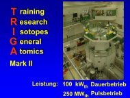
Große Fortschritte gab es in <strong>2009</strong> auch bei dem Aufbau der Massenspektrometrie- und<br />
Laserspektroskopieapparatur TRIGA-SPEC sowie der Quelle <strong>für</strong> ultrakalte Neutronen (UCN)<br />
am Forschungsreaktor TRIGA Mainz. Es erfolgten erste offline Massenmessungen an<br />
stabilen Isotopen sowie die Tests der Strahlstrecke <strong>für</strong> die kollineare Laserspektroskopie<br />
und des Gasjets <strong>für</strong> den Transport der Spaltprodukte aus der Targetkammer.<br />
In <strong>2009</strong> konnte die Berufungskommission <strong>für</strong> die W1-Professur Radiopharmazeutische<br />
Chemie ihre Tätigkeit erfolgreich beenden. Dr. Tobias Roß (ETH Zürich) hat die<br />
Juniorprofessur am 02.02.2010 angetreten. Die am Institut angesiedelte W3-Professur <strong>für</strong><br />
die Physik der schwersten Elemente wurde in einem gemeinsamen Verfahren von<br />
Universität Mainz und GSI Darmstadt am 26.11.<strong>2009</strong> ausgeschrieben.<br />
Prof. Tobias Reich wurde zum Mitglied des Scientific Advisory Committee des europäischen<br />
Network of Excellence ACTINET-I3 gewählt sowie zum Mitglied des Proposal Review<br />
Committee der Diamond Light Source in Großbritannien ernannt.<br />
Prof. Frank Rösch wurde <strong>2009</strong> in das International Advisory Board von Radiochimica Acta<br />
berufen und zum Vize-Chair der europäischen COST-Action D38 „Metal-based systems for<br />
molecular imaging applications“ gewählt. Weiterhin wurde er zum Vertreter des<br />
Fachbereichs 09 der Universität Mainz im Naturwissenschaftlich-Medizinischen<br />
Forschungszentrum gewählt.<br />
Für seine am 14.05.<strong>2009</strong> mit Auszeichnung bestandene Promotion zum Thema „On the<br />
development of novel cocaine-analogues for in vivo imaging of the dopamine transporter<br />
status“ erhielt Dr. Patrick Riß den Forschungsförderpreis der Freunde der Universität Mainz<br />
e. V. sowie den Promotionspreis der Fachgruppe Nuklearchemie der Gesellschaft Deutscher<br />
Chemiker.<br />
Die Arbeiten des Instituts wurden wiederum vielfältig finanziell gefördert. Besonderer Dank<br />
<strong>für</strong> die Förderung gilt dem Land Rheinland-Pfalz über die Johannes Gutenberg-Universität<br />
Mainz, dem Bundesministerium <strong>für</strong> Bildung und Forschung, dem Bundesministerium <strong>für</strong><br />
Wirtschaft und Technologie, der Deutschen Forschungsgemeinschaft, dem Deutschen<br />
Akademischen Austauschdienst, dem GSI Helmholtzzentrum <strong>für</strong> Schwerionenforschung<br />
GmbH, der Europäischen Gemeinschaft, der Carl-Zeiss-Stiftung, der Boehringer-Ingelheim-<br />
Stiftung sowie anderen Organisationen. Herzlicher Dank gilt auch den Gästen sowie<br />
Kolleginnen und Kollegen aus dem In- und Ausland, die <strong>2009</strong> im Institut weilten und ihre<br />
Kenntnisse und Erfahrungen mit uns teilten.<br />
Mainz, 23. Juli 2010 T. Reich
Amayri S.<br />
Beyerlein F.<br />
Buda R. A.<br />
Denschlag J.O.<br />
(pensioniert)<br />
Eberhardt K.<br />
Geppert Ch.<br />
Hampel G.<br />
Herrmann G.<br />
(emeritiert)<br />
Andjelkovic Z.<br />
Betzel T.<br />
Biegi C.<br />
Botermann B.<br />
Burchhardt C.<br />
Capito T.<br />
Cazan R.M.<br />
Eibach M.<br />
Eppard E.<br />
Even J.<br />
Fellner M.<br />
Fröhlich D.<br />
Gromm E.<br />
Hammen M.<br />
Herth M.<br />
Heß T.<br />
Hild D.<br />
Kaplan U.<br />
Breuel J.<br />
Drebert J.<br />
Gerhardt T.<br />
Handwerker C.<br />
Heiser A.<br />
Höhnemann S.<br />
Hubrath J.<br />
Janzen V.<br />
Jera R.<br />
Keller O.<br />
Wissenschaftliche Mitarbeiter<br />
Jahn M.<br />
Keller H.<br />
Kratz J. V.<br />
(pensioniert)<br />
Kratz K.-L.<br />
(pensioniert)<br />
Neugart<br />
(pensioniert) R.<br />
Novotny Ch.<br />
Diplomanden, Doktoranden und Staatsexamenskandidaten<br />
Krämer J.<br />
Kraft A.<br />
Kramer V.<br />
Krieger A.<br />
Lang T.<br />
Lauer T.<br />
Lochmann M.<br />
Loktionova N.<br />
Meister M.<br />
Moderegger D.<br />
Nagel V.<br />
Niewisch L.<br />
Ölcer A.<br />
Radchenko V.<br />
Reichert P.<br />
Riß P.<br />
Rossi D.<br />
Angestellte<br />
Kling H. O.<br />
Krille U.<br />
Lehr G.<br />
Mendel M.<br />
Müller J.<br />
Nähler A.<br />
Onasch I.<br />
Peil A.<br />
Praast B.<br />
Runke J.<br />
Nörtershäuser W.<br />
Piel M.<br />
Plonka-Spehr Ch.<br />
Reich T.<br />
Rösch F.<br />
Trautmann N.<br />
(pensioniert)<br />
Wiehl N.<br />
Scheid N.<br />
Schieferstein H.<br />
Schönberger M.<br />
Schütz Ch.<br />
Seemann J.<br />
Sieber B.<br />
Smorra Ch.<br />
Stöbener N.<br />
Tiedemann D.<br />
Vogtländer L.<br />
Wendt S.<br />
Werner St.<br />
Wunderlich T.<br />
Zakova M.<br />
Zenner J.<br />
Zimny M.<br />
Sach-Muth P.<br />
Schmidt A.<br />
Schmidt H.-M.<br />
Thörle-Pospiech P.<br />
Timpe J.-P.<br />
Widera R.<br />
Zauner S.
Gäste <strong>2009</strong><br />
Allmeroth, M. Institut <strong>für</strong> Organische Chemie, Uni Mainz<br />
Andersen, R.W. Department of Nuclearmedicine, Aaborg Hospital, Dänemark<br />
Bartl, M. Metrohm, Hohenstein<br />
Barz, M. Institut <strong>für</strong> Organische Chemie, Uni Mainz<br />
Blaum, K. Max-Planck-Institut, Heidelberg<br />
Böhm, Ch. Institut <strong>für</strong> Physik, Mainz<br />
Borisov, Y. PNPI, Gatchina, Russland<br />
Champion, J. Subatech, Nantes, Frankreich<br />
Daum, M. Paul Scherrer Institut, Villigen, Schweiz<br />
De Blois, E. Erasmus MC Rotterdam, Rotterdam, Niederlande<br />
Dreger, T. Eckert&Ziegler, Berlin<br />
Eibach, M. Institut <strong>für</strong> Physik, Mainz<br />
Eitel, G. Institut <strong>für</strong> Physik, Mainz<br />
Ferrer, R. Institut <strong>für</strong> Physik, Mainz<br />
George, S. Institut <strong>für</strong> Physik, Mainz<br />
Glowa, B. Pracownia Medycyny Nulearnej, Krakau, Polen<br />
Gölz, W. Metrohm, Hohenstein<br />
Gourni, E. Demokritos Athen, Griechenland<br />
Himmelman, J. MFT/Diagnostik, Sahlgrenska Universitetsjukhuset, Göteborg, Schweden<br />
Hinze, P. Areva NP, Erlangen<br />
Janko, M. Institut <strong>für</strong> Pathobiochemie, Mainz<br />
Jankovic, D. Vinca Institute of Nuclear Sciences, Belgrad, Serbien<br />
Jansen, K.-H. Sykam, Fürstenfeldbruck<br />
Ketelaer, J. Institut <strong>für</strong> Physik, Mainz<br />
Ketter, J. Institut <strong>für</strong> Physik, Mainz<br />
Knuth, K. Institut <strong>für</strong> Physik, Mainz<br />
Kotovskiy, N. Institut <strong>für</strong> Physik, Mainz<br />
Koumarianou, E. Institute of Atomic Energy Polatom, Otwock-Swierk, Polen<br />
Kroselj, M. University Medical Cnetre Ljubljana, Slowenien<br />
Kuczewski, B. TU Graz<br />
Kumar, V. Westmead Hospital, Sidney<br />
Luoto, P. Turku PET Centre, Turku University Hospital, Turku, Finnland<br />
Majkowska, A. Institute of Nuclear Chemistry & Technology, Warszawa, Polen<br />
Malviya, G. Dep. of Nuclear Medicine, St. Andrea Hospital, Rom, Italien<br />
Meyer, G.-J. Medizinische Hochschule Hannover<br />
Minamisono, K. Michigan State University, Michigan, USA<br />
Mohnike, W. DTZ, Berlin<br />
Montavon, G. Subatech, Nantes, Frankreich<br />
Nagel, S. KIT, Abt. Sicherheit, Karlsruhe<br />
Nagy, S. Institut <strong>für</strong> Physik, Mainz<br />
Nawroth, T. Institut <strong>für</strong> Pharmazie, Mainz<br />
Peters, T. Institut <strong>für</strong> Pharmazie, Mainz<br />
Pfister, A. Institut <strong>für</strong> Physik, Mainz<br />
Pflanzner, T. Institut <strong>für</strong> Pathobiochemie, Mainz<br />
Pruszinski, M. Institute of Nuclear Chemistry & Technology, Warszawa, Polen<br />
Reich, Ta. Institut <strong>für</strong> Geowissenschaften, Mainz<br />
Rosemann, J. Institut <strong>für</strong> Anorganische Chemie, Mainz<br />
Schausten, B. GSI Darmstadt<br />
Schmidt, T. Areva NP, Erlangen<br />
Schmidt, U. Uni Heidelberg<br />
Schnürer, P. Agilent Technologies<br />
Schwab, A. Institut <strong>für</strong> Physik, Mainz<br />
Smorra, Ch. Institut <strong>für</strong> Physik, Mainz<br />
Socan, A. Vinca Institute of Nuclear Scienes, Belgrad, Serbien<br />
Thews, O. Institut <strong>für</strong> Pathophysiologie, Mainz<br />
Volpe, R. Monash University, Australien
Waldron, B. Durham University, UK<br />
Wiltsche, H. TU Graz, Österreich<br />
Wojdowska, W. Institute of Atomic Energy, Radioisotope Centre POLATOM, Polen<br />
Yilmaz, B. Institut <strong>für</strong> Pathophysiologie, Mainz<br />
Zimontkowski, B. DTZ, Berlin
Inhaltsverzeichnis<br />
A. Kernchemie / Nuclear- and Radiochemistry<br />
A1 Production and decay of element 114: high cross sections and new nucleus 277 Hs<br />
Ch.E. Düllmann, D. Ackermann, M. Block, W. Brüchle, H.G. Essel, J.M. Gates, W.<br />
Hartmann, F.P. Heßberger, A. Hübner, E. Jäger, et al.<br />
A2 Compact Coupled to TASCA for Element 114 Chemistry<br />
A. Yakushev, J.M. Gates, A. Gorshkov, R. Graeger, A. Türler, D. Ackermann, M. Block,<br />
W. Brüchle, Ch.E. Düllmann, H.G. Essel, et al.<br />
A3<br />
244 Pu-targets for production of element 114 at TASCA<br />
K. Eberhardt, J.V. Kratz, J. Runke, Ch.E. Düllmann, B. Lommel and M. Schädel<br />
A4 Recovery of 244 Pu from irradiated targets for production of element 114<br />
J. Runke, K. Eberhardt, J.V. Kratz, Ch.E. Düllmann and M. Schädel<br />
A5 The Performance of TASCA in the 48 Ca+ 206,207,208 Pb Reactions<br />
J. Khuyagbaatar, M. Schädel, D. Ackermann, Ch.E. Düllmann, E.Jäger, F.P. Heßberger, A.<br />
Semchenkov, A. Gorshkov, R. Graeger, A. Türler, et al.<br />
A6 TASISpec – Heading towards its first experiment.<br />
L.-L. Andersson, D. Rudolph, D. Ackermann, Ch.E. Düllmann, K. Eberhardt, J. Even, U.<br />
Forsberg, J. Gellanki, J. Gerl, P. Golubev, R. Hoischen, R.-D. Herzberg, F.P. Heßberger,<br />
E. Jäger, J. Khuyagbaatar, I. Kojouharov, et al.<br />
A7 Pilot-Test Experiment with Os of a SISAK Setup for Hs-Chemistry Studies<br />
F. Samadani, J. Alstad, F. Schulz, J. Nilssen, J.P. Omtvedt, H.B. Ottesen, S. Qureshi, Ch.E.<br />
Düllmann, J.M. Gates, E. Jäger, et al.<br />
A8 Liquid – liquid – extraction with the MicroSISAK – system<br />
D. Hild, K. Eberhardt, J.V. Kratz, P. Löb, B. Werner<br />
A9 Accuracy studies and first mass measurements at TRIGA-TRAP<br />
C. Smorra, T. Beyer, D. Beck, K. Blaum, M. Block, K. Eberhardt, M. Eibach, F. Herfurth,<br />
J. Ketelaer, Sz. Nagy, et al.<br />
A10 Commissioning of TRIGA-LASER – tests and developments of the LaSpec beamline<br />
J. Krämer, K. Blaum, K. Eberhardt, Ch. Geppert, M. Hammen, A. Krieger, R. Neugart, W.<br />
Nörtershäuser, R. Sanchez, B. Sieber, and the TRIGA-SPEC collaboration<br />
A11 Towards Laser Cooling of Magnesium Ions for Sympathetic cooling of Highly Charged<br />
Ions at SPECTRAP<br />
R. Cazan, C. Geppert, W. Nörtershäuser<br />
A12 High Voltage Calibration at ISOLDE/CERN<br />
A. Krieger, Ch. Geppert, S. Rosendahl, C, Weinheimer, D. Yordanov, J. Schipper, J.<br />
Krämer and W. Nörtershäuser
A13 Electromagnetic Excitation of Neutron-Rich Ni Isotopes<br />
T. Le Bleis, D. Rossi, P. Adrich, F Aksouh, H. Alvarez-Pol, T. Aumann, J. Benlliure, M.<br />
Boehmer, K. Boretzky, E. Casarejos, et al.<br />
A14 Cubic boron nitride: A new prospective material for ultracold neutron application<br />
Yu. Sobolev, Th. Lauer, Yu. Borisov, M. Daum, N. du Fresne, L. Göltl, G. Hampel, W.<br />
Heil, A. Knecht, M. Keunecke, et al.<br />
A15 Are neutrons really neutral?<br />
C. Plonka-Spehr, A. Kraft, P. Iaydjiev, J. Klepp, V.V. Nesvizhevsky, P. Geltenbort, Th.<br />
Lauer, A. Frank<br />
A16 A new test of Time Dilation at ESR using fast 7 Li + –ions<br />
B. Botermann, T. Kühl, W. Nörtershäuser, T. Stöhlker, C. Geppert, G. Huber, S. Karpuk,<br />
C. Novotny, D. Bing, D. Schwalm, et al.<br />
A17 Precision Laser Spectoscopy of Beryllium<br />
M. Zakova, Z. Andjelkovic, J. Krämer, A. Krieger, R. Neugart, D. Tiedemann, Ch.<br />
Geppert, W. Nörtershäuser, R. Sánchez, T. Neff, K. Blaum, et al.<br />
A18 Cyclic voltammetric study of uranium in room-temperature ionic liquids<br />
A. Ölcer, T. Reich<br />
A19 Purification of 68 Ga from 68 Ge/ 68 Ga radionuclide generator combining two different<br />
columns<br />
N.S. Loktionova, A. Belozub, K.P. Zhernosekov, D.V. Filisofov, F. Rösch<br />
A20 Elution of 68 Ge/ 68 Ga radionuclide generators in “reverse” mode<br />
N.S. Loktionova, F. Rösch<br />
A21 Development of an alternative method for separation and purification of no-carrier-added<br />
arsenic-77 from bulk amounts of germanium-77<br />
V. Radchenko, M. Jahn, D. Filosofov, H. Hauser, M. Eisenhut, M. Jennewein, F. Rösch<br />
B. Radiopharmazeutische Chemie / Kernchemie in den Lebenswissenschaften<br />
Radiopharmaceutical Chemistry / Nuclear Chemistry for Life Sciences<br />
B1 Synthese von bifunktionellen DO2A-Derivaten zur multivalenten Kopplung von Targeting-<br />
Vektoren<br />
C. Burchardt, P.J. Riss, J.A. Peters, F. Rösch<br />
B2 Synthese eines chemoselektiven bifunktionellen Chelators <strong>für</strong> die molekulare Bildgebung<br />
V. Nagel, P. Riss and F. Rösch<br />
B3 Synthese von Di-Glibenclamid-DO2A-NCS<br />
M. Zimny, C. Burchardt, F. Rösch<br />
B4 Synthese und Evaluierung von 68 Ga-markierten Folsäurederivaten<br />
J. Seemann, C. Burchardt, F. Rösch, T.L. Roß
B5 Synthese des D1-Antagonisten [ 18 F]Fluorethyl-NNC 112 <strong>für</strong> die PET<br />
F. Beyerlein, M. Piel, F. Rösch<br />
B6<br />
B7<br />
68 Ga-Markierung von neuen AAZTA-Derivaten<br />
B.P. Waldron, C. Burchardt, F. Rösch, D. Parker<br />
68 Ga-Markierung von H5EPTPAC16<br />
E. Eppard, P.J. Riß, F. Rösch, C.F.G.C. Geraldes<br />
B8 Solid phase approach for the radioiodination of organotin precursors for nuclear medicine<br />
applications.<br />
S. Kuschel, C. Shea, J.S. Fowler and A.N. Gifford<br />
B9 Optimizing [ 13 N]N2 radiochemistry for nitrogen-fixation in root nodules of legumes<br />
M.C.K. Kasel, M.J. Schueller and R.A. Ferrieri<br />
B10 Synthese fluorsubstituierter 5-(Pyrimidin-4-yl)-isothiaz-3-ol-Derivate als potentielle Radioliganden<br />
<strong>für</strong> die PET<br />
V. Kramer, V. Bockhardt, H. Lüddens, F. Rösch<br />
B11 Synthese fluorsubstituierter 5-(Pyrimidin-4yl)-isooxazol-Derivate als potentielle Radioliganden<br />
<strong>für</strong> die PET<br />
V. Kramer, V. Bockhardt, H. Lüddens, F. Rösch<br />
B12 Size dependent biodistribution of HPMA-based polymeric systems using 18F-labeled<br />
polymers<br />
D. Moderegger, M. Allmeroth, R. Zentel, F. Rösch<br />
B13 Radioactive labelling of HPMA-based polymeric systems with fluorine-18 for in vivo<br />
imaging and evaluation by positron emission tomography (PET)<br />
D. Moderegger, M. Allmeroth, R. Zentel, F. Rösch<br />
B14 Radiolabelling and evaluation of a gavestinel-based ligand for imaging the NMDA-receptor<br />
status using PET<br />
T. Betzel, C. Edinger, V. Bockhard, M. Piel, G. Dannhardt, H. Lüddens, F. Rösch<br />
B15 Mikrowellengestützte 18 F-Markierung von ß-Carbolin-Alkaloiden zur Visualisierung der<br />
MAO-A<br />
H. Schieferstein, M. Piel, F. Rösch<br />
B16 68 Ga-Schiffbasen-Myokardtracer<br />
M. Zimny, M. Fellner, F. Rösch<br />
B17 68 Ga-bisphosponates for imaging bone diseases<br />
M. Fellner, R.P. Baum, V. Kubicek, P. Hermann, V. Prasad, F. Rösch<br />
B18 177 Lu-BPAMD – from bone imaging to therapy with a macrocyle-bisphosphonate ligand<br />
M. Fellner, R.P. Baum, V. Kubicek, P. Hermann and F. Rösch<br />
B19 125 I-labelling and biological evaluation of a close-borane containing Tyr3-octreotate<br />
derivative<br />
T. Betzel, F. Zoller, A. Markert, T. Heß, W. Mier, U. Haberkorn, F. Rösch
B20 Evaluation of P-Glycoprotein Modulation of [ 18 F]Fallypride<br />
M. Piel, U. Schmitt, N. Bausbacher, H.G. Buchholz, Ch. Hiemke, F. Rösch<br />
B21 Radiosynthesis and PET Imaging of [ 11 C] Valproic Acid, [ 11 C] Butyric Acid and<br />
[ 11 C]Phenylbutyric Acid<br />
N. Otto, S.W. Kim, J.S. Fowler<br />
B22 Getting Buzzed on Sugar: Proof-of-Principle that the Brain of a Bee can be imaged using<br />
18 FDG<br />
M.C.K. Kasel, C. Shea, R.A Ferrieri<br />
B23 Boron analysis of blood, tissue and cell samples by neutron autoradiography and promptgamma<br />
ray spectroscopy in combination with histological methods<br />
C. Schütz, G. Hampel, S. Werner, T. Schmitz, J.V. Kratz, K. Appelmann, R. Moss, C.<br />
Brochhausen, J. Kirkpatrick, H. Schmidberger, et al.<br />
C. Radiochemische Analytik <strong>für</strong> umweltrelevante und technische<br />
Probleme / Radiochemical Analytics for Environment and Technology<br />
C1 Resonance Ionization Spectroscopy (RIS) on 237 Np<br />
N. Stöbener, T. Gottwald, S. Raeder, G. Passler, T. Reich, N. Trautmann, K. Wendt<br />
C2 Laserspektroskopische Untersuchung des Urangehalts verschiedener Substratfolien <strong>für</strong> die<br />
effiziente Atomisation von Uran<br />
S. Raeder, A. Hakimi, G. Passler, N. Trautmann, K. Wendt<br />
C3 Bestimmung des Ionisationspotentials von Technetium und Optimierung der<br />
Nachweisgrenze <strong>für</strong> die Ultraspurenanalyse von Technetium<br />
F. Schwellnus, C. Mattolat, K. Wendt, N. Trautmann<br />
C4 Sorption von Np(V) an Opalinuston in Abhängigkeit des Hintergrundelektrolyten<br />
D.R. Fröhlich, P.J.B. Rosemann, T. Reich<br />
C5 XAS study of neptunium(V) sorbed on Opalinus Clay<br />
S. Amayri, D.R. Fröhlich, J. Drebert, T. Reich<br />
C6 Trace element and multi-isotope analysis of human head hair samples<br />
N. Scheid, S. Becker, M. Dücking, G. Hampel, T. Holdermann, J.V. Kratz, S. Schneiders, P.<br />
Weis<br />
C7 Studien zur Anwendung der Neutronenaktivierungsanalyse (NAA) hinsichtlich der<br />
Provenienzbestimmung neolithischer Hämatitartefakte<br />
D. Rieth, P. Haupt, G. Hampel, S. Zauner<br />
C8 Spurenanalyse von gammastrahlenden Radionukliden in der bodennahen Luft<br />
H. Keller, B. Praast
D. Technische Einrichtungen / Technical Facilities<br />
D1 Betrieb des Forschungsreaktors TRIGA Mainz im Jahre <strong>2009</strong><br />
G. Hampel<br />
D2 Personendosisüberwachung<br />
I. Onasch, B. Praast<br />
D3 Abgabe radioaktiver Stoffe<br />
A. Heiser, B. Praast, H. Keller<br />
E. Veröffentlichungen, Vorträge und Lehrveranstaltungen / Publications,<br />
Conference Contributions and Teaching Activities<br />
E1 Diplomarbeiten und Dissertationen<br />
E2 Veröffentlichungen und Vorträge der Mitarbeiter der bericht erstattenden Arbeitsgruppen<br />
E3 Vorträge im Seminar <strong>für</strong> Kern- und Radiochemie und im Seminar über aktuelle<br />
Themen aus Kosmochemie und Astrophysik<br />
E4 Beiträge der Dozenten des Instituts zu den Lehrveranstaltungen des Fachbereichs<br />
Chemie, Pharmazie und Geowissenschaften und des Fachbereichs Physik,<br />
Mathematik und Informatik (SS 09 und WS 09/10) sowie zur Weiterbildung
Förderungen der im Jahresbericht enthaltenen Arbeiten erfolgten durch folgende<br />
Institutionen:<br />
Land Rheinland-Pfalz:<br />
Kompetenzzentrum „Neuro-PET“ des Landes Rheinland-Pfalz<br />
Stiftung Rheinland-Pfalz <strong>für</strong> Innovation „Ankopplung einer Laserspektroskopie- und<br />
Massenspektrometrieapparatur <strong>für</strong> kurzlebige Radionuklide an den Mainzer TRIGA-Reaktor“<br />
Gemeinsames Projekt mit der Transplantantionschirurgie „Schwerpunkt Tumormedizin“<br />
Johannes Gutenberg-Universität Mainz:<br />
Forschungsfonds der Johannes Gutenberg-Universität Mainz<br />
Zentrum EMG (Elementare Kräfte und Mathematische Grundlagen)<br />
Bundesministerium <strong>für</strong> Bildung und Forschung (BMBF):<br />
Das BMBF förderte folgende Vorhaben:<br />
„Schwerste Elemente: Chemische Mikrotrennapparaturen und TASCA“<br />
„R3B-Reactions with Relativistic Radioactive Beams: Target-Recoil-Detector und NeuLAND“<br />
„Laserspektroskopie mit kontinuierlichen und gepulsten Strahlen an ISOLDE/ CERN“<br />
„FAIR/ SPARC: Entwicklungen zur Präzisions-Laser-Spektroskopie gespeicherter<br />
relativistischer Ionen“<br />
„Messung der Ladungsradien von Be-7,9,10 und des Halokerns Be-11“<br />
Bundesministerium <strong>für</strong> Wirtschaft und Technologie (BMWi):<br />
Das BMWi förderte folgendes Vorhaben:<br />
„Migration und Transport von Actiniden im natürlichen Tongestein unter Berücksichtigung von<br />
Huminstoffen und Tonorganika – Wechselwirkung von Neptunium und Plutonium mit<br />
natürlichem Tongestein“<br />
Deutsche Forschungsgemeinschaft (DFG):<br />
Graduiertenkolleg 826/3 „Spurenanalytik von Elementspezies: Methodenentwicklungen und<br />
Anwendungen“<br />
Graduiertenkolleg 1044/1 „Entwicklungsabhängige und krankheitsinduzierte Modifikationen im<br />
Nervensystem“<br />
Schwerpunktprogramm (SSP 1491) „Precision experiments in particle- and astrophysics with<br />
cold and ultra cold neutrons“
Weiterhin fördert die DFG folgende Vorhaben:<br />
„Entwicklung einer neuen Klasse von Radiopharmaka <strong>für</strong> die Positronen-Emissions-<br />
Tomographie: 70,72 As-markierte Verbindungen“<br />
„Wissenschaftleraustausch mit Russland - Metall-Chelat-Peptid-Systeme“<br />
„Synthese von hochaffinen 18 F-markierten Liganden zur Untersuchung von NMDA-Rezeptoren<br />
mittels Positronen-Emissions-Tomographie (PET)<br />
„Entwicklung eines 44 Ti/ 44 Sc-Radionuklidgenerators <strong>für</strong> den potentiellen nuklearmedizinischen<br />
Einsatz von 44 Sc-markierten PET-Radiopharmaka“<br />
„Installation und Optimierung eines Systems zur Erzeugung von ultrakalten Neutronen (UCN)<br />
am TRIGA-Reaktor sowie Studien mit UCN“<br />
“Bau einer Doppelkammer zur Speicherung von ultrakalten Neutronen <strong>für</strong> ein EDM Projekt”<br />
„Determination of the Isotopic Composition of Trace Amounts of Actinides in Environmental<br />
Micro-particles by Resonance Ionisation Mass Spectrometry (RIMS) of Sputtered Neutrals (SN)”<br />
“Test der Zeitdilatation in der speziellen Relativitätstheorie durch Laserspektroskopie an<br />
schnellen gespeicherten Ionen”<br />
Deutscher Akademischer Austauschdienst (DAAD):<br />
Internationales Studentenaustausch-Programm zwischen der Johannes Gutenberg-Universität<br />
Mainz und der State University of New York at Stony Brooks/ Brookhaven National Laboratory<br />
GSI Helmholtzzentrum <strong>für</strong> Schwerionenforschung GmbH:<br />
GSI Helmholtzzentrum <strong>für</strong> Schwerionenforschung im Rahmen der<br />
Zusammenarbeitsvereinbarungen zwischen Wissenschaftlern von Hochschulen und der GSI<br />
Helmholtz-Gemeinschaft deutscher Forschungszentren über die Helmholtz-Hochschul-<br />
Nachwuchsgruppe VH-NG-148 „Laserspektroskopie an exotischen Atomen und hochgeladenen<br />
Ionen“<br />
Helmholtz-Gemeinschaft deutscher Forschungszentren über die Helmholtz-Hochschul-<br />
Nachwuchsgruppe VH-NG-037 „Experimente mit gespeicherten und gekühlten Ionen“<br />
Europäische Gemeinschaft:<br />
„European Molecular Imaging Laboratories“<br />
Carl-Zeiss-Stiftung:<br />
Juniorprofessur: „Forschung mit ultrakalten Neutronen“<br />
Doktoranden-Stipendium: „Laserspektroskopie kurzlebiger Isotope am TRIGA<br />
Forschungsreaktor Mainz“
Boehringer-Ingelheim-Stiftung:<br />
„Borbestimmung in Gewebeproben im Rahmen der Entwicklung eines Behandlungsprotokolls<br />
<strong>für</strong> die Bor-Neutronen-Einfangtherapie an einer autotransplantierten Leber“<br />
Bundeskriminalamt (BKA):<br />
„Elementanalytik und Isotopenverhältnisbestimmung an humanbiologischen Materialien“
A.<br />
Kernchemie<br />
Nuclear- and Radiochemistry
Production and decay of element 114: high cross sections and new nucleus 277 Hs*<br />
Ch.E. Düllmann 1,# , D. Ackermann 1 , M. Block 1 , W. Brüchle 1 , H.G. Essel 1 , J.M. Gates 1,2 , W. Hartmann<br />
1 , F.P. Heßberger 1 , A. Hübner 1 , E. Jäger 1 , J. Khuyagbaatar 1 , B. Kindler 1 , J. Krier 1 , N. Kurz 1 , B.<br />
Lommel 1 , M. Schädel 1 , B. Schausten 1 , E. Schimpf 1 , J. Steiner 1 , A. Gorshkov 2 , R. Graeger 2 , A. Türler<br />
2 , A. Yakushev 2 , K. Eberhardt 3 , J. Even 3 , D. Hild 3 , J.V. Kratz 3 , D. Liebe 3 , J. Runke 3 , P. Thörle-<br />
Pospiech 3 , N. Wiehl 3 , L.-L. Andersson 4 , R.-D. Herzberg 4 , E. Parr 4 , J. Dvorak 5,6 , P.A. Ellison 5,6 , K.E.<br />
Gregorich 5 , H. Nitsche 5,6 , S. Lahiri 7 , M. Maiti 7 , J.P. Omtvedt 8 , A. Semchenkov 8 , D. Rudolph 9 , J.<br />
Uusitalo 10 , M. Wegrzecki 11<br />
1 GSI, Darmstadt, Germany; 2 TU Munich, Garching, Germany; 3 Johannes Gutenberg-University of Mainz, Germany;<br />
4 University of Liverpool, UK; 5 LBNL Berkeley, CA, USA; 6 UC Berkeley, CA, USA; 7 SINP, Kolkata, India; 8 University<br />
of Oslo, Norway; 9 Lund University, Sweden; 10 University of Jyväskylä, Finland; 11 ITE, Warsaw, Poland<br />
Introduction<br />
Discoveries of new superheavy elements (SHE) were<br />
reported from FLNR, Dubna, Russia [1], including observations<br />
of element 114 isotopes produced in the<br />
48 Ca+ 242,244 Pu reactions. Successful independent studies<br />
of some of the reactions studied in Dubna were reported<br />
[2,3], most recently also the observation of one atom each<br />
of 286,287 114 produced in the 48 Ca+ 242 Pu reaction at LBNL<br />
[4]. Predictions on the existence of an "island of stability"<br />
in the region of SHE have substantiated, despite the small<br />
number of observed events in every confirmation experiment.<br />
All successful confirmation experiments reported<br />
cross sections lower than those from FLNR by factors of<br />
two or more. Nevertheless, these cross sections are unexpectedly<br />
high compared to extrapolations from lighter<br />
systems [5], and intriguingly constant over a large range<br />
of 112�Z�118. A thorough understanding of the underlying<br />
production mechanism is still missing; location and<br />
extension of the "island of stability" in the region of<br />
spherical SHE is still far from being established. To help<br />
shedding more light on these problems, a 48 Ca+ 244 Pu experiment<br />
was performed at the gas-filled TransActinide<br />
Separator and Chemistry Apparatus (TASCA) [6,7],<br />
which was optimized for the study of 48 Ca-induced fusion<br />
reactions with actinide targets. TASCA's efficiency for<br />
this nuclear reaction type is currently unsurpassed.<br />
Experimental<br />
The UNILAC accelerated a pulsed 48 Ca beam (~2 10 12<br />
s -1 ), which passed through 244 PuO2 targets (average thickness:<br />
438 �g/cm 2 244 Pu). Beam energies inside the targets<br />
were 241.3-246.2 MeV (E*=39.8-43.9 MeV; hereafter<br />
referred to as 42-MeV run) and 236.4-241.0 MeV<br />
(E*=36.1-39.5 MeV; 38-MeV run). 2.44 10 18 (42-MeV<br />
run) and 1.15 10 18 (38-MeV run) projectiles passed<br />
through the targets. Nuclear reaction products entered<br />
* Work supported by the BMBF (06MT247I, 06MT248,<br />
06MZ223I); the GSI-F&E (MT/TÜR, MZJVKR); the<br />
Swedish and Norwegian (177538) Science Councils; the<br />
US DOE (DE-AC03-76SF00098; DE-AC02-05CH11231;<br />
NNSA Fellowship); the Govt. of India (TADDS).<br />
# c.e.duellmann@gsi.de<br />
GSITemp late2007<br />
- A1.1 -<br />
TASCA, operated in "high transmission mode" [7], and<br />
were separated in 0.8 mbar He gas. The detection system<br />
consisted of a Multi Wire Proportional Counter (MWPC)<br />
and a focal plane detector box (FPDB). The FPDB consisted<br />
of a Double Sided Silicon Strip Detector (DSSSD;<br />
pitch size: 1 mm; 144 vertical / 48 horizontal strips) and<br />
Single Sided Silicon Strip Detectors (SSSSD) mounted<br />
perpendicular in the backward hemisphere of the DSSSD<br />
[8]. The MWPC provided a signal for ions recoiling from<br />
the target and allowed distinguishing these from radioactive<br />
decays of species implanted in the DSSSD. The energy<br />
resolution of the FPDB was 25 keV FWHM for 8.1<br />
MeV �-particles fully stopped in the DSSSD and 170<br />
keV for �-particles that deposited a fraction of their energy<br />
inside the DSSSD and the remainder in the SSSSD.<br />
The detection efficiency was 72% for �-particles and<br />
100% for SF. The efficiency for focusing element 114<br />
EVRs into the DSSSD was (60�6)% [9]. Data acquisition<br />
was triggered by events registering more than 300 keV in<br />
the DSSSD or more than 500 keV in a SSSSD. More details<br />
are given in [10,11].<br />
Results<br />
We searched for decay chains from 288,289 114 and their<br />
daughters [1]. Upon identification of a chain, additional<br />
�-particles occurring in the same pixel as the chain, in<br />
between registration of the EVR and the terminating SF,<br />
were searched. Due to a damaged target, the background<br />
rate was increased during portions of the runs. SFs terminating<br />
decay chains recorded during these periods were<br />
required to occur outside beam pulses. Based on the event<br />
rate only 0.02 ( 289 114) and 0.05 ( 288 114) random chains<br />
from unrelated background events were expected. The<br />
search yielded nine EVR-�-SF chains ( 288 114) and four<br />
EVR-�-�(-�)-SF chains ( 289 114). Ten chains were measured<br />
in the 42-MeV run and three chains in the 38-MeV<br />
run (Figs. 1 and 2). The agreement of our data (Table 1)<br />
with that of [1] is good in all cases except for chain #9.<br />
The data measured for the EVR, the first, and the second<br />
�-particle suggest assigning chain #9 to 289 114� 285 112�<br />
281 Ds. 281 Ds then decayed by emission of a (8.727�0.025)-<br />
MeV �-particle 5.688 s after the decay of 285 112, during<br />
the beam-off period, where background is low. 281 Ds has
undergone SF in all ten previously observed decays [1]<br />
5. 0<br />
with T1/2= 11.1 2.<br />
7<br />
� � s. Based on background rates, the<br />
probability to register an �-like event with properties as<br />
exhibited by the observed one is only 0.1%. We thus assign<br />
it to a so far unobserved �-branch in 281 Ds. Considering<br />
this �-decay and the thirteen measured SF decays<br />
from [1] and our work, an �-decay branch b� of<br />
16<br />
9 7<br />
� � %<br />
results after correcting for detection efficiency differences<br />
for �-decay and SF. The chain was terminated 4.5 ms<br />
later by SF of new 277 Hs.<br />
The B � of element 114 EVRs in 0.8 mbar He was<br />
measured to (2.29�0.11) T m.<br />
Figure 1: Decay chains assigned to 288 114 (chains 1-8)<br />
and 289 114 (chains 9, 10) observed during the 42-MeV<br />
run. A black triangle in the lower right corner of a box<br />
indicates that the beam was off at the time of the event.<br />
Figure 2: Same as Figure 1, but showing decay chains<br />
observed during the 38-MeV run.<br />
Table 1. Decay properties ([1] and this work)<br />
Isotope Decay T1/2 (this work) T1/2 (combined)<br />
289<br />
114 �� 0.97<br />
0.97 0.<br />
32<br />
� � s<br />
288<br />
114 �� 0.24<br />
0.47 0.<br />
12<br />
� � s<br />
285<br />
112 �� 30<br />
30 10<br />
� � s<br />
284<br />
112 SF� 50 101 25<br />
� � ms<br />
281<br />
Ds SF/��91/9 20 20 7<br />
� � s<br />
277<br />
Hs SF 15<br />
3 1<br />
� � ms<br />
0.8<br />
2.1 0.<br />
4<br />
� � s<br />
0.17<br />
0.69 0.<br />
11<br />
� � s<br />
11<br />
29 6<br />
� � s<br />
24<br />
99 16<br />
� � ms<br />
5<br />
13 3<br />
� � s<br />
15<br />
3 1<br />
� � ms<br />
- A1.2 -<br />
Discussion<br />
Z=108 is a deformed proton shell closure in N~162 isotopes.<br />
The observed 277 Hs lifetime is short compared to<br />
half-lives of the Hs isotopes near the deformed N=162<br />
shell closure, indicating reduced shell stabilization in the<br />
N=169 nucleus 277 Hs. Macro-microscopic model predictions<br />
of T1/2(SF) for the neighboring isotopes are 46 ms<br />
( 276 Hs) and 0.98 ms ( 278 Hs), the geometric mean being 6.7<br />
ms [12]. This is similar to our observed lifetime. The odd<br />
neutron is expected to hinder SF decay significantly.<br />
Thus, the drop in T1/2(SF) when increasing N above 162<br />
275<br />
may be more severe than suggested by [12]. Hs<br />
(N=167) decays by �-particle emission with T1/2=0.19 s<br />
[1]. The experimental trend with prevalent �-decay in Hs<br />
isotopes with N=157-167, but predominant SF in lighter<br />
as well as in heavier isotopes is close to that in [10],<br />
which suggests dominant �-decay from N=154 to N=166<br />
but SF for N>168. This indicates that stability vanishes<br />
rapidly with increasing distance from N=162.<br />
Measured cross sections in the 38-MeV run were<br />
7. 4<br />
8.0 4.<br />
5<br />
� 4. 2<br />
� pb (3n channel) and 2.8 2.<br />
1<br />
� � pb (4n channel), and<br />
3. 3<br />
0<br />
3. 9<br />
1<br />
in the 42-MeV run, 3.5 2.<br />
� � pb (3n channel) and 9.8 3.<br />
� �<br />
pb (4n channel). Error bars include statistical uncertainties<br />
only (68.3% confidence level); the systematic uncertainty<br />
is estimated to 14%. In contrast to any other confirmation<br />
experiment, we confirm the large cross sections<br />
as reported from FLNR [1]. In fact, our measured cross<br />
sections are higher than those reported from the DGFRS.<br />
These high cross sections call for investigations of the<br />
details of the production mechanism. Production rates that<br />
follow from these values encourage using this nuclear<br />
reaction to produce relatively long-lived isotopes of element<br />
114, in particular for envisaged chemical investigations<br />
[13] or for �-spectroscopic studies that allow shedding<br />
light on the nuclear structure in this SHE region and<br />
may facilitate unique Z identification.<br />
We thank the ECR and UNILAC staff for excellent<br />
48<br />
Ca beams and H. Brand and the GSI EE department, the<br />
machine shop staff at the <strong>institut</strong>e of radiochemistry, TU<br />
Munich, and V. Gorshkov for technical support.<br />
References<br />
[1] Yu.Ts. Oganessian, J. Phys. G 34 (2007) R165.<br />
[2] S. Hofmann, EPJA 32 (2007) 251.<br />
[3] R. Eichler et al. Nature 447 (2007) 72.<br />
[4] L. Stavsetra et al., PRL 103 (<strong>2009</strong>) 132502.<br />
[5] S. Hofmann, Lect. Notes Phys. 264 (<strong>2009</strong>) 203.<br />
[6] M. Schädel, Eur. Phys. J. D 45 (2007) 67.<br />
[7] A. Semchenkov et al., NIM B 266 (2008) 4153<br />
[8] A. Gorshkov et al., in preparation for NIM A.<br />
[9] K.E. Gregorich et al., GSI Sci. Rep. 2006 (2007) 144.<br />
[10] Ch.E. Düllmann et al., submitted to PRL.<br />
[11] J.M. Gates et al., in preparation for PRC.<br />
[12] R. Smolanczuk, PRC 52 (1995) 1871.<br />
[13] A. Yakushev et al., this Scientific Report.
* Work supported by the BMBF (06MT247I, 06MT248);<br />
the GSI-F&E (MT/TÜR).<br />
† Current address: Bern University & PSI Villigen, Swit-<br />
zerland<br />
# alexander.yakushev@radiochemie.de<br />
GSITemplate2007<br />
COMPACT Coupled to TASCA for Element 114 Chemistry*<br />
A. Yakushev 1# , J.M. Gates 1,2 , A. Gorshkov 1 , R. Graeger 1 , A. Türler 1† , D. Ackermann 2 , M. Block 2 ,<br />
W. Brüchle 2 , Ch.E. Düllmann 2,3 , H.G. Essel 2 , F.P. Heßberger 2,3 , A. Hübner 2 , E. Jäger 2 , J. Khuyagbaatar<br />
2 , B. Kindler 2 , J. Krier 2 , N. Kurz 2 , B. Lommel 2 , M. Schädel 2 , B. Schausten 2 , E. Schimpf 2 , K.<br />
Eberhardt 4 , M. Eibach 4 , J. Even 4 , D. Hild 4 , J.V. Kratz 4 , L.J. Niewisch 4 , J. Runke 4 , P. Töhrle-<br />
Pospiech 4 , N. Wiehl 4 , J. Dvorak 5,6 , H. Nitsche 5,6 , J.P Omtvedt 7 , A. Semchenkov 7 , U. Forsberg 8 , D.<br />
Rudolph 8 , J. Uusitalo 9 , L.-L. Andersson 10 , R.-D. Herzberg 10 , E. Parr 10 , Z. Qin 11 , M. Wegrzecki 12<br />
1 TU Munich, Garching, Germany; 2 GSI, Darmstadt, Germany; 3 Helmholtz Institute Mainz, Germany; 4 University of<br />
Mainz, Germany; 5 LBNL, Berkeley, CA, U.S.A; 6 University of California, Berkeley, CA, U.S.A.; 7 University of Oslo,<br />
Norway; 8 Lund University, Sweden; 9 University of Jyväskylä, Finland; 10 University of Liverpool, UK; 11 IMP, Lanzhou,<br />
P.R. China, 12 ITE, Warsaw, Poland<br />
The unambiguous identification of new superheavy<br />
elements (SHE) is a very difficult task for both, physicists<br />
and chemists due to very low production rates, absence of<br />
a link to known isotopes, and unknown chemical, and<br />
decay properties. Chemical studies of SHE are of great<br />
importance because they can identify the proton number<br />
of the studied isotopes. Coupling of chemistry setups to<br />
physical recoil separators allows chemical experiments<br />
with extremely high sensitivity due to a strong suppression<br />
of unwanted byproducts in the preseparator [1]. At<br />
GSI, the new gas-filled separator TASCA has been put<br />
into operation in 2008. It is designed specifically for<br />
chemical studies of transactinides produced in nuclear<br />
fusion reactions of 48 Ca beams with actinide targets [2].<br />
The highest cross section for SHE formation was observed<br />
in the reaction 48 Ca+ 244 Pu [3,4] leading to element<br />
114 (E114). First attempts to chemically identify E114<br />
were performed by a PSI-FLNR-LLNL collaboration in<br />
Dubna, and an unexpectedly low adsorption enthalpy of<br />
element 114 on gold was reported [5], in contradiction<br />
with theoretical predictions of the trend in the binding<br />
energy, Eb, Pb>>E114>Hg>E112 [6]. These studies were<br />
performed without preseparation, and the relatively high<br />
counting rate from unwanted byproducts led to controversially<br />
discussed results. An attempt to observe E114 in the<br />
chemistry experiment after preseparation failed [7].<br />
A chemistry experiment with E114 was carried out at<br />
TASCA in <strong>2009</strong>. Because the lifetimes of even the longest-lived<br />
E114 isotopes, 288,289 114, are short [4], TASCA<br />
was operated in the Small Image Mode (SIM) [2], which<br />
focuses fusion products into a small area of about ~30x40<br />
mm 2 . The lower transmission efficiency of SIM (~35%)<br />
compared to HTM (~60%) is compensated by the smaller<br />
volume of the Recoil Transfer Chamber (RTC), which<br />
allows a faster transport of products to a detection setup.<br />
During the experiment on the synthesis of 288,289 114 [4],<br />
the operation of TASCA in SIM was successfully tested<br />
with a focal plane detector. A beam dose of 0.98 10 18 48 Ca<br />
ions at E*( 292 114)=42 MeV was acquired and two 288 114<br />
decay chains were observed. In the preparation of the<br />
experiment two RTCs made of Teflon were tested: a<br />
“small” one with a volume of 14 cm 3 and a “large” one<br />
with 29 cm 3 . Transport times and yields to COMPACT<br />
[8] were optimized for both chambers with short-lived Hg<br />
and Pb isotopes produced with 40 Ar and 48 Ca beams.<br />
Transport times of 0.6 s and 0.8 s were measured at a gas<br />
flow rate 1.3 l/min for the “small” and “large” RTC, respectively.<br />
Three different He/Ar gas mixtures with ratios<br />
of 30:70, 50:50, and 70:30 were explored. The last one<br />
was selected for the E114 chemistry experiment. Two<br />
similar COMPACT detectors connected in series were<br />
used; each detector consisted of 32 pairs of 1x1 cm 2 PIN<br />
diodes covered with a 35-nm thick gold layer. The first<br />
detector, kept at the room temperature, was connected<br />
directly to the RTC exit via a 2-cm long Teflon tube.<br />
The second detector, connected via a 30-cm long Teflon<br />
capillary, was placed downstream of the first one; a<br />
temperature gradient from +20 to 162 C was applied<br />
along it. The use of two detectors in series allows detecting<br />
species in a wide range of volatilities – from the nonvolatile<br />
Pb to the noble gas Rn (Fig. 1). With this setup<br />
the adsorption enthalpy of short-lived 288,289 114 on gold<br />
was measured with relatively high efficiency under background-free<br />
conditions. The data are under evaluation.<br />
Figure1:Pb, Hg and Rn distributions in COMPACT.<br />
References<br />
[1] Ch. E. Düllmann, Eur. Phys. J. D 45, 75 (2007).<br />
[2] A. Semchenkov et al. NIM B 266, 4153 (2008).<br />
[3] Yu. Oganessian, J. Phys. G 34, R165 (2007).<br />
[4] Ch. E. Düllmann et al. This report, p.???(2010).<br />
[5] R. Eichler et al. Radiochim. Acta, accepted (<strong>2009</strong>).<br />
[6] V. Pershina et al. J. Chem. Phys. 131, 084713 (<strong>2009</strong>).<br />
[7] D. Wittwer et al. Nucl. Instr. Meth. B 268, 28 (2010).<br />
[8] J. Dvorak et al. Phys. Rev. Lett. 97, 242501 (2006).
GSITemp late2007<br />
244 Pu-targets for production of element 114 at TASCA<br />
K. Eberhardt 1 , J. V. Kratz 1 , J. Runke 1 , Ch. E. Düllmann 2 , B. Lommel 2 and M. Schädel 2<br />
1 Institut <strong>für</strong> Kernchemie, Johannes Gutenberg-Universität, D-55128 Mainz, Germany;<br />
2 GSI Helmholtzzentrum <strong>für</strong> Schwerionenforschung GmbH, D-64291 Darmstadt, Germany<br />
Introduction: In a series of recent experiments at<br />
TASCA, production and decay as well as chemical properties<br />
of element 114 have been investigated using the<br />
244 Pu( 48 Ca,3/4n)-reaction leading to 288,289 114 [1,2]. Because<br />
the 244 Pu target material is available only in very<br />
limited amounts, the target preparation technique should<br />
give high yields. Easy and complete recovery of the target<br />
material is another pre-requisite [3]. Thus, we have chosen<br />
Molecular Plating (MP) onto 2 µm thin pinhole-free<br />
titanium foils as the target preparation technique. At<br />
TASCA, a rotating target wheel is used composed of<br />
three banana-shaped target segments with an active target<br />
area of 1.44 cm 2 each. The rotating target is confined in a<br />
nearly closed container in order to protect the beam line<br />
as well as the separator against contamination in the case<br />
that a target gets destroyed.<br />
244<br />
Pu target production and characterization: For the<br />
production of one target segment by MP about 1 mg of<br />
244<br />
Pu in the form of its nitrate is dissolved in a small volume<br />
(100-200 µl) of nitric acid in a Teflon� beaker and<br />
mixed with a surplus of isopropanol (800 µl). The mixture<br />
is then transferred into the electrochemical deposition<br />
cell (EDC) made of Teflon� which is subsequently filled<br />
up with isobutanol to a total volume of 16 ml [4]. MP is<br />
carried out by applying a voltage of 150-200 V at a<br />
maximum current density of about 1.2 mA/cm 2 . After 5-6<br />
hours plating time, deposition yields up to 90 % are<br />
achieved. The backing foils are produced by cold rolling<br />
at GSI [5]. They should be pinhole-free and are precleaned<br />
with isopropanol, 6 M hydrochloric acid and water.<br />
Prior to use, the foil integrity is checked by optical<br />
microscopy to ensure that the backing is pinhole-free. The<br />
average foil thickness is determined by weighing,<br />
whereas the homogeneity of the foil thickness is checked<br />
by �-particle energy-loss measurements. For a target<br />
backing foil with a nominal thickness of 2.2 µm deviations<br />
are in the order of � 0.2 µm.<br />
The target thickness is determined by two independent<br />
methods: (i) �-particle spectroscopy. After the deposition<br />
is completed, the target is dismounted from the EDC,<br />
dried under an infrared lamp and measured with a surface<br />
barrier �-detector at a distance of about 30 cm. (ii) The<br />
Pu-content of the solution in the EDC is determined by<br />
Neutron Activation Analysis. Subsequent to MP an aliquot<br />
of the supernatant solution in the EDC (1 ml) is irradiated<br />
for 2 h in the TRIGA Mainz research reactor with a thermal<br />
neutron flux of 7 x 10 11 cm -2 s -1 . Here, 10.5 h- 245 Pu is<br />
formed via the reaction 244 Pu(n,�) 245 Pu.<br />
The Pu content of the irradiated solution is determined by<br />
means of �-spectrometry using the prominent �-lines re-<br />
245<br />
sulting from the Pu decay at 327 keV,<br />
308 keV, and 560 keV, respectively [3]. Table 1 comprises<br />
all 244 Pu-targets produced for TASCA so far.<br />
Table 1: 244 Pu-targets for TASCA<br />
Target# Thickness [µg/cm 2 ]<br />
08-395 401<br />
08-482 502<br />
08-485 490<br />
08-486 390<br />
08-487 472<br />
09-562 673<br />
09-594 724<br />
09-623 790<br />
09-624 785<br />
The homogeneity of the Pu-layer is checked with radiography<br />
[6] using a commercial radiographic imager (FLA<br />
7000 from FUJIFILM Corp.). Figure 1 shows a picture of<br />
a target segment. Here, the brown layer indicates the Puoxide<br />
deposit. Also shown is a 3-dimensional plot of the<br />
activity distribution. With this technique it could be<br />
shown that the active target area is completely covered<br />
and, in addition, that Pu is homogeneously distributed<br />
over the entire target area. From this one can conclude<br />
that variations in target thickness are in the order of 15%.<br />
Figure 1: 244 Pu target wheel for TASCA as used for the production<br />
of element 114. The brown layer indicates the Pu-oxide<br />
deposit.<br />
References<br />
[1] Ch. E. Düllmann et al., contribution to this report.<br />
[2] A. Yakushev et al., contribution to this report.<br />
[3] J. Runke et al., contribution to this report.<br />
[4] K. Eberhardt et al., NIM A 590 (2008) 134<br />
[5] B. Lommel et al., NIM A 590 (2008) 141<br />
[6] D. Liebe et al., NIM A 590 (2008) 145<br />
Acknowledgement<br />
This work was financially supported by GSI (F&E grant<br />
MZJVKR)
GSITemplate2007<br />
Recovery of 244 Pu from irradiated targets for production of element 114*<br />
J. Runke 1,# , K. Eberhardt 1 , J. V. Kratz 1 , Ch. E. Düllmann 2 and M. Schädel 2<br />
1 Institut <strong>für</strong> Kernchemie, Johannes Gutenberg-Universität Mainz, Germany; 2 GSI, Darmstadt, Germany<br />
The 244 Pu targets [1] (PuO2 electrodeposited on Ti<br />
backing) were irradiated during recent bombardments<br />
with 48 Ca 10+ ions to produce 288, 289 114 [2,3]. During these<br />
bombardments with up to 3.6 x 10 18 ions, targets and<br />
backings underwent changes that made reprocessing and<br />
production of new targets for forthcoming experiments<br />
desirable.<br />
Figure 1: Teflon vessel.<br />
Recovery of 244 Pu from one arc-shaped segment was<br />
accomplished as follows: The Al-target frame was inserted<br />
into a Teflon vessel containing a cavity into which<br />
the frame could be inserted, see Figure 1. With a sharp<br />
knife, the target was cut out of the frame, the frame was<br />
removed, and the target together with the Ti backing was<br />
dissolved in hot conc. HCl. The dissolution of the Ti<br />
backing was incomplete. The central part of the backing<br />
that had received the highest beam intensity did not dissolve.<br />
The resulting solution with the remainder of the<br />
undissolved Ti was evaporated to near dryness, transferred<br />
into a 10 ml measuring flask and filled with 8 M<br />
HCl. An aliquot of that solution was removed, evaporated<br />
to dryness, and the α-particle activity was determined.<br />
The total activity was used for yield determination. The<br />
results indicate that more than 80 % of the Pu had been<br />
recovered.<br />
_______________<br />
*Sponsored in the frame of a GSI R&D project<br />
(MZJVKR).<br />
# runke@uni-<strong>mainz</strong>.de<br />
Counts<br />
2500<br />
2000<br />
1500<br />
1000<br />
Alphaspectrum of the Pu eluate<br />
239 Pu<br />
238 Pu<br />
500<br />
0<br />
241<br />
244 Pu<br />
Pu<br />
3,00 3,50 4,00 4,50 5,00 5,50 6,00 6,50 7,00<br />
Energy [MeV]<br />
Figure 2: Alphaspectrum of the Pu eluate.<br />
The Pu/Ti solution in 8 M HCl was transferred to a AG<br />
1x8 anion-exchange column (3 x 50 mm), and was<br />
washed subsequently with 10 x 1 ml of 8 M HCl to remove<br />
the Ti and the 241 Am from the column. Then, the Pu<br />
was eluted from the column in 8 x 1 ml of 0.5 M HCl.<br />
Figure 2 shows the spectrum of α particles of an aliquot<br />
of the eluate. Due to the isotopic composition of the plutonium<br />
(97.9 % 244 Pu, 1.3 % 242 Pu, 0.7 % 240 Pu, < 0.1%<br />
other), the main α activities are associated with 238 Pu and<br />
239 Pu. 100 µl of that solution was removed, filled up to 2<br />
ml and was irradiated with thermal neutrons in the<br />
TRIGA reactor at the Institute of Nuclear Chemistry at<br />
the University of Mainz at 100 kW together with a second<br />
reference sample containing 9.62 µg 244 Pu for 6 h. After a<br />
decay time of 18 h, both samples were assayed for the<br />
327.6 keV γ-activity of 245 Pu at a Ge detector, see Figure<br />
3. The activation analysis showed a 244 Pu recovery of 89<br />
%.<br />
Counts<br />
6000<br />
5000<br />
4000<br />
3000<br />
2000<br />
1000<br />
0<br />
Gammaspectrum of 245 Pu in the eluate<br />
308 keV<br />
327 keV<br />
560 keV<br />
0 200 400 600 800<br />
Energy [keV]<br />
Figure 3: Gammaspectrum of 245 Pu solution.<br />
References<br />
[1] K. Eberhardt et al., contribution to this report.<br />
[2] Ch. E. Düllmann et al., contribution to this report.<br />
[3] A. Yakushev et al., contribution to this report.
GSITemp late2007<br />
The Performance of TASCA in the 48 Ca+ 206,207,208 Pb Reactions<br />
J. Khuyagbaatar 1 , M. Schädel 1 , D. Ackermann 1 , Ch.E. Düllmann 1 , E. Jäger 1 , F.P. Heßberger 1 , A.<br />
Semchenkov 1,2 , A. Gorshkov 2 , R. Graeger 2 , A. Türler 2 , A. Yakushev 2 , K. Eberhardt 3 , J. Even 3 , J.V.<br />
Kratz 3 , L.-L. Andersson 4 , D. Rudolph 4 for the TASCA Collaboration<br />
1 GSI, Darmstadt, Germany; 2 Technical University München, Garching, Germany; 3 University of Mainz, Mainz, Germany;<br />
4 Lund University, Lund, Sweden<br />
The gas-filled recoil separator TASCA (TransActinide<br />
Separator and Chemistry Apparatus) was installed in a<br />
dipole-quadrupole-quadrupole configuration (DQQ) at the<br />
UNILAC at GSI [1-3]. An extensive commissioning program<br />
[4] was carried out at TASCA studying a large<br />
number of experimental parameters and nuclear reactions.<br />
Depending on the polarity of the quadrupole magnets<br />
TASCA can be operated in two modes: the so called High<br />
Transmission Mode (HTM) and the Small Image Mode<br />
(SIM). Dispersion values of 9 and 1 mm per one percent<br />
change of �� were calculated for the HTM and SIM, respectively<br />
[3]. Ion optical calculations of the HTM and<br />
SIM were performed using Monte-Carlo simulations [5].<br />
Important characteristics of TASCA in both modes were<br />
investigated using 48 Ca+Pb reactions and a 16-strip 80x35<br />
mm 2 large position-sensitive silicon-strip detector based<br />
focal plane detector (FPD). Optimal magnetic settings and<br />
gas pressures were established by centering spatial distributions<br />
of �-decaying evaporation residues in the FPD.<br />
Transmission measurements were performed with targets<br />
of well determined thicknesses.<br />
HTM:<br />
Measured spatial distributions of 254 No ions are shown in<br />
Fig. 1 for different helium gas pressures and a constant<br />
dipole magnet setting of ��=2.08 Tm. Solid curves show<br />
the calculated [5] distribution of 254 No ions in the FPD of<br />
TASCA at different gas pressures. Well centered distributions<br />
were observed in the range of 0.8 to 1.0 mbar pressure<br />
range. These distributions are in very good agreement<br />
with the calculated ones. However, at lower and<br />
higher gas pressures the 254 No distributions are horizontally<br />
shifted off-center. This means that the deflection<br />
angle of 254 No in the dipole magnet is changing. This is<br />
related to a change of average charge of 254 No ions. Such<br />
an effect was observed also at the Dubna gas-filled separator<br />
and it was explained with so-called "density effect"<br />
[6].<br />
An average value of (57±5) % for the transmission of No<br />
isotopes synthesized in 48 Ca on 206-208 Pb reactions was<br />
deduced using the cross-section data for fusionevaporation<br />
reactions from [7]. This value is in good<br />
agreement with the calculated value of about 52 % for the<br />
HTM of TASCA.<br />
SIM:<br />
To find optimal settings for the quadrupole magnets in the<br />
SIM is more difficult than for the HTM. Various settings<br />
for the quadrupole focusing were tested to obtain best<br />
values. A 40-mm diameter image size was taken as a "reference"<br />
best value. Again, deviations between this opti-<br />
mized result, which is in agreement with theoretical calculations,<br />
and distributions obtained at pressures lower<br />
than the optimal He pressures were observed.<br />
A transmission of (35±5) % was deduced at optimized<br />
SIM settings for the reaction 48 Ca+ 208 Pb. This value is in<br />
good agreement with calculated values.<br />
Fig 1: Spatial distributions of 254 No ions in the FPD at different<br />
pressures of the He filling-gas. TASCA was operated in HTM.<br />
Dashed lines show the Gaussian fit.<br />
Pure hydrogen and He-H2-mixtures were used as filling<br />
gases in both modes as well. The optimal magnetic settings<br />
and gas pressures were investigated and the corresponding<br />
average charges of nobelium ions were determined.<br />
Within a 10% uncertainty, the measured transmissions<br />
for He, H2 and mixtures of both gases were identical.<br />
When pure H2 and a mixture of He and H2 were used,<br />
we observed a better background suppression of targetlike<br />
ions as compared with pure helium.<br />
More detailed information on the TASCA performance in<br />
48 Ca+Pb reactions and average charges of the nobelium<br />
ions in various gases will be given in [8].<br />
[1] M. Schädel, Eur. Phys. J. D 45 (2007) 67. See also<br />
www.gsi.de/TASCA.<br />
[2] M. Schädel, J. Nucl. Radiochem. Sci. 8 (2007) 47.<br />
[3] A. Semchenkov et al., NIM B 266 (2008) 4153.<br />
[4] M. Schädel et al., GSI Sci. Rep. 2008 (<strong>2009</strong>) 138.<br />
[5] K.E. Gregorich et al., GSI Sci. Rep. 2006 (2007) 144.<br />
[6] Yu.Ts. Oganessian et al., PRC 64 (2001) 064309.<br />
[7] Yu.Ts. Oganessian et al., PRC 64 (2001) 054606.<br />
[8] J. Khuyagbaatar et al., to be published.
TASISpec – Heading towards its first experiment.<br />
L.-L. Andersson 1 , D. Rudolph 2 , D. Ackermann 3 , Ch.E. Düllmann 3 , K. Eberhardt 4 , J. Even 4 ,<br />
U. Forsberg 2 , J. Gellanki 2 , J. Gerl 3 , P. Golubev 2 , R. Hoischen 2,3 , R.-D. Herzberg 1 , F.P. Heßberger 3 ,<br />
E. Jäger 3 , J. Khuyagbaatar 3 , I. Kojouharov 3 , J.V. Kratz 4 , J. Krier 3 , N. Kurz 3 , E. Merchán 3 ,<br />
W. Prokopowicz 3 , M. Schädel 3 , H. Schaffner 3 , B. Schausten 3 , E. Schimpf 3 , A. Semchenkov 5,6 ,<br />
A. Türler 5,7 , H.-J. Wollersheim 3 , A. Yakushev 5 , and P. Thörle-Pospiech 4<br />
1 University of Liverpool, Oliver Lodge Laboratory, Liverpool L69 7ZE, United Kingdom; 2 Department of Physics,<br />
Lund University, S-22100 Lund, Sweden; 3 GSI Helmholtzzentrum <strong>für</strong> Schwerionenforschung GmbH, D-64291<br />
Darmstadt, Germany; 4 Universität Mainz, D-55128 Mainz, Germany; 5 Technische Universität München, D-85748<br />
Garching, Germany; 6 University of Oslo, 0315 Oslo, Norway; 7 Paul Scherrer Institute, 5232 Villigen, Switzerland<br />
TASISpec (TAsca Small Image mode Spectroscopy) [1,<br />
2] is a Si and Ge detector setup optimised for particle-γ-<br />
X-ray coincidence spectroscopy of superheavy elements<br />
in conjunction with the TASCA separator [3]. The detection<br />
system consists of 192 Si strips distributed over one<br />
double sided silicon strip detector (DSSSD) and four single<br />
sided silicon strip detectors (SSSSD). The DSSSD is<br />
the focal plane detector into which the residual nuclei are<br />
implanted and their subsequent decay products such as fission<br />
fragments or α particles are detected. The SSSSDs<br />
form a “box” upstream from the DSSSD and they are used<br />
to detect α particles which have escaped detection in the<br />
DSSSD, conversion electrons (CE) and possibly the second<br />
fission fragment. A seven-crystal Ge cluster detector<br />
is mounted directly behind the DSSSD and four clover detectors<br />
are mounted behind the four SSSSDs. The complete<br />
setup is thus composed of a total of 23 Ge crystals.<br />
The setup is constructed to enable multi-coincidence<br />
spectroscopy such as α-γ-CE and α-γ-γ with unprecedented<br />
γ-ray efficiency and thus reveal essential information<br />
necessary to build reliable level schemes for superheavy<br />
elements.<br />
During <strong>2009</strong> a thorough evaluation of the commissioning<br />
experiments was performed [2]. This involved amongst<br />
others detection efficiencies and implant-decay correlation<br />
times. As an example, the decay of 253 No has been explored.<br />
The half life of the ground state was previously determined<br />
to T 1/2 = 1.56(2) min [4]. In the present analysis<br />
the half life is determined to T 1/2 = 1.61(21) min where<br />
the uncertainty originates mainly from the small number<br />
of α particles, which could be included in the analysis. In<br />
Fig. 1 the DSSSD hitpattern is shown. It shows the pixels in<br />
which the 8.0 MeV α particles relating to the decay of the<br />
ground state of 253 No were detected. As can be seen in this<br />
figure the implants are nicely focused into a very narrow<br />
spot. Since the half life of 253 No is rather long the pixels<br />
in the very centre of the focal spot where the implantation<br />
rates are at its highest, were excluded when determining<br />
the half life. This minimises the risk of random correlations<br />
between the incoming evaporation residues and the α<br />
particles.<br />
In a commissioning run in July <strong>2009</strong> was the reaction<br />
208 Pb( 48 Ca,1n) 255 No applied to explore the benefits<br />
gained from the usage of pulse-shape electronics. Pulse<br />
shape analysis could yield particle identification due to distinct<br />
ionisation schemes in the semiconductor material for<br />
different incidenting particles, like e.g. α particles and CE.<br />
In the beginning of <strong>2009</strong> TASISpec was granted beamtime<br />
for its first main beam experiment. The experiment<br />
is scheduled for spring 2010 and will aim to explore Kisomers<br />
in 253 No in detail.<br />
y−axis (mm)<br />
60<br />
50<br />
40<br />
30<br />
20<br />
10<br />
0<br />
Excluded from the<br />
T analysis<br />
1/2<br />
0 10 20 30 40 50 60<br />
x−axis (mm)<br />
Figure 1: The position of the detected 253 No correlated alpha<br />
particles in the DSSSD. On the x and y-axis the size of<br />
the DSSSD is indicated.<br />
References<br />
[1] L. -L. Andersson et al. GSI Scientific report 2008.<br />
http://www-wnt.gsi.de/<strong>kernchemie</strong>/images/<br />
PDF 2008/NUSTAR-SHE-11.pdf<br />
[2] L. -L. Andersson et al., submitted to Nucl. Instrum. Meth.<br />
[3] A. Semchenkov et al., Nucl. Instrum. Meth. Phys. Res. B 266,<br />
4153 (2008).<br />
[4] R.-D. Herzberg et al., Eur. Phys. J. A (<strong>2009</strong>).
Pilot-Test Experiment with Os of a SISAK Setup for Hs-Chemistry Studies<br />
F. Samadani 1 , J. Alstad 1 , F. Schulz 1 , J. Nilssen 1 , J.P. Omtvedt 1 , H.B. Ottesen 1 , S. Qureshi 1 , Ch.E.<br />
Düllmann 2 , J.M. Gates 2 , E. Jäger 2 , J. Khuyagbaatar 2 , J. Krier 2 ,M. Schädel 2 , B. Schausten 2 , K. Eberhardt<br />
3 , J. Even 3 , D. Hild 3 , J.V. Kratz 3 , Ch. Roth 3 , N. Wiehl 3 , A. Türler 4 , A. Yakushev 4<br />
GSITemp late2007<br />
1 University of Oslo, Norway, 2 GSI, Darmstadt, Germany, 3 Johannes Gutenberg-<br />
University of Mainz, Germany, 4 TU Munich, Garching, Germany<br />
A liquid-liquid extraction system for investigating<br />
chemical properties of element 108, hassium, was developed<br />
[1] using �-emitting 181 Os produced at the Oslo Cyclotron<br />
Laboratory (OCL). The system is targeted for the<br />
fast solvent extraction system SISAK [2] and based on<br />
OsO4 reacting with NaOH. Such an experiment would be<br />
the first attempt to study Hs in the liquid phase. Successful<br />
Rf and Db experiments [3-5] performed at LBNL in<br />
Berkeley indicate that SISAK with its liquid scintillation<br />
detectors is sensitive enough to detect Hs, even though<br />
the Hs cross section is ~3 orders of magnitude lower.<br />
The first investigation of the reaction between HsO4<br />
and NaOH was performed in a gas phase experiment [6].<br />
The interaction of HsO4 appeared somewhat weaker with<br />
NaOH than that of OsO4, in fair agreement with theoretical<br />
predictions [7]. The liquid-liquid extraction Hsexperiment<br />
proposed in the work presented here is based<br />
on results from this gas-phase experiment. In aqueous<br />
solution, it is assumed [1] that the reactions occurring are:<br />
4(aq)<br />
NaOH(aq) � Na�OsO<br />
(OH) � ( aq)<br />
�OsO ( OH)<br />
� NaOH Na �OsO (OH) �<br />
OsO 4<br />
4<br />
� (1)<br />
Na � �<br />
(2)<br />
OsO (aq) OsO 4 (org)<br />
4 � (3)<br />
The distribution ratio between NaOH solution and toluene,<br />
which was selected as organic phase because it is<br />
also suitable as solvent for the liquid scintillation detection<br />
used by SISAK, is given by:<br />
� �<br />
� � � � � � �<br />
� OsO4<br />
org<br />
OsO � OsO (OH) � OsO (OH)<br />
D (4)<br />
� 2<br />
4 aq<br />
4<br />
4 2<br />
which can be rewritten as:<br />
D �<br />
1�<br />
K<br />
K D<br />
�<br />
OH � K K OH<br />
� � � �2 �<br />
1<br />
1<br />
2<br />
where K1, K2 and KD are equilibrium constants for reactions<br />
(1), (2), and (3), respectively. Experiments were<br />
performed in Oslo, utilizing manual extractions and SI-<br />
SAK on-line measurements to carefully study the behavior<br />
of Os in this chemical system, see Samadani et al. [1]<br />
for details. The results are summarized in Fig. 1.<br />
Based on the results from Oslo a ”proof-of-principle”<br />
experiment with �-decaying Os isotopes was performed at<br />
GSI: the full SISAK setup [8], as it would be used for a<br />
Hs experiment with double �-detector arrays to simultaneously<br />
measure both phases (for the aqueous phase done<br />
indirectly, after a second extraction step) was set up and<br />
tested. 40 Ar 11+ ions from the UNILAC irradiated a nat Ce<br />
target in the gas-filled separator TASCA (TransActinide<br />
Separator and Chemistry Apparatus) producing 172-175 Os.<br />
2<br />
4<br />
2<br />
(5)<br />
In the separator focal plane a Recoil Transfer Chamber<br />
(RTC) was mounted. It was flushed with a He/O2 gas<br />
mixture, which passed an oven (run at 600°C) mounted at<br />
the exit of the RTC to ensure fast and complete oxidation<br />
of Os. The volatile osmium tetroxide was transported to<br />
SISAK by the He/O2 gas and dissolved in NaOH solution.<br />
After extraction into toluene the �-activity was measured<br />
in on-line flow cells by liquid scintillation detection. This<br />
was the first SISAK experiment behind TASCA. Results<br />
from this run using �-decaying 172 Figure 1: Comparison of data from OCL and GSI, together<br />
with fit of eq. (5) to OCL data.<br />
Os agree well with<br />
those of �-measurements obtained in Oslo, as shown in<br />
Fig. 1. This successful experiment proved that the system<br />
is suitable for studying Hs.<br />
References<br />
[1] F. Samadani et al., "Development of a SISAK<br />
extraction system for chemical studies of element<br />
108, hassium", submitted to Radiochim. Acta.<br />
[2] J.P. Omtvedt et al., J. Alloy Comp. 271-273, 303<br />
(1998).<br />
[3] J.P. Omtvedt et al., Journ. Nucl. Radiochem.<br />
Sciences, 3, 121 (2002).<br />
[4] L. Stavsetra et al., Nucl. Instrum. Method Phys. Res.<br />
Sect. A, 543, 509 (2005).<br />
[5] J.P. Omtvedt et al. "Detection of 258 Db with the SI-<br />
SAK Liquid Scintillation Detection System", manuscript<br />
in preparation for Radiochim. Acta.<br />
[6] A. von Zweidorf et al., Radiochim. Acta, 92, 855<br />
(2004).<br />
[7] V. Pershina, Radiochim. Acta, 93, 373-376 (2005).<br />
[8] J.P Omtvedt, et al., Eur. Phys. J. D, 45, 91 (2007).
Liquid – liquid – extraction with the MicroSISAK – system<br />
D. Hild 1 , K. Eberhardt 1 , J.V. Kratz 1 , P. Löb 2 , B. Werner 2<br />
1 Institut <strong>für</strong> Kernchemie, Johannes Gutenberg-Universität, D-55128 Mainz, Germany;<br />
2 Institut <strong>für</strong> Mikrotechnik Mainz (IMM), D-55129 Mainz, Germany<br />
For studies of the chemical properties of the heaviest<br />
elements (Z > 103), it is indispensable to use fast and<br />
efficient systems, because of their short half-lives and<br />
low production rates [1]. A promising possibility for<br />
liquid chemistry in this field is the liquid – liquid –<br />
extraction by using micro reaction technology [2].<br />
Therefore the Institut <strong>für</strong> Mikrotechnik Mainz developed<br />
a device, that agitates two liquids via a static digital<br />
mixer and separates them again via a hydrophobic<br />
Teflon membrane. The past experiments have shown<br />
that the principal idea could be realised with this<br />
apparatus called MicroSISAK.<br />
The most important parameters to look at, are the<br />
separation of both liquid phases and the extraction yield.<br />
Last year, we reported on an efficient apparatus, with<br />
that the separation was increased by setting a<br />
backpressure at the outlet of the aqueous phase of our<br />
device [3]. The higher the flow rate we use to put the<br />
liquids in the microreactor, the higher is the needed<br />
backpressure to separate them. The correlation of flow<br />
rate and back pressure is shown in Figure 1 for a yield of<br />
separation not less than 98%.<br />
Backpressure in mbar<br />
500<br />
400<br />
300<br />
200<br />
100<br />
0<br />
0,0 0,4 0,8 1,2 1,6 2,0 2,4<br />
Flowrate in ml/min<br />
Figure 1. Required back pressure for a separation yield over<br />
98% depending on the flow rate<br />
The extraction yield has been studied in different phase<br />
systems (see Table 1):<br />
Table 1: reviewed extraction systems<br />
nuclid / amount aqueous solution extraction agent<br />
Hf–181 ~10 -6 mol/l H2SO4 0,5 mol/l TOA 1 mol/l in<br />
toluene<br />
Hf–181 ~10 -6 mol/l HNO3 6 mol/l DBP 0,25 mol/l in<br />
toluene<br />
Tc-99m carrier-free HNO3 0,01 mol/l TPAC 10 -4 mol/l in<br />
CHCl3<br />
The calculation of the extraction yield results from the γdecay<br />
of the used nuclide. The activity in the organic<br />
phase after mixing and separation is compared with the<br />
one in the aqueous phase before the latter is pumped in<br />
the micro reactor. After some batch experiments, the three<br />
extraction systems were tested with different flow rates.<br />
The results of two systems are compared with the batch<br />
experiments in Figure 2 and 3.<br />
Extraction yield in %<br />
100<br />
80<br />
60<br />
40<br />
20<br />
0<br />
0,0 0,4 0,8 1,2 1,6 2,0<br />
Flowrate in ml/min<br />
Figure 2. Extraction yield vs. flow rate for Hf with TOA, the red<br />
line shows the results of the batch experiments<br />
Extraction yield in %<br />
100<br />
80<br />
60<br />
40<br />
20<br />
0<br />
0,0 0,2 0,4 0,6 0,8 1,0 1,2<br />
Flow rate in ml/min<br />
Figure 3. Extraction yield vs. flow rate for Tc with TPAC, the red<br />
line shows the results of the batch experiments<br />
For these three extraction systems and the attempted flow<br />
rates, the extraction yield ranges between 15% and 45%.<br />
This is unsatisfactory.<br />
To find out, why the extraction is not as good as expected,<br />
there are experiments running to explore the effects of the<br />
contact time of the emulsion. There are also plans to run the<br />
MicroSISAK with higher temperature to check out the<br />
influence of the diffusion between the two phases.<br />
References<br />
[1] Schädel M., Angew. Chem. 2006; 118, 378 - 414<br />
[2] Ehrfeld W., Hessel V., Löwe H., Microreactors, Wiley-VCH<br />
2000<br />
[3] Hild D. et al, Institut <strong>für</strong> Kernchemie der Universität Mainz,<br />
Annual Report A18 (2008)<br />
Acknowledgement<br />
This work was financially supported by BMBF (06MZ223I).
GSITemplate2007<br />
Accuracy studies and first mass measurements at TRIGA-TRAP*<br />
C. Smorra 1,2 , T. Beyer 2,3 , D. Beck 4 , K. Blaum 2,3 , M. Block 4 , K. Eberhardt 1 , M. Eibach 1,2 ,<br />
F. Herfurth 4 , J. Ketelaer 5 , Sz. Nagy 3,4 , W. Nörtershäuser 1,4 , D. Renisch 1 and J. Runke 1<br />
1 2<br />
Institut <strong>für</strong> Kernchemie, Johannes Gutenberg-Universität, Mainz, Germany; Ruprecht Karls-Universität, Heidelberg,<br />
Germany; 3 Max-Planck-Institut <strong>für</strong> Kernphysik, Heidelberg, Germany; 4 GSI Helmholtzzentrum <strong>für</strong> Schwerionenfor-<br />
5<br />
schung, Darmstadt, Germany; Institut <strong>für</strong> Physik, Johannes Gutenberg-Universität, Mainz, Germany;<br />
Introduction: At present atomic masses are obtained<br />
with the highest precision by measuring the cyclotron<br />
frequency of a stored ion in a Penning trap [1]. TRIGA-<br />
TRAP at the research reactor TRIGA-Mainz is so far the<br />
only Penning trap mass spectrometer installed at a nuclear<br />
reactor, which provides short-lived neutron-rich radionuclides<br />
in the mass region 80 < A < 140 by thermal-neutron<br />
induced fission. The goal of TRIGA-TRAP is to perform<br />
mass measurements on these nuclides in order to contribute<br />
to improvements of nuclear structure studies, nucleosynthesis<br />
calculations and the predictive power of nuclear<br />
mass formulas [2].<br />
Results: At TRIGA-TRAP the time-of-flight ion cyclotron<br />
resonance (TOF-ICR) method is routinely used to<br />
determine cyclotron frequencies of ions produced by offline<br />
ion sources. A laser ablation ion source [4] provides<br />
carbon clusters 12 Cn + as reference ions for mass calibration<br />
and to study systematic uncertainties of the mass<br />
spectrometer. One systematic uncertainty due to nonlinear<br />
magnetic field fluctuations depends on the time<br />
elapsed between two measurements ∆t. The magnitude of<br />
this effect was determined in a long-term measurement of<br />
the cyclotron frequency of C20 + to be<br />
∆ωref −11<br />
ω<br />
ref<br />
= 4 ⋅10<br />
/ min⋅<br />
∆t.<br />
A trap misalignment leads to a systematic uncertainty<br />
in the mass calibration which increases with the mass<br />
difference of the ion of interest to the reference ion (m -<br />
mref). This so-called mass-dependent systematic effect<br />
was investigated by the determination of well-defined<br />
frequency ratios r of carbon cluster ions over a broad<br />
mass-range. About 70 frequency ratio measurements yield<br />
∆rm −9<br />
= −1.<br />
8(<br />
1)<br />
⋅10<br />
( m − m ) / u ref<br />
r<br />
as the result for the mass-dependent systematic effect at<br />
TRIGA-TRAP. After correction of these two systematic<br />
effects the residual relative systematic uncertainty of the<br />
measurements was found to be 2.5 . 10 -8 . A detailed<br />
description of these tests is given in [4].<br />
Ions for a mass measurement of 197 Au have also been<br />
produced with the laser ablation ion source using a gold<br />
foil as target. The literature value of the mass of 197 Au<br />
listed in the atomic-mass evaluation (AME) 2003 [5] was<br />
confirmed with the TRIGA-TRAP mass spectrometer.<br />
The frequency ratio to C16 + was determined with a precision<br />
of 6.9 keV (δm/m = 3.7 . 10 -8 ). A cross-check with<br />
C15 + has been performed at the same time to ensure the<br />
accuracy of the measurement. The results of the measurements<br />
are shown in Fig. 1.<br />
Figure 1: Results of the mass measurement of 197 Au + . The<br />
red lines represent the uncertainty of the literature values<br />
in [5], and the data points the results of TRIGA-TRAP.<br />
Outlook: Further offline mass measurements on lanthanoids<br />
and actinoids will be performed in 2010. Concerning<br />
the online coupling to the TRIGA reactor, a gas-jet<br />
system is being connected to an ECR ion source to transport<br />
and ionize the radionuclides. An RFQ buncher is<br />
being installed to provide cooled ion bunches for highprecision<br />
mass measurements on neutron-rich fission<br />
products.<br />
References<br />
[1] K. Blaum, Phys. Rep. 425, 1-78 (2006).<br />
[2] J. Ketelaer et al., Nucl. Inst. Meth. 594, 162-177<br />
(2008).<br />
[3] C. Smorra et al., J. Phys. B 42, 154028 (<strong>2009</strong>).<br />
[4] J. Ketelaer et al., Eur. Phys. J. D, submitted (<strong>2009</strong>).<br />
[5] G. Audi et al., Nucl. Phys. A 729, 337-676 (2003).<br />
Acknowledgements: The financial support from the<br />
Helmholtz Association for National Research Centers<br />
(HGF) under Contracts VH-NG-037 and VH-NG-148, the<br />
Max-Planck society, the 'Bundesministerium <strong>für</strong> Bildung<br />
und Forschung' under contract 06MZ91721, and the<br />
‘Stiftung Rheinland-Pfalz <strong>für</strong> Innovation’ under contract<br />
961-386261/854 is acknowledged. Sz. Nagy acknowledges<br />
support from the Alliance Program of the Helmholtz<br />
Association (HA216/EMMI).
Commissioning of TRIGA-LASER - tests and developments of the LaSpec<br />
beamline<br />
J. Krämer 1 , K. Blaum 2 , K. Eberhardt 1 , Ch. Geppert 1,3 , M. Hammen 1 , A. Krieger 1 , R. Neugart 1 , W.<br />
Nörtershäuser 1,3 , R. Sánchez 1,3 , B. Sieber 1 , and the TRIGA-SPEC collaboration<br />
1 Universität Mainz, Institut <strong>für</strong> Kernchemie, Germany; 2 Max-Planck-Institut <strong>für</strong> Kernphysik, Heidelberg, Germany;<br />
3 GSI Helmholtzzentrum <strong>für</strong> Schwerionenforschung, Darmstadt, Germany<br />
Introduction: The major parts of the TRIGA-LASER<br />
experiment for collinear laser spectroscopy on exotic fission<br />
products from the TRIGA research reactor [1] are now<br />
installed and are currently being commissioned. Besides<br />
the exploration of unknown ground in the nuclear chart<br />
with respect to spins, moments and charge radii, the experiment<br />
is the prototype of the the LaSpec experiment [2] at<br />
FAIR, GSI and will serve as a development platform until<br />
the final installation at FAIR.<br />
Experimental: For commissioning, laser spectroscopy<br />
experiments were carried out with stable Rb ions from an<br />
offline ion source. The charge exchange cell (CEC) and<br />
the optical detection system were installed at the beamline<br />
and tested. Charge exchange of the rubidium ions<br />
was accomplished with potassium vapor produced in the<br />
cell at temperatures of 100 − 200 ◦ C. A charge exchange<br />
efficiency of 99% can be reached. However, laser spectroscopy<br />
experiments will be performed with efficiencies<br />
of 20 − 40% at about 140 ◦ C to avoid artificial broadening<br />
of the resonance lines. To change the velocity of the neutralized<br />
beam for Doppler tuning, the CEC can be floated<br />
to a potential of up to 10 kV. Voltage scans can either be<br />
performed with a Heinzinger PNChp10000 high-precision<br />
high-voltage power supply programmed via GPIB or with<br />
a Kepco HV amplifier controlled by the voltage of a 16 Bit<br />
DAC. The fluorescence detector after the CEC is a combination<br />
of a light collecting elliptical mirror with a light<br />
guide and a photomultiplier. The Hamamatsu R1017 multiplier<br />
exhibits 5% quantum efficiency at the Rb transition<br />
wavelength of 780 nm.<br />
Results: The first laser spectroscopy experiments were<br />
performed with Rb atoms after charge exchange with an acceleration<br />
voltage of 10 kV and a maximum Doppler tuning<br />
voltage of ±1 kV. This range is equivalent to approximately<br />
9.5 GHz and sufficient to record the full Rb hyperfine structure<br />
of both naturally abundant isotopes 85 Rb and 87 Rb simultaneously.<br />
This is shown in Fig. 1, which shows the<br />
complete hyperfine structure as a function of the scanning<br />
voltage. The inset shows the strongest peak and demonstrates<br />
the resolution. The linewidth of 25 MHz is close to<br />
the natural linewidth of 6 MHz. The detection efficiency<br />
was optimized and a value of ǫ ≈ 1 photon / 360 atoms was<br />
achieved. The background caused by scattered laser light is<br />
about 150 kHz at 0.6 mW and therefore still too high. However,<br />
a signal-to-noise ratio of ≈ 2 in a 6 h integration time,<br />
assuming a resonance scan with 20 points, can be achieved<br />
with an atom beam intensity of 1×10 4 atoms/s. Currently, a<br />
counts<br />
90000<br />
80000<br />
70000<br />
counts<br />
180000<br />
160000<br />
1V≈10MHz<br />
side peak from CEC?<br />
300 304 308<br />
scan voltage / V<br />
87 Rb<br />
50 scans 17pA, 0.54mW laser<br />
S/N=101<br />
efficiency: 2000 atoms/photon<br />
1000 atoms/photon<br />
on the second str.peak<br />
FWHM<br />
25MHz<br />
-400 0 400<br />
scan voltage / V<br />
85 Rb<br />
Figure 1: Full hyperfine spectrum of 85 Rb and 87 Rb and enlarged<br />
scan of the strongest peak. Spectra are the sum of 10<br />
and 50 scans for the overall spectrum and the single peak,<br />
respectively (P Laser = 0.54 mW,I = 3 × 10 7 atoms/s).<br />
particle detection system for low count rates is being tested<br />
which will allow to perform particle-photon coincidence<br />
measurements to increase the sensitivity [3]. Lower straylight<br />
background and higher detection efficiency of fluorescence<br />
light are the objectives of a new light collection<br />
module which is currently being designed and simulated.<br />
Coupling to the reactor and first online experiments are expected<br />
for the end of 2010.<br />
References<br />
[1] J. Ketelaer, J. Krämer, et al., Nucl. Instrum. Methods Phys.<br />
Res., Sect. A 594 (2008) 162-177.<br />
[2] W. Nörtershäuser, P. Campbell, and the LaSpec collaboration,<br />
Hyp. Int. 171 (2006) 149.<br />
[3] D.A. Eastham, Jour. Phys. G12 (1986) L205<br />
Acknowledgment<br />
Work supported by HGF under contract VH-NG-148 and<br />
’Stiftung Rheinland-Pfalz <strong>für</strong> Innovation’ under contract<br />
961-386261/854.
Towards Laser Cooling of Magnesium Ions for Sympathetic cooling of Highly<br />
Charged Ions at SPECTRAP<br />
Radu Cazan 1,2 , Christopher Geppert 1,2 , Wilfried Nörtershäuser 1,2<br />
1 Institut <strong>für</strong> Kernchemie, Johannes Gutenberg-Universität, D-55128 Mainz, Germany; 2 GSI Helmholtzzentrum <strong>für</strong><br />
Schwerionenforschung GmbH, Darmstadt<br />
Introduction: Experiments with highly charged ions<br />
(HCI), especially with H-like and Li-like ions are an<br />
excellent testing ground for quantum electrodynamics<br />
(QED) in strong electric and magnetic fields and for<br />
precise measurements of fundamental constants. At the<br />
ESR, an experiment is being prepared to search for the<br />
hyperfine transition in lithium-like 209 Bi 82+ in order to<br />
disentangle nuclear structure from QED effects [1].<br />
Subsequent precision laser spectroscopy measurements<br />
of these transitions are prepared by the SPECTRAP<br />
collaboration [2] as part of the HITRAP project [3]. As<br />
in most experiments with trapped ions, it is necessary to<br />
reduce the temperature of the ions in order to increase<br />
the accuracy of the measurements and to diminish the<br />
magnitude of some undesirable effects such as Doppler<br />
shifts or to extend the storage time of the ions in the trap.<br />
In the SPECTRAP experiment, resistive cooling is<br />
foreseen for first experiments. Meanwhile, we are<br />
currently setting up a laser system for laser cooling of<br />
Mg + ions inside the trap, with which HCI can be quickly<br />
and efficiently cooled down sympathetically.<br />
Experimental: The laser system is composed of a 1.1 W<br />
fiber laser at 1118.54 nm and two successive second<br />
harmonic generation (SHG) ring cavities for frequency<br />
quadrupling (Fig.1). In the first SHG cavity, non-critical<br />
phase matching (NCPM) of a lithium triborate (LBO)<br />
crystal is used for doubling. The 20 mm long LBO<br />
nonlinear crystal was cut for NCPM with an optimum<br />
beam waist [4] of 28 �m. Using the ray transfer matrix<br />
analysis [5] we designed a bow tie resonator that allows<br />
us to fit the oven with the crystal kept at T = 90ºC in the<br />
middle of the short arm and to obtain the required beam<br />
waist in the crystal (see Fig. 2). The total length of the<br />
cavity is 1709 mm and has a full folding angle of 30º.<br />
The cavity length is stabilized to the laser frequency<br />
using the Hänsch-Couillaud locking scheme [6].<br />
The second SHG cavity uses critical phase matching of a<br />
�-barium borate (BBO) crystal for doubling from 559.27<br />
to 279.63 nm. A 7 mm long Brewster-cut BBO-crystal<br />
was used for type I CPM corresponding to an optimum<br />
beam waist of 16 �m [4]. We designed a bow-tie<br />
resonator capable of achieving a focus in the middle of<br />
the crystal with a waist of 16 �m in the vertical plane<br />
and 25 �m in the horizontal plane (see Fig. 2). The small<br />
astigmatism is introduced by the concave spherical<br />
mirrors, crystal faces and crystal axis. The full folding<br />
angle is 18.8º and the total length of the cavity is 504<br />
mm.<br />
Results: We obtained up to 80% coupling efficiency of<br />
the fundamental laser beam into the first cavity. With an<br />
input power level of 970 mW in front of the cavity, the<br />
maximum green output was approximately 240 mW,<br />
which is in accordance with the simulations.<br />
For the second doubler we measured a coupling efficiency<br />
of up to 80% at resonance and we were able to obtain a<br />
power level of maximum 16.7 mW for the 279 nm radiation<br />
with an input power level of approximately 210-220 mW at<br />
559 nm. Further efforts will be made in order to optimize<br />
the locking stability of both SHG cavities before the system<br />
will be used for the first time at SPECTRAP.<br />
Figure 1. Schematic layout of the experiment<br />
Figure 2. Beam waist inside the first (top) and the second<br />
(bottom) cavity<br />
References<br />
[1] V.M. Shabaev et al.: Phys.Rev.Lett. 86, 3959 (2001)<br />
[2] M. Vogel et al.: Rev.Sci.Instr. 76, 103102 (2005)<br />
[3] H.-J. Kluge et al.: Adv. Quant. Chem. 53, 83 (2008)<br />
[4] G.D. Boyd, D.A. Kleinman: J. Appl. Phys. 39, 3597 (1968)<br />
[5] T. Freeregarde et al.: Opt.Commun. 199, 435 (2001)<br />
[6] T.W. Hänsch, B. Couillaud: Opt.Commun. 35, 441 (1980)<br />
Acknowledgement<br />
This work was supported by Helmholtz Association under<br />
Contract VH-NG-148 and by the Research Center “Elementary<br />
Forces and Mathematical Foundations” (EMG) at the University<br />
of Mainz.
High Voltage Calibration at ISOLDE/CERN<br />
A.Krieger 1 , Ch. Geppert 1,2 , S. Rosendahl 3 , C. Weinheimer 3 , D. Yordanov 4 , J. Schipper 5 , J. Krämer 1<br />
and W. Nörtershäuser 1,2<br />
1 Institut <strong>für</strong> Kernchemie, Universität Mainz, Germany; 2 GSI Helmholtzzentrum <strong>für</strong> Schwerionenforschung, Darmstadt,<br />
Germany; 3 Institut <strong>für</strong> Kernphysik, Universität Münster, Germany; 4 MPI <strong>für</strong> Kernphysik, Heidelberg, Germany;<br />
5 CERN, CH-1211 Geneva 23, Switzerland<br />
Introduction: The precise knowledge of the ion<br />
source potential of better than 10 −4 is mandatory for<br />
collinear laser spectroscopy [1]. At ISOLDE/CERN [2]<br />
there are two power supplies available to accelerate ion<br />
beams up to 60 keV (later ascribed as Astec and<br />
Heinzinger). At the University of Münster a high<br />
precision voltage divider KATRIN has been build, which<br />
was specified by the PTB Braunschweig to sub-ppm<br />
accuracy and stability [3]. This device has been installed<br />
at ISOLDE to perform calibration measurements on<br />
Astec and the new Heinzinger power supply.<br />
Figure 1. Deviation of the voltage determination between the<br />
KATRIN divider and ISOLDE HV readout applied to the<br />
Heinzinger and Astec power supply. The dashed lines show the<br />
deviation between the first version of the KATRIN divider and<br />
the two Astec devices from the year 2008. At that time we were<br />
able to measure up to 35 kV only, thus we had to extrapolate<br />
up to 60 kV.<br />
Experimental: The KATRIN voltage divider scales the<br />
applied voltage down by a factor of 3636. This divided<br />
voltage is measured by a FLUKE 8508A digital<br />
reference multimeter. In order to avoid drifts within this<br />
precision voltmeter, a 10 V reference potential has been<br />
provided by a Fluke 732A high stability power supply.<br />
To calibrate the ISOLDE power supplies, the displayed<br />
potential given by the internal HV dividers from the<br />
ISOLDE controls is compared with a synchronous<br />
measurement of the KATRIN divider.<br />
Results: For the calibration measurements, the voltage<br />
of the power supplies was stepwise increased from 10<br />
kV up to its maximum of 60 kV. After setting a new<br />
voltage the power supplies had 7 minutes to thermally<br />
stabilize before we took three times synchronous voltage<br />
readings of the ISOLDE controls and the KATRIN setup<br />
in a sequence of 30 seconds. Figure 1 shows the<br />
discrepancy of the ISOLDE display against the KATRIN<br />
divider measurement. We found a disagreement of the<br />
displayed voltage by the Heinzinger power supply to the<br />
KATRIN device of 2,36(13) × 10 −4 and thus about 14<br />
volts at 60 kV. In case of the Astec power supply we<br />
observed a discrepancy of 45 V at 60 kV. This corresponds<br />
to a relative deviation of about 7,50(23) × 10 −4 . In<br />
comparison with our HV measurements of the previous<br />
Astec power supply that was replaced by the new<br />
Heinzinger device performed in 2008, the calibrations seem<br />
to be in good agreement independent of the power supplies.<br />
As the associated ISOLDE HV dividers and measurement<br />
devices, used to display the readout voltage, have not been<br />
exchanged, it seems obvious that these components are<br />
responsible for the discrepancy to the applied voltages.<br />
voltage at resonance / V<br />
47260<br />
47250<br />
47240<br />
47230<br />
43750<br />
43740<br />
43730<br />
uncorrected<br />
corrected<br />
literature value:<br />
3.084905(93) GHz<br />
Batteiger et al.<br />
Phys. Rev. A80 (<strong>2009</strong>) 022503<br />
isotope shift 24-26 Mg<br />
3.077(9) GHz<br />
24 26<br />
isotope shift 24-26 Mg<br />
3.152(9) GHz<br />
mass number A<br />
Figure 2. Discrepancy of the isotope shift 24−26 Mg measured in<br />
<strong>2009</strong> at COLLAPS/CERN to the literature value. Including the<br />
high voltage calibration decreases the difference and thus it is in<br />
agreement with the measurements from Batteiger et al.<br />
To demonstrate the importance of this calibration for<br />
collinear spectroscopy, fig.2 shows its influence on the<br />
24−26 Mg isotope shift measurements from <strong>2009</strong> [4]. The red<br />
dots ascribe the evaluated values from collinear laser<br />
spectroscopy. Here we found a substantial deviation in the<br />
order of about 80 MHz to the literature value that was<br />
measured with high accuracy in a Paul trap [5]. Including<br />
the high voltage calibration eliminates the discrepancies.<br />
These calibration measurements demonstrated the longterm<br />
stability of the power supplies and therewith our previous<br />
calibration. It eliminated discrepancies between other laser<br />
spectroscopic measurements in previous years and will<br />
improve the accuracy of further experiments.<br />
References<br />
[1] S. L. Kaufman, Optics Communications (1976) 17, 309-312<br />
[2] E. Kugler, Hyperfine Interactions (2000) 129, 23-42<br />
[3] T. Thümmler, New Journal of Physics 11 (<strong>2009</strong>) 103007<br />
[4] J. Krämer, Dissertation - in preparation (2010), Universität<br />
Mainz<br />
[5] Batteiger et al. Phys. Rev. A80 (<strong>2009</strong>) 022503<br />
Acknowledgement<br />
This work is supported by BMBF Contract No. 06TU263I,<br />
06MZ9178I, 06UL264I, Helmholtz Association Contract VH-NG-<br />
148 and Carl-Zeiss-Stiftung AZ:21-0563-2.8/197/1.
Electromagnetic Excitation of Neutron-Rich Ni Isotopes<br />
T. Le Bleis 1,2,3 , D. Rossi 4 , P. Adrich 1 , F. Aksouh 1 , H. Alvarez-Pol 5 , T. Aumann 1 , J. Benlliure 5 , M. Boehmer 6 ,<br />
K. Boretzky 1 , E. Casarejos 5 , M. Chartier 7 , A. Chatillon 1 , D. Cortina-Gil 5 , U. Datta Pramanik 8 , H. Emling 1 ,<br />
O. Ershova 1,3 , B. Fernandez-Dominguez 7 , H. Geissel 1 , M. Gorska 1 , M. Heil 1 , H. Johansson 1,9 , A. R. Junghans 10 ,<br />
A. Klimkiewicz 1,11 , O. Kiselev 1,4 , J.V. Kratz 4 , N. Kurz 1 , M. Labiche 12 , R. Lemmon 13 , Y. Litvinov 1 , K. Mahata 1 ,<br />
P. Maierbeck 6 , T. Nilsson 9 , C. Nociforo 1 , R. Palit 14 , S. Paschalis 7 , R. Plag 1,3 , R. Reifarth 1,3 , H. Simon 1 ,<br />
K. Sümmerer 1 , A. Wagner 10 , W. Walus 11 , H. Weick 1 , and M. Winkler 1<br />
1 GSI, Darmstadt, Germany; 2 IPHC, Strasbourg, France; 3 Univ. Frankfurt, Germany; 4 Univ. Mainz, Germany;<br />
5 Univ. Santiago de Compostela, Spain; 6 TU Munich, Germany; 7 Univ. Liverpool, UK; 8 SINP Kolkata, India;<br />
9 Chalmers Göteborg, Sweden; 10 FZ Dresden Rossendorf, Germany; 11 Jagiellonian Univ., Krakow, Poland; 12 Univ. Paisley,<br />
UK; 13 Daresbury, UK; 14 TIFR Mumbai, India<br />
As a response to an external electromagnetic<br />
field, a nucleus can be collectively excited to a Giant-<br />
Resonance state. The Giant Resonances, and in<br />
particular the isovector Giant Dipole Resonance (GDR),<br />
have been extensively studied both theoretically and<br />
experimentally in stable nuclei. In the early 1990s,<br />
theoretical studies predicted the presence of low-lying<br />
dipole strength below the GDR region in isospinasymmetric<br />
nuclei. This new dipole mode, usually<br />
referred to as Pygmy Dipole Resonance (PDR), has been<br />
attributed to the oscillation of a neutron- or a proton-skin<br />
against an isospin-saturated core [1]. More recently,<br />
experimental developments allowed the investigation of<br />
the dipole response of short-lived nuclei. In particular,<br />
neutron-rich Sn isotopes were studied using heavy-ioninduced<br />
electromagnetic excitation at relativistic<br />
energies in inverse kinematics at the LAND-R 3 B setup at<br />
GSI. The differential cross section dσ/dE*, which is<br />
obtained from invariant-mass reconstruction, shows the<br />
presence of low-lying strength in the dipole response<br />
that cannot be explained by the GDR alone, and which<br />
has been associated with the Pygmy Resonance<br />
mentioned above [2].<br />
In order to study the dipole response of neutronrich<br />
Ni isotopes including 68 Ni, a similar experiment has<br />
been performed by the LAND collaboration using the<br />
Coulomb excitation technique. The neutron-evaporation<br />
channels have been investigated and the strength<br />
distribution was obtained. Measurements were<br />
performed with three different targets (C, Sn and Pb) in<br />
order to distinguish electromagnetic and nuclear-induced<br />
excitations. A measurement without target yielded the<br />
background contribution. While electromagnetic<br />
excitation occurs at impact parameters larger than the<br />
sum of the radii of the colliding nuclei, the nuclear<br />
contribution stems from a narrow impact-parameter<br />
range close to the grazing impact parameter bc. This<br />
determines the target dependence of the nuclear cross<br />
section σN scaling basically with the sum of the two<br />
radii, i.e., σN ∝ AT 1/3 +AP 1/3 . In addition to this ’blackdisc’<br />
approach, we have considered a model taking into<br />
account the transparency of the nuclei for an impactparameter<br />
range Δb, yielding σN ∝ [bc – Δb/2] Δb. Here,<br />
we adopt the parameterization of bc based on empirical<br />
nuclear densities and Eikonal calculations, which has<br />
been checked against measured cross sections for<br />
electromagnetic dissociation of stable nuclei [3]. The<br />
charge dependence of the electromagnetic contribution<br />
σc ∝ ZT α was determined from a semi-classical calculation<br />
resulting in, e.g., α = 1.61(2) for the 1n cross section of<br />
68 Ni. Fits to the obtained cross sections with the three<br />
targets show that both models lead to the same results. It is<br />
then possible to determine the nuclear component of the<br />
interaction with Pb using the results obtained on C, e.g.,<br />
σN Pb = 1.57(20) σ C for 68 Ni.<br />
The preliminary analysis of both integrated and<br />
differential cross sections for electromagnetic dissociation<br />
of 67−69 Ni shows that the cross sections cannot be explained<br />
only by the excitation of the GDR with parameters from<br />
various systematics. In particular, a larger cross section in<br />
the low-energy part of the spectrum is observed, which can<br />
be described by the addition of extra dipole strength<br />
exhausting 5 to 10% of the energy-weighted sum rule. The<br />
comparison of our result, which refers to the dominant<br />
neutron-decay, with a recent (γ,γ’) measurement for 68 Ni [4]<br />
yields a decay branching ratio of about 3% for the gamma<br />
back-decay of the Pygmy resonance in this nucleus.<br />
Although the analysis is still on-going, we can say<br />
in summary that evidence for the presence of a low-energy<br />
component in the dipole-strength distribution of neutronrich<br />
Ni isotopes has been obtained indicating, a systematic<br />
nature of the PDR mode.<br />
References<br />
[1] N. Paar et al., Rep. Prog. Phys. 70 (2007) 691 and references<br />
cited therein.<br />
[2] P. Adrich et al., Phys. Rev. Lett. 95 (2005) 132501. A.<br />
Klimkiewicz et al., Phys. Rev. C 76 (2007) 051603(R).<br />
[3] T. Aumann et al., Phys. Rev. C 51 (1995) 416; C.J. Benesh<br />
et al., Phys. Rev. C 40 (1989) 1198.<br />
[4] O. Wieland et al., Phys. Rev. Lett. 102 (<strong>2009</strong>) 062502.<br />
Acknowledgements<br />
This work is supported in part by BMBF grant 06MZ2221, by the<br />
HGF Young Investigators Project VH-NG-327, and by the<br />
Alliance Program of the Helmholtz Association (HA216/EMMI).
Cubic boron nitride: A new prospective material for ultracold neutron application<br />
Yu. Sobolev †a,c , Th. Lauer a , Yu. Borisov c , M. Daum d,e,f , N. du Fresne a , L. Göltl d , G. Hampel b , W. Heil a , A. Knecht d,h , M.<br />
Keunecke i , J.V. Kratz b , T. Lang a , M. Meister a , Ch. Plonka-Spehr b , Yu. Pokotilovski j , P. Reichert b , U. Schmidt g , Th. Krist k ,<br />
N. Wiehl b , J. Zenner b,d<br />
Abstract<br />
a Institute for Physics, University of Mainz, Germany<br />
b Institute for Nuclear Chemistry, University of Mainz, Germany<br />
c Petersburg Nuclear Physics Institute, Russia<br />
d Paul Scherrer Institut, Switzerland<br />
e Physik-Department, University of Munich, Germany<br />
f Department of Physics, University of Virginia, USA<br />
g Physical Institute, University of Heidelberg, Germany<br />
h University of Zurich, Switzerland<br />
i Fraunhofer-Institut <strong>für</strong> Schicht- und Oberflächentechnik, Braunschweig, Germany<br />
j Joint Institute for Nuclear Research, Dubna, Russia<br />
k Helmholtz-Zentrum Berlin <strong>für</strong> Materialien und Energie<br />
For the first time, the neutron wall potential of natural cubic boron nitride (cBN) was measured at the ultracold neutron source of the<br />
reactor TRIGA Mainz. We used a time-of-flight method and found a value of V = 305 ± 15 neV. This is in good agreement with the result<br />
extracted from neutron reflectometry data and with theoretical expectations. Because of its high critical velocity for ultracold neutrons<br />
and its good dielectric characteristics, cubic boron nitride coatings (isotopically enriched) will be useful for a number of applications in<br />
ultracold neutron experiments.<br />
The storage of ultra cold neutrons (UCN) in material bottles<br />
is based on the reflection of the neutron caused by the strong coherent<br />
interaction with the atomic nuclei of the surface material.<br />
Quantum mechanically, this can be described by an effective potential<br />
which is commonly referred to as Fermi pseudo or optical<br />
material wall potential UF = V − W. Herein, V describes the reflectivity<br />
and depends on the atomic density Ni and on the coherent<br />
scattering length bi of the material composition:<br />
2 π � �2<br />
V = · Ni bi. (1)<br />
mn<br />
i<br />
The wall potential defines a critical velocity vc = √ 2 V/mn up to<br />
which neutrons are totally reflected from the surface. Therefore, V<br />
has to be as high as possible to increase statistics in UCN storage<br />
experiments. Besides this, certain experiments like the search for a<br />
permanent electric dipole moment of the neutron (nEDM) require<br />
additional properties of the material like high electrical resistivity,<br />
non-magnetic properties, and high vacuum compatibility.<br />
Looking for new materials, we got interested in cubic boron<br />
nitride (cBN). Due to its chemical composition and its hardness,<br />
the theoretical expectation value for natural cBN is V ≈ 320 neV.<br />
This has to be compared with the so far highest known wall potential<br />
commonly used in UCN experiments: 58 Ni with V ≈ 346 neV.<br />
Nickel, however, is conductive and magnetic.<br />
sured the energy-dependent transmission of UCN through a natural<br />
cBN sample of about 300 nm thickness coated on a silicon<br />
wafer and determined its critical velocity. The experimental setup<br />
is shown in Fig. 1. The cBN sample was placed in a foil holder<br />
in front of a chopper following a flight path of about 1 m towards<br />
the UCN detector. Fig. 2 shows the obtained time-of-flight spectra<br />
with and without sample. From these data, the velocity-dependent<br />
transmission properties of the cBN are derived.<br />
Figure 2: Obtained time-of-flight data with/out cBN sample.<br />
Details about the analysis are described in [1]. We found a value<br />
V = 305 ± 15 neV. Independently, the wall potential was also<br />
measured by cold neutron reflectometry: V = 300 ± 30 neV. Both<br />
measured values agree with each other and are in good agreement<br />
with the theoretical expectation obtained from Eq. 1. In addition,<br />
the insulator properties of the cBN sample were investigated and a<br />
value for the specific resistivity R > 3.4 × 10 16 Ω · cm was found.<br />
In conclusion, cubic boron nitride is a very promising material<br />
for coatings with high wall potential and high resistivity. Tests<br />
in the near future will focus on isotopically pure 11 cBN (Vtheor. =<br />
351 neV). With this material, the high absorption cross section of<br />
10 B can be overcome.<br />
Figure 1: Draft of the experimental setup, not at scale.<br />
—————————- • —————————-<br />
[1] Yu. Sobolev et al., NIM A 614, 461 (2010), Cubic boron ni-<br />
Using the time-of-flight (ToF) technique in a dedicated expertride: A new prospective material for ultracold neutron applicaiment<br />
at the UCN source of the reactor TRIGA Mainz, we meation † On leave from PNPI March 19, 2010
Are neutrons really neutral?<br />
C. Plonka-Spehr a , A. Kraft f , P. Iaydjiev b,c , J. Klepp d , V. V. Nesvizhevsky b , P. Geltenbort b , Th. Lauer a , A. Frank e<br />
Abstract<br />
a Institute for Nuclear Chemistry, University of Mainz, Germany<br />
b Institut Laue-Langevin, Grenoble, France<br />
c Institute for Nuclear Research and Nuclear Energy, Sofia, Bulgaria<br />
d Faculty of Physics, University of Vienna, Austria<br />
e PNPI, Gatchina, Russia<br />
f Institute for Physics, University of Mainz, Germany<br />
We developed an optical device for ultracold neutrons and investigated the influence of a tilt of its neutron guiding<br />
components. The analysis of our results [1] has shown the potential to test the electric neutrality of the free neutron<br />
on the 10 −22 qe scale with such a method.<br />
Atom and neutron neutrality as well as electric charge<br />
quantization (ECQ) are well-established experimental<br />
observations. However, their true understanding still<br />
remains a long standing question of basic interest ever<br />
since the discovery of an elementary electric charge by<br />
Milikan and others. In the present framework of the<br />
Standard Model (SM) of fundamental particle physics,<br />
ECQ implies that the charges of all known particles<br />
can be derived from integer multiples of one fundamental<br />
electric charge, namely that of the d-quark with<br />
Qd=−1/3 qe. The appearance of ECQ is associated with<br />
basic principles of the underlying structure of the SM<br />
such as gauge invariance of the Lagrangian. However,<br />
ECQ might not be a natural consequence of this model<br />
as there remains still one unknown parameter which<br />
could, in principle, lead to charge dequantization. As<br />
a consequence, so called neutral particles like the neutron<br />
could carry a small ’rest charge’.<br />
Today’s experimental accuracy on the neutron’s neutrality<br />
has reached a sensitivity of 10 −21 qe. Such direct<br />
measurements have in common that any beam deflection<br />
∆x of a charged particle depends on the square of<br />
its traveling time in an electric field E:<br />
∆x ∝<br />
q E<br />
m t2 , (1)<br />
where q is the charge and m the mass of the particle.<br />
Due to the low velocity of ultracold neutrons<br />
(UCN), < 7 m/s compared to cold or thermal neutrons,<br />
the square traveling time t 2 and hence the discovery potential<br />
of the method can be improved significantly in a<br />
UCN beam experiment.<br />
Our experimental setup can be described as a highresolution<br />
optical camera for UCN (see Fig. 1) and [1]:<br />
A neutron beam (a) is fragmented via an entrance grating<br />
(b). After a flight path along neutron guides (e)<br />
and between a situated perpendicular electric field (not<br />
shown here), the neutrons encounter a cylindrical mirror<br />
(f) which reflects the UCN back and focuses the image<br />
of the entrance grating back onto an exit grating (g). The<br />
exit grating is moved across the incident beam profile.<br />
This results in a modulation of the detected count rate<br />
as shown in our last annual report. According to (1),<br />
Email address: plonka@uni-<strong>mainz</strong>.de (C. Plonka-Spehr)<br />
Figure 1: Draft of the experiment, at scale.<br />
a presence of a neutron electric charge would lead to a<br />
shift in the observed count rate being maximum at the<br />
steep slope of the observed modulation curve.<br />
With an upgrade of this experiment in <strong>2009</strong>, we investigated<br />
the apparently most important systematic effect<br />
of a tilt of the neutron guides in z-direction. Such a<br />
tilt (which could arise e.g. from electrostatic forces between<br />
the electrodes) limits the discovery potential of<br />
the method as it would mimic an electric charge. Aiming<br />
at a sensitivity of δqn = 10 −22 qe, the acceptable limit<br />
for such a tilt was found to be on the µm-scale.<br />
Counts<br />
50<br />
40<br />
30<br />
20<br />
10<br />
0<br />
0 100 200 300 400 500<br />
Time / [ms]<br />
ToF data<br />
Gauss fit<br />
Figure 2: ToF spectrum of UCN between the two gratings.<br />
We further developed a direct measurement of the<br />
mean transit time (from which t 2 = 0.022 s 2 can be derived)<br />
in the relevant region along the situated electrodes<br />
by means of a dedicated chopper system, see Fig. 2.<br />
Further information can be found in [1].<br />
—————————- • —————————-<br />
[1] C. Plonka-Spehr et al., DOI: 10.1016/j.nima.2010.02.110<br />
Report submitted to Kernchemie / Mainz March 21, 2010
A new test of Time Dilation at ESR using fast 7 Li + -ions<br />
B. Botermann 1,2 , T. Kühl 1 , W. Nörtershäuser 1,2 , T. Stöhlker 1 , C. Geppert 2 , G. Huber 3 , S. Karpuk 3 ,<br />
C. Novotny 3 , D. Bing 4 , D. Schwalm 4 , A. Wolf 4 , G. Gwinner 5 , T. W. Hänsch 6 , S. Reinhardt 6 , and G. Saathoff 6<br />
1 GSI Helmholtzzentrum <strong>für</strong> Schwerionenforschung, D-64291 Darmstadt, Germany; 2 Institut <strong>für</strong> Kernchemie, Johannes<br />
Gutenberg-Universität, D-55128 Mainz, Germany; 3 Institut <strong>für</strong> Physik, Johannes Gutenberg-Universität, D-55128 Mainz,<br />
Germany; 4 Max-Planck-Institut <strong>für</strong> Kernphysik, Saupfercheckweg 1, D-69117 Heidelberg, Germany; 5 University of<br />
Manitoba,Winnipeg, Canada; 6 Max-Planck-Institut <strong>für</strong> Quantenoptik, Hans-Kopfermann-Str. 1, D-85748 Garching,<br />
Germany<br />
One of the fundamental principles in modern physics is<br />
Local Lorentz Invariance (LLI), which describes<br />
spacetime symmetry in the theories of the<br />
electromagnetic, the strong and the weak force as well as<br />
the local part of general relativity. Time dilation is one<br />
of the well known consequences of LLI and Special<br />
Relativity (SR), respectively, and can be probed by<br />
experiment. Such a test can be performed by<br />
simultaneous excitation of an electric dipole transition of<br />
fast moving ions. The presented experiment uses 7Li +<br />
ions, stored at the experimental storage ring (ESR) with<br />
an velocity of 33,8 % of the speed of light. The<br />
schematic setup is shown in figure 1.<br />
In the used experiment geometry the wavelengths of the<br />
excitation lasers (λa,p) and the wavelength of the dipole<br />
transition of metastable 7Li + ions at rest (λ0 = 548,5 nm)<br />
have to obey<br />
λa⋅λ p<br />
=1 (1)<br />
2<br />
λ0 if SR is valid. Here, λa (λp) denotes the wavelength of the<br />
laserbeam which is aligned antiparallel (parallel) to the<br />
flight direction of the ions. In test theories [1, 2]<br />
hypothetical deviations from SR can be addressed by a<br />
dimensionless parameter ε(β2 ) added to eq. (1)<br />
λ a⋅λ p<br />
2<br />
λ0 =1�ε�β2 � . (2)<br />
By using the Einstein clock synchronisation the<br />
experiment is not sensitve to the one-way speed of light<br />
and therefore the parameter ε(β2 ) is only dependent on<br />
orders of β2 .<br />
Due to the relativistic time dilation, the wavelengths of<br />
the exciting lasers have to obey λa,p = λ0 · γ · (1 ± β),<br />
where γ = (1 - β2 ) -1/2 is the well-known Lorentz factor<br />
and β = v/c is the velocity of the ions in terms of the<br />
speed of light. With this setup the frequencies of the<br />
lasers can be fixed down to 1 MHz which corresponds to<br />
a relative accuracy of Δν/ν = 10-9 .<br />
During the beam time in February <strong>2009</strong> the transition<br />
frequencies νa and νp of the moving ions have been<br />
determined to an overall accuracy of Δν/ν = 10-8 which<br />
is a factor 10 more accurate than the previous<br />
experiment at GSI [3]. This leads to an upper bound for<br />
the parameter ε(β 2 ) of<br />
ε�β 2 ��1,6⋅10 −8<br />
. (3)<br />
Figure 1: Schematic setup of the laser spectroscopy experiment for<br />
a test of time dilation at ESR. The laserlight for the antiparallel<br />
excitation (λa = 780 nm) of the lithium ions is provided by a diode<br />
laser, while the light for the parallel excitation (λp = 386 nm) is<br />
generated by a frequency-doubled Ti:Sa laser. The wavelengths of<br />
both lasers are stabilised to well known transitions in atomic<br />
rubidium and molecular iodine using frequency modulation<br />
spectroscopy.<br />
In the photon sector of the Standard Model Extension [4]<br />
ε(β2 ) can be expressed by an expansion in terms of β2 with a<br />
coefficent κtr<br />
ε�β 2 �=2⋅κ tr⋅β 2 �O�β 4 � . (4)<br />
This parameter is interpreted as vacuum birefringence and<br />
the presented experiment gives an upper limit for deviations<br />
from zero of<br />
κ tr�7,05⋅10 −8<br />
. (5)<br />
This is comparable with the most stringent previous test [5].<br />
Within the uncertainties of the experiment, no deviations<br />
from SR are found. In upcoming beam times, we expect to<br />
reduce the upper bound of κtr by a factor of 10.<br />
References<br />
[1] R. Mansouri et al., General Relativity and Gravitation 8, 515<br />
(1977)<br />
[2] V. Kostelecký et al., Physical Review D 39, 683 (1989)<br />
[3] C. Novotny et al., Physical Review A 80, 022107 (<strong>2009</strong>)<br />
[4] M. Tobar et al., Physical Review D 71, 025004 (2005)<br />
[5] S. Reinhardt et al., Nature Physics 3, 861 - 864 (2007)<br />
Acknowledgement<br />
This work was supported by the Helmholtz Association under<br />
Contract No. VH-NG-148, by the BMBF under Contract No.<br />
06MZ9179I and by DFG under Contract No. 789/1-1 54832o.
Precision Laser Spectroscopy of Beryllium ∗<br />
M. ˇZáková 1 , Z. Andjelkovic 1 , J. Krämer 1 , A. Krieger 1 , R. Neugart 1 , D. Tiedemann 1 , Ch. Geppert 1,2 ,<br />
W. Nörtershäuser 1,2 , R. Sánchez 1,2 , T. Neff 2 , K. Blaum 3 , M.L. Bissell 4 , M. Kowalska 4 , D. Yordanov 4 ,<br />
G.W.F. Drake 5 , F. Schmidt-Kaler 6 , Z.-C. Yan 7 , and C. Zimmermann 8<br />
1 2 3 4 Universität Mainz, Germany; GSI, Darmstadt, Germany; MPI Heidelberg, Germany; CERN, Geneva, Switzerland;<br />
5 6 7 University of Windsor, Canada; Universität Ulm, Germany; University of New Brunswick, Canada;<br />
8Universität Tübingen, Germany<br />
The neutron-rich beryllium isotopes exhibit a halo structure.<br />
The matter distribution of such isotopes is significantly<br />
larger than that of their neighbor isotopes due to<br />
a very low-density tail in the matter distribution. Important<br />
information about the nuclear structure of a halo nucleus<br />
can be obtained by measuring its nuclear charge radius,<br />
i.e. the size of the proton distribution. The difference<br />
in mean-square nuclear charge radii between two isotopes<br />
〈r2 〉A, 〈r2 〉A ′ is related to the isotope shift (IS) of an atomic<br />
transition as follows<br />
〈r 2 〉A − 〈r 2 〉A ′ = (IS − MS)/CFS. (1)<br />
For light elements, like beryllium, the mass shift (MS) has<br />
to be calculated together with the field shift constant (CFS)<br />
at least with a relative accuracy of 10 −5 . It is also required<br />
to measure the IS at the same level of accuracy.<br />
We have recently performed high-resolution collinear<br />
laser spectroscopy on a fast beryllium ion-beam and measured<br />
the IS in the 2 S 1/2 → 2 P 1/2 (D1) and 2 S 1/2 →<br />
2 P 3/2 (D2) transitions of 7,10,11 Be + with respect to 9 Be + .<br />
Here, we present the results of IS measurements in the<br />
D2 transition and extracted charge radii. The complete<br />
laser system was built and tested at GSI and Mainz University.<br />
The measurements were performed at the radioactive<br />
beam facility ISOLDE at CERN. The beryllium isotopes<br />
were laser ionized, mass separated, accelerated and delivered<br />
to the collinear beamline COLLAPS. The ions were<br />
overlapped with two laser beams, one of them propagating<br />
in the beamline collinearly and the other anticollinearly<br />
to the ion beam. The lasers were frequency stabilized to<br />
an iodine transition and to a frequency comb, respectively.<br />
Scanning across the resonances was performed with the so<br />
called Doppler tuning. More details about the experiment<br />
are reported in [1, 2]. The absolute transition frequency<br />
(ν0) was determined from the centers of gravity νc and νa<br />
in the collinear and anticollinear spectrum, respectively, according<br />
to ν0 = √ νc × νa. The centers of gravity were<br />
calculated from the individual peak centers and the corresponding<br />
hyperfine splitting and a relative accuracy of<br />
2 × 10 −9 was reached. IS were extracted from the difference<br />
in absolute transition frequency of two isotopes. The<br />
final charge radii of 2.637(55), 2.339(53) and 2.463(62) fm<br />
for 7,10,11 Be [2] were determined using mass shift calculations<br />
[4, 5], field shift constant calculations [4, 5] and ref-<br />
∗ Work supported by the Helmholtz Association (VH-NG-148), BMBF<br />
(06TU263I, 06MZ215, 06UL264I) and the EU (FP-6 EU RII3-CT-2004-<br />
506065). M.K. was supported by the EU (MEIF-CT-2006-042114).<br />
��������������������������<br />
���<br />
���<br />
���<br />
���<br />
���<br />
�����������<br />
�����������<br />
�����<br />
���������������<br />
�<br />
� � �� ��<br />
�������<br />
Figure 1: Experimental charge radii from the isotope shift<br />
measurements D1 line [1] (grey area indicates the uncertainties)<br />
and D2 line [2] compared with theoretical predictions<br />
from Fermionic Molecular Dynamics [2] and No Core<br />
Shell Model with the CD-Bonn potential [3].<br />
erence charge radius rc( 9 Be)=2.519(12) fm obtained from<br />
electron scattering. As depicted in figure 1, the charge radii<br />
from the D2 line are in a very good agreement with the<br />
charge radii obtained from the D1 line [1], but with larger<br />
uncertainties due to the unresolved hyperfine structure in<br />
the P 3/2 state. The experimental data are compared to nuclear<br />
calculations and the trend of charge radii is well reproduced,<br />
especially from new Fermionic Molecular Dynamics<br />
calculations [2]. The charge radii are decreasing<br />
from 7 Be to 10 Be which is assigned to clustering. A significant<br />
increase between 10 Be and 11 Be is mainly due to the<br />
halo neutron which moves the 10 Be core around the center<br />
of mass. According to the comparison with fragmentation<br />
experiments and the nuclear calculations, core polarization<br />
contributions are expected to be very small. From the IS<br />
in the D1 and D2 transitions we extracted splitting isotope<br />
shifts of -5.1(2.2), 3.5(2.4) and 3.6(2.5) MHz for 7,10,11 Be,<br />
respectively. These are consistent with theoretical calculations<br />
[5, 4] and provide a valuable check of the beryllium<br />
experiment.<br />
References<br />
[1] W. Nörtershäuser et al, PRL 102 (<strong>2009</strong>) 062503.<br />
[2] M. ˇZáková et al, submitted to J. Phys. G: Nucl. Phys. (2010).<br />
[3] C. Forssen et al, Phys. Rev. C 79 (<strong>2009</strong>) 021303.<br />
[4] Z.C. Yan et al, PRL 100 (2008) 243002.<br />
[5] M. Puchalski, K. Pachucki, Phys. Rev. A 79 (<strong>2009</strong>) 032510<br />
and private communication.<br />
�
Cyclic voltammetric study of uranium in room-temperature ionic liquids<br />
A. Ölcer, T. Reich<br />
Institut <strong>für</strong> Kernchemie, Johannes Gutenberg-Universität, D-55128 Mainz, Germany<br />
Introduction: Room-temperature ionic liquids (RTILs)<br />
consist of organic cations and organic or inorganic<br />
anions. Beside their fluidity over a large temperature<br />
range, they have versatile physical and chemical<br />
properties, e.g. low vapour pressure, thermical and<br />
chemical stability [1]. In our investigation, the wide<br />
electrochemical window of RTILs is of particular<br />
importance. RTILs are considered as “green solvents”<br />
and may open up new options in industrial actinide<br />
separation [2].<br />
The redox behaviour of hexavalent uranium in 1-butyl-<br />
3-methylimidazolium based RTILs has been studied,<br />
using cyclic voltammetry to achieve information about<br />
the redox couple U (+VI) /U (+IV) in the bulk solution.<br />
Experimental: In both cases uranium oxalate<br />
(UO2C2O4) has been dissolved in 1-butyl-3methylimidazolium<br />
methylsufate (BmimMsu) and 1butyl-3-methylimidazolium<br />
thiocyanate (BmimSCN)<br />
under argon atmosphere. Afterwards the solutions<br />
(c(U) ~ 8·10 -3 mol/L) have been dried under reduced<br />
pressure and heating to minimize the water content in<br />
these stock solutions. The amount of water has been<br />
determined via Karl-Fischer titration (≤ 10 ppm).<br />
For the cyclic voltammetric measurements glassy carbon<br />
has been used as working electrode, titanium as counter<br />
electrode and platinum as quasi-reference electrode<br />
(versus ferrocene/ferrocenium E 0 = 400 mV (versus<br />
SHE)[3]). All experiments have been carried out under<br />
argon atmosphere and room-temperature.<br />
I / mV<br />
0,6<br />
0,3<br />
0,0<br />
-0,3<br />
BmimMsu<br />
BmimSCN<br />
-3000 -2000 -1000 0 1000 2000 3000<br />
U / mV<br />
Figure 1 : Cyclic voltammogram of BmimMsu and<br />
BmimSCN<br />
Results: BmimMsu and BmimSCN provide an<br />
electrochemical window of 4.5 – 5.0 V (Fig. 1),<br />
therefore the reduction of U (+VI) to U (+IV) should be<br />
possible. Actually for both UO2C2O4-RTIL solutions<br />
cyclic voltammograms have been achieved, which<br />
showed a similar progression with a shift of the redox<br />
potentials (Fig. 2).<br />
i / mA<br />
1,0x10 0<br />
5,0x10 -1<br />
0,0<br />
-5,0x10 -1<br />
-1,0x10 0<br />
U(U (+IV) /U (+V) ) = 325 mV<br />
U(U (+VI) /U (+IV) ) = - 1645 mV<br />
UO 2 C 2 O 4 in BmimMsu<br />
U(U (+V) /U (+VI) ) = 1756 mV<br />
-1,5x10<br />
-3000 -2000 -1000 0 1000 2000 3000<br />
0<br />
U / mV<br />
Figure 2 : Cyclic voltammogram of the UO 2C 2O 4-BmimMsu<br />
solution (versus ferrocene/ferrocenium)<br />
With the help of the peak location, it is possible to identify<br />
a quasi-reversible reduction of uranium(VI) to uranium(IV)<br />
with an uranium(V) species as intermediate. The anodic<br />
peak potential for the U (+IV) /U (+V) oxidation is comparable to<br />
the measurements in BmimTf2N [4] and the anodic peak<br />
potential for the U (+VI) /U (+IV) reduction to the measurements<br />
in BmimCl [5]. A further criterion for a quasi-reversible<br />
process, is the linear relationship between the cathodic peak<br />
current and the root of the scan rate of the measurements<br />
(v = 1 – 100 mV/s) [6]. The potentials for the U(VI)/U(IV)<br />
redox couple are shown in Table 1 for both systems<br />
(BmimMsu and BmimSCN).<br />
Peak potential U [mV]<br />
Redox couple UO2C2O4-BmimSCN UO2C2O4-BmimMsu<br />
U (+VI) /U (+IV) -2087 -1911<br />
U (+IV) /U (+V) -96 59<br />
U (+V) /U (+VI) 1066 1490<br />
Table 1 : Potentials of the uranium redox couples (versus SHE)<br />
References<br />
[1] K. Binnemans, Lanthanides and actinides in ionic<br />
liquids, Chem. Rev. 2007, 107, 2592-2614<br />
[2] A.E. Visser, R.D. Rogers, Room-temperature ionic<br />
liquids: new solvents for f-element separations and<br />
associated solution chemistry, J. Solid State Chem. 2003,<br />
171, 109-113<br />
[3] CRC Handbook of Chemistry and Physics, 82th Edition,<br />
2001/02<br />
[4] S.I. Nikitenko et al., Spectroscopic and electrochemical<br />
studies of U(IV)-hexachloro complexes in hydrophobic<br />
room-temperature ionic liquids [BuMeIm][Tf 2N] and<br />
[MeBu 3N]]Tf 2N], Inorg. Chem. 2005, 44, 9497-9505<br />
[5] P. Giridhar et al., Electrochemical behaviour of<br />
uranium(VI) in 1-butyl-3-methylimidazolium chloride<br />
and in 0.05 M aliquat-336/chloroform, Radiochim. Acta<br />
2006, 94, 415-420<br />
[6] Y. Suzuki et. al., Electrochemical studies on uranium in<br />
the presence of organic acids, J. Nucl. Sci. Technol.<br />
2007, 44, 1227-1232
Purification of 68 Ga from 68 Ge/ 68 Ga radionuclide generator<br />
combining two different columns<br />
N.S. Loktionova 1 , A. Belozub 2 , K.P. Zhernosekov 3 , D.V. Filisofov 2 , F. Rösch 1<br />
1 Institute of Nuclear Chemistry, Johannes Gutenberg-University Mainz, Germany;<br />
2 Joint Institute of Nuclear Research, DLNP, 141980 Dubna, Russian Federation;<br />
3 Lehrstuhl <strong>für</strong> Radiochemie, Technical University of Munich, Germany<br />
Introduction: The 68 Ge/ 68 Ga radionuclide generator<br />
provides an excellent source of positron emitting 68 Ga<br />
for the routine synthesis and application of 68 Galabelled<br />
compounds using PET. However, newly<br />
available “ionic” 68 Ge/ 68 Ga radionuclide generators are<br />
not necessarily optimized for the routine synthesis of<br />
68 Ga-labelled radiopharmaceuticals in a clinical<br />
environment. The eluates have rather large volumes<br />
(up to 10 ml for a complete elution), a high concentration<br />
of H + (pH 1), a breakthrough of 68 Ge of 10 -2 %,<br />
increasing with time or usage frequency, and<br />
impurities such as stable Zn(II) generated by the decay<br />
of 68 Ga, Ti(IV) a constituent of the 68 Ge adsorption<br />
column material and Fe(III) as a general impurity.<br />
Recently, we have introduced a post-processing<br />
approach to absorb 68 Ga online from generator eluates<br />
on a small cation exchange resin (a) to purify it using<br />
HCl/acetone mixture N1 (b) and to desorb 68 Ga from<br />
the resin quantitatively using 0.4 ml of an 0.005M<br />
HCl/acetone (98.5%) solution N2 (c). The overall<br />
content of acetone in the purified 68 Ga fraction is small<br />
and non-toxic [1]. However, for some reasons it may<br />
be reasonable to reduce this amount of acetone further.<br />
Experimental: The first step of concentration and<br />
purification of the initial 68 Ge/Ga generator eluate was<br />
performed utilizing a miniaturized column with an<br />
organic cation exchanger resin (AG 50W-X8, 200-<br />
400 mesh) and HCl/acetone media. The 68 Ga eluted<br />
with 7 ml of 0.1N HCl was transferred within 1-2 min<br />
onto the chromatographic column. This represents the<br />
basic step to recover radiogallium from the generator<br />
eluate and to remove the main parts of the chemical<br />
and radiochemical impurities. In the next step, the<br />
column was eluted with 1 ml of a solution of 80%<br />
acetone and 0.15N HCl (N1). For recovery, the cation<br />
exchange column was washed with various volumes of<br />
HCl solutions of different concentration and finally<br />
with water. The 68 Ga fraction is than online transferred<br />
to a small (50 mg) column with organic anion<br />
exchanger resin (AG 1-X8, 200-400 mesh) or resins<br />
based on N,N,N’,N’-tetra-n-octyldiglycolamide<br />
(TODGA) 50–100 µm particle size.<br />
Results and Discussion: The elution profile of the<br />
68 Ga from the anion exchanger AG 1-X8 or TODGA in<br />
initial generator eluate shows highest efficacies in the<br />
first 100 µl water fraction, i.e. 74±10% in fraction #1,<br />
17±10% in fraction #2, and 6±1% in fraction #3.<br />
Altogether, about 96% of 68 Ga are being successively<br />
desorbed in only 300µl of water.<br />
(1) 7 ml 0.1 M HCl<br />
waste<br />
(1) (2) (5) (6)<br />
(4) 0.3 ml water<br />
(7) 1 ml 8 M HCl<br />
waste<br />
(3) (7)<br />
(2) 1 ml N1<br />
(3) 2 ml 5 M HCl<br />
(5) 1 ml 4 M HCl<br />
(6) 1 ml water<br />
valve 1<br />
CIEx resin<br />
valve 2<br />
(3)<br />
valve 3<br />
AIEx resin<br />
valve 4<br />
product<br />
(4)<br />
Figure 1. Sketch of a 68 Ge/ 68 Ga generator elution with cation and<br />
anion exchange columns in tandem (arrows indicate the<br />
flow directions)<br />
All 68 Ga elutes from the cation exchange cartridge and<br />
quantitatively stays on the anion exchange resin.<br />
Afterwards, 84±5% 68 Ga was eluted from the anion<br />
exchanger with 0.3 ml of H2O as described in literature<br />
[2,3]. The complete process takes only about 5 min. The<br />
high purification factor concerning 68 Ge and other<br />
metallic impurities is still preserved due to the initial<br />
cation exchange resin. With only 10±5 Bq of 68 Ge<br />
detected in the final 0.3 ml water fraction, the overall<br />
breakthrough of 68 Ge is about 10 -6 %. The pH of the final<br />
product it very acidic, but it can be increased by using<br />
0.3 ml 0.1-0.5M NaOH instead of 0.3ml water as eluent.<br />
References:<br />
[1] K. P. Zhernosekov et al., J. Nucl. Med. 48: 1741-1748<br />
[2] Hofmann M. et al., Eur. J. Nucl. Med. 2001; 28:1751–1757.<br />
[3] Meyer G.-J. et al., Eur. J. Nucl. Med. 2004; 31: 1097–1104<br />
Acknowledgement<br />
This project was supported in part by the EC via the COST action D38.
Elution of 68 Ge/ 68 Ga radionuclide generators in “reverse” mode<br />
N. S. Loktionova, F. Rösch<br />
Institute of Nuclear Chemistry, Johannes Gutenberg-University Mainz, D-55128 Mainz, Germany<br />
Introduction: The 68 Ge/ 68 Ga radionuclide generator<br />
provides an excellent source of positron emitting 68 Ga<br />
for the routine synthesis and application of 68 Galabeled<br />
compounds using PET. The eluates show a<br />
breakthrough of 68 Ge of 10 -3 to 10 -2 %, increasing with<br />
time or usage frequency, and impurities such as stable<br />
Zn II generated by the decay of 68 Ga, Ti IV a constituent<br />
of the 68 Ge adsorption column material and Fe III as a<br />
general impurity.<br />
Recently the “reverse” mode of elution for the 44 Ti/ 44 Sc<br />
radionuclide generator was investigated [1]. Here we<br />
report the evaluation and comparative study of two<br />
different modes of elution for the 68 Ge/ 68 Ga generator.<br />
Experimental: A commercial generator based on a<br />
TiO2 phase adsorbing 68 Ge IV was obtained from<br />
Cyclotron Co. Ltd. In the present study, a 300 MBqdevice<br />
was used. The generator was washed with 10 ml<br />
0.1 M HCl solution during one week in one direction<br />
(“direct elution”). In the following week, the generator<br />
was washed with 10 ml 0.1 M HCl solution the same<br />
way (direct elution before “reverse”). However,<br />
immediately after this primary elution of 68 Ga, the<br />
generator was washed with 10 ml of 0.1 N HCl in the<br />
opposite direction (“reverse elution”).. Elution of the<br />
generator was carried out three times a day. Samples<br />
were collected in 15 ml plastic vials and measured for<br />
their 68 Ga and 68 Ge content. The absolute radioactivity<br />
of 68 Ga samples was accomplished in a dose calibrator<br />
M2316 (Messelektronik Dresden GmbH). The absolute<br />
radio-activity of 68 Ge samples was measured by<br />
γ-spectro-metry using a high-purity germanium<br />
(HPGe) detector three days after elution.<br />
Results and Discussion: Figure 1 illustrates the yield<br />
of 68 Ga obtained from an increasing number of elutions<br />
of both elution modes. Figure 2 illustrates the<br />
breakthrough of 68 Ge observed in an increasing number<br />
of elutions of both elution modes.<br />
The general yield of 68 Ga did not notably change from<br />
day to day and neither within the two weeks (Fig. 1).<br />
Expectedly, in the reverse elution the 68 Ga yield was<br />
always low, representing about 10% of the initially<br />
eluted 68 Ga.<br />
Over a period of one week and 15 elution processes,<br />
the yield of 68 Ga in the direct elution and in the direct<br />
elution of the reverse mode did not significantly differ<br />
from each other.<br />
The reverse elution washes 68 Ge, which is localized on<br />
the generator column close to its breakthrough, back to<br />
a higher and “safer” position on the TiO2-matrix. This<br />
should reduce the breakthrough for the next elution.<br />
Over the 15 reverse elution processes, the 68 Ge-breakthrough<br />
in a direct elution (24 ± 4 %) following a<br />
reverse elution (16 ± 3 %) was thus reduced by 33 %.<br />
On the other hand, there was an about ten times higher<br />
“breakthrough” of 68 Ge in the reverse elution, which<br />
indicates for a strong wash-off of the 68 Ge-loading at<br />
the top of the generator column. However, the use of the<br />
“reverse” fraction from previous elution for the next<br />
direct elution should recover most of this 68 Ge-loss as it<br />
has been shown for the 44 Ti/ 44 Sc generator by the work<br />
of Filosofov et al. [1]. Alternatively, a sufficient amount<br />
of adsorbent could be installed on top of the original<br />
generator column.<br />
Yield of Ga-68 (MBq)<br />
300<br />
250<br />
200<br />
150<br />
100<br />
50<br />
0<br />
1 2 3 4 5 6 7 8 9 10 11 12 13 14 15<br />
Number of elution<br />
Fig.1. Yield of 68 Ga in the various elution modi.<br />
Breakthrough of Ge-68 (kBq)<br />
35<br />
30<br />
25<br />
20<br />
15<br />
10<br />
5<br />
0<br />
direct elution<br />
reverse elution<br />
1 2 3 4 5 6 7 8 9 10 11 12 13 14 15<br />
Number of elution<br />
direct elution<br />
reverse elution<br />
Fig. 2. Breakthrough of 68 Ge in “direct” and “reverse” elution mode.<br />
Conclusions: In this study of 15 comparative elution<br />
processes, a benefit of reduced breakthrough in the<br />
“reverse” elution mode for the 68 Ge/ 68 Ga generator of 33<br />
% could be observed.<br />
An extended study over a longer period of time including<br />
more elution processes and a reuse of the reverse elution<br />
fraction should show a beneficial effect of the reverse<br />
mode of elution also for the 68 Ge/ 68 Ga generator.<br />
In case of the 44 Ti/ 44 Sc generator an increased yield and<br />
an improved life-time of the generator has been shown<br />
over more than 50 elution processes. Furthermore, a<br />
reuse of the reverse elution fraction was applied.<br />
References:<br />
[1] D. V. Filosofov, N.S. Loktionova, F. Rösch, A 44 Ti/ 44 Sc radionuclide<br />
generator for potential application of 44 Sc-based PET-radiopharmaceuticals;<br />
Radiochimica Acta 98, 149-156 (2010)
Development of an alternative method for separation and purification of nocarrier-added<br />
arsenic-77 from bulk amounts of germanium-77.<br />
V. Radchenko 1 , M. Jahn 1 , D. Filosofov 2 , H. Hauser 3 , M. Eisenhut 3 , M. Jennewein 1 , F. Rösch 1<br />
1 Institute of Nuclear Chemistry, Johannes Gutenberg-University Mainz, Fritz-Strassmann-Weg 2, D-55128 Mainz, Germany<br />
2 Laboratory of Nuclear Problems, Joint Institute of Nuclear Research, Dubna, Russian Federation<br />
3 Radiopharmaceutical Chemistry, German Cancer Research Center, Im Neuenheimer Feld 280, 69120 Heidelberg, Germany<br />
Introduction: The element arsenic is well known as the<br />
favorite poison of the Savellis, the Borgias and Agatha<br />
Christie [1]. Another application of arsenic radioisotopes<br />
might be its use as radioactive probe in sub-toxic trace<br />
amounts for biological or medical purposes. The tracer<br />
concept of radiopharmaceutical chemistry allows the<br />
application of no-carrier-added (n.c.a.) amounts of<br />
radioactive isotopes (e.g. As-77) that are used for<br />
labeling of interesting biological carriers like monoclonal<br />
antibodies (mAb) and the imaging of their<br />
biological behavior in vivo.<br />
Experimental:<br />
Dissolving the target: The germanium metal target (100-<br />
200 mg) was dissolved in 2 ml of a 1:1-mixture of HF<br />
(48%) and hydrogen peroxide (30%). The dissolution<br />
was performed in a 15 ml plastic vial with a not tightly<br />
screwed cap at RT for about 30 min..<br />
Removing of hydrogen peroxide: After complete<br />
dissolution of the target, 5 mg of platinum on activated<br />
charcoal (Pt/C, 10%) was given to solution. As a result,<br />
degradation of the residual hydrogen peroxide occured.<br />
Full removal of H2O2 is important, because even trace<br />
amounts can change ion exchange behaviour of<br />
radioarsenic in the following purification step. Solid<br />
particles of platinum on activated charcoal were removeed<br />
by careful filtration over 30 min.<br />
Anion exchange: 1,8 ml of solution was transferred onto<br />
an anion exchange column (0,9 x 6,5 cm, AG 1X8, 200 -<br />
400 mesh) in the chloride form. Elution was performed<br />
using 1 ml fractions of a 1:1-mixture of 2M HF and 10M<br />
HCl solution. After fraction five was eluted, the eluent<br />
was changed to 7 ml 10M HCl and afterwards the<br />
column was washed with 5 ml 0,1M HCl to remove<br />
impurities of Ge-77 and Ga-72. More then 90% of the<br />
77 As(V) was eluted in fractions 10 and 11 (Fig. 1).<br />
Fixing on Bond Elut ENV column and Reduction of<br />
As(V) to As(III) with hydroxylamine: Fractions 10 and 11<br />
were combined (2 ml solution) and 2 mg of NaI was<br />
added. The solution was passed trough a BondElute<br />
column (preconditioned with 5 ml 10M HCl). As a<br />
result, 77 As was fixed on the resin. 77 As(V) was eluted<br />
with 200 µl EtOH solution. Reduction of As(V) to<br />
As(III) was performed by addition of 500 µl PBS-buffer<br />
containing 25 mM EDTA and 0,5M hydroxylamine. As<br />
a result more then 95% of arsenic were reduced to +III<br />
oxidation state.<br />
Labeling of Bevacizumab: 500 µl of the purified<br />
radioarsenic solution in the PBS fraction was combined<br />
with 500 µl of Bevacizumab solution (1.25 mg, 8 nmol).<br />
10 µl of TCEP (10 mg/ml, 420 nmol) were added and<br />
the mixture was allowed to stand at RT for 1h.<br />
Figure 1. Elution profile of 77 As(V) from a reactor irradiated nat Getarget<br />
on the anion exchange resin AG1X8.<br />
Results: The major advantage of the new separation method<br />
is an almost twofold higher yield of 70% in comparison to<br />
the previous distillation method with only 40%. The overall<br />
separation for germanium/arsenic was > 1 10 6 .<br />
A labeling test of antibodies was successfully exemplified<br />
with the monoclonal antibody Bevacizumab. The radiolabeling<br />
providing yields of � 99% within 1h incubation<br />
time at RT.<br />
One disadvantage of this method is the use of concentrated<br />
hydrofluoric acid solution in the dissolution of target and in<br />
the subsequent elution of 77 Ge from the anion exchanger.<br />
To reduce harmful risks the whole procedure and handling<br />
ask for extreme care and the use of a protective glove box.<br />
References<br />
[1] Zhu, J., et al., Nat. Rev. Cancer 2002, 2 (9), 705-13.<br />
Acknowledgement<br />
This project was financially supported by DFG grant Ro<br />
985 / 17.
B.<br />
Radiopharmazeutische<br />
Chemie / Kernchemie in den<br />
Lebenswissenschaften<br />
Radiopharmaceutical<br />
Chemistry / Nuclear Chemistry<br />
for Life Sciences
Synthese von bifunktionellen DO2A-Derivaten zur multivalenten<br />
Kopplung von Targeting-Vektoren<br />
C. Burchardt 1 , P. J. Riss 2 , J. A. Peters 3 , F. Rösch 1<br />
1 Institut <strong>für</strong> Kernchemie, Johannes Gutenberg-Universität, D-55128 Mainz, Germany<br />
2 The Wolfson Brain Imaging Center, Addenbrooke´s Hospital, CB2 0QQ, United Kingdom<br />
3 Department of Biotechnology, Technische Universität Delft, 2628 BL Delft, Niederlande<br />
Einleitung: Ein Trend in der radiopharmazeutischen<br />
Chemie ist das Verknüpfen eines bifunktionellen<br />
Chelators (BFC) mit mehreren Targeting-Vektoren.<br />
Dadurch wird die intramolekulare Konzentration an<br />
Targeting-Vektor pro Chelator erhöht, was sich in einer<br />
erhöhten Affinität zum Target auswirkt. [1] Das Prinzip<br />
zwei Targeting-Vektoren an einen BFC zu koppeln fand<br />
bereits Anwendung in Arbeiten der Arbeitsgruppe Rösch<br />
[2], allerdings war die Synthese der Derivate sehr<br />
aufwendig.<br />
Ziel dieser Arbeit war es daher, vielseitig verwendbare,<br />
einfach zu synthetisierende Chelatoren <strong>für</strong> die<br />
multivalente Kopplung von Targeting-Vektoren herzustellen.<br />
Zu diesem Zweck wurden ein bis-NCS-DO2A-<br />
Derivat zur Kopplung an primäre Amine, sowie ein bismaleimid-DO2A-Derivat<br />
zur Kopplung an Thiole,<br />
synthetisiert.<br />
O 2N<br />
H 2N<br />
H 2N<br />
SCN<br />
O<br />
O<br />
O O<br />
N<br />
O O<br />
N<br />
HO O<br />
HO O<br />
N<br />
N<br />
N<br />
N HN<br />
NH N<br />
N<br />
N<br />
N<br />
(1)<br />
N<br />
(2)<br />
N<br />
(3)<br />
N<br />
(4)<br />
N<br />
N<br />
O<br />
(5)<br />
N<br />
O<br />
N<br />
O<br />
N<br />
O<br />
O<br />
O<br />
O<br />
OH<br />
OH<br />
O<br />
NO 2<br />
NH 2<br />
NH 2<br />
NCS<br />
a)<br />
Br<br />
73 %<br />
b) Fe/CH 3COOH<br />
d)<br />
62 %<br />
c) TFA<br />
95 %<br />
S<br />
Cl Cl<br />
75 %<br />
NO 2<br />
Abbildung 1. Syntheseschema des kopplungsfähigen bis-<br />
NCS-Chelators 5.<br />
Experimentelles: Ausgehend von DO2A wurden die<br />
beiden verbleibenden Amidfunktionen mit p-Nitro-<br />
- B1 -<br />
benzylbromid in Acetonitril bei 65 °C alkyliert. Anschließend<br />
wurden die Nitrogruppen mittels Eisen/Essigsäure<br />
zum bis-p-Benzylamin 3 reduziert. Nach Abspaltung<br />
der tert.-Butyl-Schutzgruppen mittels TFA, konnte das<br />
gewünschte Produkt 5 nach Umsetzung von 4 mit<br />
Thiophosgen erhalten werden.<br />
H 2N<br />
O<br />
O<br />
O<br />
O<br />
N<br />
N<br />
O O<br />
N<br />
O O<br />
N<br />
HO O<br />
N<br />
N<br />
N<br />
N<br />
N<br />
(3)<br />
N<br />
(6)<br />
N<br />
(7)<br />
N<br />
O<br />
N<br />
O<br />
N<br />
O<br />
O<br />
O<br />
OH<br />
NH 2<br />
O<br />
N<br />
O<br />
O<br />
N<br />
O<br />
a)<br />
O<br />
O<br />
N<br />
82 %<br />
b) TFA<br />
71 %<br />
Abbildung 2. Syntheseschema des kopplungsfähigen Maleimid-<br />
Chelators 7.<br />
Außerdem wurde ausgehend vom bis-p-Benzylamin 3 das<br />
bis-Maleimid-Derivat 6 hergestellt. Nach der Entschützung<br />
mit TFA konnte auch hier das gewünschte, kopplungsfähige<br />
Produkt erhalten werden.<br />
Ergebnisse: Die Synthese der kopplungsfähigen Chelatoren<br />
gelang in guten Ausbeuten. Nun soll ihre Markierbarkeit<br />
68<br />
mit Ga getestet und die Stabilität der entstandenen<br />
Komplexe überprüft werden. Anschließend sollten durch<br />
Kopplung an Biomoleküle wie Glukosamin oder Cysteinhaltige<br />
Peptide Markierungsvorläufer <strong>für</strong> die Anwendung<br />
als PET-Tracer hergestellt werden.<br />
Literatur:<br />
[1] W. Liu et al.; Angew Chem Int Ed 48, (<strong>2009</strong>), 7346.<br />
[2] C. Burchardt et al.; Bioorg Med Chem Lett, 19, (<strong>2009</strong>), 3498.<br />
Danksagung:<br />
Diese Arbeit wurde unterstützt von der EU durch die COST<br />
Action D38.<br />
O<br />
O
Synthese eines chemoselektiven bifunktionellen Chelators<br />
<strong>für</strong> die molekulare Bildgebung<br />
V. Nagel 1 , P. Riss 2 , F. Rösch 1<br />
1 Institut <strong>für</strong> Kernchemie, Johannes Gutenberg-Universität, D-55128 Mainz, Deutschland;<br />
2 Wolfson Brain Imaging Centre, University of Cambridge, Addenbrooke‘s Hospital, Cambridge, UK<br />
Einleitung: Die molekulare Bildgebung mittels PET<br />
spielt eine bedeutende Rolle im Bereich der Tumordiagnostik.<br />
Dank seiner Verfügbarkeit über den<br />
68 Ge/ 68 Ge-Radionuklidgenerator sowie seiner guten<br />
Nuklideigenschaften, wie einer Halbwertszeit von 68<br />
Minuten, gewinnt das PET-Nuklid 68 Ga zunehmend an<br />
Bedeutung. Die Komplexierung durch einen bifunktionellen<br />
Chelator (BFC) erlaubt außerdem eine<br />
einfache und effiziente Markierung von Biomolekülen.<br />
NOTA als Ligand <strong>für</strong> 68 Ga hat den Vorteil des TACN als<br />
Grundkörper mit einer optimalen Ringgröße <strong>für</strong> das<br />
Metallion. Zusätzlich ist dieser BFC bei RT mit 68 Ga<br />
markierbar. Der verwendete Linker 2-(p-Nitrophenyl)-2bromessigsäure-tert-butylester<br />
ermöglicht eine große<br />
Vielfalt funktionelle, kopplungsfähige Gruppen in das<br />
Molekül zu integrieren: Die Synthese zum Isothiocyanat-Derivat<br />
erlaubt die Umsetzung mit primären<br />
Aminen zu Thioharnstoffen. „Site specific labelling“<br />
würde über eine Maleinimidfunktion als Teil des BFC<br />
möglich, da diese selektiv mit Thiolresten reagiert.<br />
Somit besteht die Möglichkeit einer selektiven<br />
Umsetzung des BFC mit Peptiden.<br />
Experimentelles: Die Synthese des Grundkörpers<br />
TACN erfolgte modifiziert nach Vögtle. [1] Anschließende<br />
Umsetzung mit 2-(p-Nitrophenyl)-2-bromessigsäure-tert-butylester<br />
ergab ein Gemisch aus mono- und<br />
divalentem Produkt. Beide wurden isoliert und mit tert-<br />
Butylbromacetat alkyliert. Um die Isothiocyanate zu<br />
erhalten, erfolgte eine Reduktion zur Aminofunktion und<br />
Entschützung der Säuregruppen mit TFA. Die<br />
anschließende Reaktion mit Thiophosgen sollte die<br />
kopplungsfähigen Endprodukte liefern.<br />
Für die Maleinimid-Derivate wurde die Aminofunktion<br />
mit N-Methoxycarbonylmaleinimid umgesetzt und die<br />
Säuregruppen entschützt. [2] Die Aufreinigung der<br />
Endstufen erfolgte mit HPLC.<br />
H<br />
N<br />
N<br />
H<br />
O<br />
H<br />
N<br />
Br<br />
O K<br />
+<br />
2CO3 HN<br />
NH N<br />
MeCN<br />
O<br />
O<br />
O<br />
O 2N<br />
N<br />
N<br />
N<br />
NO 2<br />
O<br />
O<br />
O<br />
O<br />
K 2CO 3<br />
Br<br />
O<br />
O<br />
O 2N<br />
O 2N<br />
O<br />
O<br />
N<br />
HN<br />
+ N<br />
O<br />
O<br />
O<br />
O<br />
2N<br />
61 % 15 %<br />
Br<br />
O 2N<br />
O<br />
O<br />
O<br />
O 2N<br />
O<br />
N<br />
N<br />
N<br />
K 2CO 3<br />
Abb. 1: Syntheseroute zum mono- und divalenten<br />
NODAPA(tert-Bu)3-NO2.<br />
O<br />
O<br />
O<br />
O<br />
- B2 -<br />
HO<br />
O<br />
O<br />
O 2N<br />
O<br />
SCN<br />
N<br />
N<br />
N<br />
N<br />
N<br />
N<br />
O<br />
O<br />
Glukosamin-<br />
Hydrochlorid<br />
O<br />
O<br />
O<br />
O<br />
OH<br />
OH<br />
H 2<br />
Pd / C<br />
CSCl 2<br />
HO<br />
S<br />
HN<br />
O<br />
NH<br />
O<br />
HO<br />
O<br />
N<br />
N<br />
N<br />
O<br />
OH<br />
OH<br />
OH HO<br />
OH<br />
OH<br />
O<br />
O<br />
H 2N<br />
O<br />
H 2N<br />
N<br />
N<br />
N<br />
N<br />
N<br />
N<br />
O<br />
O<br />
TFA<br />
O<br />
O<br />
O<br />
O<br />
OH<br />
OH<br />
TFA-Salz<br />
Abb. 2: Syntheseroute zum NODAPA-Glukosamin-Derivat.<br />
O<br />
O<br />
H 2N<br />
N<br />
N<br />
N<br />
O<br />
O<br />
O<br />
O<br />
O<br />
N<br />
O O<br />
O<br />
H 2O/ Dioxan<br />
N<br />
O<br />
O<br />
O<br />
N<br />
Abb. 3: Syntheseroute zum NODAPA-Maleinimid-Derivat.<br />
Für das divalente Molekül wurde jeweils analog vorgegangen.<br />
Ergebnisse: Die Syntheseroute zum NODAPA(tert-Bu)3-<br />
NO2 konnte optimiert werden. Es gelang außerdem beide<br />
Maleinimid-Derivate zu synthetisieren; das divalente<br />
Derivat wurde bereits mit 68 Ga markiert (50°C, 60%). Die<br />
Umsetzung der NCS-Derivate mit Glukosamin steht noch<br />
aus.<br />
Literatur:<br />
[1] W. Raßhofer, et al., Liebigs Ann. Chem., 1340 (1977).<br />
[2] J. Schlesinger, et al., Bioconjug. Chem. 20, 1340 (<strong>2009</strong>).<br />
O<br />
N<br />
HO<br />
O<br />
O<br />
N<br />
O<br />
N<br />
N<br />
N<br />
N<br />
O<br />
O<br />
O<br />
O<br />
O<br />
O<br />
TFA<br />
OH<br />
OH
Synthese von Di-Glibenclamid-DO2A-NCS<br />
M. Zimny, C. Burchardt, F. Rösch<br />
Institut <strong>für</strong> Kernchemie, Johannes Gutenberg-Universität, D-55128 Mainz, Germany<br />
Einleitung: Diabetes mellitus ist eine Krankheit des<br />
Stoffwechselsystems an der 2006 allein in Deutschland<br />
acht Millionen Menschen erkrankt waren. Weltweit<br />
entsprechen 246 Millionen Erkrankte etwa 6% der<br />
Weltbevölkerung. Die Tendenz ist weiterhin steigend,<br />
weshalb man mittlerweile schon von einer „Epidemie<br />
des 21. Jahrhunderts spricht". Man unterteilt <strong>für</strong> die<br />
praktische Therapie in absoluten (Diabetes mellitus 1)<br />
und relativem Insulinmangel (Diabetes mellitus 2). Form<br />
1 ist eine Autoimmunerkrankung, die meist schon im<br />
kindlichen Alter auftritt und bei der die<br />
Insulinproduzierenden β-Zellen der Bauchspeicheldrüse<br />
zerstört werden. Da sich der Diabetes erst manifestiert,<br />
wenn bereits 80% der Zellen zerstört sind, ist hier eine<br />
frühzeitige Diagnose von allerhöchster Priorität. Form 2<br />
beruht auf verschiedenen Ursachen, resultiert meist aber<br />
aus einem altersbedingten Insulinmangel. Sulfonylharnstoffderivate,<br />
zu denen auch Glibenclamid gehört,<br />
werden in der Therapie des Diabetes mellitus 2<br />
eingesetzt. Sie binden an die SUR-Untereinheit der<br />
ATP-sensitiven K + -Kanäle der β-Zellen und steuern<br />
somit die Insulinausschüttung. Besonders auffällig ist<br />
die hohe Bindungsaffinität von Glibenclamid<br />
(KD < 10 nM). Glibenclamid stellt daher ein sehr<br />
interessantes Zielmolekül <strong>für</strong> die nicht-invasive Untersuchung<br />
des β-Zellen-Status mittels Positronenemissionstomographie<br />
(PET) dar [1]. Erste 18 F-markierte<br />
Glibenclamid-Derivate zeigten jedoch eine zu hohe<br />
Anreicherung in der Leber im Vergleich zu der<br />
gewünschten Aufnahme im Pankreas [2]. Die erhöhte<br />
Leberaufnahme soll durch neue weniger lipophile<br />
Glibenclamid-Derivate vermieden werden. Die erhöhte<br />
Polarität soll hierbei durch eine Kopplung an den<br />
makrozyklischen bifunktionellen Chelator DO2A-NCS<br />
gelingen. Der Chelator DO2A ermöglicht die<br />
Radiomarkierung mit dem Positronenstrahler 68 Ga.<br />
Experimental:<br />
Die Synthese der kopplungsfähigen Glibenclamid-<br />
Derivate erfolgte ausgehend von 5-Brom/bzw. 5-<br />
Chlorhydroxybenzoesäure. Dabei wurde die Carboxyfunktion<br />
zunächst als Methylester geschützt und dann<br />
mit dem zuvor synthetisierten Linker 1 verknüpft.<br />
HO NH 2<br />
C 10H18O5<br />
HO N<br />
H<br />
O<br />
O<br />
CBr 4<br />
PPh 3<br />
DCM<br />
Figure 1. Synthese des t Boc geschützten Linkers.<br />
O<br />
Br N<br />
H<br />
1<br />
Anschließend wurde der Ester wieder gespalten und die<br />
Säure mit 4-(2-Aminoethyl)-benzsulfonamid gekoppelt.<br />
Die nachfolgende Kopplung mit N-Cyclohexylformamid<br />
erfolgte Kupfer (I)- katalysiert.<br />
O<br />
- B3 -<br />
OH O<br />
OH O<br />
1<br />
OH<br />
H2SO4 O K2CO3 Methanol Aceton<br />
Cl<br />
Cl<br />
O<br />
O<br />
NH<br />
O<br />
Cl<br />
O<br />
N<br />
H<br />
O<br />
S<br />
NH2<br />
O<br />
O<br />
O NH<br />
O<br />
O<br />
O NH<br />
Cl<br />
O<br />
Cl<br />
O<br />
O<br />
O<br />
N<br />
H<br />
NaOH<br />
O<br />
O<br />
Dioxan, Wasser<br />
Figure 2. Syntheseshema des Glibenclamidderivats.<br />
NH<br />
O<br />
Cl<br />
O O<br />
S<br />
N N<br />
O H H<br />
Die t Boc-Schutzgruppe des Linkers wurde mittels TFA<br />
abgespalten. Die Kopplung an den DO2A-NCS-Chelator<br />
wurde im Basischen durchgeführt. Anschließend wurden<br />
erste Markierungsversuche mit 68 Ga durchgeführt.<br />
H H<br />
O<br />
N N<br />
S<br />
O O<br />
Figure 3.<br />
Chelator<br />
S<br />
HN<br />
H<br />
N<br />
O<br />
N<br />
H<br />
O<br />
Cl<br />
Cl<br />
O<br />
O<br />
HO<br />
O<br />
N<br />
H<br />
S<br />
N NH<br />
H<br />
N N<br />
N N<br />
OH<br />
O<br />
O O<br />
S<br />
N<br />
O<br />
H<br />
N<br />
H<br />
68 Ga-markierungsfähiger Di-Glibenclamid-DO2A-<br />
Results: Es konnte erfolgreich das Di-Addukt des<br />
Glibenclamid-DO2A-NCS synthetisiert werden. Im<br />
Folgenden soll die Markierung systematisch bei<br />
unterschiedlichen Bedingungen (Lösunsgsmittel, pH-Wert,<br />
Temperatur, Zeit) optimiert werden. Es soll eine geeignete<br />
Aufreinigungsmöglichkeit gefunden werden, die eine hohe<br />
radiochemische Reinheit gewährleistet. Anschließend wird<br />
der neue Radioligand in in vitro Affintitätsstudien getestet.<br />
Ein erfolgreicher Kandidat wird weiter auf sein Potential als<br />
PET Tracer zum in vivo β-Zellen-Imaging getestet.<br />
References<br />
[1] C.-Y. Shiue, A. Schmitz, R. Schirrmacher, G. G. Shiue,<br />
A. Alavi, Curr. Med. Chem.– Immun., Endoc. & Metab.<br />
Agents, 2004, 4, 271-280.<br />
[2] R.Schirrmacher, Dissertation 19XX, Universität.<br />
O<br />
OH
Synthese und Evaluierung von 68 Ga-markierten Folsäurederivaten<br />
J. Seemann, C. Burchardt, F. Rösch, T. L. Roß<br />
Institut <strong>für</strong> Kernchemie, Johannes Gutenberg-Universität Mainz, D-55128 Mainz<br />
Einleitung: Folsäure (Abb. 1) ist auch unter dem Namen<br />
Vitamin B9 bekannt und muss mit der Nahrung<br />
aufgenommen werden, da dem menschlichen Körper<br />
gewisse Schlüsselenzyme <strong>für</strong> die Biosynthese fehlen.<br />
Folsäure stellt einen wichtigen Faktor bei der de novo<br />
DNA-Synthese dar. Um Folsäure aus der Blutbahn in die<br />
Zelle zu transportieren bestehen zwei Möglichkeiten, der<br />
Folatrezeptor FR und der Folsäuretransporter (reduced<br />
folate carrier, RFC).<br />
N<br />
H 2<br />
HN<br />
O<br />
N<br />
N N<br />
HN<br />
N<br />
H<br />
Abb. 1. : Struktur der Folsäure.<br />
O<br />
O OH<br />
�<br />
Auf gesunden Zellen wird der Folatrezeptor nicht<br />
signifikant exprimiert, während eine große Zahl an<br />
humanen Tumorzellen ihn überexprimieren, um ihren<br />
hohen Bedarf an Folsäure zu decken. In der Niere erfolgt<br />
mittels FR eine Rückresorption von Folsäure, um sie<br />
nicht in unnötigen Mengen auszuscheiden, wodurch man<br />
hier eine physiologische Expression des FR findet.<br />
Durch seine geringe Expression in gesunden Gewebe<br />
und Überexpression auf vielen Tumorzellen, stellt der<br />
FR ein ideales onkologisches Target zur Diagnose wie<br />
auch zur Therapie dar.<br />
In der onkologischen molekularen Bildgebung sind<br />
Folate schon weitverbreitet [1]. Auch erste 18 F-Folate <strong>für</strong><br />
die PET wurden in den letzten Jahren entwickelt [2,3].<br />
Bislang zeigen sie aber alle noch gewisse Defizite.<br />
Ziel dieser Arbeit ist es, verschiedene 68 Ga-markierte<br />
Folsäurederivate <strong>für</strong> die PET zu entwickeln und sie mit<br />
Hilfe von in vivo µ-PET Studien zu evaluieren.<br />
Die Synthese der Tracer soll mittels einer click-Reaktion<br />
durchgeführt werden. Diese Reaktion vom Typ einer<br />
Kupfer(I)-katalysierten 1,3-dipolaren Cycloaddition<br />
erlaubt eine regiospezifische Kopplung von Aziden und<br />
Alkinen zu 1,4-Triazolen. Zudem sind die Umsetzungen<br />
orthogonal zu den meisten anderen funktionellen<br />
Gruppen und können ohne Schutzgruppenchemie<br />
durchgeführt werden. Der Katalysator ermöglicht<br />
überdies schnelle und fast quantitative Umsetzungen<br />
unter sehr milden Bedingungen [4].<br />
Experimentelles: Zunächst wurden verschiedene Spacer<br />
als Bindeglied zwischen Folsäure und dem<br />
bifunktionellen Chelator DO2A (Abb. 2) synthetisiert.<br />
Die Wahl fiel einerseits auf einen aliphatischen<br />
Hexinspacer, der mit einer click-Reaktion an die<br />
Folsäure angefügt werden kann. Andererseits wurde ein<br />
Triethylenglykolspacer [4] synthetisiert (Abb. 3), der<br />
mittels Amidbindung ein Derivat des DO2A mit<br />
Folsäure verknüpft.<br />
�<br />
OH<br />
O<br />
NH<br />
N<br />
O O<br />
O<br />
N<br />
H<br />
N<br />
O<br />
Abb. 2. : Struktur des Chelators DO2A ( t Boc-geschützt).<br />
Die Folsäure selbst wurde durch Anfügen eines Spacers mit<br />
Azidfunktion modifiziert und somit zur click-Reaktion<br />
befähigt.<br />
Insbesondere soll auch der Einfluss von weiteren Spacervariationen<br />
in Länge und Art auf Bindungseigenschaften<br />
und Pharmakokinetik der Radiofolate untersucht werden.<br />
HO<br />
- B4 -<br />
N 3<br />
O<br />
MesCl<br />
TEA<br />
0 °C<br />
TFA<br />
rt<br />
3 h<br />
O<br />
Mes<br />
N<br />
H 2<br />
O<br />
O<br />
O<br />
O<br />
OH<br />
+<br />
O<br />
CH 2<br />
O<br />
O O<br />
But<br />
O<br />
O<br />
O<br />
t-Bu<br />
O O<br />
But-<br />
Na<br />
THF<br />
rt<br />
O<br />
H 2<br />
Raney Ni<br />
rt<br />
2 d<br />
O O<br />
HO<br />
HO<br />
N<br />
H 2<br />
Abb. 3 : Synthese des Triethylenglykolspacers.<br />
O<br />
O<br />
NaN 3<br />
DMF<br />
60 °C<br />
15 h<br />
O<br />
O<br />
O O<br />
But<br />
O<br />
O O<br />
Ergebnisse und Ausblick: Bisher konnten verschiedene<br />
Chelator- und Spacerbausteine erfolgreich synthetisiert<br />
werden. Nach Aufreinigung werden diese durch<br />
Verknüpfung mittels click-Reaktion bzw. Amidbindung an<br />
das entsprechende Azido-Folat gekoppelt.<br />
Die so gewonnen Vorläufermoleküle werden anschließend<br />
mit 68 Ga markiert. Verfügbare 68 Ga-Folate werden in in<br />
vitro Zellversuchen und in vivo µ-PET Studien evaluiert.<br />
Literatur:<br />
[1] Ke C.Y. et al.,, Nucl. Med. Biol. 2003, 30, 811.<br />
[2] Ross T. L. et al., Bioconjugate Chem. 2008, 19, 2462.<br />
[3] Bettio A. et al., J. Nucl. Med. 2006, 47, 6, 1153.<br />
[4] Ross T. L., Curr. Radiopharm. 2010, 3, xxx.<br />
[5] Herzner H., Kunz H., Carbohydrate Research 2007 , 342, 541.<br />
Danksagung:<br />
Die Autoren danken der Merck Eprova AG <strong>für</strong> die Bereitstellung<br />
von Pteroinsäurederivaten.<br />
But-<br />
O
Synthese des D1-Antagonisten [ 18 F]Fluorethyl-NNC 112 <strong>für</strong> die PET<br />
F. Beyerlein, M. Piel, F. Rösch<br />
Institut <strong>für</strong> Kernchemie, Johannes Gutenberg-Universität, D-55128 Mainz, Germany.<br />
Einleitung: Das dopaminerge System stellt eines der<br />
wichtigsten Neurotransmittersysteme dar. Es hat großen<br />
Einfluss auf die Regulation der Motorik, Motivation,<br />
Emotionen, Wahrnehmung und neuroendokrinen Sekretion.<br />
Ist es gestört, können sich neurologische und<br />
psychologische Erkrankungen wie Schizophrenie oder<br />
Morbus Parkinson entwickeln. Ein Krankheitsbild, in<br />
dem das dopaminerge System eine große Rolle spielt, ist<br />
die Schizophrenie. Sie betrifft etwa 0,7% der Weltbevölkerung.<br />
PET-Studien haben gezeigt, dass die Dopamin-Ausschüttung<br />
im präfrontalen Cortex bei Schizophrenie-Patienten<br />
reduziert ist und die D1-Rezeptor-<br />
Dichte dort kompensatorisch erhöht ist. Die Therapie der<br />
Schizophrenie erfolgt bis heute jedoch ausschließlich mit<br />
Arzneistoffen, die am D2-Rezeptor wirken. Da die<br />
genaue Rolle der D1-Rezeptoren im komplexen<br />
Mechanismus der Schizophrenie noch nicht aufgeklärt<br />
ist, besteht auf diesem Gebiet weiterer<br />
Forschungsbedarf. Um hierzu die Vorteile der PET<br />
nutzen zu können, ist ein geeignetes Tracer-Molekül<br />
nötig [1].<br />
Versuche zur Bestimmung der Struktur-Wirkungs-<br />
Beziehung von D1-Rezeptorliganden ergaben, dass<br />
selektive Liganden eine Phenylethylamin-Struktur, eine<br />
Catechol-Struktur, bei der eine der Hydroxygruppen<br />
durch ein Halogen substituiert werden kann, und einen<br />
hydrophoben Substituent am β-C-Atom der Ethylamin-<br />
Struktur enthalten müssen [2].<br />
Motivation: Der auch heute noch wichtigste PET-Tracer<br />
<strong>für</strong> D1-Rezeptoren ist [ 11 C]SCH 23390, obwohl er nur<br />
niedrige D1-Affinität und eine geringe Selektivität<br />
gegenüber dem 5HT2A-Rezeptor aufweist. Eine<br />
deutliche Verbesserung konnte mit der Entwicklung des<br />
Tracers [ 11 C]NNC 112 erreicht werden. Neben einer<br />
hohen Affinität (0,18 nM) weist er auch eine deutlich<br />
höhere Selektivität auf [3]. Trotzdem wurde noch keine<br />
Markierung mit 18 F durchgeführt. Daher sollte ein<br />
geeigneter Markierungsvorläufer synthetisiert und durch<br />
Markierung mit [ 18 F]FETos das [ 18 F]Fluorethyl-NNC<br />
112 erhalten werden.<br />
Experimentelles: Sowohl <strong>für</strong> die Synthese des<br />
Markierungsvorläufers als auch <strong>für</strong> die inaktive<br />
Referenzverbindung ist die Darstellung einer reaktiven<br />
Kopplungskomponente (1) nötig. Da<strong>für</strong> wurde Ortho-<br />
Kresol mit 2-Bromacetaldehyd-dieethylacetal zum Ether<br />
umgesetzt und anschließend mit Polyphosphorsäure<br />
cyclisiert. Durch Bromierung der Seitenkette wurde<br />
Brommethylbenzofuran erhalten, welches weiter zum<br />
Aldehyd oxidiert und anschließend zum Epoxid 1<br />
umgesetzt wurde. 1 wurde anschließend mit 2-(3-<br />
Chloro-4-methoxyphenyl)ethylenamin-hydrochlorid<br />
umgesetzt. Das gewünschte Kopplungsprodukt konnte<br />
bisher jedoch noch nicht isoliert werden.<br />
Eine anschließende Cyclisierung zum Benzazepin-<br />
Derivat liefert den gewünschten Vorläufer <strong>für</strong> eine<br />
- B5 -<br />
Fluoethylierung mit [ 18 F]FETos. Für die Darstellung der<br />
inaktiven Referenzverbindung wird es mit 1-Brom-2fluorethan<br />
umgesetzt. In einem letzten Reaktionsschritt soll<br />
die Methylschutzgruppe an der Hydroxygruppe abgespalten<br />
werden.<br />
P 4O 10, H 3PO 4<br />
Hexamethylentetramin<br />
CHCl3,AcOH O O<br />
OH O<br />
Br<br />
K2CO3,DMF O<br />
O<br />
O<br />
AIBN, NBS<br />
NaOH, CH 2Cl 2<br />
Abb. 1. Synthese der Kopplungskomponente 1<br />
Abb. 2. Syntheseschema des Markierungsvorläufers <strong>für</strong><br />
[ 18 F]Fluorethyl-NNC 112<br />
Ergebnisse: Die Kopplungskomponente 1 konnte mit einer<br />
Gesamtausbeute von 9% über 5 Stufen erhalten werden.<br />
Eine alternative Syntheseroute ausgehend von<br />
Methylsalicylat führte nicht zum Erfolg. Die Reaktion<br />
beider Komponenten führte bisher immer zu einer<br />
doppelten Kopplung mit zwei Molekülen von 1 an die<br />
Aminfunktion. Durch Einführen einer Schutzgruppe am<br />
Amin soll die Kopplung im Verhältnis 2:1 in Zukunft<br />
unterbunden werden.<br />
Literatur<br />
[1] Howes, OD; Kapur, S; Schizophrenia Bulletin 35, <strong>2009</strong>, 549.<br />
[2] Nichols, DE; Mailman, RB; Tropsha, A; J. Med. Chem.42,<br />
1999, 3217.<br />
[3] Abi-Dargham, A et al., J. Cereb. Blood Flow Metab. 27, 2007,<br />
1733.<br />
S<br />
I<br />
O<br />
O<br />
O<br />
Br<br />
(1)<br />
O<br />
O
68 Ga-Markierung von neuen AAZTA-Derivaten<br />
B. P. Waldron 2 , C. Burchardt 1 , F. Rösch 1 , D. Parker 2<br />
1 Institut <strong>für</strong> Kernchemie, Johannes Gutenberg-Universität, D-55128 Mainz, Germany;<br />
2 Department of Chemistry, Durham University, Durham DH1 3LE, United Kingdom<br />
68<br />
Einleitung: Gallium gewinnt dank seiner hervorragenden<br />
Verfügbarkeit über den 68 Ge/ 68 Ga-Generator<br />
und der damit einhergehenden Kostenersparnis im<br />
Vergleich zu Zyklotron-produzierten Positronemittern<br />
als PET-Nuklid immer mehr an Bedeutung. Da Gallium<br />
als Element der 3. Hauptgruppe keine kovalenten<br />
Bindungen zu biologisch relevanten Molekülen ausbilden<br />
kann, wird ein bifunktioneller Chelator (BFC)<br />
benötigt um Biomoleküle mit 68 Ga markieren zu können.<br />
Ein geeigneter BFC sollte in der Lage sein kinetisch und<br />
thermodynamisch stabile Komplexe mit 68 Ga zu bilden.<br />
Eine sehr einfache Markierbarkeit, bestenfalls analog zu<br />
den vom SPECT-Nuklid 99m Tc bekannten Kit-Systemen,<br />
wäre sehr wünschenswert. Ein potentieller BFC <strong>für</strong> 68 Ga<br />
ist das sogenannte AAZ3A, ein Strukturisomer des<br />
bekannten BFC NOTA. In dieser Studie wurden fünf<br />
verschiedene AAZTA-Derivate (Abb. 1) [1] mit 68 Ga<br />
markiert [2] und ihre strukturellen Einflüsse auf die<br />
Markierbarkeit miteinander verglichen.<br />
O<br />
O<br />
OH<br />
OH<br />
HO<br />
N<br />
HO<br />
N<br />
O<br />
N<br />
N<br />
O<br />
N<br />
N<br />
O<br />
O<br />
OH<br />
HN<br />
NH<br />
OH<br />
O<br />
O<br />
HO<br />
HN<br />
O<br />
N<br />
N<br />
O<br />
OH<br />
OH<br />
O<br />
HN<br />
HN<br />
N<br />
N<br />
O<br />
O<br />
HO<br />
N<br />
HN<br />
NH<br />
O<br />
N<br />
N<br />
O<br />
O<br />
OH<br />
AAZTA AAZ3A MeAAZ3A<br />
L1 5<br />
Abbildung 1. Strukturformeln der 68 Ga-markierten AAZTA-<br />
Derivate.<br />
Experimentelles: Jeweils 20 �g Chelator wurden in<br />
5 mL Millipore Wasser gegeben und 3 min auf die<br />
jeweilige Markierungstemperatur (RT, 50 °C, 90 °C)<br />
vortemperiert. Anschließend erfolgte die Zugabe von<br />
50 �L aufgereinigtem Generatoreluat. Der resultierende<br />
pH-Wert lag bei 2,3. Über einen Zeitraum von 10 min<br />
wurden Aliquots von 2 �L aus der Markierungslösung<br />
entnommen und die radiochemische Ausbeute (RCA)<br />
mittels Radio-DC (Silica-Gel, Laufmittel: Citrat-Puffer<br />
pH 4) bestimmt.<br />
OH<br />
O<br />
Perce ntage 68 Ga complexe d (%)<br />
Percentage 68 Ga comple xed (%)<br />
- B6 -<br />
100<br />
80<br />
60<br />
40<br />
20<br />
0<br />
0 2 4 6 8 10<br />
100<br />
80<br />
60<br />
40<br />
20<br />
Time (mins)<br />
AAZTA<br />
298 K<br />
323 K<br />
363 K<br />
0<br />
0 2 4 6 8 10<br />
Time (mins)<br />
MeAAZ3A<br />
298 K<br />
323 K<br />
363 K<br />
Perce ntage 68 Ga complexe d (%)<br />
Perce ntage 68 Ga complexe d (%)<br />
100<br />
80<br />
60<br />
40<br />
20<br />
AAZ3A<br />
298 K<br />
323 K<br />
363 K<br />
0<br />
0 2 4 6 8 10<br />
100<br />
80<br />
60<br />
Time (mins)<br />
40<br />
20<br />
0<br />
L<br />
298 K<br />
323 K<br />
363 K<br />
0 2 4 6 8 10<br />
1<br />
Time (mins)<br />
Abbildung 2. Markierungskinetiken der Derivate AAZTA,<br />
AAZ3A, MeAAZ3A und L1 bei RT (rot), 50 °C (blau) und 90 °C<br />
(grün).<br />
Ergebnisse: Die Derivate AAZTA, MeAAZ3A sowie L1<br />
zeigten alle sehr gute Markierbarkeit bei 50 und 90 °C. Der<br />
Chelator 5 ließ sich erwartungsgemäß nicht markieren, da<br />
alle seine Carboxylvalenzen durch Schutzgruppen blockiert<br />
wurden. Die zusätzliche Methylgruppe in MeAAZ3A zeigte<br />
im Vergleich zu AAZ3A einen positiven Effekt auf die<br />
Radiomarkierung und erlaubt schnellere Kinetiken und<br />
höhere RCA. AAZTA bildete bei der Markierung verschiedene<br />
Isomere, die sich auf die Komplexierung durch<br />
unterschiedliche Carboxylgruppen zurückführen lassen. Die<br />
Derivate AAZTA und L1 zeigten außerdem noch sehr gute<br />
Markierungsausbeuten bei RT, was diese Derivate<br />
besonders interessant macht.<br />
In weiteren Untersuchungen soll nun die Stabilität der<br />
gebildeten 68 Ga-Komplexe in Challenge-Studien mit DTPA<br />
und APO-Transferrin bei 37 °C untersucht werden. Falls<br />
sich die Stabilität der Komplexe als ausreichend <strong>für</strong> eine<br />
PET-Untersuchung herausstellt, sollen bifunktionelle<br />
Chelatoren der entsprechenden Derivate synthetisiert<br />
werden. Die Einführung einer funktionellen, kopplungsfähigen<br />
Gruppe könnte dann beispielsweise an Stelle der<br />
freien Methylgruppe oder im Fall des Derivats L1 über die<br />
para-Position der Benzylamide erfolgen.<br />
Literatur:<br />
[1] G. Gugliotta et al.; Bioorg Med Chem Lett. 19 (<strong>2009</strong>) 3442.<br />
[2] K. Zhernosekov et al.; J Nucl Med. 48 (2007) 1741.<br />
Danksagung:<br />
Die Autoren bedanken sich bei der EU <strong>für</strong> die Unterstützung durch<br />
die COST Action D38.
68 Ga-Markierung von H5EPTPAC16<br />
E. Eppard 1 , P.J. Riß 2 , F. Rösch 1 , C.F.G.C. Geraldes 3<br />
1 Institut <strong>für</strong> Kernchemie, Johannes Gutenberg-Universität, D-55128 Mainz, Germany;<br />
2 The Wolfson Brain Imaging Centre, University of Cambridge, Addenbrooke's Hospital, Cambridge, United Kingdom;<br />
3 Departemento de Bioquimica, Universidade de Coimbra, Coimbra, República Portuguesa<br />
Einleitung: Das von S. Torres et al. beschriebene<br />
[Gd(EPTPAC16)(H2O)] 2- ist ein sehr potentes MRT-<br />
Kontrastmittel. 1,2 Es ist in der Lage Mizellen<br />
auszubilden und zeigt in der Leber einen beständigen<br />
positiven Kontrast-Effekt in T1-gewichteten Bildern.<br />
Außer in der Leber reichert sich Gd(EPTPAC16)(H2O)] 2auch<br />
in der Lunge mit langer Retentionszeit an. Auf<br />
diesem Anreicherungseffekt beruht die Anwendung von<br />
99m Tc-MAA, einem SPECT-Tracer in der Lungerperfusionsdiagnostik.<br />
Für die MRT hat dieser Effekt keine<br />
Bedeutung da aufgrund der Knappheit an Protonen in<br />
der Lunge kein Kontrast-Effekt auftritt. Über das<br />
Ersetzen des Gadoliniums im [Gd(EPTPAC16)(H2O)] 2durch<br />
68 Ga könnte die Anreicherung der Mizellen in der<br />
Lunge mittels PET dargestellt und zur Untersuchung der<br />
Lungenperfusion herangezogen werden, um so die<br />
bessere Auflösung der PET im Vergleich zur SPECT<br />
mittels 99m Tc-markierten Partikeln (z.B 99m Tc-MAA)<br />
zugänglich zu machen.<br />
Abbildung 1. Das Kontrastmittel [Gd(EPTPAC16)(H2O)] 2- und<br />
der entsprechende 68 Ga(EPTPAC16)-Komplex; R = C17H33O2.<br />
Experimentelles: In der Radiomarkierung des<br />
H5EPTPAC16 wurde das generatorproduzierte und<br />
gereinigte 68 Ga verwendet. 3 Für die Bestimmung der<br />
Reaktionskinetiken der Komplexbildung wurde ein<br />
Aliquot der Stammlösung von 1 mg H5EPTPAC16 in<br />
1 mL Wasser zu 500 �L Wasser bei pH 4 gegeben.<br />
Durch Zugabe von max. 400 �L der Elutionslösung<br />
wurde die Reaktion gestartet.<br />
radiochemische Ausbeute [%]<br />
110<br />
100<br />
90<br />
80<br />
70<br />
60<br />
50<br />
40<br />
30<br />
20<br />
10<br />
0<br />
0 5 10 15 20<br />
Reaktionszeit [min]<br />
Abbildung 2. Kinetik der pH-Abhängigkeit der Komplexbildung<br />
(60 nmol, 40 °C)<br />
pH 2<br />
pH 3<br />
pH 4<br />
pH 5<br />
- B7 -<br />
Zu entsprechenden Zeitpunkten (0 – 20 min) wurde je ein<br />
Aliquot entnommen und auf 20 �L 1 M Na3Citrat-Lösung<br />
gegeben und mittels Radio-DC analysiert. Zur Evaluierung<br />
der Komplexstabilität des<br />
68 Ga(EPTPAC16) wurden<br />
Challenge-Experimente gegen unterschiedliche andere<br />
Chelatoren unter physiologischen Bedingungen (pH 7,4,<br />
37 °C) durchgeführt. Dazu wurden Aliquote der<br />
Markierungslösung zu PBS-Lösungen von NOTA, EDTA,<br />
EuCl3, Apo-Transferrin und Gd(NO3)3 gegeben. Über eine<br />
Zeit von 15 - 120 min wurden Aliquote der inkubierten<br />
Lösungen entnommen und die Änderung der radiochemischen<br />
Ausbeute per Radio-DC bestimmt.<br />
radiochemische Ausbeute [%]<br />
100<br />
90<br />
80<br />
70<br />
60<br />
50<br />
40<br />
30<br />
1mM 1 mikroM 1mM 1mikroM 1mM 1mikroM 1mg/mL<br />
NOTA NOTA EDTA EDTA EuCl3 EuCl3 Apo-Trans<br />
Abbildung 3. Verleich der Abnahme der radiochemischen<br />
Ausbeute bei Zugabe von NOTA, EDTA, EuCl3 und apo-<br />
Transferrin nach 180 min.<br />
Ergebnisse: Die Markierung von H5EPTPAC16 mit 68 Ga zu<br />
einem 68 Ga(EPTPAC16)-Komplex konnte erfolgreich<br />
durchgeführt werden. Unter optimalen Bedingungen<br />
(60 nmol Ligand, pH 4, 40 °C) konnte eine radiochemische<br />
Ausbeute von bis zu 96% in � 5 min erzielt werden. Des<br />
Weiteren konnte gezeigt werden, dass der Galliumkomplex<br />
nicht gegen Transchelatierung und Transmetallierung stabil<br />
ist und nicht durch eine Trägerung stabilisiert werden kann<br />
ohne die Markierungsausbeute nachhaltig zu vermindern.<br />
Zur Stabilisierung soll der offenkettige Chelator durch<br />
einen Ring (NOTA) ersetzt werden. Anschließend erfolgt<br />
die Charakterisierung und Evaluierung des neuen Komplexbildners<br />
in Hinblick auf seine Fähigkeit zur Mizellenbildung.<br />
Literatur<br />
[1] Torres S. et al. NMR in Biomedicine, 21, 322-336, 2008<br />
[2] Torres S. et al. European Journal of Chemistry, 12, 940-948,<br />
2006<br />
[3] Zhernosekov K. et al. Journal of Labelled Compounds and<br />
Radiopharmaceuticals, 48, 188, 2005<br />
Acknowledgement<br />
Diese Arbeit wurde gefördert von der Dr. Georg Scheuing Stiftung
Solid phase approach for the radioiodination of organotin precursors<br />
for nuclear medicine applications.<br />
Sonja Kuschel 1 , Colleen Shea 2 , Joanna S Fowler 2 and Andrew N Gifford 2 .<br />
1 Johannes Gutenberg-Universität, Mainz<br />
2 Medical Department, Brookhaven National Laboratory, Upton, NY 11973<br />
Objectives: Organotin compounds are popular as<br />
precursors for the electrophilic radioiodination and<br />
radiofluorination of aromatic compounds due to<br />
their stability in long-term storage combined with<br />
the high yields of radiolabeled product obtained.<br />
However, for nuclear medicine applications, a<br />
major drawback in using these precursors is the<br />
difficulty in fully removing the highly toxic<br />
organotin from the radiolabeled product.<br />
Scheme 1. Synthesis of a solid phase supported labeling<br />
precursor.<br />
In this study we investigated a solid phase route<br />
(scheme 1) for producing radioiodinated aromatic<br />
derivatives in which the organotin leaving group is<br />
tethered to a resin support, thus facilitating its<br />
separation from the final product (Scheme 2).<br />
Scheme 2. Solid phase supported iodination and corresponding<br />
125 I-labeling.<br />
As solid support a novel totally PEG (polyethylene<br />
glycol) based resin was used with a high stability<br />
and very good swelling properties in aqueous<br />
solutions.<br />
Methods: Tributyltin chloride in which the tin was<br />
bound to a resin support via one of the alkyl groups<br />
was prepared.<br />
For a model compound to test radiolabeling<br />
conditions, the resin was used to prepare resinbound<br />
tributyltin methyl benzoate. This was<br />
- B8 -<br />
prepared via transmetalation and a subsequent<br />
transesterification step from a commercially<br />
available zinc precursor.<br />
Results: Exposure of the resin-bound compound to<br />
electrophilic radioiodine ( 125 I) in the presence of an<br />
oxidant liberated free radiolabeled methyl<br />
[ 125 I]iodobenzoate in approximately 55% radiochemical<br />
yield. HPLC analysis of the released<br />
product indicated no evidence of co-release of the<br />
resin-bound organotin precursor.<br />
Conclusions: Resin-bound organotin pre-cursor in<br />
which the compound to be labeled is tethered to the<br />
support via the tin leaving group can be used to<br />
produce radioiodine-labeled aromatic compounds in<br />
a single step and with high specific activity. This<br />
approach is thus expected to bypass the need for<br />
HPLC purification steps to remove toxic unreacted<br />
organotin compounds from the radiolabeled product<br />
prior to administration to patients.<br />
Acknowledgements:<br />
This work was part of a student exchange program<br />
between the Institut <strong>für</strong> Kernchemie, Johannes<br />
Gutenberg-Universität Mainz and the Medical<br />
Department, Brookhaven National Laboratory,<br />
Upton, NY.
Optimizing [ 13 N]N2 radiochemistry for nitrogen-fixation in root nodules of<br />
legumes<br />
Mirjam C.K. Kasel 1 , Michael J. Schueller 2 and Richard A. Ferrieri 2<br />
1 Fachbereich Chemie, Johannes Gutenberg Universität, 55099 Mainz, Germany;<br />
2 Brookhaven National Laboratory, Medical Department, 11973 Upton, NY, USA<br />
Introduction: Nitrogen is the key element that most<br />
often limits plant growth, productivity and crop quality<br />
in many agricultural ecosystems. Legumes have the<br />
ability to fix atmospheric N2 through an energetically<br />
expensive pathway involving a symbiosis in their roots<br />
with certain Rhizobia that manifest as nodules affixed to<br />
primary and lateral root structures belowground. In light<br />
of the growing concerns over climate change and how<br />
elevated atmospheric CO2 and/or frequent drought<br />
conditions will impact our future agro-economy, there is<br />
a need to improve upon basic tools and technology that<br />
will enable the reliable measurement of these basic<br />
processes within intact plants. The aim of the present<br />
work was to explore the radiochemistry conditions for<br />
synthesizing 13 N[N2] in a state and quality that is suitable<br />
for administration to plant root nodules enabling studies<br />
of nitrogen fixation.<br />
Experimental: 13 N(t1/2 = 9.97 min) was produced via<br />
the 16 O(p,n) 13 N nuclear reaction using distilled water in a<br />
2.5 mL volume high-pressure liquid target and 17 MeV<br />
protons (50 μAּmin) on the TR-19 cyclotron at<br />
Brookhaven National Laboratory, USA. The 13 N was<br />
recovered as [ 13 N]NO3 - and chemically reduced to<br />
[ 13 N]NH3 gas using about 4 g Devarda’s Alloy with 350<br />
mg grounded KOH 1 . Within 3 minutes EOB we were<br />
able to recover on average 55 mCi of [ 13 N]NO3 - .<br />
13 KOH 13<br />
NO3 Devarda's Alloy ( 50% Cu, 45% Al, 5%<br />
Zn) NH3 13 14<br />
13<br />
NH3 NH3 3NaOBr NN 3NaBr<br />
H2O<br />
The evolving [ 13 N]NH3 out of reaction 1 was distilled<br />
under Ar-flow and trapped in a 2 nd reaction vessel<br />
containing 0.7 mL volume of various concentrated<br />
NH4Cl-solutions. At the completion of [ 13 N]NH3<br />
trapping, NaOBr solution was remotely introduced into<br />
the 2 nd reaction vessel, which was made according to the<br />
procedure by Vaalburg et al. 2 . During oxidation, the<br />
evolving [ 13 N]N2 gas filled a disposable 60 mL syringe,<br />
which, once filled, was counted for total radioactivity. A<br />
0.1 mL volume aliquot was withdrawn for radio GC<br />
analysis on a HP 5860A gas chromatograph equipped<br />
with a Spherocarb column (1/8-in o.d. x 10-ft stainless<br />
steel, 80/100 mesh) coupled to a TCD. The system was<br />
operated isothermally using He-carrier gas at 30<br />
mLּmin -1 and maintaining instrument zones to the<br />
following: injector (42 o C); column oven (24 o C); and<br />
TCD (250 o C). The outflow of the TCD was connected in<br />
series with a Geiger counter enabling direct<br />
measurements of mass and associated radioactivity (Fig.<br />
1). All [ 13 N]N2 data was decay corrected to EOB.<br />
[1]<br />
[2]<br />
- B9 -<br />
Figure 1. Radio GC: showing mass elution profiles (O2: 3.72<br />
min, N2: 4.14 min and radioactive [ 13 N]N 2: 4.22 min)<br />
Figure 2. Plot of RCY (as % [ 13 N]NO 3 - ) vs. [ 13 N]N2 SPA.<br />
Figure 3. Influence of carrier NH 4Cl and excess molar<br />
equivalents of NaOBr on [ 13 N]N 2 RCY.<br />
Results: The radiochemical yield (RCY) averaged about<br />
11.5%, and the absolute specific activity (SPA) was on<br />
average 6.72E10 -6 Ci/μmol. Typically, we were able to<br />
prepare ~7 mCi of gaseous [ 13 N]N2 in less than 20 mL of<br />
gas (at STP) within 13 min from the EOB. There is a<br />
positive correlation between RCY and tracer SPA (Fig. 2).<br />
Increasing amounts of carrier NH4Cl enhanced the RCY<br />
(Fig. 3), but we see a greater effect on RCY by increasing<br />
the amount of excess hypobromite. In addition to<br />
macroscopic amounts of N2, we also saw O2 in all samples<br />
(Fig. 1). The O2 production must be intimately tied to the<br />
oxidation chemistry converting NH3 to N2, although we can<br />
not offer detailed mechanistic insight at this time.<br />
References<br />
[1] R.A. Ferrieri & A.P. Wolf , Radiochim. Acta. 1983, 34, 69-83.<br />
[2] W. Vaalburg, A. Steenhoek, A.M.J. Paans, R. Peset, S.<br />
Reiffers, M.G. Woldring, J. Label Cmpds. & Radiopharm.<br />
1981, 18, 303-308.<br />
Acknowledgement<br />
This research was supported in part by the US Department of<br />
Energy, Office of Biological and Enviromental Research under<br />
contract DE-AC02-98CH10886, and in part by DAAD for a<br />
student fellowship (supporting Kasel). I want to thank Richard<br />
Ferrieri, PhD, for being a great mentor, and Joanna Fowler, PhD,<br />
the imaging group at BNL and Prof. Dr. Frank Rösch, for enabling<br />
my stay at BNL.
Synthese fluorsubstituierter 5-(Pyrimidin-4-yl)-isothiaz-3-ol-Derivate als<br />
potentielle Radioliganden <strong>für</strong> die PET<br />
V. Kramer 1 , V. Bockhardt 2 , H. Lüddens 2 , F. Rösch 1<br />
1 Institut <strong>für</strong> Kernchemie, Johannes Gutenberg-Universität, D-55128 Mainz, Germany;<br />
2 Klinik und Poliklinik <strong>für</strong> Psychiatrie, Unikliniken Mainz, Johannes Gutenberg-Universität, D-55128 Mainz, Germany<br />
Einleitung: Die Positronen-Emissions-Tomographie<br />
spielt bei der Diagnose und Therapiekontrolle von<br />
neurodegenerativen und psychischen Erkrankungen eine<br />
wichtige Rolle. Hochaffine, kompetitive Rezeptorliganden<br />
sind zur Messung endogener Neurotransmitterkonzentrationen<br />
besonders wichtig, und wurden bereits<br />
<strong>für</strong> das serotonerge und dopaminerge Nervensystem<br />
beschrieben. Bis heute gibt es jedoch keine<br />
vergleichbaren Liganden <strong>für</strong> das GABAerge Nervensystem,<br />
was <strong>für</strong> die Behandlung von Erkrankungen wie<br />
Alzheimer, Epilepsie, Depressionen und Neurosen<br />
sowie zur Erforschung des ZNS besonders wichtig<br />
wäre.<br />
In den letzten Jahren sind durch Arbeiten von Frolund et<br />
al. neue hochaffine GABAA-Rezeptorliganden, basierend<br />
auf einer Isothiazol-Grundstruktur, in den Vordergrund<br />
des Forschungsinteresses gerückt [1] . Diese<br />
zeichnen sich gegenüber den 5-(Pyrimidin-4-yl)-isooxazol-Derivaten<br />
durch ihre deutlich bessere Affinität aus.<br />
Ziel dieser Arbeit war es daher fluorierte 4-(Naphthylmethyl)-5-(pyrimidin-4-yl)-isothiaz-3-ol-Derivate<br />
als<br />
potentielle Radioliganden <strong>für</strong> die PET zu entwickeln.<br />
Experimentelles: Im Vordergrund der organischen<br />
Synthesen stand zunächst die Synthese des geschützten,<br />
iodierten 5-(Pyrimidin-4-yl)-isothiaz-3-ols (8) (siehe<br />
Abb. 1).<br />
O<br />
O<br />
HN<br />
O<br />
O<br />
N<br />
N<br />
O<br />
O<br />
O<br />
S<br />
(5)<br />
(6)<br />
f)<br />
O<br />
N<br />
S<br />
NH 2<br />
OH<br />
O<br />
a) b)<br />
O<br />
N<br />
e)<br />
O<br />
O<br />
N<br />
(1) (2)<br />
(4)<br />
NH 2<br />
g)<br />
O<br />
N<br />
O<br />
N<br />
S<br />
h)<br />
O<br />
(7)<br />
O<br />
d)<br />
O<br />
O<br />
N<br />
N<br />
(3)<br />
N<br />
I<br />
c)<br />
(8)<br />
O<br />
O<br />
O<br />
N<br />
S<br />
Abbildung 1: Synthese von 4-Iodo-3-isopropoxy-5-(methoxycarbonylpyrimidin-4-yl)-isothiazol:<br />
a) BzBr, K 2CO 3, THF,<br />
98%; b) 1. LiAlH 4, THF 2. (COCl) 2, DMSO, TEA, 74%; c)<br />
Ph 3PCH 2CONH 2, THF, 62%; d) ClCOOEt, THF, 58%; e)<br />
AcSH/EtOAc, 57%; f) 1. NaOH 2. H 2O 2 3. SO 2Cl 2, 84%; g)<br />
iPrBr, K 2CO 3, THF, 50%; h) ICl, HAc, 36%.<br />
Dazu wurde zunächst Methylisonipecotat mit einer<br />
Benzylgruppe geschützt, mit LiAlH4 zum Alkohol<br />
reduziert und nach Swern zum Aldehyd (2) oxidiert.<br />
Dieses wurde in einer Wittig-Reaktion zum Propenamid<br />
(3) umgesetzt und anschließend mit Methylchlorformiat<br />
NH 2<br />
- B10 -<br />
umgeschützt. Nach sauer-katalysierter Addition von<br />
Thioessigsäure wurde der Ringschluss zum Isothiazol (6) in<br />
einer mehrstufigen Eintopfsynthese mit NaOH, H2O2 und<br />
SO2Cl2 durchgeführt. Nach dem Einführen einer Isopropyl-<br />
Schutzgruppe wurde der Aromat (7) mit Iodchlorid iodiert,<br />
und konnte <strong>für</strong> die nachfolgenden Kopplungsreaktionen<br />
weiter verwendet werden.<br />
Als weitere Kopplungskomponenten wurden in Position 1<br />
substituierte 2-Formylnaphthalin-Derivate (A1-A4) ausgehend<br />
von 1-Hydroxynaphthalin-2-carbonsäure und<br />
1-Brom-2-methylnaphthalin synthetisiert. Ein Syntheseschema<br />
der verschiedenen Aldehyde ist in Abb. 2 gezeigt.<br />
OH<br />
O<br />
OH<br />
a)<br />
OH<br />
O<br />
O<br />
(A1)<br />
d)<br />
b)<br />
c)<br />
O<br />
b)<br />
O<br />
O<br />
O<br />
O O<br />
O<br />
OH<br />
F<br />
F<br />
F<br />
O<br />
O<br />
c)<br />
d)<br />
O O<br />
O<br />
OH<br />
(A2) (A3)<br />
Br<br />
Br<br />
(A4)<br />
e)<br />
f)<br />
Br<br />
Br O<br />
g)<br />
F O<br />
Abbildung 2: Synthese der 2-Formylnaphthalinderivate: a)<br />
Me 2SO 4, THF, 85%; b) RBr, LiH, DMF, 90-95%; c) LiAlH 4,<br />
THF, 90%; d) NaOCl, TEMPO, 87%; e) NBS, AIBN, 90%; f)<br />
NaOAc, HAc, 96%; g) TBAF, THF, reflux, 50%<br />
Ergebnisse: Die Synthese des Grundkörpers (8) konnte mit<br />
guten Ausbeuten durchgeführt werden. Die Ausbeuten der<br />
Wittig-Reaktion und der aromatischen Iodierung lagen<br />
jedoch dabei deutlich unter den Literaturausbeuten. Im Falle<br />
der Iodierung ist dies auf das Sublimieren des Iodchlorids<br />
bei 80 °C zurückzuführen. Bei niedrigeren Temperaturen<br />
geht die Reaktionsausbeute ebenfalls zurück.<br />
Ausblick: Die dargestellten Aldehyde sollen an den<br />
Grundkörper gekoppelt, die Addukte anschließend<br />
entschützt und auf ihre Affinitäten hin untersucht werden.<br />
References<br />
[1] Frolund et al.; J Med Chem; 2006; 49; 1388<br />
Br
Synthese fluorsubstituierter 5-(Pyrimidin-4-yl)-isooxazol-Derivate als potentielle<br />
Radioliganden <strong>für</strong> die PET<br />
V. Kramer 1 , V. Bockhardt 2 , H. Lüddens 2 , F. Rösch 1<br />
1 Institut <strong>für</strong> Kernchemie, Johannes Gutenberg-Universität, D-55128 Mainz, Germany;<br />
2 Klinik und Poliklinik <strong>für</strong> Psychiatrie, Unikliniken Mainz, Johannes Gutenberg-Universität, D-55128 Mainz, Germany<br />
Einleitung: Die Positronen-Emissions-Tomographie<br />
spielt bei der Diagnose und Therapiekontrolle von<br />
neurodegenerativen und psychischen Erkrankungen eine<br />
wichtige Rolle. Hochaffine, kompetitive Rezeptorliganden<br />
sind zur Messung endogener Neurotransmitterkonzentrationen<br />
besonders wichtig, und wurden bereits<br />
<strong>für</strong> das serotonerge und dopaminerge Nervensystem<br />
beschrieben. Bis heute gibt es jedoch keine<br />
vergleichbaren Liganden <strong>für</strong> das GABAerge Nervensystem,<br />
was <strong>für</strong> die Behandlung von Erkrankungen wie<br />
Alzheimer, Epilepsie, Depressionen und Neurosen<br />
sowie zur Erforschung des ZNS besonders wichtig<br />
wäre.<br />
In den letzten Jahren wurde eine Vielzahl neuer<br />
hochaffiner und kompetitiver Rezeptorliganden an den<br />
GABAA-Rezeptor beschrieben, welche sich von der<br />
Grundstruktur des 4PIOLs ableiten [1] . Ziel dieser Arbeit<br />
war es daher neue fluorierte 5-(Pyrimidin-4-yl)isooxazol-Derivate<br />
als potentielle Radioliganden <strong>für</strong> die<br />
PET zu entwickeln.<br />
Experimentelles: Die Synthese der Referenzverbindungen<br />
(5a) und (5b) wurde nach einer modifizierten<br />
Literatursynthese durchgeführt. Dazu wurde<br />
zunächst die Aminofunktion des Isonipecotats als<br />
Ethylcarbamat geschützt und anschließend die freie<br />
Säure mit DCC und Meldrums Säure in den β-Ketoester<br />
(2) überführt. Dieser wurde mit Natriumethanolat<br />
deprotoniert und mit den entsprechenden Alkylbromiden<br />
in α-Position alkyliert. Bildung des Amids mit<br />
Hydroxylamin bei -30 °C und anschließendes Erhitzen<br />
auf 80 °C in konz. Salzsäure ergab die entsprechenden<br />
Isooxazole (4a) und (4b). Durch Abspalten der<br />
Schutzguppe in 48% HBr in Essigsäure wurden die<br />
Referenzverbindungen (5a) und (5b) erhalten (siehe<br />
Abb. 1).<br />
HN<br />
HN<br />
R<br />
O<br />
(5a);(5b)<br />
a: R = Br<br />
OH<br />
OH<br />
N<br />
O<br />
O<br />
O O<br />
a) OH b)<br />
O<br />
O N<br />
O N<br />
O (1) O (2)<br />
e)<br />
O<br />
O<br />
N<br />
R<br />
(4a);(4b)<br />
OH<br />
N<br />
O<br />
d)<br />
O<br />
O<br />
N<br />
O<br />
c)<br />
R<br />
(3a);(3b)<br />
b: R = Fluorethoxy<br />
Abbildung 1: Synthese der Referenzverbindungen: a) Ethylchlorformiat,<br />
Na 2CO 3, THF, 99%; b) DCC, DMAP, Meldrums<br />
Säure, 71%; c) NaOEt, EtOH, RBr, 56%; d) H 2NOH, NaOH,<br />
dann HCl konz., 40%; e) HBr/HOAc, 91%<br />
O<br />
O<br />
- B11 -<br />
Die in vitro-Affinitäten der neu synthetisierten Liganden<br />
wurden mittels eines „radioligand binding assays“ gegen<br />
[ 3 H]Muscimol bestimmt. Dabei wurden sowohl Membranen<br />
von Hirnzelllysaten der Ratte, als auch GABAA-Rezeptor<br />
exprimierende Zellen verwendet.<br />
Als Maß <strong>für</strong> die Lipophilie wurden die Log P-Werte mittels<br />
HPLC und Sörensenpuffer als Laufmittel bestimmt.<br />
Ergebnisse: Die Synthese der Referenzverbindungen<br />
konnte mit guten Ausbeuten durchgeführt werden.<br />
Lediglich bei dem Ringschluss zum Isoxazol konnte die<br />
Literaturausbeute nicht reproduziert werden, da Hydroxylamin<br />
bevorzugt mit der reaktiveren Ketogruppe des β-Ketoesters<br />
(2) reagiert. Die Referenzverbindungen (5a) und (5b)<br />
wurden in einer Gesamtausbeute von 12% bzw. 16%<br />
erhalten und die chemische Reinheit mittels HPLC<br />
bestimmt. Diese lag <strong>für</strong> beide Verbindungen bei ≥ 96%.<br />
Die mittels in vitro binding assay bestimmten Affinitäten<br />
zur GABA binding site des GABAA-Rezeptors und die<br />
Lipohilie der Referenzverbindungen sind in Tabelle 1<br />
dargestellt.<br />
Tabelle 1: Affinitäten und Lipophilie der Referenzverbindungen<br />
Verbindung Ki [nM] Log P<br />
(5a) 14 1,1<br />
(5b) 145 0,9<br />
Für Verbindung (5a), welche hier als Standard dienen soll,<br />
wurde ein Ki-Wert von 14 nM bestimmt, was mit den in der<br />
Literatur angegebenen Werten (10 nM) gut<br />
übereinstimmt [1] . Die Affinität des fluorethoxysubstituierten<br />
Liganden (5b) wurde mit 145 nM bestimmt und ist im<br />
Vergleich zu Verbindung (5a) deutlich erniedrigt, was auf<br />
den größeren Platzbedarf zurückzuführen ist. Dies ist in<br />
gewisser Weise verwunderlich, da in Position 1 des<br />
Naphthylrings auch sterisch anspruchsvolle Gruppen wie<br />
Phenylringe toleriert werden.<br />
Die Log-P Werte liegen in einem sehr niedrigen Bereich,<br />
was auf eine positive Ladung am Stickstoff des Piperidinrings<br />
bewrikt wird. Daraus könnte eine geringe Passage der<br />
Blut-Hirn-Schranke resultieren.<br />
Ausblick: Zur Verbesserung der biologischen Eigenschaften<br />
sollen neue Liganden mit einem geringeren<br />
Raumanspruch des Fluorlabels sowie einer auf Isothiazolen<br />
basierenden Grundstruktur entwickelt werden.<br />
References<br />
[1] Frolund et al.; J Med Chem; 2005; 48; 427.
Size dependent biodistribution of HPMA-based polymeric systems using<br />
18 F-labeled polymers<br />
Dorothea Moderegger 1 , Mareli Allmeroth 2 , Rudolf Zentel 2 , Frank Roesch 1<br />
1 Institute of Nuclear Chemistry, Johannes Gutenberg University of Mainz, D-55128 Mainz, Germany;<br />
2 Institute of Organic Chemistry, Johannes Gutenberg University of Mainz, D-55128 Mainz, Germany<br />
Introduction: During the last decades the development<br />
of polymeric nanomedicines for medical diagnosis and<br />
treatment is of increasing interest in pharmaceutical<br />
research. 1 Using synthetic polymers for therapeutic<br />
purposes offers the possibility of combining various<br />
functionalities among a single molecule e.g. for drugtargeting,<br />
solubility or modulation of bioavailability. In<br />
this respect, the biocompatible polymeric backbone<br />
N-(2-hydroxypropyl)methacrylamide (HPMA) is a<br />
promising candidate for applications in the field of<br />
nanoparticular drug-delivery systems and has already<br />
been extensively studied in vivo. 2 Such polymers<br />
accumulate in the diseased region due to an enhanced<br />
permeability and retention effect in tumor tissue.<br />
Nevertheless, detailed knowledge about the biodistribution<br />
of polymeric drug-delivery systems in living<br />
organisms are required to optimize polymeric systems<br />
for medical applications.<br />
On this basis, a better understanding about biodistribution<br />
processes and also about the tumor accumulation<br />
in vivo can be realized by radioactive labeling in<br />
combination with non-invasive imaging techniques.<br />
Experimental: Well defined HPMA-based random<br />
copolymers of different molecular weights (Mw=12 kDa<br />
and 77 kDa) synthesized via the RAFT polymerization<br />
technique 3 were labeled with the positron emitting<br />
isotope fluorine-18 using the secondary labeling synthon<br />
2-[ 18 F]fluoroethyl-1-tosylate ([ 18 F]FETos). For labeling<br />
purposes the polymeric precursors were functionalized<br />
with ~ 4% tyramine moieties thus offering a reactive site<br />
for the prosthetic labeling procedure using [ 18 F]FETos.<br />
The radioactive coupling step was performed using a<br />
solution of 3 mg polymer, 1 µL 5N NaOH and<br />
[ 18 F]FETos in 1 mL of DMSO (figure 1). The clear<br />
solution was kept at 120 °C for 15 min. The reaction<br />
mixture was purified using size exclusion chromatography<br />
(HiTrap Desalting Column, Sephadex G-25<br />
Superfine, column volume 5 mL; flow: 1 mL/min<br />
physiological saline) leading to a pure solution of the<br />
18<br />
F-labeled polymer. For biodistribution studies<br />
~ 20 MBq of the purified polymer solution was injected<br />
intravenously via the tail vein in tumor bearing<br />
Copenhagen rats (R3327-AT1 dunning prostate<br />
carcinoma). At 2h after administration the rats were<br />
sacrificed and dissected. Samples of various organs were<br />
taken, weighted and their radioactivity was measured by<br />
a �-counter.<br />
Figure 1. Radioactive labeling of polymers using [ 18 F]FETos.<br />
Results: To understand how the molecular weight affects<br />
the biological uptake, different HPMA-based polymeric<br />
systems were 18 F-labeled successfully and evaluated in vivo.<br />
Biodistribution studies clearly showed higher renal<br />
clearance (kidney, urine) for lower MW polymer of 12 kDa<br />
(below the renal excretion threshold for HPMA copolymer)<br />
whereas higher MW polymer of 77 kDa showed increased<br />
accumulation in liver and spleen. Accumulation of both<br />
polymeric systems in lung, muscle, heart, intestines and<br />
testes is low. The uptake in tumor tissue is slightly higher<br />
for the polymer of 77 kDa (0.20% ID/g tissue). Better<br />
tumor accumulation might be achieved by the attachment of<br />
targeting vectors to the polymer backbone.<br />
%ID/g tissue<br />
- B12 -<br />
25<br />
20<br />
15<br />
10<br />
5<br />
0<br />
lung<br />
liver<br />
spleen<br />
kidney<br />
muscle<br />
heart<br />
blood<br />
urine<br />
intestine<br />
Mw=12.000 g/mol<br />
Mw=77.000 g/mol<br />
Figure 2. Biodistribution of 18 F-labeled HPMA-based polymeric<br />
systems (MW = 12 kDa and 77 kDa) in various organs 2h p.i.<br />
References<br />
[1] R. Duncan, Nat. Rev. Cancer 2006, 6 (9), 688-701.<br />
[2] P.A. Vasey et al., Clin. Cancer Res. 1999, 5 (1), 83-94.<br />
[3] P. Theato, J. Polym. Sci. Part A: Polym. Chem. 2008, 46,<br />
6677-6687.<br />
Acknowledgement<br />
This work was financially supported by SAMT.<br />
testis<br />
tumor
Radioactive labeling of HPMA-based polymeric systems with fluorine-18 for<br />
in vivo imaging and evaluation by positron emission tomography (PET)<br />
Dorothea Moderegger 1 , Mareli Allmeroth 2 , Rudolf Zentel 2 , Frank Roesch 1<br />
1 Institute of Nuclear Chemistry, Johannes Gutenberg University of Mainz, D-55128 Mainz, Germany;<br />
2 Institute of Organic Chemistry, Johannes Gutenberg University of Mainz, D-55128 Mainz, Germany<br />
Introduction: During the last decades polymer<br />
therapeutics became more and more an emerging field of<br />
interest. 1 An example which has already been<br />
intensively studied in clinical trials is the biocompatible<br />
polymeric backbone N-(2-hydroxypropyl)-methacrylamide<br />
(HPMA). 2 Nevertheless, detailed knowledge<br />
about the biodistribution of polymeric drug-delivery<br />
systems in living organisms is still lacking. Especially<br />
information about the tumor accumulation in vivo due to<br />
the enhanced permeability and retention effect are of<br />
major interest. Here, positron emission tomography<br />
(PET) as non-invasive, molecular whole body imaging<br />
technique offers a great opportunity to visualize the in<br />
vivo behavior of radioactively labeled polymeric<br />
structures.<br />
Experimental: Well defined HPMA-based random<br />
copolymers of different molecular weights (Mw=12 kDa<br />
and 77 kDa) synthesized via the RAFT polymerization<br />
technique 3 were labeled with the positron emitting<br />
isotope fluorine-18 using the secondary labeling synthon<br />
2-[ 18 F]fluoroethyl-1-tosylate ([ 18 F]FETos). For labeling<br />
purposes, the polymeric precursors were functionalized<br />
with ~ 4% tyramine moieties thus offering a reactive site<br />
for the prosthetic labeling procedure using [ 18 F]FETos.<br />
The radioactive coupling step was performed using a<br />
solution of 3 mg polymer, 1 µL 5N NaOH and<br />
[ 18 F]FETos in 1 mL of DMSO (figure 1). The clear<br />
solution was kept at 120 °C for 15 min. The reaction<br />
mixture was purified using size exclusion chromatography<br />
(HiTrap Desalting Column, Sephadex G-25<br />
Superfine, column volume 5 mL; flow: 1 mL/min<br />
physiological saline) leading to a pure solution of the<br />
18<br />
F-labeled polymer. For kinetic PET studies, the<br />
animals (tumor bearing Copenhagen rats, R3327-AT1<br />
dunning prostate carcinoma) were anaesthetized with<br />
pentobarbital and a catheter was inserted into the left<br />
jugular vein for radiotracer application. Listmode<br />
acquisition was started with the injection of 25-35 MBq<br />
of the purified polymer solution. The tracer distribution<br />
was followed for 2h p.i. Thereafter, a whole body scan<br />
of the rat was performed. After the experiment, the rats<br />
were sacrificed and an urine sample was taken, and the<br />
radioactivity was measured with an �-counter.<br />
Figure 1. Radioactive labeling of polymers using [ 18 F]FETos.<br />
Results: To understand how the molecular weight<br />
affects the tumor distribution, different HPMA-based<br />
polymeric systems were labeled successfully and<br />
- B13 -<br />
evaluated in vivo using µPET imaging. The PET studies<br />
with tumor bearing rats showed relative activities compared<br />
to reference tissue (testes) of 250% for the higher MW<br />
polymer (77 kDa) and 200% for lower MW polymer<br />
(12 kDa). Dynamic µPET scans of the tumors over 60-<br />
120 min p.i. are shown in figure 2 (sagittal cross sections of<br />
the tumor tissue).<br />
Figure 2. Dynamic µPET scans over 60-120 min after injection of<br />
18 F-labeled HPMA-based polymeric systems: left: sagittal cross<br />
section of tumor tissue after administration of labeled polymer of<br />
MW = 12 kDa, right: 77 kDa labeled polymer.<br />
Expectedly, analysis of urine probes taken after experiments<br />
clearly showed higher renal clearance of the lower MW<br />
polymer (69% ID/g) compared to the higher MW polymer<br />
(4% ID/g). These findings confirm the known renal<br />
excretion threshold for HPMA copolymers of 40 kDa. 4<br />
References<br />
[1] R. Duncan, Nat. Rev. Cancer 2006, 6 (9), 688-701.<br />
[2] P.A. Vasey et al., Clin. Cancer Res. 1999, 5 (1), 83-94.<br />
[3] P. Theato, J. Polym. Sci. Part A: Polym. Chem. 2008, 46,<br />
6677-6687.<br />
[4] M. J. Vincent et al., Adv. Drug Deliv. Rev. <strong>2009</strong>, 61, 1117-<br />
1120.<br />
Acknowledgement<br />
This work was financially supported by SAMT.
Radiolabelling and evaluation of a gavestinel-based ligand for imaging<br />
the NMDA-receptor status using PET<br />
T. Betzel 1 , C. Edinger 2 , V. Bockhard 3 , M. Piel 1 , G. Dannhardt 2 , H. Lüddens 3 , F. Rösch 1<br />
1 Institute of Nuclear Chemistry, Johannes Gutenberg-University, D-55128 Mainz, Germany; 2 Institute of Pharmacy,<br />
Johannes Gutenberg-University, D-55128 Mainz, Germany, 3 Institute of Psychology, Johannes Gutenberg-University,<br />
D-55128 Mainz, Germany<br />
Introduction: The NMDA-receptor is involved in a<br />
multiplicity of neurological functions in the central<br />
nervous system. Due to its role in neurodegenerative<br />
disorders such as cerebral ischemia, epileptical seizures,<br />
Parkinson’s and Alzheimer’s disease it is of major<br />
importance [1]. But also in terms of neuroexcitatory<br />
effects such as neuronal development, learning and<br />
neuronal plasticity, the NMDA-receptor is of high<br />
interest [2]. The aim of this work is to design a ligand<br />
for imaging the NMDA-receptor status via PET.<br />
Experimental: From biological studies of a series of<br />
fluoroethoxy substituted gavestinel derivatives the most<br />
promising candidate with low nanomolar affinity was<br />
chosen for 18 F-labelling. Two corresponding precursor<br />
molecules (precursor A, ethyl ester and precursor B,<br />
carboxylic acid) were synthesized in a seven or eight<br />
step reaction, respectively (Figure 1).<br />
6 steps<br />
Cl<br />
Cl<br />
Cl<br />
Cl<br />
N<br />
H<br />
precursor B<br />
N<br />
H<br />
O<br />
COOEt<br />
O<br />
H 2N<br />
OH<br />
COOH<br />
N<br />
H<br />
H 2N<br />
OH<br />
OH<br />
TBDPSCl<br />
NaOH<br />
THF / H 2O<br />
OTBDPS<br />
Cl<br />
Cl<br />
Cl<br />
Cl<br />
N<br />
H<br />
N<br />
H<br />
precursor A<br />
Figure 1. Reaction scheme of the precursors<br />
O<br />
NH 4F<br />
EtOH<br />
COOEt<br />
O<br />
COOEt<br />
N<br />
H<br />
N<br />
H<br />
OTBDPS<br />
The 18 F-labelling was achieved by the use of 2-<br />
[ 18 F]fluoroethyltosylate via the reaction of [ 18 F]F - and<br />
ethylene ditosylate (Figure 2).<br />
Cl<br />
Cl<br />
N<br />
H<br />
O<br />
COOR<br />
N<br />
H<br />
OH<br />
TosO<br />
5 N NaOH<br />
DMSO<br />
[ 18 F]F - , [K2.2.2],<br />
K 2CO3<br />
18 F<br />
Cl<br />
TosO<br />
Cl<br />
N<br />
H<br />
OTos<br />
O<br />
COOR<br />
N<br />
H<br />
a) R =<br />
O<br />
OH<br />
b) R = H<br />
18 F<br />
H 2<br />
C CH3<br />
Figure 2. Reaction scheme of the radiosyntheses<br />
a) [ 18 F]fluoroethylation of the ester, using 0.95 eq. 5 N NaOH<br />
b) [ 18 F]fluoroethylation of the carboxylic acid, using 1.9 eq.<br />
5 N NaOH<br />
Radiochemistry: According to the optimized labelling<br />
process (Figure 3), precursor B (2 mg, 5,11 µmol) was<br />
dissolved in DMSO (300 µL), mixed with 5 N sodium<br />
hydroxide solution (1.94 µL, 1.9 eq.) and heated to<br />
100 °C. 2-[ 18 F]fluoroethyltosylate in DMSO (700 µL)<br />
- B14 -<br />
was added and the mixture was stirred at 100 °C for 20 min.<br />
To quench the reaction, the mixture was diluted with water<br />
(1 mL). The isolation of the [ 18 F]-labelled product was<br />
performed on a semipreparative Dionex HPLC system<br />
equipped with a LiChrosphere column (250 mm � 10 mm)<br />
by means of an eluent system of a mixture of NaOAc buffer<br />
(0.25 M + 5 mL/L CH3COOH, pH 5.01) and acetonitrile at<br />
a flow of 4 mL/min. The HPLC product fraction (10 mL)<br />
was diluted with water (40 mL), passed through a strata-X<br />
cartridge and washed with water (10 mL). The product was<br />
eluted with EtOH (2 mL). The solvent was evaporated in<br />
vacuo and the product was formulated in isotonic NaClsolution.<br />
Quality control was performed on a Sykam HPLC<br />
system, using the above mentioned solvent system and a<br />
LiChrosphere column (250 mm � 4 mm) at a flow of<br />
1 mL/min. Additionally, a quality control by radio TLC was<br />
performed (Rf = 0.48, petrol ether / ethyl acetate 1 / 2 + 3 %<br />
formic acid). The new radiotracer was applied to a small<br />
animal µ-PET study using a healthy rat.<br />
Figure 3. Results of the optimization of the 18 F-labelling of both<br />
precursor molecules<br />
Results: Two new precursors were<br />
successfully synthesized. Corresponding<br />
radiolabelling via 2-<br />
[ 18 F]fluoroethyltosylate resulted in<br />
radiochemical yields of 70 – 80 %. In<br />
a small animal µ-PET study the<br />
tracer, containing the carboxylic acid,<br />
showed no brain uptake. It is assumed<br />
that the new radiotracer is not able to<br />
sufficiently penetrate the blood-brain-barrier. In further<br />
studies, autoradiographic experiments and an alternative<br />
pro-drug design of the tracer are being planned.<br />
References<br />
[1] Cooke SF, Bliss TVP, Brain, 2006; 129: 1659 - 1673<br />
[2] Gozlan H, Ben-Ari Y, Trends in TiPS, 1995; 11: 368 - 374<br />
Acknowledgement<br />
This work was financially supported by DFG grant RO 985/20-1.
Mikrowellengestützte 18 F-Markierung von �-Carbolin-Alkaloiden zur<br />
Visualisierung der MAO-A<br />
H. Schieferstein, M. Piel, F. Rösch<br />
Institut <strong>für</strong> Kernchemie, Johannes Gutenberg-Universität, D-55128 Mainz, Germany<br />
Einleitung:<br />
Zum besseren Verständnis von neurologischen<br />
Krankheitsbildern wie Depressionen oder Schizophrenien<br />
stellt die Monoamin-Oxidase A (MAO-A) ein<br />
interessantes Target dar. Die Bestimmung von MAO-A<br />
Konzentrationen und Aktivitäten im Gehirn mittels<br />
bildgebender Verfahren ist hier von besonders großem<br />
Interesse. [1] Zur Visualisierung wurden bereits PET-<br />
Studien mit O-(Methyl-[ 11 C])harmin und N-(Methyl-<br />
[ 11 C]]methyl-harmin durchgeführt. [2] Aufgrund der<br />
kurzen Halbwertszeit von Kohlenstoff-11 und der<br />
Notwendigkeit eines Zyklotrons in unmittelbarer Nähe<br />
zum Patienten wurden zur Optimierung der Leitstruktur<br />
fluorierte Harminderivate mit verschiedenen<br />
Polyethylenglykol (PEG) Spacern synthetisiert und erste<br />
in vitro Evaluierungen durchgeführt. [3] Die<br />
Harminderivate Fluorethylharmol, Fluor-PEG2-harmol<br />
und Fluor-PEG3-harmol zeigten vielversprechende<br />
Eigenschaften, so dass diese und analoge Derivate in in<br />
vivo PET-Studien weiter untersucht werden sollen.<br />
Bisher konnten nur schlechte Ausbeuten erzielt werden,<br />
so dass Studien im Kleintier-PET nur schwer möglich<br />
waren.<br />
Experimentalteil: An die phenolische Hydroxygruppe<br />
des Harmols wurden PEG-Spacer verschiedener Längen<br />
gekoppelt, wodurch die Lipophilie und somit auch die<br />
unspezifische Bindung erniedrigt werden soll. Für die<br />
nukleophile Direktfluorierung wurde eine Tosylabgangsgruppe<br />
verwendet (Schema 1). Zur Optimierung der<br />
radiochemischen Ausbeute (RCA) wurde die Markierung<br />
unter konventionellem Heizen mit der mikrowellengestützten<br />
Markierung verglichen. [ 18 F]Fluorethylharmin<br />
wurde mittels 2-[ 18 F]Fluorethyltosylat ([ 18 F]FETos)<br />
markiert. Anschließend wurden in vivo Kleintier-PET-<br />
Studien an weiblichen Spraque-Dawley Ratten<br />
durchgeführt.<br />
R<br />
O<br />
N<br />
H<br />
N<br />
R 1 =<br />
R 2 =<br />
R 3 =<br />
H<br />
O<br />
O<br />
O<br />
O<br />
OTos<br />
O<br />
OTos<br />
Schema 1. Vorläufer <strong>für</strong> die Markierung mit [ 18 F]FETos<br />
(R1) und die direkte 18 F-Markierung (R2; R3).<br />
Ergebnisse: Im Rahmen der Optimierung der<br />
Markierungsbedingungen konnten die RCA des PEG3-<br />
Derivats in der Mikrowelle auf 45% (Ölbad 20%,<br />
150 C) und des PEG4-Derivats auf 25% (Ölbad 0 %,<br />
150 °C) gesteigert werden. Weiterhin konnten durch die<br />
Verwendung der Mikrowelle die eingesetzten<br />
Kryptofix ® 2.2.2 - und Basenzusätze deutlich erniedrigt<br />
werden, was eine vereinfachte chromatographische<br />
Aufreinigung der markierten Produkte ermöglichte. Die<br />
- B15 -<br />
Darstellung des [ 18 F]Fluorethylharmins mittels [ 18 F]FETos<br />
erlaubte eine RCA von 95 %. Des Weiteren wurden<br />
Kleintier-PET-Studien durchgeführt, die zeigen, dass sich<br />
lediglich das [ 18 F]Fluorethylharmin in MAO-A-reichen<br />
Regionen im Gehirn akkumuliert, wobei es aber auch zu<br />
einer hohen unspezifischen Bindung im Gehirn kommt, was<br />
quantitative Aussagen über die MAO-A Konzentration<br />
erschwert. Die mit Polyethylenglykol derivatisierten<br />
Harminderivate zeigten aufgrund einer unzureichenden<br />
Blut-Hirn-Schranken-Gängigkeit keine spezifische<br />
Anreicherung im Gehirn. Bei allen Harminderivaten konnte<br />
jedoch eine Affinität zu den im peripheren Gewebe<br />
vorhandenen Monoamin-A Oxidasen festgestellt werden.<br />
Dies ermöglichte die Visualisierung der MAO-A<br />
Konzentration im Herzen, in der Leber und im<br />
Gastrointestinaltrakt (Abb. 1).<br />
A) B) C)<br />
D)<br />
Abb 1. A) [ 18 F]Fluorethylharmin reichert sich an MAO-A<br />
reichen Regionen im Gehirn an, jedoch auch mit einer hohen<br />
unspezifischen Bindung. B) [ 18 F]Fluor-PEG3-harmol und C)<br />
[ 18 F]Fluor-PEG4-harmol können die Blut-Hirn-Schranke nicht<br />
ausreichend passieren. D) Anreicherungsmuster des<br />
[ 18 F]Fluorethylharmins in MAO-A-reichen Regionen im<br />
peripheren Gewebe.<br />
Ausblick: Mittels Blockade-Studien soll die unspezifische<br />
Bindung des [ 18 F]Fluorethylharmins ermittelt werden. Des<br />
Weiteren soll die Bindung aller Harmin-Derivate an<br />
Myocardzellen in vitro untersucht werden.<br />
References<br />
[1] J. Fowler et al., Methods, 2002, 27, 263.<br />
[2] M. Bergström et al., Nucl. Med. Biol., 1997, 24, 381.<br />
[3] E. Blom et al., J. Label. Compds. Radiopharm., 2008, 51, 277.
68 Ga-Schiffbasen-Myokardtracer<br />
M. Zimny, M. Fellner, F. Rösch<br />
Institut <strong>für</strong> Kernchemie, Johannes Gutenberg-Universität, D-55128 Mainz, Germany<br />
Einleitung: Die KHK (koronare Herzkrankheit)<br />
bezeichnet die mit Abstand häufigste Todesursache in<br />
den Industrienationen. Dabei sorgen arteriosklerotische<br />
Ablagerungen innerhalb der Gefäßwände <strong>für</strong> eine<br />
Minderdurchblutung des Herzgewebes und eine Beeinträchtigung<br />
der Sauerstoffversorgung (Ischämie). Folgen<br />
sind beispielsweise Herzrhythmusstörungen, Angina<br />
Pectoris, Herzinsuffizienz und Herzinfarkt.<br />
Veränderungen der myokardialen Perfusion zeigen den<br />
Verlauf der Krankheit an und erlauben eine Risikobewertung<br />
des Patienten. Des Weiteren ist es wichtig<br />
zwischen irreversiblem Narbengewebe und reversiblen<br />
Ischämien unterscheiden zu können, damit man die<br />
Chancen und Risiken einer revaskulisierenden Intervention<br />
abschätzen kann. Ziel ist es einen lipophile,<br />
monokationische Radiotracer zu entwickeln, der eine<br />
hohe first-pass Extraktion im Herzen aufweist.<br />
Idealerweise sollte eine stabile Aufnahme von 2-3% der<br />
injizierten Dosis der Substanz in den Herzmuskel<br />
erfolgen.<br />
Experimentalteil:<br />
Neun verschiedene Aldehyde wurden mit einem Amin-<br />
Grundgerüst gekoppelt und anschließend mit 68 Ga<br />
markiert. Die Markierung wurde <strong>für</strong> 10 min bei 80 °C in<br />
HEPES-Puffer durchgeführt. Die Aufreinigung erfolgt<br />
über eine C-18-Kartusche.<br />
R 3<br />
R 2<br />
R 3<br />
N<br />
R 1<br />
R<br />
R<br />
N N<br />
R 1 = -H, -OH, -NH2 R 2 = -H, -OH, -N(Et) 2<br />
R 3 = -H, -OH, -NO2, -Br, -OCH3, -OCH2CH3, -tertButyl<br />
Figure 1. Darstellung von neun verschiedenen Schiffbasen<br />
Die Lipophilie <strong>für</strong> jeden neuen Radiotracer wurde<br />
vierfach bestimmt. Dazu wurde das Kartuschen-Eluat<br />
der jeweiligen 68 Ga-Schiffbase eingeengt und der<br />
Tracer mit PBS Puffer aufgenommen. Je 0,7 ml<br />
Lösung wurden in ein Reaktionsgefäß mit 0,7 ml<br />
Octanol gegeben und <strong>für</strong> 2 min geschüttelt.<br />
Anschließend wurden die Phasen mittels Zentrifuge<br />
getrennt und die Radioaktivität entsprechender Aliqots<br />
im Gamma-Counter gemessen. 0,4 ml der Octanollösung<br />
wurden dann wiederholt zu einem Gemisch aus<br />
0,3 ml Octanol und 0,7 ml PBS-Puffer gegeben, erneut<br />
geschüttelt, getrennt und vermessen. Dieses Vorgehen<br />
wurde dreimal wiederholt.<br />
Zudem wurde mittels Zellversuchen die Aufnahme der<br />
68 Ga-Schiffbasen in Rattenherzzellen untersucht. Dazu<br />
wurden das ethanolische Kartuschen-Eluat mittels<br />
R<br />
R<br />
R<br />
R<br />
N<br />
R<br />
R<br />
- B16 -<br />
isotonischer Kochsalzlösung verdünnt und zu etwa 2 Mio.<br />
Zellen ins Medium gegeben. Ein Teil der Zellen diente als<br />
negative Vergleichsproben. Hier<strong>für</strong> wurde das<br />
Membranpotential der Zellen mittels des Ionophors<br />
Valinomycin vorab zerstört. Dementsprechend sollte die<br />
Zellaufnahme bei diesen Proben verringert sein.<br />
Ergebnisse: Abbildung 2 zeigt die Lipophilieunterschiede<br />
der 68 Ga-Schiffbasen-Verbindungen. Man sieht, dass sich<br />
die Lipophilie jeder Verbindung zwischen 1,4 und 2,9<br />
befindet. Dies entspricht den zuvor festgelegten<br />
Zielvorgaben.<br />
3<br />
2.5<br />
2<br />
1.5<br />
1<br />
0.5<br />
0<br />
Lipophilie<br />
G1L1 G1L2 G1L3 G1L7 G1L9 G1L10 G1L11 G1L12<br />
Figure 2. Lipophilie der Schiffbase-Verbindungen<br />
Figure 3. Ergebnisse der Zellversuche der Schiffbasen-<br />
Verbindungen<br />
In den in vitro Versuche mit Ratten-Myokardzellen zeigten<br />
die Verbindungen G1L10 und G1L12 die höchste<br />
50<br />
40<br />
30<br />
%<br />
20<br />
10<br />
0<br />
G1L1<br />
G1L2<br />
G1L3<br />
G1L7<br />
G1L9<br />
Zelluptake<br />
G1L10<br />
G1L11<br />
zellaufnahme.<br />
Des Weiteren konnte die Aufnahme bei allen Verbindungen<br />
durch eine Vorbehandlung der Zellen mit dem Ionophor<br />
Valinomycin deutlich reduziert werden, so dass gezeigt<br />
werden konnte, dass es sich um einen aktive Aufnahme der<br />
neuen 68 Ga-Radiotracer durch die Myokardzellen handelt.<br />
Ausblick<br />
Die Verbindung G1L10 wird in in vivo Kleintier-µ-PET<br />
Studien eingesetzt.<br />
References<br />
[1] Hsaio, Mathias, Wey, Fanwick, Green, Nuclear Medicine and<br />
Biology, 36, <strong>2009</strong>, 39-45<br />
G1L12<br />
mit Valinomycin<br />
ohne Valinomycin
68 Ga-bisphosponates for imaging bone diseases<br />
M. Fellner 1 , R. P. Baum 2 , V. Kubicek 3 , P. Hermann 3 , V. Prasad 2 , F. Rösch 1<br />
1 Institute of Nuclear Chemistry, University of Mainz, Germany<br />
2 Department of Nuclear Medicine, Center for PET/CT, Zentralklinik Bad Berka, Germany<br />
3 Department of Inorganic Chemistry, University of Prague, Czech Republic<br />
99m<br />
Introduction: As Tc-bisphosphonates are well<br />
established tracers for the diagnosis of osteoblastic bone<br />
metastases using SPECT. Analogue attempts for PET<br />
68<br />
using Ga tracers would be potentially useful.<br />
Molecules combining a bisphosphonate bone-targeting<br />
structure and a macrocyclic complexing moiety for<br />
trivalent gallium could be considered as interesting<br />
vectors. A novel 68 Ga-DOTA-based bisphosphonate<br />
(BPAMD) was successfully synthesised and evaluated in<br />
vivo in humans.<br />
Experimentalteil: Using the cation exchanger method<br />
for purification and concentration of the 68 Ge/ 68 Ga<br />
generator eluate the ligand BPAMD was 68 Ga -labelled<br />
in 85% yield within 10 min. The product was purified<br />
using a Strata-X-C cartridge for removing small amounts<br />
of free 68 Ga. After purification the acidic pH was<br />
adjusted and the product was passed trough a sterile<br />
filter. Quality control was performed by different radio-<br />
TLC systems and HPLC.<br />
Results: In a first human in vivo study, the 68 Ga-<br />
BPAMD (MIP 50 min. p.i., 462 MBq) was injected i.v.<br />
into a patient with known extensive bone metastases of<br />
prostate cancer, as detected by [ 18 F]fluoride-PET. 68 Ga-<br />
BPAMD revealed intense accumulation in multiple<br />
osteoblastic lesions in the central skeleton, ribs, and<br />
proximal extremities. Metastases were detected in the<br />
whole skeleton with higher SUVmax (77.1 and 62.1 in<br />
the 10th thoracic and L2 vertebra) values compared to<br />
[ 18 F]fluoride (39.1 and 39.2).<br />
Conclusions: Advantages of this new bone imaging<br />
PET-tracer are the very high target-to-soft-tissue ratios<br />
and ultrafast clearance, its ease of use and the generatorbased<br />
availability of 68 Ga which becomes especially<br />
important in these days of 99m Tc shortage. While<br />
[ 18 F]fluoride is adsorbed on bone surface and is related<br />
to blood flow, the bisphosphonate 68 Ga-BPAMD is taken<br />
up also by osteoclasts reflecting the farnesyl diphosphate<br />
synthase enzyme dynamics in the HMG-CoA reductase<br />
pathway. Since this pathway is mainly responsible for<br />
the osteoclastic bone destruction, 68 Ga-BPAMD may be<br />
superior in osteoclastic lesions.<br />
Finally, 68 Ga-BPAMD seems to be an ideal PET -tracer<br />
to plan and monitor bisphosphonate therapy in several<br />
bone disorders like osteoporosis, osteitis deformans,<br />
bone metastases, multiple myeloma, osteogenesis<br />
imperfecta, etc. and also to monitor radionuclide therapy<br />
for bone palliation.<br />
- B17 -<br />
Figure 1: 68 Ga-BPAMD PET (MIP 50 min. p.i. of 468 MBq):<br />
intense accumulation of tracer in multiple osteoblastic lesions in<br />
the central skeleton, ribs, and proximal extremities (a).<br />
68 Ga-BPAMD PET/CT (sagittal image): SUVmax 77.1<br />
([ 18 F]fluoride: 39.1) in 10 th thoracic vertebra (b). [ 18 F]fluoride<br />
ion PET (MIP 90 min. p.i. of 270 MBq): SUVmax 39.2 ( 68 Ga-<br />
BPAMD 62.1) in L2 vertebra.<br />
Figure 2: Comparison of 68 Ga-BPAMD (a) and [ 18 F]fluoride ion<br />
(b) coronal PET images: Similar high image quality, but slightly<br />
higher uptake in normal bone (e.g. ribs and left proximal humerus)<br />
in the sodium fluoride study; faint renal uptake of the 68 Galabelled<br />
tracer. Anterior and posterior images of the post therapy<br />
scans obtained 24 hrs after palliative radionuclide therapy with<br />
Sm-153 EDTMP are shown (c) on the right.<br />
References<br />
[1] Fellner M, Baum RP, Peters JA, Lukeš I, Hermann P, Prasad<br />
V, Rösch F. Eur J Nucl Med Mol Imaging 2010; online first<br />
[2] Kubicek V, Rudovsky J, Kotek J, Hermann P, Vander Elst L,<br />
Muller RN, Kolar ZI, Wolterbeek HT, Peters JA, Lukes I. J<br />
Am Chem Soc 2005;127(47):16477-85<br />
[3] Vitha T; Kubicek V; Hermann P; Vander Elst L; Muller RN;<br />
Kolar ZI; Wolterbeek HT; Breeman WAP; Lukes I; Peters JA.<br />
J Med Chem 2008;51(3):677-83
177 Lu-BPAMD – from bone imaging to therapy<br />
with a macrocycle-bisphosphonate ligand<br />
M. Fellner 1 , R. P. Baum 2 , V. Kubicek 3 , P. Hermann 3 , F. Rösch 1<br />
1 Institute of Nuclear Chemistry, University of Mainz, Germany<br />
2 Department of Nuclear Medicine, Center for PET/CT, Zentralklinik Bad Berka, Germany<br />
3 Department of Inorganic Chemistry, University of Prague, Czech Republic<br />
Introduction: 68 Ga-BPAMD (BPAMD = (4-{[bis-<br />
(phosphonomethyl))carbamoyl]methyl}-7,10-bis(carboxymethyl)-1,4,7,10-tetraazacyclododec-1-yl)acetic<br />
acid) was used in a first human in vivo study for<br />
diagnosis of osteoblastic bone metastases. The DOTAbased<br />
bisphosphonate ligand BPAMD may also be<br />
suitable for complexation with therapeutic radionuclides<br />
such as 177 Lu. The same ligand thus may be used for<br />
diagnosis, dosimetry calculation, therapy and therapy<br />
control via PET.<br />
HO 2C<br />
HO 2C<br />
N N<br />
N<br />
N<br />
BPAMD<br />
CO 2H<br />
O<br />
N<br />
H<br />
PO 3H 2<br />
PO 3H 2<br />
Figure 1: Ligand BPAMD used for labelling with 177 Lu<br />
Experimental: 177 Lu was provided in 0.04 M HCl via<br />
the 176 Yb(n,γ) 177 Yb � 177 Lu production process. The<br />
radiolabelling was optimized using different amounts of<br />
ligand in acetate buffer solution at pH 4-5 and different<br />
temperatures of 25 – 95 °C. For quality control different<br />
radio-TLC systems were used (Silica TLC plates or<br />
ITLC strips with citrate buffer at pH 4). The stability of<br />
177 Lu-BPAMD was investigated for a period of one<br />
week.<br />
Results: BPAMD can be labelled in almost quantitative<br />
radiochemical yield using about 100 nmol of BPAMD in<br />
less than 30 min in acetate buffer solution at 90 °C.<br />
177 Lu-BPAMD shows radiolysis within hours under high<br />
activity concentrations of 15 GBq/mL. Generally, radical<br />
scavengers such as ethanol or gentisic acid can be<br />
applied to suppress radiolysis. However, ethanol alone<br />
could not sufficiently preclude the radiolysis here.<br />
Therefore, the product solution was dilution directly<br />
after labelling and by an addition of gentisic acid the<br />
degradation could be minimized to only 1-2% within<br />
24h. These findings were further transferred towards a<br />
protocol for human application and endoradiotherapy<br />
with 177 Lu-BPAMD. Two patients with bone metastases<br />
were treated with 5 GBq of 177 Lu-BPAMD each and<br />
monitored by SPECT over 24h. Both showed significant<br />
enrichment of the radiotracer on bone lesions.<br />
Conclusions: BPAMD exhibits the potential to be used<br />
for diagnosis and therapy monitoring (by means of PET<br />
with 68 Ga) and radionuclide therapy itself (with 177 Lu).<br />
The uptake is reflecting the farnesyl diphosphate<br />
synthase enzyme dynamics in the HMG-CoA reductase<br />
pathway. Since this pathway is mainly responsible for<br />
the osteoclastic bone destruction, BPAMD is a<br />
promising ligand for 177 Lu-therapy of bone lesions.<br />
- B18 -<br />
Figure 2: 177 Lu-BPAMD SPECT (5 GBq) showing intense<br />
accumulation of tracer in multiple osteoblastic lesions and fast<br />
clearance from blood pool. 19h post injection almost no<br />
background is visible.<br />
Figure 3: Second patient treated with 177 Lu-BPAMD shows 22h<br />
post injection also high accumulation of the therapeutic agent in<br />
bone lesions, whereas in the rest of the skeleton there is only very<br />
low radioactivity detected.<br />
References<br />
[1] Fellner M, Baum RP, Peters JA, Lukeš I, Hermann P, Prasad<br />
V, Rösch F. Eur J Nucl Med Mol Imaging 2010; online first<br />
[2] Kubicek V, Rudovsky J, Kotek J, Hermann P, Vander Elst L,<br />
Muller RN, Kolar ZI, Wolterbeek HT, Peters JA, Lukes I. J<br />
Am Chem Soc 2005;127(47):16477-85<br />
[3] Vitha T; Kubicek V; Hermann P; Vander Elst L; Muller RN;<br />
Kolar ZI; Wolterbeek HT; Breeman WAP; Lukes I; Peters JA.<br />
J Med Chem 2008;51(3):677-83
125 I-labelling and biological evaluation of a closo-borane containing<br />
Tyr 3 -octreotate derivative<br />
T. Betzel 1 , F. Zoller 2,3 , A. Markert 2 , T. Heß 1 , W. Mier 3 , U. Haberkorn 2,3 , F. Rösch 1<br />
1 Institut <strong>für</strong> Kernchemie, Johannes Gutenberg-Universität, D-55128 Mainz, Germany; 2 Deutsches Krebsforschungszentrum, Klinische<br />
Kooperationseinheit Nuklearmedizin, Im Neuenheimer Feld 280, D-69120 Heidelberg, 3 Radiologische Klinik, Nuklearmedizin,<br />
Universitätsklinkum Heidelberg, Im Neuenheimer Feld 400, D-69120 Heidelberg<br />
Introduction: Despite the improvements in cancer<br />
therapy during the last years, high-grade gliomas and<br />
many other types of cancer are still extremely resistant<br />
to all current forms of therapy. Boron neutron capture<br />
therapy (BNCT) provides a way to destroy cancer cells<br />
without damaging healthy tissue. However, BNCT in<br />
practice is still limited because of the lack of boron<br />
containing compounds which selectively deliver boron<br />
to cancer cells [1]. Since many neuroendocrine tumours<br />
show an overexpression of the somatostatin receptor, it<br />
was our aim to synthesize compounds, which contain a<br />
large number of boron atoms which are still highly<br />
affine towards this transmembrane receptor. The<br />
synthetic peptide Tyr 3 -octreotate (Tyr 3 -TATE) was<br />
chosen as highly affine tumour targeting vector (TTV)<br />
which was coupled with a (sarcosine)5-(glycine)1-spacer<br />
and linked with a closo-borane conjugated compound.<br />
The obtained affinity data (1.8 ± 0.3 nM) demonstrate<br />
that the use of a spacer between TTV and closo-borane<br />
conjugated compound is an option to avoid a loss of the<br />
biological affinity of closo-borane conjugated peptides<br />
[2]. Due to these promising results biological studies<br />
were performed by the use of a [ 125 I]-labelled closoborane<br />
conjugated Gly-(Sar)5-Tyr 3 -TATE.<br />
Experimental: Peptide synthesis has been achieved on a<br />
fully automated synthesizer using Fmoc/ t Bu-building<br />
blocks. On-resin disulfide formation were performed by<br />
simultaneous deprotection and oxidation of the<br />
Cys(Acm) protecting groups. Afterwards, the closoborane<br />
compound was conjugated. Purification of the<br />
final product was achieved by RP-HPLC after cleavage<br />
from the solid support.<br />
[ 125 I]-labelling of the tyrosine moiety was achieved in a<br />
buffered DMSO solution. The product was purified by<br />
RP-HPLC [3]. Serum stability of the labelled compound<br />
was determined in human serum incubated at 37 °C and<br />
analysed by radio-RP-HPLC. For binding studies,<br />
700000 cells (AR42J) were seeded into 6-well plates and<br />
cultivated at 37 °C for 24 h. 125 I-labelled peptide was<br />
added to the cells (1 � 10 6 cpm/well) and incubated for<br />
appropriate times. Radioactivity was determined with a<br />
γ-counter and calculated as percentage applied dose per<br />
10 6 cells.<br />
O<br />
HN<br />
N<br />
O<br />
O<br />
O<br />
N<br />
N<br />
O<br />
O<br />
N<br />
N<br />
O<br />
B 10H 10<br />
O<br />
O<br />
N<br />
H<br />
HO<br />
O<br />
H<br />
N<br />
H<br />
N<br />
O<br />
OH<br />
S<br />
S<br />
Figure 1: [ 125 I]-labelled closo-borane conjugated<br />
Gly-(Sar)5-Tyr 3 -TATE.<br />
O<br />
N<br />
H<br />
N<br />
H<br />
O<br />
O<br />
H<br />
N<br />
OH<br />
125 I<br />
H<br />
N<br />
HN<br />
O<br />
O<br />
NH<br />
NH 2<br />
- B19 -<br />
Results: Radioiodination of the tyrosine moiety of the<br />
closo-borane conjugated Tyr 3 -TATE gave a labelling yield<br />
of 88 ± 15 % and an overall radiochemical yield (RCY) of<br />
45 ± 15 % after HPLC-purification. HPLC quality controls<br />
of the labelled compound verified a radiochemical purity of<br />
> 99 %. The incubation in human serum demonstrated a<br />
biological half life of 31 h. The AR42J-cell assay showed a<br />
binding of the closo-borane conjugate of up to 12 %<br />
compared to the Tyr 3 -octreotate with 44 % after 4 h.<br />
Figure 2: Stability of the [ 125 I]-labelled closo-borane conjugated<br />
Tyr 3 -TATE in human serum incubated at 37 °C.<br />
Figure 3: Cellular binding of the [ 125 I]-labelled closo-borane<br />
conjugated Tyr 3 -TATE and its substructures.<br />
Conclusions: A closo-borane Tyr 3 -TATE derivative has<br />
been successfully labelled in good RCY and very high<br />
radiochemical purity, ready for its usage in biological<br />
evaluations. The serum stability and cellular binding<br />
properties reveal the potential application in BNCT.<br />
Further evaluations such as organ distribution,<br />
pharmacokinetics and molecular imaging studies are in<br />
progress.<br />
References<br />
[1] M. F. Hawthorne, Mol Med Today 1998, 4, 174.<br />
[2] T. Betzel, T. Hess, B. Waser, J. C. Reubi, F. Roesch,<br />
Bioconjug Chem 2008, 19, 1796.<br />
[3] W. H. Bakker, E. P. Krenning, W. A. Breeman, J. W. Koper,<br />
P. P. Kooij, J. C. Reubi, J. G. Klijn, T. J. Visser, R. Docter,<br />
S. W. Lamberts, J Nucl Med 1990, 31, 1501.<br />
Acknowledgement<br />
This work was financially supported by DFG grant RO985/22-1.
Evaluation of P-Glycoprotein Modulation of [ 18 F]Fallypride<br />
Piel, Markus 1 ; Schmitt, Ulrich 2 ; Bausbacher, Nicole 3 ; Buchholz, Hans-Georg 3 ; Hiemke, Christoph 2 ; Rösch, Frank 1<br />
1 Institue of Nuclear Chemistry, Johannes Gutenberg-University Mainz, 55128 Mainz, Germany; 2 Department of<br />
Psychiatry and Psychotherapy, Universitätsmedizin Mainz, 55131 Mainz, Germany; 3 Department of Nuclear Medicine,<br />
Universitätsmedizin Mainz, 55131 Mainz, Germany.<br />
Introduction: P-gp is a member of the highly conserved<br />
superfamily of ATP-binding cassette (ABC) transporter<br />
proteins and acts as an active efflux pump for a wide<br />
range of compounds, including a number of drugs and<br />
steroid hormones. P-gp at the BBB thus regulates<br />
intracerebral concentrations and may affect the PET<br />
imaging of ligands that are substrates of this transporter<br />
[1]. Therefore, we examined possible interactions between<br />
P-gp and [ 18 F]fallypride, which is a routinely used high<br />
affinity ligand to determine the D2-receptor status in vivo<br />
[2].<br />
Experimental: [ 18 F]fallypride was synthesised as described<br />
elsewhere [3] and formulated in isotonic saline<br />
solution containing 10% ethanol. 25-30 MBq of the tracer<br />
were then applied to Sprague Dawley rats. Half of them<br />
were treated with the P-gp inhibitor cyclosporine A (50<br />
mg/kg i.p.) 30 min before injection of [ 18 F]fallypride. In a<br />
second experimental series, P-gp KO (mdr1a/b -/-) and<br />
wild type mice, with or without cyclosporine A treatment,<br />
were used in ex vivo biodistribution studies to determine<br />
the brain uptake of [ 18 F]fallypride.<br />
Results: In Sprague Dawley rats, µPET imaging showed<br />
the same trend in uptake kinetics of [ 18 F]fallypride for<br />
both groups (cf. Figure 1). In the control group, a BP of<br />
13.7 and a SUV of 2.5 at 60 min was observed in the<br />
striatum, while the blockade group gave a BP of 8.7 and a<br />
SUV of 4.2 in the striatum. In the mouse models the<br />
determined brain uptake resulted in 2.62 %ID/g in KO,<br />
2.29 %ID/g in treated and 1.68 %ID/g in untreated wild<br />
type mice.<br />
Standard Uptake Value<br />
6<br />
5<br />
4<br />
3<br />
2<br />
1<br />
0<br />
0 500 1000 1500 2000 2500 3000 3500<br />
Time p.i. [sec]<br />
striatum unblocked<br />
cerebellum unblocked<br />
striatum blocked with CsA<br />
cerebellum blocked with CsA<br />
Figure 1. Plot of striatal and cerebrellar [ 18 F]Fallypride uptake<br />
for blocked and unblocked Sprague Dawley rats<br />
- B20 -<br />
Conclusions: In the rat model, the treated animals showed<br />
a reduction of the BP compared to the untreated animals,<br />
due to an increased unspecific uptake in the cerebellum<br />
(cf. Figure 2). Besides, generally higher SUVs were found<br />
which result from a global increase of radioactivity in the<br />
brain under blockade conditions. In mice, a similar effect<br />
showed an increasing uptake from wild type to treated and<br />
KO mice. It is assumed that not all P-gp transporters could<br />
be blocked by cyclosporine A, resulting in an additionally<br />
increased uptake in KO mice. These results indicate that<br />
[ 18 F]fallypride is a substrate of P-gp and therefore its<br />
uptake is modulated by this efflux pump.<br />
control blocked P-gp<br />
Figure 2: [ 18 F]Fallypride sum images (0-60 min p.i.) of untreated<br />
(left) and with cyclosporine A treated (right) Sprague Dawley<br />
rats.<br />
References<br />
[1] Uhr M, Tontsch A, Namendorf C, Ripke S, Lucae S, Ising<br />
M, Dose T, Ebinger M, Rosenhagen M, Kohli M, Kloiber S,<br />
Salyakina D, Bettecken T, Specht M, Pütz B, Binder E B,<br />
Müller-Myhsok B, Holsboer F (2008) Polymorphisms in the<br />
Drug Transporter Gene ABCB1 Predict Antidepressant<br />
Treatment Response in Depression, Neuron 57: 203-209.<br />
[2] Mukherjee Y, Yang Z-Y, Das M K, Brown T (1995)<br />
Fluorinated benzamide neuroleptics—III. Development of<br />
(S)-N-[(1-allyl-2-pyrrolidinyl)methyl]-5-(3-[ 18 F]fluoropropyl)-2,3-dimethoxybenzamide<br />
as an improved dopamine D-2<br />
receptor tracer, Nucl. Med. Biol. 22: 282-296.<br />
[3] Siessmeier T, Zhou Y, Buchholz HG, Landvogt C,<br />
Vernaleken I, Piel M, Schirrmacher R, Rösch F,<br />
Schreckenberger M, Wong DF, Cumming P, Grunder G,<br />
Bartenstein P (2005) Parametric mapping of binding in<br />
human brain of D-2 receptor ligands of different affinities, J.<br />
Nucl. Med. 46: 964-972.<br />
[4] Schmitt U, Abou El-Ela A, Guo1 L J, Glavinas H, Krajcsi P,<br />
Baron J M, Tillmann C, Hiemke C, Langguth P, Härtter S<br />
(2006) Cyclosporine A (CsA) affects the pharmacodynamics<br />
and pharmacokinetics of the atypical antipsychotic amisulpride<br />
probably via inhibition of P-glycoprotein (P-gp), J.<br />
Neural Transm. 113: 787-801.
Radiosynthesis and PET Imaging of [ 11 C]Valproic Acid, [ 11 C] Butyric Acid and<br />
[ 11 C]Phenylbutyric Acid<br />
Introduction: Three fatty acids, butyric acid (BA),<br />
4-phenylbutyric acid (PBA), valproic acid (VPA) are<br />
bioavailable by the oral route and approved for the<br />
treatment of a variety of human diseases. They are also<br />
well-known as histone deacetylase (HDAC) inhibitors.<br />
For example, BA has been investigated for the treatment<br />
of colorectal carcinoma and PBA has been used in urea<br />
cycle disorder treatment and has recently been reported<br />
to increase the lifespan in drosophila, which was<br />
associated with the global alteration of histone<br />
acetylation pattern. Similarly, VPA has been used for<br />
the treatment of seizure disorders for many decades.<br />
Thus their pharmacokinetics are of intrinsic interest and<br />
we hypothesized that an examination of their<br />
pharmacokinetics and biodistribution of these<br />
compounds ‘on the whole organism level’ using PET<br />
would provide non-invasive insight into the involvement<br />
of epigenetics and other mechanisms on their<br />
therapeutic and side effects.<br />
Experimental: [ 11 C]BA, [ 11 C]PBA were synthesized<br />
with the corresponding Grignard reagents, n-propyl<br />
magnesium chloride, 3-phenylbutylmagneisum bromide<br />
in anhydrous THF using carrier free [ 11 C]CO2 at room<br />
temperature (decay-corrected radiochemical yield,<br />
15-90% at EOB). The synthesis of carrier-added<br />
[ 11 C]VPA was performed by saturating a solution of<br />
THF with cold gaseous CO2 at -78°C and subsequent<br />
trapping of [ 11 C]CO2 which was then reacted with<br />
4-heptylmagnesiumbromide. Following acidic<br />
hydrolysis afforded carrier-added [ 11 C]VPA in a<br />
radiochemical yield of 6-12%.<br />
A<br />
B<br />
C<br />
11 O<br />
CO2 H + /H2O Mg<br />
Cl<br />
THF, RT<br />
O Mg * Cl<br />
Br Mg Br<br />
Mg<br />
11<br />
CO2 +<br />
O H /H2O THF<br />
RT<br />
Mg Br<br />
11<br />
CO2<br />
THF<br />
RT<br />
THF<br />
35°C, 5h<br />
*<br />
O Mg<br />
Br<br />
RT<br />
*<br />
Nicola Otto 1 , Sung Won Kim 2 , Joanna S. Fowler 2<br />
1 Johannes Gutenberg-Universität Mainz, 55099 Mainz, Germany<br />
2 Brookhaven National Laboratory, 11973 Upton, NY, USA<br />
RT<br />
O<br />
OH<br />
O<br />
* OH<br />
O H O<br />
O OH<br />
Mg<br />
Br<br />
+ /H2O * *<br />
RT<br />
Fig. 1: Radiosynthesis of [ 11 C]BA (A), [ 11 C]VPA (B) and<br />
[ 11 C]PBA (C).<br />
Purification was performed by semi-preparative<br />
RP-HPLC. Radiochemical purity was greater than 99%.<br />
Radiosynthesis was accomplished within 40 minutes<br />
from EOB including sterile formulation. The<br />
pharmacokinetics of all three acids was examined in<br />
baboons using PET.<br />
Results: The determined log P value for [ 11 C]BA was 0.06<br />
and for [ 11 C]VPA it was 0.26. The plasma protein binding<br />
was 74 % for [ 11 C]VPA and 18 % for [ 11 C]BA. Both<br />
[ 11 C]VPA and [ 11 C]BA showed low uptake in the brain.<br />
Peripherally, [ 11 C]VPA showed exceptionally high uptake in<br />
heart. [ 11 C]BA is rapidly metabolized to a radioactive<br />
metabolite within 10 min.<br />
%ID/cc<br />
0.008<br />
0.006<br />
0.004<br />
0.002<br />
0.000<br />
C-11 BA<br />
C-11 VA<br />
C-11 PBA<br />
0 20 40 60 80<br />
Fig. 2: Time-Activity-Curve shows whole brain uptake for<br />
[ 11 C]BA, [ 11 C]VPA and [ 11 Time (min)<br />
C]PBA.<br />
Fig: 3: PET images (body scan) after injection of [ 11 C]BA (A),<br />
[ 11 C]VPA (B) (avg. 30-90 min), top row: transaxial plane, bottom<br />
row: coronal plane, (liv): liver, (hrt): heart, (sp): spleen, (kd):<br />
kidneys, (pc): pancreas.<br />
Conclusions: The three fatty acids showed totally different<br />
metabolism and pharmacokinetics. In order to validate the<br />
binding of the fatty acids as HDAC-specific additional<br />
studies are required. For [ 11 C]BA, the analysis of HDAC<br />
interaction is also complicated by the rapid metabolism.<br />
Additional studies are planned to determine the radioactive<br />
metabolites of [ 11 C]BA and to further investigate the<br />
pharmacokinetics and metabolism of the fatty acids.<br />
References:<br />
[1] Nalivaeva N., Trends Pharmacol Sci <strong>2009</strong>; 30, 509<br />
[2] Thangaraju M., J Gastrointest Surg 2008; 12, 1773<br />
[3] Rosenberg G., Cell Mol Life Sci 2007; 64, 2090<br />
[4] Neu H., J Label Compd Radiopharm 1997; 39, 509<br />
[5] Kihlberg T., Nucl Med Biol 1994; 21, 1067<br />
Acknowledgements: This work was supported by DAAD, NIH<br />
and DOE. Special thanks to Sung Won Kim for helpful discussions<br />
and advice, Joanna S. Fowler, David L. Alexoff, Michael Schueller,<br />
Frank Rösch and the PET Imaging Group at Brookhaven National<br />
Laboratory.<br />
- B21 -
Getting Buzzed on Sugar: Proof-of-Principle that the Brain of a Bee can be<br />
imaged using 18 FDG<br />
Mirjam C. K. Kasel 1 , Colleen Shea 2 and Richard A. Ferrieri 2<br />
1 Fachbereich Chemie, Johannes Gutenberg Universität, 55099 Mainz, Germany;<br />
2 Brookhaven National Laboratory, Medical Department, 11973 Upton, NY, USA<br />
Introduction: Plants enjoy a rich evolutionary benefit in<br />
their ability to synthesize thousands of compounds both<br />
for defense against biotic and abiotic stressors, and for<br />
sexual reproduction and outcrossing. In the genus<br />
Nicotiana, nicotine, a powerful alkaloid, is produced in<br />
the plant's roots and delivered to the aboveground tissues<br />
where it acts as a defense toxin. However, recent<br />
findings show that floral nectar can contain up to 2.5<br />
ppm of this compound and modify the feeding behavior<br />
of pollinators such as the honey bee. 1,2 Nicotine is well<br />
known for its strong addictive qualities in<br />
mammals, yet little is known about the<br />
effects it can have on the brain chemistry<br />
of the bee. The purpose of this project was<br />
N<br />
N<br />
CH3<br />
to determine whether the brain of a bee could be imaged<br />
using short-lived radiotracers.<br />
2-[ 18 F]fluoro-2-desoxy-glucose ( 18 FDG), a radioactive<br />
analog of glucose first developed at BNL in mid-70's, is<br />
used to image sugar metabolism in vivo in humans using<br />
H<br />
OH<br />
Positron Emission Tomography<br />
HO<br />
O<br />
(PET). H<br />
HO<br />
H H<br />
18<br />
H<br />
F<br />
OH<br />
18 FDG's popularity has grown<br />
world-wide for its use in diagnostic PET<br />
imaging, and has become commercially available to<br />
most hospitals. However, commercial<br />
18 FDG is<br />
formulated in isotonic saline. To render the tracer in a<br />
state suitable for insect ingestion, we developed a rapid<br />
desalination method.<br />
18<br />
Experimental: Desalination of FDG using Ion<br />
Retardation: ‘Cold’ functionality test of the resin: 1 mL<br />
of a saline solution (containing 2 mg/mL glucose) was<br />
passed through a column containing 3 grams of ion<br />
retardation resin (BioRad, AG®11A8, Richmond,<br />
Calif.), then rinsed with DI water. The conductivity of<br />
eluting fractions was measured and tested for glucose<br />
levels using a colorimetric assay (p-anisidine reagent)<br />
(Table 1). Prepared columns were used with commercial<br />
18<br />
FDG-formulation (10 mCi in 0.5 mL saline) and rinsed<br />
with and additional 2 mL of DI water to elute the sugar.<br />
The extract was refluxed to dryness at 100°C under an<br />
argon sweep and the 18 FDG reconstituted in 200 µL of<br />
40% aqueous sucrose. This process took roughly 30<br />
minutes to complete enabling 5.75 mCi of reconstituted<br />
18<br />
FDG for insect feeding.<br />
Administration of Tracer: A free-flying bumble bee<br />
(Bombus terrestris) was caught the day prior to the study<br />
and deprived of food for 12 hours. The bee was allowed<br />
to free-feed from drops of the prepared 18 FDG sucrose<br />
solution (50µl of initial 200µL, ~ 1.43 mCi) for 3 hours<br />
prior to sacrificing the insect and imaging. The drops of<br />
radioactive sugar were consumed after 2 hours and were<br />
replaced with a source of non-radioactive sucrose water<br />
(40%) to rinse proboscis and mouth of the bee.<br />
- B22 -<br />
Dissection and Imaging: After feeding, the bee was<br />
sacrificed using liquid nitrogen, thawed and washed with<br />
water to remove surface contamination from feeding. Legs<br />
and wings were removed from the body. The bee was<br />
imaged using positron autoradiography exposing plates for<br />
25 minutes in order to gain sufficient sensitivity for<br />
radioactivity distribution. The body was then dissected and<br />
counted using a 4-π gamma counter sensitive to the<br />
annihilation gammas.<br />
Results: Desalination of 18 FDG<br />
Table 1: Ion Retardation Resin<br />
Sample Conductivity Sugar Test<br />
Saline/glucose solution<br />
(2mg/ml) 1mL<br />
3.09mS positive<br />
sample flow-through<br />
1 mL<br />
283µS positive<br />
1st water fraction 1mL 800µS positive<br />
2nd water fraction 1mL 200µS positive, but less intense<br />
complete desalination was not achieved, but sufficient<br />
for insect ingestion.<br />
Bee Image<br />
The radioactivity in the head area<br />
was 24% higher than that in the<br />
lower body section according to<br />
dissection and static count data.<br />
Image resolution wasn’t<br />
sufficient to enable us to<br />
distinguish fine structure within<br />
the insect's brain region.<br />
However, we were able to<br />
distinguish between the insect's<br />
crop and stomach area and the<br />
general brain region. This study<br />
demonstrates the potential to<br />
measure total bee brain activity<br />
using 18 FDG.<br />
References<br />
[1] Afik, O.; Inbar, M.; Ne’eman, G.; Shafir, S.; Izhaki, I.,(poster),<br />
Chemical Ecology Meeting, Neuchatel, Switzerland (<strong>2009</strong>).<br />
[2] Singaravelan, N.; Inbar, M, Ne’eman, G.; Distl, M.; Wink, M.;<br />
Izhaki, I., J.Chem.Ecol., 32, 1, 2006.<br />
[3] Saji, H.; Magata, Y.; Yamada, Y.; Tajima, K.; Yonekura, Y.;<br />
Konishi, J.; Ohmomo, Y.; Yokoyama, A., Chem.Pharm.Bull.,<br />
40, 743-736, 1992.<br />
[4] Gaidos, Susan; ScienceNews; 174, 16, 2008.<br />
Acknowledgement<br />
Research was supported in part by the U.S. DOE, Office of<br />
Biological and Environmental Research, and in part by Deutscher<br />
Akademischer Austauschdienst.
Boron analysis of blood, tissue and cell samples by neutron autoradiography and<br />
prompt-gamma ray spectroscopy in combination with histological methods<br />
C. Schütz 1 , G.Hampel 1 , S.Werner 1 , T. Schmitz 1 , J.V. Kratz 1 , K. Appelmann 2 , R. Moss 2 ,<br />
C. Brochhausen 3 , J. Kirkpatrick 3 , H. Schmidberger 4 , B. Kuczewski 5 , G. Otto 6<br />
1 Institute for Nuclear Chemistry, University of Mainz, Fritz-Strassmann-Weg 2,D-55099 Mainz, Germany<br />
2 Joint Research Center (JRC) of the European Comission, NL-1755 ZG Petten, the Netherlands 3 Department of<br />
Pathology, University of Mainz, Langenbeckstr. 1, D-55099 Mainz, Germany<br />
4 Department of Radiooncology, University of Mainz, Langenbeckstr. 1, D-55131 Mainz, Germany<br />
5 Department of Chemistry, University of Köln, Zülpicher Strasse 45, D-50674 Köln, Germany<br />
6 Department of Hepatobiliary, Pancreatic and Transplantation Surgery, University of Mainz, Langenbeckstr. 1, D-<br />
55131 Mainz, Germany<br />
Due to the encouraging results of the BNCT research<br />
on non-resectable liver-metastases of colorectal<br />
carcinoma in Pavia, Italy, within the recent years, a<br />
close cooperation was formed between the University<br />
of Mainz, Germany, and the University of Pavia. At<br />
present, a cooperative, clinical study with up to 15<br />
patients is carried out to determine the boron uptake in<br />
both healthy and tumour tissue of the liver.<br />
Three methods for tissue analysis have been selected:<br />
Neutron-autoradiography using CR-39 films, prompt<br />
gamma neutron activation analysis (PGRA) and ICP-<br />
MS. CR-39 films overlaid with tissue slices containing<br />
10 B are irradiated at the TRIGA Mainz and the TRIGA<br />
Pavia and then analysed. The method for quantitative<br />
neutron capture radiographic analysis (QNCR) was<br />
developed in cooperation with the BNCT groups in<br />
Pavia and Bremen, Germany, whereas PGRA is<br />
performed in cooperation with the JRC in Petten, The<br />
Netherlands. Blood samples were taken during the<br />
operations to provide data for the analysis of the drug’s<br />
kinetics (Fig.2).<br />
B-10 conc (ppm)<br />
25.00<br />
20.00<br />
15.00<br />
10.00<br />
5.00<br />
0.00<br />
0 20 40 60 80 100 120 140<br />
time (min)<br />
Patient 1<br />
Patient 2<br />
Patient 3<br />
Fig.2: Blood-Boron concentration vs infusion time<br />
All samples are provided by the hospital of the<br />
University of Mainz. After a BPA infusion of 200<br />
mg/kg body weight, liver samples were taken from<br />
different positions and depths of the organ, then frozen<br />
in liquid nitrogen and cut to thin slices (20 µm), blood<br />
samples are taken in 30 min intervals. The tumour<br />
areas and the healthy tissue were categorised as T+<br />
(highly proliferating tumour tissue), T (proliferating<br />
tumour tissue), T- (degrading tumour tissue), T- / Nec<br />
- B23 -<br />
(degrading tumour tissue, partly necrotic), Nec<br />
(necrotic tissue) and N (healthy tissue), respectively.<br />
The data vary greatly between the different kinds of<br />
tissue, according to their state of proliferation. As<br />
anticipated, the uptake is highest for highly<br />
proliferating tumour tissue and lowest for necrotic<br />
tissue, ranging from 1.5 to 4.0 compared to healthy<br />
liver tissue. The resulting uptake ratios are shown in<br />
table 1.<br />
Patient 1 before perf. after perfusion<br />
Tissue cat. T+ /T T- T-/Nec Nec N<br />
Con.(µg/g) --- --- n.f. 17.95 12.90 3.22 7.86<br />
Patient 2 before perf. after perfusion<br />
Tissue cat. T N T+ T T- Nec N<br />
Con.(µg/g) 19.91 10.26 24.33 20.23 n.f. n.f. 10.58<br />
Patient 3 before perf. after perfusion<br />
Tissue cat. Nec N T+ T T- Nec N<br />
Con.(µg/g) 4.70 9.06 25.15 n.f. 10.88 4.70 6.37<br />
Table 1: Results from the tissue samples of the first three<br />
patients (ppm 10 B, n.f.: no samples of such kind were found).<br />
Autoradiographic analysis demonstrated that the uptake<br />
behaviour of BPA into tumour cells is strongly<br />
dependent on the state of proliferation; we found<br />
uptake ratios between 1.5 to 4.0. Therefore, we are stil<br />
not able to predict the outcome of a therapeutic attempt<br />
for all kinds of cell structures.<br />
We are able to perform analysis with a resolution of<br />
about 500 µm, which means we are able to distinguish<br />
different cell clusters; this enables us to determine the<br />
10 B concentration in tissue in a much more<br />
differentiated way compared to bulk analysis<br />
procedures. In addition, the biological features of<br />
tumour proliferation in the samples were categorised in<br />
order to correlate tumour growth with boron uptake.<br />
The results from blood sample analysis, carried out<br />
with PGRA, showed maximum boron concentrations of<br />
20 – 25 ppm after two hours infusion time, furthermore<br />
the blood-concentration curves reflect very well the<br />
course of surgery.
C.<br />
Radiochemische Analytik <strong>für</strong><br />
umweltrelevante und<br />
technische Probleme<br />
Radiochemical Analytics for<br />
Environment and Technology
Resonance Ionization Spectroscopy (RIS) on 237 Np<br />
N. Stöbener 1 , T. Gottwald 2 , S. Raeder 2 , G. Passler 2 , T. Reich 1 , N. Trautmann 1 , K. Wendt 2<br />
1 Institut <strong>für</strong> Kernchemie, Johannes Gutenberg-Universität, D-55128 Mainz, Germany;<br />
2 Institut <strong>für</strong> Physik, Johannes Gutenberg-Universität, D-55128 Mainz, Germany<br />
Introduction: 237 Np (T1/2 = 2.14�10 6 a) will be a main<br />
contributor to the radiotoxicity of spent nuclear fuel for<br />
periods exceeding one million years. Therefore, research<br />
on the geochemical properties and the migration<br />
behaviour of neptunium is important for the safety<br />
assessment of proposed nuclear waste repositories. Since<br />
the concentrations of neptunium in the aquifer in case of<br />
a leakage of a repository are expected to be less than<br />
10 -10 mol�L -1 , very sensitive analytical methods are<br />
needed for detection and quantification of<br />
237 Np.<br />
Previous studies by J. Riegel et al. 1 demonstrated the<br />
application of resonance ionization mass spectrometry<br />
(RIMS) with dye lasers for ultratrace analysis of<br />
neptunium. Meanwhile, this setup has been replaced by a<br />
more reliable all solid state laser system consisting of<br />
three titanium-sapphire (Ti:Sa) lasers pumped jointly by<br />
a Nd:YAG laser. Since these lasers are not capable of<br />
producing light of the wavelengths used previously,<br />
resonance ionization spectroscopy (RIS) was applied to<br />
identify high lying energy levels and autoionizing states<br />
which can be used for the development of three step<br />
excitation schemes for RIMS with Ti:Sa lasers.<br />
Experimental: The laser setup used for RIS consisted of<br />
three Ti:Sa lasers. While two of theses lasers contained<br />
lyot filters and an etalon and were operated at fixed<br />
wavelengths, the third Ti:Sa laser was equipped with an<br />
optical grading and was tuneable continuously over a<br />
broad wavelength range. Blue laser light, which is<br />
necessary for the first excitation step of the neptunium<br />
atoms into levels with an energy of about 25000 cm -1 ,<br />
was produced by frequency doubling of the light of one<br />
Ti:Sa laser in a BBO crystal. Samples for RIS were<br />
prepared by applying about 10 14 atoms of 237 Np as<br />
neptunium(V) nitrate solution onto a small piece of<br />
zirconium foil. After heating to dryness, the samples<br />
were placed in a graphite furnace. During resistive<br />
heating of the furnace, the neptunium ions were reduced<br />
by the zirconium and an atomic beam of neptunium was<br />
emitted. These neptunium atoms were subsequently<br />
ionized by light from the Ti:Sa lasers; the ions formed<br />
were detected with a quadrupole mass spectrometer.<br />
Since many suitable energy levels are known for the first<br />
excitation step 2 , one laser was operated at different fixed<br />
wavelengths to populate several of these odd parity<br />
states. For excitation of unknown high lying energy<br />
levels, the continuously tuneable grating assisted laser<br />
was scanned and NIR light of the third Ti:Sa was used<br />
for non-resonant ionization of the highly excited atoms.<br />
Search for autoionizing states was performed similarly<br />
by populating several of the newly identified high lying<br />
levels with light from a laser operating at a fixed<br />
wavelength and scanning of the grating assisted Ti:Sa<br />
laser.<br />
- C1 -<br />
Results: Numerous high-lying energy levels and<br />
autoionizing states of neptunium have been identified (s.<br />
fig. 1 and 2). Also, several excitation schemes suitable for<br />
resonance ionization mass spectrometry with three Ti:Sa<br />
lasers have been developed. Two examples are shown in<br />
fig. 3. Saturation of the optical transitions is usually<br />
possible with moderate laser power for the first two<br />
excitation steps.<br />
Figure 1. Spectrum of high lying levels of 237 Np accessible by<br />
resonant excitation from an energy level at 24798 cm -1<br />
Figure 2. Spectrum of autoionizing states excited from a high<br />
lying energy level at 37789 cm -1<br />
Figure 3. Two possible schemes for resonant excitation and<br />
ionization of 237 Np populating autoionizing states above the<br />
ionization potential at 50535 cm -1 .<br />
References<br />
[1] J. Riegel et al., Appl. Phys. B 56 (1993), 275-280<br />
[2] J. Blaise, J.-F. Wyart, Selected Constants, Energy Levels and<br />
Atomic Spectra, Tables Internationales de Constantes,<br />
Université P. et M. Curie, Paris 1992<br />
Acknowledgement<br />
This work was financially supported by DFG graduate school 826<br />
“Spurenanalytik von Elementspezies”.
Laserspektroskopische Untersuchung des Urangehalts verschiedener<br />
Substratfolien <strong>für</strong> die effiziente Atomisation von Uran<br />
S. Raeder, A. Hakimi, G. Passler, N. Trautmann*, K. Wendt<br />
Institut <strong>für</strong> Physik, *Institut <strong>für</strong> Kernchemie, Johannes Gutenberg-Universität Mainz<br />
Die Isotopenzusammensetzung einer uranhaltigen<br />
Probe liefert wichtige Informationen<br />
über ihre Herkunft und Historie. Dabei ist das<br />
Isotopenverhältnis 236 U/ 238 U von besonderem<br />
Interesse. Das Isotop 236 U kommt in der Natur<br />
nur mit einer extrem geringen relativen<br />
Häufigkeit von
Bestimmung des Ionisationspotentials von Technetium und<br />
Optimierung der Nachweisgrenze <strong>für</strong> die Ultraspurenanalyse<br />
von Technetium*<br />
F. Schwellnus 1 , C. Mattolat 1 , K. Wendt 1 , N. Trautmann 2<br />
1 Institut <strong>für</strong> Physik und 2 Institut <strong>für</strong> Kernchemie, Johannes Gutenberg-Universität, D-55128 Mainz<br />
Das �-aktive Isotop 99g Tc (T1/2=2,1∙10 5 a) wird<br />
bei der Spaltung von 235 U durch thermische<br />
Neutronen mit einer Ausbeute von etwa 6%<br />
gebildet. Dies macht Technetium zu einem<br />
Hauptbestandteil nuklearen Abfalls. Daher ist<br />
es von großer Bedeutung, das chemische Verhalten<br />
von Technetium und die Mobilität unter<br />
umweltrelevanten Bedingungen zu erforschen.<br />
Das Ionisationspotential des Technetiumatoms<br />
ist bislang nur mit vergleichsweise geringer<br />
Präzision bekannt [1,2].<br />
Bestimmung des Ionisationspotentials<br />
Das Ionisationspotential von 99g Tc wurde über<br />
Rydbergkonvergenzen gegen den Grundzustand,<br />
sowie gegen den bekannten ersten und<br />
zweiten angeregten Zustand des Ions neu<br />
bestimmt. Die Untersuchung wurde mittels<br />
Resonanzionisations-Spektroskopie (RIS) mit<br />
dem gepulsten Ti:Sa-Lasersystem der Arbeitsgruppe<br />
LARISSA des Instituts <strong>für</strong> Physik<br />
durchgeführt. Aus dem Grundzustand von<br />
Technetium wurde zunächst der Zustand<br />
4d 5 5s5p 6 P7/2 und anschließend die Zustände<br />
4d 5 5s5d<br />
6 D9/2 oder 4d 6 5d<br />
6 P5/2 mit<br />
frequenzverdoppeltem Laserlicht der Wellenlängen<br />
429,82 nm (1. Schritt) und 395,15<br />
bzw. 461,81 nm (2. Schritte) bevölkert. Aus<br />
diesen Zuständen wurden Rydbergzustände<br />
zum Grundzustand oder respektive zu den<br />
bekannten ersten und zweiten angeregten<br />
Zuständen des Technetiumions mit Laserlicht<br />
zwischen 830 und 900 nm angeregt. Abbildung<br />
1 zeigt die Rydbergserien zu den beiden<br />
angeregten Zuständen des Ions. Aus der<br />
Anregungsenergie der Zustände konnte mittels<br />
der Rydberg-Ritz Formel das Ionisationspotential<br />
von Technetium zu 7.119391(15) eV<br />
bestimmt werden.<br />
Bestimmung einer Nachweisgrenze<br />
Am RISIKO-Massenseparator des Instituts <strong>für</strong><br />
Physik wurde die Resonanzionisation an<br />
Technetium <strong>für</strong> die Ultraspurenanalyse<br />
entwickelt. Das hierzu eingesetzte Anregungsschema<br />
wurde auf Basis der<br />
vorgenannten spektroskopischen Resultate<br />
ausgewählt. Für die Anregung und Ionisation<br />
wurden hochrepetierende Ti:Saphir-Laser bei<br />
429,82 nm (1. Schritt), 395,15 nm (2. Schritt)<br />
und 838,24 nm (3. Schritt) eingesetzt. Mit den<br />
verfügbaren Laser-leistungen (jeweils 0,6 W,<br />
0,3 W bzw. 3,2 W) konnten alle Anregungsschritte<br />
gesättigt werden. Es wurde eine<br />
optimierte Ionenquelle aus Graphit verwendet,<br />
welche die effiziente Resonanzionisation bei<br />
geringem Untergrund durch Oberflächenionisation<br />
erlaubt. Die Ionisationseffizienz<br />
wurde zu etwa 4% bestimmt; mit einer<br />
synthetischen Probe von 1∙10 9 Atomen 99g Tc<br />
konnte eine Nachweisgrenze von 7∙10 5<br />
Atomen erreicht werden.<br />
Literatur<br />
[1] C. Moore, Bur. Stand. Circular 467 (1958)<br />
[2] W. Finkelnburg and W. Humbach, Naturwissenschaften<br />
42, 35 (1955)<br />
* Gefördert im Rahmen eines Projektes des Bundesministerium<br />
<strong>für</strong> Bildung und Forschung (Projekt 06MZ215)<br />
Abbildung 1: Rydbergserie zu angeregten Zuständen des Technetiumions<br />
- C3 -
Sorption von Np(V) an Opalinuston in Abhängigkeit des Hintergrundelektrolyten<br />
D.R. Fröhlich, P.J.B. Rosemann, T. Reich<br />
Institut <strong>für</strong> Kernchemie, Johannes Gutenberg-Universität, D-55128 Mainz, Germany<br />
Natürliche Tongesteine werden in verschiedenen<br />
Ländern Europas als potentielles Wirtgestein eines<br />
radioaktiven Endlagers diskutiert. Bei der Planung<br />
einer solchen Endlagerstätte müssen Pu und die<br />
minoren Actiniden Am, Cm und Np aufgrund ihrer<br />
hohen Radiotoxizität berücksichtigt werden. Im<br />
Falle des Neptuniums betrifft das das langlebige<br />
Nuklid Np-237 (t1/2 = 2,1x10 6 a).<br />
Als natürliches Referenztongestein wurde<br />
Opalinuston (OPA, aerob, BHE-241) aus Mont Terri<br />
(Schweiz) verwendet. Das Opalinuston-Porenwasser<br />
(pH = 7,6) enthält verschiedene Salze, wobei die<br />
Hauptbestandteile NaCl, CaCl2 und MgCl2 sind [1].<br />
Während in den meisten Sorptionsstudien mit<br />
Neptunium vor allem NaClO4 als<br />
Hintergrundelektrolyt verwendet, wird ist wenig<br />
darüber bekannt, wie sich der Hintergrundelektrolyt<br />
selbst auf die Sorption auswirkt. Da Porenwasser<br />
von seiner Zusammensetzung vergleichsweise<br />
kompliziert ist, wurden Sorptionskurven von<br />
~10 µM Np(V) zum einen mit den beiden<br />
Hauptbestandteilen des Porenwassers (NaCl, CaCl2)<br />
als Hintergrundelektrolyt und zum anderen mit<br />
NaClO4 bei verschiedenen Ionenstärken und<br />
pH ≈ 7,6 an Luft aufgenommen und die Ergebnisse<br />
mit der Sorption von 8 µM Np(V) an OPA in<br />
Porenwasser verglichen. Um einen möglichen<br />
Einfluß der unterschiedlichen Speziation von Np(V)<br />
in den verschiedenen Medien auf die Sorption zu<br />
berücksichtigen, wurde die Speziation mit Hilfe von<br />
Visual Minteq V. 2.52 [2] berechnet (siehe<br />
Tabelle 1).<br />
Tabelle 1: Speziation von 8 µM Np(V) in verschiedenen<br />
Medien an Luft bei pH = 7,6 (alle Angaben in %)<br />
NpO 2 + NpO2CO 3 - NpO2Cl<br />
NaClO 4 (I = 0,1) 97,3 2,7 -<br />
NaClO 4 (I = 0,4) 97,3 2,6 -<br />
NaCl (I = 0,1) 94,5 2,7 2,8<br />
NaCl (I = 0,4) 89,3 2,5 8,2<br />
CaCl 2 (I = 0,1) 95,4 2,7 1,9<br />
CaCl 2 (I = 0,4) 91,8 2,5 5,7<br />
Porenwasser (I = 0,4) 91,1 2,5 6,4<br />
Tabelle 1 zeigt, dass sich die Speziation von Np(V)<br />
in den verschiedenen Medien nicht stark<br />
unterscheidet. In den chloridhaltigen Lösungen tritt<br />
lediglich ein zusätzlicher Anteil von 2 - 8 %<br />
Monochloro-Spezies auf; die Hauptspezies bleibt<br />
jedoch in allen Fällen NpO2 + , so dass die Speziation<br />
des Np(V) in den Lösungen keinen großen Einfluß<br />
auf die Sorption haben sollte.<br />
In Abbildung 1 kann man gut erkennen, dass die<br />
Wahl des Hintergrundelektrolyten jedoch einen<br />
deutlichen Einfluß auf die Neptuniumsorption hat.<br />
Die Verteilungskoeffizienten Kd in den Lösungen<br />
mit den 1:1 Elektrolyten NaCl und NaClO4<br />
- C4 -<br />
übersteigen die entsprechenden Kd-Werte in CaCl2<br />
und Porenwasser um eine Größenordnung.<br />
log(K d / l/kg)<br />
3,0<br />
2,8<br />
NaClO (I = 0,1) 4<br />
NaCl (I = 0,1) 1<br />
NaClO (I = 0,4)<br />
4<br />
NaCl (I = 0,4)<br />
2<br />
2,6 CaCl (I = 0,1) 2 CaCl (I = 0,4)<br />
2<br />
2,4<br />
2,2<br />
2,0<br />
1,8<br />
1,6<br />
1,4<br />
1,2<br />
1,0<br />
Porenwasser<br />
0 2 4 6 8 10 12 14 16 18 20 22<br />
Opalinustonmenge / g/l<br />
Abbildung 1: Logarithmische Auftragung der K d-Werte<br />
<strong>für</strong> die Sorption von Np(V) an OPA in verschiedenen<br />
Hintergrundelektrolyten in Abhängigkeit der Tonmenge<br />
Die aus den in Abbildung 1 gezeigten<br />
Sorptionsdaten berechneten mittleren Kd-Werte sind<br />
in Tabelle 2 zusammengefasst. Während bei NaClO4<br />
die Erhöhung der Ionenstärke zu einer Änderung des<br />
Kd-Wertes um einen Faktor von 2 führt, ist bei den<br />
chloridhaltigen Elektrolyten kein signifikanter<br />
Einfluss der Ionenstärke zu erkennen. Die Werte in<br />
CaCl2-Lösung stimmen gut mit denen in<br />
Porenwasser überein.<br />
Die Wahl des Hintergrundelektrolyten hat somit<br />
einen starken Effekt auf die Sorption von Np(V) an<br />
OPA. Des Weiteren erlauben die gemessenen Kd-<br />
Werte den Schluß, dass das Sorptionsverhalten von<br />
Np(V) an OPA wesentlich durch die im Porenwasser<br />
enthaltenen Ca 2+ - und Cl - -Ionen beeinflusst wird.<br />
Tabelle 2: Mittlere Verteilungskoeffizienten <strong>für</strong> die<br />
Sorption von ~8 µM Np(V) an Opalinuston an Luft<br />
K d / l/kg pH [Np] / mol/l<br />
NaClO 4 (I = 0,1) 133 ± 9* 7,8 1x10 -5<br />
NaClO 4 (I = 0,4) 231 ± 13* 7,7 1x10 -5<br />
NaCl (I = 0,1) 214 ± 12* 7,8 9x10 -6<br />
NaCl (I = 0,4) 224 ± 17* 7,8 9x10 -6<br />
CaCl 2 (I = 0,1) 45 ± 8* 7,4 8x10 -6<br />
CaCl 2 (I = 0,4) 34 ± 3* 7,2 8x10 -6<br />
Porenwasser (I = 0,4) 25 ± 5 [3] 7,6 8x10 -6<br />
* Fehler entspricht Standardabweichung der Werte aus Abb. 1<br />
Literatur:<br />
[1] L.R. Van Loon, et al., J. Contam. Hydrol. 61, 73-83 (2003).<br />
[2] J.P. Gustafsson, http://www.lwr.kth.se/English/OurSoftware/<br />
vminteq (2007).<br />
[3] T. Wu, et al., Environ. Sci. Technol., 43, 6567-6571 (<strong>2009</strong>).<br />
Diese Arbeit wurde finanziert durch das<br />
Bundesministerium <strong>für</strong> Wirtschaft und Technologie<br />
(Förderkennzeichen 02E10166) und das interdisziplinäre<br />
Graduiertenkolleg 826 der DFG.
XAS study of neptunium(V) sorbed on Opalinus Clay<br />
S. Amayri, D.R. Fröhlich, J. Drebert, T. Reich<br />
Institut <strong>für</strong> Kernchemie, Johannes Gutenberg-Universität, D-55128 Mainz, Germany<br />
Introduction: Clay formations are considered in several<br />
European countries as a potential host rock and as<br />
backfill material for the disposal of spent nuclear fuel<br />
[1]. Sorption and diffusion are the main transport<br />
processes of toxic and long-lived radionuclides such as<br />
neptunium ( 237 Np, t1/2 = 2.1 × 10 6 a) in argillaceous host<br />
rock materials [2]. To understand the sorption<br />
mechanism of Np(V) on a natural clay at a molecularlevel,<br />
X-ray Absorption Spectroscopy (XAS)<br />
measurements on Np(V) sorbed on Opalinus clay (OPA)<br />
were performed. Np LIII-edge Extended X-ray<br />
Absorption Fine Structure (EXAFS) spectra provided<br />
information about the local environment of Np sorbed<br />
onto OPA. The Np LIII-edge X-ray Absorption Near<br />
Edge Structure (XANES) was used to identify the Np<br />
oxidation state in the samples.<br />
Experimental: The sorption of Np(V) on<br />
aerobic/anaerobic OPA powders (from Mont Terri,<br />
Switzerland) has been investigated by batch experiments<br />
as a function of pH, partial pressure of CO2, in the<br />
absence and presence of ambient O2 in different<br />
background electrolytes (see Tab. 1). For XAS<br />
measurements six wet-paste samples with different<br />
amounts of Np(V) sorbed were prepared from a 23.5<br />
mM 237 Np(V) stock solution. The stock solution had<br />
been purified from traces of 239 Pu and 233 Pa. The total<br />
Np(V) concentration in each sample was 8.8·10 -6 M. The<br />
neptunium uptake of the samples as measured by both<br />
liquid scintillation counting and γ-ray spectroscopy was<br />
between 52 and 121 ppm (Table 1). Aerobic OPA<br />
powder (BHE-421) was prepared in air and anaerobic<br />
OPA powder (BDR-2) was prepared under inert gas<br />
atmosphere from two different OPA bore cores,<br />
respectively. XAS measurements (XANES and EXAFS)<br />
were performed on the Rossendorf Beamline (BM20) at<br />
the European Synchrotron Radiation Facility (ESRF).<br />
The Np LIII-edge (17625 eV) spectra were recorded in<br />
fluorescence mode using a 13-element Ge solid-state<br />
detector at room temperature. The software packages<br />
EXAFSPAK and FEFF 8.20 were used for the analysis<br />
of the EXAFS data using atomic clusters based on the<br />
crystal structures of NaNpO2(CO3) and<br />
(UO2)2SiO4·2H2O where U was replaced by Np to<br />
model a possible Np-Si/Al interaction with the OPA<br />
surface.<br />
Table 1. Summary of the wet-paste samples prepared at<br />
8.8·10 -6 M Np(V) for XAS measurements.<br />
Sample OPA CO2/Ar pH Electrolyte<br />
Np<br />
loading / ppm<br />
1 aerobic pCO2=10 -3.5 2<br />
atm 8.5<br />
anaerobic pCO2=10<br />
Sat. CaCO3 91<br />
-2.3 3<br />
atm 8.5<br />
aerobic pCO2=10<br />
Sat. CaCO3 109<br />
-3.5 atm 8.5 0.1 M NaClO4 108<br />
4 anaerobic Ar-atmosph. 8.5 0.1 M NaClO4 121<br />
5 aerobic Ar-atmosph. 8.5 0.1 M NaClO4 110<br />
6 anaerobic Ar-atmosph. 7.6 OPA pore water 52<br />
Results: Figure 1 shows the raw Np LIII-edge k 3 -<br />
weighted EXAFS data, the best theoretical model, and<br />
the corresponding Fourier transforms of all samples 1-6.<br />
As can be seen from Fig. 1, samples 2, 4, and 6 of<br />
anaerobic OPA prepared in different background<br />
electrolytes show similar EXAFS spectra and have a<br />
different EXAFS pattern compared to samples 1, 3, 5 of<br />
aerobic OPA.<br />
χ(k)·k 3<br />
- C5 -<br />
2<br />
3<br />
4<br />
5<br />
6<br />
1<br />
O<br />
ax<br />
O<br />
eq<br />
O<br />
C dis<br />
Data<br />
Fit<br />
3 4 5 6 7 8 9 0 1 2 3 4 5 6<br />
k (Å -1 )<br />
R+Δ (Å)<br />
Figure 1. Np LIII-edge k 3 -weighted EXAFS spectra (left)<br />
and the corresponding Fourier transform magnitudes<br />
(right) of samples 1-6.<br />
In previous batch experiments under anaerobic<br />
conditions, a stronger sorption of Np(V) on OPA was<br />
found possibly due to reduction of Np(V) to Np(IV).<br />
XANES measurements (not shown) show that the<br />
oxidation state (V) of Np was dominating in all samples<br />
(same Np-Oax, Np-Oeq distances, see Tab. 2). The<br />
reduction of Np(V) to Np(IV) under anaerobic conditions<br />
could not be confirmed.<br />
Table 2. EXAFS structural parameters for samples 1-6.<br />
FT<br />
2Oax 5Oeq C Odis<br />
Sample<br />
R R N a R N a R<br />
ΔE0/eV<br />
1 1.85 2.46 -8.1<br />
2 1.83 2.52 2.4 2.98 2.4 4.25 -8.9<br />
3 1.83 2.45 -10.3<br />
4 1.85 2.54 2.5 3.00 2.5 4.26 -6.6<br />
5 1.86 2.41 -9.2<br />
6 1.85 2.51 1.9 3.00 1.9 4.24 -9.5<br />
NpO2(H2O)4 + [3] 1.82 2.49<br />
NpO2(CO3)3 5- [4] 1.86 2.53 2.7 2.98 3.0 4.22<br />
a) Coordination numbers for C and Odis, were linked together and<br />
adjusted as one parameter during the fit, ΔR= ± 0.02 Å.<br />
As shown in Tab. 2, the values of Np coordination shells<br />
and bond distances of samples 2, 4, and 6 are similar and<br />
consistent with the formation of a Np(V) carbonato<br />
species at the OPA surface. The average Np-Oeq bond<br />
distance of samples 1, 3 and 5 is somewhat shorter than in<br />
the Np(V) aquo ion. This could indicate inner-sphere<br />
sorption of a Np(V) at the OPA surface.<br />
References<br />
[1] Nagra, Project Opalinus Clay - safety report: Nagra Technical<br />
Report NTB 02-05, Nagra: Wettingen, Switzerland, 2002.<br />
[2] T. Brasser et al., GRS report, GRS-247, Germany, 2008.<br />
[3] T. Reich et al., Radiochim. Acta 88 (2000) 633.<br />
[4] D. L. Clark et al., J. Am. Chem. Soc. 118 (1996) 2089.<br />
Acknowledgement<br />
The authors would like to thank Ch. Marquard (FZK) for providing<br />
OPA, ESRF for provision of synchrotron beam time, and the<br />
ROBL team for assistance during the EXAFS measurements. This<br />
work was supported by the BMWi grant No. 02E10166.
Trace element and multi-isotope analysis of human head hair samples<br />
N. Scheid 1,2 , S. Becker 2 , M. Dücking 2 , G. Hampel 1 , T. Holdermann 2 , J.V. Kratz 1 , S. Schneiders 2 , P. Weis 2<br />
1 Institute of Nuclear Chemistry, Johannes Gutenberg-University, D-55128 Mainz, Germany; 2 Federal Criminal Police<br />
Office Wiesbaden, Forensic Science Institute, D-65173 Wiesbaden, Germany<br />
Introduction: In cooperation with the BKA Wiesbaden,<br />
this project is dealing with the investigation of human<br />
head hair. The aim of these examinations is to obtain<br />
information about the travelling behaviour of a person<br />
using spatially resolved analysis of biological samples.<br />
The question to be answered is whether it is possible to<br />
see a change of someone’s habitat through the elemental<br />
or isotopic composition of his hair.<br />
For the elemental analysis, several methods are applied:<br />
inductively coupled plasma mass spectrometry (ICP-<br />
MS), laser ablation-ICP-MS (LA-ICP-MS) and instrumental<br />
neutron activation analysis (INAA). Isotope ratio<br />
mass spectrometry (IRMS) and thermal ionization mass<br />
spectrometry (TIMS) are used for measuring the isotopic<br />
composition of carbon, nitrogen, hydrogen, oxygen,<br />
strontium and lead in the hair.<br />
For each analysis, appropriate sample preparation methods<br />
are needed. In the following, only the INAA method<br />
is described in more detail.<br />
Experimental: For INAA, the hair samples were irradiated<br />
without further washing procedure in order to avoid<br />
loss of soluble elements in the first tests. The samples<br />
were ground and homogenised using a vibrating mill.<br />
Two different irradiation programmes (long and short)<br />
and three different γ - spectrometry measurement routines<br />
were applied. Irradiation was performed at the<br />
research reactor TRIGA Mark II in Mainz [1]. These<br />
programmes were matched with the different radioactive<br />
half-lives of the elements [2].<br />
During the long irradiation programme, samples of<br />
70 mg were irradiated in the rotary specimen rack for six<br />
hours (thermal neutron flux 0. 7 ⋅ 10 12 cm -2 s -1 ). After a<br />
waiting time of one to two days, the samples were measured<br />
with a high purity germanium (HPGe) semiconductor<br />
detector for one hour, followed by measurements of<br />
eight hours.<br />
For the short irradiation of one minute, a 30 mg aliquot<br />
of each sample was taken. This irradiation was carried<br />
out in the pneumatic specimen tube with a thermal neutron<br />
flux of 1. 6 ⋅ 10 12 cm -2 s -1 . The samples of this short<br />
irradiation were measured for ten minutes after a decay<br />
time of approx. two minutes.<br />
High purity liquid standards were used for the quantification.<br />
These standards were irradiated in the same procedures<br />
as the hair samples. For data evaluation, the<br />
software Genie 2000 Version 3.0 (Canberra Eurisys) was<br />
used.<br />
Results of the first INAA tests: To test the ability of<br />
this analytical method to accurately determine element<br />
concentrations, two standard human hair reference materials<br />
of the China National Analysis Center for Iron and<br />
Steel [NCS DC 73347 and NCS ZC 81002b] and two<br />
hair standards of the International Atomic Energy<br />
Agency (Wien, Austria) [IAEA-085 and IAEA-086]<br />
were analysed.<br />
- C6 -<br />
For these four standards, good recoveries were achieved. A<br />
comparison of some experimental results of NCS DC 73347<br />
with the values given in the certificate is listed in Table 1.<br />
The concentrations of Ca, Mn, Na, Zn and As are in good<br />
agreement with the certified values. The concentrations<br />
found for K and Br are close to the information values<br />
given in the certificate. For Al, no concentration is specified<br />
in the certificate. The Cu content is below the limit of quantification.<br />
For Zn, two nuclides were measured. The precision<br />
for Zn-69m is significantly better than for Zn-65. The<br />
results achieved by the measurement sequences 6h-1h and<br />
6h-8h are very similar. Therefore, it may only be necessary<br />
to concentrate on one of these two measurement routines in<br />
the future.<br />
1min - DC73347 certificate<br />
10min [µg/g] [µg/g]<br />
Al<br />
17181 ±<br />
871<br />
-<br />
Ca 2933 ± 578 2900 ± 300<br />
Cu - 10,6 ± 1,2<br />
Mn 5,90 ± 0,53 6,3 ± 0,8<br />
6h - 1h<br />
DC73347<br />
[µg/g]<br />
certificate<br />
[µg/g]<br />
6h - 8h<br />
DC73347<br />
[µg/g]<br />
certificate<br />
[µg/g]<br />
Na 139,4 ± 3,7 152 ± 17 Na 139,7 ± 5,1 152 ± 17<br />
K 15,7 ± 2,2 [20] K 17,6 ± 2,2 [20]<br />
Zn-65<br />
196,0 ±<br />
54,46<br />
190 ± 9 Zn-65 157,5 ± 18,6 190 ± 9<br />
Zn- 69m 173,2 ± 6,6 190 ± 9 Zn- 69m 175,4 ± 6,8 190 ± 9<br />
As 0,23 ± 0,02 0,28 ± 0,05 As 0,23 ± 0,02<br />
0,28 ±<br />
0,05<br />
Br 0,24 ± 0,06 [0,36] Br 0,29 ± 0,03 [0,36]<br />
Table 1. Element concentrations of human hair reference material<br />
NCS DC 73347 determined by using INAA and compared with<br />
the certificate (right column). Concentrations in brackets [] are<br />
information values.<br />
Conclusion: For all analytical techniques mentioned above,<br />
sample preparation methods were developed and first tests<br />
were realised. By using ICP-MS, LA-ICP-MS and INAA,<br />
the elemental composition of hair samples could be determined.<br />
Also, the results of IRMS and TIMS are promising.<br />
Thus, these analytical methods seem to be suitable for the<br />
investigation of human head hair.<br />
In the future, further analyses of more hair samples will be<br />
performed. Still, the development of an adequate interpretation<br />
method for the results will be a major task.<br />
References<br />
[1] Hampel, G., Eberhardt, K., Trautmann, N., 2006. Der TRIGA<br />
Forschungsreaktor Mainz, atw internationale Zeitschrift <strong>für</strong><br />
Kernenergie 51 (5), 328-330.<br />
[2] Reus, U., Westmeier, W., 1983. Catalog of gamma rays from<br />
radioactive decay part II. At. Data Nucl. Data Tables 29 (1),<br />
193-406.<br />
Acknowledgement<br />
This work was financially supported by BKA Wiesbaden.
Studien zur Anwendung der Neutronenaktivierungsanalyse (NAA) hinsichtlich<br />
der Provenienzbestimmung neolithischer Hämatitartefakte<br />
D. Rieth 1 , P. Haupt 1 , G. Hampel 2 , S. Zauner 2<br />
Institut <strong>für</strong> Vor- und Frühgeschichte, Johannes Gutenberg-Universität Mainz, D-55116 Mainz, Germany<br />
Institut <strong>für</strong> Kernchemie, Johannes Gutenberg-Universität Mainz, D-55128 Mainz, Germany<br />
Einleitung: Die im Rahmen einer Magisterarbeit durchgeführte<br />
Untersuchung beschäftigte sich mit der Möglichkeit der<br />
Herkunftsbestimmung archäologischer Hämatitartefakte mittels<br />
NAA. Hämatit fand schon seit dem Paläolithikum lange<br />
vor seiner Nutzung als Eisenerz Verwendung. In der steinzeitlichen<br />
Kunst wurde es als Farbstoff etwa in den Höhlen von<br />
Lascaux oder Altamira benutzt [1]. Auch im Grabritus wurde<br />
Hämatit, vermutlich als rituelles Substitut <strong>für</strong> Blut beigegeben<br />
[2]. Ein Herkunftsnachweis war bislang nur durch optische<br />
oder pauschalchemische Vergleiche mit bekannten Vorkommen<br />
in der Nähe möglich, wobei mögliche Handelswege über<br />
größere Strecken nicht in die Untersuchungen einbezogen<br />
werden konnten [3]. Abbauspuren sind durch spätere Überprägungen<br />
wie Eisenbergbau archäologisch nur selten nachweisbar,<br />
weshalb die Zuweisung eines Fundes zu einem heute<br />
noch vorhandenen Vorkommen als Hinweis auf antiken Abbau<br />
dienen kann. Die vorliegende Arbeit versucht die Frage zu<br />
klären, ob die NAA als Methode zur Lösung dieses Problems<br />
beitragen kann.<br />
Durchführung am TRIGA Mainz: Während der Studie<br />
wurden 27 Fundobjekte von 9 Fundstellen in Rheinland-Pfalz<br />
und Hessen untersucht, wobei von jedem Stück 3 Proben zur<br />
Bestrahlung am Forschungsreaktor TRIGA Mainz entnommen<br />
wurden. Die Proben wurden jeweils einer Kurz- sowie einer<br />
Langzeitbestrahlung (1 Minute per Rohrpost bzw. 6 Stunden<br />
im Bestrahlungskarussell) bei einer Reaktorleistung von 100<br />
kW unterzogen und anschließend mit Messzeiten von 10 bzw.<br />
30 Minuten sowie 1 bzw. 8 Stunden am Gamma-Messplatz<br />
analysiert. Dadurch sollten möglichst viele Elemente nachgewiesen<br />
werden, wobei 36 bestimmt werden konnten. Zunächst<br />
wurde die Elementzusammensetzung hinsichtlich ihrer Homogenität<br />
innerhalb eines Vorkommens bzw. einer geologischen<br />
Kleinregion und ihrer Unterscheidbarkeit zu anderen<br />
Fundplätzen untersucht. Die Auswertung wurde wegen der<br />
relativ geringen Zahl an Proben ohne statistische Hilfsmittel<br />
durchgeführt. Zu diesem Zweck wurde versucht, charakteristische<br />
Muster von verschiedenen Element- und deren Konzentrationsverhältnissen<br />
zu ermitteln.<br />
W/U<br />
Abbildung 1: Konzentrationsverhältnis von W/U in Abhängigkeit des<br />
Konzentrationsverhältnisses von As/Sb <strong>für</strong> Proben aus Lascaux und<br />
Altamira.<br />
- C7 -<br />
Ergebnis: Anhand einzelner Elemente oder absoluter Konzentrationen<br />
konnte keine eindeutige Aussage getroffen werden,<br />
da die natürliche Varianz der absoluten Konzentrationen<br />
zu groß ist. Erst durch den Vergleich verschiedener Elementverhältnisse<br />
konnte sowohl eine Gruppierung regional zusammengehörender<br />
sowie eine Unterscheidung ortsfremder<br />
Hämatitvorkommen erreicht werden. Vor allem das As/Sb /<br />
W/U- (Abb.1) und das Ba/Cs / Hf/Ta-Verhältnis (Abb. 2)<br />
zeigen die erhoffte Gruppierung der Proben nach Herkunftsregionen.<br />
Der einzelne Ausreißer (Abb.2 rechts oben), der<br />
sich in seiner Elementzusammensetzung deutlich von den<br />
übrigen dieser neolithischen Fundstelle unterschied, kann als<br />
Hinweis auf ein Importstück gedeutet werden.<br />
Abbildung 2: Konzentrationsverhältnis von Hf/T in Abhängigkeit<br />
des Konzentrationsverhältnisses Ba/Cs <strong>für</strong> Proben aus Lascaux und<br />
Altamira.<br />
Zusammenfassung:<br />
Das wichtigste Kriterium zur Differenzierung verschiedener<br />
Vorkommen ist, wie die durchgeführten Untersuchungen<br />
zeigen, das Arsen/Antimon / Wolfram/Uran- Verhältnis, das,<br />
größtenteils eigenständig zur Analyse genutzt werden kann. In<br />
Kombination mit weiteren Verhältnissen wie etwa Barium/Caesium<br />
/Hafnium/Tantal kann eine höhere Genauigkeit<br />
bei der Charakterisierung einer Lagerstätte erreicht werden.<br />
Somit ist die Anwendung der NAA zur Herkunftsbestimmung<br />
an Hämatit geeignet.<br />
Literatur:<br />
[1] Lorblanchet M., Höhlenmalerei. Ein Handbuch (Sigmaringen<br />
1997).<br />
[2] Timm K., Blut und rote Farbe im Totenkult. Ethnographisch-<br />
Archäologische Zeitschrift 5, 1964, 43-49.<br />
[3] Audouin F. / Plisson H., Les Ocres et leurs temoins au<br />
paleolithique en France: Enquete et expériences sur leur validité<br />
archaeologique, Cahiers du centre de Recherches Préhistoriques<br />
1982, 33-80.<br />
Danksagung:<br />
Proben <strong>für</strong> diese Arbeit wurden vom Landesamt <strong>für</strong> Denkmalpflege<br />
Hessen, Schloß Biebrich sowie vom Landesamt <strong>für</strong><br />
Geologie und Bergbau Rheinland-Pfalz zur Verfügung gestellt.
Seit Beginn des Jahres 1994 werden die aerosolpartikelgetragenen<br />
Radionuklide aus der bodennahen Luft<br />
mittels einer großvolumigen Sammelapparatur mit einem<br />
Luftdurchsatz von etwa 90.000 m³/Woche auf Filtern<br />
abgeschieden, die wöchentlich gewechselt werden.<br />
Nach dem Pressen der Filter können die gamma-strahlenden<br />
Nuklide ohne weitere Probenvorbereitung direkt gemessen<br />
werden.<br />
Nach einer Abklingzeit von etwa einer Stunde wurde<br />
zunächst eine Kurzzeitmessung mit einer Messzeit von 10<br />
Stunden durchgeführt, um eventuell vorhandene kurzlebige<br />
Radionuklide nachweisen zu können. Aufgrund der hohen<br />
Aktivitäten der natürlichen Radionuklide lagen die<br />
Erkennungsgrenzen je nach Nuklid bei 2,6 µBq/m³ <strong>für</strong> Co-<br />
57 bis 320 µBq/m³ <strong>für</strong> Ce-143.<br />
Bei der Langzeitmessung mit einer Messzeit von 60<br />
Stunden wurden nach dem Zerfall der kurzlebigen<br />
natürlichen Radionuklide, abhängig von der Gesamtaktivität,<br />
die auf dem Filter abgeschieden wurde, folgende<br />
Erkennungsgrenzen erreicht:<br />
Cs-137 : 0,17 – 1,15 µBq/m³<br />
I-131 : 0,63 – 1,87 µBq/m³<br />
Zr-95 : 0,38 – 1,20 µBq/m³<br />
Der Grundpegel der Cs-137-Aktivitätskonzentration war im<br />
Vergleich zu den vergangenen Jahren unverändert. Die<br />
natürlichen Radionuklide Be-7 und Na-22 werden durch<br />
Aktivitätskonzentration in Bq/m³ (Cs-137, Na-22)<br />
Spurenanalyse von gammastrahlenden Radionukliden in der bodennahen Luft<br />
1E-4<br />
1E-5<br />
1E-6<br />
1E-7<br />
H. Keller, B. Praast<br />
Kernreaktionen (Spallation) in der Stratosphäre gebildet<br />
und gelangen durch Luftmassenaustausch in die bodennahe<br />
Luft. Die Werte der Be-7-Aktivitätskonzentrationen liegen<br />
wie schon in den vergangenen Jahren bei einigen mBq/m³,<br />
die des Na-22 bei maximal 0,74 µBq/m³ (siehe Abb. 1).<br />
Als mögliche Quellen der Cs-137-Kontamination in der<br />
bodennahen Luft kommen Resuspension, großräumiger<br />
Transport von Luftmassen aus Gebieten mit höherer<br />
Bodenkontamination und Austausch stratosphärischer<br />
Luftmassen in Frage [1,2]. Die Beiträge der einzelnen<br />
Quellen sind von den örtlichen meteorologischen<br />
Gegebenheiten und der Menge der infolge des<br />
Reaktorunfalls in Tschernobyl lokal deponierten Aktivität<br />
[3] abhängig.<br />
Literatur:<br />
[1] Hötzl H., Rosner G. und Winkler R.: Sources of Present<br />
Chernobyl-derived Caesium Concentrations in Surface Air<br />
and Deposition Samples, The Science of the Total<br />
Environment 119, 231-242, 1992<br />
[2] Kulan, A: Seasonal Be-7 and Cs-137 activities in surface air<br />
before and after the Chernobyl event, Journal of<br />
Environmental Radioactivity 90-2, 140-150, 2006<br />
[3] Der Bundesminister <strong>für</strong> Umwelt, Naturschutz und<br />
Reaktorsicherheit (Hrsg.): Veröffentlichungen der Strahlenschutzkommission<br />
Band 7, Auswirkungen des Reaktorunfalls<br />
in Tschernobyl auf die Bundesrepublik Deutschland, 47,<br />
Gustav Fischer Verlag, Stuttgart, New York, 1987<br />
1 /9 4 1 /9 5 1 /9 6 1 /9 7 1 /9 8 1 /9 9 1 /0 0 1 /0 1 1 /0 2 1 /0 3 1 /0 4 1 /0 5 1 /0 6 1 /0 7 5 3 /0 7 5 2 /0 8<br />
94 95 96 97 98 99 00 01 02 03 04 05 06 07 08 09<br />
Sammelzeitraum (Kalenderjahr)<br />
1E-2<br />
1E-3<br />
1E-4<br />
1E-5<br />
Aktivitätskonzentration<br />
in Bq/m³ (Be-7)<br />
Cs-137<br />
Na-22<br />
Abb.1 : Zeitlicher Verlauf der Wochenmittelwerte der Aktivitätskonzentrationen von Cs-137, Be-7 und Na-22 an der Messstation<br />
des Instituts <strong>für</strong> Kernchemie der Johannes Gutenberg-Universität Mainz in den Jahren 1994 bis <strong>2009</strong>.<br />
- C8 -<br />
Be-7
D.<br />
Technische Einrichtungen<br />
Technical Facilities
Betrieb des Forschungsreaktors TRIGA Mainz<br />
Der Forschungsreaktor TRIGA Mark II wurde im Jahre<br />
<strong>2009</strong> schwerpunktmäßig im Dauerbetrieb mit 100 kW<br />
Leistung gefahren. Die Betriebsdaten des Reaktors<br />
sind in Tabelle 1 zusammengefaßt. Gegenüber 2008<br />
ist die Zahl der Betriebstage und der Betriebstunden<br />
leicht zurückgegangen. Der Grund hier<strong>für</strong> liegt in<br />
betriebsfreien Zeiten, die <strong>für</strong> Wartungsarbeiten und <strong>für</strong><br />
Aufbauten von Experimenten am Reaktor benötigt<br />
wurden. Insgesamt sind die Schwankungen in der<br />
Betriebszeit in den letzten Jahren allerdings gering.<br />
Die Impulsbestrahlungen haben gegenüber 2008 um<br />
etwa 42% abgenommen. Die Ursache hier<strong>für</strong> liegt<br />
darin, dass das Experiment mit ultrakalten Neutronen<br />
am Strahlrohr C verstärkt im Dauerbetrieb<br />
durchgeführt wurde. Die freigesetzte Energie und der<br />
Spaltstoffverbrauch haben sich auf Grund des<br />
kürzeren Dauerbetriebes im Vergleich zum letzten<br />
Jahr erniedrigt und sind mit den Werten des Jahres<br />
2006 vergleichbar. Seit Inbetriebnahme des TRIGA<br />
Mainz im August 1965 sind 176,8 g U-235 abgebrannt<br />
worden. Änderungen in der Kernbeladung erfolgten in<br />
<strong>2009</strong> nicht, d.h. der Kern des Mainzer Reaktors war<br />
seit Februar 2007 mit 76 Brennelementen beladen.<br />
In Tabelle 2 ist die Zahl der Bestrahlungen in den verschiedenen<br />
Bestrahlungspositionen aufgeführt. Am<br />
häufigsten wurde, wie in den Vorjahren, das Bestrahlungskarussell<br />
genutzt mit einer im Vergleich zum<br />
Vorjahr um etwa 500 erniedrigten Anzahl von<br />
Bestrahlungen. Hier hat sich, nach einer Hochphase in<br />
den Jahren 2007 und 2008, die Zahl der<br />
Bestrahlungen wieder auf den langjährigen Mittelwert<br />
von ca. 1500 eingestellt. Im Vergleich zum Vorjahr hat<br />
ebenfalls die Anzahl an Rohrpostbestrahlungen<br />
abgenommen und die Anzahl der Bestrahlungen im<br />
Zentralen Bestrahlungsrohr hat sich gegenüber 2008<br />
halbiert. In der thermischen Säule sind in <strong>2009</strong> mit 529<br />
etwa genauso viele Bestrahlungen durchgeführt<br />
worden, wie in den Jahren 1965 bis 2003<br />
zusammengenommen.<br />
Am Strahlrohr A wurden Experimente zur<br />
Untersuchung der chemischen Eigenschaften der<br />
Tabelle 1: Betriebsdaten des Reaktors<br />
G. Hampel<br />
Institut <strong>für</strong> Kernchemie, Universität Mainz<br />
schwersten Elemente durchgeführt. Am Strahlrohr B<br />
befindet sich in Zusammenarbeit mit dem MPI <strong>für</strong><br />
Kernphysik in Heidelberg, der Universität Heidelberg<br />
sowie der Helmholtz-Nachwuchsgruppe im Institut <strong>für</strong><br />
Kernchemie eine Anlage im Aufbau, bei der mittels<br />
Penningfallen-Massenspektrometrie und kollinearer<br />
Laserspektroskopie die Grundzustandseigenschaften<br />
neutronenreicher Kerne mit höchster Präzision<br />
ermittelt werden sollen.<br />
Am Strahlrohr C ist in Kooperation mit dem Institut <strong>für</strong><br />
Physik der Universität Mainz und der TU München<br />
eine Apparatur zur Erzeugung von ultrakalten Neutronen<br />
(UCN) aufgebaut, die <strong>für</strong> verschiedene<br />
Experimente betrieben wurde. Die UCN-Quelle wurde<br />
im Vergleich zu 2007 und 2008 nicht nur im<br />
Pulsbetrieb, sondern verstärkt im Dauerbetrieb<br />
eingesetzt. Die UCN-Quelle an Strahlrohr D befindet<br />
sich im Aufbau. Hier laufen die Vorbereitungen <strong>für</strong><br />
eine neue und im Vergleich zur Strahlrohr C leistungsstärkere<br />
Quelle.<br />
Die Thermische Säule wurde in <strong>2009</strong> im Rahmen des<br />
Projektes zur Behandlung von Lebermetastasen<br />
mittels Bor-Neutronen-Einfang-Therapie verstärkt zur<br />
Bestrahlung von borhaltigen Gewebeschnitten<br />
eingesetzt, mit dem Ziel, radiographisch die<br />
Borkonzentration in den Proben zu bestimmen.<br />
Zusätzlich wurden Bestrahlungen von Zellkulturen<br />
durchgeführt, um die biologische Wirkung der<br />
Neutronenstrahlen auf die Zellen zu bestimmen.<br />
Dadurch verdreifachten sich die Bestrahlungen an der<br />
thermischen Säule.<br />
Die Gesamtzahl der Bestrahlungen hat in <strong>2009</strong><br />
abgenommen, welches durch den verstärkten Einsatz<br />
der thermischen Säule zurückzuführen ist. Das<br />
Spektrum der Reaktornutzer (Tabelle 3) ist bei den<br />
externen Nutzern allerdings kaum verändert.<br />
Im Jahre <strong>2009</strong> besichtigten ca. 740 Personen den<br />
Reaktor (Tabelle 4), wobei etwa 80 Stunden <strong>für</strong><br />
Führungen und Erläuterungen der Forschungsarbeiten<br />
am hiesigen Institut aufgewandt wurden.<br />
Betriebsdaten von 1965 2004 2005 2006 2007 2008 <strong>2009</strong> insgesamt<br />
bis 2003<br />
Betriebstage 7372 188 202 205 191 202 194 8554<br />
Betriebsstunden 37214 918 898 944 924 970 805 42673<br />
Impulse 14820 112 266 746 593 237 138 16912<br />
davon Reaktivität bis 1,25 $ 335 4 17 6 26 13 4 405<br />
1,50 $ 11666 82 228 673 446 157 81 13333<br />
1,75 $ 236 4 13 6 8 6 4 277<br />
2,00 $ 2583 22 8 61 113 61 49 2897<br />
Freigesetzte Energie (MWh) 2945,5 82,2 77,5 66,9 74,2 83,4 65,62 3395,32<br />
davon im Dauerbetrieb 2910,5 82 77,1 65,7 73,1 82,9 65,33 3356,63<br />
im Impulsbetrieb 35 0,2 0,4 1,2 1,1 0,5 0,29 38,69<br />
Spaltstoffverbrauch [g U-235] 153,2 4,3 4 3,5 3,9 4,4 3,5 176,8<br />
Tabelle 2: Zahl der Bestrahlungen in den verschiedenen Bestrahlungspositionen des Reaktors<br />
Bestrahlungsposition von 1965 2004 2005 2006 2007 2008 <strong>2009</strong> insgesamt<br />
bis 2003<br />
Bestrahlungskarussell 54970 1505 1618 1460 1947 2044 1488 65032<br />
Rohrpost 1, 2 und 3 62867 22 241 364 520 597 375 64986<br />
Schnelle Rohrpostanlagen 145329 61 0 0 0 0 0 145390<br />
Strahlrohre 10155 121 747 1139 853 293 136 13444<br />
Zentrales Bestrahlungsrohr 2683 61 71 53 83 119 59 3129<br />
Reaktortank (auf dem Kern) 1240 0 0 0 0 0 0 1240<br />
Thermische Säule 563 0 63 53 69 171 529 1448<br />
alle Bestrahlungspositionen 277807 1770 2740 3069 3472 3224 2587 294669<br />
- D1.1 -
Tabelle 3: Benutzer des Reaktors im Jahre <strong>2009</strong><br />
Universität Mainz Zahl der Bestrahlungen<br />
Institut <strong>für</strong> Kernchemie 1625<br />
Externe Bestrahler Zahl der Bestrahlungen<br />
BASF Ludwigshafen 4<br />
Curt-Engelhorn-Zentrum Mannheim 913<br />
TH Darmstadt 1<br />
AREVA NP GmbH Karlstein 16<br />
TRACERO Oldenburg 2<br />
TU Graz 23<br />
Universität Marburg 3<br />
_________________<br />
insgesamt: 2587<br />
=================<br />
Vorjahr: 3224<br />
Tabelle 4: Besucher des Reaktors im Jahre <strong>2009</strong><br />
Datum: Besucher: Anzahl:<br />
20.01.<strong>2009</strong> C.v. Ossietzky - Schule 13<br />
21.01.<strong>2009</strong> Uni - Mainz, Lehramt-Biologie 39<br />
28.01.<strong>2009</strong> Landesfeuerwehr Koblenz 16<br />
03.02.<strong>2009</strong> Tag der offenen Tür 68<br />
09.02.<strong>2009</strong> Gymnasium Saarlouis 22<br />
11.02.<strong>2009</strong> Kur<strong>für</strong>st-Balduin-Gymnasium 37<br />
16.02.<strong>2009</strong> FTU - Karlsruhe 24<br />
17.02.<strong>2009</strong> Strahlenschutz-Kurs <strong>für</strong> Lehrkräfte 62<br />
26.02.<strong>2009</strong> Privatgymnasium Marienstatt 50<br />
04.03.<strong>2009</strong> Priv. Hildegardisschule Bingen 15<br />
09.03.<strong>2009</strong> Theresianum - Mainz 10<br />
10.03.<strong>2009</strong> Kerschensteiner Schule 11<br />
13.03.<strong>2009</strong> Kernchemisches Praktikum 18<br />
03.04.<strong>2009</strong> Kernchemisches Praktikum 16<br />
28.04.<strong>2009</strong> Helmholzgesellschaft 13<br />
13.05.<strong>2009</strong> Wirtschaftsrat 14<br />
17.06.<strong>2009</strong> Elisabethschule Marburg 27<br />
27.06.<strong>2009</strong> KinderUni 17<br />
08.07.<strong>2009</strong> Gymnasium Phillippinum 59<br />
09.07.<strong>2009</strong> Studentendelegation aus India 45<br />
14.07.<strong>2009</strong> Ein Tag am TRIGA Mainz 8<br />
21.08.<strong>2009</strong> Kernchemisches Praktikum 18<br />
21.08.<strong>2009</strong> NATLAB Mainz 16<br />
03.09.<strong>2009</strong> Grundkurs „Fachkunde im Strahlenschutz“ 16<br />
16.09.<strong>2009</strong> Ministerium f. Umwelt und Forsten 8<br />
02.10.<strong>2009</strong> Young Researchers NCT-Meeting 25<br />
16.10.<strong>2009</strong> Kernchemisches Praktikum 18<br />
27.10.<strong>2009</strong> Personalabteilung 14<br />
04.11.<strong>2009</strong> Freunde der Universität 13<br />
04.11.<strong>2009</strong> Uni Mainz, Besuche in der Mittagspause 16<br />
18.11.<strong>2009</strong> Universitätsmedizin / MTRA-Schule 11<br />
insgesamt 739<br />
- D1.2 -
Personendosisüberwachung<br />
I. Onasch, B. Praast<br />
Ergebnisse der amtlichen Personendosisüberwachung in den Jahren 1999 bis <strong>2009</strong> (Jahresdosen in mSv). Die<br />
untere Nachweisgrenze der Personendosis bei monatlicher Auswertung beträgt 0,1 mSv.<br />
2000 2001 2002 2003 2004 2005 2006 2007 2008 <strong>2009</strong><br />
Anzahl der ausgewerteten Dosimeter<br />
866 954 934 862 903 961 993 1006 1146 1206<br />
Jahresdosen in<br />
mSv<br />
Anzahl der Personen<br />
5,0 - 6,0 - - - - - - - 1 -<br />
4,0 - 5,0 - - - 1 - - - - - -<br />
3,0 - 4,0 2 2 - 1 - - - - - -<br />
2,0 - 3,0 - - 1 - 1 2 2 2 3 2<br />
1,0 - 2,0 1 - 1 2 3 - - 2 3 5<br />
0,5 - 1,0 1 - 2 3 1 1 2 3 5 1<br />
0,1 - 0,5 6 4 7 9 17 25 8 26 14 15<br />
Von im Mittel 101 überwachten Personen im Jahr <strong>2009</strong> erhielten 78 Personen Jahresdosen, die sämtlich<br />
unterhalb der Nachweisgrenze (< 0,1 mSv / Monat) lagen.<br />
- D2 -
Über die Abluft wurden im Berichtsjahr 147 GBq Edelgase<br />
abgegeben (Tab.1). Dabei handelte es sich ausschließlich<br />
um das durch Neutronenaktivierung des in der Luft<br />
enthaltenen Argons gebildete kurzlebige Isotop Ar-41<br />
(t1/2 = 1,83 h). Dies entspricht 29 % des genehmigten<br />
Wertes. Die aerosolpartikelgebundene Aktivität lag<br />
unterhalb der geforderten Nachweisgrenzen. Die Abluftmenge<br />
betrug 6,7 E+07 m³.<br />
genehmigt abgegeben<br />
Edelgase 500 GBq 147 GBq<br />
Aerosole 0,5 GBq < NWG<br />
Tabelle 1: Mit der Abluft im Jahr <strong>2009</strong> abgeleitete Aktivität<br />
Mit dem Abwasser wurden im Berichtsjahr keine radioaktiven<br />
Stoffe abgegeben.<br />
An die Landessammelstelle Rheinland-Pfalz wurden folgende<br />
radioaktive Stoffe als Abfälle abgeliefert:<br />
Nuklid<br />
Aktivität<br />
in Bq<br />
Nuklid<br />
Aktivität<br />
in Bq<br />
H-3 1,88E+05 Cs-134 1,50E+05<br />
Na-22 4,40E+01 Cs-137 9,60E+06<br />
Sc-46 1,00E+03 Ba-133 5,58E+02<br />
Ti-44 1,43E+04 Ce-139 3,22E+02<br />
Mn-54 1,08E+04 Ce-144 1,60E+04<br />
Fe-55 6,61E+03 Pm-143 6,71E+01<br />
Fe-59 5,78E+01 Pm-147 7,19E+04<br />
Co-56 3,44E+03 Eu-152 1,10E+05<br />
Co-57 7,44E+03 Eu-154 1,83E+04<br />
Co-58 4,27E+03 Eu-155 3,05E+04<br />
Co-60 2,24E+06 Gd-153 1,89E+01<br />
Zn-65 2,59E+05 Tb-160 6,69E+01<br />
Ge-68 5,04E+05 Lu-173 9,11E+03<br />
As-73 2,41E+05 Lu-176 8,78E+01<br />
Se-75 4,79E+03 Hf-172 8,41E+02<br />
Sr-85 5,67E+01 Hf-175 3,11E+01<br />
Y-88 1,77E+02 Hf-181 3,55E+00<br />
Zr-95 1,40E+02 Ta-182 2,16E+03<br />
Rh-101 6,23E+03 Os-185 4,87E+01<br />
Ru-106 2,53E+03 Ir-192 4,91E+01<br />
Ag-108M 5,87E+03 Bi-207 4,45E+03<br />
Ag-110M 6,87E+02 Ra-226 2,54E+05<br />
Sn-113 8,37E+03 Ac-227 9,75E+04<br />
Sb-124 5,90E+02 Th-228 1,99E+06<br />
Sb-125 2,34E+04 Th-232 8,51E+05<br />
Te-123M 6,78E+01 Pa-231 7,23E+03<br />
Abgabe radioaktiver Stoffe<br />
A. Heiser, B. Praast, H. Keller<br />
- D3 -<br />
Nuklid<br />
Aktivität<br />
in Bq<br />
Nuklid<br />
Aktivität<br />
in Bq<br />
U-235 1,14E+05 Cm-244 9,75E+02<br />
U-238 6,50E+04 Cm-246 2,00E+04<br />
Np-237 1,21E+06 Cm-248 1,10E+04<br />
Pu-238 1,09E+03 Cf-249 1,31E+06<br />
Pu-239 1,01E+05 Cf-250 1,45E+04<br />
Pu-240 1,00E+04 Cf-251 7,42E+03<br />
Pu-242 1,00E+04 Cf-252 6,51E+03<br />
Am-241 6,12E+04<br />
Tabelle 2: Mit dem Abfall im Jahr <strong>2009</strong> abgegebene Aktivität
E.<br />
Veröffentlichungen, Vorträge<br />
Lehrveranstaltungen<br />
Publications, Conference Contributions<br />
Teaching Activities
Diplom<br />
Eppard, Elisabeth<br />
68 Ga-Markierung von H5 EpTPAC16<br />
Korntheuer, Jens<br />
Analytik von 127 I und 124 I in Umweltproben<br />
Kraft, Andreas<br />
Untersuchung systematischer Effekte <strong>für</strong> ein<br />
Experiment zur Bestimmung einer möglichen<br />
elektrischen Ladung des freien Neutrons<br />
Moderegger, Dorothea<br />
Synthese sekundärer Markierungs-synthos<br />
zur 18 F-Markierung HPMA-basierter Polymere<br />
sowie erste Evaluierung <strong>für</strong> die molekulare<br />
Bildgebung<br />
Meister, Michael<br />
Aufbau eines Sputterlabors und Herstellung<br />
von Leitern ultrakalter Neutronen<br />
Nagel, Verena<br />
Synthese eines chemoselektiven<br />
bifunktionoellen Chelators <strong>für</strong> die molekulare<br />
Bildgebung<br />
Reichert, Peter<br />
Aufbau einer Sputteranlage<br />
Schieferstein, Hanno<br />
Synthese, Fluor-18 Markierung und<br />
Evaluierung von MAO-A Inhibitoren<br />
Schönberger, Matthias<br />
Development of (11C) SSR 149415 as a<br />
Potential Radiotracer for Positron Emission<br />
Tomography<br />
Stöbener, Nils<br />
Spurenanalyse von Neptunium mit<br />
Resonanzionisations-Massenspektrometrie<br />
Vogtländer, Lisa Üyen<br />
Untersuchung von Photonendosis und<br />
Neutronenfluss im 20 x 20 x 127 cm 3<br />
Bestrahlungskanal in der thermischen Säule<br />
des TRIGA Reaktors an der Johannes<br />
Gutenberg-Universität Mainz<br />
Zenner, Johannes<br />
Konstruktion und Test eines Setups zur<br />
Polarisation ultrakalter Neutronen<br />
Diplomarbeiten und Dissertationen<br />
Zimny, Melanie<br />
68<br />
Synthese und Ga-Markierung von<br />
Sulfonylharnstoff-Derivaten<br />
Dissertationen<br />
Capito, Tanja<br />
18<br />
Neue F-markierte Liganden zur<br />
Visualisierung des GABA/�5-Rezeptorstatus<br />
mittels PET<br />
Herth, Matthias<br />
18<br />
Synthesis and evaluation of Fradiopharmaceuticals<br />
with in the serotonergie<br />
receptor system for molecular imaging<br />
Heß, Tobias<br />
Synthese, Tritierung,<br />
99m<br />
Tc-Markierung und<br />
Evaluierung von Benzamid-Derivaten zur<br />
Visualisierung der D2 / D3-Rezeptoren<br />
Jahn, Markus<br />
Nicht geträgerte Radioarsenisotope: Ihre<br />
Herstellung, Abtrennung und Markierung von<br />
Proteinen<br />
Tiedemann, Dirk<br />
Nuclear Charge Radius Determination of<br />
7,10 Be and the One-Neutron Halo Nucleus<br />
11 Be<br />
Wendt, Sonja<br />
Sorption and Direct Speciation of<br />
Neptunium(V) on Aluminium Oxide and<br />
Montmorillonite<br />
Wunderlich, Thomas<br />
Anwendung der CE-ICP-MS und RIMS sowie<br />
Entwicklung und Anwendung der CE-RIMS<br />
auf Umweltproben
Veröffentlichungen und Vorträge der Mitarbeiter der bericht erstattenden Arbeitsgruppen<br />
I. Altarev, G. Ban, G. Bison, K. Bodek, M.<br />
Burghoff, M. Cvijovic, M. Daum, P. Fierlinger, E.<br />
Gutsmiedl, G. Hampel, et al.<br />
Towards a new measurement of the neutron<br />
electric dipole moment<br />
Nucl. Instr. Methods Phys. Res. A 611, 133<br />
(<strong>2009</strong>)<br />
I. Altarev, C.A. Baker, G. Ban, K. Bodek, M.<br />
Burghoff, M. Daum, P. Fierlinger, P. Geltenbort,<br />
K. Green, M.G.D. van der Grinten, et al.<br />
Neutron to mirror-neutron oscillations in the<br />
presence of mirror magnetic fields<br />
Phys. Rev. D 80, 032003, 08 (<strong>2009</strong>)<br />
A. Anghel, F. Atchison, B. Blau, B. van den<br />
Brandt, M. Daum, R. Doelling, M. Dubs, P.A.<br />
Duperrex, A. Fuchs, D. George, et al.<br />
The PSI ultra-cold neutron source<br />
Nucl. Instr. Methods Phys. Res. A 611, 272<br />
(<strong>2009</strong>)<br />
F. Atchison, B. Blau, K. Bodek, B. van den<br />
Brandt, T. Brys, M. Daum, P. Fierlinger, A. Frei,<br />
P. Geltenbort, P. Hautle, et al.<br />
Investiation of solid d2, o2 and cd4 for ultracold<br />
neutron production<br />
Nucl. Instr. Methods Phys. Res. A 611, 252<br />
(<strong>2009</strong>)<br />
F. Atchison, B. Blau, A. Bollhalder. M. Daum, P.<br />
Fierlinger, P. Geltenbort, G. Hampel, M.<br />
Kasprzak, K. Kirch, S. Kchli, et al.<br />
Transmission of very slow neutrons through<br />
material foils and its influence on the design of<br />
ultracold neutron sources<br />
Nucl. Instr. Methods Phys. Res. A 608, 144, 9<br />
(<strong>2009</strong>)<br />
K. Bodek, St. Kistryn, M. Kuzniak, J. Zeijma, M.<br />
Burghoff, S. Knappe-Grueneberg, T. Sander-<br />
Thoemmes, A. Schnabel, L. Trahms, G. Ban, et<br />
al.<br />
Additional results from the first dedicated search<br />
for neutron-mirror-neutron oscillations<br />
Nucl. Instr. Methods Phys. Res. A 611, 141<br />
(<strong>2009</strong>)<br />
C. Burchardt, P.J. Riss, F. Zoller, S. Maschauer,<br />
O. Prante, T. Kuwert, F. Roesch<br />
[ 68 Ga]Ga-DO(2)A-(OBu-l-tyr)(2): synthesis, 68 Garadiolabeling<br />
and in vitro studies of a novel 68 Ga-<br />
DO(2)A-tyrosine conjugate as potential tumor<br />
tracer for PET.<br />
Bioorg. Med. Chem. Lett. 19, 3498 (<strong>2009</strong>)<br />
Veröffentlichungen<br />
I. Conejos Sanchez, G. Hampel, St. Zauner, J.<br />
Riederer<br />
Reverse paintings on glass—A new approach for<br />
dating and localization<br />
Applied Radiation and Isotopes 67, 2113 (<strong>2009</strong>)<br />
N. Erdmann, J. V. Kratz, N. Trautmann, G. Passler<br />
Resonance ionization mass spectrometry of ion<br />
beam sponttered neutrals for element- and<br />
isotope-selective analysis of plutonium in microparticles<br />
Anal. Bioanal. Chem. 395, 1911 (<strong>2009</strong>)<br />
S. Filipp, J. Klepp, C. Plonka, U. Schmidt, P.<br />
Geltenbort, Y. Hasegawa, H. Rauch<br />
Characterization of a new ultra-cold neutron<br />
sotrage setup for arbitrary 3d-spin control<br />
Nucl. Instr. Methods Phys. Res. A 598, 571 (<strong>2009</strong>)<br />
S. Filipp, J. Klepp, Y. Hasegawa, C. Plonka-Spehr,<br />
U. Schmidt, P. Geltenbort, H. Rauch<br />
Experimental demonstration of the stability of<br />
berry’s phase for a spin-1/2 particle<br />
Phys. Rev. Lett. 102, 030404, 01 (<strong>2009</strong>)<br />
K. T. Flanagan, P. Vingerhoets, M. Avgoulea, J.<br />
Billowes, M. L. Bissell, K. Blaum, B. Cheal, M. De<br />
Rydt, V. N. Fedosseev, D. H. Forest, et al.<br />
Nuclear Spins and Magnetic Moments of 71,73,75 Cu:<br />
Inversion of �2p3/2 and �1f5/2 Levels in 75 Cu<br />
Phys. Rev. Lett. 103, 142501 (<strong>2009</strong>)<br />
G. Hampel, M. Blaickner, J. Knorr, J.V. Kratz, A.<br />
Lizón Aguilar, S. Minouchehr, S. Nagels, G. Otto,<br />
H. Schmidberger, C. Schütz, et al.<br />
Irradiation facility at the TRIGA Mainz for<br />
treatment of liver metastases<br />
Appl. Radiat. Isot. 67, 238 (<strong>2009</strong>)<br />
M.M. Herth, M. Barz, D. Moderegger, M.<br />
Allmeroth, M. Jahn, O. Thews, R. Zentel, F.<br />
Roesch<br />
Radioactive labeling of defined HPMA-based<br />
polymeric structures using [ 18 F]FETos for in vivo<br />
imaging by Positron Emission Tomography.<br />
Biomacromolecules 10, 1697 (<strong>2009</strong>)<br />
M.M. Herth, M. Piel, F. Debus, U. Schmitt, H.<br />
Lüddens, F. Rösch<br />
Preliminary in vivo and ex vivo evaluation of the 5-<br />
HT2A imaging probe [ 18 F]MH.MZ<br />
Nucl. Med. Biol. 36, 447 (<strong>2009</strong>)<br />
M.M. Herth, V. Kramer, M. Piel, M. Palner, P.J.<br />
Riss, G.M. Knudsen, F. Rösch
Synthesis and in vitro affinities of various MDL<br />
100907 derivatives as potential 18 F-radioligands<br />
for 5-HT2A receptor imaging with PET<br />
Bioorg. Med. Chem. 17, 2989 (<strong>2009</strong>)<br />
J. Jakubek, P. Schmidt-Wellenburg, P.<br />
Geltenbort, M. Platkevic, C. Plonka-Spehr, J.<br />
Solc, T. Soldner<br />
A coated pixel device TimePix with micron spatial<br />
resolution for UCN detection<br />
Nucl. Instr. Methods Phys. Res. A 600, 651<br />
(<strong>2009</strong>)<br />
J. Jakubek, M. Platkevic, P. Schmidt-Wellenburg,<br />
P. Geltenbort, C. Plonka-Spehr, M. Daum<br />
Position-sensitive spectroscopy of ultracold<br />
neutrons with Timepix pixel detector<br />
Nucl. Instr. Methods Phys. Res. A 607, 45, 8<br />
(<strong>2009</strong>)<br />
Y. Kasamatsu, A. Toyoshima, M. Asai, K.<br />
Tsukada, Z. Li, Y. Ishii, H. Toume, T. K. Sato, T.<br />
Kikuchi, I. Nishinaka, et al.<br />
Anionic Fluoro Complex of Element 105, Db<br />
Chem. Lett. 38, 1084 (<strong>2009</strong>)<br />
A. Klimkiewicz, N. Paar, P. Adrich, M. Fallot, T. le<br />
Bleis, D. Rossi, K. Boretzky, T. Aumann, H.<br />
Alvarez-Pol, F. Aksouh, et al.<br />
Pygmy Dipole Strength in Exotic Nuclei and the<br />
Equation of State, Nuclear Structure and<br />
Dynamics ‘09<br />
AIP Conf. Proc. 1165, 181 (<strong>2009</strong>)<br />
J. Krämer, K. Blaum, M. De Rydt, K.T. Flanagan,<br />
Ch. Gepper, M. Kowalska, P. Lievens, R.<br />
Neugart, G. Neyens, W. Nörtershäuser, et al.<br />
Nuclar Ground-State Spin and Magentic Moment<br />
of 21 Mg<br />
Phys. Lett. B 678, 465 (<strong>2009</strong>)<br />
A. Lizon Aguilar, G. Hampel, B. Wortmann<br />
Boron Neutron Capture Therapy at the TRIGA<br />
Mainz, in Requirements for Boron Neutron<br />
Capture Therapy (BNCT) at a Nuclear Research<br />
Reactor<br />
European Communities, <strong>2009</strong>, ISBN 987-92-79-<br />
12431-0<br />
E. Mané, J. Billowes, K. Blaum, P. Campbell, B.<br />
Cheal, P. Delahaye, K. T. Flanagan, D. H. Forest,<br />
H. Franberg, C. Geppert, et al.<br />
An ion cooler-buncher for high-sensitivity collinear<br />
laser spectroscopy at ISOLDE<br />
Eur. Phys. J. A 42, 503 (<strong>2009</strong>)<br />
S. Nagels, G. Hampel, J.V. Kratz, A. Lizón<br />
Aguilar, S. Minouchehr, G. Otto, H.<br />
Schmidberger, C. Schütz, L. Vogtländer, B.<br />
Wortmann<br />
Determination of the irradiation field at the<br />
research reactor TRIGA Mainz for BNCT<br />
Appl. Radiat. Isot. 67, 242 (<strong>2009</strong>)<br />
W. Nörtershäuser, D. Tiedemann, M. Záková, Z.<br />
Andjelkovic, K. Blaum, M.L. Bissell, R. Cazan,<br />
G.W.F. Drake, C. Geppert, M. Kowalska, et al.<br />
Nuclear Charge Radii of 7,9,10 Be and the oneneutron<br />
halo nucleus 11 Be<br />
Phys. Rev. Lett. 102, 062503 (<strong>2009</strong>)<br />
W. Nörtershäuser, P. Müller<br />
Neutronenhalos in neuem Licht. Kernphysik mit<br />
exotischen Isotopen<br />
Physik in unserer Zeit 40, 96 (<strong>2009</strong>)<br />
C. Novotny, B. Bernhardt, D. Bing, G. Ewald, C.<br />
Geppert, G. Gwinner, G. Huber, S. Karpuk, H.-J.<br />
Kluge, T. Kühl, et al.<br />
Towards a precision test of time dilation at high<br />
velocity<br />
Can. J. Phys. 87, 749 (<strong>2009</strong>)<br />
C. Novotny, G. Huber, S. Karpuk, S. Reinhardt, D.<br />
Bing, D. Schwalm, A. Wolf, B. Bernhardt, T. W.<br />
Hänsch, R. Holzwarth et al.<br />
Sub-Doppler laser spectroscopy on relativistic<br />
beams and tests of Lorentz invariance<br />
Phys. Rev. A 80, 022107 (<strong>2009</strong>)<br />
T. Reich, S. Amayri, J. Drebert, D. Fröhlich, L.R.<br />
Van Loon, T. Wu<br />
Sorption and diffusion of Np(V) in Opalinus clay<br />
Geochim. Cosmochim. Acta 73S1, A 1084 (<strong>2009</strong>)<br />
P.J. Riss, F. Rösch<br />
Efficient microwave-assisted direct radiosynthesis<br />
of [ 18 F]PR04.MZ, and [ 18 F]LBT999: Selective<br />
dopamine transporter ligands for qunatitative<br />
molecular imaging by means of PET.<br />
Bioorg. Med. Chem. 17, 7630 (<strong>2009</strong>)<br />
P.J. Riss, V. Soskic, A. Schrattenholz, F. Roesch<br />
Synthesis and radiosynthesis of<br />
N 5 -[ 18 F]fluoroethyl-pirenzepine and its metabolite<br />
N 5 -[ 18 F]fluoroethyl-LS 75<br />
J. Label Compd. Radiopharm. 52, 576 (<strong>2009</strong>)<br />
P.J. Riss, F. Debus, R. Hummerich, U. Schmidt,<br />
P. Schloss, H. Lueddens, F. Roesch<br />
Ex vivo and in vivo evaluation of [ 18 F]PR04.MZ in<br />
rodents: a selective dopamine transporter imaging<br />
agent .<br />
Chem. Med. Chem. 4,1480 (<strong>2009</strong>)<br />
P.J. Riss, J.M. Hooker, D. Alexoff, S.W. Kim, J.S.<br />
Fowler, F. Rösch<br />
[ 11 C]PR04.MZ, a promising DAT ligand for low<br />
concentration imaging: Synthesis, efficient 11 C-Omethylation<br />
and initial small animal PET studies.<br />
Bioorg. Med. Chem. Lett. 19, 04343 (<strong>2009</strong>)<br />
R. Sánchez, M. Žáková, Z. Andjelkovic, B. A.<br />
Bushaw, K. Dasgupta, G. Ewald, Ch. Geppert, H.-<br />
J. Kluge, J. Krämer, M. Nothhelfer, et al.<br />
Absolute frequency measurements on the 2S -><br />
3S transition of lithium-6,7<br />
New J. Phys. 11, 0073016 (<strong>2009</strong>)
R. Sánchez, M. Záková, Ch. Geppert, J. Krämer,<br />
A. Krieger, D. Tiedemann, and W. Nörtershäuser<br />
Frequency-comb-based measurements of lithium<br />
and beryllium isotopes for nuclear structure<br />
studies<br />
Can. J. Phys. 87, 825 (<strong>2009</strong>)<br />
N. Scheid, S. Becker, M. Dücking, G. Hampel, J.<br />
V. Kratz, P. Watzke, P. Weis, S. Zauner<br />
Forensic investigation of brick stones using<br />
instrumental neutron activation analysis (INAA),<br />
laser ablation – inductively coupled plasma –<br />
mass spectrometry (LA-ICP-MS) and x-ray<br />
fluorescence analysis (XRF)<br />
Appl. Radiat. Isot. 67, 2128 (<strong>2009</strong>)<br />
A.Yu. Teterin, M.V. Ryzhkov, Yu.A. Teterin, K.I.<br />
Maslakov, T. Reich, S.L. Molodtsov<br />
Emission of ThO2 valence electrons upon<br />
excitation with synchrotron radiation near the<br />
O4,5(Th) resonance absorption threshold<br />
Radiochem. 51, 560 (<strong>2009</strong>)<br />
K. Tsukada, H. Haba, M. Asai, A. Toyoshima, K.<br />
Akiyama, Y. Kasamatsu, I. Nishinaka, S.<br />
Ichikawa, K. Yasuda, Y. Miyamoto, et al.<br />
Adsorption of Db and its homologues Nb and Ta,<br />
and the pseudo-homologue Pa on anionexchange<br />
resin in HF solution<br />
Radiochim. Acta 97, 83 (<strong>2009</strong>)<br />
T. Wu, S. Amayri, T. Reich<br />
Neptunium(V) sorption onto gibbsite<br />
Radiochim. Acta 97, 99 (<strong>2009</strong>)<br />
T. Wu, S. Amayri, J. Drebert, L.R. Van Loon, T.<br />
Reich<br />
Neptunium(V) sorption and diffusion in Opalinus<br />
clay<br />
Environ. Sci. Technol. 43, 6567 (<strong>2009</strong>)<br />
Z.-C. Yan, W. Nörtershäuser, G. W. F. Drake<br />
Erratum: High Precision Atomic Theory for Li and<br />
Be + : QED Shifts and Isotope Shifts<br />
Phys. Rev. Lett. 102, 249903(E) (<strong>2009</strong>)
Fremantle, Australien: The University of Western<br />
Australia, Department of Nuclear Medicine,<br />
05.01.<strong>2009</strong><br />
F. Rösch*<br />
An introduction to 68 Ga-radiopharmaceutical<br />
chemistry<br />
Sydney, Australien: Sydney Westmead Hospital,<br />
07.-09.01.<strong>2009</strong><br />
F. Rösch<br />
68 Ga vs. therapy generator / post-processing<br />
new tracers<br />
Münster: Seminar am Institut <strong>für</strong> Kernphysik<br />
der Universität Münster, 19.01.<strong>2009</strong><br />
Ch. Geppert*<br />
The BeTINa-Experiment at CERN-ISOLDE<br />
Darmstadt: GSI Kolloquium, 27.01.<strong>2009</strong><br />
J.V. Kratz*<br />
Chemical Properties of the Superheavy Elements<br />
Mainz: Tag der offenen Tür der Universität<br />
Mainz, 03.02.<strong>2009</strong><br />
F. Rösch, M. Piel<br />
Radiopharmazeutische Chemie zur molekularen<br />
Bildgebung (PET-Imaging)<br />
Darmstadt: AP-Seminar GSI-Darmstadt,<br />
04.02.<strong>2009</strong><br />
M. Zakova<br />
Nuclear charge radii of 7,10 Be and the one neutron<br />
halo-nucleus 11 Be<br />
Kaiserslautern: Strahlenschutzkurs, Universität<br />
Kaiserslautern, 20.02.<strong>2009</strong><br />
H. Keller*<br />
Materialien und Einrichtungen <strong>für</strong> das Radionuklidlabor<br />
H. Keller*<br />
Kontamination von Personen und Sachen,<br />
Dekontamination<br />
Wien: SAAGAS 22, 25.-27.02.<strong>2009</strong><br />
G. Hampel, M. Feige<br />
Untersuchungen zum zeitlichen Verlauf der<br />
Elementkonzentrationen während der Traubenreife<br />
und der Mostfermentation mittels NAA<br />
---------------<br />
+ Vortragender unterstrichen, falls nicht an<br />
erster Stelle aufgeführt<br />
* auf Einladung<br />
Vorträge +<br />
J. Hampel, H. Gerstenberg , G. Hampel , J. V.<br />
Kratz, St. Reber, E. Schmich, N. Wiehl<br />
Neutronenaktivierungsanalyse an Silicium <strong>für</strong><br />
Solarzellen<br />
J. Riederer, I. Conejos Sánchez, G. Hampel,<br />
St. Zauner<br />
Die Datierung und Lokalisierung von Hinterglasmalereien<br />
mit Hilfe der Neutronenaktivierungsanalyse<br />
N. Scheid , St. Becker , M. Dücking , G. Hampel<br />
, J. V. Kratz , P. Watzke , P. Weis , St.<br />
Zauner<br />
Kriminaltechnische Untersuchung von Ziegelsteinen<br />
N. Wiehl, S. Ochse<br />
Analyse von Fensterfolien <strong>für</strong> ultrakalte Neutronen<br />
St. Zauner, J. Dolata , G. Hampel , P. Haupt ,<br />
A. Siebert<br />
Multielementanalyse römischer Ziegel<br />
Bern, Schweiz: Seminar des Paul Scherrer<br />
Instituts, Dept. <strong>für</strong> Chemie und Biochemie,<br />
06.03.<strong>2009</strong><br />
M. Pruczynski, N.S. Loktionova, D.V. Filosofov,<br />
F. Rösch*<br />
Potential medical application of Ti-44:<br />
44 Ti/ 44 Sc radionuclide generators for 44 Sc-PET<br />
radiopharmaceuticals<br />
Hamburg: 73. Jarhestagung der DPG und<br />
DPG Frühjahrstagung <strong>2009</strong>, 02.-06.03.<strong>2009</strong><br />
Z. Andjelkovic et al.<br />
Ion Capture and non-destructive detection in<br />
the SPECTRAP experiment<br />
M. Eibach et al.<br />
Penning trap mass measurements and laser<br />
spectroscopy on neutron-rich fission products<br />
extracted from the research reactor TRIGA<br />
Mainz<br />
Ch. Geppert*<br />
Frequenzkammgestützte Laserspektroskopie<br />
kurzlebiger Isotope zur Kernladungsradiusbestimmung<br />
des Halokerns 11 Be<br />
T. Gottwald, J. Lassen, C. Mattolat, G. Passler,<br />
S. Raeder, T. Reich, N. Stöbener, K.<br />
Wendt<br />
Laserresonanzionisationsspektroskopie an<br />
Neptunium
Ch. Novotny<br />
Doppler-freie Spektroskopie an schnellen Lithium-Ionen<br />
am Experimentier-Speicherring der<br />
GSI<br />
S. Raeder, T. Gottwald, S. Rothe, N. Stöbener,<br />
S. Fies, V. Sonnenschein, C. Mattolat, N.<br />
Trautmann, K. Wendt<br />
Spektroskopie an den Aktiniden Th, U und Np<br />
B. Sommer<br />
Das SPECTRAP-Experiment und erste Tests<br />
mit einer Offline-Ionenquelle<br />
N. Stöbener, T. Gottwald, S. Raeder, R. Buda,<br />
G. Passler, T. Reich, N. Trautmann, K. Wendt<br />
Resonanzionisations-Massenspektrometrie zur<br />
Ultraspurenbestimmung von Neptunium<br />
M. Zakova<br />
High-resolution collinear laser spectroscopy as<br />
a tool for precise high-voltage determination<br />
Bochum: DPG Spring Meeting of the DPG<br />
Division Hadronic and Nuclear Physics, 16.-<br />
20.03.<strong>2009</strong><br />
M. Elvers, T. Aumann, D. Bemmerer, K. Boretzky,<br />
J. Enders, J. Hehner, M. Heil, J.V.<br />
Kratz, W. Prokopowicz, R. Reifarth, D. Rossi,<br />
G. Schrieder, D. Stach, A. Wagner, D. Yakorew,<br />
A. Zilges fort he R³B – Collaboration<br />
Studies of multigap resistive plate chamber<br />
prototypes for the new NeuLAND detector at<br />
the R³B experiment at FAIR<br />
A.Krieger<br />
Hochspannungskalibration durch kollineare<br />
Laserspektroskopie an ISOLDE/CERN<br />
Th. Lauer<br />
Cubic bron nitride – a new material for ultracold<br />
neutron application<br />
D. Rossi for the LAND–S287-S295-Collaboration<br />
Elektromagnetic excitation of nickel nuclei at<br />
LAND/R³B<br />
Y. Sobolev<br />
Comparison of solid hydrogen and solid deuterium<br />
as UCN converter with the TOF method<br />
D. Tiedemann<br />
Bestimmung der Kernladungsradien von 7,10 Be<br />
und dem Neutronen-Halokern 11 Be<br />
D. Yakorev, T. Aumann, D. Bemmerer, K. Boretzky,<br />
T. Cowan, M. Elvers, J. Hehner, M.<br />
Heil, J.V. Kratz, W. Prokopowicz, R. Reifarth,<br />
D. Rossi, G. Schrieder, D. Stach, A. Wagner,<br />
A. Zilges fort he R³B – Collaboration<br />
Timing measurements at ELBE on multigap<br />
resistive plate chamber protoypes for Neu-<br />
LAND<br />
Darmstadt : NUSTAR Annual Meeting, GSI<br />
Darmstadt, 26.03.<strong>2009</strong><br />
W. Nörtershäuser*<br />
Collinear Laser Spectroscopy at its Best:<br />
Charge Radii of Be-7, 9, 10 and the Halo Nucleus<br />
Be-11<br />
Leipzig: Workshop des BMWi - Verbundprojektes<br />
“Wechselwirkung Transport von Actiniden<br />
im natürlichen Tongestein unter Berücksichtigung<br />
von Huminstoffen und Tonorganika“, 07.-<br />
08.04.<strong>2009</strong><br />
T. Wunderlich<br />
Sorption von Np und Pu an Opaliunston unter<br />
aeroben und anaeroben Bedingungen<br />
Darmstadt: Wissenschaft <strong>für</strong> alle, GSI Darmstadt,<br />
22.04.<strong>2009</strong><br />
W. Nörtershäuser*<br />
Es werde Licht! Wie Laser uns helfen die Welt<br />
zu verstehen – Anwendungen des Lasers an<br />
den Grenzen des Wissens<br />
Warschau, Polen: COST Chemistry D 38;<br />
Metal-Based Systems for Molecular Imaging<br />
Applications, 25.-27.04.<strong>2009</strong><br />
M. Fellner, W. Dillenburg, O. Thews, F. Renz,<br />
F. Rösch<br />
Syntheses and preliminary application of 68 Gaschiff<br />
base derivatives for in vivo imaging of<br />
the p-Glycoprotein status in tumours<br />
M. Fellner, N. Loktionova, P. Riß, O. Thews, I.<br />
Lukes C.F.G.C. Geraldes, F. Rösch<br />
68 Ga-labelling and in vivo studies of new phosphonate-structures<br />
(BPAMD, BPAPD and<br />
BPPED) for bone tumour imaging<br />
E. Koumarianou, R. Mikolajczak, F. Rösch.<br />
N.S. Loktionova. D. Pawlak, C. Zikos, P.<br />
Bouziotis and S.C. Archimandritis<br />
In vitro evaluation of Sc-44 and Ga-68 labelled<br />
DOTA-Bombesin in PC-3 cells<br />
N. Loktionova, D.V. Filosofov, M. Pruszynski,<br />
F. Rösch<br />
Design and performance of a 5 mCi 44 Ti/ 44 Sc<br />
radionuclide generator<br />
A. Majkowska, M. Pruszynski, N. Loktionova,<br />
P. Riß, F. Rösch, A. Bilewicz<br />
Labelling and stability of tri- and tetraaza macrocyclic<br />
complexes of 44 Sc<br />
J. Notni, P. Hermann, J. Havlickova, V. Kubicek,<br />
J. Kotek, J. Plutnar, I. Lukes, N. Loktionova,<br />
P. Riß, F. Rösch<br />
PrP9: A novel 68 Ga chelator suitable for preparation<br />
of multimeric bioconjugates<br />
P. Riß, F. Zoller, F.- P. Montforts, F. Rösch<br />
Direct labelling with 68 Ga under anhydrous<br />
conditions: N.c.a. 68 Ga(acac)3
M. Zimny, C. Burchardt, P. Riß, F. Rösch<br />
D02A-cross-linked sulfonylurea derivatives for<br />
the visualisation of the pancreatic �-cell mass<br />
in vivo with PET<br />
F. Zoller, P. Riß, F.-P. Montsforts, F. Rösch<br />
On the feasibility of tumour and arteriosclerosis<br />
imaging using 68 Ga-imaging agents derived<br />
from macrocyclic tetrapyrroles<br />
Gießen: Atomphysik Seminar, Universität Gießen,<br />
07.05.<strong>2009</strong><br />
W. Nörtershäuser<br />
Atomkern mit Heiligenschein - Laserspektroskopische<br />
Bestimmung des Kernladungsradius<br />
von Be-11<br />
Aachen: RWTH Aachen, Institut <strong>für</strong> Anorganische<br />
Chemie, 08.05.<strong>2009</strong><br />
T. Reich *<br />
Strukturuntersuchungen an Actiniden mittels<br />
Röntgenabsorptionsspektroskopie (EXAFS/<br />
XANES)<br />
Berlin: 7. Berliner PET/CT-Symposium,<br />
13.05.<strong>2009</strong><br />
F. Rösch*<br />
Neue Tracer <strong>für</strong> die PET/CT-Untersuchung aus<br />
Generatorsystemen: Medizinischer/radiopharmazeutischer<br />
Fortschritt und seine wirtschaftliche<br />
Eignung<br />
Darmstadt; NUSTAR Seminar, GSI Darmstadt,<br />
13.05.<strong>2009</strong><br />
W. Nörtershäuser*<br />
The nuclear charge radius of Be-11 and recent<br />
achievements in high-resolution laser spectroscopy<br />
at GSI<br />
Nantes, Frankreich: 1 st TARCC International<br />
Workshop on targeted radionuclide therapy<br />
“Advances in targeted radionuclide therapy”,<br />
25. -26.05 <strong>2009</strong><br />
F. Koumarianou, R. Mikolajczak, F. Rösch,<br />
N.S. Loktionova, D. Pawlak, C. Zikos, P.<br />
Bouziotis, S.C. Archimandritis<br />
In vitro Evaluation of Sc-44 and Ga-68 labelled<br />
DOTA-Bombesin in PC-3 cells<br />
Grand Rapids, Michigan, USA: 8 th Intern. Conf.<br />
on Radio Active Nuclear Bemas (RNB8), 26.-<br />
30.05.<strong>2009</strong><br />
W. Nörtershäuser<br />
Nuclear Charge Radii of 7,9,10 Be and the One-<br />
Neutron Halo Nucleus 11 Be<br />
Barcelona, Spain: 4 th European Molecular Imaging<br />
Meeting, 27. – 30.05.<strong>2009</strong><br />
P.J. Riß*, C. Burchhardt, M. Fellner, F. Zoller,<br />
F. Rösch<br />
Non-Peptidic 68 Ga Imaging Agents<br />
Graz, Österreich: Workshop zu ICP-OES Analytik<br />
<strong>für</strong> Bioproben an der Technischen Universität<br />
Graz, 06.-10.06.<strong>2009</strong><br />
Ch. Schütz, B. Kuczewski, F. Enzmann, S.<br />
Minouchehr, L. Vogtländer, C. Grunewald, T.<br />
Nawroth, G. Hampel, J.V. Kratz, G. Otto, H.<br />
Schmidberger, Ch. Brochausen, F. Bittinger, D.<br />
Gabel<br />
BNCT in Mainz<br />
St. Petersburg, Russland: 7 th International<br />
Workshop “Ultra Cold & Cold Neutrons. Physics<br />
& Sources”, 08.-14.06.<strong>2009</strong><br />
Th. Lauer<br />
Cubic boron nitride – a new material for ultracold<br />
neutron application<br />
Ch. Plonka-Spehr<br />
Charge quantization and test of the neutron<br />
charge with ultracold neutrons<br />
Heidelberg: Bothe Kolloquium, MPIK Heidelberg,<br />
10.06.<strong>2009</strong><br />
W. Nörtershäuser*<br />
The nuclear charge radius of Be-11 and recent<br />
achievements in high-resolution laser spectroscopy<br />
at GSI<br />
Toronto, Kanada: SNM 56 th Annual Meeting,<br />
13.-17.06.<strong>2009</strong><br />
M. Herth, F. Debus, M. Piel, H. Lüddens, F.<br />
Rösch, V. Kramer<br />
18 18<br />
F-Labeling and evaluation of novel Flabelled<br />
MDL 100907 derivatives as potential<br />
5-HT2A antagonists<br />
I. Vernaleken, M. Raptis, P. Bartenstein, D.<br />
Wong, W. Schaefer, F. Rösch, G. Gründer<br />
D2/3-Receptor availability in schizophrenia<br />
decreases with progression of psychosis<br />
Mainz: Treffpunkt der Wissenschaft <strong>2009</strong> “Zeit<br />
Reise”, Öffentlicher Abendvortrag, 18.06.<strong>2009</strong>
N. Trautmann*<br />
70 Jahre Kernspaltung: „Wie alles begann“<br />
Havana, Kuba: Congreso Oncología Habana<br />
<strong>2009</strong>, 18.-21.06.<strong>2009</strong><br />
F. Rösch*<br />
The potential of generator-derived 68 Ga for<br />
PET<br />
Poznan, Polen: VIII Intern. Workshop: Application<br />
of lasers and storage devices in atomic<br />
nuclei research – Recent achievements and<br />
future prospects, 22.-25.06.<strong>2009</strong><br />
R. Cazan<br />
Towards sympathetic cooling of trapped ions<br />
with laser-cooled Mg + ions for mass spectrometry<br />
and laser spectroscopy<br />
J. Krämer<br />
Collinear Laser Spectroscopy on Neutron-rich<br />
Fission Products at the Research Reactor<br />
TRIGA Mainz<br />
A. Krieger<br />
High voltage calibration at ISOLDE/CERN using<br />
collinear spectroscopy<br />
M. Nothhelfer<br />
On the long and winding road towards HFS-<br />
Spectoscopy of Li-like Bismuth<br />
W. Nörtershäuser*<br />
Laser spectroscopy on relativistic lithium<br />
beams for a test of Lorentz invariance<br />
W. Nörtershäuser*<br />
Laser spectroscopy of halo nuclei<br />
R. Sánchez<br />
High-Precision Laser Spectroscopy on the 2S –<br />
3S Transition of 7 Li and 6 Li<br />
M. Vogel<br />
Blind spectroscopy – Precision laser spectroscopy<br />
without optical detection<br />
Edmonton, Canada:18 th International Symposium<br />
on Radiopharmaceutical Sciences, 12.-<br />
17. 07.<strong>2009</strong><br />
T. Betzel, C. Edinger, G. Dannhardt, F. Rösch<br />
Syntheses and biological evaluation of new<br />
compounds as potential imaging agents for the<br />
NMDA-Receptor<br />
C. Burchardt, P.Riß, F. Zoller, S. Maschauer,<br />
O. Prante, T. Kuwert, F. Roesch<br />
Novel dimeric gallium-68 labeled tyrosinederivatives<br />
– Potential tumor tracers for PET<br />
C. Burchardt, P. Riß, F. Rösch<br />
Systematic Gallium-68 labeling studies exempllified<br />
for a novel DO2A-tyrosine derivative<br />
M. Fellner, W. Dillenburg, O. Thews, F. Renz,<br />
F. Rösch<br />
Syntheses and preliminary application of 68 Ga -<br />
Schiff base derivatives for in vivo imaging of<br />
the P-glycoprotein status in tumours<br />
M. Fellner, N. Loktionova, P. Riß, O. Thews, I.<br />
Lukes, C. Geraldes, F. Rösch<br />
Phosphonate complexes of Gallium-68 for<br />
bone tumour imaging<br />
M. Fellner, W. Dillenburg, H. Buchholz, M.<br />
Schreckenberger, F. Renz, F. Rösch, O.<br />
Thews<br />
Imaging of changes in P-glycoprotein activity in<br />
vivo with 68 Ga-Schiff base derivatives<br />
M. Herth, M. Barz, M. Jahn, V. Kramer, R.<br />
Zentel, F. Rösch<br />
Radioactive labeling of defined HPMA-based<br />
polymeric structures: using ( 18 F)FETOS for in<br />
vivo imaging by Positron Emission Tomography<br />
(PET)<br />
M. Herth, F. Debus, V. Kramer, M. Piel, H.<br />
Lueddens, F. Rösch<br />
18 18<br />
F-Labeling and evaluation of novel Flabeled<br />
MDL 100907 derivatives<br />
M. Herth, V. Kramer, F. Rösch<br />
Synthesis of novel WAY 100635 derivatives<br />
containing a norbornene group and radiofluoroination<br />
of ( 18 F)AH1.MZ<br />
M. Herth, V. Kramer, M. Piel, M. Palner, P. Riß,<br />
G. Knudsen, F. Rösch<br />
Synthesis and in vitro affinities of various MDL<br />
100907 derivatives as potential<br />
18 F-<br />
radioligands for 5-HT2A receptor imaging with<br />
PET<br />
T. Hess, C. Meisterhans, F. Rösch<br />
Synthesis of ( 3 H)Fallypride using ( 3 H)Methyl<br />
Nosylate<br />
E. Koumarianou, R. Mikolajczak, F. Rösch, N.<br />
Loktionova, D. Pawlek, C. Zikos, P. Bouziotis,<br />
S. Archimandritis<br />
Comparative in vitro evaluation of DOTA-<br />
Bombesin analog labelled with yttrium-90, lutetium-177,<br />
gallium-68 and scandium-44<br />
V. Kramer, M. Herth, F. Debus, M. Palner, G.<br />
Knudsen, H. Lueddens, F. Rösch<br />
Radiolabeling and evaluation of MDL 100, 907<br />
derivates as potential 18 F-radioligands to determine<br />
changes in endogenous serotonin<br />
V. Kramer, M. Herth, M. Palner, G. Knudsen,<br />
F. Rösch<br />
Synthesis and structure activity relationships of<br />
new 5-HT2A receptor antagonists combining<br />
the structure of (R)-MH.MZ, altanserine and<br />
SR 46349B<br />
N. Loktionova, D. Filosofov, F. Rösch
Preperation of a 5 mCi prototype 44 Ti/ 44 Sc radionuclide<br />
generator<br />
N. Loktionova, D. Filosofov, M. Pruszynski, F.<br />
Rösch<br />
Post-processing of 44 Ti/ 44 Sc-radionuclide generator<br />
for medical application<br />
N. Loktionova, M. Pruszynski, A. Majkowska,<br />
P. Riß, F. Rösch<br />
Labeling<br />
DOTATOC<br />
and stability studies of<br />
44<br />
Sc-<br />
P. Riß, F. Rösch<br />
A time-effective microwave enhanced fluorination<br />
method: Entries for high direct labelling<br />
yields of tropanes<br />
P. Riß, C. Kroll, V. Nagel, F. Rösch<br />
NODAPA-OH and NODAPA-NCS: Mono- and<br />
multimeric six-coordinate Ga-chelators<br />
P. Riß, F. Rösch<br />
68<br />
Synthesis and Ga-radiolabelling of N3S3,<br />
N303 and NO3-Type bifunctional chelators<br />
P. Riß, C. Kroll, F. Rösch<br />
68<br />
Synthesis and Ga-Radiolabelling of 2desoxyglucose<br />
conjugated macrocyclic chelators<br />
U. Schmitt, M. Herth, D. Lee, M Piel, H. Buchholz,<br />
F. Rösch, C. Hiemke, F. Debus<br />
Determination of possible P-glycoprotein interaction<br />
of a novel 5-HT2A ligand [ 18 F]MH.MZ<br />
F. Zoller, P. Riß, F. Montforts, F. Rösch<br />
( 68 Ga) FZ.MZ: A potential imaging agent for<br />
arterioscleotic plaques<br />
San Francisco, USA: Actinides <strong>2009</strong>, 12.-<br />
17.07.<strong>2009</strong><br />
R.A. Buda, N. Erdmann, J. Galy, J.V. Kratz, K.<br />
Lützenkirchen, K. Mayer, A. Nicholl, P. Thörle-<br />
Pospiech, G. Rasmussen, N. Trautmann, M.<br />
Wallenius, T. Wunderlich<br />
Nuclear reactors during World War II in Germany;<br />
reality or fiction<br />
T. Wunderlich, R.A. Buda, J.V. Kratz, N.<br />
Trautmann<br />
Studies of the system plutonium-pore wateropalinus<br />
clay by CE-ICP-MS and CE-RIMS<br />
Tübingen: Institutsseminar des Physikalischen<br />
Instituts, 24.07.<strong>2009</strong><br />
W. Nörtershäuser*<br />
Precision laser spectroscopy of light exotic<br />
isotopes<br />
Camerino, Italien: 14th International Conference<br />
on X-ray Absorption Fine Structure, 26.-<br />
31.07.<strong>2009</strong><br />
T. Reich, S. Amayri, J. Drebert, D. Fröhlich, T.<br />
Wu<br />
EXAFS study of Np(V) sorption onto gibbsite<br />
(�-Al(OH)3)<br />
Frankfurt, Deutschland: Wissenschaftsforum<br />
Chemie <strong>2009</strong>, 30.08.-02.09.<strong>2009</strong><br />
S. Amayri, D. Fröhlich, T. Wu, J. Drebert, L.R.<br />
Van Loon, T. Reich<br />
Untersuchung der Sorption und Diffusion von<br />
Neptunium (V) in Opalinuston<br />
R.A. Buda, J.V. Kratz, P. Thörle, N. Trautmann,<br />
T. Wunderlich, N. Erdmann, K.<br />
Lützenkirchen, K. Mayer, A. Nicholl, G. Rasmussen,<br />
M. Wallenius<br />
Ein Kernreaktor in Deutschland während des 2.<br />
Weltkrieges: Realität oder Fiktion?<br />
J. Even, J.V. Kratz, W. Brüchle, R.A. Buda, Ch.<br />
Düllmann, K. Eberhardt, E. Jäger, J.<br />
Khuyagbaatar, J. Krier, T. Lauer, M. Mendel,<br />
M. Schädel, B. Schausten., E. Schimpf, A.<br />
Türler, V. Vilas, N. Wiehl, T. Wunderlich, A.<br />
Yakushev<br />
Auf dem Weg zur elektrolytischen Abscheidung<br />
von Hassium – Element 108<br />
D. Hild, K. Eberhardt, J.V. Kratz, P. Löb, B.<br />
Werner, G. Skarnemark<br />
MikroSISAK – Entwicklungen zur kontinuierlichen<br />
Flüssig-Flüssig Extraktion im Bereich von<br />
µl.<br />
A. Ölcer, J. Drebert, T. Reich<br />
Spektroskopische Untersuchungen von<br />
Uran(VI) in ionischen Flüssigkeiten auf 1-Butyl-<br />
3-methylimidazolium-Basis<br />
P. Riß*<br />
On the development of novel cocaineanalogues<br />
for in vivo imaging of the dopamine<br />
transporter status<br />
F. Rösch*<br />
Radiopharmaceuticals for Targeted Radionuclide<br />
Therapy: Design and Application<br />
N. Scheid, S. Becker, M. Dücking, P. Weis, G.<br />
Hampel, J.V. Kratz, S. Zauner<br />
Kriminaltechnische Untersuchung von Ziegelsteinen:<br />
Methodenvergleich zwischen LA-ICP-<br />
MS, NAA und RFA<br />
Ch. Schütz, L. Vogtländer, G. Hampel, F.<br />
Enzmann, J.V. Kratz, S. Minoucher, H.<br />
Schmidberger, G. Otto, S. Bortolussi, S. Stella,<br />
S. Altieri, S. Nagels, B. Wortmann<br />
Bor-Neutroneneinfangtherapie am TRIGA<br />
Mainz<br />
N. Stöbener, T. Gottwald, S. Raeder, G.<br />
Passler, T. Reich, N. Trautmann, K. Wendt<br />
Ultraspurenanalyse von Neptunium mit Resonanzionisations-Massenspektrometrie
Piaski, Polen: XXXI Mazurian Lakes Conference<br />
on Physics: Nuclear Physics and the<br />
Road to FAIR, 02.09.<strong>2009</strong><br />
W. Nörtershäuser*<br />
Nuclear Charge Radii of Light Halo Nuclei<br />
Leuven, Belgien: 16 th Euroschool on Exotic<br />
Beams <strong>2009</strong>, 04.-11.09.<strong>2009</strong><br />
W. Nörtershäuser*<br />
Lecture series: Lasers used in radioactive<br />
beam experiments<br />
Kjeller, Norwegen: IAEA, September <strong>2009</strong><br />
P. Riß, F. Zoller, F.-P. Montforts, F. Rösch<br />
Post-processing of 68 Ge/ 68 Ga RNG for labeling<br />
under anhydrous conditions: n.c.a. 68 Ga(acac)3<br />
Mainz: 12th Workshop on Progress in Analytical<br />
Methodologies for Trace Metal Speciation<br />
TraceSpec <strong>2009</strong>, 15.-18.09.<strong>2009</strong><br />
D. Fröhlich, S. Amayri, J. Drebert, T. Reich<br />
Sorption and speciation of neptunium(V) on<br />
Opalinus clay<br />
T. Reich * , S. Amayri, S. Wendt, B. Baeyens, R.<br />
Dähn, M.H. Bradbury, A.C. Scheinost<br />
Speciation and surface complexation modeling<br />
of Np(V) sorption on montmorillonite<br />
N. Stöbener, T. Gottwald, S. Raeder, G.<br />
Passler, T. Reich, N. Trautmann, K. Wendt<br />
Trace analysis of neptunium with resonance<br />
ionization mass spectrometry<br />
Kennewick, WA, USA: 12 th International Conference<br />
on the Chemistry and Migration Behavior<br />
of Actinides and Fission Products in the<br />
Geosphere, 20.-25.09.<strong>2009</strong><br />
S. Amayri, D.R. Fröhlich, T. Wu, J. Drebert,<br />
L.R. Van Loon, T. Reich<br />
Sorption and diffusion behavior of neptunium(V)<br />
in Opalinus clay<br />
D.R. Fröhlich, S. Amayri, J. Drebert, T. Reich<br />
Study of neptunium(V) sorption on Opalinus<br />
clay under aerobic/anaerobic conditions<br />
E. Gromm, R.A. Buda. J.V. Kratz<br />
CE-DAD-ICP-MS for determination of complex<br />
formation constants for the complexation of<br />
lanthanides with humic substances<br />
T. Reich, S. Amayri, S. Wendt, B. Baeyens, R.<br />
Dähn, M.H. Bradbury, A.C. Scheinost<br />
Spectroscopic study and surface complexation<br />
modeling of Np(V) sorption on montmorillonite<br />
S. Sachs, T. Reich, G. Bernhard<br />
Study of the role of sulfur functionalities in<br />
humic acids for uranium(VI) complexation<br />
Mainz: Veranstaltung des AHSC Mainz,<br />
22.09.<strong>2009</strong><br />
N. Trautmann*<br />
Die Entdeckung der Kernspaltung und ihre<br />
Folgen – Segen und/oder Fluch?<br />
Davos, Schweiz: Goldschmidt <strong>2009</strong>, 21.-<br />
26.06.<strong>2009</strong><br />
T. Reich, S. Amayri, J. Drebert, D. Fröhlich,<br />
L.R. Van Loon, T. Wu<br />
Sorption and diffusion of Np(V) in Opalinus clay<br />
Schellerhau: 17. Jahrestagung der Arbeitsgemeinschaft<br />
Radiochemie/Radiopharmazie, 24.-<br />
26.09.<strong>2009</strong><br />
D. Moderegger, M. Herth, F. Rösch<br />
Markierung HPMA-basierter Polymere mit<br />
[ 18 F]FETos zur in vivo Bildgebung mittels Positronen<br />
Emissions Tomografie (PET)<br />
H. Schieferstein, M. Piel, F. Rösch<br />
Mikrowellengestützte 18 F-Markierung von �-<br />
Carbolin-Alkaloiden<br />
Mainz: Training School, COST Action BM0607,<br />
Targeted Radionuclide Therapy (TRNT), 28.-<br />
30.09.<strong>2009</strong><br />
F. Rösch<br />
Eluate processing via cation exchange chromatography<br />
procedure<br />
Mainz, Young Researchers NCT Meeting,<br />
29.09.-02.10.<strong>2009</strong><br />
C. Grunewald, T. Nawroth, T. Peters, P. Langguth,<br />
G. Hampel<br />
Radiation induced effects on cell vitality<br />
T. Peters, T. Nawroth, P. Langguth, C. Grunewald,<br />
G. Hampel<br />
Radiotherapeutic strategies for brain cancer –<br />
NCT development based on a glioblastoma cell<br />
model<br />
T. Schmitz, P. Lindemann, L. Vogtländer, G.<br />
Hampel, J.V. Kratz<br />
Neutron detectors and monitoring for BNCT at<br />
the TRIGA Mainz<br />
Ch. Schütz, B. Kuczewski, F. Enzmann, S.<br />
Minouchehr, L. Vogtländer, G. Hampel, J.V.<br />
Kratz, G. Otto, H. Schmidberger, Ch.<br />
Brochhausen, F. Bittinger, D. Gabel<br />
Boron analysis in human blood and tissue by<br />
QNCR and ICP-MS/OES<br />
St. Werner, C. Grunewald, Ch. Schütz, T. Nawroth,<br />
T. Peters, P. Langguth, G. Hampel, J.V.<br />
Kratz<br />
Growing curves and uptake of boron durgs of<br />
human hepatoma cells
Paul Scherrer Institut, Villigen, Schweiz:<br />
Festkollognium anlässlich der Emeritierung von<br />
Heinz W. Gäggeler<br />
J.V. Kratz*<br />
Schwere Erinnerungen an Schwere Elemente<br />
Mainz: Workshop des BMWi - Verbundprojektes<br />
„Wechselwirkung Transport von Actiniden<br />
in natürlichen Tongestein unter Berücksichtigung<br />
von Huminstoffen und Tonorganika“, 06.-<br />
07.10.<strong>2009</strong><br />
S. Amayri<br />
Wechselwirkung von Neptunium und Plutonium<br />
mit Opalinuston<br />
Barcelona, Spanien: EANM`09, 10.-14.10.<strong>2009</strong><br />
E. Koumarianou, R. Mikolajczak, D. Pawlak, F.<br />
Rösch, N. Loktionova, C. Zikos, S.C.<br />
Archimandritis<br />
44 Sc and 68 Ga versus 90 Y and 177 Lu labelled<br />
DOTA- Bombesin and its in vitro evaluation in<br />
PC-3 cells<br />
F. Rösch*<br />
Radionuclides for targeted radionuclide therapy:<br />
expectations and limitations<br />
Mainz: Seventh Workshop on the Chemistry of<br />
the Heaviest Elements, 11.-13.10.<strong>2009</strong><br />
K. Eberhardt<br />
Coupling of TASCA with SHIPTRAP<br />
J. Even<br />
Underpotential deposition – a new method to<br />
study the chemistry of hassium<br />
D. Hild<br />
MicroSISAK – Improvements of a device for<br />
continuous liquid – liquid extraction on a microliter<br />
scale<br />
J.V. Kratz<br />
The Helmholtz – Institut Mainz: Structure,<br />
Symmetry and Stability<br />
J. Runke<br />
Preparation of 244 Pu targets by electrodeposition<br />
Mainz: EMG Klausurtagung, 21.-22.10.<strong>2009</strong><br />
Ch. Novotny<br />
Test of Time Dilatation in SRT at the GSI Storage<br />
Ring<br />
M. Zakova<br />
Nuclear charge radius determination of the<br />
halo nucleus Be-11<br />
J. Zenner<br />
nEDM – search for the neutron Electric Dipole<br />
Moment<br />
Kapstadt, Südafrika: 11 th Neutron and Ion Dosimetry<br />
Symposium (NEUDOS-11), 12.-16.<br />
10.<strong>2009</strong><br />
M. Blaickner, G. Hampel, J.V. Kratz, S.<br />
Minouchehr, S. Nagels, G. Otto, H. Schmidberger,<br />
C. Schütz, L. Vorgländer<br />
MCNP simulations fort he BNCT treatment<br />
planning of the project on liver metaastases at<br />
the TRIGA Mainz<br />
C. Grunewald, T. Nawroth, T. Peters, P. Langguth,<br />
G. Hampel, Ch. Schütz, H. Schmidberger<br />
Radiobiological experiments with cells from<br />
hepatocellular carcinoma for BNCT<br />
G. Hampel, Ch. Schütz, L. Vogtländer, J.V.<br />
Kratz, S. Altieri, S. Bortolussi, C. Grunewald, T.<br />
Nawroth, S. Minouchehr, H. Schmidberger, M.<br />
Blaickner, S. Nagels, G. Otto<br />
Boron Neutron Capture Therapy (BNCT) at the<br />
research reactor TRIGA Mainz<br />
N. Scheid, S. Becker, M. Dücking, G. Hampel,<br />
J.V. Kratz, P. Watzke, P. Weis, S. Zauner<br />
Investigation of brick stones using instrumental<br />
neutron activation analysis (INAA), laser ablation<br />
– inductively coupled plasma – mass spectrometry<br />
(LA-ICP-MS) and X-ray fluorescence<br />
analysis (XRF)<br />
Ch. Schütz, St. Werner, G. Hampel, J.V. Kratz,<br />
C. Grunewald, T. Nawroth, S. Altieri, S.<br />
Bortolussi, S. Minouchehr, G. Otto, H. Schmidberger,<br />
Ch. Brochhausen, M. Blaickner<br />
Application of BNCT at research reactors:<br />
Feasibility study for liver cancer at the TRIGA<br />
Mainz<br />
Ch. Schütz, G. Hampel, K. Bartholomew, J.V.<br />
Kratz, S. Altieri, S. Bortolussi, Ch.<br />
Brochhausen, S. Minouchehr, H. Schmidberger,<br />
B. Kuczewski, G. Otto<br />
Determination of boron in tissue for BNCT<br />
Tokai, JAEA, Japan: The 120 th Nuclear Chemistry<br />
Group Seminar, 28.10.<strong>2009</strong><br />
J. Even<br />
Liquid phase chemistry behind TASCA<br />
Cartagena, Colombia: ALASBIM 03.-<br />
07.11.<strong>2009</strong><br />
F. Rösch*, M. Fellner, N. Loktionova, O.<br />
Thews, P. Herrmann, J.A. Peters, R.P. Baum<br />
Bisphosphonates conjugated to macrocyclic<br />
ligands: 68 Ga-based PET/CT imaging tracers<br />
vs. 177 Lu-based therapy
M. Cecilia, H. Amaral, M. Tomicic, J. Bolelli, J.<br />
Ribbeck, I. Coudeu, F. Rösch, R. Pruzzo, F.<br />
Redondo, H. Lavados<br />
Radiopharmaceutical procedures to label peptidic<br />
somatostatin deivatives with 68 Ge/ 68 Ga<br />
generator derived 68 Ga<br />
Cartagena de Indias, Colombia: ICRT, 05.-<br />
07.11.<strong>2009</strong><br />
F. Rösch*<br />
68 Ga-labelled bone tracers<br />
F. Rösch*<br />
68 Ge/Ga generators: From chemical to medical<br />
systems<br />
Kjeller, Norwegen: IAEA Technical Meeting on<br />
Evaluation and Validation of Radioisotope<br />
Generator based Radiotracers for Industrial<br />
Application, 07.-11.11.<strong>2009</strong><br />
F. Rösch<br />
Post-processing of<br />
68<br />
Ge/Ga Radionuclide<br />
Generators for labeling under anhydrous conditions:<br />
n.c.a. 68 Ga(acac)3<br />
Uppsala, Schweden: Uppsala Universitet,<br />
26.11.<strong>2009</strong><br />
F. Rösch*<br />
Some applications of isotopic tracers<br />
Bad Honnef: Jahrestagung des Komitees <strong>für</strong><br />
Hadronen und Kerne (KHuK), 11.12.<strong>2009</strong><br />
W. Nörtershäuser*<br />
Bericht zur ISOLDE<br />
Tallahassee, Florida, USA: Direct Reactions<br />
with Exotic Beams, 16.-19.12.<strong>2009</strong><br />
D. Rossi<br />
Measurement of the Dipole Response of Neutron<br />
– rich<br />
Nickel Isotopes at LAND/R³B<br />
Berlin: IAG – „PET/CT (PET/MRT) – Sparpotenzial<br />
<strong>für</strong> die GKV, Benefit <strong>für</strong> den Patienten,<br />
21.12.<strong>2009</strong><br />
F. Rösch<br />
Radiopharmazeutische Prinzipien der PET
R. Baum (Zentralklinik Bad Berka)<br />
Molekulare Bildgebung (Rezeptor-PET/CT) und<br />
Therapie (PRRT) neuroendokriner Tumoren<br />
G. Bergmann (FZ Dresden.Rossendorf)<br />
Kleintier-PET in der Tumorforschung<br />
B. Botermann (Universität Mainz)<br />
Test der Einsteinschen Relativitätstherorie an<br />
schnellen Li-Ionen<br />
P. Fierlinger (TU München)<br />
Messung des elektrischen Dipolmoments von Xenon<br />
A. Franck (JINR Dubna, Russland)<br />
Precise UCN spectrometry with Fabry-Perrot interferometers<br />
D. Föhlich (Universität Mainz)<br />
Untersuchungen zur Sorption von Neptunium an<br />
Opalinuston<br />
H. Gerstenberg (FRM II, München)<br />
Si-Dotierung und weitere industrielle Aufgaben am<br />
FRM II<br />
H. Hübner (Universität Erlangen)<br />
Spezifität und Selektivität neuartiger GPCR-<br />
Liganden<br />
V. Kumar (Dep. of Nuclear Medicine, Sydney)<br />
Molecular imaging of infection using new SPECT<br />
and PET radiopharmacueticals<br />
T. Lauer (Universität Mainz)<br />
Ultrakalte Neutronen in Mainz – 3 Jahre in 45 Minuten<br />
K. Lützenkirchen (ITU Karlsruhe)<br />
Nuklear Forensik<br />
M. Meister, P. Reichert (Universität Mainz)<br />
Beschichtungen an der neuen Sputteranlage <strong>für</strong><br />
UCN-Komponenten<br />
K. Meyer (Friedrich-Alexander-Universität Erlangen-<br />
Nürnberg)<br />
Highlights in Uranium Coordination Chemistry<br />
W. Morell (AREVA NP GmbH)<br />
Renaissance der Kernenergie<br />
Th. Nawroth (Universität Mainz)<br />
Radiobilogy for BNCT<br />
S. Nievaart (Hochflussreaktor Petten, Niederlande)<br />
Optimal neutron characteristics fort he BNCT treatment<br />
of a whole liver, full of metastases<br />
Vorträge<br />
im Seminar <strong>für</strong> Kern- und Radiochemie<br />
O. Prante (Universitätsklinikum Erlangen)<br />
Entwicklung neuer PET-Tracer: Subtyp-selektive<br />
Liganden und Glycopeptide<br />
P. Riß (Universität Mainz)<br />
On the development of novel cocaine-analogues for<br />
in vivo imaging of the dopamine transporter status<br />
M. Scholz (GSI Darmstadt)<br />
Modellierung der biologischen Wirkung von Ionenstrahlen:<br />
Grundlagen und mögliche Anwendungen in<br />
der Bor-Neutronen-Einfangtherapie<br />
N. Stöbener (Universität Mainz)<br />
Ultraspurenanalyse von Neptunium mit Hilfe der<br />
RIMS<br />
M. Wang (Institute of Modern Physics, Lanzhou,<br />
China)<br />
Progress in Atomic Mass Evaluation<br />
Ch. Weber (Ludwig-Maximilians-Universität München)<br />
Präzisionsmassenmessungen an kurzlebigen Nukliden<br />
G. Weckwerth (Universität Köln)<br />
Herkunft von Feinstaub in Umweltzonen<br />
N. Wiehl (Universität Mainz)<br />
Es muss nicht immer Origin sein – preiswerte Softwarealternativen<br />
<strong>für</strong> den Institutsalltag<br />
J. Zenner, Th. Lang (Universität Mainz)<br />
Spin-Manipulation und Detektion von ultrakalten<br />
Neutronen
Beiträge der Dozenten des Instituts zu den Lehrveranstaltungen des Fachbereichs Chemie,<br />
Pharmazie und Geowissenschaften und des Fachbereichs Physik, Mathematik und Informatik<br />
(SS 09 und WS 09/10)<br />
sowie zur Weiterbildung<br />
Vorlesungen, Seminare, Kurse und Praktika in Kernchemie:<br />
Einführung in die Kernchemie J.V. Kratz<br />
(mit Übungen)<br />
Chemie und Kernchemie der schwersten Elemente Ch. Düllmann<br />
J.V. Kratz<br />
Kernreaktionen J.V. Kratz<br />
Radiopharmazeutische Chemie F. Rösch<br />
Chemie der Actiniden T. Reich<br />
Spektroskopie der Actiniden T. Reich<br />
Physik des Lasers W. Nörtershäuser, T. Kühl<br />
Laserspektroskopie und Massenspektrometrie W. Nörtershäuser<br />
an gespeicherten Teilchen F. Herfurth<br />
Fundamentale Experimente mit ultrakalten Neutronen Ch. Plonka-Spehr<br />
Stromerzeugung – Atomenergie bis Windturbine C. Walther<br />
Einführung in die Kosmochemie III + IV U. Ott<br />
Modul Kernchemie I J.V. Kratz, F. Rösch,<br />
T. Reich, Ch. Düllmann,<br />
W. Nörtershäuser,<br />
G. Hampel, H. Keller<br />
K. Eberhardt, M. Piel<br />
Modul Kernchemie II J.V. Kratz<br />
F. Rösch<br />
T. Reich<br />
M. Piel<br />
Modul Radiopharmazeutische Chemie I F. Rösch<br />
G. Dannhardt<br />
M. Piel<br />
Modul Radiopharmazeutische Chemie II F. Rösch<br />
G. Dannhardt<br />
W. Kiefer<br />
M. Piel
Seminar über laufende Arbeiten im Institut <strong>für</strong> Kernchemie J.V. Kratz<br />
F. Rösch<br />
T. Reich<br />
W. Nörtershäuser<br />
Ch. Plonka-Spehr<br />
Seminar <strong>für</strong> Kern- und Radiochemie J.V. Kratz<br />
F. Rösch<br />
T. Reich<br />
W. Nörtershäuser<br />
Ch. Plonka-Spehr<br />
Seminar über aktuelle Themen aus der U. Ott<br />
Kern- und Kosmochemie K.-L. Kratz<br />
Kernchemisches Praktikum I J.V. Kratz<br />
(72 Teilnehmer) F. Rösch<br />
T. Reich<br />
Kernchemisches Praktikum II J.V. Kratz<br />
(11 Teilnehmer) F. Rösch<br />
T. Reich<br />
Ch. Plonka-Spehr<br />
Grundkurs im Strahlenschutz zum Erwerb der Fachkunde H. Keller<br />
nach § 30 Strahlenschutzverordnung J.V. Kratz<br />
(16 Teilnehmer) F. Rösch<br />
sowie weitere<br />
Referenten<br />
Aktualisierung der Fachkunde im Strahlenschutz H. Keller<br />
(9 Teilnehmer)<br />
Kurs Fachkunde im Strahlenschutz <strong>für</strong> Lehramtskandidaten G. Hampel<br />
der Chemie und Physik (unter Mitwirkung des Ministeriums K. Eberhardt<br />
<strong>für</strong> Umwelt und Forsten, Mainz) H. Keller<br />
(63 Teilnehmer) sowie weitere Referenten<br />
Reaktorpraktikum G. Hampel<br />
(16 Teilnehmer) K. Eberhardt<br />
H. Keller<br />
Internationaler Studienaustausch F. Rösch<br />
(4 Teilnehmer) M. Piel<br />
J. Fowler<br />
„Praktikum mit Physiklaboranten“ Umgang mit offenen G. Hampel<br />
radioaktiven Stoffen am Beispiel der Neutronenaktivierungsanalyse<br />
(18 Teilnehmer)
Schüler besuchen den Forschungsreaktor TRIGA: G. Hampel<br />
„Ein Tag am Forschungsreaktor TRIGA“<br />
(6 Teilnehmer)<br />
Mitwirkung an Diplomprüfungen, in denen Kernchemie als viertes Fach gewählt wurde:<br />
52 Prüfungen<br />
Mitwirkung an Wiederholungsprüfungen Diplom Chemie, Physik<br />
3 Prüfungen<br />
Mitwirkung an Promotionen, in denen Kernchemie als Haupt- oder Nebenfach gewählt wurde:<br />
19 Prüfungen<br />
Lehrveranstaltungen in Chemie:<br />
Vorlesung Chemie <strong>für</strong> Physiker, Geologen und Mineralogen I T. Reich<br />
(mit Übungen)<br />
Vorlesung Chemie <strong>für</strong> Physiker, Geologen und Mineralogen II F. Rösch<br />
(mit Übungen)<br />
Allgemeines anorganisch-chemisches Praktikum <strong>für</strong> T. Reich<br />
Geologen und Mineralogen sowie weitere Assistenten<br />
(44 Teilnehmer)<br />
Allgemeines anorganisch-chemisches Praktikum <strong>für</strong> T. Reich<br />
Physiker und Meteorologen sowie weitere Assistenten<br />
(2 Teilnehmer)<br />
Vordiplom in Physik (Fach: Chemie): 49 Prüfungen<br />
Vordiplom in Geologie-Paläontologie (Fach: Chemie): 56 Prüfungen<br />
Vordiplom Philosophie: 1 Prüfung