Squamous cell carcinoma arising from an epidermal inclusion cyst ...
Squamous cell carcinoma arising from an epidermal inclusion cyst ...
Squamous cell carcinoma arising from an epidermal inclusion cyst ...

You also want an ePaper? Increase the reach of your titles
YUMPU automatically turns print PDFs into web optimized ePapers that Google loves.
www.najms.org North Americ<strong>an</strong> Journal of Medical Sciences 2010 J<strong>an</strong>uary, Volume 2. No. 1.<br />
Case Report OPEN ACCESS<br />
<strong>Squamous</strong> <strong>cell</strong> <strong>carcinoma</strong> <strong>arising</strong> <strong>from</strong><br />
<strong>an</strong> <strong>epidermal</strong> <strong>inclusion</strong> <strong>cyst</strong>: A case report<br />
Sonia Ziadi, Mounir Trimeche, Faten Hammedi, Baddredine Sriha, Wafa Jomaa, Moncef Mokni, Sadok Korbi<br />
Department of Pathology, Farhat Hached Hospital, Sousse, Tunisia.<br />
Citation: Ziadi S, Trimeche M, Hammedi F, Sriha B, Jomaa W, Mokni M, Korbi S. <strong>Squamous</strong> <strong>cell</strong> <strong>carcinoma</strong> <strong>arising</strong> <strong>from</strong><br />
<strong>an</strong> <strong>epidermal</strong> <strong>inclusion</strong> <strong>cyst</strong>: A case report. North Am J Med Sci 2010; 2: 46-47.<br />
Availability: www.najms.org<br />
ISSN: 1947 – 2714<br />
Abstract<br />
Context: Cut<strong>an</strong>eous <strong>epidermal</strong> <strong>cyst</strong>s are common lesions, but fortunately, malign<strong>an</strong>t tr<strong>an</strong>sformation of their epithelium is<br />
rare. There are few case reports in the literature concerning malign<strong>an</strong>t tr<strong>an</strong>sformation of <strong>an</strong> <strong>epidermal</strong> <strong>cyst</strong> into squamous<br />
<strong>cell</strong> <strong>carcinoma</strong>. We present a case of squamous <strong>cell</strong> <strong>carcinoma</strong> <strong>arising</strong> <strong>from</strong> <strong>an</strong> <strong>epidermal</strong> <strong>inclusion</strong> <strong>cyst</strong> <strong>an</strong>d describe the<br />
clinical <strong>an</strong>d histopathologic findings. Case Report: A tumour measuring 1.5cm was surgically excised. Based on the<br />
histopathologic findings of the tumour, this case was diagnosed as squamous <strong>cell</strong> <strong>carcinoma</strong> that arises in <strong>an</strong> <strong>epidermal</strong> <strong>cyst</strong>.<br />
Malign<strong>an</strong>t ch<strong>an</strong>ge had not been suspected until histological examination revealed it. Nine months after tumour resection,<br />
the patient is free of disease. Conclusion: malign<strong>an</strong>t tr<strong>an</strong>sformation of <strong>an</strong> <strong>epidermal</strong> <strong>inclusion</strong> <strong>cyst</strong> is rare; this case<br />
illustrates the import<strong>an</strong>ce of routine histology in excision of <strong>epidermal</strong> <strong>cyst</strong>s.<br />
Keywords: Epidermal <strong>cyst</strong>, <strong>carcinoma</strong>, histopathology.<br />
Correspondence to: Dr Faten Hammedi, Department of Pathology, Farhat Hached Hospital, 4000 Sousse, Tunisia. Tel.:<br />
+216 22 88 65 78, Fax: +216 73 210 355, Email: faten_hammedi@yahoo.fr<br />
Introduction<br />
Epidermal <strong>cyst</strong>s (or epidermoid <strong>cyst</strong>s) are commonly<br />
encountered in practice. Malign<strong>an</strong>t tr<strong>an</strong>sformation of<br />
<strong>epidermal</strong> <strong>cyst</strong> is uncommon. There are few reports of<br />
such malign<strong>an</strong>t tr<strong>an</strong>sformation in the literature [1, 2].<br />
We report a new case of a head <strong>epidermal</strong> <strong>inclusion</strong> <strong>cyst</strong><br />
which underwent a malign<strong>an</strong>t ch<strong>an</strong>ge 3 years after the<br />
initial detection of the lesion, <strong>an</strong>d discuss the<br />
clinicopathologic features of this rare entity.<br />
Case Report<br />
A 50-year-old m<strong>an</strong>, without previous medical or family<br />
history, presented with nodule (1.5 cm x 1 cm) on the head.<br />
The duration of the lesion was 3 years. Physical<br />
examination revealed a firm, mobile, mass, measuring 1.5<br />
cm in diameter. The overlying skin appeared normal. This<br />
was diagnosed as sebaceous <strong>cyst</strong>. The tumour was<br />
completely resected. Macroscopic examination showed a<br />
nodular lesion measuring 1.5 cm in diameter. The<br />
overlying skin appeared intact. When bisected, the cut<br />
surface displayed a <strong>cyst</strong>ic lesion containing much<br />
46<br />
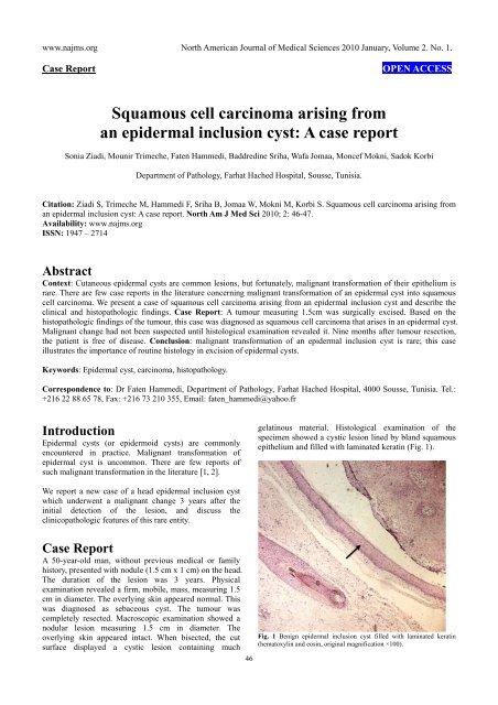
gelatinous material. Histological examination of the<br />
specimen showed a <strong>cyst</strong>ic lesion lined by bl<strong>an</strong>d squamous<br />
epithelium <strong>an</strong>d filled with laminated keratin (Fig. 1).<br />
Fig. 1 Benign <strong>epidermal</strong> <strong>inclusion</strong> <strong>cyst</strong> filled with laminated keratin<br />
(hematoxylin <strong>an</strong>d eosin, original magnification ×100).
www.najms.org North Americ<strong>an</strong> Journal of Medical Sciences 2010 J<strong>an</strong>uary, Volume 2. No. 1.<br />
There were several small scattered isl<strong>an</strong>ds of severely<br />
atypical squamous epithelium. These areas of typical<br />
<strong>epidermal</strong> <strong>cyst</strong> were juxtaposed with zones that displayed<br />
marked nuclear irregularity with mitotic activity <strong>an</strong>d <strong>an</strong><br />
infiltrative growth pattern (Fig. 2). Based on these findings,<br />
the diagnosis of squamous <strong>cell</strong> <strong>carcinoma</strong> <strong>arising</strong> in <strong>an</strong><br />
epidermoid <strong>cyst</strong> was made.<br />
Fig. 2 Malign<strong>an</strong>t squamous <strong>cell</strong> <strong>carcinoma</strong> <strong>arising</strong> <strong>from</strong> the <strong>epidermal</strong><br />
<strong>cyst</strong>; with mitotic activity <strong>an</strong>d <strong>cell</strong>ular atypia (hematoxylin <strong>an</strong>d eosin,<br />
original magnification ×400).<br />
Immunohistochemical detection of p53 <strong>an</strong>tigen was found<br />
only in <strong>cell</strong> squamous <strong>carcinoma</strong> (Fig. 3). The surgical<br />
margins were free. The patient has not received <strong>an</strong>y<br />
adjuv<strong>an</strong>t therapy. Nine months after tumour resection, the<br />
patient is free of disease.<br />
Fig. 3 Immunohistochemistry showing positivity of the tumor <strong>cell</strong>s for<br />
P53 protein (original magnification ×200).<br />
Discussion<br />
An <strong>epidermal</strong> <strong>inclusion</strong> <strong>cyst</strong> is a widespread benign<br />
intradermal lesion, <strong>an</strong>d may occur <strong>an</strong>ywhere in the body. A<br />
47<br />
<strong>carcinoma</strong> <strong>arising</strong> in a preexisting <strong>epidermal</strong> <strong>inclusion</strong> <strong>cyst</strong><br />
or sebaceous <strong>cyst</strong> is uncommon. We know of few detailed<br />
case reports of <strong>carcinoma</strong> <strong>arising</strong> in epidermoid <strong>cyst</strong>s [1,<br />
2]. Reported rates of malign<strong>an</strong>t tr<strong>an</strong>sformation of<br />
<strong>epidermal</strong> <strong>cyst</strong> into cut<strong>an</strong>eous squamous <strong>cell</strong> <strong>carcinoma</strong><br />
r<strong>an</strong>ge <strong>from</strong> 0.011 to 0.045% [2, 3]. Most cases were on the<br />
head <strong>an</strong>d neck, while the other lesions were on the trunk or<br />
limb [4, 5]. The size of the lesions r<strong>an</strong>ged <strong>from</strong> 1.5 to 10<br />
cm (me<strong>an</strong>, 5cm) <strong>an</strong>d the duration of lesions r<strong>an</strong>ged <strong>from</strong> 2<br />
to 132 months (me<strong>an</strong>, 33.5 months) [3, 4].<br />
In our case, the size <strong>an</strong>d duration of the lesion were 1.5 cm<br />
<strong>an</strong>d 3 years respectively. There is no gender or racial<br />
predisposition, <strong>an</strong>d all ages may be affected [2]. Clinically,<br />
it is difficult to differentiate between a benign <strong>an</strong>d<br />
malign<strong>an</strong>t <strong>cyst</strong>ic lesion. Histological examination<br />
normally yields the diagnosis. The nature of the stimulus<br />
for malign<strong>an</strong>t tr<strong>an</strong>sformation is uncertain [6]. In this case<br />
it is probable that the loss of the wild-type p53 is a critical<br />
event responsible for malign<strong>an</strong>t tr<strong>an</strong>sformation.<br />
The clinical course, prognosis, approach, <strong>an</strong>d optimal<br />
m<strong>an</strong>agement of this disease entity are not well-established.<br />
<strong>Squamous</strong> <strong>cell</strong> <strong>carcinoma</strong> arise in <strong>an</strong> <strong>epidermal</strong> <strong>cyst</strong> is rare.<br />
We emphasize that all resected skin <strong>cyst</strong>ic specimens<br />
should undergo further microscopic examination to avoid<br />
<strong>an</strong>y misdiagnosis.<br />
References<br />
1. Jehle KS, Shakir AJ, Sayegh. <strong>Squamous</strong> <strong>cell</strong><br />
<strong>carcinoma</strong> <strong>arising</strong> in <strong>an</strong> epidermoid <strong>cyst</strong>. Br J Hosp<br />
Med 2007; 68: 446.<br />
2. Chiu MY, Ho ST. <strong>Squamous</strong> <strong>cell</strong> <strong>carcinoma</strong> <strong>arising</strong><br />
<strong>from</strong> <strong>an</strong> <strong>epidermal</strong> <strong>cyst</strong>. Hong Kong Med J 2007; 13:<br />
482-484.<br />
3. López-Ríos F, Rodríguez-Peralto JL, Castaño E,<br />
Benito A. <strong>Squamous</strong> <strong>cell</strong> <strong>carcinoma</strong> <strong>arising</strong> in a<br />
cut<strong>an</strong>eous <strong>epidermal</strong> <strong>cyst</strong>: case report <strong>an</strong>d literature<br />
review. Am J Dermatopathol. 1999; 21: 174-177.<br />
4. Bhatt V, Ev<strong>an</strong>s M, Malins THE. <strong>Squamous</strong> <strong>cell</strong><br />
<strong>carcinoma</strong> <strong>arising</strong> in the lining of <strong>an</strong> epidermoid <strong>cyst</strong><br />
within the sublingual gl<strong>an</strong>d--a case report. Br J Oral<br />
Maxillofac Surg 2008; 46: 683-685.<br />
5. Cameron DS, Hilsinger RL Jr. <strong>Squamous</strong> <strong>cell</strong><br />
<strong>carcinoma</strong> in <strong>an</strong> <strong>epidermal</strong> <strong>inclusion</strong> <strong>cyst</strong>: case report.<br />
Otolaryngol Head Neck Surg 2003; 129: 141-143.<br />
6. Morg<strong>an</strong> MB, Stevens GL, Somach S, T<strong>an</strong>nenbaum M.<br />
Carcinoma <strong>arising</strong> in epidermoid <strong>cyst</strong>: a case series<br />
<strong>an</strong>d aetiological investigation of hum<strong>an</strong><br />
papillomavirus. Br J Dermatol 2001; 145: 505-506.