universite louis pasteur – strasbourg 1 - THESES ET MEMOIRES DE ...
universite louis pasteur – strasbourg 1 - THESES ET MEMOIRES DE ...
universite louis pasteur – strasbourg 1 - THESES ET MEMOIRES DE ...
You also want an ePaper? Increase the reach of your titles
YUMPU automatically turns print PDFs into web optimized ePapers that Google loves.
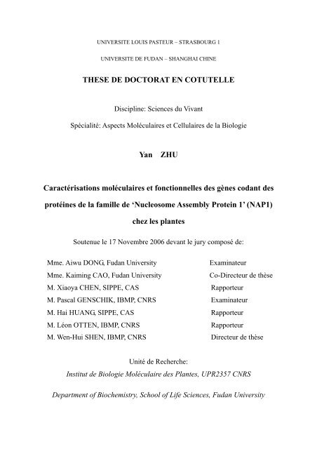
UNIVERSITE LOUIS PASTEUR <strong>–</strong> STRASBOURG 1<br />
UNIVERSITE <strong>DE</strong> FUDAN <strong>–</strong> SHANGHAI CHINE<br />
THESE <strong>DE</strong> DOCTORAT EN COTUTELLE<br />
Discipline: Sciences du Vivant<br />
Spécialité: Aspects Moléculaires et Cellulaires de la Biologie<br />
Yan ZHU<br />
Caractérisations moléculaires et fonctionnelles des gènes codant des<br />
protéines de la famille de ‘Nucleosome Assembly Protein 1’ (NAP1)<br />
chez les plantes<br />
Soutenue le 17 Novembre 2006 devant le jury composé de:<br />
Mme. Aiwu DONG, Fudan University Examinateur<br />
Mme. Kaiming CAO, Fudan University Co-Directeur de thèse<br />
M. Xiaoya CHEN, SIPPE, CAS Rapporteur<br />
M. Pascal GENSCHIK, IBMP, CNRS Examinateur<br />
M. Hai HUANG, SIPPE, CAS Rapporteur<br />
M. Léon OTTEN, IBMP, CNRS Rapporteur<br />
M. Wen-Hui SHEN, IBMP, CNRS Directeur de thèse<br />
Unité de Recherche:<br />
Institut de Biologie Moléculaire des Plantes, UPR2357 CNRS<br />
Department of Biochemistry, School of Life Sciences, Fudan University
Acknowledgements<br />
I am deeply indebted to my supervisors Dr. SHEN Wen-Hui (IBMP-CNRS, France), Dr. DONG Aiwu<br />
and Prof. CAO Kaiming (Fudan University, China), whose guidance, suggestion and encouragement helped<br />
me throughout the research and thesis writing. Their wide knowledge has been of great value for me.<br />
I would also like to thank the other members of my thesis committee: Dr. GENSCHIK Pascal, Prof.<br />
OTTEN Léon, Dr. CHEN Xiaoya and Dr. HUANG Hai, who accepted to be the “Examinateur” or<br />
“Rapporteur” and took effort in reading and writing.<br />
I thank all of my colleagues in lab 606 of IBMP-CNRS for their help, support and valuable hints, and<br />
working with them gave me the feeling of being in a great family. They are: WOLFF Michel, MEYER<br />
Denise, YU Yu, ZHAO Zhong, XU Lin, RUAN Ying and LIU Shiming. At the same time, I would also<br />
like to thank the members in the lab of Fudan University, who give me much help and courage. They are:<br />
LIU Ziqiang, YU Fang, DING Bo.<br />
Many more persons helped me in various ways during my thesis research. I wish to thank Marie-claire,<br />
Esther et al. (IBMP-CNRS, France) and ZHAN Shuxuan, GE Xiaochun et al. (Fudan University, China)<br />
for their friendly help. I also thank all the gardeners for their hard work.<br />
Finally, I would like to give my special thanks to my parents whose constant encouragement and patient<br />
love enabled me to complete this research. I wish to share this happy moment with them.<br />
ZHU Yan<br />
Oct 2006
TABLE OF CONTENTS<br />
I ABBREVIATIONS………………………………………………………………………….1<br />
II INTRODUCTION………………………………………………………………………….2<br />
II-1 Chromatin………………………………………………………………………………..2<br />
II-1-1 Heterochromatin and euchromatin……………………………………………………..2<br />
II-1-2 Chromatin folding………………………………………………………………………3<br />
II-2 Nucleosome……………………………………………………………………………….3<br />
II-2-1 Nucleosome core……………………………………………………………………….3<br />
II-2-2 Nucleosome surface…………………………………………………………………….5<br />
II-2-3 Variant nucleosomes……………………………………………………………………7<br />
II-3 Nucleosome assembly and histone chaperone…………………………………………7<br />
II-3-1 Nucleosome assembly…………………………………………………………………..8<br />
II-3-2 H3-H4 histone chaperones…………………………………………………………...10<br />
II-3-2-1 CAF-1 (Chromatin Assembly Factor-1)……………………………………………10<br />
II-3-2-2 ASF1 (Anti-Silencing Function 1)…………………………………………………12<br />
II-3-2-3 HIRA (Histone Regulation A)………………………………………………...…….13<br />
II-3-3 H2A-H2B histone chaperones………………………………………………………....14<br />
II-3-3-1 NAP1 (Nucleosome Assembly Protein 1) family…………….……………………...14<br />
II-3-3-2 Histone-binding activities of NAP1 family…………………………………………16<br />
II-3-3-3 Subcellular localization of NAP1 family……………………………………………16<br />
II-3-3-4 Post-translational modification of NAP1 family……………………………………17<br />
II-3-3-5 Chaperone activity of NAP1 family………………………………………………...18<br />
II-3-3-6 Transcriptional regulation by NAP1 family…………………………………………18<br />
II-3-3-7 In vivo function of NAP1 family…………………………………………...……….19<br />
II-4 Histone chaperones beyond nucleosome assembly…………………………………...20<br />
II-4-1 Small subunit of CAF-1………………………………………………………………..20<br />
II-4-2 Interaction of NAP1 family with B-type cyclin……………………………………….21<br />
II-4-3 PP2A inhibitory activity of S<strong>ET</strong>……………………………………………………….21<br />
II-4-4 INHAT activity of S<strong>ET</strong>………………………………………………………………...23<br />
II-5 Objectives of thesis……………………………………………………………………..24<br />
III RESULTS………………………………………………………………………………...25<br />
III-1 Molecular characterization of rice and tobacco NAP1 proteins…………………...25<br />
III-2 Molecular and functional characterization of Arabidopsis NAP1-related proteins<br />
(NRPs)…………………………………………………………………………………….47<br />
IV GENERAL DISCUSSION AND PERSPECTIVES…………………………………..70<br />
IV-1 Histone-binding and self-association of NAP1 family proteins in plant………….70
IV-2 Subcellular localization of plant NAP1 family……………………………………...70<br />
IV-3 Chromatin remodeling in root development………………………………………..71<br />
IV-4 In vitro and in vivo chromatin-based functions…………………………………….74<br />
IV-5 Perspectives…………………………………………………………………………..77<br />
V MATERIALS AND M<strong>ET</strong>HODS…………………………………………………………79<br />
V-1 Materials………………………………………………………………………………...79<br />
V-1-1 Plant materials…………………………………………………………………………79<br />
V-1-2 Bacteria strains…………………………………………………………………………79<br />
V-1-3 Cloning and expression vectors………………………………………………………..80<br />
V-1-4 Antibiotics……………………………………………………………………………...82<br />
V-1-5 Chemicals………………………………………………………………………………83<br />
V-2 Methods………………………………………………………………………………….84<br />
V-2-1 General techniques in molecular biology……………………………………………...84<br />
V-2-2 Tobacco BY2 (TBY2) cells culture and transformation………………………………91<br />
V-2-3 Arabidopsis thaliana culture, callus regeneration and transformation………………..92<br />
V-2-4 Pull down assay………………………………………………………………………..93<br />
V-2-5 In vitro Casein kinase 2α (CK2α) kinase assay……………………………………….94<br />
V-2-6 Chromatin immunoprecipitation (CHIP)……………………………………………...95<br />
V-2-7 Comet assay……………………………………………………………………………96<br />
VI REFERENCES………………………………………………………………………...97
I Abbreviations<br />
AP ammonium persulfate<br />
ATP adenosine triphosphate<br />
bp base pair<br />
BSA bovine serum albumin<br />
CTAB cetyl trimethyl ammonium bromide<br />
Da Dalton<br />
DAPI 4',6-Diamidino-2-phenylindole<br />
DNA deoxyribonucleic acid<br />
dNTP deoxyribonucleotide triphosphate<br />
DTT dithiothreitol<br />
EB ethidium bromide<br />
EDTA ethylene diamine tetra acetic acid<br />
EGTA ethylene glycol tetra acetic acid<br />
GFP green fluorescent protein<br />
GST Glutathion-S-Transferase<br />
hr hour<br />
IPTG isopropyl-beta-D-thiogalactopyranoside<br />
min minute<br />
MS Murashige and Skoog<br />
nt nucleotide<br />
PAGE polyacrylamide gel electrophoresis<br />
PBS phosphate buffered saline<br />
PCR polymerase chain reaction<br />
PMSF phenyl methyl sulphonyl fluoride<br />
PVDF polyvinylidene difluoride<br />
RNA ribonucleic acid<br />
RT reverse transcription<br />
SDS sodium dodecyl sulphate<br />
sec second<br />
TAE Tris-acetate-EDTA buffer<br />
TBE Tris-borate-EDTA buffer<br />
TBS Tris buffered saline<br />
TE Tris-EDTA buffer<br />
TEMED N,N,N',N'-Tetramethylethylenediamine<br />
Tris Tris(hydroxymethyl)aminomethane<br />
UV ultra violet<br />
YFP yellow fluorescent protein<br />
1/106
II-1 Chromatin<br />
II INTRODUCTION<br />
In eukaryotic cells, chromatin comprises huge DNA information ranging from 10 million to<br />
100 billion bp. This “database” is confined in a “library” of nucleus with only a few<br />
micrometers in diameter (Richmond, 2006). The marvelous engineering of hierarchical<br />
chromatin folding is hence critical for its condensation and package to fit into this small<br />
volume. Chromatin folding is participated by mammoth amounts of basic protein histones and<br />
other functional nuclear proteins, mainly through largely unknown and complex mechanisms<br />
of protein-protein and protein-DNA interactions (Luger and Hansen, 2005). All the cellular<br />
processes targeting DNA as substrates, such as replication, recombination, repair and<br />
transcription, should overcome the structural barrier of chromatin structure. It becomes clear<br />
that any mechanism with the potential ability to alter the levels of chromatin compaction<br />
could inherently regulate DNA accessibility (Luger, 2006).<br />
II-1-1 Heterochromatin and euchromatin<br />
By using DNA coloration, chromatin in interphase can be visualized at microscopic level into<br />
two relatively distinct forms, heterochromatin and euchromatin (Fransz et al., 2003).<br />
Heterochromatin is initially referred as those chromatin regions that remain densely stained<br />
and highly condensed throughout the cell cycle. With the increasing knowledge on chromatin,<br />
heterochromatin is assigned with several additional features, such as inactive in transcription<br />
and late in replication (Hennig, 1999). On the contrary, euchromatin is loosely packed during<br />
interphase and contains genes actively transcribed.<br />
Heterochromatin can be either constitutive or facultative (Brown, 2002). Constitutive<br />
heterochromatin is highly condensed and consists of high amount of repeated DNA sequences,<br />
such as those present in the centromeric, pericentromeric and subtelomeric regions, whose<br />
decondensation only occurs during DNA replication in late S-phase. Facultative<br />
heterochromatin is not a permanent feature but is seen in some cells at certain time, and<br />
2/106
contains loci activated in special stage and repressed in others (Brown, 2002). In recent years,<br />
heterochromatin studies have led to a dramatic advance in understanding epigenetic control of<br />
gene activity (Lund et al., 2004; Bernstein and Allis, 2005). Nevertheless, the precise<br />
molecular interactions and structural changes at chromatin higher-order folding remain barely<br />
known (Grigoryev et al., 2006).<br />
II-1-2 Chromatin folding<br />
The fundamental unit of chromatin is the nucleosome, which introduces supercoil responsible<br />
for the first level of DNA packaging and results in condensation for over 5 times in length.<br />
With the help of linker histones occupying the positions between nucleosomes, nucleosomes<br />
in the structure of “beads-on-a-string” are further compacted into fiber structure with 30 nm<br />
of diameter, in a way currently undetermined (Luger and Harsen, 2005). This level of<br />
compaction reflects the major euchromatic structure containing loci actively transcribed, and<br />
it is considered that most nuclear machineries targeting DNA carry on their functions<br />
efficiently on this level of “workshops” (Hayes and Harsen, 2001). Those domains embracing<br />
inert loci or intergenic repeat sequences are further condensed into higher hierarchical<br />
structures with the participation of other nuclear proteins, forming heterochromatin<br />
(Woodcock, 2006) (see Figure II-1).<br />
II-2 Nucleosome<br />
II-2-1 Nucleosome core<br />
Nucleosome core is composed of histone octamer wrapped by approximate 146 bp of DNA in<br />
about 1.75 turns. Between nucleosome cores are found the linker histones (named as H1/H5)<br />
and linker DNA that is about 160 bp (yeast) to more than 200 bp (higher organisms) in length<br />
(Hayes and Harsen, 2001; Luger, 2006).<br />
Histone octamer consists of two molecules of each of core histones H2A, H2B, H3 and H4.<br />
As the most slowly evolved proteins, histones present similar structures containing 2 distinct<br />
domains: the histone fold domain formed by three α-helices connected by two loops, and the<br />
3/106
Hierarchical DNA compaction in nucleus<br />
Nucleus (human)<br />
Mitotic chromosome<br />
Chromatin fiber<br />
Nucleosome<br />
Base pair<br />
400,000 fold<br />
10,000 fold<br />
35 fold<br />
6 ~ 11 fold<br />
Figure II-1 DNA compaction in nucleus<br />
Each level of DNA strucuture and compaction are shown, and size of corresponding<br />
organization are illustrated.<br />
4/106
N-terminal tail domain consisting of 15-30 basic amino acids and protruding out of<br />
nucleosome surface (Khorasanizadeh, 2004). Biochemical studies have shown that in<br />
solutions of moderate salt and in the absence of DNA, the H3-H4 complex forms a tetramer<br />
and H2A-H2B complex forms a dimer in the “handshake” arrangement. These components<br />
then associate together to form the histone octamer in the presence of DNA or in buffered<br />
solutions containing more than 1M NaCl. H3 has a unique role within the nucleosome, as<br />
there is a 2-fold symmetry in the nucleosome organized directly along the dimer interface of<br />
the two H3 histones. Apart from heterodimerization with H4, H3 also forms direct contacts<br />
with histone H2A whereby the H2A-H2B dimers position themselves (Luger, 2006) (see<br />
Figure II-2).<br />
More than 120 direct atomic interactions between histones and DNA backbone, and nearly<br />
equal number of water-mediated interactions are distributed at the 14 super-helix locations at<br />
the interface of these sub-complexes, while multiple electrostatic effect, hydrophobic effect<br />
and hydrogen bonds are required for nucleosome formation (Luger, 2003; Kamakaka and<br />
Biggins, 2005). Although nucleosome is the basic and repeating unit of chromatin and the<br />
sequence of DNA wrapped around histone octamer greatly varies, no obvious change in<br />
overall nucleosome structure depending on DNA sequence has been found in the crystal<br />
studies in the past decades (Luger, 2006). Currently it is considered that solvent water<br />
contributes greatly to the structural similarity by adapting histone surfaces to conformational<br />
variations in DNA (Davey et al., 2002). Nevertheless, as the basic components of nucleosome,<br />
histones have critical contribution in nucleosome structure. It is certain that modification on<br />
histone residues or alteration in primary sequence would affect nucleosome dynamics<br />
possibly via causing subtle structural variation or recruiting different chromatin-modifying<br />
factors, and hence influence the DNA accessibility and thereafter higher architecture.<br />
II-2-2 Nucleosome surface<br />
Histone N-terminal tails that protrude from nucleosome core contain the main sites for several<br />
covalent post-translational modifications, such as methylation on lysines and arginines,<br />
acetylation on lysines, phosphorylation on serines and threonines (Peterson and Laniel, 2004).<br />
5/106
H3-H4<br />
tetramer<br />
Histone<br />
octamer<br />
H2A-H2B<br />
dimer<br />
Figure II-2 Histone octamer assembly<br />
Histone H3 and H4 form tetramer, while H2A and H2B form dimer. The 2-fold<br />
symmetry organized directly along the dimer interface of the two H3 histones is<br />
marked in dashed. Via interaction between H2A and H3, two dimers of H2A-H2B<br />
position themselves and form histone octamer.<br />
6/106
These modifications are considered to be involved in the regulation of chromatin dynamics, in<br />
the ways of controlling the folding of the nucleosomal array into higher order structures, and<br />
mediating signaling for cellular processes. It is widely accepted that these covalent<br />
modifications function as “a histone code” in two levels: as a short-term signal to activate or<br />
repress specific gene(s) in response to cellular signaling, and as a stable marking system<br />
during cellular differentiation to inheritably determine specific chromatin states in an<br />
epigenetic manner (Loyola and Almouzni, 2004).<br />
II-2-3 Variant nucleosomes<br />
Besides canonical histones, most eukaryotes have evolved various histone variants that are<br />
non-allelic with the major ones. In general, canonical histone genes have multiple copies<br />
highly similar in sequence, primary expression during S-phase in cell cycle, and the products<br />
are incorporated in the chromatin throughout the genome. In contrast, histone variants usually<br />
present as single-copy genes that escape the strict restriction in S-phase and can be expressed<br />
during the whole cell cycle (Kamakaka and Biggins, 2005). After transferred into nucleus,<br />
histone variants are destined to being incorporated in specific functional domains in<br />
chromatin to carry out their corresponding cellular functions. In some cells, histone variants<br />
can especially replace the canonical histone positions and promote nucleosomal and further<br />
chromatic structure changes resulting in differentiated cellular functions. Variants of histones<br />
H2A and H3 have been known for decades, and recently they have emerged to the forefront of<br />
chromatin research because they are intimately involved in the specification of alternative<br />
chromatin states (Henikoff et al., 2004).<br />
II-3 Nucleosome assembly and histone chaperones<br />
The whole chromatin is replicated during S-phase in cell cycle. Pre-existed nucleosomes are<br />
randomly transferred to both daughter strands after the passing of replication fork machinery,<br />
while the other half of the nucleosome complement is made from newly synthesized and<br />
nuclear-imported histones in a reaction known as de novo nucleosome assembly (Verreault,<br />
2000).<br />
7/106
Histones are highly positively charged due to basic residues rich in composition. While<br />
directly interacting with naked DNA in physiologic conditions, histones would cause<br />
precipitation due to opposing charges rather than functional interaction. Various histone<br />
chaperones are evolutionally introduced to prevent the electrostatic dysfunction. These<br />
chaperones, mainly as negatively charged molecules, interact with histones possibly via<br />
masking basic charges and promote the intrinsic character of histones for their deposition onto<br />
DNA and nucleosome assembly. Importantly, they are not the components consisted in the<br />
final assembly products (Polo and Almouzni, 2006). To date, increasing number of histone<br />
chaperones are successively isolated and identified (see Table II-1).<br />
II-3-1 Nucleosome assembly<br />
In S-phase, the bulk of canonical histones are imported into nucleus and deposited onto<br />
nascent DNA for nucleosome assembly tightly coupled with DNA replication, which is<br />
termed as replication-coupled (RC) pathway. Besides RC, canonical histones can also be<br />
incorporated during processes independent of replication, such as transcription and DNA<br />
repair, which is correspondingly termed as replication-independent (RI) pathway (Verreault,<br />
2000). Histone variants are generally incorporated into nucleosomes mainly in RI pathway<br />
because of its constitutive expression pattern and specific cellular functions.<br />
No matter in pathway of RC or RI, as well as by use of the canonical or variant, histones are<br />
assembled into mature and functional nucleosomes in a stepwise course, as presented in<br />
current models. First, (H3-H4)2 tetramer is deposited onto naked DNA to form<br />
sub-nucleosomal particles via histone chaperone with preference to H3-H4, due to its higher<br />
affinity for DNA as compared to histone chaperone. Second, two dimers of H2A-H2B are<br />
deposited onto tetramer via histone chaperone with preference to H2A-H2B, due to their<br />
higher affinity for sub-nucleosomal particles as compared to either histone chaperone or DNA.<br />
However, this nascent state of nucleosome is immature and irregularly positioned along the<br />
template, and needs chromatin remodeling to form regular nucleosome array and functional<br />
regions, which is finally processed in the presence of ACF (ATP-utilizing chromatin<br />
8/106
Table II-1, Histone Chaperones<br />
Histone Chaperone Human Mouse Xenopus Drosophila Yeast Arabidopsis Related function<br />
CAF-1<br />
p150<br />
p60<br />
p150<br />
p60<br />
p150<br />
p60<br />
p150<br />
p105<br />
Cac1<br />
Cac2<br />
FAS1<br />
FAS2<br />
chromatin assembly coupled to DNA synthesis,<br />
maintenance of silent chromatin states, DNA repair<br />
stable<br />
RbAp48 RbAp48 RbAp48 NURF-55 Cac3 MSI1<br />
Asf1<br />
ASF1a<br />
ASF1b<br />
ASF1a<br />
ASF1b<br />
---<br />
ASF-1 ASF-1<br />
ASF1a<br />
ASF1b<br />
putative histone transfer, synergy with CAF-1 activities<br />
HIRA HIRA HIRA HIRA HIRA<br />
Hir1<br />
Hir2<br />
HIRA chromatin assembly independent of DNA replication<br />
N1/N2 --- --- N1/N2 --- --- --storage<br />
of H3/H4 pools in Xenopus oocyte to be used for<br />
chromatin assembly<br />
NAP-1 NAP-1 NAP-1 NAP-1 NAP-1 NAP-1 --- deposition of H2A/H2B, exchange of H2A/H2B variant<br />
S<strong>ET</strong><br />
TAF1α<br />
TAF1β<br />
TAF1α<br />
TAF1β<br />
TAF1α<br />
TAF1β<br />
S<strong>ET</strong> --- ---<br />
deposition of H2A/H2B, PP2A inhibitor, INHAT<br />
Nucleoplasmin --- --- Nucleoplasmin Nucleoplasmin --- --storage<br />
of H2A/H2B pools in Xenopus oocyte to be used for<br />
chromain assembly, sperm chromatin decondensation<br />
9/106
emodeling factors) (Nakagawa et al., 2001; Tyler, 2002) (see Figure II-3 and following<br />
Introduction).<br />
II-3-2 H3-H4 histone chaperones<br />
There are many currently identified chaperones preferentially binding H3 and H4, such as<br />
CAF-1 (Chromatin Assembly Factor-1), RCAF (Replication-Coupling Assembly Factor),<br />
HIRA (Histone Regulation A) and N1/N2. CAF-1 is the best-documented chaperone well<br />
studied in divergent species, and is considered to be involved in the RC chromatin assembly,<br />
DNA repair and heterochromatin silencing; while RCAF is an intermediate and co-operator<br />
with CAF-1 participating in the replication. HIRA was found in the nucleosome assembly<br />
complex containing H3 variant H3.3, and responsible for its deposition and the corresponding<br />
RI pathway. N1/N2 role is speculated as species-specific histone storage, for its peculiar<br />
abundance in Xenopus oocyte.<br />
II-3-2-1 CAF-1 (Chromatin Assembly Factor-1)<br />
CAF-1 was initially identified by biochemical fractionation of extracts derived from human<br />
HeLa cells as an activity that allowed chromatin assembly coupled to DNA replication in the<br />
SV40 viral system (Smith and Stillman, 1989). With H3 and specifically acetylated isoforms<br />
of H4, i.e. acetylated lysine 5 and 12 in histone tail, CAF-1 forms CAC (Chromatin Assembly<br />
Complex), which was isolated from nuclei of human cells and could assemble nucleosomes in<br />
a RC manner (Verreault et al., 1996). CAF-1 is evolutionarily conserved, and in most species,<br />
CAF-1 consists of three components: the large, mid and small subunits. In humans, the<br />
subunits correspond to p150, p60 and p48, in budding yeast to Cac1, Cac2 and Cac3, and in<br />
Arabidopsis to FAS1, FAS2 and MSI1 (Ridgway and Almouzni, 2000).<br />
p150, the largest subunit of CAF-1, interacts directly with PCNA (Proliferating Cell Nuclear<br />
Antigen), an accessory factor in DNA polymerase complex, which elucidates the molecular<br />
mechanism whereby CAF-1 is coupled to replication fork, and provides the first molecular<br />
link between nucleosome assembly and DNA replication (Shibahara and Stillman, 1999).<br />
10/106
Redistribution of parental histones<br />
PCNA<br />
RCAF<br />
H3-H4<br />
CAF-1<br />
Replication de novo<br />
H2A-H2B<br />
Transcription<br />
NAP1<br />
Figure II-3 Current model of participation of histone chaperone in<br />
nucleosome assembly<br />
Parental nucleosomes are redistributed into daughter strands at random. For de<br />
novo assembly, (H3-H4) tetramer is firstly deposited onto naked DNA facilitated<br />
2<br />
by histone chaperones with preference to H3-H4; next, two dimers of H2A-H2B<br />
are deposited onto tetramer facilitated by histone chaperone with preference to<br />
H2A-H2B; finally, nascent nucleosomes are slided by ATP-utilizing chromatin<br />
remodeling complex for regular nucleosome array. Transcription activation needs<br />
the initial removal of H2A-H2B dimers for consequential activities.<br />
11/106<br />
S<strong>ET</strong><br />
Nucleosome sliding
Deletion of mammalian CAF-1 causes S-phase arrest and cell death, indicating the vital<br />
importance of CAF-1 replication-dependent assembly activity (Hoek and Stillman, 2003; Ye<br />
et al., 2003; Nabatiyan and Krude, 2004). In contrast, loss of CAF-1 is not lethal in<br />
Arabidopsis thaliana and Saccharomyces cerevisiae, implying uncertain pathways bypass<br />
CAF-1 function in plant and yeast cells to survive (Schönrock et al., 2006).<br />
Loss of CAF-1 is not lethal in yeast, but results in transcriptional release of some silenced<br />
genes at telomeres and increases the UV radiation sensitivity, suggested CAF-1 is required for<br />
the stable inheritance of transcriptionally repressed chromatin and responses to DNA damage<br />
(Monson et al., 1997; Enomoto and Berman, 1998; Game and Kaufman, 1999). Functional<br />
genomic analysis of fas1 and fas2 mutants showed that CAF-1 was needed for the complete<br />
compaction of heterochromatin in Arabidopsis (Schönrock et al., 2006), and loss of CAF-1<br />
function could cause the release of certain repressive transcription such as TSI, as well as the<br />
increased sensitivity to genotoxic stress (Takeda et al., 2004). Furthermore, phenotype<br />
analysis of fas mutants shows that CAF-1 maintains the cellular and functional organization<br />
of both shoot apical meristem (SAM) and root apical meristem (RAM). The mutants are<br />
defective in maintenance of the expression states of WUSCHEL (WUS) in SAM and<br />
SCARECROW (SCR) in RAM, suggesting that CAF-1 plays a critical role in the organization<br />
of SAM and RAM during post-embryonic development by facilitating stable maintenance of<br />
gene expression states (Kaya et al., 2001).<br />
II-3-2-2 ASF1 (Anti-Silencing Function 1)<br />
The analysis of factors that are required in addition to CAF-1 for DNA replication-coupled<br />
chromatin assembly led to the identification of RCAF (Replication-Coupling Assembly Factor)<br />
complex from crude Drosophila embryo extract (Tyler et al., 1999). Drosophila RCAF<br />
complex comprises histone H3 and H4 and the homologue of yeast ASF1 (Anti-Silencing<br />
Function 1), which was previously identified as a late G1 phase-specific gene that derepresses<br />
transcriptional silencing when over-expressed (Le et al., 1997). The specific acetylation<br />
patterns on H3 and H4 in RCAF complex are identical to those of newly synthesized histones<br />
to be assembled onto replicating DNA.<br />
12/106
The eukaryotic ASF1 proteins are highly conserved through evolution in structure and<br />
function (Tamburini et al., 2005). Yeast and Drosophila encode one ASF1 protein, while<br />
human genome contains two Asf1 homologues, Asf1a and Asf1b. In contrast to CAF-1<br />
complex, RCAF/ASF1 is unable on its own to promote RC nucleosome deposition (Loyola<br />
and Almouzni, 2004). It functions synergistically with CAF-1 complex during in vitro<br />
chromatin assembly coupled to DNA replication and repair (Tyler et al., 2001). Although 2<br />
Asf1 homologues, Asf1a and Asf1b, are present in the Arabidopsis genome, no functional<br />
studies have been reported yet.<br />
Currently, RCAF/ASF1 is proposed to transfer nascent histones to CAF-1, the specialized<br />
histone deposition factor complex. Thus it would be placed in a perfect position to monitor<br />
the fluctuation in the histone pools in a cycling somatic cell, and represent the prototype of a<br />
histone donor chaperone that could effectively ensure a constant supply of histones at sites of<br />
nucleosome assembly (Loyola and Almouzni, 2004).<br />
II-3-2-3 HIRA (Histone Regulation A)<br />
HIR/HIRA (Histone Regulation) genes were initially identified in yeast as negative regulators<br />
of histone gene expression (Sherwood and Osley, 1991). They represent a conserved family of<br />
proteins found in different species including yeast, Drosophila, Xenopus, mouse and human<br />
(Loyola and Almouzni, 2004). Saccharomyces cerevisiae contains two homologues, HIR1 and<br />
HIR2; while in vertebrates there is only one, HIRA. Its ectopic over-expression leads to a<br />
transcriptional down-regulation of histone genes (Nelson et al., 2002). HIRA was found in<br />
complex with histone variant H3.3 to facilitate chromatin assembly independent of DNA<br />
synthesis, distinct from the RC histone chaperone CAF-1 (Ray-Gallet et al., 2002; Tagami et<br />
al., 2004). The knockout of HIRA in Arabidopsis is embryo lethal. However, a decrease in the<br />
level of HIRA resulted in dramatic modifications in leaves in transgenic plant (Phelps-Durr et<br />
al., 2005)<br />
13/106
II-3-3 H2A-H2B histone chaperones<br />
In the current knowledge, the main histone chaperones of H2A-H2B are nucleoplasmin and<br />
NAP1 (Nucleosome Assembly Protein 1). Nucleoplasmin is the typical Xenopus chaperone<br />
involved in histone storage in cooperation with N1/N2, and is not conserved in yeast and<br />
plants. NAP1 is considered as a highly conserved histone chaperone from yeast to human<br />
with preference to H2A-H2B dimer to facilitate the chromatin assembly and play roles in<br />
chromatin remodeling during cell proliferation and gene regulation.<br />
II-3-3-1 NAP1 (Nucleosome Assembly Protein 1) family<br />
NAP1 was first identified as a cellular factor that facilitates the nucleosome assembly in vitro<br />
by introducing supercoils into relaxed circular DNA in the presence of purified core histones<br />
(Ishimi et al., 1983). Only one NAP1 gene is present in yeast, however, higher eukaryotes,<br />
from Drosophila to human, have evolved NAP1 families comprising multiple members.<br />
Based on the result from yeast NAP1 protein crystal structure recently determined (Park and<br />
Luger, 2006a), while its N- and C-terminal regions are largely disordered, the central region is<br />
considered as stable structural entity, which was previously proved sufficient for histone<br />
binding and nucleosome assembly. It can be divided into two domains: domain I consists of<br />
one long α-helix flanked by two shorter α-helices, responding for protein dimerization.<br />
Domain II is a four-stranded anti-parallel β-sheet that is protected by α-helices at its<br />
underside, responding for protein-protein interaction (Park and Luger, 2006a).<br />
According to the protein structure, another protein S<strong>ET</strong> (SE Translocation) can also be<br />
classified into NAP1 family since it shares the central region, with difference in N- and<br />
C-terminus (Park and Luger, 2006b) (see Figure II-4). S<strong>ET</strong> protein is not found in yeast, but<br />
conserved in multicellular eukaryotes. It was initially identified as the product of a gene that<br />
was fused to the can gene as a result of a translocation in a patient with acute undifferentiated<br />
leukemia (von Lindern et al., 1992). In addition, it was independently characterized as a<br />
cellular factor required for the replication of purified adenovirus core particles in vitro,<br />
14/106
Figure II-4 Structural organization of NAP1 family (Park and Luger, 2006b)<br />
(A) Overall structure of yeast NAP1. Subdomains are represented in different<br />
colors. Secondary structure assignments and amino acid numbers (in intervals of<br />
10) are indicated below. The unstructured acidic C-terminal domain that is not a<br />
part of current model is indicated with a red dotted line. (B) Alignment of NAP<br />
family member, yeast NAP1 and human S<strong>ET</strong> are shown. The position and length<br />
of the acidic C-terminal domain are indicated.<br />
15/106
whereby it acquired the nomenclature of TAF1 (Template Activating Factor 1) (Matsumoto et<br />
al., 1993).<br />
II-3-3-2 Histone-binding activities of NAP1 family<br />
The ability to bind histones is the prerequisite property for the histone chaperones to carry on<br />
their function. Drosophila NAP1 protein was found associated with histone H2A and H2B in<br />
a crude whole-embryo extract by co-immunoprecipitation assay (Ito et al., 1996).<br />
Furthermore, density gradient analysis showed that, although capable of binding all four<br />
histones in vitro, NAP1 exhibited a greater affinity for histone H2A-H2B, however in the<br />
presence of H3-H4, H2A-H2B were transferred from NAP1 to the sub-nucleosomal particles<br />
(Nakagawa et al., 2001). Similar results were obtained for mouse and human proteins,<br />
showing that those NAP1 homologues are conserved in this activity (Ishimi et al., 1987).<br />
Nevertheless, yeast NAP1 preferentially binds the H3-H4 tetramer in vitro, which is mediated<br />
by the N-terminal histone tails (McBryant et al., 2003); in addition, it is also capable of<br />
binding linker histone H1 (Kepert et al., 2005). Human S<strong>ET</strong> interacts with core histones with<br />
a preference for histone H3-H4, which is also considered via the interaction with N-terminus<br />
of H3 (Schneider et al., 2004). The significance of these findings for the in vivo function of<br />
NAP1 family remains to be determined.<br />
II-3-3-3 Subcellular localization of NAP1 family<br />
As a histone chaperone, NAP1 family members should not only interact with free histones in<br />
the cytoplasm, but also participate in the assembly of nucleosomes in the nucleus. Studies of<br />
NAP1 localization in Drosophila embryo revealed that NAP1 is present in the nucleus during<br />
S phase but predominantly in the cytoplasm during G2 phase (Ito et al., 1996). Similar result<br />
was found for hNAP1L4 (human NAP1-like 4) (Rodriguez et al., 2000).<br />
Import of nascent histones into nucleus is mediated by importin Kap (Karyopherin) proteins.<br />
The N-terminal tails of H2A and H2B serve as NLS (Nuclear Localization Signal) recognized<br />
by certain Kap proteins in yeast, while NAP1 plays a role to bridge the Kap-histone<br />
16/106
interaction to increase specificity of histone cargo (Mosammaparast et al., 2002). Intriguingly,<br />
yeast NAP1 was immuno-stained predominantly in the cytoplasm. Deletion analysis has<br />
uncovered that its N-terminal leucine-rich NES (Nuclear Export Signal) was functional to<br />
export the protein out of nucleus via exportin Crm1p, resulting in its nucleocytoplasmic<br />
shuttling, which is important for its function during mitotic progression (Mosammaparast et<br />
al., 2002; Miyaji-Yamaguchi et al., 2003). Mammalian S<strong>ET</strong> was predominantly located in the<br />
nucleus (Nagata et al., 1998).<br />
II-3-3-4 Post-translational modification of NAP1 family<br />
Post-translational modifications on protein could bring influence over its function and/or<br />
cellular localization. The prominent modification on NAP1 group members is<br />
phosphorylation (Adachi et al., 1994; Kellogg et al., 1995a; Li et al., 1999; Rodriguez et al.,<br />
2000). Xenopus NAP1 could be specifically phosphorylated by Cyclin B/p34 cdc2 in vitro, but<br />
not by Cyclin A/ p34 cdc2 , suggesting its potential modulation by specific cyclin during cell<br />
cycle for further cellular functions (Kellogg et al., 1995a). It was found that Drosophila<br />
NAP1, human NAP1 and hNAP1L4 could be phosphorylated by CK2 (Casein Kinase 2) in<br />
vitro and in vivo, a ubiquitous second message-independent serine/threonine-specific kinase.<br />
Furthermore, the subcellular localization of hNAP1L4 is associated with its phosphorylation<br />
state: it is phosphorylated and localized in the cytoplasm at the G0/G1 boundary; while at G1/S<br />
boundary it is dephosphorylated and transported into nucleus to carry out its histone<br />
chaperone function (Rodriguez et al., 2000). Although human S<strong>ET</strong> was phosphorylated in<br />
vivo, the kinase and its influence on S<strong>ET</strong> activity still remain obscure (Adachi et al., 1994).<br />
Poly-glutamylation is an unusual modification by adding variable number of glutamyl units<br />
on protein side chains, which is previously only detected on tubulin. Interestingly, various<br />
animal NAP1 proteins, from Drosophila to human, were found poly-glutamylated in vivo,<br />
however, by distinct pathway and for potentially distinct functions (Regnard et al., 2000). The<br />
discovery of poly-glutamylation on NAP1, which was previously considered as<br />
tubulin-specific, implies their potential cytological relevance.<br />
17/106
II-3-3-5 Chaperone activity of NAP1 family<br />
NAP1 family members alone could deposit core histones onto DNA and assembly<br />
nucleosome in vitro (Kawase et al., 1996; Gamble et al., 2005). However, except yeast NAP1,<br />
most members could reconstitute nucleosomes along DNA template only with random<br />
distribution and failed to result in periodic arrays of nucleosomes resembling those functional<br />
and native chromatin in vivo (Tyler, 2002). Although yeast NAP1 could accomplish regularly<br />
spaced nucleosome assembly, the nucleosomal array presented relatively short spacing<br />
interval distinct from the normal ones observed in vivo and unlikely rendered functional<br />
structure and dynamics. These results suggested that, as histone chaperone, NAP1 family<br />
members along could not remodel the nascent nucleosomes into active and functional arrays,<br />
which should be cooperated with ACF (Tyler, 2002).<br />
One important characteristic of NAP1 family is that they can bind basic protein other than<br />
histones. During spermatogenesis in Xenopus, histones, especially H2A-H2B, are largely<br />
replaced with various SP (Sperm-specific basic Protein), which is tightly related to the highly<br />
condensation in sperm chromatin structure. Incubation of NAP1 family member can result the<br />
interaction of chaperone with all kinds of SP proteins and their release from the highly<br />
packaged structure, and hence the final decondensation of sperm chromatin (Matsumoto et al.,<br />
1999).<br />
The adenovirus genome complexed with viral core proteins is the template for transcription of<br />
early genes and the first round of replication in adenovirus-infected human cells. NAP1<br />
family members can interact with adenovirus core protein and be involved in remodeling of<br />
the adenovirus chromatin-like structure for replication and transcription (Kawase et al., 1996).<br />
II-3-3-6 Transcriptional regulation by NAP1 family<br />
During transcription, chromatin regions have to adopt a more open conformation, and<br />
transcription factor and nucleosomal histones compete for the regulatory region on the<br />
template. Removal of one H2A-H2B dimer from the histone octamer seems to be essential for<br />
18/106
the transcriptional initiation and elongation (Belotserkovskaya et al., 2003). Yeast NAP1<br />
could reversibly remove and replace histone H2A-H2B dimer or variant dimer from<br />
assembled nucleosomes, resulting in active histone exchange (Park et al., 2005). Similar<br />
property was also found in human NAP1 and S<strong>ET</strong> (Okuwaki et al., 2005).<br />
It was proved that NAP1 family members interact with p300/CBP (Corticosteroid-Binding<br />
Protein) in vivo and in vitro, one transcriptional co-activator with intrinsic histone<br />
acetyltransferases activity (Shikama et al., 2000; Asahara et al., 2002; Karetsou et al., 2005).<br />
They form multi-component ternary complex with histones and regulate gene expression,<br />
likely via modifying acetylation on histones followed by transferring H2A-H2B dimers from<br />
nucleosomes to NAP1 family members (Ito et al., 2000).<br />
Besides the common regulation, other examples of respective involvement of NAP1 in<br />
promoter-specific transcription regulation have been published. In a yeast two-hybrid screen,<br />
human NAP1 was shown to form a complex with the transcriptional activator E2 (Rehtanz et<br />
al., 2004). A complex of human NAP1, E2 and p300 was shown to activate transcription in<br />
vitro. Over-expression of Xenopus NAP1 affects the expression of several potential target<br />
genes in a tissue-specific manner (Steer et al., 2003). Human S<strong>ET</strong> plays as a cofactor along<br />
with other transcription factors to specifically regulate P450c17 transcription (Compagnone et<br />
al., 2000). It also regulates KA11 gene-promoter-specific transcription by binding adaptor<br />
protein Fe65 (Telese et al., 2005).<br />
II-3-3-7 In vivo function of NAP1 family<br />
NAP1-regulated gene transcription in chromatin context is exemplified by the genome-wide<br />
expression analysis of nap1 in Saccharomyces cerevisiae. In Δnap1 strain about 10% of all<br />
yeast open reading frames change the expression level more than 2-fold compared with<br />
wild-type strain in micro-array analysis. These genes are generally clustered, suggesting the<br />
specific manner of NAP1 regulation (Ohkuni et al., 2003).<br />
Knockout targeting of NAP1 gene in Drosophila resulted in embryonic lethal or poorly viable<br />
19/106
adult escapers (Lankenau et al., 2003). mNAP1L2 (mouse NAP1-like 2) is an X-chromosome<br />
linked gene specifically expressed in neurons. Inactivated mNAP1L2 by gene targeting would<br />
lead to embryonic lethality from mid-gestation onwards, suggesting it represents a class of<br />
tissue-specific factors to regulate neuronal cell proliferation (Rogner et al., 2000).<br />
Presenilin plays critical roles in the genesis of Alzheimer’s disease and in LIN-12/Notch<br />
signaling during development. While in screen for genes that influence presenilin level or<br />
activity in Caenorhabditis elegans, it was found that substitution mutant of spr-2 (suppressor<br />
of presenilin-2) suppresses the egg-laying defective phenotype caused by a null allele of the<br />
sel-12 presenilin gene. SPR-2 was identified as the homologue of S<strong>ET</strong> (Wen et al., 2000).<br />
II-4 Histone chaperones beyond nucleosome assembly<br />
II-4-1 Small subunit of CAF-1<br />
As the small subunit of CAF-1, p48/Cac3/MSI1 is a conserved protein from yeast to human,<br />
and has also been found associated with chromatin regulatory complexes. HAT-B (B-type<br />
histone acetyltransferases), which acetylate K5 and K12 of newly synthesized histone H4, are<br />
heterodimeric complexes composed of a catalytic subunit Hat1p and an accessory protein p48<br />
that facilitates contact between the complex and histones (Parthun et al., 1996). In addition,<br />
p48 protein binds to RPD3 histone deacetylases and is part of several RPD3-containing<br />
complexes (Vermaak et al., 1999).<br />
p48/Cac3/MSI1 also functions together with Enhancer of Zeste [E(Z)]-type Polycomb group<br />
histone methyltransferases in mammals, insects and plants. Polycomb group proteins are<br />
required for the maintenance of transcriptional repression of genes for developmental<br />
regulators, which often are Homeobox transcription factors in animals and MADS box<br />
transcription factors in plants (Ringrose and Paro, 2004). p48/Cac3/MSI1 and RPD3 histone<br />
deacetylases interact with Retinoblastoma tumor suppressor protein pRB in mammals and<br />
plants, and mediate the transcriptional repression of pRB target genes (Nicolas et al., 2001;<br />
Kennedy et al., 2001). Taken together, p48/Cac3/MSI1 activities, which are beyond<br />
20/106
nucleosome assembly, are of central importance for cell proliferation and differentiation.<br />
II-4-2 Interaction of NAP1 family with B-type cyclin<br />
In addition to the histone chaperone activity, NAP1 was also found involved in the mitotic<br />
event via interaction with B-type cyclins (Kellogg et al., 1995a). Yeast NAP1 was found to<br />
play an important role in budding yeast cyclin Clb2 function in suppressing polar bud growth<br />
(Kellogg and Murray, 1995b). Yeast cells lacking NAP1 showed in clumps of connected cells<br />
with elongated shapes, and the deleting nap1 in Clb2-dependent cells had a much more<br />
dramatic effect that formed large clumps of interconnected cells that had an unusual elongated<br />
morphology and temperature sensitive for growth (Kellogg et al., 1995a). This abnormal<br />
phenotype is greatly related to the microtubule dynamics that is regulated by B-type cyclins<br />
during mitosis, and NAP1 may play a role in controlling microtubule stability (Kellogg and<br />
Murray, 1995b).<br />
Although no S<strong>ET</strong> exists in yeast, Xenopus S<strong>ET</strong> can interact specially with cyclin B, similar<br />
with NAP1 (Kellogg et al., 1995a), suggesting that S<strong>ET</strong> may also be involved in the<br />
regulation pathway of cyclin B on cell cycle. Cyclins perform function via interacting with<br />
distinct CDK (cyclin-dependent kinase) at appropriate period, and modulating its substrate<br />
specificity to activate corresponding pathways. CDK activity can be regulated by CKI (CDK<br />
Inhibitor) that associates with cyclin-CDK complexes, such as p21 Cip1 . S<strong>ET</strong> along, or in<br />
cooperation with p21 Cip1 , can inhibit cyclin B-CDK1 activity and regulate the transition of<br />
G2/M in cell cycle (Canela et al., 2003). Furthermore, S<strong>ET</strong> can interact with GAPDH<br />
(glyceraldehyde 3-phosphate dehydrogenase), which can reverse the S<strong>ET</strong> inhibition on<br />
cyclin-CDK1 activity in a dose-dependent manner (Carujo et al., 2006). Finally, S<strong>ET</strong> can<br />
reverse the inhibition of cyclin E-CDK2 induced by p21 Cip1 , and hence regulate the transition<br />
of G1/S in cell cycle (Estanyol et al., 1999).<br />
II-4-3 PP2A inhibitory activity of S<strong>ET</strong><br />
A truncated form of S<strong>ET</strong> was first isolated as a potent heat-stable protein inhibitor of PP2A<br />
21/106
(Protein Phosphatase 2A) (Li et al., 1995). PP2A is a major serine/threonine phosphatase that<br />
regulates diverse cellular processes. Physiological targets of this phosphatase include<br />
cell-surface receptors and ion channels, protein kinases involved in mitogenic signaling and<br />
cell cycle, key regulatory enzymes and proteins involved in metabolism, and numerous<br />
transcription factors (Al-Murrani et al., 1999). Subsequent study indicated that S<strong>ET</strong> could<br />
potently and specifically inhibit PP2A activity at nanomolar concentrations similar with the<br />
truncated form (Li et al., 1996). This inhibition is likely conserved since studies in human<br />
malaria parasite, Plasmodium falciparum, showed that S<strong>ET</strong> can also inhibit its parasitic PP2A<br />
activity potently (Dobson et al., 2003).<br />
c-Jun is one proto-oncogene, as a major component of AP-1 (Activator Protein-1)<br />
transcription factor complex. PP2A can markedly down-regulate the concentration and DNA<br />
binding of c-Jun and subsequent AP-1 transcriptional activity, while S<strong>ET</strong> can reverse this<br />
repression via its inhibition on PP2A. Further experiments showed that the effect of S<strong>ET</strong> and<br />
PP2A might be mediated, in part, by changing the phosphorylation of c-Jun at Ser 63<br />
(Al-Murrani et al., 1999). S<strong>ET</strong> can also modulate the PP2A effect on the balance of<br />
cytochrome P450c17 metabolism between cortisol synthesis and sex steroids production<br />
(Pandey et al., 2003).<br />
Transcriptional activation of the heat shock genes during the heat shock response in<br />
Drosophila has been intimately linked to phosphorylation of histone H3 at serine 10, whereas<br />
repression of non-heat-shock genes correlates with dephosphorylation of histone H3, which is<br />
accomplished by PP2A. Mutants in PP2A display reduced genome-wide H3<br />
dephosphorylation, and sites of H3 phosphorylation that do not contain heat shock genes<br />
remain transcriptionally active during heat shock in PP2A mutants. Interestingly, S<strong>ET</strong> is<br />
found recruited to the heat shock gene loci following heat shock, and potentially prevent the<br />
PP2A repression and promote the activation. Aberrant S<strong>ET</strong> activity results in misregulation of<br />
the catalytic activity of PP2A, producing abnormal patterns of gene expression (Nowak et al.,<br />
2003). Human leukemic HRX fusion proteins derived from a chromosomal translocation in<br />
acute leukemia formed a heterocomplex with S<strong>ET</strong> and PP2A. HRX fusion proteins might<br />
22/106
function in conjunction with S<strong>ET</strong> and PP2A to deregulate cell proliferation and cell cycle<br />
controls, resulting in leukemia (Adler et al., 1997).<br />
II-4-4 INHAT activity of S<strong>ET</strong><br />
S<strong>ET</strong> is found as a subunit of human cellular complex INHAT (Inhibitor of Acetyltransferases)<br />
that potently inhibits the histone acetyltransferase activity of p300/CBP and PCAF (Seo et al.,<br />
2001). However, S<strong>ET</strong> alone, also contain the INHAT activity attributed by its C-terminal<br />
acidic tail, which is essential for histone binding. It was proposed that S<strong>ET</strong> inhibits histone<br />
acetylation by masking histone tails from the histone acetyltransferases such as p300. In<br />
addition, S<strong>ET</strong> was shown to associate with histone deacetylases in vitro and in vivo,<br />
suggesting that S<strong>ET</strong> could integrate chromatin hypo-acetylation and transcriptional repression<br />
(Kutney et al., 2004).<br />
Histone hypo-acetylation and DNA hyper-methylation are hallmarks of gene silencing. S<strong>ET</strong>,<br />
as an inhibitor of histone acetyltransferase, can inhibit active demethylation of DNA resulting<br />
in gene silencing, integrating DNA methylation and transcriptional silencing (Cervoni et al.,<br />
2002).<br />
S<strong>ET</strong> INHAT activity is not limited in histones but also directly on transcription factor. The<br />
Sp/KLF (for Sp1- and Krüppel-like factor) family of zinc finger transcription factors is<br />
important in development, differentiation, and oncogenic processes. S<strong>ET</strong> can interact with the<br />
DNA-binding domain (DBD) of transcription factors Sp1 and KLF5, and mask them from<br />
acetylation by co-activator/acetylase p300 and hence from activation. It is a new pathway for<br />
regulation of DNA-binding transcription factor on DBD through interaction and coupled<br />
acetylation by two opposing regulatory factors of a coactivator/acetylase and a negative<br />
cofactor harboring activity to inhibit acetylation (Miyamoto et al., 2003; Suzuki et al., 2003).<br />
This notion is further supported by the interaction of S<strong>ET</strong> with estrogen receptor α-associated<br />
proteins via DBD, and the inhibition of acetylation and hence transactivation (Loven et al.,<br />
2003).<br />
23/106
II-5 Objectives of thesis<br />
As histone chaperones, NAP1 family members contain histone-binding and chromatin<br />
assembly and remodeling activities, as well as functions in many other biologic processes.<br />
Although much progress has been achieved in molecular and biochemical characterization of<br />
NAP1 family member in yeast and animal, intriguingly, no study had been published in the<br />
field of plant, except one report on the isolation of soybean cDNA encoding a NAP1<br />
homologue (Yoon et al., 1995). My thesis work focused on molecular and functional<br />
characterization of plant NAP1 homologues. We first want to know the conservation of NAP1,<br />
their histone binding activity and their expression during the cell cycle to insight into<br />
H2A/H2B chaperones in plants. This work uncovers a complex pattern of intracellular<br />
localization of different plant NAP1 proteins, which intrigued us to investigate nuclear<br />
transport of these proteins and their binding proteins. Finally, by reverse genetics in the model<br />
plant Arabidopsis, we investigated function of NAP1 genes in plant growth and development.<br />
Together my thesis work provides novel insights into the dynamic regulation of chromatin<br />
structure for plant development.<br />
24/106
III RESULTS<br />
III-1 Molecular characterization of rice and tobacco NAP1 proteins<br />
My thesis starts from the molecular characterization of plant NAP1 proteins. The<br />
monocotyledon plant rice and the dicotyledon plant tobacco were initially chosen to<br />
characterize. My contributions to the work included cDNA isolation, gene expression analysis,<br />
plasmid construction, recombinant protein production, protein phosphorylation in vitro and<br />
microscopy observation. The corresponding results were published in Article 1 and Article 2.<br />
(Article 1-Planta)<br />
(Article 2-Plant Physiology)<br />
25/106
[Signalement bibliographique ajouté par : ULP <strong>–</strong> SCD <strong>–</strong> Service des thèses électroniques]<br />
Regulation of biosynthesis and intracellular localization of rice and tobacco homologues<br />
of nucleosome assembly protein 1<br />
Aiwu Dong, Yan Zhu, Yu Yu, Kaiming Cao, Chongrong Sun et Wen-Hui Shen<br />
Planta, 2003, Vol. 216, Pages 561-570<br />
Pages 561 à 570 :<br />
La publication présentée ici dans la thèse est soumise à des droits détenus par un éditeur<br />
commercial.<br />
Pour les utilisateurs ULP, il est possible de consulter cette publication sur le site de l'éditeur :<br />
http://www.springerlink.com/content/tel99u7fkkta8dcd/fulltext.html<br />
Il est également possible de consulter la thèse sous sa forme papier ou d'en faire une demande<br />
via le service de prêt entre bibliothèques (PEB), auprès du Service Commun de<br />
Documentation de l'ULP: peb.sciences@scd-ulp.u-strasbg.fr
[Signalement bibliographique ajouté par : ULP <strong>–</strong> SCD <strong>–</strong> Service des thèses électroniques]<br />
Interacting Proteins and Differences in Nuclear Transport Reveal Specific Functions for<br />
the NAP1 Family Proteins in Plants<br />
Aiwu Dong, Ziqiang Liu, Yan Zhu, Fang Yu, Ziyu Li, Kaiming Cao, et Wen-Hui Shen<br />
Plant Physiology, 2005, Vol. 138, Pages 1446<strong>–</strong>1456<br />
Pages 1446 à 1456 :<br />
La publication présentée ici dans la thèse est soumise à des droits détenus par un éditeur<br />
commercial.<br />
Pour les utilisateurs ULP, il est possible de consulter cette publication sur le site de l'éditeur :<br />
http://www.plantphysiol.org/cgi/content/full/138/3/1446<br />
Il est également possible de consulter la thèse sous sa forme papier ou d'en faire une demande<br />
via le service de prêt entre bibliothèques (PEB), auprès du Service Commun de<br />
Documentation de l'ULP: peb.sciences@scd-ulp.u-strasbg.fr
III-2 Molecular and functional characterization of Arabidopsis NAP1-related proteins<br />
(NRPs)<br />
Due to the poor genetic approaches in rice and tobacco, for further analysis of biological roles<br />
of NAP1, I chose to work on the model plant Arabidopsis thaliana, which contains 4 genes<br />
encoding NAP1 and 2 genes encoding NAP1-related proteins (NRP1 and NRP2).<br />
One or several T-DNA mutant lines corresponding to each gene were searched from the<br />
Arabidopsis Stock Centers and identified. Single mutant with loss-of-function of any of these<br />
genes did not show any phenotype under our in vitro culture and greenhouse growth<br />
conditions, then I crossed single mutants to generate double mutants. The NRP double mutant<br />
clearly showed short-root phenotype. My work was subsequently focused on the<br />
characterization of this mutant. The results obtained were described in Article 3 and Article 4.<br />
(Article 3-Plant Cell)<br />
(Article 4, submitted)<br />
47/106
[Signalement bibliographique ajouté par : ULP <strong>–</strong> SCD <strong>–</strong> Service des thèses électroniques]<br />
Arabidopsis NRP1 and NRP2 Encode Histone Chaperones and Are Required for<br />
Maintaining Postembryonic Root Growth<br />
Yan Zhu, Aiwu Dong, Denise Meyer, Olivier Pichon, Jean-Pierre Renou, Kaiming Cao,<br />
et Wen-Hui Shen<br />
The Plant Cell, 2006, Vol. 18, Pages 2879-2892<br />
Pages 2879 à 2892 :<br />
La publication présentée ici dans la thèse est soumise à des droits détenus par un éditeur<br />
commercial.<br />
Pour les utilisateurs ULP, il est possible de consulter cette publication sur le site de l'éditeur :<br />
http://www.plantcell.org/cgi/content/full/18/11/2879<br />
Il est également possible de consulter la thèse sous sa forme papier ou d'en faire une demande<br />
via le service de prêt entre bibliothèques (PEB), auprès du Service Commun de<br />
Documentation de l'ULP: peb.sciences@scd-ulp.u-strasbg.fr
Addenda<br />
Chromatin remodeling in Arabidopsis root growth<br />
Yan Zhu 1,2 , Aiwu Dong 2 , and Wen-Hui Shen 1,*<br />
1 Institut de Biologie Moléculaire des Plantes, laboratoire propre du CNRS (UPR 2357)<br />
conventionné avec l'Université Louis Pasteur (Strasbourg 1), 12 rue du Général<br />
Zimmer, 67084 Strasbourg Cédex, France;<br />
2 Department of Biochemistry, School of Life Sciences, Fudan University, Shanghai<br />
200433, PR China;<br />
* Correspondence to: Wen-Hui Shen; Institut de Biologie Moléculaire des Plantes,<br />
laboratoire propre du CNRS (UPR 2357) conventionné avec l'Université Louis<br />
Pasteur (Strasbourg 1), 12 rue du Général Zimmer, 67084 Strasbourg Cédex,<br />
France ; Tel. : +33 3 88417283 ; Fax : +33 3 88614442 ; Email: Wen-<br />
Hui.Shen@ibmp-ulp.u-strasbg.fr<br />
KEY WORDS : Arabidopsis thaliana, histone chaperone, chromatin, transcription,<br />
root growth<br />
Addendum to : Arabidopsis NRP1 and NRP2 encode histone chaperones and<br />
are required for maintaining post-embryonic root growth<br />
Y. Zhu, A. Dong, D. Meyer, O. Pichon, J.-P. Renou, K. Cao and W.-H. Shen<br />
Plant Cell 2006 Nov 22; [Epub ahead of print]<br />
62/106
ABSTRACT<br />
The basic structural unit of chromatin is the nucleosome, which consists of 146 bp of<br />
DNA wrapped around the histone octamer constituted by two molecules each of<br />
histones H2A, H2B, H3 and H4. Nucleosome assembly/disassembly/reassembly<br />
processes occur primarily during DNA replication and also during transcription, DNA<br />
repair and recombination. Several chromatin-remodeling factors had been previously<br />
shown to have pleiotropic roles in different processes of plant growth and<br />
development. We have recently demonstrated that the Arabidopsis NRP1 and NRP2<br />
genes encode H2A/H2B chaperones and are required for the maintenance of post-<br />
embryonic root growth. The nrp1-1nrp2-1 double mutant plants specifically showed a<br />
short-root phenotype in normal growth conditions. They were also hypersensitive to<br />
DNA damage and showed release of transcriptional gene silencing. We propose that<br />
NRP1 and NRP2 act as histone H2A/H2B chaperones in nucleosome assembly,<br />
playing critical roles for a correct genome transcription in the maintenance of root<br />
growth.<br />
63/106
In plants as in animals, development is regulated by differential gene expression<br />
whereby cells acquire specific fates (1). Plant development takes place essentially<br />
postembryonically. During embryogenesis, only the basic body plan is established,<br />
with small groups of cells called apical meristems at both ends of the body axis.<br />
During postembryonic development, the shoot apical meristem (SAM) at the apical<br />
end and the root apical meristem (RAM) at the basal end continuously provide cells<br />
to maintain stem cells as well as to initiate organogenesis which leads to the<br />
development of the aerial parts and the subterranean root system of the plant,<br />
respectively. Chromatin structure is fundamental to transcription. Similar chromatin<br />
gene mutations that cause embryonic lethality in animals frequently result in plants<br />
that are detectably modified but viable, making it relatively easy to study the effects of<br />
these genes in development in plants. Chromatin-remodeling factors have been<br />
shown to play crucial roles in divers processes of plant development, including leaf<br />
patterning, shoot organogenesis, flowering time control, gametogenesis and seed<br />
reproduction (2-5).<br />
By reverse genetic analysis, we demonstrated that the nrp1-1nrp2-1 mutant<br />
containing loss-of-function of both NRP1 and NRP2 genes has a short-root<br />
phenotype. NRP1 and NRP2 proteins show highest homology with the animal<br />
S<strong>ET</strong>/TAF-I/I2 PP2A proteins, which are involved in nucleosome assembly by<br />
chaperoning histones H2A and H2B (6). Consistently, NRP1 and NRP2 were found<br />
to bind histones, preferentially H2A and H2B than H3. They are localized primarily in<br />
the nucleus and they bind chromatin in planta. Additional supports of NRP1 and<br />
NRP2 in chromatin remodeling were the observations that the nrp1-1nrp2-1 mutant<br />
plants exhibit perturbed genome transcription, release of heterochromatic gene<br />
silencing and hypersensitive response to DNA damage.<br />
64/106
In spite of the fundamental role of NRP1 and NRP2 in chromatin remodeling,<br />
the phenotype of the nrp1-1nrp2-1 mutant is remarkably root specific, the embryos<br />
and the aerial organs (leaves, rosettes, inflorescences, flowers and fruits) developed<br />
normally in the mutant plants. Arrest of cell cycle progression at G2/M and disordered<br />
cellular organization were observed in the mutant roots. In this addendum, we show<br />
that the mutant root segments, when cultured in vitro, had similar capacity in callus<br />
formation than the wild-type root segments (Figure 1A), and that roots regenerated in<br />
vitro from hypocotyls were also arrested in elongation in the mutant but not in the<br />
wild-type (Figure 1B). These new data further strengthen the root specificity of<br />
requirement of NRP1 and NRP2 in the maintenance of cell proliferation. The specific<br />
short-root phenotype of the nrp1-1nrp2-1 mutant is in sharp contrast to the pleiotropic<br />
phenotypes of the fas1 and fas2 mutants, which include stem fasciation, abnormal<br />
phyllotaxy, modified leaf shape, reduced growth and size of all organs (7, 8). FAS1<br />
and FAS2 encode subunits of the CAF1 complex, which chaperones histones H3 and<br />
H4 in nucleosome assembly. It is possible that H2A/H2B and H3/H4 contribute to<br />
different levels of nucleosome assembly/disassembly and more importantly additional<br />
chaperones are likely involved in chaperoning histones in nucleosome assembly in<br />
Arabidopsis.<br />
Several ethylene-responsive genes encoding transcription factors were<br />
upregulated in the nrp1-1nrp2-1 mutant seedlings. However, the mutant seedlings<br />
showed normal ethylene triple response (data not shown) and the treatment by the<br />
ethylene biosynthesis inhibitor AVG (L-a-(2-amino-ethoxyvinyl)-glycine) could not<br />
sufficiently rescue the short-root phenotype (Figure 1C). These latter observations<br />
indicate that the modifications of ethylene-responsive genes are not enough to<br />
explain the nrp1-1nrp2-1 mutant phenotype. Among the other differentially expressed<br />
65/106
genes found in the nrp1-1nrp2-1 mutant seedlings, GLABRA2 (GL2) was<br />
downregulated whereas PL<strong>ET</strong>HORA2 (PLT2) was upregulated. GL2 encodes a<br />
homeodomain transcription factor and represses root hair formation (9), its<br />
downregulation correlated with the high proliferation of root hairs in the nrp1-1nrp2-1<br />
mutant. PLT2 encodes an AP2-type transcription factor and plays crucial roles in<br />
stem cell specification and maintenance in the RAM (10), its perturbed expression<br />
could have significantly contributed to the nrp1-1nrp2-1 mutant root phenotype.<br />
NRP1 and NRP2 proteins were found to bind chromatin at GL2 and PLT2, thus might<br />
regulate directly expression of these genes. Chromatin organization at GL2 was also<br />
affected in fas2 mutant roots (11). In addition to dynamics of nucleosome<br />
assembly/disassembly, histone acetylation/deacetylation and ATP-dependent<br />
chromatin remodeling also play important roles in root growth and development (12,<br />
13). Future experiments will further explore epigenetic regulation in root growth and<br />
development, particularly in response to intrinsic and environmental factors.<br />
NRP1 and NRP2 are expressed not only in roots but also in leaves, stems<br />
and flowers. In addition, NRP1 and NRP2 can form homomeric and heteromeric<br />
protein complexes, and NRP1 and NRP2 show functional redundancy in root growth.<br />
Nonetheless, molecular phenotype differed between the nrp1-1 and nrp2-1 single<br />
mutants: a significantly higher number of genes was detected by transcriptome<br />
analysis to be differentially expressed in the nrp2-1 than in the nrp1-1 mutant<br />
seedlings. Further work is required to understand both redundant and unique<br />
molecular functions of NRP1 and NRP2. The close homologues of NRP1 and NRP2<br />
are NAP1-group proteins, which include four members (AtNAP1;1 to AtNAP1;4) in<br />
Arabidopsis. Different members of the NAP1-group proteins in rice and tobacco have<br />
distinct subcellular localizations and show different affinity to different types of<br />
66/106
histones (14, 15). Genetic and molecular characterization of this group of genes in<br />
Arabidopsis will help to understand their roles in epigenetic inheritence, particularly<br />
interesting in comparison with NRP1 and NRP2 in plant growth and development.<br />
References<br />
1. Meyerowitz EM. Plants compared to animals: the broadest comparative study<br />
of development. Science 2002; 295:1482<br />
2. Guyomarc'h S, Bertrand C, Delarue M, Zhou DX. Regulation of meristem<br />
activity by chromatin remodelling. Trends Plant Sci 2005; 10:332-8.<br />
3. Hsieh TF, Fischer RL. Biology of chromatin dynamics. Annu Rev Plant Biol<br />
2005; 56:327-51.<br />
4. Zhao Z, Yu Y, Meyer D, Wu C, Shen WH. Prevention of early flowering by<br />
expression of FLOWERING LOCUS C requires methylation of histone H3 K36.<br />
Nat Cell Biol 2005; 7:1256-60.<br />
5. Baurle I, Dean C. The timing of developmental transitions in plants. Cell 2006;<br />
125:655-64.<br />
6. Park YJ, Luger K. Structure and function of nucleosome assembly proteins.<br />
Biochem Cell Biol 2006; 84:549-58.<br />
7. Leyser, HMO, Furner IJ. Characterisation of three shoot apical meristem<br />
mutants of Arabidopsis thaliana. Development 1992; 116:397-403.<br />
8. Kaya H, Shibahara KI, Taoka KI, Iwabuchi M, Stillman B, Araki T. FASCIATA<br />
genes for chromatin assembly factor-1 in Arabidopsis maintain the cellular<br />
organization of apical meristems. Cell 2001; 104:131-42.<br />
67/106
9. Ohashi Y, Oka A, Rodrigues-Pousada R, Possenti M, Ruberti I, Morelli G,<br />
Aoyama T. Modulation of phospholipid signaling by GLABRA2 in root-hair<br />
pattern formation. Science 2003; 300,1427-1430.<br />
10. Aida M, Beis D, Heidstra R, Willemsen V, Blilou I, Galinha C, Nussaume L,<br />
Noh YS, Amasino R, Scheres B. The PL<strong>ET</strong>HORA genes mediate patterning of<br />
the Arabidopsis root stem cell niche. Cell 2004; 119:109-20.<br />
11. Costa S, Shaw P. Chromatin organization and cell fate switch respond to<br />
positional information in Arabidopsis. Nature 2006; 439:493-6.<br />
12. Xu CR. Liu C, Wang YL, Li LC, Chen WQ, Xu ZH, Bai SN. Histone acetylation<br />
affects expression of cellular patterning genes in the Arabidopsis root<br />
epidermis. Proc Natl Acad Sci USA 2005; 102:14469-74.<br />
13. Fukaki H, Taniguchi N, Tasaka M. PICKLE is required for SOLITARY-<br />
ROOT/IAA14-mediated repression of ARF7 and ARF19 activity during<br />
Arabidopsis lateral root initiation. Plant J 2006; 48:380-9.<br />
14. Dong A, Zhu Y, Yu Y, Cao K, Sun C, Shen WH. Regulation of biosynthesis<br />
and intracellular localization of rice and tobacco homologues of nucleosome<br />
assembly protein 1. Planta 2003; 216: 561-70.<br />
15. Dong A, Liu Z, Zhu Y, Yu F, Li Z, Cao K, Shen WH. Interacting proteins and<br />
differences in nuclear transport reveal specific functions for the NAP1 family<br />
proteins in plants. Plant Physiol. 2005; 138: 1446-1456.<br />
68/106
Figure 1. Comparison of callus and root growth between the wild-type (WT) and the<br />
nrp1-1nrp2-1 mutant. (A) Representatives of callus regeneration and growth from<br />
root segments cultured in the presence of 2.3 µM 2,4-D, 11.4 µM IAA and 3.2 µM<br />
BAP. (B) Representatives of root regeneration and growth from hypocotyls<br />
cultured in the presence of 5.7 µM IAA and 1 µM IBA. Arrows indicate roots<br />
arrested in growth. Photographs were made 2 weeks after culture. (C) Root<br />
elongation of plants grown at different concentrations of the ethylene biosynthesis<br />
inhibitor AVG. The mean value from 20 plants is shown. Vertical bars represent<br />
standard deviations.<br />
69/106
IV GENERAL DISCUSSION AND PERSPECTIVES<br />
IV-1 Histone-binding and self-association of NAP1 family proteins in plant<br />
Our pull-down assays showed that NRP proteins, as well as tobacco NAP1 proteins, can bind<br />
histone H2A and H2B. Furthermore, this interaction seems preferential, since NRP proteins<br />
interact barely with H3. In vitro binding assay showed that, despite of distinct relative binding<br />
strengths, plant NAP1 proteins bind core histones as well as the linker histone H1. Since<br />
animal NAP1 can modulate the binding of H1 to chromatin, it will be interesting to test the<br />
role of plant NAP1 family in the construction and remodeling of chromatin mediated by H1 in<br />
the cellular processes and plant growth. Taken together, the histone-binding property of plant<br />
NAP1 family members we analyzed support their role as histone chaperones.<br />
We also showed that NRP1 and NRP2 could form homo- and heteromeric protein complexes,<br />
as well as tobacco NAP1 proteins. Consistently, previous work showed that human S<strong>ET</strong><br />
proteins could form homo- and heterodimer (Matsumoto et al., 1999; Miyaji-Yamaguchi et al.,<br />
1999), while yeast and Drosophila NAP1 proteins could form polymeric complexes<br />
(McBryant and Peersen, 2004; Ito et al., 1996). It is considered that the long α-helix in<br />
N-terminal NAP domain is responsible for forming protein dimer (Park and Luger, 2006a),<br />
and interestingly, this long α-helix is well conserved in plant NAP1 group. It was considered<br />
that animal NAP1 proteins exist as dimer to participate in the H2A-H2B nuclear import and<br />
transcription regulation (Tóth et al., 2005); in addition, dimerization of human S<strong>ET</strong> protein is<br />
critical for its chromatin remodeling activity (Miyaji-Yamaguchi et al., 1999). It is likely that<br />
NAP1 family members in plant also carry on their function in the form of dimer.<br />
IV-2 Subcellular localization of plant NAP1 family<br />
By using GFP-tagged fusion protein, the subcellular localization analysis shows that NRP1<br />
and NRP2 proteins are mainly localized in the nucleus, like the animal homologue S<strong>ET</strong>. This<br />
is quite consistent with their function of histone chaperone in chromatin remodeling and<br />
transcription regulation.<br />
70/106
Except OsNAP1;3, all the plant NAP1 proteins we analyzed contain a conserved and typical<br />
NLS (KKKPKK), in addition, there is a second NLS (KPKKK) in C-terminal tail of<br />
OsNAP1;1. The common NLS (KKKPKK) does not seem functional since it could not import<br />
its proteins into the nucleus. The nuclear localization of OsNAP1;1 depends on the second<br />
NLS, which is supported by substitution mutant, while the mechanism of nuclear-import of<br />
OsNAP1;3 remains unclear yet.<br />
We showed that OsNAP1;1 and NtNAP1;1 could shuttle between the cytoplasm and the<br />
nucleus. Approaches of mutagenesis and use of inhibitor proved that the nuclear-export of the<br />
two proteins is mediated by NES (Nuclear Export Signal), and the result indirectly proved<br />
that NtNAP1;1 can also be imported into nucleus, however, this nuclear localization seems<br />
transient and quickly reversed by exportins. Surprisingly, the other nucleocytoplasmic protein<br />
OsNAP1;3 is not affected by NES mutagenesis and inhibitor treatment. There are two<br />
explanations for the distinct subcellular localization of NAP1 proteins. One is that all the<br />
NAP1 proteins can be imported into nucleus but are exported by distinct mechanisms.<br />
OsNAP1;1 and NtNAP1;1 are via their NES recognized by exportins while others are via<br />
unknown and independent pathways, however with inefficiency in OsNAP1;3. The other<br />
explanation is that besides OsNAP1;1, only NtNAP1;1 and OsNAP1;3 can be imported into<br />
the nucleus to some extent, while the mechanisms of their nuclear exporting are still distinct.<br />
Further study on differential NAP1-binding proteins may help to explain it. After all, the<br />
divergent compartmentation of NAP1 proteins suggests that, as a whole family, the activities<br />
of NAP1 proteins are beyond nucleosome assembly.<br />
Our result is the first systematic research on plant NAP1 family, and for the first time presents<br />
the variation in subcellular compartmentation of its members in plant, helps to guide the<br />
future research to discover the functional regulation of individual plant NAP1 family member.<br />
IV-3 Chromatin remodeling in root development<br />
Arabidopsis roots are tip-growing structures that grow in length through the action of tip<br />
meristems. Behind the root cap, the root tip is organized along its length into successive<br />
71/106
meristematic zone, elongation zone and<br />
differentiation zone (Scheres et al., 2002).<br />
Furthermore, transverse sections showed<br />
that the radial organization of the mature<br />
tissues in wild-type roots consists of the<br />
epidermis, cortex, endodermis and<br />
pericycle, which surround the vascular<br />
cylinder.<br />
The elongation zone in the nrp1-1nrp2-1<br />
double mutant is greatly reduced<br />
compared to that of wild-type roots.<br />
Transverse sections revealed that cell<br />
division in mutant roots was abnormal<br />
and cell organization in each layer was irregular, which were much more pronounced in<br />
sections of the differentiation zone. We introduced cell division marker CYCB1:GUS into<br />
double mutant by cross to analyze the role of NRP1 and NRP2 in cell cycle progression in<br />
proximal meristem cells. It was found that higher number of GUS positive cells were<br />
observed in the mutant root tips at later growth stages (from 8 DAG) compared to the<br />
wild-type root tips, suggesting that G2/M arrest occurred in the mutant.<br />
Since human S<strong>ET</strong> protein can interact with Cyclin B and inhibit Cyclin B-CDK1 activity and<br />
regulate the transition of G2/M in cell cycle (Canela et al., 2003), we initially want to verify<br />
the conservation of this pathway in plant. In our pull-down assays, tobacco cyclin<br />
Nicta;CYCB1;1 binds the NAP1 proteins, but fails to bind NRP1 or NRP2. In addition,<br />
expression pattern analysis shows that, like Cyclin B, NRP are ubiquitously expressed in all<br />
the tissues tested. The model that direct interaction of NRP proteins with Cyclin B exerts the<br />
subsequent inhibitory effect on its activity seems unlikely to explain the current short-root<br />
phenotype. The occurrence of G2/M arrest in the mutant should be contributed by other<br />
mechanisms.<br />
72/106
Ectopic long hairs are found irregularly positioned in the mutant root, in parallel, ectopic<br />
expression of GL2 (GLABRA 2) gene is also found in the mutant by RT-PCR. GL2 is a<br />
homeobox gene involved in development of root epidermis in Arabidopsis (Masucci et al.,<br />
1996a). Wild-type epidermis is composed of two cell types, root-hair cells and hairless cells,<br />
which are located at distinct positions within the root, implying that positional cues control<br />
cell-type differentiation. GL2 gene is preferentially expressed in the differentiating hairless<br />
cells of the wild-type, and appears to act in a cell-position-dependent manner to suppress hair<br />
formation in differentiating hairless cells (Masucci et al., 1996a). The phenotype of the double<br />
mutant is quite consistent with the down-regulation of GL2 gene in our findings<br />
It is considered that hormones ethylene and auxin act downstream of GL2 to promote root<br />
hair outgrowth during epidermis development in the Arabidopsis root (Masucci and<br />
Schiefelbein, 1996b). Furthermore, ethylene and auxin are known to regulate root hair<br />
elongation (Pitts et al., 1998), as well as root elongation (Swarup et al., 2002; Alonso et al.,<br />
2003), while the molecular mechanisms by which ethylene and auxin interact to regulate these<br />
73/106
processes, agonistically in some cases and antagonistically in others, remain largely unknown<br />
(Stepanova et al., 2005). Interestingly, 3 ethylene-responsive proteins are up-regulated to<br />
more than 2-fold in the double mutant in our microarray. This finding promotes us to<br />
investigate the involvement of ethylene and auxin in the NRP-dependent root growth.<br />
We have tried several chemicals related to ethylene and auxin in the rescue assays, however,<br />
neither the ethylene biosynthesis inhibitor AVG, nor the synthetic auxins or auxin transport<br />
inhibitors, can rescue the short-root phenotype of nrp1-1nrp2-1 double mutant. Furthermore,<br />
the mutant shows similar triple response with the wild-type, such as apical hook maintenance<br />
in the dark growing environment, and opened hook and shortened hypocotyl in the presence<br />
of auxin transport inhibitor NPA (Achard et al., 2003). Taken together, these results show that<br />
simple regulation of ethylene and auxin concentration or compartmentation cannot rescue the<br />
mutant phenotype caused by the loss-of-function of NRPs. There are two possibilities: NRPs<br />
likely accomplish their function in the pathways independent of ethylene or auxin signaling,<br />
or, NRPs function in the downstream and loss-of-function of NRPs constitutively activate or<br />
repress some consequent pathways resulting in the phenotype.<br />
IV-4 In vitro and in vivo chromatin-based functions<br />
Our results established the interaction of plant NRP proteins with chromatin. YFP:NRP fusion<br />
protein can interact with chromatin region corresponding to the loci whose expression are<br />
regulated in the double mutant, such as GL2; however, exception is also detected at some<br />
genes whose expression are unchanged. At the pericentromeric heterochromatin, YFP:NRP<br />
can bind TSI but to a less extent Ta3 and the centromeric 180 bp-repeats. Taken together, NRP<br />
protein interacts with chromatin with no uniform distribution over the genome.<br />
Study of animal S<strong>ET</strong> showed that it was a multifunctional protein. It alone, or forming<br />
complex INHAT with other subunits, could mask histone tail from acetylation by histone<br />
acetyltransferase p300/CBP and PCAF, and hence repress in vitro transcription mediated by<br />
the participation of co-activators with histone acetyltransferase activity (Seo et al., 2001).<br />
During the heat shock response in Drosophila, repression of non-heat-shock genes correlates<br />
74/106
with PP2A activities by dephosphorylation of histone H3 Ser10, while S<strong>ET</strong> is found recruited<br />
to the heat shock gene loci and potentially prevents the PP2A repression and promotes the<br />
activation (Nowak et al., 2003). S<strong>ET</strong> is required for transcription in vitro of chromatin<br />
templates as histone chaperone (Gamble et al., 2005), and is also found associated with<br />
various transcription factors and chromatin remodeling factors to influence their activities in<br />
distinct mechanisms (Al-Murrani et al., 1999; Pandey et al., 2003; Loven et al., 2003;<br />
Miyamto et al., 2003; Suzuki et al., 2003; Kutney et al., 2004). In summary, the<br />
chromatin-association state of S<strong>ET</strong> is quite divergent depending on its histone-binding<br />
activities and/or mediation via other factors, and its function in gene expression depends on<br />
its dynamic state and divergent associated partners. It may help to explain our failure to set up<br />
one concise platform to link the NRP binding and NRP-regulated genes.<br />
In nrp1-1nrp2-1 double mutant, 102 genes were found differentially expressed, some of<br />
which are good candidates responsible for the phenotype. However, this number is lower than<br />
those reported for fas mutants, 483 genes in fas1 and 204 genes in fas2 (Schönrock et al.,<br />
2006). In addition, the specific short-root phenotype of nrp1-1nrp2-1 double mutant is distinct<br />
from the phenotype of fas1 and fas2 which is pleiotropic in the whole plant including<br />
fasciation. In addition, the identified genes barely overlap in these two categories of mutants,<br />
suggesting that they likely act independently, and NRP1 and NRP2 function in maintaining<br />
post-embryonic growth in a more specific manner.<br />
Perturbed gene expression in double mutant spreads into the heterochromatic region, such as<br />
TSI, in the presence of genotoxic stress. This release was previously found in mutants<br />
defective in factors participated in DNA/chromatin replication and assembly, such as FAS1,<br />
FAS2, TSK/MGO3/BRU1 and RNR, which encode the larger two subunits of CAF-1, one<br />
novel factor involved in histone chaperoning together with CAF-1, and ribonucleotide<br />
reductase respectively (Takeda et al., 2004; Wang and Liu, 2006), implying NRP proteins are<br />
involved in the maintenance of heterochromatin silencing in epigenetic inheritance. Previous<br />
research has suggested that stochastic release of TGS in tsk/mgo3/bru1 and mutants of fas and<br />
rnr caused meristem fasciation resulting from the ectopic expression of WUSCHEL (WUS), a<br />
75/106
key meristem regulator (Mayer et al., 1998; Kaya et al., 2001; Takeda et al., 2004; Wang and<br />
Liu, 2006). It is also likely that ectopic release of some critical transcription factor(s) in<br />
double mutant would be collectively responsible for the short-root phenotype.<br />
It is considered that chromatin assembly/disassembly is essential for DNA damage response<br />
and epigenetic inheritance in DNA repair. tsk/mgo3/bru1, and mutants of rnr and fas, all show<br />
higher sensitivity to DNA damage, which is also found in mutant defective in TEBICHI (TEB)<br />
that encodes protein with helicase and DNA polymerase domains (Inagaki et al., 2006). Our<br />
result shows that n5n6 double mutant is more sensitive to genotoxic stress such as bleomycin<br />
and UV treatment, indicating that the mutant genome is also more sensitive to DNA damage.<br />
In view of the common release of TGS and genotoxic sensitivity, it is reasonable to postulate<br />
that affected chromatin assembly would influence not only the epigenetic inheritance, but also<br />
the maintenance of chromatin architecture and global stability.<br />
The results of CYCB1:GUS accumulation in the above-mentioned mutants furthermore give<br />
support to the critical role of chromatin assembly. Mutant tsk/mgo3/bru1, tebichi and fas, as<br />
well as n5n6, all show the accumulation of GUF-positive cells, indicating the arrest of G2/M<br />
in these mutants. G2/M checkpoint ensures correct and complete DNA synthesis and<br />
chromatin duplication before the entry into mitosis and cytokinesis, and B-type cyclins are<br />
involved in this checkpoint activation (Stark and Taylor, 2006). The accumulation of<br />
CYCB1:GUS and activation of G2/M checkpoint, together with the unstable chromatin<br />
structure to genotoxic stress, and incorrect epigenetic inheritance, could be considered as an<br />
interplaying consequences, resulting from the inhibition of chromatin duplication caused by<br />
affected chromatin assembly. This potential interplay is illustrated in the following model. As<br />
histone chaperones, NRP and CAF-1 cooperate with other factors, such as BRI1, TEBICHI<br />
and RNR, to guarantee the correct chromatin assembly and further influence chromatin<br />
structure, as well as epigenetic inheritance and cell cycle checkpoint.<br />
76/106
IV-5 Perspectives<br />
chromatin structure<br />
and stability<br />
G 2/M checkpoint<br />
chromatin assembly<br />
CAF-1<br />
NRP<br />
etc.<br />
BRU1<br />
TEBICHI<br />
RNR<br />
etc.<br />
epigenetic<br />
inheritance<br />
Despite that our work provides novel insight into dynamic regulation of chromatin structure<br />
for plant development, to fully understand H2A-H2B histone chaperone, we still lack their<br />
biochemical and functional characterizations. Obviously, those direct proofs of their activities<br />
in nucleosome assembly and involvement in transcription regulation would greatly help us to<br />
better understand the plant H2A-H2B histone chaperones function and their effect in<br />
chromatin dynamics during plant growth and development.<br />
Our initial approach of reverse genetics in studying NAP1 function in Arabidopsis showed<br />
that single mutant of NAP1 family member didn’t show any phenotype. It will be interesting<br />
for the further analysis of double, triple and quadruple mutants of this gene group to clarify<br />
their biological functions. In addition, as H2A-H2B histone chaperone, the phenotype of<br />
nrp1-1nrp2-1 double mutant is much less severe that those of any of H3-H4 histone<br />
chaperones: the loss-of-function of HIRA or ASF1 in Arabidopsis results in lethality, and<br />
mutation in CAF-1 function shows pleiotropic effect in the shoot and root. Considering the<br />
high similarity of protein sequence of plant NAP1 family, and the redundant function of<br />
NAP1 and S<strong>ET</strong> in animal, it will be interesting to test the overlap and redundancy of function<br />
77/106
of plant NAP1 family in plant growth and development.<br />
Animal S<strong>ET</strong> has many unique activities beyond histone chaperone, which include INHAT,<br />
PP2A inhibition. It was reported that ectopic histone acetylation can affect the expression of<br />
root patterning genes GL2 and other genes involved in epidermal cell differentiation in<br />
Arabidopsis (Xu et al., 2005). Ectopic PP2A activity can cause pleiotropic effects including<br />
inhibition of root growth (Garbers et al., 1996). Whether these activities, as well as the<br />
potential ones unknown but unique to plants, contribute to the function of either NRP protein<br />
in a similar or preferential manner, needs further investigation.<br />
78/106
V-1 Materials<br />
V-1-1 Plant materials<br />
V MATERIALS AND M<strong>ET</strong>HODS<br />
a) Nicotiana tabacum L. cv. Bright Yellow 2 (BY2) cell<br />
Tobacco BY2 cell line shows high synchronization ability and growth rate, by 80-100 fold<br />
once a week. High cell cycle synchrony can be easily obtained after aphidicolin treatment. In<br />
addition, it is easy for transformation and stable transgenic maintenance, making it powerful<br />
in exploring molecular and cellular biology of plant cells.<br />
b) Arabidopsis thaliana Columbia (Col-0)<br />
As the model organism of flowering plants, Arabidopsis thaliana has many advantages: small<br />
genome, rapid life cycle and prolific seed production, as well as efficient transformation<br />
approaches and amounts of available mutant lines, which facilitate studies of cellular and<br />
molecular biology in plant. Distinct Arabidopsis ecotypes show different organ shapes and<br />
growth characteristics due to different genetic background. Columbia (Col), with sequenced<br />
genome in Arabidopsis Genome Initiative (AGI), is one most popular ecotype, and Col-0, one<br />
of Col accessions, is used here as background line.<br />
V-1-2 Bacteria strains<br />
V-1-2-1 Escherichia Coli<br />
a) Top10<br />
Strain Top10 is ideal for high-efficiency cloning and plasmid propagation. In addition to<br />
support of blue/white screening, mutations recA1 and endA1 in Top10 increase insert stability<br />
and improve plasmid DNA quality in preparation.<br />
79/106
) DH5α<br />
Strain DH5α is recA <strong>–</strong> endA <strong>–</strong> mutant like Top10 and gives high transformation efficiency and<br />
plasmid yield. It can also be used as host for protein expression of pGEX series of vectors<br />
such as pGEX-4T-1.<br />
c) BL21 (<strong>DE</strong>3)<br />
Strain BL21 (<strong>DE</strong>3) allows highly efficient protein expression under control of T7 promoter. It<br />
is used as host for protein expression of p<strong>ET</strong> system such as p<strong>ET</strong>14b.<br />
V-1-2-2 Agrobacterium tumefaciens<br />
a) GV3101<br />
C58, Ti plasmid cured, resistant to rifampicin, in use for mediating Arabidopsis<br />
transformation.<br />
b) LBA4404<br />
pAL4404 plasmid, resistant to rifampicin, in use for mediating tobacco BY2 cells<br />
transformation or others.<br />
V-1-3 Cloning and expression vectors<br />
a) pGEM®-T and pGEM®-T Easy Vector (Promega)<br />
Both vectors are prepared for PCR product cloning by use of 3’ terminal thymidine. They<br />
contain multiple restriction sites within α-peptide coding region of β-galactosidase, allowing<br />
color screening on indicator plates for insertional inactivation. Ampicillin resistance gene is<br />
for selection.<br />
b) pEYFP-pEYFP<br />
80/106
pEYFP-pEYFP contains two EYFP cDNAs in tandem under promoter lac. Three multiple<br />
cloning sites are located between these cDNAs and promoter, allowing selective construction<br />
of EYFP-fusion protein. Ampicillin resistance gene is for selection.<br />
c) p<strong>ET</strong>-14b (Novagen)<br />
p<strong>ET</strong> system is powerful for cloning and expression of recombinant proteins. p<strong>ET</strong>-14b is one<br />
vector that allows N-terminal fusion to cleavable His-tag sequence for rapid affinity<br />
purification. Ampicillin resistance gene is for its selection. After cloning, plasmids are<br />
transformed into expression hosts such as <strong>DE</strong>3 containing a chromosomal copy of T7 RNA<br />
polymerase gene under lacUV5 control, and induced by IPTG for expression.<br />
d) pGEX-4T-1 (Amersham Biosciences)<br />
pGEX-4T-1 vector is for high-level expression of GST-fusion protein under tac promoter<br />
inducible by IPTG. One recognition site for thrombin is introduced for GST-tag cleavage.<br />
Ampicillin resistance gene is for selection.<br />
e) pER8<br />
pER8 is one binary expression vector developed for plant transformation, which adopts the<br />
XVE, LexA-Vp16-ER system strictly induced by estrogen receptor-based chemical.<br />
Spectinomycin and hygromycin resistance genes are for selection in agrobacteria and plants,<br />
respectively.<br />
81/106
V-1-4 Antibiotics<br />
Name Stock Concentration Company<br />
Ampicillin 100 mg/l Duchefa<br />
Carbenicillin 500 mg/l Duchefa<br />
Chloramphenicol 34 mg/l Duchefa<br />
Gentamycin 20 mg/l Duchefa<br />
Hygromycin 40 mg/l Duchefa<br />
Kanamycin 100 mg/l Duchefa<br />
Rifampicin 50 mg/l Duchefa<br />
Spectinomycin 100 mg/l Duchefa<br />
82/106
V-1-5 Chemicals<br />
Name Full Name Company M.W. Function<br />
1-NAA naphthalene-1-acetic acid SERVA 186.2<br />
Synthetic auxin, auxin carrier-independent,<br />
diffused into nucleus<br />
1-NOA 1-naphthoxyacetic acid ALDRICH 202.2 Auxin influx inhibitor<br />
2,4-D 2,4-dichlorophenoxyacidic acid SERVA 221.0<br />
Synthetic auxin, auxin carrier-mediated,<br />
blocked by 1-NOA<br />
6-BAP 6-benzylaminopurine<br />
(S)-trans-2-Amino-4-(2-<br />
SERVA 225.3 Cytokinin growth regulator<br />
AVG aminoethoxy)-3-butenoic acid<br />
hydrochloride<br />
SIGMA 196.6 Ethylene synthesis inhibitor<br />
β -Estradiol β -Estradiol<br />
ICN<br />
Biomedicals,<br />
Inc.<br />
272.4 Transgenic plant gene induction<br />
Cantharidin Cantharidin SIGMA 196.2 Inhibitor of PP2A<br />
IAA Indole-3-acetic acid SERVA 175.2 Cytokinin growth regulator<br />
IBA Indole-3-butyric acid SERVA 203.2 Auxin growth regulator<br />
NPA N-1-naphthylphthalamic acid DUCHEFA 291.3 Auxin efflux inhibitor<br />
83/106
V-2 Methods<br />
V-2-1 General techniques in molecular biology<br />
V-2-1-1 Vector construction<br />
V-2-1-1-1 PCR (Polymerase Chain Reaction)<br />
PCR reaction comprises following components: 1x amplification buffer, 1.5-3 mM MgCl2,<br />
200 μM dNTPs Mix, 0.4 μM forward primer, 0.4 μM reverse primer, 1-5 unit(s) thermostable<br />
DNA polymerase and suitable template DNA. Reaction volume is usually adjusted up to<br />
20-50 µl. After first denaturation treatment, PCR reaction enters repeating cycles including<br />
denaturation (94°C, 30 sec), annealing (30 sec) and elongation (72°C). Optimal elongation<br />
time and annealing temperature varies in distinct reaction conditions. Normally, every 1 kb<br />
more of PCR product needs about 1 min more of elongation, and PCR products are purified<br />
by the kit “Nucleospin Extract II” (Macherey-Nagel).<br />
V-2-1-1-2 Semi-quantitative PCR<br />
Same PCR reactions are established in several tubes. During PCR program, at the end of<br />
desired cycle, pause the PCR machine and pick out one tube quickly, then continue the<br />
program to the next step.<br />
V-2-1-1-3 Restriction enzyme digestion<br />
Digestion reaction comprises approximately 200 ng of DNA with 1-2 unit(s) of restriction<br />
enzyme(s) in optimal reaction buffer at suggested temperature. Generally, 1 hr is sufficient for<br />
most digestion. Released DNA fragment is purified by the kit “Nucleospin Extract II”<br />
(Macherey-Nagel).<br />
V-2-1-1-4 Gel electrophoresis<br />
DNA samples are resolved in agarose gel prepared in TAE or TBE buffer. The higher<br />
84/106
concentration of agarose gel resolves the smaller fragment of DNA sample. With the staining<br />
of ethidium bromide, agarose gel can be read under UV light.<br />
1 x TAE: 40 mM Tris-acetate; 2 mM EDTA.<br />
1 x TBE: 89 mM Tris-borate; 2 mM EDTA.<br />
V-2-1-1-5 Ligation<br />
DNA ligation comprises approximate 60 ng of insertion DNA and 20 ng of linear vector DNA<br />
in 1 x ligation buffer with 1 unit of T4 DNA ligase (Fermentas). Usually overnight at 16°C<br />
results in maximal ligation efficiency. The ligation product can be transformed into competent<br />
E. coli host by heat shock or electroporation. Before electroporation, ligation product needs to<br />
be desalted usually via ethanol precipitation.<br />
V-2-1-1-6 Competent cell preparation and transformation<br />
V-2-1-1-6-1 Heat shock using calcium chloride<br />
A single bacterial colony is inoculated in 3 ml of LB and incubated by vigorous agitation at<br />
37°C overnight. 0.5 ml of overnight culture is inoculated and propagated in 50 ml of fresh LB<br />
in a 250-ml flask at 37°C for about 2 hr until OD600 reaches 0.2-0.3. The culture is fully<br />
chilled on ice and centrifuged at 3,000 rpm for 10 min at 4°C. The pellets are gently<br />
resuspended in 10 ml of ice-cold fresh 0.1 M CaCl2 solution, and kept on ice for 30 min. After<br />
another centrifugation at 3,000 rpm for 10 min at 4°C, the pellets are resuspended gently in 2<br />
ml of ice-cold 0.1 M CaCl2 solution. The CaCl2-treated cells can be used directly for<br />
transformation or dispensed into aliquots and frozen at -70°C.<br />
Mix each 200 µl of competent cells with plasmid DNA or ligation product in sterile tubes by<br />
swirling gently and store on ice for 30 min. Transfer the tubes in a preheated 42°C water bath<br />
and keep for exactly 90 sec. Do not shake the tubes. Rapidly transfer the tubes to an ice bath<br />
and allow cells to chill for 1-2 min. Add 800 µl of LB medium to each tube and incubate<br />
cultures for 1 hr at 37°C to allow the bacteria to recover and express antibiotic resistance<br />
85/106
marker encoded by plasmid. Transfer the appropriate volume of transformed competent cells<br />
onto agar LB medium containing the appropriate antibiotic. Invert the plates and incubate at<br />
37°C. Transformed colonies should appear in 12-16 hr.<br />
V-2-1-1-6-2 Electroporation<br />
A single bacterial colony is inoculated in 3 ml of LB. After overnight agitation at 37°C, 3 ml<br />
of culture is diluted 100 times with fresh LB and incubated by agitation at 37°C until OD600<br />
reaches 0.5. The culture is chilled on ice for 10 min and centrifuged at 5,000 rpm for 10 min<br />
at 4°C. The cells are washed twice by ice-cold pure H2O, and once by ice-cold sterile 10%<br />
glycerol. Finally cells are resuspended in 1 ml of ice-cold 10% glycerol. The<br />
electrocompetent cells can be used immediately. For storage, 40 µl aliquots of cell suspension<br />
are dispensed in each tubes, frozen quickly in liquid nitrogen and stored in a -70°C freezer.<br />
40 µl of electrocompetent cells mixed with plasmid DNA or purified ligation products are<br />
pipetted into pre-cooled electroporation cuvettes. Transformation is carried by Bio-Rad<br />
electroporation apparatus delivering an electrical pulse of 25-μF capacitance, 2.5 kV and<br />
200/400 Ω resistance (200 Ω for E.coli and 400 Ω for Agrobacterium). The DNA-cell mixture<br />
is added with 1 ml of LB medium and removed from electroporation cuvette, followed by<br />
incubation for 1 hr at 37°C to allow bacteria to recover. Recovered culture is then transferred<br />
onto agar LB medium containing appropriate antibiotics. Transformed colonies should appear<br />
in 12-16 hr (E. coli) or after 36 hr (Agrobacterium).<br />
V-2-1-1-7 Mini-preparation of plasmid DNA by alkaline lysis with SDS<br />
Inoculate a single bacterial colony in 3 ml of LB containing appropriate antibiotic and<br />
incubate by vigorous shaking at 37°C overnight. Harvest bacteria by centrifugation at<br />
maximum speed for 30 sec and resuspend in ice-cold 100 µl of alkaline lysis solution I by<br />
vigorous vortexing. Add 200 µl of freshly prepared alkaline lysis solution II and mix by<br />
inverting tube 6-8 times and keep on ice for 3-5 min. Do not vortex. Add 150 µl of ice-cold<br />
solution III, invert tube 6-8 times and keep on ice for another 3-5 min. Centrifuge bacterial<br />
86/106
lysate at maximum speed for 5 min and transfer the supernatant to a fresh tube. Add an equal<br />
volume of phenol:chloroform (1:1 v/v), mix organic and aqueous phases by vortexing and<br />
then centrifuge the emulsion at maximum speed for 2 min. Transfer the aqueous upper layer<br />
to a fresh tube and precipitate by adding 2 volumes of 100% ethanol. DNA pellet is visible<br />
after centrifugation at maximum speed for 5 min. Wash DNA pellet with 70% ethanol. After<br />
air-dry, DNA pellet is finally dissolved in 50 µl of distilled H2O containing 10 μg/ml<br />
DNase-free RNase A (Fermentas).<br />
Alkaline Lysis Solution I: 50 mM Glucose; 10 mM EDTA; 25 mM Tris-Cl (pH 8.0)<br />
Alkaline Lysis Solution II: 0.2 M NaOH; 1% (w/v) SDS.<br />
Alkaline Lysis Solution III: 3 M potassium acetate; 11.5% acetic acid.<br />
Alternatively, plasmid DNA can be prepared via the kit “NucleoSpin Plasmid QuickPure”<br />
(Macherey-Nagel).<br />
V-2-1-2 Genomic DNA isolation<br />
V-2-1-2-1 CTAB method<br />
Grind plant tissue in liquid nitrogen to fine powder. Homogenize about 200 mg of fine<br />
powder in 600 µl of pre-heated 2x CTAB Buffer and incubate in 65°C for 15 min. Add equal<br />
volume of chloroform/isoamyl alcohol (24:1, v/v) to form an emulsion by thorough vortex.<br />
After centrifugation at 11,000 g for 1 min, transfer supernatant to a new tube, and add 1/5<br />
volume of 5% CTAB Solution and mix well. Perform another chloroform/isoamyl alcohol<br />
extraction. Mix final supernatant gently with equal volume of CTAB Precipitation Buffer and<br />
keep for 15 min. After centrifugation at 11,000 g for 1 min, re-hydrate DNA pellet in<br />
high-salt TE Buffer. Perform another precipitation by adding 2 volumes of 100% ethanol and<br />
keep at -20°C for more than 30 min. Centrifuge DNA pellet at 11,000 g for 15 min and wash<br />
with 80% ethanol. Finally the pellet is briefly dried and dissolved in 50 µl of 0.1x TE<br />
containing 100 μg/ml DNase-free RNase A (Fermentas). The quantity and quality of genomic<br />
DNA are analyzed by gel electrophoresis in 0.3% agarose.<br />
87/106
2x CTAB Buffer: 2% CTAB; 100 mM Tris-Cl (pH8.0); 20 mM EDTA; 1.4 M NaCl.<br />
5% CTAB Solution: 5% CTAB in 0.35 M NaCl.<br />
CTAB Precipitation Buffer: 1% CTAB; 50 mM Tris-Cl (pH 8.0); 10 mM EDTA.<br />
High-Salt TE Buffer: 10 mM Tris-Cl (pH 8.0); 1 mM EDTA; 1 M NaCl.<br />
0.1x TE: 1 mM Tris-Cl (pH 8.0); 0.1 mM EDTA.<br />
V-2-1-2-2. Urea method (for large scale of T-DNA insertion mutants screening)<br />
Grind one rosette leaf or one inflorescence in a tube with 600 µl of Urea Lysis Buffer, and<br />
extract once by equal volume of phenol:chloroform (1:1, v/v) by vigorous vortex. After<br />
centrifugation at 12,000 rpm for 7 min, transfer supernatant to a new tube and mix with 1/10<br />
volume of 3 M sodium acetate and 1 volume of isopropyl alcohol, and keep at -20°C for 20<br />
min. Centrifuge DNA pellet at 12,000 rpm for 5 min and wash by 70% ethanol. After air-dry,<br />
re-hydrate pellet with 50 µl of distilled H2O.<br />
Urea Lysis Buffer: 7.5 M Urea; 0.2 M Tris-Cl (pH 8.0); 50 mM EDTA; 2% Sodium Lauroyl<br />
Sarcosine (SLS)<br />
V-2-1-3 RNA isolation and analysis<br />
V-2-1-3-1 Total RNA extraction<br />
Grind plant tissue in liquid nitrogen to fine powder. Homogenize 100 mg of fine powder in 1<br />
ml of Trizol Reagent (Invitrogen) by vigorous vortex and keep at room temperature for 5 min.<br />
Add 200 µl of chloroform and keep at room temperature for 2-3 min. After centrifugation at<br />
12,000 g for 15 min at 4°C, transfer supernatant to a new tube and add 500 µl of isopropyl<br />
alcohol for precipitation at room temperature for 10 min. After centrifugation at 12,000 g for<br />
10 min at 4°C, RNA pellet is visible and washed with 1 ml of 75% ethanol by centrifugation<br />
at no more than 7,500 g for 5 min at 4°C. Finally, air-dry and dissolve RNA pellet in 20 µl<br />
RNase-free H2O; 55°C water bath for 5 min helps the RNA dissolution. The concentration of<br />
RNA is measured by absorbance at 260 nm (1 unit of OD260 = 40 μg/ml of RNA).<br />
88/106
V-2-1-3-2 RT-PCR<br />
To avoid DNA interference, Reaction Mixture I contains 5 μg of RNA, 5 µl of 5x Buffer and<br />
1 µl of RQ1 DNase (Promega), with H2O up to 18.75 µl. After incubation at 37°C for 10 min,<br />
DNase activity is inactivated by incubation at 56°C for 10 min. Add Reaction Mixture II then:<br />
2.5 µl of 0.1 M DTT, 2.5 µl of 10 mM dNTPs, 1 µl of 0.5 μg/µl Oligo poly-dT and 0.25 µl of<br />
40 U/µl RNase Inhibitor (Promega). After incubation at 42°C for 2 min, add 1 µl of<br />
Improm-II Reverse Transcriptase (Promega) into the mixture. Keep reaction of reverse<br />
transcription at 42°C for 60 min and then at 70°C for 10 min to inactivate RTase activity. The<br />
cDNA synthesized in the reaction can be used as template in following PCR.<br />
V-2-1-4 Recombinant protein expression, purification and detection<br />
V-2-1-4-1 Protein expression<br />
Transform constructed expression plasmids into appropriate host cells. Propagate fresh<br />
transformants in fresh liquid LB medium in large flask to provide aeration. After culture<br />
reaches OD600 0.4~0.6, add IPTG to a final concentration of 0.4-1 mM and keep induction at<br />
28°C for 2 hr more. Harvest cells for immediate purification or frozen at -70°C. Expression of<br />
recombinant protein is detected by SDS-PAGE.<br />
V-2-1-4-2 Protein purification<br />
V-2-1-4-2-1 His-tag fusion proteins<br />
Collect bacteria (about 1000 OD600) by centrifugation at 3,000 rpm for 10 min at 4°C and then<br />
resuspend in 20 ml of ice-old Balance Buffer plus 0.5 mM EDTA, 100 μg/ml lysozyme and<br />
protease inhibitor cocktail (Roche), and lyse by French Press. After centrifugation at 20,000 g<br />
for 30 min at 4°C, load supernatant onto Ni 2+ -Sepharose Fast Flow column (Amersham<br />
Biosciences), which has been previously coupled with 0.1 M NiSO4 and pre-equilibrated with<br />
Balance Buffer. Wash column extensively with Balance Buffer and Wash Buffer. The<br />
retained fusion protein is eluted from the column with Elution Buffer.<br />
89/106
Balance Buffer: 20 mM Tris<strong>–</strong>Cl (pH 8.0); 0.5 M NaCl; 0.2% Tween 20; 5% glycerol; 1 mM<br />
DTT; 1mM PMSF<br />
Wash Buffer: His-tag lysis buffer plus 60 mM imidazole<br />
Elution Buffer: 20 mM Tris-Cl (pH 8.0); 0.15 M NaCl; 0.3 M imidazole; 1 mM DTT; 1 mM<br />
PMSF; 10% glycerol.<br />
V-2-1-4-2-2 GST fusion proteins<br />
Harvest cells and resuspend in 20 ml of ice-cold Lysis Buffer plus 100 μg/ml lysozyme and<br />
protease inhibitor cocktail (Roche), and lyse by French Press. After centrifugation at 20,000 g<br />
for 20 min at 4°C, load supernatant onto a glutathione-Sepharose-4B column (Amersham<br />
Biosciences), which has been pre-equilibrated with Lysis Buffer. After extensive wash by<br />
Lysis Buffer without glycerol, the retained GST-fusion protein is eluted from the column with<br />
Elution Buffer.<br />
PBS: 140 mM NaCl; 2.7 mM KCl; 10 mM Na2HPO4; 1.8 mM KH2PO4; pH 7.3<br />
Lysis buffer: 1 x PBS; 0.1% Triton X-100; 1 mM EDTA; 1 mM DTT; 1 mM PMSF; 10%<br />
Glycerol<br />
Elution Buffer: 50 mM Tris-Cl; 10 mM glutathione; pH 7.5<br />
V-2-1-4-2-3 SDS-PAGE<br />
Using “Mini-protein II” system (Bio-Rad), SDS-PAGE is performed to separate proteins<br />
mainly based on molecular weight. The protein samples are treated in 1x SDS loading buffer<br />
at 95°C for 10 min. After preparation of resolving gel and stacking gel, load protein samples<br />
onto the gel and run in 1x SDS electrophoresis buffer at 100 V to separate samples. To<br />
visualize proteins, the gel is stained with coomassie blue staining buffer and destained with<br />
destaining buffer.<br />
Resolving Gel: 10-15% Acrylamide/Bis-acrylamide (29:1); 375 mM Tris-Cl (pH8.8); 0.1%<br />
SDS; 0.1% AP; 0.4 µl/ml TEMED<br />
90/106
Stacking Gel: 5% Acrylamide/Bis-acrylamide (29:1); 125 mM Tris-Cl (pH6.8); 0.1% SDS;<br />
0.1% AP; 1 µl/ml TEMED<br />
1x SDS Loading Buffer: 50 mM Tris-Cl (pH6.8); 100 mM DTT; 2% SDS; 0.1%<br />
Bromophenol blue; 10% glycerol<br />
1x SDS Electrophoresis Buffer: 25 mM Tris; 250 mM Glycine; 0.1% SDS<br />
Coomassie blue staining buffer: 0.1% Commassie Blue R-250, 40% ethanol, 2% Acetic acid<br />
Destaining Buffer: 7% ethanol, 7% acetic acid<br />
V-2-1-4-2-4 Western blot<br />
After SDS-PAGE, the gel is equilibrated in Transfer Buffer for at least 10 min before transfer.<br />
Immobilon-p PVDF transfer membrane (Millipore) is pre-wetted in 100% methanol, rinsed in<br />
water for 5 min and equilibrated in transfer buffer for at least 10 min before use. Then transfer<br />
proteins from gel to PVDF membrane at 270 mA for 2 hr at 4°C.<br />
Wash membrane in TTBS for 5 min, block in milk-TTBS for 1 hr, then incubate in primary<br />
antibody diluted in milk-TTBS at 4°C overnight. After washing 3 times with milk-TTBS,<br />
incubate membrane in secondary antibody diluted in milk-TTBS at room temperature for 1 hr,<br />
then wash membrane once with milk-TTBS, 3 times with TTBS and once with TBS. Finally<br />
detect signal on membrane by the ECL western blot detection kit (Amersham Biosciences).<br />
Transfer buffer: 25 mM Tris; 192 mM Glycine; 15% Methanol.<br />
TBS buffer: 20 mM Tris-HCl (pH7.4); 150 mM NaCl.<br />
TTBS buffer: TBS buffer plus 0.1% Triton X-100.<br />
Milk-TTBS: TTBS buffer containing 5% non-fat milk powder.<br />
V-2-2 Tobacco BY2 (TBY2) cells culture and transformation<br />
V-2-2-1 TBY2 cell culture<br />
TBY2 cell suspension is maintained by weekly subculture, agitating on a rotary shaker at<br />
130 rpm at 27°C in the dark.<br />
91/106
TBY2 medium: 4.3 g/l MS (M 0221, Duchefa); 30 g/l Sucrose; 100 mg/l Myo-Inositol; 1 mg/l<br />
Thiamine; 0.2 mg/l 2,4-D; 200 mg/l KH2PO4; (9 g/l Agar); pH 5.8.<br />
V-2-2-2 TBY2 cell transformation<br />
Inoculate a single colony of Agrobacterium tumefaciens LBA4404 in 2 ml of YEB in<br />
presence of antibiotics and incubate at 28˚C for 24-48 hr. Then the bacteria are activated by<br />
inoculating 50 μl of culture into 2 ml of fresh of YEB with the same antibiotics and incubated<br />
at 28˚C overnight. Harvest bacteria cells by centrifugation at 3,000 rpm for 10 min, and wash<br />
once with 10 mM MgSO4. Finally resuspend bacteria pellet in 2 ml of 10 mM MgSO4.<br />
Acetosyringone is added to bacteria suspension to a final concentration of 200 μM and kept at<br />
least 2 hr before use.<br />
For transformation, incubate 5 ml of 3-day-old TBY2 cells with 100 µl of acetosyringone-<br />
treated Agrobacterium. After incubation for 48 hr at 27°C in the dark, wash cells in 25 ml of<br />
TBY2 medium for 4 times. Add antibiotics in the last wash. The cells are then plated onto<br />
solid TBY2 medium complemented with antibiotics. Callus, which appear after 2-3 weeks, are<br />
transferred onto new plates and are cultured independently.<br />
V-2-3 Arabidopsis thaliana culture, callus regeneration and transformation<br />
V-2-3-1 Arabidopsis culture<br />
Treat Arabidopsis seeds with 70% ethanol containing 0.2% Tween-20 by rotating on a wheel<br />
for no more than 10 min. After that, wash seeds with 96% ethanol twice and dry up<br />
completely. Then spread seeds carefully on the plate containing Arabidopsis medium. Wrap<br />
plates with breathing tape and keep in cold room for a couple of days before transferring to<br />
growth room.<br />
Arabidopsis Medium: 4.9 g/l MS (M 0255, Duchefa); 10 g/l Sucrose; 9 g/l Agar; pH 5.7<br />
92/106
V-2-3-2 Explant and callus regeneration<br />
After seedlings are grown until the first 3 true leaves appear, cut segments from cotyledon,<br />
leaves(1 st <strong>–</strong>3 rd ), hypocotyls and root, and further cut by knives for more wound areas. Sterilely<br />
transfer these segments to Arabidopsis Medium containing corresponding phytohormones to<br />
induce distinct regeneration. For root induction, 1 mg/L IAA and 0.2 mg/L IBA are included;<br />
for shoot induction, 0.54 μM 1-NAA and 3.2 μΜ BAP are included; and for callus<br />
regeneration, 2.3 μM 2,4-D, 11.4 μM IAA and 3.2 μM BAP are included. After transfer, these<br />
segments are maintained in growth room for 2 weeks more.<br />
V-2-3-3 Arabidopsis transformation by flower dip method<br />
Inoculate a single colony of transformed Agrobacterium tumefaciens in 3 ml YEB in presence<br />
of antibiotics, and incubate at 28˚C overnight. This 3 ml of overnight culture is then diluted in<br />
300 ml YEB with the same antibiotics and incubated at 28˚C for another 16-24 hr until the<br />
OD600 reaches about 1.2-1.6. Harvest bacteria cells by centrifugation at 5,000 rpm for 10 min,<br />
and wash once with 100 ml of 10 mM MgSO4 and finally resuspended in 10 ml of 10 mM<br />
MgSO4. Acetosyringone is added to bacteria suspension to a final concentration of 200 μM<br />
and treated at least 20 min before flower dip.<br />
Dilute 10 ml of bacteria suspension into 200~250 ml with Infiltration Medium. Following<br />
dipping flowers in Infiltration Medium for 1-2 min, put the plant immediately in small<br />
“growth chamber”, which helps to keep a high humidity. After 24 hr, open the door of the<br />
“growth chamber” to allow the dipped flowers and the coming-up flowers from the same stem<br />
to set seeds. The seeds are collected after plants dry up.<br />
Infiltration Medium: 2.15 g/l MS (M 0221, Duchefa); 5 g/l Sucrose; 0.01 mg/l BAP; 0.05%<br />
Sylwett; pH 5.7.<br />
V-2-4 Pull down assay<br />
Stably transformed and estradiol-inducible tobacco BY2 cells or Arabidopsis expressing GFP<br />
93/106
or GFP fusion proteins are induced by 4 μM estradiol for 24 hr and harvested. After grinding<br />
cells in liquid nitrogen, 2 g fine powder is homogenized in 10 ml GST Pull Down buffer plus<br />
protease inhibitor cocktail (Roche). DNase I (Roche) is added to 10 μg/ml as a final<br />
concentration to release chromatin proteins. The whole cell lysate is centrifuged at 20,000 g<br />
for 30 min at 4°C. Collect supernatant and mix with Glutathion-sepharose beads pre-fixed<br />
with GST or GST fusion proteins. After rotating for 2 hr at 4°C on a wheel, the beads are<br />
washed for four times with GST Pull Down Buffer. After washing, specifically bound<br />
proteins are eluted from the beads by GST Pull Down Buffer containing 1 M NaCl and then<br />
precipitated by 10% TCA. The bound proteins are analyzed by SDS-PAGE and western blot<br />
using polyclonal antibody against GFP (Molecular Probe Inc.) with 1:5000 dilution.<br />
GST Pull Down Buffer: 25 mM Tris-HCl (pH 7.5); 100 mM NaCl; 15 mM MgCl2; 10 mM<br />
EDTA; 5 mM EGTA; 1 mM NaF; 1 mM DTT; 0.1% Tween-20; 5% glycerol<br />
V-2-5 In vitro Casein kinase 2α (CK2α) kinase assay<br />
V-2-5-1 Kinase and substrate<br />
CK2α cDNA is obtained by RT-PCR from Oryza sativa, and cloned into pGEX-4T-1 by<br />
BamH1 and XhoI, resulting as pGEX-4T-1-OsCK2α. After freshly transformed into DH5α<br />
host cell, recombinant proteins are expressed and purified as methods mentioned above.<br />
Coding regions of rice and tobacco NAP1 cDNAs are cloned into p<strong>ET</strong>-14b by using NdeI and<br />
BamHI restriction sites. After freshly transformed into BL21 (<strong>DE</strong>3), recombinant proteins are<br />
expressed and purified as methods mentioned above.<br />
V-2-5-2 Kinase assay<br />
Incubate 2 μg of recombinant NAP1 protein with 2 ng of purified recombinant GST-OsCK2α<br />
in 20 μl of Kinase Buffer in presence of 5 μCi 32 P-γ-ATP at 30°C for 20 min. Casein<br />
(Sigma-Aldrich Chimie, Saint Quentin Fallavier, France) is used as a control substrate.<br />
Reaction products are separated on 12% to 15% SDS-PAGE gels. Proteins on the gel are<br />
visualized by Coomassie Brilliant Blue R250 staining, while radioactive products by<br />
94/106
auto-radiography.<br />
Kinase Buffer: 50 mM Tris-Cl (pH 7.5); 10 mM MgCl2; 0.1 M NaCl; 1 mM DTT<br />
V-2-6 Chromatin immunoprecipitation (CHIP)<br />
20-day-old Arabidopsis seedlings are used for CHIP analysis. Dip seedlings in Fix Buffer in<br />
vacuum for 10 min for crosslink, and then add glycine to a final concentration of 0.125 M and<br />
for additional 5 min in vacuum. 2 g of plant material is ground for each immunoprecipitation<br />
and is homogenized in 10 ml Lysis Buffer plus 10% glycerol and protease inhibitor cocktail<br />
(Roche). After homogenization, centrifuge the whole lysate at 3,500 rpm at 4°C for 10 min.<br />
Resuspend the pellet in 1 ml Lysis Buffer. DNA is sheared by sonication to approximately<br />
500~1000 bp fragments. After centrifugation at maximum speed for 10 min at 4°C, pre-clear<br />
the supernatant by adding 40 µl of Protein A beads (Amersham Biosciences) and rotate at 4°C<br />
for 1 hr. After centrifugation at 500 g for 2 min at 4°C, mix the supernatant with appropriate<br />
antibody at 1:100 dilution, followed by incubation at 4°C overnight. Collect the immune<br />
complexes by adding 40 µl of Protein A beads and rotate at 4°C for 1 hr. Wash Protein A<br />
beads with 1 ml of each of the following wash buffers at 4°C for 5 min: 1 x Lysis Buffer; 1 x<br />
High Salt Wash Buffer; 1 x LiCl Wash Buffer and 2 x TE. The immune complexes are then<br />
eluted twice with 200 µl of elution buffer from the beads, by incubation at 65°C for 15 min<br />
with agitation. A total of 16 µl 5 M NaCl is then added to about 400 µl elution products, and<br />
protein-DNA crosslink is reversed by incubation at 65°C for 5~6 hr. Following<br />
phenol/chloroform extraction and ethanol precipitation, the pellet is washed with 70% ethanol<br />
and dissolved in 30 µl H2O.<br />
Fix buffer: 0.4M sucrose; 10 mM Tris-HCl (pH 8.0); 1 mM EDTA; 1 mM PMSF; 1.0%<br />
formaldehyde.<br />
Lysis Buffer: 50 mM HEPES (pH 7.5); 150 mM NaCl; 1 mM EDTA; 1% Triton X-100; 5<br />
mM β-mercaptoethanol.<br />
High Salt Wash Buffer: Lysis Buffer containing 500 mM NaCl.<br />
LiCl Wash Buffer: 0.25 M LiCl; 0.5% NP-40; 1 mM EDTA; 10 mM Tris-HCl (pH 8.0); 5<br />
95/106
mM β-mercaptoethanol.<br />
Elution buffer: 1% SDS; 0.1 M NaHCO3.<br />
V-2-7 Comet assay<br />
Harvest plant material and briefly rinse in liquid MS medium, carefully dry with a paper<br />
towel and then immediately use for comet assay or freeze in liquid nitrogen and store at<br />
<strong>–</strong>80°C. Microscopic slides were pre-coated with a layer of 1% normal melting point agarose<br />
dissolved in water and thoroughly dried (such as air-dried overnight).<br />
Slice plant material within 300-400 μl of PBS containing 50 mM EDTA on ice with a fresh<br />
razor blade. Filter suspension with 100 μm nylon mesh to remove visible pellet. Mix 40 μl of<br />
filtered suspension of nuclei with same volume of liquid 1% low melting point agarose at<br />
42°C and quickly drop separately on 2 pre-coated slides, then cover with a 22 mm*22 mm<br />
cover-glass and keep on ice.<br />
After 30 min, take off the cover-glass, then subject the nuclei to lysis in High Salt Buffer for<br />
20 min at room temperature. Equilibrate the slides with ice-cold TBE buffer for 5 min for 3<br />
times, then electrophorese at room temperature in TBE at 17 mA for 10 min.<br />
To clear gels of starch grains present in the nuclear suspension, keep the slides for 10 min in<br />
1% Triton X-100 prior to dehydration for 2*5 min in 70 and 96% ethanol and air-drying(such<br />
as air-dried overnight). These slides could be kept for a long time at room temperature. Dry<br />
agarose gels are stained with 20 μl of fresh EtBr solution, covered with a cover-glass and used<br />
for evaluation. The comets were viewed with a Nikon E800 epifluorescence microscope and<br />
analyzed with the CometScore. (http://autocomet.com)<br />
Each experimental point is represented by the mean value(standard deviation =S.D.) of 400<br />
data from 4 comet gels, based on the median values of the percentage of DNA tail of 100<br />
individual comets per gel.<br />
High Salt: 2.5 M NaCl, 10 mM Tris-HCl, pH 7.5, 100 mM EDTA<br />
96/106
VI REFERENCES<br />
Achard, P., Vriezen, W.H., Van Der Straeten, D. and Harberd, N.P. (2003) Ethylene regulates<br />
Arabidopsis development via the modulation of <strong>DE</strong>LLA protein growth repressor function.<br />
Plant Cell 15, 2816-2825.<br />
Adachi, Y., Pavlakis, G. N. and Copeland, T. D. (1994) Identification of in vivo<br />
phosphorylation sites of S<strong>ET</strong>, a nuclear phosphoprotein encoded by the translocation<br />
breakpoint in acute undifferentiated leukemia. FEBS Let. 340, 231-235.<br />
Adler, H.T., Nallaseth, F.S., Walter, G. and Tkachuk, D.C. (1997) HRX leukemic fusion<br />
proteins form a heterocomplex with the leukemia-associated protein S<strong>ET</strong> and protein<br />
phosphatase 2A. J. Biol. Chem. 272, 28407-28414.<br />
Al-Murrani, S.W.K., Woodgett, J.R. and Damuni, Z. (1999) Expression of I2 PP2A , an inhibitor<br />
of protein phosphatase 2A, induces c-Jun and AP-1 activity. Biochem. J. 341, 293-298.<br />
Alonso, J.M., Stepanova, A.N., Solano, R., Wisman, E., Ferrari, S., Ausubel, F.M. and Ecker,<br />
J.R. (2003) Five components of the ethylene-response pathway identified in a screen for weak<br />
ethylene-insensitive mutants in Arabidopsis. Proc. Natl. Acad. Sci. USA 100, 2992-2997.<br />
Asahara, H., Tartare-Deckert, S., Nakagawa, T., Ikehara, T., Hirose, F., Hunter, T., Ito, T. and<br />
Montminy, M. (2002) Dual roles of p300 in chromatin assembly and transcriptional activation<br />
in cooperation with nucleosome assembly protein 1 in vitro. Mol. Cell. Biol. 22, 2974-2983.<br />
Belotserkovskaya, R., Oh, S., Bondarenko, V. A., Orphanides, G., Studitsky, V. M. and<br />
Reinberg, D. (2003) FACT facilitates transcription-dependent nucleosome alteration.<br />
Science 301, 1090<strong>–</strong>1093.<br />
Bernstein, E. and Allis, C.D. (2005) RNA meets chromatin. Genes Dev. 19, 1635-1655.<br />
Brown, T.A. (2002) Genomes (second edition).<br />
Canela N., Rodriguez-Vilarrupla, A.R., Estanyol, J.M., Díaz, C., Pujol, M.J., Agell, N. and<br />
Bachs, O. (2003) The S<strong>ET</strong> protein regulates G2/M transition by modulating cyclin<br />
B-cyclin-dependent kinase 1 activity. J. Biol. Chem. 278, 1158-1164.<br />
Carujo, S., Estanyol, J.M., Ejarque, A., Agell, N., Bachs, O. and Pujol, M.J. (2006)<br />
Glyceraldehyde 3-phosphate dehydrogenase is a S<strong>ET</strong>-binding protein and regulates cyclin<br />
B-cdk1 activity. Oncogene 25, 4033-4042.<br />
Cervoni, N., Detich, N., Seo, S.-B., Chakravarti, D. and Szyf, M. (2002) The oncoprotein<br />
Set/TAF-Iβ, an inhibitor of histone acetyltransferase, inhibits active demethylation of DNA,<br />
97/106
integrating DNA methylation and transcriptional silencing. J. Biol. Chem. 277, 25026-25031.<br />
Compagnone, N.A., Zhang, P., Vigne, J.L. and Mellon, S.H. (2000) Novel role for the nuclear<br />
phosphoprotein S<strong>ET</strong> in transcriptional activation of P450c17 and initiation of<br />
neurosteroidogenesis. Mol. Endocrinol. 14, 875-888.<br />
Davey, C.A., Sargent, D.F., Luger, K., Maeder, A.W. and Richmond, T.J. (2002) Solvent<br />
mediated interactions in the structure of the nucleosome core particle at 1.9 Å resolution. J.<br />
Mol. Biol. 319, 1097-1113.<br />
Dobson, S., Kumar, R., Bracchi-Ricard, V., Freeman, S., Al-Murrani, S.W.K., Johnson, C.,<br />
Damuni, Z., Chakrabarti, D. and Barik, S. (2003) Characterization of a unique aspartate-rich<br />
protein of the S<strong>ET</strong>/TAF-family in the human malaria parasite, Plasmodium falciparum, which<br />
inhibits protein phosphatase 2A. Mol. Biochem. Parasitology 126, 239-250.<br />
Enomoto, S. and Berman, J. (1998) Chromatin assembly factor-1 contributes to the<br />
maintenance, but not the re-establishment, of silencing at the yeast silent mating loci. Genes<br />
Dev. 12, 219-232.<br />
Estanyol, J.M., Jaumot, M., Casanovas, O., Rodriguez-Vilarrupla, A., Agell, N. and Bachs, O.<br />
(1999) The protein S<strong>ET</strong> regulates the inhibitory effect of p21 Cip1 on cyclin E-cyclin-dependent<br />
kinase 2 activity. J. Biol. Chem. 274, 33161-33165.<br />
Fransz, P., Soppe, W. and Schubert, I. (2003) Heterochromatin in interphase nuclei of<br />
Arabidopsis thaliana. Chromo. Res. 11, 227-240.<br />
Gamble, M.J., Erdjument-Bromage, H., Tempst, P., Freedman, L.P. and Fisher, R.P. (2005)<br />
The histone chaperone TAF-I/S<strong>ET</strong>/INHAT is required for transcription in vitro of chromatin<br />
templates. Mol. Cell. Biol. 25, 797-807.<br />
Game, J.C. and Kaufman, P.D. (1999) Role of Saccharomyces cerevisiae chromatin assembly<br />
factor-I in repair of ultraviolet radiation damage in vivo. Genetics 151, 485-497.<br />
Garbers, C., DeLong, A., Deruere, J., Bernasconi, P. and Soll, D. (1996) A mutant in protein<br />
phosphatase 2A regulatory subunit A affects auxin transport in Arabidopsis. EMBO J. 15,<br />
2115-2124.<br />
Grigoryev, S.A., Bulynko, Y.A. and Popova, E.Y. (2006) The end adjusts the means:<br />
Heterochromatin remodeling during terminal cell differentiation. Chromo. Res. 14, 53-69.<br />
Hayes, J.J. and Hansen, J.C. (2001) Nucleosomes and the chromatin fiber. Curr. Opin. Genet.<br />
Dev. 11, 124-129.<br />
Henikoff, S., Furuyama, T. and Ahmad, K. (2004) Histone variants, nucleosome assembly<br />
98/106
and epigenetic inheritance. Trends Genet. 20, 320-326.<br />
Hennig, W. (1999) Heterochromatin. Chromosoma 108, 1-9.<br />
Hoek, M. and Stillman, B. (2003) Chromatin assembly factor 1 is essential and couples<br />
chromatin assembly to DNA replication in vivo. Proc. Natl. Acad. Sci. USA. 100,<br />
12183-12188.<br />
Inagaki, S., Suzuki, T., Ohto, M., Urawa, H., Horiuchi, T., Nakamura, K. and Morikami, A.<br />
(2006) Arabidopsis TEBICHI, with helicase and DNA polymerase domains, is required for<br />
regulated cell division and differentiation in meristems. Plant Cell 18, 879-892.<br />
Ishimi, Y., Yasuda, H., Hirosumi, J., Hanaoka, F. and Yamada, M. (1983) A protein which<br />
facilitates assembly of nucleosome-like structure in vitro in mammalian cells. J. Biochem.<br />
94, 735-744.<br />
Ishimi, Y., Kojima, M., Yamada, M. and Hanaoka, F. (1987) Binding mode of<br />
nucleosome-assembly protein (AP-I) and histones. Eur. J. Biochem. 162, 19-24.<br />
Ito, T., Bulger, M., Kobayashi, R. and Kadonaga, J.T. (1996) Drosophila NAP-1 is a core<br />
histone chaperone that functions in ATP-facilitated assembly of regularly spaced nucleosomal<br />
arrays. Mol. Cell. Biol. 16, 3112-3124.<br />
Ito, T., Ikehara, T., Nakagawa, T., Lee, K.W. and Muramatsu, M. (2000) p300-mediated<br />
acetylation facilitates the transfer of histone H2A-H2B dimers from nucleosomes to a histone<br />
chaperone. Genes Dev. 14, 1899-1907.<br />
Kamakaka, R.T. and Biggins, S. (2005) Histone variants: deviants? Genes Dev. 19,<br />
295-316.<br />
Karetsou, Z., Martic, G., Sflomos, G. and Papamarcaki, T. (2005) The histone chaperone<br />
S<strong>ET</strong>/TAF-Ibeta interacts functionally with the CREB-binding protein. Biochem. Biophys. Res.<br />
Commun. 335, 322-327.<br />
Kawase, H., Okuwaki, M., Miyaji, M., Ohba, R., Handa, H., Ishimi, Y., Fujii-Nakata, T.,<br />
Kikuchi, A. and Nagata, K. (1996) NAP-I is a functional homologue of TAF-I that is required<br />
for replication and transcription of the adenovirus genome in a chromatin-like structure.<br />
Genes. Cells. 1, 1045-1056.<br />
Kaya, H., Shibahara, K.I., Taoka, K.I., Iwabuchi, M., Stillman, B. and Araki, T. (2001)<br />
FASCIATA genes for chromatin assembly factor-1 in Arabidopsis maintain the cellular<br />
organization of apical meristems. Cell 104, 131-142.<br />
Kellogg, D.R., Kikuchi, A., Fujii-Nakata, T., Turck, C.W. and Murray, A.W. (1995a)<br />
99/106
Members of the NAP/S<strong>ET</strong> family of proteins interact specifically with B-type cyclins. J. Cell<br />
Biol. 130, 661-673.<br />
Kellogg, D.R. and Murray, A.W. (1995b) NAP1 acts with Clb2 to perform mitotic functions<br />
and to suppress polar bud growth in budding yeast. J. Cell Biol. 130, 675-685.<br />
Kennedy, B.K., Liu, O.W., Dick, F.A., Dyson, N., Harlow, E. and Vidal, M. (2001) Histone<br />
deacetylases-dependent transcriptional repression by pRB in yeast occurs independently of<br />
interaction through the LXCXE binding cleft. Proc. Natl. Acad. Sci. USA 98, 8720-8725.<br />
Kepert, J.F., Mazurkiewicz, J., Heuvelman, G.L., Toth, K.F. and Rippe, K. (2005) NAP1<br />
modulates binding of linker histone H1 to chromatin and induces an extended chromatin fiber<br />
conformation. J. Biol. Chem. 280, 34063-34072.<br />
Khorasanizadeh, S. (2004) The nucleosome: from genomic organization to genomic<br />
regulation. Cell 116, 259-272.<br />
Kutney, S.N., Hong, R., Macfarlan, T. and Chakravarti, D. (2004) A signaling role of<br />
histone-binding proteins and INHAT subunits pp32 and Set/TAF-Iβ in integrating chromatin<br />
hypoacetylation and transcriptional repression. J. Biol. Chem. 279, 30850-30855.<br />
Lankenau, S., Barnickel, T., Marhold, J., Lyko, F., Mechler, B.M., and Lankenau, D-H. (2003)<br />
Knockout targeting of the Drosophila Nap1 gene and examination of DNA repair tracts in the<br />
recombination products. Genetics 163, 611-623.<br />
Le, S., Davis, C., Konopka, J.B. and Sternglanz, R. (1997) Two new S-phase specific genes<br />
from Saccharomyces cerevisiae. Yeast 13, 1029-1042.<br />
Li, M., Guo, H. and Damuni, Z. (1995) Purification and characterization of two potent<br />
heat-stable protein inhibitors of protein phosphatase 2A from bovine kidney. Biochemistry 34,<br />
1988-1996.<br />
Li, M., Makkinje, A. And Damuni, Z. (1996) The myeloid leukemia-associated protein S<strong>ET</strong> is<br />
a potent inhibitor of protein phosphatase 2A. J. Biol. Chem. 271, 11059-11062.<br />
Li, M., Strand, D., Krehan, A., Pyerin, W., Heid, H., Neumann, B. and Mechler, B.M. (1999)<br />
Casein Kinase 2 binds and phosphorylates the Nucleosome Assembly Protein-1 (NAP1) in<br />
Drosophila melanogaster. J. Mol. Biol. 293, 1067-1084.<br />
Loven, M.A., Muster, N., Yates, J.R. and Nardulli, M. (2003) A novel estrogen receptor<br />
α-associated protein, template-activating factor Iβ, inhibits acetylation and transactivation.<br />
Mol. Endocrinology 17, 67-78.<br />
Loyola, A. and Almouzni, G. (2004) Histone chaperones, a supporting role in the limelight.<br />
100/106
Biochim. Biophys. Acta 1677, 3-11.<br />
Luger, K. (2003) Structure and dynamic behavior of nucleosomes. Curr. Opin. Genet. Dev. 13,<br />
127-135.<br />
Luger, K. and Hansen, J.C. (2005) Nucleosome and chromatin fiber dynamics. Curr. Opin.<br />
Struct. Biol. 15, 188-196.<br />
Luger, K. (2006) Dynamic nucleosomes. Chromosome Research 14, 5-16.<br />
Lund, A.H. and van Lohuizen, M. (2004) Epigenetics and cancer. Genes Dev. 18, 2315-2335.<br />
Masucci, J.D., Rerie, W.G., Foreman, D.R., Zhang, M., Galway, M.E., Marks, M.D. and<br />
Schiefelbein, J.W. (1996a) The homeobox gene GLABRA 2 is required for position-dependent<br />
cell differentiation in the root epidermis of Arabidopsis thaliana. Development 122,<br />
1253-1260.<br />
Masucci, J.D. and Schiefelbein, J.W. (1996b) Hormones act downstream of TTG and GL2 to<br />
promoter root hair outgrowth during epidermis development in the Arabidopsis root. Plant<br />
Cell 8, 1505-1517.<br />
Matsumoto, K., Nagata, K., M. Ui and F. Hanaoka. (1993) Template activating factor I, a<br />
novel host factor required to stimulate the adenovirus core DNA replication. J. Biol. Chem.<br />
268, 10582-10587.<br />
Matsumoto, K., Nagata, K., Miyaji-Yamaguchi, M.M., Kikuchi, A. and Tsujimoto, M. (1999)<br />
Sperm chromatin decondensation by template activating factor I through direct interaction<br />
with basic proteins. Mol. Cell. Biol. 19, 6940-6952.<br />
Mayer, K.F., Schoof, H., Haecker, A., Lenhard, M., Jurgens, G. and Laux, T. (1998) Role of<br />
WUSCHEL in regulating stem cell fate in the Arabidopsis shoot meristem. Cell 95, 805-815.<br />
McBryant, S.J., Park, Y.J., Abernathy, S.M., Laybourn, P.J., Nyborg, J.K. and Luger, K.<br />
(2003) Preferential binding of the histone (H3-H4)2 tetramer by NAP1 is mediated by the<br />
amino-terminal histone tails. J. Biol. Chem. 278, 44574-44583.<br />
McBryant, S.J. and Peersen, O.B. (2004) Self-association of the yeast nucleosome assembly<br />
protein 1. Biochemistry 43, 10592-10599.<br />
Miyaji-Yamaguchi, M., Okuwaki, M. and Nagata, K. (1999) Coiled-coil structure-mediated<br />
dimerization of template activating factor-I is critical for its chromatin remodeling activity. J.<br />
Mol. Biol. 290, 547-557.<br />
Miyaji-Yamaguchi, M., Kato, K., Nakano, R., Akashi, T., Kikuchi, A. and Nagata, K. (2003)<br />
101/106
Involvement of nucleocytoplasmic shuttling of yeast Nap1 in mitotic progression. Mol. Cell.<br />
Biol. 23, 6672-6684.<br />
Miyamoto, S., Suzuki, T., Muto, S., Aizawa, K., Kimura, A., Mizuno, Y., Nagino, T., Imai,<br />
Y., Adachi, N., Horikoshi, M. and Nagai, R. (2003) Positive and negative regulation of the<br />
cardiovascular transcription factor KLF5 by p300 and the oncogenic regulator S<strong>ET</strong> through<br />
interaction and acetylation on the DNA-binding domain. Mol. Cell. Biol. 23, 8528-8541.<br />
Monson, E.K., de Bruin, D. and Zakian, V.A. (1997) The yeast Cac1 protein is required for<br />
the stable inheritance of transcriptionally repressed chromatin at telomeres. Proc. Natl. Acad.<br />
Sci. USA 94, 13081-13086.<br />
Mosammaparast, N., Ewart, C.S. and Pemberton, L.F. (2002) A role for nucleosome assembly<br />
protein 1 in the nuclear transport of histone H2A and H2B. EMBO J. 21, 6527-6538.<br />
Nabatiyan, A. and Krude, T. (2004) Silencing of Chromatin Assembly Factor 1 in human<br />
cells leads to cell death and loss of chromatin assembly during DNA synthesis. Mol. Cell. Biol.<br />
24, 2853-2862.<br />
Nagata, K., Saito, S., Okuwaki, M., Kawase, H., Furuya, A., Kusano, A., Hanai, N., Okuda, A.<br />
and Kikuchi, A. (1998) Cellular localization and expression of template-activating factor 1 in<br />
different cell types. Exp. Cell Res. 240, 274-281.<br />
Nakagawa, T., Bulger, M., Muramatsu, M., and Ito, T. (2001) Multistep chromatin assembly<br />
on supercoiled plasmid DNA by nucleosome assembly protein-1 and ATP-utilizing chromatin<br />
assembly and remodeling factor. J. Biol. Chem. 276, 27384-27391.<br />
Nelson, D.M., Ye, X., Hall, C., Santos, H., Ma, T., Kao, G.D., Yen, T.J., Harper, J.W. and<br />
Adams, P.D. (2002) Coupling of DNA synthesis and histone synthesis in S phase independent<br />
of cyclin/cdk2 activity. Mol. Cell. Biol. 22, 7459-7472.<br />
Nicolas, E., Ait-Si-Ali, S. and Trouche, D. (2001) The histone deacetylases HDAC3 targets<br />
RbAp48 to the Retinoblastoma protein. Nucleic. Acids. Res. 29, 3131-3136.<br />
Nowak, S.J., Pai, C.-Y. and Corces, V.G. (2003) Protein phosphatase 2A activity affects<br />
histone H3 phosphorylation and transcription in Drosophila melanogaster. Mol. Cell. Biol. 23,<br />
6129-6138.<br />
Ohkuni, K., Shirahige, K. and Kikuchi, A. (2003) Genome-wide expression analysis of NAP1<br />
in Saccharomyces cerevisiae. Biochem. Biophys. Res. Commun. 306, 5-9.<br />
Okuwaki, M., Kato, K., Shimahara, H., Tate, S. and Nagata, K. (2005) Assembly and<br />
disassembly of nucleosome core particles containing histone variants by human nucleosome<br />
assembly protein I. Mol. Cell. Biol. 25, 10639-10651.<br />
102/106
Pandey, A.V., Mellon, S.H. and Miller, W.L. (2003) Protein Phosphatase 2A and<br />
phosphoprotein S<strong>ET</strong> regulate androgen production by P450c17. J. Biol. Chem. (2003) 278,<br />
2837-2844.<br />
Park, Y.-J., Chodaparambil, J.V., Bao, Y., McBryant, S.J. and Luger, K. (2005) Nucleosome<br />
assembly protein 1 exchanges histone H2A-H2B dimers and assists nucleosome sliding. J.<br />
Biol. Chem. 280, 1817-1825.<br />
Park, Y.-J. and Luger, K. (2006a) The structure of nucleosome assembly protein 1. PNAS 103,<br />
1248-1253.<br />
Park, Y.-J. and Luger, K. (2006b) Structure and function of nucleosome assembly proteins.<br />
Biochem. Cell Biol. 84, 549-558.<br />
Parthun, M.R., Widom, J. and Gottschling, D.E. (1996) The major cytoplasmic histone<br />
acetyltransferases in yeast: links to chromatin replication and histone metabolism. Cell 87,<br />
85-94.<br />
Peterson, C.L. and Laniel, M.A. (2004) Histones and histone modifications. Curr. Biol. 14,<br />
R546-551.<br />
Phelps-Durr, T.L., Thomas, J., Vahab, P. and Timmermans, M.C. (2005) Maize rough sheath<br />
2 and its Arabidopsis orthologue ASYMM<strong>ET</strong>RIC LEAVES 1 interact with HIRA, a predicted<br />
histone chaperone, to maintain knox genes silencing and determinacy during organogenesis.<br />
Plant Cell 17, 2886-2898.<br />
Pitts, R.J., Cernac, A. and Estelle, M. (1998) Auxin and ethylene promote root hair elongation<br />
in Arabidopsis. Plant J. 16, 553-560.<br />
Polo, S.E. and Almouzni, G. (2006) Chromatin assembly: a basic recipe with various flavours.<br />
Curr. Opin. Genet. Dev. 16, 104-111.<br />
Ray-Gallet, D., Quivy, J.P., Scamps, C., Martini, E.M., Lipinski, M., and Almouzni, G.<br />
(2002). HIRA is critical for a nucleosome assembly pathway independent of DNA synthesis.<br />
Mol. Cell 9, 1091<strong>–</strong>1100.<br />
Regnard, C., Desbruyeres, E., Huet, J-C., Beauvallet, C., Pernollet, J-C. and Edde, B. (2000)<br />
Polyglutamylation of nucleosome assembly proteins. J. Biol. Chem. 275, 15969-15976.<br />
Rehtanz, M., Schmidt, H.M., Warthorst, U. and Steger, G. (2004) Direct interaction between<br />
nucleosome assembly protein 1 and the papillomavirus E2 proteins involved in activation of<br />
transcription. Mol. Cell. Biol. 24, 2153-2168.<br />
Richmond, T.J. (2006) Genomics: predictable packaging. Nature 442, 750-752.<br />
103/106
Ridgway, P. and Almouzni, G. (2000) CAF-1 and the inheritance of chromatin states: at the<br />
crossroads of DNA replication and repair. J. Cell. Sci. 113, 2647-2658.<br />
Ringrose, L. and Paro, R. (2004) Epigenetic regulation of cellular memory by the Polycomb<br />
and Trithorax group proteins. Annu. Rev. Genet. 38, 413-443.<br />
Rodriguez, P., Pelletier, J., Price, G.B. and Zannis-Hadjopoulos M. (2000) NAP-2: Histone<br />
chaperone function and phosphorylation state through the cell cycle. J.Mol.Boil. 298,<br />
225-238.<br />
Rogner, U.C., Spyropoulos, D.D., Le Novere, N., Changeux, J-P. and Avner, P. (2000)<br />
Control of neurulation by the nucleosome assembly protein-1-like 2. Nat. Genet. 25, 431-435.<br />
Scheres, B., Benfey, P. and Dolan, L. (2002) The Arabidopsis Book. (American Society of<br />
Plant Biologists)<br />
Schneider, R., Bannister, A.J., Weise, C. and Kouzarides, T. (2004) Direct binding of INHAT<br />
to H3 tails disrupted by modifications. J. Biol. Chem. 279, 23859-23862.<br />
Schönrock, N., Exner, V., Probst, A., Gruissem, W. and Hennig, L. (2006) Functional<br />
genomic analysis of CAF-1 mutants in Arabidopsis thaliana. J. Biol. Chem. 281, 9560-9568.<br />
Seo, S.-B., McNamara, P., Heo, S., Turner, A., Lane, W.S. and Chakravarti, D. (2001)<br />
Regulation of histone acetylation and transcription by INHAT, a human cellular complex<br />
containing the Set oncoprotein. Cell 104, 119-130.<br />
Sherwood, P.W. and Osley, M.A. (1991) Histone regulatory (hir) mutations suppress delta<br />
insertion alleles in Saccharomyces cerecisiae. Genetics 128, 729-738.<br />
Shibahara, K. and Stillman, B. (1999) Replication-dependent marking of DNA by PCNA<br />
facilitates CAF-1-coupled inheritance of chromatin. Cell 96, 575-585.<br />
Shikama, N., Chan, H.M., Krstic-Demonacos, M., Smith, L., Lee, C.-W., Cairns, W. and La<br />
Thangue, N.B. (2000) Functional interaction between nucleosome assembly proteins and<br />
p300/CREB-binding protein family coactivators. Mol. Cell. Biol. 20, 8933-8943.<br />
Smith, S. and Stillman, B. (1989) Purification and characterization of CAF-1, a human cell<br />
factor required for chromatin assembly during DNA replication in vitro. Cell 58, 15-25.<br />
Stark, G.R. and Taylor, W.R. (2006) Control of the G2/M transition. Mol. Biotechnol. 32,<br />
227-248.<br />
Steer, W.M., Abu-Daya, A., Brickwood, S.J., Mumford, K.L., Jordanaires, N. and Mitchell, J.<br />
(2003) Xenopus nucleosome assembly protein becomes tissue-restricted during development<br />
104/106
and can alter the expression of specific genes. Mech. Dev. 120, 1045-1057.<br />
Stepanova, A.N., Hoyt, J.M., Hamitton, A.A. and Alonso, J.M. (2005) A link between<br />
ethylene and auxin uncovered by the characterization of two root-specific ethylene-insensitive<br />
mutants in Arabidopsis. Plant Cell 17, 2230-2242.<br />
Suzuki, T., Muto, S., Miyamoto, S., Aizawa, K., Horikoshi, M. and Nagai, R. (2003)<br />
Functional interaction of the DNA-binding transcription factor Sp1 through its DNA-binding<br />
domain with the histone chaperone TAF-I. J. Biol. Chem. 278, 28758-28764.<br />
Swarup, R., Parry, G., Graham, N., Allen, T. and Bennett, M. (2002) Auxin cross-talk:<br />
integration of signaling pathways to control plant development. Plant Mol. Biol. 49, 411-426.<br />
Tagami, H., Ray-Gallet, D., Almouzni, G. and Nakatani, Y. (2004) Histone H3.1 and H3.3<br />
complexes mediate nucleosome assembly pathways dependent or independent of DNA<br />
synthesis. Cell 116, 51-61.<br />
Takeda, S., Tadele, Z., Hofmann, I., Probst, A.V., Angelis, K.J., Kaya, H., Araki, T.,<br />
Mengiste, T., Scheid, O.M., Shibahara, K., Scheel, D. and Paszkowski, J. (2004) BRU1, a<br />
novel link between responses to DNA damage and epigenetic gene silencing in Arabidopsis.<br />
Genes Dev. 18, 782-793.<br />
Tamburini, B.A., Carson, J.J., Adkins, M.W. and Tyler, J.K. (2005) Functional conservation<br />
and specialization among eukaryotic Anti-Silencing Function 1 histone chaperones.<br />
Eukaryotic Cell 4, 1583-1590.<br />
Telese, F., Bruni, P., Donizetti, A., Gianni, D., d’Ambrosio, C., Scaloni, A. (2005)<br />
Transcription regulation by the adaptor protein Fe65 and the nucleosome assembly factor S<strong>ET</strong>.<br />
EMBO Rep. 6, 77-82.<br />
Tóth, K.F., Mazurkiewicz, J. and Rippe, K. (2005) Association states of nucleosome assembly<br />
protein 1 and its complexes with histones. J. Biol. Chem. 280, 15690-15699.<br />
Tyler, J.K., C. R. Adams, S.-R. Chen, R. Kobayashi, R. T. Kamakaka, and J. T. Kadonaga.<br />
(1999) The RCAF complex mediates chromatin assembly during DNA replication and repair.<br />
Nature 402, 555<strong>–</strong>560.<br />
Tyler, J.K., Collins, K.A., Prasad-sinha, J., Amiott, E., Bulger, M., Harte, P.J., Kobayashi, R.<br />
and Kadonaga, J.T. (2001) Interaction between the Drosophila CAF-1 and ASF1 chromatin<br />
assembly factors. Mol. Cell. Biol. 21, 6574-6584.<br />
Tyler, J.K. (2002) Chromatin assembly: Cooperation between histone chaperones and<br />
ATP-dependent nucleosome remodeling machines. Eur. J. Biochem. 269, 2268-2274.<br />
105/106
Vermaak, D., Wade, P.A., Jones, P.L., Shi, Y.B. and Wolffe, A.P. (1999) Functional analysis<br />
of the SIN3-histone deacetylases RPD3-RbAp48-histone H4 connection in the Xenopus<br />
oocyte. Mol. Cell. Biol. 19, 5847-5860.<br />
Verreault, A., Kaufman, P.D., Kobayashi, R. and Stillman, B. (1996) Nucleosome assembly<br />
by a complex of CAF-1 and acetylated histones H3/H4. Cell 87, 95-104.<br />
Verreault, A. (2000). De novo nucleosome assembly: new pieces in an old puzzle. Genes Dev.<br />
14, 1430<strong>–</strong>1438.<br />
von Lindern, M., van Baal, S., Wiegant, J., Raap, A., Hagemeijer, A. and Grosveld, G. (1992)<br />
Can, a putative oncogene associated with myeloid leukemogenesis, may be activated by<br />
fusion of its 3’ half to different genes: characterization of the set gene. Mol. Cell. Biol. 12,<br />
3346-3355.<br />
Wang, C. and Liu, Z. (2006) Arabidopsis ribonucleotide reductases are critical for cell cycle<br />
progression, DNA damage repair, and plant development. Plant Cell 18, 350-365.<br />
Wen, C., Levitan, D., Li, X. and Greenwald, I. (2000) spr-2, a suppressor of the egg-laying<br />
defect caused by loss of sel-12 presenilin in Caenorhabditis elegans, is a member of the S<strong>ET</strong><br />
protein subfamily. PNAS 97, 14524-14529.<br />
Woodcock, C.L. (2006) Chromatin architecture. Curr. Opin. Struct. Biol. 16, 213-220.<br />
Xu, C.-R., Liu, C., Wang, Y.-L., Li, L.-C., Chen, W.-Q., Xu, Z.-H. and Bai, S.-N. (2005)<br />
Histone acetylation affects expressin of cellular patterning genes in the Arabidopsis root<br />
epidermis. PNAS 102, 14469-14474.<br />
Ye, X., Franco, A.A., Santos, H., Nelson, D.M., Kaufman, P.D. and Adams, P.D. (2003)<br />
Defective S phase chromatin assembly causes DNA damage, activation of the S phase<br />
checkpoint, and S phase arrest. Mol. Cell 11, 341-351.<br />
Yoon, H.W., Kim, M.C., Lee, S.Y., Hwang, I., Bahk, J.D., Hong, J.C., Ishimi, Y. and Cho,<br />
M.J. (1995) Molecular cloning and functional characterization of a cDNA encoding<br />
nucleosome assembly protein 1 (NAP-1) from soybean. Mol. Gen. Genet. 249, 465-473.<br />
106/106


