pcna expression in epithelial linings of odontogenic cysts - EJUM ...
pcna expression in epithelial linings of odontogenic cysts - EJUM ...
pcna expression in epithelial linings of odontogenic cysts - EJUM ...
You also want an ePaper? Increase the reach of your titles
YUMPU automatically turns print PDFs into web optimized ePapers that Google loves.
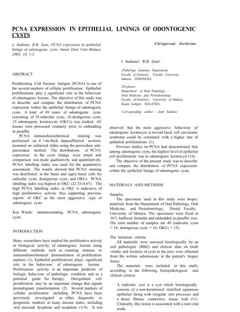
PCNA EXPRESSION IN EPITHELIAL LININGS OF ODONTOGENIC<br />
CYSTS<br />
J. Sudiono, R.B. Za<strong>in</strong>. PCNA <strong>expression</strong> <strong>in</strong> <strong>epithelial</strong><br />
l<strong>in</strong><strong>in</strong>gs <strong>of</strong> <strong>odontogenic</strong> <strong>cysts</strong>. Annal Dent Univ Malaya<br />
2003; 10: 1-5.<br />
ABSTRACT<br />
Proliferat<strong>in</strong>g Cell Nuclear Antigen (PCNA) is one <strong>of</strong><br />
the several markers <strong>of</strong> cellular proliferation. Epithelial<br />
proliferations play a significant role <strong>in</strong> the behaviour<br />
<strong>of</strong> <strong>odontogenic</strong> lesions. The objective <strong>of</strong> this study was<br />
to describe and compare the distribution <strong>of</strong> PCNA<br />
<strong>expression</strong> with<strong>in</strong> the <strong>epithelial</strong> l<strong>in</strong><strong>in</strong>gs <strong>of</strong> <strong>odontogenic</strong><br />
<strong>cysts</strong>. A total <strong>of</strong> 49 cases <strong>of</strong> <strong>odontogenic</strong> <strong>cysts</strong><br />
consist<strong>in</strong>g <strong>of</strong> 18 radicular <strong>cysts</strong>, 16 dentigerous <strong>cysts</strong>,<br />
15 <strong>odontogenic</strong> kerato<strong>cysts</strong> (OKCs) was studied. All<br />
tissues were processed rout<strong>in</strong>ely prior to embedd<strong>in</strong>g<br />
<strong>in</strong> paraff<strong>in</strong>.<br />
PCNA immunohistochemical sta<strong>in</strong><strong>in</strong>g was<br />
performed on 4 !-tm thick deparaff<strong>in</strong>ized sections<br />
mounted on sial<strong>in</strong>ized slides us<strong>in</strong>g the peroxidase antiperoxidase<br />
method. The distributions <strong>of</strong> PCNA<br />
<strong>expression</strong> <strong>in</strong> the <strong>cysts</strong> l<strong>in</strong><strong>in</strong>gs were noted and<br />
comparison was made qualitatively and quantitatively.<br />
PCNA labell<strong>in</strong>g <strong>in</strong>dex was used for the quantitative<br />
assessment. The results showed that PCNA sta<strong>in</strong><strong>in</strong>g<br />
was distributed <strong>in</strong> the basal and supra basal cells for<br />
radicular <strong>cysts</strong>, dentigerous <strong>cysts</strong>, and OKCs. PCNA<br />
labell<strong>in</strong>g <strong>in</strong>dex was highest <strong>in</strong> OKC (22.33±4.07). The<br />
high PCNA labell<strong>in</strong>g <strong>in</strong>dex <strong>in</strong> OKC is <strong>in</strong>dicative <strong>of</strong><br />
high proliferative activity thus support<strong>in</strong>g previous<br />
reports <strong>of</strong> OKC as the most aggressive type <strong>of</strong><br />
<strong>odontogenic</strong> <strong>cysts</strong>.<br />
Key Words: immunosta<strong>in</strong><strong>in</strong>g, PCNA, <strong>odontogenic</strong><br />
<strong>cysts</strong>.<br />
INTRODUCTION<br />
Many researchers have studied the proliferative activity<br />
or biological activity <strong>of</strong> <strong>odontogenic</strong> lesions us<strong>in</strong>g<br />
different methods such as count<strong>in</strong>g mitoses or<br />
immunohistochemical demonstration <strong>of</strong> proliferation<br />
markers (1). Epithelial proliferations playa significant<br />
role <strong>in</strong> the behaviour <strong>of</strong> <strong>odontogenic</strong> lesions.<br />
Proliferation activity is an important predictor <strong>of</strong><br />
biologic behaviour <strong>of</strong> pathologic condition and as a<br />
potential guide for therapy. Deregulated cell<br />
proliferation may be an important change that signals<br />
premalignant transformation (2). Several markers <strong>of</strong><br />
cellular proliferation <strong>in</strong>clud<strong>in</strong>g PCNA have been<br />
previously <strong>in</strong>vestigated as either diagnostic or<br />
prognostic markers <strong>in</strong> many disease states, <strong>in</strong>clud<strong>in</strong>g<br />
oral mucosal dysplasia and neoplasm (3-9). It was<br />
J. Sudiono l , R.B. Za<strong>in</strong> 2<br />
Orig<strong>in</strong>al Article<br />
JPathology Anatomy Department,<br />
Faculty <strong>of</strong> Dentistry, Trisakti University,<br />
Jakarta, INDONESIA<br />
2Pr<strong>of</strong>essor<br />
Department <strong>of</strong> Oral Pathology,<br />
Oral Medic<strong>in</strong>e and Periodontology,<br />
Faculty <strong>of</strong> Dentistry, University <strong>of</strong> Malaya,<br />
Kuala Lumpur, MALAYSIA.<br />
Correspond<strong>in</strong>g author - Janti Sudiono<br />
observed that the more aggressive behaviour <strong>of</strong><br />
<strong>odontogenic</strong> keratocyst <strong>in</strong> nevoid basal cell carc<strong>in</strong>oma<br />
syndrome could be correlated with a higher rate <strong>of</strong><br />
<strong>epithelial</strong> proliferations (1).<br />
Previous studies on PCNA had demonstrated that<br />
among <strong>odontogenic</strong> <strong>cysts</strong>, the highest level <strong>of</strong> <strong>epithelial</strong><br />
cell proliferation was <strong>in</strong> <strong>odontogenic</strong> keratocyst (10).<br />
The objective <strong>of</strong> the present study was to describe<br />
and compare the distribution <strong>of</strong> PCNA <strong>expression</strong><br />
with<strong>in</strong> the <strong>epithelial</strong> l<strong>in</strong><strong>in</strong>gs <strong>of</strong> <strong>odontogenic</strong> <strong>cysts</strong>.<br />
MATERIALS AND METHODS<br />
Samples<br />
The specimens used <strong>in</strong> this study were biopsy<br />
materials from the Department <strong>of</strong> Oral Pathology, Oral<br />
Medic<strong>in</strong>e, and Periodontology, Dental Faculty,<br />
University <strong>of</strong> Malaya. The specimens were fixed <strong>in</strong><br />
10 % buffered formal<strong>in</strong> and embedded <strong>in</strong> paraff<strong>in</strong> wax.<br />
The total number <strong>of</strong> samples are 49 (radicular <strong>cysts</strong><br />
= 18, dentigerous <strong>cysts</strong> = 16, OKCs = 15).<br />
The <strong>in</strong>clusion criteria<br />
All materials were assessed histologically by an<br />
oral pathologist (RBZ) and cl<strong>in</strong>ical data on tooth<br />
vitality and location <strong>of</strong> <strong>cysts</strong> <strong>in</strong> the jaws were obta<strong>in</strong>ed<br />
from the written submissions <strong>in</strong> the patient's biopsy<br />
forms.<br />
The materials were <strong>in</strong>cluded <strong>in</strong> this stud'y<br />
accord<strong>in</strong>g to the follow<strong>in</strong>g histopathological and<br />
cl<strong>in</strong>ical criteria:<br />
A radicular cyst is a cyst which histologically<br />
consists <strong>of</strong> a non-kerat<strong>in</strong>ised stratified squamous<br />
<strong>epithelial</strong> l<strong>in</strong><strong>in</strong>g with irregular rete processes and<br />
a dense fibrous connective tissue wall (11).<br />
Cl<strong>in</strong>ically, this lesion is associated with a non-vital<br />
tooth.
2 Annals <strong>of</strong> Dentistry, University <strong>of</strong> Malaya, Vol. 10 2003<br />
A dentigerous cyst is a cyst which<br />
histologically consists <strong>of</strong> a non-kerat<strong>in</strong>ised<br />
stratified squamous <strong>epithelial</strong> l<strong>in</strong><strong>in</strong>g and a fibrous<br />
connective tissue wall (ll). Cl<strong>in</strong>ically/<br />
radiologically, this lesion is associated with the<br />
crown <strong>of</strong> an impacted tooth.<br />
An <strong>odontogenic</strong> keratocyst is a cyst which<br />
histologically consists <strong>of</strong> a parakerat<strong>in</strong>ized or<br />
orthokerat<strong>in</strong>ized stratified squamous epithelium<br />
with pallisad<strong>in</strong>g <strong>of</strong> basal cell nuclei, absence <strong>of</strong><br />
rete processes and there is a fibrous connective<br />
tissue wall (ll). Cl<strong>in</strong>ically/radiologically, this is<br />
a lesion <strong>in</strong> the jaw bone.<br />
Demographic characteristics <strong>of</strong> the samples<br />
The mean ages <strong>of</strong> the patients with radicular,<br />
dentigerous, and <strong>odontogenic</strong> <strong>cysts</strong> were 33.00± 14.54;<br />
35.5±17.97; 31.6±16.75 years respectively. Table 1<br />
showed the ethnic and gender distribution <strong>of</strong> the<br />
samples. All the patients with cystic lesions was<br />
predom<strong>in</strong>antly <strong>in</strong> the Ch<strong>in</strong>ese ethnic group with female<br />
slightly higher, which were 61% (11/18) for radicular<br />
cyst, 63% (10/16) for dentigerous cyst, 67% (10/15)<br />
for OKCs.<br />
Immunohistochemical sta<strong>in</strong><strong>in</strong>g <strong>of</strong> PCNA<br />
The specimens were fixed <strong>in</strong> 10% buffered<br />
formal<strong>in</strong> and embedded <strong>in</strong> paraff<strong>in</strong> wax. PCNA<br />
immunohistochemistry sta<strong>in</strong><strong>in</strong>g was performed on 4<br />
/lm thick deparaff<strong>in</strong>ised sections mounted on sial<strong>in</strong>ised<br />
slides and dried at 56°C for 30 m<strong>in</strong>utes.<br />
After dewax<strong>in</strong>g and rehydrat<strong>in</strong>g, the sections were<br />
immersed <strong>in</strong> 4.28 mM citrate buffer at pH6 and treated<br />
by microwave for 12 m<strong>in</strong>utes at 100°C. This method<br />
was used to expose more antigenic sites <strong>in</strong> the tissue.<br />
The sections were then immersed <strong>in</strong> 3 % H 20 2 <strong>in</strong><br />
methanol to block endogenous peroxidase activity,<br />
followed by <strong>in</strong>cubation <strong>in</strong> normal rabbit serum (NRS)<br />
1: 20 for 20 m<strong>in</strong>utes to block background peroxidase<br />
activity. Sections were <strong>in</strong>cubated for two hours <strong>in</strong> a<br />
primary antibody (monoclonal anti PCNA antibody,<br />
clone PC-1O,Dako, A/S Denmark, 1: 100). The<br />
primary antibody which was bound to PCNA was<br />
detected us<strong>in</strong>g a secondary antibody (Biot<strong>in</strong>ylated<br />
rabbit anti-mouse). The reaction products were<br />
visualised with an immunoperoxidase method us<strong>in</strong>g<br />
Avid<strong>in</strong> Biot<strong>in</strong> Complex-HRP (ABC) which can be used<br />
for all monoclonal antibodies. The peroxidase activity<br />
was developed and visualised by a chromogen solution<br />
consist<strong>in</strong>g <strong>of</strong> 0.05 % diam<strong>in</strong>e benzid<strong>in</strong>e hydrochloride<br />
(DAB) for 10 m<strong>in</strong>utes to obta<strong>in</strong> a brown reaction<br />
product. Sections were counter sta<strong>in</strong>ed with Meyer's<br />
haematoxyl<strong>in</strong> and mounted <strong>in</strong> a mount<strong>in</strong>g medium dibutyl<br />
phthalate <strong>in</strong> xylene. The negative control<br />
consisted <strong>of</strong> omitt<strong>in</strong>g the primary and secondary<br />
antibody. All dilutions were performed us<strong>in</strong>g Tris<br />
Buffered Sal<strong>in</strong>e.<br />
Evaluations<br />
The qualitative evaluation <strong>in</strong>cludes the record<strong>in</strong>g<br />
<strong>of</strong> the presence or absence <strong>of</strong> the PCNA sta<strong>in</strong><strong>in</strong>g and<br />
describ<strong>in</strong>g its distribution. The quantitative evaluation<br />
was done by count<strong>in</strong>g the number <strong>of</strong> positive nuclei<br />
under 20X objective <strong>in</strong> comparison with the total<br />
number <strong>of</strong> <strong>epithelial</strong> cells for three randomly selected<br />
fields.<br />
RESULTS<br />
Table 1. Ethnic and gender distribution <strong>of</strong> the samples<br />
The nuclear PCNA reactivity was detected as granular<br />
sta<strong>in</strong><strong>in</strong>g throughout the nucleoplasm. The PCNA<br />
positive nuclei (PCNA +) were distributed <strong>in</strong> the basal<br />
and supra basal cells for radicular <strong>cysts</strong>, dentigerous<br />
<strong>cysts</strong>, and OKCs (Figure 1 - 3).<br />
Ethnic Radicular (N=18) Dentigerous (N=16) OKC (N=15) Total (N=49)<br />
Groups n (%) n (%) n (%) n (%)<br />
Malay<br />
Male 0(0.00) 4 (25.00) 0(0.00) 4 (8.16)<br />
Female 1 (5.56) a (0.00) 0(0.00) 1 (2.04)<br />
Ch<strong>in</strong>ese<br />
Male 4 (22.22) 7 (43.75) 3 (20.00) 14 (28.57)<br />
Female 7 (38.89) 3 (18.75) 7 (46.67) 17 (34.69) .<br />
Indians<br />
Male 4 (22.22) 1 (6.25) 2 (13.33) 7 (14.29)<br />
Female 2 (11.11) 0(0.00) 3 (20.00) 5 (10.20)<br />
Others<br />
Male 0(0.00) 1 (6.25) 0(0.00) 1 (2.04)<br />
Female 0(0.00) 0(0.00) 0(0.00) 0(0.00)<br />
Total<br />
Male 8 (44.44) 13 (81.25) 5 (33.33) 26 (53.06)<br />
Female 10 (55.56) 3 (18.75) a (66.67) 23 (46.94)
Figure l: PCNA + nuclei (arrows) <strong>in</strong> basal and<br />
suprabasal cells <strong>of</strong> radicular cyst l<strong>in</strong><strong>in</strong>g.<br />
L = lumen E = epithelium<br />
C=connective tissue (Orig<strong>in</strong>al magnification 50x)<br />
Figure 2: PCNA + nuclei (arrows) <strong>in</strong> the basal and<br />
suprabasal cells <strong>of</strong> dentigerous cyst l<strong>in</strong><strong>in</strong>g.<br />
L=lumen E=epithelium<br />
C = connective tissue (Orig<strong>in</strong>al magnification lOOx)<br />
Figure 3: PCNA + nuclei (arrows) <strong>in</strong> the<br />
kerat<strong>in</strong>ised <strong>epithelial</strong> l<strong>in</strong><strong>in</strong>g <strong>of</strong> OKC<br />
L=lumen E=epithelium<br />
C = connective tissue (Orig<strong>in</strong>al magnification lOOx)<br />
PCNA <strong>expression</strong> <strong>in</strong> <strong>epithelial</strong> l<strong>in</strong><strong>in</strong>gs <strong>of</strong> <strong>odontogenic</strong> <strong>cysts</strong> 3<br />
Table 2. PCNA labell<strong>in</strong>g <strong>in</strong>dex for radicular, dentigerous,<br />
and <strong>odontogenic</strong> kerato<strong>cysts</strong><br />
Type <strong>of</strong> lesions Number <strong>of</strong> PCNN cells<br />
(n=61) lesions (mean ± SO)<br />
Radicular <strong>cysts</strong> 6 (33.3%) 10.39 ± 2.18<br />
(n=18)<br />
Dentigerous <strong>cysts</strong> 8 (50.0%) 10.79 ± 1.59<br />
(n=16)<br />
Odontogenic keratocyst 7 (46.7%) 22.33 ± 4.07<br />
(n=15)<br />
Table 2 shows that the PCNA labell<strong>in</strong>g <strong>in</strong>dex<br />
(mean <strong>of</strong> PCNA + cells) for OKC was the highest<br />
(22.33±4.07) among all the <strong>odontogenic</strong> <strong>cysts</strong> followed<br />
by that <strong>of</strong> dentigerous cyst (10.79 ± 1.59) and that <strong>of</strong><br />
radicular cyst (10.39±2.18).<br />
DISCUSSION<br />
Radicular and dentigerous <strong>cysts</strong> and OKCs are the most<br />
common type <strong>of</strong> <strong>odontogenic</strong> <strong>cysts</strong> found <strong>in</strong> dental<br />
practice. These <strong>cysts</strong> have different biologic activities.<br />
Most radicular <strong>cysts</strong> grow slowly and do not atta<strong>in</strong> a<br />
large size. As the epithelium desquamates <strong>in</strong>to the<br />
lumen, the prote<strong>in</strong> content <strong>in</strong>creased. Fluid enters the<br />
lumen <strong>in</strong> an attempt to equalize the osmotic pressure<br />
and slow enlargement occurs (12-13). The dentigerous<br />
cyst appears to have greater potential for<br />
ameloblastomatous transformation than all other jaw<br />
<strong>cysts</strong> (12-15). The cl<strong>in</strong>icopathological features <strong>of</strong> OKC<br />
are quite different with that <strong>of</strong> other <strong>odontogenic</strong> <strong>cysts</strong>.<br />
The OKC shows a different growth mechanism and<br />
biologic behaviour from the more common dentigerous<br />
and radicular <strong>cysts</strong>. Radicular and dentigerous <strong>cysts</strong><br />
cont<strong>in</strong>ue to enlarge as a result <strong>of</strong> <strong>in</strong>creased osmotic<br />
pressure with<strong>in</strong> the lumen <strong>of</strong> the cyst, while <strong>in</strong> OKC<br />
the growth may be related to unknown factors <strong>in</strong>herent<br />
<strong>in</strong> the epithelium itself or enzymatic activity <strong>in</strong> the<br />
fibrous wall. Accord<strong>in</strong>g to Labban and Aghabeigi<br />
(1990), the presence <strong>of</strong> fenestrated capillaries <strong>in</strong> the<br />
keratocyst may <strong>in</strong>dicate a rapid transfer <strong>of</strong> fluid to meet<br />
the demand <strong>of</strong> the relatively active proliferat<strong>in</strong>g<br />
epithelium which may be prompted by growth factors<br />
released from platelets <strong>in</strong> those thrombosed vessels<br />
(16). The OKC tends to expand antero-posteriorly <strong>in</strong><br />
medulla without any bony expansion. -Accord<strong>in</strong>g to<br />
Waldron (1995), the aggressiveness <strong>of</strong> OKC may be<br />
due to many factors such as it be<strong>in</strong>g a multilocular cyst<br />
and the presence <strong>of</strong> growth <strong>epithelial</strong> factors with<strong>in</strong> its<br />
<strong>epithelial</strong> l<strong>in</strong><strong>in</strong>g (13). Browne and Gough (1972), and<br />
Regezi and Sciubba (1992) described that <strong>odontogenic</strong><br />
<strong>cysts</strong> with ortho or parakerat<strong>in</strong>ized epithelium as<br />
hav<strong>in</strong>g a higher recurrence rate thus the high<br />
proliferative activity <strong>of</strong> the rema<strong>in</strong><strong>in</strong>g <strong>epithelial</strong> cells<br />
after enucleation <strong>of</strong> cyst (12, 17).
4 Annals <strong>of</strong> Dentistry, University <strong>of</strong> Malaya, Vol. 10 2003<br />
There were no records <strong>in</strong> the' biopsy forms<br />
<strong>in</strong>dicat<strong>in</strong>g that the OKCs <strong>in</strong> this study is part <strong>of</strong> the<br />
nevoid basal cell carc<strong>in</strong>oma syndrome. In view <strong>of</strong> the<br />
rarity <strong>of</strong> this syndrome, it was assumed that most <strong>of</strong><br />
the OKCs <strong>in</strong> this study were non-syndromic.<br />
The PCNA labell<strong>in</strong>g <strong>in</strong>dex <strong>of</strong> OKCs <strong>in</strong> this study<br />
was found to be the highest, as compared to that <strong>of</strong><br />
dentigerous and radicular cyst (Table 2). This was <strong>in</strong><br />
agreement with the study by Li et al (1995) (18) where<br />
another proliferation marker Ki-67 was used. These<br />
results were <strong>in</strong> keep<strong>in</strong>g with the cl<strong>in</strong>ically more<br />
aggressive (proliferative) behaviour <strong>of</strong> OKC as<br />
compared to that <strong>of</strong> dentigerous and radicular <strong>cysts</strong>.<br />
Li et al (1994) (10) had shown a supra basal<br />
distribution <strong>of</strong> PCNA positive cells <strong>in</strong> OKC whilst for<br />
dentigerous cyst the distribution was <strong>in</strong> basal area.<br />
These distributions <strong>of</strong> PCNA positive cells <strong>in</strong> OKC and<br />
dentigerous cyst were also shown <strong>in</strong> this study. In this<br />
study there was a basal and suprabasal distribution <strong>of</strong><br />
PCNA positive cells for OKC and dentigerous cyst. In<br />
the radicular cyst, the distribution <strong>of</strong> PCNA positive<br />
cells was also <strong>in</strong> a basal and supra basal location.<br />
This study also showed generally variable<br />
reactivity with many samples show<strong>in</strong>g less <strong>in</strong>tense<br />
PCNA <strong>expression</strong>. Thus the number <strong>of</strong> samples with<br />
<strong>in</strong>tense PCNA <strong>expression</strong>s was low: 33.33% for<br />
radicular <strong>cysts</strong>, 50% dentigerous <strong>cysts</strong>, and 47%<br />
OKCs. This may be due to the fact that most <strong>of</strong> the<br />
specimens used were archival material (1981-1996)<br />
where there was no <strong>in</strong>dication <strong>of</strong> their fixation time.<br />
Accord<strong>in</strong>g to Yu et al (1991) (7), to obta<strong>in</strong> a good<br />
result, the fixation time must not be more than 48<br />
hours (6-48 hours). This view was also supported by<br />
Tsuji et al (1992 (19) who found that PCNA PC-lO<br />
reactivity was specific <strong>in</strong> the range <strong>of</strong> 6 to 48 hours<br />
<strong>in</strong> 10 % formal<strong>in</strong> fixed paraff<strong>in</strong> sections. Pellegr<strong>in</strong>i<br />
et al (1995) (20) reported that PCNA is very sensitive<br />
to the concentration and duration <strong>of</strong> formal<strong>in</strong> fixation,<br />
and also with the <strong>in</strong>cubation time <strong>of</strong> primary antibody.<br />
Another reason for the variable reactivity is due to<br />
PCNA <strong>in</strong>stability and this varies from cell to cell<br />
with<strong>in</strong> the same specimen result<strong>in</strong>g <strong>in</strong> strong to weak<br />
immunosta<strong>in</strong><strong>in</strong>g and render<strong>in</strong>g evaluation difficult.<br />
The weak PCNA sta<strong>in</strong><strong>in</strong>g observed may be due to the<br />
long half life <strong>of</strong> the PCNA prote<strong>in</strong> (Hall et aI, 1990)<br />
(5).<br />
CONCLUSIONS<br />
The labell<strong>in</strong>g <strong>in</strong>dex (mean) <strong>of</strong> PCNA positive cells <strong>in</strong><br />
OKC was the highest among <strong>odontogenic</strong> <strong>cysts</strong>. The<br />
highest PCNA labell<strong>in</strong>g <strong>in</strong>dex <strong>in</strong> OKC <strong>in</strong> this present<br />
study supported the previous reports that the OKC is<br />
the most aggressive type <strong>of</strong> <strong>odontogenic</strong> <strong>cysts</strong>.<br />
REFERENCES<br />
1. El Murtadi A, Grehan D, Toner M, Mc Cartan<br />
BE. PCNA sta<strong>in</strong><strong>in</strong>g <strong>in</strong> syndrome and non<br />
syndrome <strong>odontogenic</strong> kerato<strong>cysts</strong>. Oral Surg Oral<br />
Med Oral Pathol 1996; 81: 217-220.<br />
2. Mighell A. Proliferat<strong>in</strong>g cell nuclear antigen.<br />
Oral Surg Oral Med Oral Pathol 1995; 30: 3-4.<br />
3. Garcia RL, Coltrera MD, Gown AMI. Analysis<br />
<strong>of</strong> proliferative grade us<strong>in</strong>g anti PCNA/cycl<strong>in</strong><br />
monoclonal antibodies <strong>in</strong> fixed, embedded tissues.<br />
Comparison with flow cytometric analysis. Am J<br />
Patho11984; 134: 733-739.<br />
4. Dawson AE, Norton JA, We<strong>in</strong>ber DS.<br />
Comparative assessment <strong>of</strong> proliferation and DNA<br />
content <strong>in</strong> breast carc<strong>in</strong>oma by image analysis and<br />
flow cytometry. Am J Patho11990; 136: 115-124.<br />
5. Hall PA, Levison DA, Woods AL. Proliferat<strong>in</strong>g<br />
cell nuclear antigen (PCNA) immunolocalization<br />
<strong>in</strong> paraff<strong>in</strong> sections an <strong>in</strong>dex <strong>of</strong> cell proliferation<br />
with evidence <strong>of</strong> deregulated <strong>expression</strong> <strong>in</strong> some<br />
neoplasms. J Patho11990; 162: 285-294.<br />
6. Allegranza A, Girlando S, Arrigont GL.<br />
Proliferat<strong>in</strong>g cell nuclear antigen <strong>expression</strong> <strong>in</strong><br />
central nervous system neoplasms. Virchows Arch<br />
(A) 1991; 419: 417-423.<br />
7. Yu CC, Hall PA, Fletcher CDM, Camplejohns<br />
RS, Waseem NH, Lane DP, Levison DA.<br />
Haemangiopericytomas: the prognostic value <strong>of</strong><br />
immunohistochemical sta<strong>in</strong><strong>in</strong>g with a monoclonal<br />
antibody to proliferat<strong>in</strong>g cell nuclear antigen<br />
(PCNA). Histopathology 1991; 19: 29-33.<br />
8. Ogawa I, Takata T, Miyauchi M, Ito H, Ijuh<strong>in</strong> N,<br />
Nikai H. Proliferat<strong>in</strong>g cell nuclear antigen<br />
(PCNA) as a marker <strong>of</strong> proliferat<strong>in</strong>g cells <strong>in</strong><br />
salivary gland tumors. Jpn J Oral Bioi 1992; 34:<br />
454-457.<br />
9. Yu C and Filipe M. Update on proliferation<br />
associated antibodies applicable to formal<strong>in</strong> fixed<br />
paraff<strong>in</strong>-embedded tissue and their cl<strong>in</strong>ical<br />
applications. Histochem 1993; 25: 843-853.<br />
10. Li TJ, Browne RM and Matthews JB.<br />
Quantification <strong>of</strong> PCNA + cells with<strong>in</strong> <strong>odontogenic</strong><br />
jaw cyst epithelium. J Oral Pathol Med 1994; 23:<br />
184-189.<br />
11. Kramer IRH, Piridborg 11, Shear M. Histological<br />
typ<strong>in</strong>g <strong>of</strong> <strong>odontogenic</strong> tumours. Second edition.<br />
Spr<strong>in</strong>ger-Verlag, Berl<strong>in</strong>, 1992; 34-42.
12. Regezi JA and Sciuba 1. Cl<strong>in</strong>ocal-pathologic<br />
correlation. Second edition. Saunders.<br />
Philadelphia, 1993; 322-343.<br />
13. Waldron CA. "Odontogenic cyst and tumours". In<br />
Oral and Maxill<strong>of</strong>acial Pathology. Edited by<br />
Neville BW, Damm DD, Allen OM, Bouquot JE.<br />
1995; 103-106.<br />
14. Rob<strong>in</strong>son L and Mart<strong>in</strong>ez MG. Unicystic<br />
ameloblastoma. A prognostically dist<strong>in</strong>ct entity.<br />
Cance~ 1977; 40: 278-285.<br />
15. Gardner 1981. Plexiform Unicystic<br />
Ameloblastoma. A Diagnostic Problem <strong>in</strong><br />
Dentigerous Cysts. Cancer 1981; 15: 1359-1363.<br />
16. Labban NG and Aghabeigi B. A Comparative<br />
Stereologic and Ultrastructural Study <strong>of</strong> Blood<br />
Vessels <strong>in</strong> Odontogenic Kerato<strong>cysts</strong> and<br />
Dentigerous Cysts. J Oral Pathol Med 1990; 19:<br />
442-446.<br />
PCNA <strong>expression</strong> <strong>in</strong> <strong>epithelial</strong> l<strong>in</strong><strong>in</strong>gs <strong>of</strong> <strong>odontogenic</strong> <strong>cysts</strong> 5<br />
17. Browne RM and Gough NG. Malignant Change<br />
<strong>in</strong> Odontogenic Cyst. Cancer. 1972; 29: 1199-<br />
1207.<br />
18. Li TJ, Browne RM and Matthews JE. Expression<br />
<strong>of</strong> proliferat<strong>in</strong>g cell nuclear antigen (PCNA) and<br />
Ki-67 <strong>in</strong> unicystic ameloblastoma. J Histopathol<br />
1995; 26: 219-228.<br />
19. Tsuji T, Sasaki K, Kimura Y, Yamada K, Mori M,<br />
Sh<strong>in</strong>ozaki F. Measurement <strong>of</strong> proliferat<strong>in</strong>g cell<br />
nuclear antigen (PCNA) and its cl<strong>in</strong>ical application<br />
<strong>in</strong> oral cancers. J Oral Maxill<strong>of</strong>ac Surg 1992; 21:<br />
369-372.<br />
20. Pellegr<strong>in</strong>i W, Facchetti F, Marocolo D, Salvi L,<br />
Capucci A, Tironi A, Rossi G. Assessment <strong>of</strong> cell<br />
proliferation <strong>in</strong> normal and pathological bone<br />
marrow biopsies: a study us<strong>in</strong>g double sequential<br />
immunophenotyp<strong>in</strong>g on paraff<strong>in</strong> sections. J<br />
Histopathol 1995; 27: 397-405.


