Anti-plasmodial action of de novo-designed ... - Malaria Journal
Anti-plasmodial action of de novo-designed ... - Malaria Journal
Anti-plasmodial action of de novo-designed ... - Malaria Journal

You also want an ePaper? Increase the reach of your titles
YUMPU automatically turns print PDFs into web optimized ePapers that Google loves.
<strong>Anti</strong>-<strong>plasmodial</strong> <strong>action</strong> <strong>of</strong> <strong>de</strong> <strong>novo</strong>-<strong>de</strong>signed,<br />
cationic, lysine-branched, amphipathic,<br />
helical pepti<strong>de</strong>s<br />
Kaushik et al.<br />
Kaushik et al. <strong>Malaria</strong> <strong>Journal</strong> 2012, 11:256<br />
http://www.malariajournal.com/content/11/1/256
Kaushik et al. <strong>Malaria</strong> <strong>Journal</strong> 2012, 11:256<br />
http://www.malariajournal.com/content/11/1/256<br />
RESEARCH Open Access<br />
<strong>Anti</strong>-<strong>plasmodial</strong> <strong>action</strong> <strong>of</strong> <strong>de</strong> <strong>novo</strong>-<strong>de</strong>signed,<br />
cationic, lysine-branched, amphipathic,<br />
helical pepti<strong>de</strong>s<br />
Naveen K Kaushik, Jyotsna Sharma and Dinkar Sahal *<br />
Abstract<br />
Background: A lack <strong>of</strong> vaccine and rampant drug resistance <strong>de</strong>mands new anti-malarials.<br />
Methods: In vitro blood stage anti-<strong>plasmodial</strong> properties <strong>of</strong> several <strong>de</strong> <strong>novo</strong>-<strong>de</strong>signed, chemically synthesized,<br />
cationic, amphipathic, helical, antibiotic pepti<strong>de</strong>s were examined against Plasmodium falciparum using SYBR<br />
Green assay. Mechanistic <strong>de</strong>tails <strong>of</strong> anti-<strong>plasmodial</strong> <strong>action</strong> were examined by optical/fluorescence microscopy<br />
and FACS analysis.<br />
Results: Unlike the monomeric <strong>de</strong>capepti<strong>de</strong>s {(Ac-GXRKXHKXWA-NH2)(X=F,ΔF) (Fm , ΔFm IC50 >100 μM)}, the<br />
lysine-branched,dimeric versions showed far greater potency {IC50 (μM) Fd 1.5 , ΔFd 1.39}. The more helical<br />
and proteolytically stable ΔFd was studied for mechanistic <strong>de</strong>tails. ΔFq, a K-K2 <strong>de</strong>ndrimer <strong>of</strong> ΔFm and (ΔFm)2<br />
a linear dimer <strong>of</strong> ΔFm showed IC50 (μM) <strong>of</strong> 0.25 and 2.4 respectively. The healthy/infected red cell selectivity<br />
indices were >35 (ΔFd), >20 (ΔFm)2 and 10 (ΔFq). FITC-ΔFd showed rapid and selective accumulation in<br />
parasitized red cells. Overlaying DAPI and FITC florescence suggested that ΔFd binds DNA. Trophozoites and<br />
schizonts incubated with ΔFd (2.5 μM) egressed anomalously and Band-3 immunostaining revealed them not<br />
to be associated with RBC membrane. Prematurely egressed merozoites from pepti<strong>de</strong>-treated cultures were<br />
foundtobeinvasionincompetent.<br />
Conclusion: Good selectivity (>35), good resistance in<strong>de</strong>x (1.1) and low cytotoxicity indicate the promise <strong>of</strong> ΔFd<br />
against malaria.<br />
Keywords: Anomalous egress, <strong>Anti</strong>-<strong>plasmodial</strong> pepti<strong>de</strong>s, De <strong>novo</strong> pepti<strong>de</strong> <strong>de</strong>sign, Kinetics <strong>of</strong> pepti<strong>de</strong> uptake,<br />
Pepti<strong>de</strong> binding to DNA, Plasmodium falciparum<br />
Background<br />
The <strong>de</strong>vastating diseases caused by protozoan parasites<br />
are a major bur<strong>de</strong>n <strong>of</strong> the tropics, and in particular,<br />
Plasmodium falciparum, the causative agent <strong>of</strong><br />
falciparum malaria, creates a serious public health problem<br />
in many areas <strong>of</strong> the <strong>de</strong>nsely populated <strong>de</strong>veloping<br />
world. The wi<strong>de</strong>spread resistance <strong>of</strong> P. falciparum to<br />
chloroquine (CQ), which has spread from Asia to Africa,<br />
has ren<strong>de</strong>red the drug ineffective against the most dangerous<br />
Plasmodium strain in many affected regions <strong>of</strong> the<br />
world. Unfortunately, CQ-resistance is associated with<br />
cross-resistance to other quinoline drugs, such as quinine<br />
* Correspon<strong>de</strong>nce: dinkar@icgeb.res.in<br />
<strong>Malaria</strong> Research Group, International Centre for Genetic Engineering and<br />
Biotechnology, Aruna Asaf Ali Marg, New Delhi 110067, India<br />
and amodiaquine [1]. Plasmodium falciparum is genetically<br />
diverse and has multiple in<strong>de</strong>pen<strong>de</strong>nt origins <strong>of</strong> mutations<br />
in genes that confer resistance to wi<strong>de</strong>ly used<br />
anti-malarial drugs [2]. Left with just artemisinin to fight<br />
against malaria, Arata Kochi, Director <strong>of</strong> the <strong>Malaria</strong><br />
Division at the World Health Organization, had felt compelled<br />
to say “if we lose artemisinin, we will no longer have<br />
an effective cure for malaria” [3]. However, most recently,<br />
alarming signs <strong>of</strong> clinical resistance against artemisinin, in<br />
the form <strong>of</strong> <strong>de</strong>layed parasite clearance, are being observed<br />
in the bor<strong>de</strong>r between Cambodia and Thailand [4,5]. The<br />
challenge <strong>of</strong> <strong>de</strong>signing an effective vaccine along traditional<br />
lines against malaria is that many P. falciparum<br />
proteins are highly polymorphic and their functions are<br />
redundant [6]. More than 200 million new malaria cases<br />
© 2012 Kaushik et al.; licensee BioMed Central Ltd. This is an Open Access article distributed un<strong>de</strong>r the terms <strong>of</strong> the Creative<br />
Commons Attribution License (http://creativecommons.org/licenses/by/2.0), which permits unrestricted use, distribution, and<br />
reproduction in any medium, provi<strong>de</strong>d the original work is properly cited.
Kaushik et al. <strong>Malaria</strong> <strong>Journal</strong> 2012, 11:256 Page 2 <strong>of</strong> 15<br />
http://www.malariajournal.com/content/11/1/256<br />
reported annually is a challenge [7] that un<strong>de</strong>rscores the<br />
urgent requirement for new drugs against malaria.<br />
Pepti<strong>de</strong>s are an essential component <strong>of</strong> <strong>de</strong>fence mechanism<br />
<strong>of</strong> all life forms and anti-microbial pepti<strong>de</strong>s are<br />
evolutionarily ancient biological weapons. Their wi<strong>de</strong>spread<br />
distribution throughout the living kingdom suggests<br />
that anti-microbial pepti<strong>de</strong>s may have served a<br />
fundamental role in the successful evolution <strong>of</strong> complex<br />
multi-cellular organisms [8]. Despite their ancient lineage,<br />
anti-microbial pepti<strong>de</strong>s have remained effective <strong>de</strong>fensive<br />
weapons, <strong>de</strong>feating the general belief that bacteria, fungi<br />
and viruses can and will <strong>de</strong>velop resistance to any conceivable<br />
substance. Among other differences, uniquely anionic<br />
charge on bacterial surface is a curious feature that distinguishes<br />
the prokaryotic bacteria from their eukaryotic<br />
counterparts [9]. <strong>Anti</strong>-microbial pepti<strong>de</strong>s gain selectivity<br />
from their ability to target this previously un<strong>de</strong>rappreciated<br />
‘microbial Achille’s heel’ [10-12]. Interestingly,<br />
a seminal feature <strong>of</strong> the malaria parasite-infected red cell<br />
is reflected in an altered asymmetry <strong>of</strong> lipid composition<br />
in its cell surface membrane. In contrast to the uninfected,<br />
healthy red cell, the malaria-infected red cell shows a<br />
translocation <strong>of</strong> the anionic phosphatidylserine from the<br />
inner leaflet to the outer leaflet <strong>of</strong> the bi-layer [13]. As a<br />
result, the FITC-Annexin negative, healthy red cell now<br />
turns to become FITC-Annexin positive [14]. Thus a<br />
Plasmodium-infected red cell seems to mimic the anionic<br />
surface charge that characterizes the bacterial cell surface.<br />
In principle, this is expected to make malaria-infected red<br />
cells become vulnerable to the <strong>action</strong> <strong>of</strong> anti-microbial<br />
pepti<strong>de</strong>s. In<strong>de</strong>ed, naturally occurring or modified pepti<strong>de</strong>s,<br />
such as <strong>de</strong>rmaseptin [15], oligoacyllysine [16], cyclosporin<br />
A [17], cecropin A [18], NK-2 [14] and meucin [19], have<br />
been found to display in vitro anti-malarial activity. Some<br />
membrane-active, hydrophobic pepti<strong>de</strong>s <strong>of</strong> fungal origin<br />
have also been found to exhibit in vitro anti-malarial <strong>action</strong><br />
[20]. However, many <strong>of</strong> these naturally occurring pepti<strong>de</strong>s<br />
suffer from drawbacks such as poor potency, stability<br />
and selectivity [21]. Therefore, in a bid to improve their<br />
performance, efforts are being ma<strong>de</strong> to engineer pepti<strong>de</strong>s<br />
in diverse ways with the aim <strong>of</strong> reducing their size, improving<br />
their stability against proteases and enhancing<br />
their selectivity [22-24]. The structure activity relationships<br />
<strong>of</strong> a series <strong>of</strong> <strong>de</strong> <strong>novo</strong>-<strong>de</strong>signed, conformationallyconstrained<br />
helical, amphipathic, cationic pepti<strong>de</strong>s against<br />
bacteria have earlier been reported [25]. In the present<br />
work, the potent anti-<strong>plasmodial</strong> <strong>action</strong> <strong>of</strong> these pepti<strong>de</strong>s<br />
against both CQ-sensitive and CQ-resistant strains <strong>of</strong> P.<br />
falciparum are being reported. The results indicate that a<br />
lysine-branched, dimeric pepti<strong>de</strong> ΔFd, which is highly potent<br />
(IC 50 1.39 μM) across CQ-sensitive and CQ-resistant<br />
strains {Resistance In<strong>de</strong>x (IC 50 CQ resistant strain/ IC 50<br />
CQ sensitive strain)1.1} <strong>of</strong> P. falciparum, fairly selective<br />
against parasitized red blood cells {Selectivity In<strong>de</strong>x (HC 50<br />
URBC/IC50 P. falciparum) >35) and fairly non toxic to<br />
mammalian HeLa cells (TC 50 >25 μM), stalls parasite<br />
growth by causing arrest <strong>of</strong> ring stage parasite, anomalous<br />
egress <strong>of</strong> trophozoites and premature egress <strong>of</strong> schizonts<br />
that fail to produce invasion competent merozoites.<br />
Methods<br />
Pepti<strong>de</strong>s<br />
Pepti<strong>de</strong>s ΔFm, (ΔFm) 2, ΔFd, ΔFq, Fm, Fd, D-Lys- ΔFd,<br />
prochitinase and E30 (Table 1) were synthesized by<br />
Fmoc chemistry-based, manual, solid-phase synthesis.<br />
Di<strong>de</strong>hydrophenylalanine (ΔF) was chosen since it is a<br />
conformationally-constrained amino acid residue with a<br />
proven reputation to confer helical character to pepti<strong>de</strong>s.<br />
FITC <strong>de</strong>rivatizations <strong>of</strong> (ΔFm) 2 and ΔFd were done after<br />
linking aminohexanoic acid to the N terminus. Pepti<strong>de</strong>s<br />
were purified to >95% homogeneity by RPHPLC and<br />
characterized by mass spectroscopy and circular dichroism<br />
as <strong>de</strong>scribed previously [25]. Chromatographic and<br />
mass spectral characterization is given as follows: RPHPLC<br />
pr<strong>of</strong>iles <strong>of</strong> control pepti<strong>de</strong>s prochitinase, E30 and bovine insulin<br />
(Additional file 1); ΔFm and ΔFd (Additional file 2)<br />
Electro Spray Mass Spectroscopy (ESMS) pr<strong>of</strong>iles <strong>of</strong> prochitinase,<br />
E30 and bovine insulin (Additional file 3); ESMS<br />
pr<strong>of</strong>iles <strong>of</strong> ΔFm and ΔFd (Additional file 4); MALDI <strong>of</strong><br />
(ΔFm) 2 (Additional file 5), RPHPLC and mass spectral<br />
data for ΔFq (Additional file 6) and ESMS pr<strong>of</strong>iles <strong>of</strong> Fm,<br />
D-Lys- ΔFd and Fd (Additional file 7). Pepti<strong>de</strong>s corresponding<br />
to prochitinase, E30 (a 30 residues-long pepti<strong>de</strong><br />
from Hepatitis E virus ORF3) (synthesized and characterized<br />
in house) and bovine insulin (Sigma) were used<br />
as controls in experiments on ΔFd mediated selective<br />
haemolysis <strong>of</strong> infected red cells. FITC-Insulin (Sigma)<br />
was used as control in experiments done to study the<br />
uptake <strong>of</strong> FITC tagged (ΔFm) 2 and ΔFd by Plasmodiuminfected<br />
RBCs.<br />
In vitro cultivation <strong>of</strong> Plasmodium falciparum<br />
Chloroquine-sensitive (3D7) and CQ-resistant (Dd2 and<br />
INDO) strains <strong>of</strong> P. falciparum were maintained in continuous<br />
culture according to the method <strong>of</strong> Trager and<br />
Jensen [26] with minor modifications. Cultures were maintained<br />
in fresh group O +ve human erythrocytes suspen<strong>de</strong>d<br />
at 4% haematocrit in complete medium {16.2 g/L RPMI<br />
1640 containing 25 mM HEPES, 11.11 mM glucose<br />
(Gibco), 0.2% sodium bicarbonate (Sigma), 0.5% Albumax I<br />
(Gibco), 45 μg/litre hypoxanthine (Sigma) and 50 μg/litre<br />
gentamicin (Gibco)} and incubated at 37°C un<strong>de</strong>r a gas<br />
mixture 5% O2, 5%CO2, and 90% N2. Every day, the spent<br />
medium was replaced by fresh complete medium to propagate<br />
the culture. For INDO strain in culture medium, albumax<br />
was replaced by 10% pooled human serum (Innovative<br />
Research) as suggested by MR4 [27]. Parasitaemia was<br />
monitored by microscopic examination <strong>of</strong> Giemsa-stained
Kaushik et al. <strong>Malaria</strong> <strong>Journal</strong> 2012, 11:256 Page 3 <strong>of</strong> 15<br />
http://www.malariajournal.com/content/11/1/256<br />
Table 1 In-vitro blood stage anti<strong>plasmodial</strong> activities, resistance and selectivity indices <strong>of</strong> pepti<strong>de</strong>s against different<br />
strains <strong>of</strong> P. falciparum<br />
Pepti<strong>de</strong>s Pepti<strong>de</strong> Sequence and <strong>de</strong>sign IC50 P. falciparum (μM)<br />
3D7 Dd2 INDO<br />
Resistance in<strong>de</strong>x<br />
IC50Dd2/ IC503D7 HC50 URBC<br />
μM<br />
Fm Ac-GFRKFHKFWA-NH2 >100 >100 - >100<br />
ΔFm Ac-GΔFRKΔFHKΔFWA-NH2 >100 >100 - - >100<br />
ΔFd 1.39 ± 0.1 1.6 ± 0.09 1.5 ± 0.075 1.15 >50 ( >35)*<br />
D-Lys-ΔFd** 1.8 ± 0.07 - - - >50 (> 27)<br />
Fd 1.5 ± 0.08 - - - >50 (>33)<br />
(ΔFm)2 Ac-GΔFRKΔFHKΔFWAAGΔFRKΔFHKΔFWA-NH2 2.4 ± 0.15 2.5 ± 0.13 - 1.04 >50 (>20)<br />
ΔFq 0.25 ±0.02 - - - 2.5 ±0.13 (10)<br />
Prochitinase EEPHKAASAEGKK > 40 - - - > 40<br />
E30 NPPDHSAPLGATRPSAPPLPHVVDLPQLGP > 40 - - - > 40<br />
Insulin<br />
GIVEQCCASVCSLYQLENYCN<br />
FVNQHLCGSHLVEALYLVCGERGFFYTPKA<br />
blood smears. Synchronized ring stage parasite was<br />
obtained by 5% sorbitol treatment [28]. Trophozoites<br />
and schizont-stage parasites were enriched by using<br />
Percoll gradient [29].<br />
Drug dilutions<br />
Stock solutions <strong>of</strong> pepti<strong>de</strong>s and CQ were prepared in<br />
water (milli-Q gra<strong>de</strong>) while artemisinin stock solution<br />
was in dimethyl sulphoxi<strong>de</strong> (DMSO). All stocks were<br />
then diluted with culture medium to achieve the<br />
required drug concentrations. The concentration <strong>of</strong> pepti<strong>de</strong><br />
solution in water was based on A 280 [E (M -1 cm -1 )<br />
ΔF (di<strong>de</strong>hydrophenylalanine) 19,000, W (Tryptophan)<br />
5,000)]. Thus E 280 were 62,000, 124,000, 124,000 and<br />
248,000 for ΔFm, (ΔFm) 2, ΔFd and ΔFq respectively.<br />
The concentration <strong>of</strong> FITC-pepti<strong>de</strong>s was based on A495<br />
[E(M -1 cm -1 ) FITC 77,000 for (ΔFm) 2 with one FITC and<br />
154,000 for ΔFd with two FITC per molecule]. Drugs<br />
>40 - - - >40<br />
Artemisinin 0.015 0.016 0.015 1<br />
Chloroquine 0.04 0.16 0.5 4<br />
* Hemolytic Selectivity in<strong>de</strong>x (HC50URBC/ IC50Pf3D7) is shown in parenthesis, **: K D refers to Lysine <strong>of</strong> D configuration. Values <strong>of</strong> standard <strong>de</strong>viation given as ± are<br />
based on three in<strong>de</strong>pen<strong>de</strong>nt observations.<br />
and pepti<strong>de</strong>s solutions were placed in 96-well flat bottom<br />
tissue culture gra<strong>de</strong> plates (Corning).<br />
Assay for anti-<strong>plasmodial</strong> activity<br />
For drug screening, SYBR green I based fluorescence<br />
assay was used as <strong>de</strong>scribed previously by Smilkstein<br />
et al. [30]. Sorbitol synchronized ring stage parasites<br />
(haematocrit: 2%, parasitaemia: 1%, 100 μl) un<strong>de</strong>r normal<br />
culture conditions were incubated in the absence or<br />
presence <strong>of</strong> increasing concentrations <strong>of</strong> pepti<strong>de</strong>s in<br />
water. CQ and artemisinin were used as positive controls.<br />
Vehicle control 0.4% DMSO (which was found to<br />
be non-toxic to parasite) was used in case <strong>of</strong> artemisinin.<br />
After 48 hr <strong>of</strong> incubation 100 μl <strong>of</strong> SYBR Green I buffer<br />
[0.2 μl <strong>of</strong> 10,000 X SYBR Green I (Invitrogen) per ml <strong>of</strong><br />
lysis buffer {Tris (20 mM; pH 7.5), EDTA (5 mM),<br />
saponin (0.008%; wt/vol), and Triton X-100 (0.08%;<br />
vol/vol)}] was ad<strong>de</strong>d to each well, mixed twice gently
Kaushik et al. <strong>Malaria</strong> <strong>Journal</strong> 2012, 11:256 Page 4 <strong>of</strong> 15<br />
http://www.malariajournal.com/content/11/1/256<br />
with multi-channel pipette and incubated in dark at 37 0 C<br />
for 1 h. Fluorescence was measured with a Victor fluorescence<br />
multi-well plate rea<strong>de</strong>r (Perkin Elmer) with excitation<br />
and emission wavelength centred at 485 and 530 nm,<br />
respectively. Fluorescence counts for CQ (0.1 μM for 3D7,<br />
1 μM for INDO) were subtracted from counts in each<br />
well. The fluorescence counts were plotted against the<br />
drug concentration and IC 50 (the 50% inhibitory concentration)<br />
was <strong>de</strong>termined by analysis <strong>of</strong> dose–response<br />
curves. Results <strong>of</strong> the above mentioned fluorescencebased<br />
assay were validated microscopically by examination<br />
<strong>of</strong> Giemsa-stained smears <strong>of</strong> pepti<strong>de</strong>-treated parasite cultures.<br />
Statistical significance <strong>of</strong> relative potencies <strong>of</strong> pepti<strong>de</strong>s<br />
was <strong>de</strong>termined by stu<strong>de</strong>nt’sT test.<br />
In vitro stage <strong>de</strong>pen<strong>de</strong>nce <strong>of</strong> <strong>action</strong><br />
Stage specificity <strong>of</strong> <strong>action</strong> <strong>of</strong> ΔFd on the parasite’s blood<br />
stage life cycle was <strong>de</strong>termined by microscopic analysis <strong>of</strong><br />
the effect <strong>of</strong> ΔFd on each <strong>of</strong> the three stages (ring, trophozoite<br />
and schizont) <strong>of</strong> the parasite life cycle. Synchronized<br />
stages were obtained by sorbitol-mediated synchronization<br />
repeated thrice (synchronization 1, medium washed, incubation<br />
for 3 hr, 37 0 C, synchronization 2, medium washed<br />
and culture allowed to grow in complete medium for<br />
48 hr. At this stage the culture was synchronized a third<br />
time to obtain highly synchronized ring stage culture).<br />
This culture was grown for 24 hr and 38 hr to obtain<br />
trophozoite and schizont stage cultures respectively. Both<br />
trophozoite and schizont enriched cultures were subjected<br />
to Percoll gradient centrifugation to obtain highly purified<br />
parasites <strong>of</strong> specific stages. Giemsa-stained smears were<br />
microscopically observed over 2,000 RBCs to obtain differential<br />
counts.<br />
Cultures (1% parasitaemia, 2% haematocrit) at each <strong>of</strong><br />
the above mentioned stages were see<strong>de</strong>d in 96-well<br />
plates containing different concentrations <strong>of</strong> ΔFd and<br />
the plates incubated for 12 h (schizont), 24 h (trophozoite)<br />
and 48 h (ring) un<strong>de</strong>r standard culture condition.<br />
Smears were drawn, Giemsa-stained and analysed microscopically.<br />
Stage-specificity <strong>of</strong> <strong>action</strong> was assessed by observing<br />
the stage transitions in drug-treated samples<br />
against untreated controls.<br />
Cytotoxic activity <strong>of</strong> ΔFd on HeLa cells using MTT assay<br />
The cytotoxic effects <strong>of</strong> ΔFd on mammalian cells was<br />
assessed by functional assay as <strong>de</strong>scribed [31] using<br />
HeLa cells cultured in RPMI containing 10% fetal bovine<br />
serum, 0.21% sodium bicarbonate (Sigma) and 50 μg/mL<br />
gentamycin (complete medium). Briefly, cells (10 4 cells/<br />
200 μl/well) were see<strong>de</strong>d into 96- well flat-bottom tissue<br />
culture plates in complete medium. Pepti<strong>de</strong> solutions<br />
were ad<strong>de</strong>d after 24 hr <strong>of</strong> seeding and incubated for<br />
48 hr in a humidified atmosphere at 37°C and 5% CO 2.<br />
DMSO (as positive inhibitor) was ad<strong>de</strong>d at 10%. Twenty<br />
microlitres <strong>of</strong> a stock solution <strong>of</strong> MTT (5 mg/mL in 1X<br />
phosphate buffered saline) was ad<strong>de</strong>d to each well, gently<br />
mixed and incubated for another 4 hr. After spinning<br />
the plate at 1500 rpm for 5 min, supernatant was<br />
removed and 100 μl <strong>of</strong> DMSO (stop agent) was ad<strong>de</strong>d.<br />
Formation <strong>of</strong> formazon was read on a microtiter plate<br />
rea<strong>de</strong>r (Versa max tunable multi-well plate rea<strong>de</strong>r) at<br />
570 nm. The 50% cytotoxic concentration (TC 50) <strong>of</strong> drug<br />
was <strong>de</strong>termined by analysis <strong>of</strong> dose–response curves.<br />
Haemolysis assay<br />
Selectivity <strong>of</strong> haemolysis by pepti<strong>de</strong>s for infected erythrocytes<br />
(PRBC) vs uninfected erythrocytes (URBC)<br />
was examined by incubating the test molecules with<br />
URBCs and PRBCs respectively in phosphate-buffered<br />
saline (PBS). Briefly, fresh RBCs were spin washed (1600<br />
RPM; 5 min) three times in PBS and re-suspen<strong>de</strong>d in<br />
PBS at 2% haematocrit. A 100 μl suspension was ad<strong>de</strong>d<br />
to 96-well plate containing the pepti<strong>de</strong>s at different concentrations.<br />
PBS alone (for baseline values) and 0.4%<br />
Triton X-100 in PBS (for 100% haemolysis) were used as<br />
controls. After incubation at 37°C for 3 hr, the samples<br />
were centrifuged and supernatant was used to <strong>de</strong>termine<br />
the haemolytic activity measured in terms <strong>of</strong> haemoglobin<br />
release as monitored by A415. Triton-treated control<br />
samples were diluted 10-fold before reading absorbance.<br />
Base line value (PBS control,
Kaushik et al. <strong>Malaria</strong> <strong>Journal</strong> 2012, 11:256 Page 5 <strong>of</strong> 15<br />
http://www.malariajournal.com/content/11/1/256<br />
and floo<strong>de</strong>d with CY3 labelled anti-mouse antibody<br />
(Sigma)(1: 500 dilution in 1% BSA/PBS,1 hr, 37 0 Cindark),<br />
(d) PBS washed and floo<strong>de</strong>d with DAPI (4, 6-diamidino-2phenylindole)<br />
(invitogen) (500 ng/ml, 10 min, 37 0 C). After<br />
a final PBS wash the smears were observed un<strong>de</strong>r Nikon<br />
eclipse fluorescence microscope.<br />
For studying pepti<strong>de</strong> localization, P. falciparum cultures<br />
were individually incubated with FITC-ΔFd (2 μM), FITC-<br />
(ΔFm) 2 (2 μM) or FITC-Insulin (3 μM) a) alone and b) together<br />
with DAPI in complete medium at 37°C for 30 min<br />
and the cells were spin washed (1,600 RPM, 5 min) twice<br />
with 1 X PBS to reduce background fluorescence. The<br />
cells were smeared on a glass sli<strong>de</strong>, and fluorescence was<br />
visualized by using the respective filter settings for FITC<br />
and DAPI.<br />
For studying the selectivity and route <strong>of</strong> transport <strong>of</strong><br />
ΔFd into the red cell-resi<strong>de</strong>nt malaria parasite, URBC and<br />
PRBC were incubated with FITC-ΔFd (4 μM) in parallel<br />
sets at 4°C vs at room temperature (25°C) for specified<br />
times and spin washed (1,600 RPM, 2 min) with complete<br />
medium (3 X 200 μl). The cells were smeared on a glass<br />
sli<strong>de</strong> and both bright field images and fluorescence images<br />
(using FITC filter) were captured at 100 X magnification<br />
using Nikon eclipse fluorescence microscope. The s<strong>of</strong>tware<br />
Adobe Photoshop was used to overlay the fluorescence<br />
image on the bright field image.<br />
Kinetics <strong>of</strong> pepti<strong>de</strong> uptake<br />
Kinetics <strong>of</strong> FITC-labelled pepti<strong>de</strong> uptake was studied<br />
using Flow cytometer (BD FACS callibur). FITC- ΔFd<br />
% Growth<br />
(3 μM) was incubated for indicated time intervals with<br />
synchronized rings (~7% parasitaemia, 2% haematocrit)<br />
and synchronized trophozoites (~20% parasitaemia,<br />
2% haematocrit) stage cultures in a total volume <strong>of</strong><br />
100 μl. Cells were spin washed (1 min) with 1 ml<br />
PBS and samples injected into FACS.<br />
Results<br />
Inhibition <strong>of</strong> Plasmodium falciparum growth by pepti<strong>de</strong>s<br />
The anti-<strong>plasmodial</strong> activities <strong>of</strong> the <strong>de</strong> <strong>novo</strong>-<strong>de</strong>signed,<br />
synthetic pepti<strong>de</strong>s ΔFm, Fm, ΔFd, D-Lys-ΔFd, Fd, (ΔFm) 2<br />
and ΔFq (Table 1), were <strong>de</strong>termined by quantitative SYBR<br />
Green I based estimation <strong>of</strong> DNA replication after one<br />
<strong>de</strong>velopmental cycle (48 hr) as a measure <strong>of</strong> growth<br />
(see Figure 1 for growth inhibition pr<strong>of</strong>iles <strong>of</strong> ΔFm,<br />
(ΔFm) 2, ΔFd and ΔFq). In contrast to the monomers<br />
ΔFm/Fm (IC 50 > 100 μM), the dimers showed potent<br />
{IC50 :(ΔFm)2 2.4 μM, ΔFd 1.39 μM, D-Lys-ΔFd 1.8 μM,<br />
Fd 1.5 μM} dose <strong>de</strong>pen<strong>de</strong>nt anti-<strong>plasmodial</strong> <strong>action</strong><br />
against the growth <strong>of</strong> CQ-sensitive, blood stage parasite<br />
(P. falciparum 3D7) in culture. Interestingly the K-K2<br />
branched tetrameric <strong>de</strong>ndrimer ΔFq with IC 50 0.25 μM<br />
turned out to be the most potent anti-<strong>plasmodial</strong> in<br />
the present series. The progressive increment in anti<strong>plasmodial</strong><br />
potency with valency <strong>of</strong> the pepti<strong>de</strong>s suggests<br />
an oligomeric state <strong>of</strong> the pepti<strong>de</strong> is associated<br />
with potency. The haemolysis-based selectivity indices<br />
for the potent pepti<strong>de</strong>s were >35 (ΔFd), >20 (ΔFm) 2 ,<br />
>27 (D-Lys-ΔFd), >33 (Fd) and 10 (ΔFq). The favourable<br />
in<strong>de</strong>x <strong>of</strong> >35 for ΔFd became the reason to study this<br />
(ΔFm (48 h)<br />
(ΔFm) 2 (48 h)<br />
(ΔFm) 2 (96 h)<br />
ΔFd (48 h)<br />
ΔFd (96 h)<br />
ΔFq (48 h)<br />
Pepti<strong>de</strong> (µM)<br />
Figure 1 Multivalent cationic, amphipathic helical pepti<strong>de</strong>s are potent inhibitors <strong>of</strong> the growth <strong>of</strong> malaria parasite in culture.<br />
Dose-<strong>de</strong>pen<strong>de</strong>nt effects <strong>of</strong> ΔFm (monomer), [(ΔFm) 2 and ΔFd] (dimers) and ΔFq (quadrumer) on the growth <strong>of</strong> ring-stage synchronized<br />
Plasmodium falciparum (3D7) culture <strong>of</strong> malaria parasite. The anti-<strong>plasmodial</strong> potency increases in going from monomer ΔFm, to dimers [ΔFd,<br />
and (ΔFm) 2] and the quadrumer ΔFq. The marginal difference in the comparative growth inhibition pr<strong>of</strong>iles <strong>of</strong> the two dimers at 48 hr vs 96 hr<br />
suggests that there is predominantly early <strong>de</strong>ath. Each data point represents the mean+/− SD <strong>of</strong> three replicates.<br />
()<br />
()<br />
100
Kaushik et al. <strong>Malaria</strong> <strong>Journal</strong> 2012, 11:256 Page 6 <strong>of</strong> 15<br />
http://www.malariajournal.com/content/11/1/256<br />
A<br />
B<br />
C<br />
Ring Stage synchronized culture after 48 ha<br />
Untreated Fd 1.56 M (IC50) 3.12 M (IC80)<br />
Trophozoite stage synchronized culture after 24h<br />
Untreated Fd 2.5 µM (IC70)<br />
Schizont stage Synchronized culture after 6 – 12 h.<br />
0 h 6h<br />
9 h 12 h<br />
Control<br />
Fd 2.5 µM<br />
Figure 2 Microscopy <strong>of</strong> anti-<strong>plasmodial</strong> <strong>action</strong> <strong>of</strong> ΔFd on Plasmodium falciparum 3D7. (A) Untreated or ΔFd-treated, ring-stage<br />
synchronized cultures (parasitaemia 1%) were observed after 48 hr. Untreated culture shows high ring-stage parasitaemia, ΔFd IC 50 and IC 80<br />
treated cultures show low trophozoite-stage and low ring-stage arrested parasitaemia respectively, (B) Untreated or ΔFd-treated trophozoite-stage<br />
synchronized cultures were observed after 24 hr. Untreated culture shows intracellular rings while the ΔFd-treated culture shows anomalously<br />
egressed trophozoites. Note the selectivity in <strong>action</strong> on parasitized cells with no effect on uninfected cells. (C) Untreated or ΔFd-treated schizont stage<br />
synchronized cultures were observed at 6–12 hr. While schizonts with the characteristic rosette arrangement <strong>of</strong> merozoites are intracellular at 6 hr in<br />
untreated culture, they have (a) prematurely egressed and (b) lost the rosette arrangement <strong>of</strong> merozoites in the pepti<strong>de</strong> treated culture (For zoom <strong>of</strong><br />
the images, see additional file 10, panel A). At 12 hr while the merozoites in control have inva<strong>de</strong>d fresh red cells to form rings, the merozoites <strong>of</strong><br />
pepti<strong>de</strong> treated cultures have failed to inva<strong>de</strong> and form rings (For quantitative account <strong>of</strong> <strong>de</strong>crease in invasion events , see additional file 10, panel C).
Kaushik et al. <strong>Malaria</strong> <strong>Journal</strong> 2012, 11:256 Page 7 <strong>of</strong> 15<br />
http://www.malariajournal.com/content/11/1/256<br />
pepti<strong>de</strong> in greater <strong>de</strong>tail. Further, (ΔFm)2 was studied<br />
since it was interesting to compare a linear dimer with a<br />
branched dimer. The K-K 2 <strong>de</strong>ndrimeric quadrumer was<br />
not studied in <strong>de</strong>tail due to its poor haemolytic in<strong>de</strong>x. Several<br />
control pepti<strong>de</strong>s (Table 1) including sequences corresponding<br />
to prochitinase, E30, and bovine insulin showed<br />
no inhibition up to a concentration <strong>of</strong> 40 μM. Interestingly,<br />
both (ΔFm) 2 and ΔFd retained their anti-<strong>plasmodial</strong> potencies<br />
also against the CQ-resistantDd2strainresultinginresistance<br />
in<strong>de</strong>x values <strong>of</strong> ~1 (Table 1). The more potent,<br />
branched dimer ΔFd showed IC 50 value <strong>of</strong> 1.5 μM against<br />
the highly CQ-resistant INDO strain <strong>of</strong> P. falciparum.<br />
These results showed that dimerization potentiates anti<strong>plasmodial</strong><br />
activity by more than 50-fold over the corresponding<br />
monomer. The small but significant difference<br />
(stu<strong>de</strong>nt’s T test p: 0.013) between the potencies<br />
<strong>of</strong> the linear [IC 50: (ΔFm) 2 2.4 μM] and branched<br />
(IC 50: ΔFd 1.39 μM) dimers suggests that the mo<strong>de</strong><br />
<strong>of</strong> dimerization may also play a subtle role in modulation<br />
<strong>of</strong> potency. When examined for comparative<br />
potency in 48 hr (one cycle) vs 96 hr (two cycles)<br />
assays, only marginal increments in potency [1.5 fold<br />
(ΔFm) 2, and 1.1 fold ΔFd] were observed at 96 hr<br />
(Figure 1).<br />
Ring vs trophozoite: selectivity in the <strong>action</strong> <strong>of</strong> ΔFd<br />
In or<strong>de</strong>r to find whether there was ring vs trophozoite selectivity<br />
in the <strong>action</strong> <strong>of</strong> ΔFd, microscopic evaluation <strong>of</strong> its<br />
<strong>action</strong> was studied against parasitized red cells synchronized<br />
at ring (Figure 2A) and trophozoite (Figure 2B)<br />
stages respectively. When the ring stage parasite culture<br />
was treated with IC 50 dose <strong>of</strong> ΔFd, it was observed<br />
(Figure 2A) that, after 48 hr <strong>of</strong> culture, the rings had progressed<br />
only up to the trophozoite stage suggesting biochemical<br />
arrest and the resulting interception <strong>of</strong> the<br />
progression to the schizont stage. Further at IC 80, it was<br />
observed that the rings did not mature even to the trophozoite<br />
stage and the arrest <strong>of</strong> the parasite cycle was at<br />
the ring stage. Since ΔFd at its IC 50 caused arrest at<br />
trophozoite stage (Figure 2A), the effect <strong>of</strong> ΔFd at its<br />
IC 70 (2.5 μM) was tested on cultures synchronized at<br />
trophozoite stage. It was interesting to see (Figure 2B) that<br />
the pepti<strong>de</strong> caused anomalous egress <strong>of</strong> trophozoites.<br />
Even as 95% <strong>of</strong> trophozoites were found to be extracellular<br />
(Additional file 8); this phenomenon was not<br />
a consequence <strong>of</strong> non-specific haemolysis since uninfected<br />
red cells were not affected (Figure 2B). Thus it appears<br />
that at ~ IC 80 rings are metabolically arrested and the<br />
RBCs harbouring them are not lysed while such doses<br />
cause selective lysis <strong>of</strong> parasitized RBCs that harbour trophozoites.<br />
The observation <strong>of</strong> MSP3 staining in ~ 40% <strong>of</strong><br />
the anomalously egressed trophozoites (Additional file 9)<br />
is worth noting.<br />
ΔFd is fairly non toxic to mammalian HeLa cells<br />
Toxicity <strong>of</strong> ΔFd to mammalian cells was examined by<br />
MTT assay. HeLa cells incubated with varying concentrations<br />
(2.5-25 μM) <strong>of</strong> ΔFd (Figure 3) did not show any<br />
toxicity. This suggests that the therapeutic in<strong>de</strong>x (TC 50<br />
Mammalian cells/IC 50 P.falciparum) <strong>of</strong> this pepti<strong>de</strong><br />
(>16) is promising.<br />
ΔFd causes premature egress <strong>of</strong> undifferentiated Schizonts<br />
While it is unnatural for trophozoites to egress, the egress<br />
<strong>of</strong> schizonts is a natural process that leads to increased<br />
parasitemia. It was therefore interesting to find the effect<br />
<strong>of</strong> ΔFd on egress <strong>of</strong> schizonts. As shown (Figure 2C), the<br />
pepti<strong>de</strong> treated cultures showed premature egress at 6 h<br />
at a time when the schizonts in the control culture were<br />
intracellular. A close look at the schizonts <strong>of</strong> the control<br />
and the pepti<strong>de</strong> treated cultures (see Additional file 10,<br />
panel A) revealed that (a) The characteristic symmetric<br />
rosette arrangement <strong>of</strong> merozoites seen in control at 6 hr<br />
and 9 hr is absent in the prematurely egressed schizonts<br />
<strong>of</strong> the pepti<strong>de</strong> treated culture and (b) the well differentiated<br />
merozoites <strong>of</strong> the control are invasion competent<br />
which enables them to form new rings while the merozoites<br />
<strong>of</strong> the pepti<strong>de</strong>-treated culture are invasion disabled<br />
resulting in no new infections <strong>of</strong> red blood cells. Interestingly<br />
merozoites from both the control and pepti<strong>de</strong> treated<br />
schizonts were found to be MSP3+ (Additional file 9).<br />
Selective haemolytic effect <strong>of</strong> ΔFd<br />
The anomalous egress <strong>of</strong> trophozoites via haemolysis<br />
motivated an examination <strong>of</strong> the selectivity in the <strong>action</strong><br />
<strong>of</strong> ΔFd against parasitized (PRBC) vs uninfected red cells<br />
(URBC) over a range <strong>of</strong> pepti<strong>de</strong> concentrations. As<br />
shown (Figure 4A), while the URBCs showed consi<strong>de</strong>rable<br />
resistance to lysis, the PRBCs showed increasing<br />
lysis both with increasing concentration <strong>of</strong> pepti<strong>de</strong> and<br />
also with increasing parasitaemia. It may be noted that<br />
the observed lysis is proportional to the percentage <strong>of</strong><br />
Figure 3 Histogram showing results <strong>of</strong> MTT assay measuring<br />
viability <strong>of</strong> HeLa cells incubated with ΔFd at different<br />
concentrations. Data shows mean and standard <strong>de</strong>viation <strong>of</strong> three<br />
in<strong>de</strong>pen<strong>de</strong>nt observations.
Kaushik et al. <strong>Malaria</strong> <strong>Journal</strong> 2012, 11:256 Page 8 <strong>of</strong> 15<br />
http://www.malariajournal.com/content/11/1/256<br />
A<br />
% Hemolysis<br />
B<br />
C<br />
% Trophozoites/Rings<br />
Insulin<br />
10% P<br />
Ø<br />
Trophs<br />
(intracellular)<br />
Rings<br />
(intracellular)<br />
Trophs<br />
(extracellular)<br />
Ø<br />
Ø<br />
Pepti<strong>de</strong> (µM)<br />
Fd<br />
20% P<br />
(15% Trophs)<br />
Fd<br />
10% P<br />
(7 % Trophs)<br />
Fd<br />
URBC<br />
Fd (µM)<br />
Figure 4 ΔFd causes selective haemolysis <strong>of</strong> parasitized red<br />
cells leading to anomalous egress <strong>of</strong> trophozoites. (A) Samples<br />
<strong>of</strong> mixed stage parasite culture at different parasitaemia (P)<br />
(% figures on respective curves) were incubated (3 hr) with the<br />
indicated concentrations <strong>of</strong> pepti<strong>de</strong> and percentage haemolysis<br />
estimated by A415. Control pepti<strong>de</strong> (insulin) with infected red cells<br />
(10% trophozoite-stage parasitaemia, solid line); ΔFd with URBC,<br />
(dashed line); ΔFd with 10% parasitaemia (rings 3%, trophozoites 7%,<br />
dashed dotted line); and ΔFd with 20% parasitaemia (rings 5%,<br />
trophozoites 15%, dotted line). Two other control pepti<strong>de</strong>s<br />
(prochitinase and E30) behaved like insulin (data not shown).<br />
(B) Shows microscopic analysis <strong>of</strong> the selective sensitivity <strong>of</strong><br />
trophozoites ( , anomalously egressed) vs rings (*, intracellular) at<br />
12 μM ΔFd, (C) shows dose-<strong>de</strong>pen<strong>de</strong>nt selective effect <strong>of</strong> ΔFd on<br />
anomalous egress <strong>of</strong> trophozoites but not rings, monitored<br />
microscopically after incubation (3 hr). Data shown were obtained<br />
after counting 2,000 erythrocytes.<br />
trophozoites in the cultures tested. Thus in the two<br />
mixed cultures shown in Figure 4A, the percentage<br />
haemolysis values <strong>of</strong> 7% and 16% correspond to percentage<br />
trophozoite populations <strong>of</strong> ~ 7% and 15%, respectively.<br />
Further microscopic evaluation <strong>of</strong> mixed parasite<br />
cultures treated with ΔFd (12 μM) revealed (Figure 4B)<br />
that only the trophozoites and not the rings were<br />
observed to be extracellular. In or<strong>de</strong>r to find if the<br />
observed lysis <strong>of</strong> infected cells was specific to ΔFd or<br />
would any pepti<strong>de</strong> in general cause similar lysis,<br />
three control pepti<strong>de</strong>s (insulin, E30 and prochitinase)<br />
(Figure 4A) were tested and found not to show any haemolysis<br />
up to a concentration <strong>of</strong> 40 μM. Microscopic examination<br />
<strong>of</strong> ~ 2,000 cells from infected red cell cultures<br />
revealed a pepti<strong>de</strong> concentration <strong>de</strong>pen<strong>de</strong>nt inverse relation<br />
between intracellular vs extracellular trophozoites<br />
(Figure 4C). Also evi<strong>de</strong>nt from this figure is the stability <strong>of</strong><br />
ring-infected cells up to 12 μM <strong>of</strong>ΔFd.<br />
Anomalously egressed trophozoites are not surroun<strong>de</strong>d<br />
by host cell membrane<br />
Immunostaining with band 3 antibody was done in or<strong>de</strong>r<br />
to find if the trophozoites egressed in response to ΔFd<br />
were free or packaged in host cell membrane. As shown<br />
in Figure 5, while the untreated culture showed the DAPI<br />
Figure 5 ΔFd-mediated parasite egressed from red blood cells<br />
are not coated with host cell membrane. Bright field optical<br />
images (top panel) show haemozoin crystals that are intracellular in<br />
control and appear to be extracellular in ΔFd (12.5 μM)-treated<br />
sample. Panel 2 shows DAPI stained nuclei <strong>of</strong> the malaria parasite.<br />
Panel 3 (immunostaining with band 3 antibody) indicates that band<br />
3 (red) was seen in all cells. Panel 4 (overlay <strong>of</strong> DAPI and band 3)<br />
indicates that the parasites (staining blue) in control panel are<br />
intracellular and flanked by band 3 stain. But the ΔFd-treated<br />
parasites, which egressed anomalously, are extracellular and not<br />
flanked by band 3.
Kaushik et al. <strong>Malaria</strong> <strong>Journal</strong> 2012, 11:256 Page 9 <strong>of</strong> 15<br />
http://www.malariajournal.com/content/11/1/256<br />
stained parasites to be intracellular and flanked by band 3<br />
staining (red), the egressed extracellular trophozoites in<br />
the ΔFd-treated culture (12.5 μM) had no band 3 staining<br />
A<br />
B<br />
C<br />
around them. This observation suggests that egress does<br />
not involve host cell membrane and is likely to be<br />
mediated via lysis <strong>of</strong> the host cell.<br />
Figure 6 Cellular localization <strong>of</strong> FITC-labelled pepti<strong>de</strong>s in Plasmodium falciparum-infected red blood cells. (A) FITC-ΔFd (3 μM) was<br />
incubated with parasitized culture (30 min, 37°C). Fluorescence image overlaid on optical image shows that FITC-ΔFd exhibits selective entry into<br />
parasitized RBCs. Arrow heads:red (trophozoites showing haemozoin), blue (likely to be ring stages with low fluorescence). (B) FITC-ΔFd and FITC-<br />
(ΔFm)2 but not FITC-insulin are internalized by PRBC. The overlay <strong>of</strong> FITC fluorescence (green) with DAPI (blue) suggests that these two pepti<strong>de</strong>s<br />
bind to DNA <strong>of</strong> the parasite. (C)Transport <strong>of</strong> FITC-ΔFd (4 μM) from RBC surface to parasite: ΔFd has selective affinity for infected RBC (PRBC)<br />
surface. The slow entry <strong>of</strong> FITC-ΔFd into PRBC at 4°C becomes fast at 25°C.
Kaushik et al. <strong>Malaria</strong> <strong>Journal</strong> 2012, 11:256 Page 10 <strong>of</strong> 15<br />
http://www.malariajournal.com/content/11/1/256<br />
Dimers ΔFd and (ΔFm)2 show selective penetration into<br />
Plasmodium-infected RBCs<br />
To gain a better un<strong>de</strong>rstanding <strong>of</strong> the anti-<strong>plasmodial</strong><br />
<strong>action</strong> <strong>of</strong> the two dimeric pepti<strong>de</strong>s, localization <strong>of</strong><br />
pepti<strong>de</strong>s was studied using fluorophore-labelled pepti<strong>de</strong>s.<br />
Fluorescence microscopy with FITC-labelled ΔFd<br />
showed that this pepti<strong>de</strong> was selective in targeting the<br />
parasite insi<strong>de</strong> the infected RBC (Figure 6A). Colocalization<br />
<strong>of</strong> FITC florescence (green) with DAPI florescence<br />
(blue) (Figure 6B) indicated that the two pepti<strong>de</strong>s<br />
bind to the DNA <strong>of</strong> the malaria parasite. In or<strong>de</strong>r<br />
to check whether entry <strong>of</strong> the two dimers was specific or<br />
would any other pepti<strong>de</strong> also enter parasitized cells,<br />
FITC-labelled insulin was examined for uptake by the<br />
parasitized cells. Fluorescence microscopy revealed that<br />
there was no accumulation <strong>of</strong> FITC-insulin in parasitized<br />
cells suggesting specificity in uptake and anti-<strong>plasmodial</strong><br />
<strong>action</strong> <strong>of</strong> the two dimeric pepti<strong>de</strong>s. Since no fluorescence<br />
was observed on the red cell surface even as there was<br />
intense fluorescence intracellularly, it was surmised that<br />
the uptake <strong>of</strong> the pepti<strong>de</strong> may be faster than the time<br />
(30 min) given for the experiment. In or<strong>de</strong>r to capture<br />
early events in transfer <strong>of</strong> the pepti<strong>de</strong> from the RBC surface<br />
to the parasite, a comparative uptake study at 4°C<br />
vs at 25°C was performed. As shown (Figure 6C), while<br />
the URBC showed no staining, the PRBC at 4°C showed<br />
predominantly surface staining with a modicum <strong>of</strong> intracellular<br />
staining. However PRBC at 25°C showed a transition<br />
from surface to intracellular staining at 10 min<br />
which became completely intracellular at 30 min. Thus<br />
it appears that parasite-infected red blood cells are well<br />
geared for rapid uptake <strong>of</strong> this pepti<strong>de</strong>.<br />
Uptake kinetics <strong>of</strong> ΔFd<br />
In or<strong>de</strong>r to assess the kinetics <strong>of</strong> uptake <strong>of</strong> the fluorescently<br />
tagged pepti<strong>de</strong> into the infected red cells, a time<strong>de</strong>pen<strong>de</strong>nt<br />
analysis <strong>of</strong> the phenomenon was studied by<br />
FACS. Monitoring the uptake in ring-synchronized cultures<br />
(Figure 7) revealed a low uptake (1.97%) at the first<br />
minute rising to ~3% at 20 min. In contrast to rings, the<br />
analogous pepti<strong>de</strong> uptake by trophozoites was found to<br />
be fast at the very first minute (7.6%) with further substantial<br />
rise to 22% at 20 min.<br />
Discussion<br />
The success <strong>of</strong> antibiotics is based upon the characteristic<br />
molecular targets that distinguish the prokaryotic<br />
bacteria from the nucleated eukaryotic cells [32,33]. Cationic,<br />
amphipathic helical, antibiotic pepti<strong>de</strong>s also seem<br />
to gain specificity by exploiting the fact that bacteria<br />
have a prepon<strong>de</strong>rance <strong>of</strong> anionic lipids, such as phosphatidylglycerol<br />
and bis(phosphatidyl)glycerol (cardiolipin),<br />
conferring a negative charge on their surface. In contrast,<br />
their eukaryotic counterparts have a high <strong>de</strong>nsity<br />
<strong>of</strong> zwitterionic lipids such as phosphatidylcholine and<br />
phosphatidylethanolamine, enabling their surfaces to be<br />
largely neutral [34,35]. A well-studied and yet curious<br />
feature <strong>of</strong> the human red blood cell is the transition<br />
from FITC-Annexin negative to FITC-Annexin positive<br />
status upon infection with the malaria parasite [14]. It is<br />
Figure 7 Kinetics <strong>of</strong> ΔFd entry into parasitized red blood cells (synchronized rings (~7% parasitaemia) and synchronized trophozoites<br />
(~20% parasitaemia). Uptake <strong>of</strong> fluorescently tagged FITC-ΔFd (2.5 μM) was monitored by flow cytometry at 25°C. Panels A and B <strong>de</strong>pict the<br />
pepti<strong>de</strong> uptake pr<strong>of</strong>iles obtained with rings and trophozoites, respectively. Zero minute pr<strong>of</strong>iles correspond to samples not treated with pepti<strong>de</strong>.<br />
Figures against percentage gated indicate the number <strong>of</strong> cells stained above the threshold line. Note (a) the fast uptake and progressive increase<br />
in the number <strong>of</strong> fluorescent signals with time, and (b) faster uptake by trophozoites compared to ring-stage parasitized cells.
Kaushik et al. <strong>Malaria</strong> <strong>Journal</strong> 2012, 11:256 Page 11 <strong>of</strong> 15<br />
http://www.malariajournal.com/content/11/1/256<br />
well known that this phenomenon is caused by the<br />
translocation <strong>of</strong> the anionic phosphatidylserine from the<br />
inner to the outer leaflet <strong>of</strong> the lipid bilayer. Thus infection<br />
with Plasmodium confers an anionic character to<br />
the red blood cell giving it a sha<strong>de</strong> <strong>of</strong> semblance to a<br />
bacterial membrane. Focusing on the altered membrane<br />
asymmetry seen in the infected red cell, the interesting<br />
anti-<strong>plasmodial</strong> properties <strong>of</strong> several <strong>de</strong> <strong>novo</strong>-<strong>de</strong>signed,<br />
cationic, amphipathic, helical, bonafi<strong>de</strong> membrane-active<br />
anti-bacterial pepti<strong>de</strong>s have been examined in the<br />
present studies.<br />
The first observation <strong>of</strong> the comparative anti-<strong>plasmodial</strong><br />
potencies <strong>of</strong> two monomeric (ΔFm, Fm) and four dimeric<br />
pepti<strong>de</strong>s {ΔFd, Fd, D-Lys-ΔFd, (ΔFm) 2}(Table 1)<br />
indicated that the dimers (IC 50 1.39- 2.4 μM) were<br />
about two or<strong>de</strong>rs <strong>of</strong> magnitu<strong>de</strong> more potent than the<br />
monomers (IC 50 >100 μM). Among the dimeric pepti<strong>de</strong>s<br />
{ΔFd: IC 50 1.39 μM, D-Lys-ΔFd: IC 50 1.8 μM,<br />
(ΔFm)2: IC50 2.4 μM, Fd: IC50 1.5 μM}, the lysinebranched<br />
ΔFd was chosen for <strong>de</strong>tailed mechanistic studies<br />
since it had favourable features <strong>of</strong> anti-<strong>plasmodial</strong><br />
potency and selectivity in<strong>de</strong>x (>35). Interestingly, in<br />
going from this bivalent-branched dimer ΔFd to the tetravalent<br />
K-K 2-branched quadrumer ΔFq (IC 50: 0.25μM), a<br />
further six-fold potentiation was observed. However, as<br />
shown in Table 1, this potentiation was associated with a<br />
<strong>de</strong>cline in selectivity in<strong>de</strong>x from >35 (ΔFd) to 10 (ΔFq).<br />
Nevertheless, the trend <strong>of</strong> increasing anti-<strong>plasmodial</strong> potency<br />
with increasing valency (monomer to dimer to<br />
quadrumer) <strong>of</strong> the pepti<strong>de</strong> suggests that oligomerization<br />
on cell surfaces may play an important role in the anti<strong>plasmodial</strong><br />
<strong>action</strong> <strong>of</strong> these cationic, amphipathic pepti<strong>de</strong>s.<br />
It is important to note that crystal structures <strong>of</strong> several<br />
ΔF-containing pepti<strong>de</strong>s have revealed the propensity <strong>of</strong><br />
this planar aromatic residue to engage in long-range, multicentred<br />
inter<strong>action</strong>s (N-H...O, C-H...O, C-H...π, and<br />
N-H...π) that can stabilize oligomeric states like the ΔF<br />
zipper [36] in the absence <strong>of</strong> linker, or the helical hairpins<br />
in the presence <strong>of</strong> appropriate linker [37,38]. The coming<br />
together <strong>of</strong> optimal values <strong>of</strong> anti-<strong>plasmodial</strong> potency and<br />
selectivity indices (haemolytic selectivity in<strong>de</strong>x >35 and<br />
mammalian cell cytotoxicity in<strong>de</strong>x >16) in the lysinebranched<br />
dimeric ΔFd became the motivation to unravel<br />
mechanistic <strong>de</strong>tails <strong>of</strong> the anti-<strong>plasmodial</strong> <strong>action</strong> <strong>of</strong> this<br />
pepti<strong>de</strong>. (ΔFm) 2, the corresponding linear dimer, was also<br />
studied in some experiments to explore if the mo<strong>de</strong> <strong>of</strong><br />
dimerization may influence the anti-<strong>plasmodial</strong> <strong>action</strong>s <strong>of</strong><br />
these two dimeric pepti<strong>de</strong>s.<br />
The essentiality <strong>of</strong> apicoplast, an organelle <strong>of</strong> cyanobacterial<br />
origin in the malaria parasite, is well known to<br />
make the parasite vulnerable to antibiotics,such as tetracycline,<br />
clindamycin and thiostrepton [39,40], which are<br />
known to cause <strong>de</strong>layed <strong>de</strong>ath in malaria parasite. This<br />
phenomenon, caused by targeting <strong>of</strong> the apicoplast or<br />
the mitochondrion, is characterized by a significantly<br />
lower IC 50 post second cycle (at 96 hr) vs the first cycle<br />
(at 48 hr) [41]. In or<strong>de</strong>r to find if the anti-<strong>plasmodial</strong><br />
pepti<strong>de</strong>s un<strong>de</strong>r study may be targeting organelles such<br />
as the apicoplast <strong>of</strong> the malaria parasite, comparative<br />
anti-<strong>plasmodial</strong> potencies against P. falciparum 3D7<br />
were <strong>de</strong>termined at both 48 hr and 96 hr. Since the data<br />
(Figure 1) did not show a significant reduction <strong>of</strong> IC 50 at<br />
96 hr, the possibility that ΔFd and (ΔFm) 2 may cause early<br />
<strong>de</strong>ath by influencing several other targets besi<strong>de</strong>s the apicoplast<br />
and the mitochondria cannot be ruled out.<br />
In trying to gain a better un<strong>de</strong>rstanding <strong>of</strong> the probable<br />
mechanisms that confer the malaria parasite growth<br />
inhibitory properties on the dimeric pepti<strong>de</strong> ΔFd, the<br />
pepti<strong>de</strong>-treated samples were examined by microscopy.<br />
As shown (Figure 2A), in comparison to the untreated control<br />
(high ring-stage parasitaemia), while the IC 50-treated<br />
ring stage synchronized culture was found to have the initial<br />
low parasitaemia (1%) and growth arrest at trophozoite<br />
stage, the IC 80 treated sample was found to have the intial<br />
parasitaemia (1%) with the few parasitized cells showing<br />
arrested, probably <strong>de</strong>ad pyknotic ring forms. Interestingly,<br />
the microscopic examination <strong>of</strong> the ΔFd-treated,<br />
trophozoite-enriched culture (Figure 2B) showed the presence<br />
<strong>of</strong> extra erythrocytic Giemsa-positive trophozoites<br />
alongsi<strong>de</strong> uninfected red blood cells. In<strong>de</strong>ed manual<br />
counting <strong>of</strong> a large number <strong>of</strong> fields (Additional file 8)<br />
indicated that over 95% <strong>of</strong> the trophozoites were in fact<br />
extracellular. The presence <strong>of</strong> extracellular trophozoites in<br />
the midst <strong>of</strong> intact uninfected red blood cells was suggestive<br />
<strong>of</strong> the selective haemolytic <strong>action</strong> <strong>of</strong> ΔFd on parasitized<br />
cells causing anomalous release <strong>of</strong> trophozoites following<br />
24-hr incubation.<br />
The transition <strong>of</strong> ring stage to trophozoite stage in<br />
presence <strong>of</strong> ΔFd at its IC 50, indicates that while this low<br />
dose is sufficient to arrest trophozoites, it is clearly not<br />
sufficient to halt the ring from moving to the trophozoite<br />
stage (Figure 2A). This heightened sensitivity <strong>of</strong><br />
trophozoite-stage cultures over ring-stage cultures may<br />
be related to the enormous red cell reorganizational<br />
changes associated with a fast feeding, actively metabolizing<br />
and replicating life style <strong>of</strong> trophozoite in comparison<br />
with the more se<strong>de</strong>ntary ring stage. The selective<br />
lysis <strong>of</strong> trophozoite-bearing cells (Figure 4B) also suggests<br />
that such remo<strong>de</strong>lling <strong>of</strong> the trophozoite harbouring<br />
red cell membrane [42] may be ren<strong>de</strong>ring it more<br />
vulnerable to the <strong>action</strong> <strong>of</strong> membrane active pepti<strong>de</strong>s<br />
like the ΔFd. The greater vulnerability <strong>of</strong> trophozoite<br />
bearing over ring-bearing red cells is evi<strong>de</strong>nt also from<br />
the fact that pepti<strong>de</strong>-mediated haemolysis is directly proportional<br />
to the percentage trophozoites in mixed stage<br />
culture samples (Figure 4A).<br />
Trophozoite egress, induced by the pepti<strong>de</strong>, is not natural<br />
to the life cycle <strong>of</strong> the malaria parasite. Hence, it
Kaushik et al. <strong>Malaria</strong> <strong>Journal</strong> 2012, 11:256 Page 12 <strong>of</strong> 15<br />
http://www.malariajournal.com/content/11/1/256<br />
was important to find if host cell membranes-may be<br />
involved in the process. To address this issue, the<br />
pepti<strong>de</strong>-treated sample was exposed to immunostaining<br />
with band 3 antibody. As shown (Figure 5), the egressed<br />
trophozoites were not flanked by band 3 staining suggesting<br />
that the process is more likely to be caused by<br />
lysis <strong>of</strong> the infected host cell. A closer examination <strong>of</strong><br />
the phenomenon <strong>of</strong> ΔFd-mediated anomalous egress <strong>of</strong><br />
trophozoites revealed that in trophozoite-ring mixed culture<br />
exposed to FITC-ΔFd at low concentrations (2 μM)<br />
and short time (30 min) (Figure 5A), the pepti<strong>de</strong> seems<br />
to enter and attack the parasite from within without<br />
causing immediate lysis <strong>of</strong> the infected red cell. However<br />
when trophozoite-stage culture was exposed to ΔFd for<br />
longer times (24 hr), at similar low concentrations<br />
(2 μM), this pepti<strong>de</strong> seemed to cause selective lysis <strong>of</strong><br />
parasitized cells leading to anomalous egress <strong>of</strong> trophozoites<br />
(Figure 2B). Further, at higher concentrations<br />
(12.0 μM) this pepti<strong>de</strong> caused selective lysis <strong>of</strong> red cells<br />
harbouring trophozoites within 3 hr (Figure 4B).<br />
Unlike trophozoites, schizonts have an intrinsic program<br />
<strong>of</strong> egress that causes the release <strong>of</strong> numerous merozoites<br />
leading to infection <strong>of</strong> fresh red cells causing<br />
amplification <strong>of</strong> infection and increasing the severity <strong>of</strong><br />
disease. Hence it was interesting to find if ΔFd may perturb<br />
the programmed process <strong>of</strong> egress in schizonts. As<br />
shown (Figure 2C and Additional file 10), this pepti<strong>de</strong><br />
caused premature egress <strong>of</strong> schizonts. As a consequence,<br />
the egressed schizonts which showed lumps <strong>of</strong> amplified<br />
DNA did not exhibit the characteristic symmetrically<br />
organized rosette appearance <strong>of</strong> merozoites seen in the<br />
untreated control schizont. Further the merozoites from<br />
the pepti<strong>de</strong> treated culture showed a significantly low invasion<br />
efficiency in comparison to the control merozoites<br />
(Additional file 10). Figure 8 summarizes the<br />
versatility <strong>of</strong> ΔFd to target each stage <strong>of</strong> the life cycle <strong>of</strong><br />
P. falciparum in characteristic and <strong>de</strong>cisive ways with<br />
good selectivity.<br />
<strong>Malaria</strong> parasites go to extraordinary means to modify<br />
RBC membrane, which separates them from the external<br />
world. These modifications inclu<strong>de</strong> a marked increase in<br />
erythrocyte membrane fluidity [43-46], alterations in<br />
host cell lipid fatty acid composition [47,48] and<br />
phospholipid-transbilayer distribution [49], enhancement<br />
<strong>of</strong> the rate <strong>of</strong> lipid transbilayer movement [50,51] and<br />
increased permeability through newly formed pores on<br />
the erythrocyte membranes [52,53]. As a part <strong>of</strong> these<br />
major re-organizational events, the malaria-infected red<br />
cell is well known to exhibit a translocation <strong>of</strong> the anionic<br />
phosphatidylserine from the inner leaflet to the<br />
outer leaflet <strong>of</strong> the bi-layer [13,54]. This more negative<br />
cell surface may provi<strong>de</strong> the force for the fast and specific<br />
uptake <strong>of</strong> cationic pepti<strong>de</strong>s by the malaria-infected<br />
red cell. Previous studies have indicated that high levels<br />
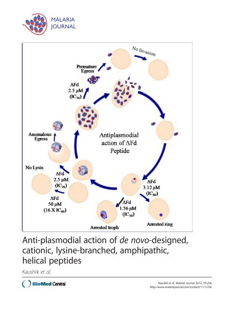
Figure 8 Mo<strong>de</strong>l <strong>of</strong> anti<strong>plasmodial</strong> <strong>action</strong> <strong>of</strong> ΔFd. ΔFd causes<br />
growth arrest <strong>of</strong> rings, anomalous egress <strong>of</strong> trophozoites and<br />
premature egress <strong>of</strong> schizonts. Its IC80 (3.12 μM) and IC50<br />
(1.56 μM) cause arrest <strong>of</strong> rings and trophozoites respectively and its<br />
IC70 (2.5 μM) causes the anomalous egress <strong>of</strong> trophozoites and<br />
premature egress <strong>of</strong> schizonts. In both cases the parasite fails to<br />
proliferate since egressed trophozoites cannot differentiate into<br />
schizonts and the premature, undifferentiated egressed schizonts<br />
seem to release merozoites that are invasion incompetent. The<br />
pepti<strong>de</strong> shows good selectivity against parasitized RBCs since 16X<br />
IC80 fails to lyse healthy RBCs.<br />
<strong>of</strong> cellular uptake can be achieved through the inclusion<br />
<strong>of</strong> cationic residues into arginine-based pepti<strong>de</strong> oligomers<br />
[55]. The positive molecular charge facilitates<br />
charge-driven uptake through the plasma membrane,<br />
which exhibits a potential gradient that can electrophorese<br />
cationic species from the extracellular space into the<br />
cell [56,57]. Interestingly, a recent study has <strong>de</strong>monstrated<br />
that membrane asymmetry can be altered and<br />
maintained in the altered state by externally ad<strong>de</strong>d poly-<br />
L-lysine [58]. The combined microscopic (Figure 6) and<br />
FACS analysis (Figure 7) suggests that ΔFd enters the<br />
infected cells and stains rings and trophozoites within a<br />
few minutes. Thus it is quite likely that ΔFd and (ΔFm) 2,<br />
the two cationic dimeric pepti<strong>de</strong>s studied here, in close<br />
resemblance to poly-L-lysine, may first home on those<br />
infected red blood cells that show slightly more anionic<br />
character as a result <strong>of</strong> alterations in membrane asymmetry<br />
and binding <strong>of</strong> these cationic pepti<strong>de</strong>s could<br />
further enhance and maintain this anionic character<br />
facilitating the stronger binding and faster internalization<br />
<strong>of</strong> pepti<strong>de</strong>s into the infected cells.
Kaushik et al. <strong>Malaria</strong> <strong>Journal</strong> 2012, 11:256 Page 13 <strong>of</strong> 15<br />
http://www.malariajournal.com/content/11/1/256<br />
The ability <strong>of</strong> ΔFd to cross the host red cell membrane,<br />
the parasitophorous vacuole membrane, the parasite<br />
plasma membrane and also the parasite nuclear<br />
membrane to reach the nucleus <strong>of</strong> the parasite<br />
(Figure 6B), indicates its resemblance to cell-penetrating<br />
pepti<strong>de</strong>s which are known to have a lipophilic-cationic<br />
character. Even as the pepti<strong>de</strong> was apparently targeting<br />
the DNA <strong>of</strong> the parasite, the absence <strong>of</strong> FITC-ΔFd on<br />
the host red cell membrane or all the subsequent membranes<br />
mentioned above was puzzling. It was surmised<br />
that these localizations may have been missed due to the<br />
rapidity <strong>of</strong> the process <strong>of</strong> pepti<strong>de</strong> uptake. In or<strong>de</strong>r to<br />
capture some stages preceding the intranuclear entry <strong>of</strong><br />
the pepti<strong>de</strong>, the pepti<strong>de</strong>-staining experiment was performed<br />
as a function <strong>of</strong> both time and temperature. As<br />
shown (Figure 6C), the images captured at 4°C (30 min)<br />
in<strong>de</strong>ed showed predominant staining on the host cell<br />
surface. In contrast, the images corresponding to 25°C<br />
(10 min) and 25°C (30 min) showed progressively<br />
greater staining <strong>of</strong> the intracellular parasite nuclear material.<br />
These results suggest that this pepti<strong>de</strong> crosses<br />
several membranes <strong>of</strong> the infected red cells before<br />
entering the nucleus.<br />
The most probable reasons for the significantly<br />
enhanced potency <strong>of</strong> the dimers ΔFd/Fd over the monomers<br />
ΔFm/Fm inclu<strong>de</strong> increased membrane binding<br />
and permeabilization, enhanced binding affinity for<br />
DNA and proteins and enhanced biochemical stability<br />
against <strong>de</strong>gradation by proteases. These properties originating<br />
from increased avidity and affinity <strong>of</strong> inter<strong>action</strong>s<br />
unique to dimeric pepti<strong>de</strong>s and absent in<br />
monomeric pepti<strong>de</strong>s have been <strong>de</strong>scribed previously<br />
[25]. In studies on the antibiotic <strong>action</strong> <strong>of</strong> these pepti<strong>de</strong>s it<br />
has previously been observed that the requirements <strong>of</strong> helicity<br />
for potent antibiotic <strong>action</strong> are much higher for the<br />
gram positive Staphylococcus.aureus than is the case with<br />
the Gram-negative Escherichia coli. In contrast, as shown<br />
in the present study, all dimers {(ΔFd, Fd, D-Lys-ΔFd,<br />
(ΔFm) 2} are nearly equipotent against P. falciparum<br />
(Table 1). This suggests that different conformational and<br />
topological properties <strong>of</strong> pepti<strong>de</strong>s may be important for<br />
their activity against different organisms.<br />
Some important features <strong>of</strong> these pepti<strong>de</strong>s as drugs<br />
against malaria inclu<strong>de</strong> their favourable resistance indices<br />
(Table 1) that allow them to rapidly kill both drug-sensitive<br />
and drug-resistant strains <strong>of</strong> malaria parasite with equal potencies,<br />
their amphipathic nature that gives them drug-like<br />
character, and their ability to permeabilize and penetrate<br />
biological membranes, which allows them to attack target<br />
cells both from the surface as well as intracellularly. In<br />
addition, the presence <strong>of</strong> the conformationally constrained,<br />
non-protein, amino acid di<strong>de</strong>hydrophenylalanine in both<br />
ΔFd and (ΔFm) 2 provi<strong>de</strong>s consi<strong>de</strong>rable protection against<br />
proteolytic <strong>de</strong>gradation [25]. Even as these two dimeric<br />
pepti<strong>de</strong>s <strong>of</strong>fer similar pr<strong>of</strong>iles <strong>of</strong> anti-<strong>plasmodial</strong> <strong>action</strong>s, a<br />
judicious choice for further improvisation should be the<br />
branched dimer ΔFd over the linear dimer (ΔFm) 2 since<br />
(a) the former is little more potent against P. falciparum,<br />
(b) the branched dimer is more stable against proteases<br />
[25], and (c) the branched dimer has better economics <strong>of</strong><br />
production since the time it takes to synthesize a<br />
branched dimer is half as much as the time it takes to<br />
assemble a linear dimer.<br />
Conclusion<br />
This study reports the anti-<strong>plasmodial</strong> <strong>action</strong> <strong>of</strong> ΔFd, a<br />
<strong>de</strong> <strong>novo</strong>-<strong>de</strong>signed, cationic, lysine-branched amphipathic,<br />
helical pepti<strong>de</strong>. In vitro assays suggest good selectivity<br />
(>35), good resistance in<strong>de</strong>x (1.1) and low mamamalian<br />
cell cytotoxicity, as a promise <strong>of</strong> ΔFd against malaria.<br />
The strategy adopted by ΔFd to inhibit the growth <strong>of</strong><br />
malaria parasite appears to be broadly two-fold: (a) involving<br />
growth arrest without causing lysis <strong>of</strong> red cell<br />
(at IC 50-IC 100), and (b) anomalous egress <strong>of</strong> trophozoites<br />
and premature egress <strong>of</strong> undifferentiated schizonts<br />
leading to <strong>de</strong>ath <strong>of</strong> the parasite (at > IC 100).<br />
Additional files<br />
Additional file 1: RPHPLC pr<strong>of</strong>iles <strong>of</strong> control pepti<strong>de</strong>s.<br />
Additional file 2: RPHPLC pr<strong>of</strong>iles <strong>of</strong> ΔFm and ΔFd.<br />
Additional file 3: ESMS <strong>of</strong> RPHPLC purified prochitinase, E30<br />
and Insulin.<br />
Additional file 4: ESMS <strong>of</strong> RPHPLC purified ΔFm and ΔFd.<br />
Additional file 5: MALDI mass spectrum (Bruker Daltonics Flex<br />
analysis) <strong>of</strong> RPHPLC purified linear dimeric pepti<strong>de</strong>.<br />
Additional file 6: Chromatographic and mass spectral<br />
characterization <strong>of</strong> ΔFq.<br />
Additional file 7: ESMS <strong>of</strong> RPHPLC purified Fm, Fd and D-Lys-ΔFd.<br />
Additional file 8: Microscopic differential counts <strong>of</strong> ΔFd (2.5 μM)<br />
treated trophozoites after 24 h.<br />
Additional file 9: ΔFd treated schizonts express MSP3.<br />
Additional file 10: ΔFd causes premature egress <strong>of</strong> schizonts.<br />
Abbreviations<br />
CQ: Chloroquine; ΔF: Di<strong>de</strong>hydrophenylalanine; ΔFm: ΔF containing<br />
monomeric <strong>de</strong>capepti<strong>de</strong>; ΔFd: Lysine branched dimer <strong>of</strong> ΔFm; (ΔFm)2: Linear<br />
dimer <strong>of</strong> ΔFm; DAPI: 4',6-diamidino-2-phenylindole; FACS: Fluorescence<br />
activated cell sorter; FITC: Fluorescein isothiocyanate;<br />
P. falciparum: Plasmodium falciparum; IC100: Inhibitory concentration causing<br />
100% inhibition <strong>of</strong> growth; PRBC: Parasitized red blood cell; URBC: Uninfected<br />
red blood cell; Troph: Trophozoite; ΔFq:<br />
The K-K2<strong>de</strong>ndrimer presenting a quadrumer form <strong>of</strong> ΔFm.<br />
Competing interests<br />
The authors <strong>de</strong>clare that they have no competing interests.<br />
Authors’ contributions<br />
NKK and JS carried out the experiments to <strong>de</strong>termine the anti<strong>plasmodial</strong><br />
potencies <strong>of</strong> different pepti<strong>de</strong>s, NKK performed mechanistic experiments<br />
including FACS and immun<strong>of</strong>luorescence microscopy, DS conceived <strong>of</strong> the<br />
study, participated in its <strong>de</strong>sign, coordination and brain storming and drafted<br />
the manuscript. All authors read and approved the final manuscript.
Kaushik et al. <strong>Malaria</strong> <strong>Journal</strong> 2012, 11:256 Page 14 <strong>of</strong> 15<br />
http://www.malariajournal.com/content/11/1/256<br />
Acknowledgements<br />
We thank MR4 who generously provi<strong>de</strong>d the chloroquine-resistant Dd2 and<br />
INDO strains used in the study. Thanks to X Su and the late Dr. David<br />
Walliker who <strong>de</strong>posited these strains with MR4, BEI Resources Repository,<br />
NIAID, NIH:. Our thanks to the anonymous reviewers for their critical and<br />
thoughtful comments that have enriched the manuscript enormously. We<br />
thank Dr. Pawan Malahotra for anti-band 3 antibody, Sumit Rathore for FACS<br />
analysis, Dr. Maryam Imam for providing anti MSP3 antibody, Dr. Aparna<br />
Anantharaman for MTT assay and Dr.Anil Sharma for help with statistical<br />
analysis. NKK thanks Indian Council for Medical Research (ICMR), New Delhi,<br />
for Senior Research fellowship. We thank the ICGEB, New Delhi for internal<br />
funding.<br />
Received: 8 May 2012 Accepted: 13 July 2012<br />
Published: 1 August 2012<br />
References<br />
1. Tinto H, Rwagacondo C, Karema C, Mupfasoni D, Vandoren W, Rusanganwa<br />
E, Erhart A, Van Overmeir C, Van Marck E, D'Alessandro U: In-vitro<br />
susceptibility <strong>of</strong> Plasmodium falciparum to mono<strong>de</strong>sethylamodiaquine,<br />
dihydroartemisinin and quinine in an area <strong>of</strong> high chloroquine<br />
resistance in Rwanda. Trans R Soc Trop Med Hyg 2006, 100:509–514.<br />
2. Mu J, Ferdig MT, Feng X, Joy DA, Duan J, Furuya T, Subramanian G, Aravind<br />
L, Cooper RA, Wootton JC, Xiong M, Su XZ: Multiple transporters<br />
associated with malaria parasite responses to chloroquine and quinine.<br />
Mol Microbiol 2003, 49:977–989.<br />
3. Jacqueline R: Halt called on single-drug antimalarial prescriptions. Nature<br />
2006, doi:10.1038/news060116-13.<br />
4. Dondorp AM, Nosten F, Yi P, Das D, Phyo AP, Tarning J, Lwin KM, Ariey F,<br />
Hanpithakpong W, Lee SJ, Ringwald P, Silamut K, Imwong M, Chotivanich K,<br />
Lim P, Herdman T, An SS, Yeung S, Singhasivanon P, Day NP, Lin<strong>de</strong>gardh N,<br />
Socheat D, White NJ: Artemisinin resistance in Plasmodium falciparum<br />
malaria. N Engl J Med 2009, 361:455–467.<br />
5. White NJ: Artemisinin resistance--the clock is ticking. Lancet 2010,<br />
376:2051–2052.<br />
6. Crompton PD, Pierce SK, Miller LH: Advances and challenges in malaria<br />
vaccine <strong>de</strong>velopment. J Clin Invest 2010, 120:4168–4178.<br />
7. WHO: World <strong>Malaria</strong> Report 2011, Issue Dec. 2011. http://www.who.int/<br />
mediacentre/factsheets/fs094/en/in<strong>de</strong>x.html.<br />
8. Yount NY, Yeaman MR: Emerging themes and therapeutic prospects for<br />
anti-infective pepti<strong>de</strong>s. Annu Rev Pharmacol Toxicol 2012, 52:337–360.<br />
9. Yeaman MR, Yount NY: Mechanisms <strong>of</strong> antimicrobial pepti<strong>de</strong> <strong>action</strong> and<br />
resistance. Pharmacol Rev 2003, 55:27–55.<br />
10. Epand RM, Shai Y, Segrest JP, Anantharamaiah GM: Mechanisms for the<br />
modulation <strong>of</strong> membrane bilayer properties by amphipathic helical<br />
pepti<strong>de</strong>s. Biopolymers 1995, 37:319–338.<br />
11. Fe<strong>de</strong>r R, Dagan A, Mor A: Structure-activity relationship study <strong>of</strong><br />
antimicrobial <strong>de</strong>rmaseptin S4 showing the consequences <strong>of</strong><br />
pepti<strong>de</strong> oligomerization on selective cytotoxicity. JBiolChem2000,<br />
275:4230–4238.<br />
12. Zasl<strong>of</strong>f M: <strong>Anti</strong>microbial pepti<strong>de</strong>s <strong>of</strong> multicellular organisms. Nature 2002,<br />
415:389–395.<br />
13. Sherman IW, Prudhomme J, Tait JF: Altered membrane phospholipid<br />
asymmetry in Plasmodium falciparum-infected erythrocytes.<br />
Parasitol Today 1997, 13:242–243.<br />
14. Gelhaus C, Jacobs T, Andra J, Leippe M: The antimicrobial pepti<strong>de</strong> NK-2,<br />
the core region <strong>of</strong> mammalian NK-lysin, kills intraerythrocytic<br />
Plasmodium falciparum. <strong>Anti</strong>microb Agents Chemother 2008, 52:1713–1720.<br />
15. Ghosh JK, Shaool D, Guillaud P, Ciceron L, Mazier D, Kustanovich I, Shai Y,<br />
Mor A: Selective cytotoxicity <strong>of</strong> <strong>de</strong>rmaseptin S3 toward intraerythrocytic<br />
Plasmodium falciparum and the un<strong>de</strong>rlying molecular basis. J Biol Chem<br />
1997, 272:31609–31616.<br />
16. Radzishevsky I, Krugliak M, Ginsburg H, Mor A: <strong>Anti</strong><strong>plasmodial</strong> activity <strong>of</strong><br />
lauryl-lysine oligomers. <strong>Anti</strong>microb Agents Chemother 2007, 51:1753–1759.<br />
17. Azouzi S, El Kirat K, Morandat S: The potent antimalarial drug cyclosporin<br />
A preferentially <strong>de</strong>stabilizes sphingomyelin-rich membranes. Langmuir<br />
2010, 26:1960–1965.<br />
18. Boman HG, Wa<strong>de</strong> D, Boman IA, Wahlin B, Merrifield RB: <strong>Anti</strong>bacterial and<br />
antimalarial properties <strong>of</strong> pepti<strong>de</strong>s that are cecropin-melittin hybrids.<br />
FEBS Lett 1989, 259:103–106.<br />
19. Gao B, Xu J: Rodriguez M<strong>de</strong>l C, Lanz-Mendoza H, Hernan<strong>de</strong>z-Rivas R, Du<br />
W, Zhu S: Characterization <strong>of</strong> two linear cationic antimalarial pepti<strong>de</strong>s in<br />
the scorpion Mesobuthus eupeus. Biochimie 2010, 92:350–359.<br />
20. Nagaraj G, Uma MV, Shivayogi MS, Balaram H: <strong>Anti</strong>malarial activities <strong>of</strong><br />
pepti<strong>de</strong> antibiotics isolated from fungi. <strong>Anti</strong>microb Agents Chemother 2001,<br />
45:145–149.<br />
21. Hancock RE, Sahl HG: <strong>Anti</strong>microbial and host-<strong>de</strong>fense pepti<strong>de</strong>s as new<br />
anti-infective therapeutic strategies. Nat Biotechnol 2006, 24:1551–1557.<br />
22. Dathe M, Wieprecht T: Structural features <strong>of</strong> helical antimicrobial<br />
pepti<strong>de</strong>s: their potential to modulate activity on mo<strong>de</strong>l membranes and<br />
biological cells. Biochim Biophys Acta 1999, 1462:71–87.<br />
23. Giangaspero A, Sandri L, Tossi A: Amphipathic alpha helical antimicrobial<br />
pepti<strong>de</strong>s. Eur J Biochem 2001, 268:5589–5600.<br />
24. Malina A, Shai Y: Conjugation <strong>of</strong> fatty acids with different lengths<br />
modulates the antibacterial and antifungal activity <strong>of</strong> a cationic<br />
biologically inactive pepti<strong>de</strong>. Biochem J 2005, 390:695–702.<br />
25. Dewan PC, Anantharaman A, Chauhan VS, Sahal D: <strong>Anti</strong>microbial <strong>action</strong> <strong>of</strong><br />
prototypic amphipathic cationic <strong>de</strong>capepti<strong>de</strong>s and their branched<br />
dimers. Biochemistry 2009, 48:5642–5657.<br />
26. Trager W, Jensen JB: Human malaria parasites in continuous culture.<br />
Science 1976, 193:673–675.<br />
27. MR4. http://www.mr4.org/MR4ReagentsSearch/Results.aspx?BEINum=MRA-<br />
819&Template=parasites.<br />
28. Lambros C, Van<strong>de</strong>rberg JP: Synchronization <strong>of</strong> Plasmodium falciparum<br />
erythrocytic stages in culture. J Parasitol 1979, 65:418–420.<br />
29. Riva<strong>de</strong>neira EM, Wasserman M, Espinal CT: Separation and concentration<br />
<strong>of</strong> schizonts <strong>of</strong> Plasmodium falciparum by Percoll gradients. J Protozool<br />
1983, 30:367–370.<br />
30. Smilkstein M, Sriwilaijaroen N, Kelly JX, Wilairat P, Riscoe M: Simple and<br />
inexpensive fluorescence-based technique for high-throughput antimalarial<br />
drug screening. <strong>Anti</strong>microb Agents Chemother 2004, 48:1803–1806.<br />
31. Mosmann T: Rapid colorimetric assay for cellular growth and survival:<br />
application to proliferation and cytotoxicity assays. J Immunol Meth 1983,<br />
65:55–63.<br />
32. Yonath A: Polar bears, antibiotics, and the evolving ribosome (Nobel<br />
Lecture). Angew Chem Int Ed Engl 2010, 49:4341–4354.<br />
33. Matsuzaki K: Why and how are pepti<strong>de</strong>-lipid inter<strong>action</strong>s utilized for<br />
self-<strong>de</strong>fense? Magainins and tachyplesins as archetypes. Biochim Biophys<br />
Acta 1999, 1462:1–10.<br />
34. Shai Y: Mechanism <strong>of</strong> the binding, insertion and <strong>de</strong>stabilization <strong>of</strong><br />
phospholipid bilayer membranes by alpha-helical antimicrobial and cell<br />
non-selective membrane-lytic pepti<strong>de</strong>s. Biochim Biophys Acta 1999,<br />
1462:55–70.<br />
35. Yang L, Weiss TM, Lehrer RI, Huang HW: Crystallization <strong>of</strong> antimicrobial pores<br />
in membranes: magainin and protegrin. Biophys J 2000, 79:2002–2009.<br />
36. Ramagopal UA, Ramakumar S, Mathur P, Joshi R, Chauhan VS:<br />
Dehydrophenylalanine zippers: strong helix-helix clamping through a<br />
network <strong>of</strong> weak inter<strong>action</strong>s. Protein Eng 2002, 15:331–335.<br />
37. Ramagopal UA, Ramakumar S, Sahal D, Chauhan VS: De <strong>novo</strong> <strong>de</strong>sign and<br />
characterization <strong>of</strong> an apolar helical hairpin pepti<strong>de</strong> at atomic resolution:<br />
Comp<strong>action</strong> mediated by weak inter<strong>action</strong>s. Proc Natl Acad Sci U S A<br />
2001, 98:870–874.<br />
38. Rudresh, Ramakumar S, Ramagopal UA, Inai Y, Goel S, Sahal D, Chauhan VS:<br />
De <strong>novo</strong> <strong>de</strong>sign and characterization <strong>of</strong> a helical hairpin eicosapepti<strong>de</strong>;<br />
emergence <strong>of</strong> an anion receptor in the linker region. Structure 2004,<br />
12:389–396.<br />
39. Fichera ME, Roos DS: A plastid organelle as a drug target in<br />
apicomplexan parasites. Nature 1997, 390:407–409.<br />
40. Ralph SA, D'Ombrain MC, McFad<strong>de</strong>n GI: The apicoplast as an antimalarial<br />
drug target. Drug Resist Updat 2001, 4:145–151.<br />
41. Dahl EL, Rosenthal PJ: Multiple antibiotics exert <strong>de</strong>layed effects against<br />
the Plasmodium falciparum apicoplast. <strong>Anti</strong>microb Agents Chemother 2007,<br />
51:3485–3490.<br />
42. Hanssen E, McMillan PJ, Tilley L: Cellular architecture <strong>of</strong> Plasmodium<br />
falciparum-infected erythrocytes. Int J Parasitol 2010, 40:1127–1135.<br />
43. Allred DR, Sterling CR, Morse PD 2nd: Increased fluidity <strong>of</strong> Plasmodium<br />
berghei-infected mouse red blood cell membranes <strong>de</strong>tected by electron<br />
spin resonance spectroscopy. Mol Biochem Parasitol 1983, 7:27–39.<br />
44. Howard RJ, Sawyer WH: Changes in the membrane microviscosity <strong>of</strong><br />
mouse red blood cells infected with Plasmodium berghei <strong>de</strong>tected
Kaushik et al. <strong>Malaria</strong> <strong>Journal</strong> 2012, 11:256 Page 15 <strong>of</strong> 15<br />
http://www.malariajournal.com/content/11/1/256<br />
using n-(9-anthroyloxy) fatty acid fluorescent probes. Parasitology<br />
1980, 80:331–342.<br />
45. Sherman IW, Greenan JR: Altered red cell membrane fluidity during<br />
schizogonic <strong>de</strong>velopment <strong>of</strong> malarial parasites (Plasmodium falciparum<br />
and P. lophurae). Trans R Soc Trop Med Hyg 1984, 78:641–644.<br />
46. Taraschi TF, Parashar A, Hooks M, Rubin H: Perturbation <strong>of</strong> red cell<br />
membrane structure during intracellular maturation <strong>of</strong> Plasmodium<br />
falciparum. Science 1986, 232:102–104.<br />
47. Vial HJ, Philippot JR, Wallach DF: A reevaluation <strong>of</strong> the status <strong>of</strong><br />
cholesterol in erythrocytes infected by Plasmodium knowlesi and<br />
P. falciparum. Mol Biochem Parasitol 1984, 13:53–65.<br />
48. Vial HJ, Thuet MJ, Broussal JL, Philippot JR: Phospholipid biosynthesis by<br />
Plasmodium knowlesi-infected erythrocytes: the incorporation <strong>of</strong><br />
phospohlipid precursors and the i<strong>de</strong>ntification <strong>of</strong> previously un<strong>de</strong>tected<br />
metabolic pathways. J Parasitol 1982, 68:379–391.<br />
49. Gupta CM, Mishra GC: Transbilayer phospholipid asymmetry in<br />
Plasmodium knowlesi-infected host cell membrane. Science 1981,<br />
212:1047–1049.<br />
50. Joshi P, Dutta GP, Gupta CM: An intracellular simian malarial parasite<br />
(Plasmodium knowlesi) induces stage-<strong>de</strong>pen<strong>de</strong>nt alterations in<br />
membrane phospholipid organization <strong>of</strong> its host erythrocyte. Biochem J<br />
1987, 246:103–108.<br />
51. Schwartz RS, Olson JA, Raventos-Suarez C, Yee M, Heath RH, Lubin B, Nagel RL:<br />
Altered plasma membrane phospholipid organization in Plasmodium<br />
falciparum-infected human erythrocytes. Blood 1987, 69:401–407.<br />
52. Ginsburg H, Kutner S, Zangwil M, Cabantchik ZI: Selectivity properties <strong>of</strong><br />
pores induced in host erythrocyte membrane by Plasmodium falciparum.<br />
Effect <strong>of</strong> parasite maturation. Biochim Biophys Acta 1986, 861:194–196.<br />
53. Kutner S, Ginsburg H, Cabantchik ZI: Permselectivity changes in malaria<br />
(Plasmodium falciparum) infected human red blood cell membranes.<br />
J Cell Physiol 1983, 114:245–251.<br />
54. Vial HJ, Ancelin ML: <strong>Malaria</strong>l lipids. An overview. Subcell Biochem 1992,<br />
18:259–306.<br />
55. Wen<strong>de</strong>r PA, Mitchell DJ, Pattabiraman K, Pelkey ET, Steinman L, Rothbard JB:<br />
The <strong>de</strong>sign, synthesis, and evaluation <strong>of</strong> molecules that enable or<br />
enhance cellular uptake: peptoid molecular transporters. Proc Natl Acad<br />
Sci U S A 2000, 97:13003–13008.<br />
56. Rothbard JB, Jessop TC, Lewis RS, Murray BA, Wen<strong>de</strong>r PA: Role <strong>of</strong><br />
membrane potential and hydrogen bonding in the mechanism <strong>of</strong><br />
translocation <strong>of</strong> guanidinium-rich pepti<strong>de</strong>s into cells. J Am Chem Soc<br />
2004, 126:9506–9507.<br />
57. Terrone D, Sang SL, Roudaia L, Silvius JR: Penetratin and related cellpenetrating<br />
cationic pepti<strong>de</strong>s can translocate across lipid bilayers in the<br />
presence <strong>of</strong> a transbilayer potential. Biochemistry 2003, 42:13787–13799.<br />
58. Brown KL, Conboy JC: Electrostatic induction <strong>of</strong> lipid asymmetry. JAm<br />
Chem Soc 2011, 133:8794–8797.<br />
doi:10.1186/1475-2875-11-256<br />
Cite this article as: Kaushik et al.: <strong>Anti</strong>-<strong>plasmodial</strong> <strong>action</strong> <strong>of</strong> <strong>de</strong><br />
<strong>novo</strong>-<strong>de</strong>signed, cationic, lysine-branched, amphipathic, helical pepti<strong>de</strong>s.<br />
<strong>Malaria</strong> <strong>Journal</strong> 2012 11:256.<br />
Submit your next manuscript to BioMed Central<br />
and take full advantage <strong>of</strong>:<br />
• Convenient online submission<br />
• Thorough peer review<br />
• No space constraints or color figure charges<br />
• Immediate publication on acceptance<br />
• Inclusion in PubMed, CAS, Scopus and Google Scholar<br />
• Research which is freely available for redistribution<br />
Submit your manuscript at<br />
www.biomedcentral.com/submit