Proliferation, apoptosis, and manganese superoxide ... - Erbeofficinali
Proliferation, apoptosis, and manganese superoxide ... - Erbeofficinali
Proliferation, apoptosis, and manganese superoxide ... - Erbeofficinali

Create successful ePaper yourself
Turn your PDF publications into a flip-book with our unique Google optimized e-Paper software.
3�-End labelling of DNA in apoptotic cells<br />
Apoptosis was determined morphologically in mesothelioma tissue<br />
<strong>and</strong> in mesothelioma cells by in situ labelling of the 3� ends of the<br />
DNA fragments (TUNEL) using the ApopTag <strong>apoptosis</strong> detection kit<br />
(Oncor, Gaithersburg, MD). Sections from the tissue <strong>and</strong> cell blocks<br />
were dewaxed in xylene, rehydrated in ethanol, <strong>and</strong> incubated with 20<br />
�g/ml proteinase K (Boehringer-Mannheim) at room temperature for<br />
15 min. Incubation in 2% hydrogen peroxide in PBS was performed<br />
to block endogenous peroxidase activity, after which slides were<br />
exposed to a terminal transferase enzyme <strong>and</strong> digoxigenin-labelled<br />
nucleotides. Thereafter, slides were incubated with an anti-digoxigenin<br />
antibody labelled with peroxidase, <strong>and</strong> the colour was developed<br />
with diaminobenzidine–hydrogen peroxide. Samples were<br />
lightly counterstained with haematoxylin.<br />
Light microscopic evaluation<br />
Each section was evaluated blindly by 2 authors. The extent of cell<br />
proliferation was calculated as the percentage of Ki-67-positive tumour<br />
cells. Apoptotic cells with clearly labeled nuclei <strong>and</strong> smaller<br />
apoptotic bodies were counted. At least 500 consecutive tumour cells<br />
were counted in each sample, <strong>and</strong> the percentages of positive cells<br />
were expressed as proliferation or apoptotic indices. The extent of<br />
necrosis was assessed by a morphometric method described previously<br />
(Eerola et al., 1997). Briefly, a Nikon (Tokyo, Japan) Labophot<br />
light microscope was employed connected via a video camera (Panasonic<br />
F10, Osaka, Japan) to a TV monitor (Sony, Tokyo, Japan)<br />
equipped with a net of 50 points adherent to the screen. The total<br />
magnification for counting the volume density was �40, <strong>and</strong> all<br />
available sections from the tumours were counted in each case.<br />
Evaluation of MnSOD staining intensity has been reported previously<br />
(Kahlos et al., 2000). In that study, MnSOD was assessed semiquantitatively<br />
by grading the staining intensity of the tumour cells in<br />
mesothelioma sections <strong>and</strong> simultaneously estimating the proportion<br />
of positive cells of all tumour cells of the section. This semi-quantitation<br />
was expressed as negative (0), weak (�1), moderate (�2), or<br />
intense (�3) according to the staining intensity in tumour cells <strong>and</strong> the<br />
proportion of positive tumour cells.<br />
Detection of caspase 3 activation by Western blotting<br />
The apoptotic response of mesothelioma cells to epirubicin was<br />
also assessed by detecting caspase 3 activation using polyclonal<br />
PROLIFERATION AND APOPTOSIS IN MESOTHELIOMA<br />
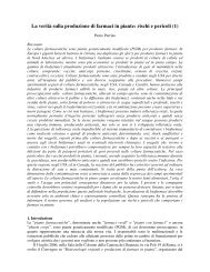
FIGURE 1 – Immunostaining of MnSOD (a) <strong>and</strong> Ki-67 (c) in a patient with epithelial malignant mesothelioma showing high MnSOD in<br />
association with low cell proliferation. In another patient with epithelial mesothelioma, low MnSOD expression (b) is associated with a high<br />
proliferation index (47%) (d) (Magnification �142).<br />
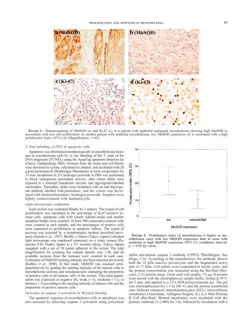
FIGURE 2 – <strong>Proliferation</strong> index of mesothelioma is higher in mesothelioma<br />
cases with low MnSOD expression than in cases with<br />
moderate or high MnSOD expression (95% CI, confidence interval;<br />
p � 0.02 by t-test).<br />
rabbit anti-human caspase 3 antibody (CPP32; PharMingen, San<br />
Diego, CA). According to the manufacturer, the antibody detects<br />
both the 32 kDa inactive pro-enzyme <strong>and</strong> the fragmented active<br />
unit of 17 kDa. Cell pellets were suspended in sterile water, <strong>and</strong><br />
the protein concentration was measured using the Bio-Rad (Hercules,<br />
CA) protein assay. From each cell sample, 75 �g of protein<br />
were mixed with the electrophoresis sample buffer, boiled at 95°C<br />
for 5 min, <strong>and</strong> applied to a 12% SDS-polyacrylamide gel. The gel<br />
was electrophoresed for 1.5 hr (90 V) <strong>and</strong> the protein transferred<br />
onto Hybond enhanced chemiluminescence (ECL) nitrocellulose<br />
membranes (Amersham, Arlington Heights, IL) in a Mini-Protean<br />
II Cell (Bio-Rad). Blotted membranes were incubated with the<br />
primary antibody (1:2,000) for 1 hr, followed by incubation with a<br />
39