Your Patient Images, All In One Place. - Kowa
Your Patient Images, All In One Place. - Kowa
Your Patient Images, All In One Place. - Kowa

You also want an ePaper? Increase the reach of your titles
YUMPU automatically turns print PDFs into web optimized ePapers that Google loves.
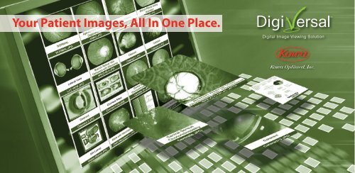
<strong>Your</strong> <strong>Patient</strong> <strong>Images</strong>, <strong>All</strong> <strong>In</strong> <strong>One</strong> <strong>Place</strong>.<br />
Digital Image Viewing Solution
Fully <strong>In</strong>tegrated<br />
Image VIew<strong>In</strong>g:<br />
A patient file displays thumbnails of all the patient’s images from<br />
virtually all diagnostic equipment. Click on the thumbnail to view<br />
a full-sized image in the review software.<br />
• Connects your practice’s instruments and files to a single database<br />
• Automatically converges patient images into an individual file from<br />
virtually all diagnostic equipment<br />
• <strong>All</strong>ows for connectivity with other practices or medical facilities<br />
dIgIVersal promIses....<br />
To comprehensively manage your patients’ diagnostic<br />
imagery by converging sources as varied as digital<br />
fundus cameras, slit lamps, visual field, OCTs and more,<br />
from initial testing through point of care, and even<br />
across different disciplines.<br />
We resolve all of these needs with a simple, single<br />
platform that’s a dependable and cost-effective<br />
turnkey solution for your practice.<br />
At <strong>Kowa</strong> Optimed, we’re fulfilling the promise of<br />
CONSOLIDATED IMAGE VIEWING, with the <strong>Kowa</strong><br />
DigiVersal Digital Image Viewing Solution<br />
System, capable of converging patient images<br />
from multiple diagnostic instruments onto a<br />
single screen – quickly and seamlessly.
Image VIew<strong>In</strong>g<br />
solutIon<br />
Use the drawing feature to create notations on images that are<br />
automatically stored in the patient folder.<br />
• Automatic simultaneous, instant annotation to track<br />
disease status<br />
• <strong>All</strong>ows you to use your personal shorthand and symbols<br />
• Saves time by avoiding repetitive input<br />
• Outstanding <strong>Patient</strong> Education Tool
when better care...<br />
Just some of the DigiVersaladvantages<br />
you’ll see<br />
For imaging a complete converging and viewing platform<br />
that lets you manage and view image files from virtually<br />
any instrument or other source, regardless of brand or<br />
model, within a single screen.<br />
Draw-Itgraphics technology allows you to create sophisticated<br />
real-time drawings at the click of a mouse, permitting automatic<br />
documentation to track the progression of conditions and<br />
pathologies. It’s unique to DigiVersal — and is fully embedded<br />
with all other imaging data in your patients’ files.<br />
Our EyeSend technology has proven to be a major breakthrough in<br />
multi-location file transference, letting you converge your informationtagged<br />
images and transfer them to a central database. It’s<br />
available as both a component of DigiVersal and also as a<br />
standalone solution soon.<br />
Digital Image Viewing Solution
s<strong>In</strong>gle screen<br />
VIew<strong>In</strong>g<br />
...drIVes smarter<br />
bus<strong>In</strong>ess and<br />
enhanced patIent care<br />
By making your practice smarter, nimbler and more<br />
cost-efficient, DigiVersal helps put the focus where<br />
it belongs — on more effective patient care!<br />
Saves time & expedites process, by eliminating re-entry<br />
of data and patient information at various stages of care,<br />
and allowing quick click throughs to the exact image or<br />
combination you’re after whenever you need it.<br />
Streamlines image files by eliminating the need for bulky paperbased<br />
charts and hardcopy images, substituting an elegant<br />
and efficient unified image viewing system that’s literally “proven<br />
in practice.”<br />
Puts our world-class expertise to work for you: <strong>Kowa</strong> Optimed has<br />
built a reputation for innovation and superior customer support,<br />
based on our intimate understanding of practitioners’ medical<br />
and business needs. So when you implement DigiVersal you’re<br />
really putting our proven proficiency to work in your office — a<br />
responsibility we take very seriously.
Compare images from different instruments simultaneously. This shows<br />
a fundus image on the left and an OCT scan on the right, allowing for<br />
easy comparison between structure and function.<br />
• Side by side comparison eliminates time wasted switching<br />
between screens and applications<br />
• Convergence of multiple types of diagnostic information<br />
allows for accurate and focused comparison.<br />
dIgIVersal beneFIts to practIce<br />
the s<strong>In</strong>gle screen solutIon<br />
• Makes your practice more time and cost efficient<br />
• <strong>All</strong>ows you to focus on patient care, not paper-care<br />
• Databases are searchable by either<br />
patient, date range, image capture type or <strong>Patient</strong> ID<br />
• View 4-up — color / red / green / blue separation<br />
• Drag and drop images to quickly to generate<br />
referral letters and reports<br />
• <strong>In</strong>terfaces with EMR software<br />
• Reliable customer service<br />
• Puts <strong>Kowa</strong> Optimed’s world class expertise to<br />
work for you
Create your own stereo pairs by simply linking images of the same eye,<br />
which were taken on the same day.<br />
• See changes over time in stereo by simply scrolling up and<br />
down to view the oldest to the most recent images<br />
• View a complete history of a patient's stereo pairs<br />
sImultaneous<br />
Image VIew<strong>In</strong>g:<br />
View images from different instruments simultaneously. This shows<br />
topography on the left and specular microscopy on the right.<br />
• Side by side comparison allows for easy analysis<br />
• View images from different instruments without<br />
switching applications.
Partial Equipment List<br />
Follow Us:<br />
Aberrometer<br />
Bausch & Lomb<br />
• Zywave II<br />
marco<br />
• OPD-Scan II<br />
Corneal Topographer<br />
Bausch & Lomb<br />
• Orbscan<br />
Perimeter – Visual Field<br />
Carl Zeiss Meditec<br />
• HFA-730 i<br />
• HFA-740 i<br />
• HFA II 745 i<br />
• HFA II 750 i<br />
• HFA 7xx<br />
Haag-Streit<br />
• Octopus 900<br />
Reichert<br />
• Foresee PHP<br />
OCT<br />
Optovue<br />
• RTVue (with PC)<br />
topcon<br />
• 3D OCT 2000<br />
Carl Zeiss<br />
(Humphrey)<br />
• Cirrus HD-OCT<br />
Model 4000<br />
• Stratus OCT<br />
Model 3000<br />
Scanning Laser<br />
Polarimeter<br />
Carl Zeiss<br />
(Humphrey)<br />
• GDx<br />
Retina Tomographer<br />
Heidelberg<br />
Engineering<br />
• HRT-II (with PC)<br />
Retinal Cameras<br />
optos<br />
• Panoramic 200<br />
Carl Zeiss<br />
(Humphrey)<br />
• Visucam PRO NM<br />
• FF 450plus IR<br />
Nidek<br />
• AFC-230/210<br />
• TRC-NW8<br />
<strong>Kowa</strong><br />
• Genesis-D<br />
• Genesis-Df<br />
• nonmyd a-d<br />
• nonmyd a-d<br />
(5 mega)<br />
Ask us about compatibility with your equipment.<br />
<strong>Kowa</strong> Optimed, <strong>In</strong>c. • 20001 South Vermont Ave. • Torrance, CA 90502<br />
Toll Free: 800-966-5692 • Phone: 310-327-1913 • www.kowa-usa.com<br />
• nonmyd a-dIII<br />
• nonmyd 7<br />
• nonmyd WX 3D<br />
• VX-10<br />
• VX-10a<br />
• VX-10i<br />
Scheimpflug<br />
Oculus<br />
• Pentacam<br />
(with PC)<br />
Slit Lamps<br />
cso<br />
• SL 980<br />
• SL 990<br />
Haag-Streit<br />
• BQ 900<br />
<strong>Kowa</strong><br />
• SL-15<br />
marco<br />
• G5<br />
topcon<br />
• SL-D2<br />
Specular Microscope<br />
Konan<br />
• NSP-9900<br />
Biometery<br />
Carl Ziess<br />
(Humphrey)<br />
• IOL Master<br />
Ultrasound<br />
Quantel<br />
• Axis II<br />
accutome<br />
• A-Scan Plus<br />
Authorized Representative