The effect of dose reduction on the detection - Dentomaxillofacial ...
The effect of dose reduction on the detection - Dentomaxillofacial ...
The effect of dose reduction on the detection - Dentomaxillofacial ...

You also want an ePaper? Increase the reach of your titles
YUMPU automatically turns print PDFs into web optimized ePapers that Google loves.
RESEARCH<br />
<str<strong>on</strong>g>The</str<strong>on</strong>g> <str<strong>on</strong>g>effect</str<strong>on</strong>g> <str<strong>on</strong>g>of</str<strong>on</strong>g> <str<strong>on</strong>g>dose</str<strong>on</strong>g> <str<strong>on</strong>g>reducti<strong>on</strong></str<strong>on</strong>g> <strong>on</strong> <strong>the</strong> detecti<strong>on</strong> <str<strong>on</strong>g>of</str<strong>on</strong>g> anatomical<br />
structures <strong>on</strong> panoramic radiographs<br />
G Kaeppler *,1 , K Dietz 2 and S Reinert 3<br />
1 Department <str<strong>on</strong>g>of</str<strong>on</strong>g> Oral Radiology, School <str<strong>on</strong>g>of</str<strong>on</strong>g> Dental Medicine, University <str<strong>on</strong>g>of</str<strong>on</strong>g> Tübingen, Germany; 2 Department <str<strong>on</strong>g>of</str<strong>on</strong>g> Medical Biometry,<br />
University <str<strong>on</strong>g>of</str<strong>on</strong>g> Tübingen, Germany; 3 Department <str<strong>on</strong>g>of</str<strong>on</strong>g> Oral and Maxill<str<strong>on</strong>g>of</str<strong>on</strong>g>acial Plastic Surgery, University <str<strong>on</strong>g>of</str<strong>on</strong>g> Tübingen, Germany<br />
Introducti<strong>on</strong><br />
Objectives: <str<strong>on</strong>g>The</str<strong>on</strong>g> aim was to evaluate <strong>the</strong> <str<strong>on</strong>g>effect</str<strong>on</strong>g> <str<strong>on</strong>g>of</str<strong>on</strong>g> <str<strong>on</strong>g>dose</str<strong>on</strong>g> <str<strong>on</strong>g>reducti<strong>on</strong></str<strong>on</strong>g> <strong>on</strong> diagnostic accuracy using<br />
different screen–film combinati<strong>on</strong>s and digital techniques for panoramic radiography.<br />
Methods: Five observers assessed 201 pairs <str<strong>on</strong>g>of</str<strong>on</strong>g> panoramic radiographs (a total <str<strong>on</strong>g>of</str<strong>on</strong>g> 402 panoramic<br />
radiographs) taken with <strong>the</strong> Orthophos Plus w (Sir<strong>on</strong>a, Bensheim, Germany), for visualizati<strong>on</strong> <str<strong>on</strong>g>of</str<strong>on</strong>g> 11<br />
anatomical structures <strong>on</strong> each side, using a 3-point scale 21, 0 and 1. Two radiographs <str<strong>on</strong>g>of</str<strong>on</strong>g> each<br />
patient were taken at two different times (c<strong>on</strong>venti<strong>on</strong>al setting and setting with decreased <str<strong>on</strong>g>dose</str<strong>on</strong>g>, d<strong>on</strong>e<br />
by increasing tube potential settings or halving tube current). To compare <strong>the</strong> <str<strong>on</strong>g>dose</str<strong>on</strong>g> at different tube<br />
potential settings <str<strong>on</strong>g>dose</str<strong>on</strong>g>–length product was measured at <strong>the</strong> sec<strong>on</strong>dary collimator. Films with<br />
medium and regular intensifying screens (high and low tube potential settings) and storage phosphor<br />
plates (low tube potential setting, tube current setting equivalent to regular intensifying screen and<br />
halved) were compared. <str<strong>on</strong>g>The</str<strong>on</strong>g> five observers made 27 610 assessments. Intrarater agreement was<br />
expressed by Cohen’s kappa coefficient.<br />
Results: <str<strong>on</strong>g>The</str<strong>on</strong>g> results dem<strong>on</strong>strated an equivalence <str<strong>on</strong>g>of</str<strong>on</strong>g> regular screens (low tube potential setting)<br />
and medium screens (high and low tube potential settings). A significant difference existed between<br />
medium screens (low tube potential setting, mean score 0.92) and <strong>the</strong> group <str<strong>on</strong>g>of</str<strong>on</strong>g> regular film–screen<br />
combinati<strong>on</strong>s at high tube potential settings (mean score 0.89) and between all film–screen<br />
combinati<strong>on</strong>s and <strong>the</strong> digital system irrespective <str<strong>on</strong>g>of</str<strong>on</strong>g> exposure (mean score below 0.82). <str<strong>on</strong>g>The</str<strong>on</strong>g>re were<br />
no significant differences between medium and regular screens (mean score 0.88 to 0.92) for<br />
assessment <str<strong>on</strong>g>of</str<strong>on</strong>g> <strong>the</strong> period<strong>on</strong>tal ligament space, but <strong>the</strong>re was a significant difference compared with<br />
<strong>the</strong> digital system (mean score below 0.76). <str<strong>on</strong>g>The</str<strong>on</strong>g> kappa coefficient for intrarater agreement was<br />
moderate (0.55).<br />
C<strong>on</strong>clusi<strong>on</strong>s: New regular intensifying screens can replace medium screens at low tube potential<br />
settings. Digital panoramic radiographs should be taken at low tube potential levels with an exposure<br />
equivalent at least to a regular intensifying screen.<br />
Dentomaxill<str<strong>on</strong>g>of</str<strong>on</strong>g>acial Radiology (2006) 35, 271–277. doi: 10.1259/dmfr/16653683<br />
Keywords: radiography, panoramic; kV setting; intensifying screens; storage phosphor radiography<br />
Possibilities <str<strong>on</strong>g>of</str<strong>on</strong>g> <str<strong>on</strong>g>dose</str<strong>on</strong>g> <str<strong>on</strong>g>reducti<strong>on</strong></str<strong>on</strong>g> in radiography have been<br />
provided by a new type <str<strong>on</strong>g>of</str<strong>on</strong>g> film, 1,2 a new screen–film<br />
system 3,4 or by <strong>the</strong> use <str<strong>on</strong>g>of</str<strong>on</strong>g> a digital system. 5,6 In <strong>the</strong> case <str<strong>on</strong>g>of</str<strong>on</strong>g><br />
new screen–film systems or digital systems <strong>the</strong> questi<strong>on</strong><br />
<str<strong>on</strong>g>of</str<strong>on</strong>g> <strong>the</strong> detecti<strong>on</strong> <str<strong>on</strong>g>of</str<strong>on</strong>g> anatomical structures becomes <strong>the</strong> focus<br />
<str<strong>on</strong>g>of</str<strong>on</strong>g> attenti<strong>on</strong>.<br />
*Corresp<strong>on</strong>dence to: Priv.-Doz. Dr. Gabriele Kaeppler, Department <str<strong>on</strong>g>of</str<strong>on</strong>g> Oral<br />
Radiology, School <str<strong>on</strong>g>of</str<strong>on</strong>g> Dental Medicine, Osianderstr. 2-8, University <str<strong>on</strong>g>of</str<strong>on</strong>g> Tübingen,<br />
D-72076 Tübingen, Germany; E-mail: gabriele.kaeppler@med.uni-tuebingen.de<br />
Received 30 June 2005; revised 10 October 2005; accepted 23 October 2005<br />
Dentomaxill<str<strong>on</strong>g>of</str<strong>on</strong>g>acial Radiology (2006) 35, 271–277<br />
q 2006 <str<strong>on</strong>g>The</str<strong>on</strong>g> British Institute <str<strong>on</strong>g>of</str<strong>on</strong>g> Radiology<br />
http://dmfr.birjournals.org<br />
Film–screen systems <str<strong>on</strong>g>of</str<strong>on</strong>g> speed class 200 have been<br />
accepted in extraoral radiography. <str<strong>on</strong>g>The</str<strong>on</strong>g> problem up to now<br />
has been that a regular screen has not been able to gain<br />
uniform acceptance in maxill<str<strong>on</strong>g>of</str<strong>on</strong>g>acial radiology because <str<strong>on</strong>g>of</str<strong>on</strong>g> a<br />
c<strong>on</strong>cern about a reduced recogniti<strong>on</strong> <str<strong>on</strong>g>of</str<strong>on</strong>g> clinical structures<br />
with this screen.<br />
<str<strong>on</strong>g>The</str<strong>on</strong>g> most comm<strong>on</strong>ly used tube potential is 70 kV in<br />
88%. 4 An increase in tube potential and a <str<strong>on</strong>g>reducti<strong>on</strong></str<strong>on</strong>g> <str<strong>on</strong>g>of</str<strong>on</strong>g><br />
milliamperes could reduce <strong>the</strong> radiati<strong>on</strong> <str<strong>on</strong>g>dose</str<strong>on</strong>g> required but<br />
could also result in poorer image c<strong>on</strong>trast and image<br />
quality, whose <str<strong>on</strong>g>effect</str<strong>on</strong>g>s <strong>on</strong> <strong>the</strong> patient radiograph should be<br />
assessed.
272<br />
<str<strong>on</strong>g>The</str<strong>on</strong>g> aim <str<strong>on</strong>g>of</str<strong>on</strong>g> this study was to compare image quality, as<br />
measured by detecti<strong>on</strong> <str<strong>on</strong>g>of</str<strong>on</strong>g> anatomic structures, in panoramic<br />
radiographs made with different radiati<strong>on</strong> <str<strong>on</strong>g>dose</str<strong>on</strong>g>s. Doses<br />
were varied by altering <strong>the</strong> image receptor (various film–<br />
screen combinati<strong>on</strong>s and digital imaging) and tube<br />
potential and/or tube current settings.<br />
Patients and methods<br />
Two panoramic radiographs each <str<strong>on</strong>g>of</str<strong>on</strong>g> 201 patients (402<br />
radiographs), taken in <strong>the</strong> Department <str<strong>on</strong>g>of</str<strong>on</strong>g> Oral Radiology <str<strong>on</strong>g>of</str<strong>on</strong>g><br />
<strong>the</strong> Centre <str<strong>on</strong>g>of</str<strong>on</strong>g> Dentistry at two different times in a<br />
randomized order with c<strong>on</strong>venti<strong>on</strong>al settings and with a<br />
reduced <str<strong>on</strong>g>dose</str<strong>on</strong>g>, were used for this study. Both radiographs<br />
were taken <strong>on</strong> <strong>the</strong> basis <str<strong>on</strong>g>of</str<strong>on</strong>g> current medical indicati<strong>on</strong> in<br />
accordance with <strong>the</strong> German X-ray rules. 7 No additi<strong>on</strong>al<br />
radiographs were taken for <strong>the</strong> study.<br />
<str<strong>on</strong>g>The</str<strong>on</strong>g> two radiographs <str<strong>on</strong>g>of</str<strong>on</strong>g> <strong>the</strong> same patient were taken<br />
with a high tube potential setting ($80 kV) and a low tube<br />
potential setting (#75 kV) and also with a high and low<br />
tube current setting, depending <strong>on</strong> <strong>the</strong> screen used, medium<br />
or regular, respectively.<br />
All <str<strong>on</strong>g>of</str<strong>on</strong>g> <strong>the</strong> radiographs were made with Orthophos Plus w<br />
(Sir<strong>on</strong>a, Bensheim, Germany) and <strong>on</strong>e <str<strong>on</strong>g>of</str<strong>on</strong>g> <strong>the</strong> following<br />
image receptor systems: Kodak T-MAT G RA films<br />
(Kodak, Chal<strong>on</strong>-sur-Sa<strong>on</strong>e, France), equipped with a<br />
medium intensifying screen (Ko200); Agfa Dentus Ortholux<br />
(Agfa, Mortsel, Belgium), equipped with a regular<br />
intensifying screen (plastic cassette; AgKu); Kodak<br />
Ektavisi<strong>on</strong> G, equipped with an Ektavisi<strong>on</strong> intensifying<br />
screen (regular screen, Ektav); and <strong>the</strong> DenOptix w<br />
(Gendex, VixWin 2000, Hamburg, Germany; low tube<br />
potential setting; DenO) storage phosphor system. DenOptix<br />
w used VixWin 2000 s<str<strong>on</strong>g>of</str<strong>on</strong>g>tware. <str<strong>on</strong>g>The</str<strong>on</strong>g> original image had a<br />
16-bit resoluti<strong>on</strong>; <strong>the</strong> images were assessed <strong>on</strong> <strong>the</strong> m<strong>on</strong>itor<br />
with 8-bit resoluti<strong>on</strong>. <str<strong>on</strong>g>The</str<strong>on</strong>g> DenOptix w plates were scanned<br />
at 300 dpi. <str<strong>on</strong>g>The</str<strong>on</strong>g> films were developed at 358C with Agfa<br />
developing and fixing soluti<strong>on</strong> (Agfa G 334 fixer, Agfa G<br />
138 developer; Agfa-Gevaert, Mortsel, Belgium) in <strong>the</strong><br />
Protec 45 compact developing machine (Du P<strong>on</strong>t de<br />
Nemours, Bad Homburg, Germany).<br />
<str<strong>on</strong>g>The</str<strong>on</strong>g> standard settings (kV, mA) resulted from previous<br />
<str<strong>on</strong>g>dose</str<strong>on</strong>g> measurements in <strong>the</strong> film plane at low and high tube<br />
potential-settings and previous in vitro assessments. 8 For<br />
<strong>the</strong> patient radiographs <strong>the</strong> kV/mA settings (mean values)<br />
were adjusted to 66/16 (kV/mA) and 69/15 (Kodak T-MAT<br />
G RA, medium screen), 70/8 (Agfa Dentus Ortholux and<br />
Kodak Ektavisi<strong>on</strong> G, both with regular screens; Denoptix w<br />
storage phosphor plates), 70/4 (Denoptix w ) and 80/6<br />
(Kodak T-MAT G RA, medium screen) and 80/5 (Agfa<br />
Dentus Ortholux, regular screen). As <strong>the</strong> in vitro results<br />
were insufficient for high tube potential settings and digital<br />
radiography, <strong>on</strong>ly 70 kV was used for <strong>the</strong> digital patient<br />
radiographs taken by DenOptix w .<br />
To compare <strong>the</strong> <str<strong>on</strong>g>dose</str<strong>on</strong>g> at different tube potential settings<br />
<str<strong>on</strong>g>dose</str<strong>on</strong>g>–length product (DLP) was measured at <strong>the</strong> sec<strong>on</strong>dary<br />
collimator (Quart dido/time 22P; Quart, Zorneding,<br />
Germany). <str<strong>on</strong>g>The</str<strong>on</strong>g> following exposure settings in panoramic<br />
radiography were used for measuring <strong>the</strong> DLP (median <str<strong>on</strong>g>of</str<strong>on</strong>g><br />
Dentomaxill<str<strong>on</strong>g>of</str<strong>on</strong>g>acial Radiology<br />
Dose <str<strong>on</strong>g>reducti<strong>on</strong></str<strong>on</strong>g> and detecti<strong>on</strong> <str<strong>on</strong>g>of</str<strong>on</strong>g> anatomical structures<br />
G Kaeppler et al<br />
five measured values): 66 kV/16 mA (DLP 6.35 mGy cm,<br />
medium screen), 69 kV/15 mA (DLP 6.43 mGy cm,<br />
medium screen), 70 kV/8 mA (DLP 3.53 mGy cm, regular<br />
screen and storage phosphor plate), 70 kV/4 mA (DLP<br />
1.75 mGy cm, storage phosphor plate), 80 kV/6 mA (DLP<br />
3.48 mGy cm, medium screen), 80 kV/5 mA (DLP 2.91<br />
mGy cm, regular screen), 80 kV/4 mA (DLP 2.28 mGy cm,<br />
regular screen).<br />
Films combined with a medium screen (high and low<br />
tube potential settings) could be compared with films<br />
combined with a regular screen (high and low tube<br />
potential settings) and storage phosphor plates (low tube<br />
potential setting, tube current setting equivalent to regular<br />
screen and at half <strong>the</strong> <str<strong>on</strong>g>dose</str<strong>on</strong>g>).<br />
Several combinati<strong>on</strong>s could be distinguished from <strong>the</strong><br />
paired radiographs:<br />
(1) Medium screen–film systems (speed class 250), low<br />
tube potential setting compared with high tube<br />
potential setting;<br />
(2) Medium intensifying screen compared with regular<br />
intensifying screen (both at low tube potential<br />
settings);<br />
(3) Regular screen–film systems (speed class 400), low<br />
tube potential setting compared with high tube<br />
potential setting;<br />
(4) Digital panoramic radiograph, <str<strong>on</strong>g>dose</str<strong>on</strong>g> in accordance<br />
with <strong>the</strong> regular intensifying screen and low tube<br />
potential setting, compared with film and regular<br />
intensifying screen;<br />
(5) Digital panoramic radiograph, <str<strong>on</strong>g>dose</str<strong>on</strong>g> equivalent to<br />
half <str<strong>on</strong>g>of</str<strong>on</strong>g> regular intensifying screen and low tube<br />
potential setting, compared with film and regular<br />
intensifying screen.<br />
Assessment <str<strong>on</strong>g>of</str<strong>on</strong>g> <strong>the</strong> digital panoramic radiographs was<br />
performed directly <strong>on</strong> <strong>the</strong> m<strong>on</strong>itor screen (M<strong>on</strong>itor 21 00<br />
F77-T95, FlexScan F77S, EIZO; Eizo Nanao Technologies<br />
Inc., CA). <str<strong>on</strong>g>The</str<strong>on</strong>g> film radiographs were assessed using<br />
a film viewer (Ella Type N6.1; Fa. Ella, Mérieux,<br />
France).<br />
Assessment <str<strong>on</strong>g>of</str<strong>on</strong>g> <strong>the</strong> radiographs was d<strong>on</strong>e by five<br />
observers, all <str<strong>on</strong>g>of</str<strong>on</strong>g> whom had at least 7 years experience in<br />
assessing radiographs. Two <str<strong>on</strong>g>of</str<strong>on</strong>g> <strong>the</strong> observers were oral<br />
surge<strong>on</strong>s and three were training to become oral or<br />
maxill<str<strong>on</strong>g>of</str<strong>on</strong>g>acial surge<strong>on</strong>s. <str<strong>on</strong>g>The</str<strong>on</strong>g> procedure was in line with<br />
o<strong>the</strong>r studies assessing anatomical structures. 9–12<br />
<str<strong>on</strong>g>The</str<strong>on</strong>g> following 11 anatomical structures were assessed<br />
<strong>on</strong> <strong>the</strong> radiographs: Periapical regi<strong>on</strong> <str<strong>on</strong>g>of</str<strong>on</strong>g> 13 and 36,<br />
period<strong>on</strong>tal ligament space (PDL) <str<strong>on</strong>g>of</str<strong>on</strong>g> tooth 26 and<br />
46, floor <str<strong>on</strong>g>of</str<strong>on</strong>g> <strong>the</strong> maxillary sinus, floor <str<strong>on</strong>g>of</str<strong>on</strong>g> <strong>the</strong> nasal cavity,<br />
c<strong>on</strong>dyle, dentine–enamel juncti<strong>on</strong> (DEJ) <str<strong>on</strong>g>of</str<strong>on</strong>g> tooth 46,<br />
mandibular canal (ro<str<strong>on</strong>g>of</str<strong>on</strong>g>, floor), and <strong>the</strong> trabecular structure<br />
<str<strong>on</strong>g>of</str<strong>on</strong>g> <strong>the</strong> mandibular horiz<strong>on</strong>tal ramus. Three scores were<br />
used for assessing <strong>the</strong> radiographs (“structure well<br />
visible” ¼ 1, “structure partly visible” ¼ 0, “structure not<br />
or hardly visible” ¼ 21).<br />
Tooth 47 could be substituted for tooth 46 if <strong>the</strong> latter<br />
was crowned and, likewise, tooth 37 or 47 could be<br />
substituted for a missing 36. <str<strong>on</strong>g>The</str<strong>on</strong>g> ro<str<strong>on</strong>g>of</str<strong>on</strong>g> and floor <str<strong>on</strong>g>of</str<strong>on</strong>g> <strong>the</strong>
mandibular canal were assessed caudally to <strong>the</strong> roots <str<strong>on</strong>g>of</str<strong>on</strong>g> <strong>the</strong><br />
last lower molar (right side).<br />
For c<strong>on</strong>trol <str<strong>on</strong>g>of</str<strong>on</strong>g> <strong>the</strong> intraobserver and interobserver<br />
differences each observer had to assess 100 panoramic<br />
radiographs in a rerun, resulting in an additi<strong>on</strong>al 1100<br />
assessments by each observer. Altoge<strong>the</strong>r <strong>the</strong> five observers<br />
voted 27 610 times, including 22 110 in <strong>the</strong> first<br />
sessi<strong>on</strong> (402 radiographs £ 11 structures £ 5 observers)<br />
and 5500 in <strong>the</strong> sec<strong>on</strong>d sessi<strong>on</strong>.<br />
<str<strong>on</strong>g>The</str<strong>on</strong>g> statistical analysis was undertaken with JMP w<br />
(Versi<strong>on</strong> 5.0.1.2; SAS Institute Inc., Cary, NC). An<br />
analysis <str<strong>on</strong>g>of</str<strong>on</strong>g> variance (with Tukey’s HSD post-hoc test,<br />
P , 0.05) (HSD ¼ h<strong>on</strong>est significant difference) was<br />
performed, based <strong>on</strong> <strong>the</strong> anatomical structures, <strong>the</strong> film–<br />
screen combinati<strong>on</strong>s, <strong>the</strong> digital system, tube potential<br />
setting and <strong>the</strong> observers (interobserver and intraobserver<br />
differences). Intrarater agreement was expressed by<br />
Cohen’s kappa coefficient. <str<strong>on</strong>g>The</str<strong>on</strong>g> six-point scale as proposed<br />
by Landis and Koch 13 was used to interpret <strong>the</strong> kappa<br />
coefficient.<br />
Results<br />
Frequency <str<strong>on</strong>g>of</str<strong>on</strong>g> visibility scores for five film–screen<br />
combinati<strong>on</strong>s and two combinati<strong>on</strong>s <str<strong>on</strong>g>of</str<strong>on</strong>g> <strong>the</strong> storage<br />
phosphor system are given in Table 1.<br />
No statistically significant difference was found<br />
between panoramic radiographs at high tube potential<br />
settings and speed class 250 and panoramic radiographs at<br />
low tube potential settings and speed class 250 or 400<br />
(Table 2). <str<strong>on</strong>g>The</str<strong>on</strong>g> assessment revealed no <str<strong>on</strong>g>reducti<strong>on</strong></str<strong>on</strong>g> in <strong>the</strong><br />
detecti<strong>on</strong> <str<strong>on</strong>g>of</str<strong>on</strong>g> anatomical details (Tukey HSD-test,<br />
P , 0.05). <str<strong>on</strong>g>The</str<strong>on</strong>g> panoramic radiographs <str<strong>on</strong>g>of</str<strong>on</strong>g> patients were<br />
<str<strong>on</strong>g>of</str<strong>on</strong>g> equal quality for regular intensifying screens at low tube<br />
potential settings and medium intensifying screens at low<br />
and high tube potential settings (Figure 1, Table 2).<br />
<str<strong>on</strong>g>The</str<strong>on</strong>g>re was a significant difference between <strong>the</strong> medium<br />
film–screen combinati<strong>on</strong> at a low tube potential setting<br />
(mean score 0.92) and <strong>the</strong> regular film–screen combinati<strong>on</strong><br />
at 80 kV (AgKu80/5, mean score 0.89) and <strong>the</strong><br />
storage phosphor system at 70 kV and exposure equivalent<br />
to a regular intensifying screen or half <str<strong>on</strong>g>of</str<strong>on</strong>g> it (DenO70/8,<br />
DenO70/4; mean score below 0.821; Table 2).<br />
Table 1 Frequency <str<strong>on</strong>g>of</str<strong>on</strong>g> scores for five film–screen combinati<strong>on</strong>s and two<br />
combinati<strong>on</strong>s <str<strong>on</strong>g>of</str<strong>on</strong>g> <strong>the</strong> storage phosphor system<br />
System kV mA p(1) p(0) p(21) n<br />
DenO 70 4 0.820 0.153 0.0270 1595<br />
DenO 70 8 0.832 0.157 0.0108 2310<br />
AgKu 80 5 0.900 0.090 0.0098 2640<br />
Ko200 80 6 0.912 0.084 0.0036 3355<br />
AgKu 70 8 0.913 0.081 0.0063 8855<br />
Ektav 70 8 0.924 0.070 0.0063 1595<br />
Ko200 66/69 15, 16 0.924 0.072 0.0040 7260<br />
Total 27610<br />
p(1), structure well visible; p(0), structure partly visible; p(21), structure<br />
not or hardly visible; Ektav, Ektavisi<strong>on</strong> G (Ektavisi<strong>on</strong> screen); Ko200,<br />
Kodak T-Mat G RA (medium screen); AgKu, Agfa Dentus Ortholux<br />
(regular screen, plastic cassette); DenO, DenOptixw storage phosphor<br />
radiography system<br />
Dose <str<strong>on</strong>g>reducti<strong>on</strong></str<strong>on</strong>g> and detecti<strong>on</strong> <str<strong>on</strong>g>of</str<strong>on</strong>g> anatomical structures<br />
G Kaeppler et al 273<br />
Table 2 Mean scores for <strong>the</strong> seven systems (n ¼ 27610 scores)<br />
Abbreviati<strong>on</strong> System * * *<br />
Mean<br />
scores<br />
Ko200 Medium film–screen, 66 kV,<br />
69 kV<br />
A 0.920<br />
Ektav Regular film–screen, 70 kV A B 0.918<br />
Ko200 Medium film–screen, 80 kV A B 0.908<br />
AgKu Regular film–screen, 70 kV A B 0.907<br />
AgKu Regular film–screen, 80 kV B 0.890<br />
DenO Storage phosphor plate, 70 kV C 0.821<br />
DenO Storage phosphor plate, 70 kV<br />
(1/2 mA)<br />
C 0.793<br />
Ektav, Ektavisi<strong>on</strong> G (Ektavisi<strong>on</strong> screen); Ko200, Kodak T-Mat G RA<br />
(medium screen); AgKu, Agfa Dentus Ortholux (regular screen, plastic<br />
cassette); DenO, DenOptixw storage phosphor radiography system; 1/2<br />
mA, mA halved<br />
*Levels not c<strong>on</strong>nected by <strong>the</strong> same letter are significantly different. <str<strong>on</strong>g>The</str<strong>on</strong>g><br />
same letters (A, B, C) in fr<strong>on</strong>t <str<strong>on</strong>g>of</str<strong>on</strong>g> <strong>the</strong> row mean scores indicate that <strong>the</strong>re<br />
are no significant differences am<strong>on</strong>g those rows<br />
<str<strong>on</strong>g>The</str<strong>on</strong>g> Kodak T-MAT G RA film (Ko200, 66 kV, 69 kV)<br />
got a poor assessment in 4 parts per thousand<br />
( p(21) ¼ 0.0040), <strong>the</strong> Ektavisi<strong>on</strong> (Ektav) and Ortholux<br />
(AgKu) films in 6.3 parts per thousand ( p(21) ¼ 0.0063),<br />
i.e. <strong>on</strong>e-third worse than Kodak T-MAT G RA (medium<br />
intensifying screen). Never<strong>the</strong>less <strong>the</strong> differences in parts<br />
per thousand were small (Table 1).<br />
When assessing <strong>the</strong> anatomical structures <strong>on</strong> <strong>the</strong><br />
panoramic views <strong>the</strong>re were significant differences<br />
between <strong>the</strong> specific combinati<strong>on</strong>s with films or storage<br />
phosphor radiography, particularly c<strong>on</strong>cerning <strong>the</strong> periapical<br />
regi<strong>on</strong> 13 (Table 3; Ektavisi<strong>on</strong>, mean score 0.952;<br />
DenOptix 70/8, mean score 0.743), <strong>the</strong> floor and ro<str<strong>on</strong>g>of</str<strong>on</strong>g> <str<strong>on</strong>g>of</str<strong>on</strong>g> <strong>the</strong><br />
mandibular canal (Table 4; Kodak T-MAT G, at 69 kV,<br />
mean score 0.933; Kodak T-MAT G, 80 kV, mean score<br />
0.682. DenOptix 70/4, mean score 0.724 and 0.352), and<br />
<strong>the</strong> floor <str<strong>on</strong>g>of</str<strong>on</strong>g> <strong>the</strong> nasal cavity (Table 5; Ektavisi<strong>on</strong>, mean<br />
score 1.000, DenOptix 70/8, mean score 0.867).<br />
<str<strong>on</strong>g>The</str<strong>on</strong>g> periapical regi<strong>on</strong> <str<strong>on</strong>g>of</str<strong>on</strong>g> 13 was not visible in equal<br />
quality with all systems, a fact shown by <strong>the</strong> significant<br />
differences between <strong>the</strong> digital system and almost all film–<br />
screen combinati<strong>on</strong>s (Table 3). Only in comparis<strong>on</strong> with<br />
<strong>the</strong> medium screen–film combinati<strong>on</strong> exposed with high<br />
tube potential (Ko200, 80 kV 6 mA, mean score 0.846) did<br />
<strong>the</strong> digital system show no significant difference (DenO<br />
70/4, mean score 0.766, Tukey HSD test, P , 0.05).<br />
In assessing <strong>the</strong> floor <str<strong>on</strong>g>of</str<strong>on</strong>g> <strong>the</strong> mandibular canal <strong>the</strong>re was<br />
a significant difference between <strong>the</strong> DenOptix w storage<br />
phosphor radiographs exposed with half <str<strong>on</strong>g>of</str<strong>on</strong>g> <strong>the</strong> <str<strong>on</strong>g>dose</str<strong>on</strong>g> <str<strong>on</strong>g>of</str<strong>on</strong>g> a<br />
regular screen (DenO70/4, mean score 0.724) and all film–<br />
screen combinati<strong>on</strong>s. For <strong>the</strong> ro<str<strong>on</strong>g>of</str<strong>on</strong>g> <str<strong>on</strong>g>of</str<strong>on</strong>g> <strong>the</strong> mandibular canal<br />
<strong>the</strong>re was no significant difference between <strong>the</strong> levels <str<strong>on</strong>g>of</str<strong>on</strong>g><br />
exposure for <strong>the</strong> digital radiographs with half or corresp<strong>on</strong>ding<br />
exposure to <strong>the</strong> regular screen (DenO70/8 and<br />
DenO70/4) but a significant difference between DenOptix w<br />
radiographs exposed with half <str<strong>on</strong>g>of</str<strong>on</strong>g> <strong>the</strong> <str<strong>on</strong>g>dose</str<strong>on</strong>g> <str<strong>on</strong>g>of</str<strong>on</strong>g> a regular<br />
screen (DenO70/4, mean score: 0.352) and nearly all film–<br />
screen combinati<strong>on</strong>s (Table 4, best combinati<strong>on</strong> Kodak<br />
T-MAT G RA, 80 kV, mean score: 0.682).<br />
Nei<strong>the</strong>r was any significant difference revealed in <strong>the</strong><br />
rating <str<strong>on</strong>g>of</str<strong>on</strong>g> <strong>the</strong> floor <str<strong>on</strong>g>of</str<strong>on</strong>g> <strong>the</strong> nasal cavity and <strong>the</strong> maxillary<br />
sinus for DenOptix w exposed with half or equivalent<br />
Dentomaxill<str<strong>on</strong>g>of</str<strong>on</strong>g>acial Radiology
274<br />
Dose <str<strong>on</strong>g>reducti<strong>on</strong></str<strong>on</strong>g> and detecti<strong>on</strong> <str<strong>on</strong>g>of</str<strong>on</strong>g> anatomical structures<br />
G Kaeppler et al<br />
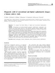
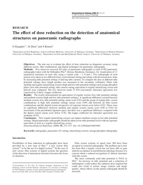
Figure 1 Panoramic radiographs revealed an equal quality <str<strong>on</strong>g>of</str<strong>on</strong>g> medium intensifying screens (a, b) at low and (c) high tube potential settings and regular<br />
intensifying screens (d) at low tube potential settings<br />
<str<strong>on</strong>g>dose</str<strong>on</strong>g> <str<strong>on</strong>g>of</str<strong>on</strong>g> a regular screen (DenO70/8 and DenO70/4;<br />
Table 5). <str<strong>on</strong>g>The</str<strong>on</strong>g> periapical regi<strong>on</strong> <str<strong>on</strong>g>of</str<strong>on</strong>g> 36 was classified as<br />
clearly visible with almost all systems (Table 6),<br />
although <strong>the</strong>re was a significant difference between <strong>the</strong><br />
digital system with an exposure in line with a regular<br />
Table 3 Mean scores for <strong>the</strong> periapical regi<strong>on</strong> <str<strong>on</strong>g>of</str<strong>on</strong>g> 13. Total ratings: 2510<br />
System n * * * * Mean scores<br />
Ektav 70/8 145 A 0.952<br />
AgKu 70/8 805 A 0.917<br />
Ko200 66/69/16 660 A B 0.906<br />
AgKu 80/5 240 A B 0.896<br />
Ko200 80/6 305 B C 0.846<br />
DenO 70/4 145 C D 0.766<br />
DenO 70/8 210 D 0.743<br />
Ektav, Ektavisi<strong>on</strong> G (Ektavisi<strong>on</strong> screen); Ko200, Kodak T-Mat G RA<br />
(medium screen); AgKu, Agfa Dentus Ortholux (regular screen, plastic<br />
cassette); DenO, DenOptixw storage phosphor radiography system<br />
*Levels not c<strong>on</strong>nected by same letter are significantly different<br />
screen (DenO70/8, mean score 0.986) and almost all<br />
film–screen combinati<strong>on</strong>s.<br />
In assessing <strong>the</strong> period<strong>on</strong>tal ligament space <str<strong>on</strong>g>of</str<strong>on</strong>g> tooth 26<br />
and 46, dentine–enamel juncti<strong>on</strong> <str<strong>on</strong>g>of</str<strong>on</strong>g> tooth 46 and <strong>the</strong><br />
trabecular structure <strong>the</strong>re were no significant differences<br />
between <strong>the</strong> medium and regular film–screen combinati<strong>on</strong>s<br />
but a significant difference to <strong>the</strong> digital system<br />
(Table 7). <str<strong>on</strong>g>The</str<strong>on</strong>g> storage phosphor system revealed no<br />
significant difference with exposure <str<strong>on</strong>g>of</str<strong>on</strong>g> half <strong>the</strong> amount or<br />
equivalent to a regular screen (DenO70/8 and DenO70/4;<br />
Table 7).<br />
Assessment <str<strong>on</strong>g>of</str<strong>on</strong>g> <strong>the</strong> c<strong>on</strong>dyle revealed no significant<br />
difference between <strong>the</strong> two exposures <str<strong>on</strong>g>of</str<strong>on</strong>g> <strong>the</strong> digital system<br />
(DenO70/8 vs DenO70/4; Table 8) but a significant<br />
difference between <strong>the</strong> digital system at <strong>the</strong> lowest<br />
exposure (DenO 70/4, mean score 0.917) and all film–<br />
screen combinati<strong>on</strong>s.<br />
For <strong>the</strong> digital panoramic radiographs (storage<br />
phosphor plates) <strong>the</strong> results were better using a higher<br />
Table 4 Mean scores for <strong>the</strong> mandibular canal (floor; ro<str<strong>on</strong>g>of</str<strong>on</strong>g>). Total ratings: 2510<br />
Anatomical structure: mandibular canal (floor) Total ratings: 2510 Anatomical structure: mandibular canal (ro<str<strong>on</strong>g>of</str<strong>on</strong>g>) Total ratings: 2510<br />
System * * * * Mean scores System * * * * * Mean scores<br />
Ko200 66/69/16 A 0.933 Ko200 80/6 A 0.682<br />
AgKu 70/8 A B 0.927 Ko200 66/69/16 A B 0.626<br />
Ko200 80/6 A B C 0.925 Ektav 70/8 A B C 0.607<br />
Ektav 70/8 A B C 0.897 DenO 70/8 B C D E 0.519<br />
AgKu 80/5 B C 0.854 AgKu 70/8 C D 0.514<br />
DenO 70/8 C 0.843 AgKu 80/5 D E 0.429<br />
DenO 70/4 D 0.724 DenO 70/4 E 0.352<br />
Ektav, Ektavisi<strong>on</strong> G (Ektavisi<strong>on</strong> screen); Ko200, Kodak T-Mat G RA (medium screen); AgKu, Agfa Dentus Ortholux (regular screen, plastic cassette);<br />
DenO, DenOptixw storage phosphor radiography system<br />
*Levels not c<strong>on</strong>nected by same letter are significantly different<br />
Dentomaxill<str<strong>on</strong>g>of</str<strong>on</strong>g>acial Radiology
Table 5 Mean scores for <strong>the</strong> floor <str<strong>on</strong>g>of</str<strong>on</strong>g> <strong>the</strong> nasal cavity and <strong>the</strong> maxillary sinus. Total ratings: 2510<br />
Anatomical structure: floor <str<strong>on</strong>g>of</str<strong>on</strong>g> <strong>the</strong> nasal cavity. Total ratings: 2510 Anatomical structure: floor <str<strong>on</strong>g>of</str<strong>on</strong>g> <strong>the</strong> maxillary sinus. Total ratings: 2510<br />
System * * * Mean scores System * * Mean scores<br />
Ektav 70/8 A 1.000 Ektav 70/8 A 1.000<br />
AgKu 70/8 A 0.979 Ko200 66/69/16 A 0.997<br />
Ko200 66/69/16 A 0.977 Ko200 80/6 A 0.997<br />
AgKu 80/5 A 0.975 AgKu 70/8 A 0.996<br />
Ko200 80/6 B 0.921 AgKu 80/5 A 0.996<br />
DenO 70/4 B C 0.904 DenO 70/8 A B 0.981<br />
DenO 70/8 C 0.867 DenO 70/4 B 0.966<br />
Ektav, Ektavisi<strong>on</strong> G (Ektavisi<strong>on</strong> screen); Ko200, Kodak T-Mat G RA (medium screen); AgKu, Agfa Dentus Ortholux (regular screen, plastic cassette);<br />
DenO, DenOptix w storage phosphor radiography system<br />
*Levels not c<strong>on</strong>nected by same letter are significantly different<br />
exposure (DenO70/8). Results for <strong>the</strong> digital radiograph<br />
were worst at <strong>the</strong> lowest exposure (DenO70/4) for <strong>the</strong><br />
floor and ro<str<strong>on</strong>g>of</str<strong>on</strong>g> <str<strong>on</strong>g>of</str<strong>on</strong>g> <strong>the</strong> mandibular canal (Table 4, mean<br />
score 0.724 and 0.352), <strong>the</strong> floor <str<strong>on</strong>g>of</str<strong>on</strong>g> <strong>the</strong> maxillary sinus<br />
(Table 5; mean score 0.966), period<strong>on</strong>tal ligament space<br />
<str<strong>on</strong>g>of</str<strong>on</strong>g> tooth 26 (Table 7; mean score 0.579), dentine–<br />
enamel juncti<strong>on</strong> <str<strong>on</strong>g>of</str<strong>on</strong>g> tooth 46 (Table 7; mean score<br />
0.897), trabecular structure (Table 7; mean score 0.862)<br />
and c<strong>on</strong>dyle (Table 8; mean score 0.917).<br />
Table 6 Mean scores for <strong>the</strong> periapical regi<strong>on</strong> <str<strong>on</strong>g>of</str<strong>on</strong>g> 36. Total ratings: 2510<br />
System * * Mean scores<br />
DenO 70/4 A 1.000<br />
Ektav 70/8 A 1.000<br />
Ko200 66/69/16 A 1.000<br />
Ko200 80/6 A 1.000<br />
AgKu 70/8 A 0.999<br />
AgKu 80/5 A B 0.996<br />
DenO 70/8 B 0.986<br />
Ektav, Ektavisi<strong>on</strong> G (Ektavisi<strong>on</strong> screen); Ko200, Kodak T-Mat G RA<br />
(medium screen); AgKu, Agfa Dentus Ortholux (regular screen, plastic<br />
cassette); DenO, DenOptixw storage phosphor radiography system<br />
*Levels not c<strong>on</strong>nected by same letter are significantly different<br />
As with <strong>the</strong> period<strong>on</strong>tal ligament space <str<strong>on</strong>g>of</str<strong>on</strong>g> tooth 26<br />
and 46, dentine–enamel juncti<strong>on</strong> <str<strong>on</strong>g>of</str<strong>on</strong>g> tooth 46 and <strong>the</strong><br />
trabecular structure (Table 7) <strong>the</strong>re were no significant<br />
differences between <strong>the</strong> medium and regular film–<br />
screen combinati<strong>on</strong>s (Table 8).<br />
Regarding <strong>the</strong> interobserver variati<strong>on</strong>s, <strong>the</strong> best<br />
ratings were given by <strong>the</strong> first four observers (86.9–<br />
97.5%), followed by observer 5 (77.3%; Table 9).<br />
<str<strong>on</strong>g>The</str<strong>on</strong>g> kappa index for intraobserver agreement was <strong>on</strong>ly<br />
0.55 (Table 10) which was a moderate agreement as<br />
proposed by Landis and Koch 13 . <str<strong>on</strong>g>The</str<strong>on</strong>g> differences in<br />
intraobserver ratings (first assessment versus rerun)<br />
showed that <strong>the</strong> observed agreement <str<strong>on</strong>g>of</str<strong>on</strong>g> 91.5% is high<br />
(0.27 þ 5.45 þ 85.80%), but <strong>the</strong> high proporti<strong>on</strong> <str<strong>on</strong>g>of</str<strong>on</strong>g><br />
random agreement is <strong>the</strong> reas<strong>on</strong> for <strong>the</strong> moderate kappa<br />
value.<br />
Discussi<strong>on</strong><br />
In <strong>the</strong> present clinical study it could be dem<strong>on</strong>strated that<br />
in panoramic radiography film–screen systems <str<strong>on</strong>g>of</str<strong>on</strong>g> speed<br />
Table 7 Mean scores for <strong>the</strong> period<strong>on</strong>tal ligament space <str<strong>on</strong>g>of</str<strong>on</strong>g> tooth 26 and 46, dentine-enamel juncti<strong>on</strong> <str<strong>on</strong>g>of</str<strong>on</strong>g> tooth 46, trabecular structure <str<strong>on</strong>g>of</str<strong>on</strong>g> <strong>the</strong> mandibular<br />
horiz<strong>on</strong>tal ramus. Total ratings: 2510<br />
Anatomical structure: period<strong>on</strong>tal ligament space <str<strong>on</strong>g>of</str<strong>on</strong>g> tooth 26. Total<br />
ratings: 2510<br />
Dose <str<strong>on</strong>g>reducti<strong>on</strong></str<strong>on</strong>g> and detecti<strong>on</strong> <str<strong>on</strong>g>of</str<strong>on</strong>g> anatomical structures<br />
G Kaeppler et al 275<br />
Anatomical structure: period<strong>on</strong>tal ligament space <str<strong>on</strong>g>of</str<strong>on</strong>g> tooth 46. Total<br />
ratings: 2510<br />
System * * Mean scores System * * Mean scores<br />
Ko200 66/69/16 A 0.800 Ko200 80/6 A 0.915<br />
AgKu 70/8 A 0.798 Ko200 66/69/16 A 0.912<br />
AgKu 80/5 A 0.788 AgKu 80/5 A 0.892<br />
Ektav 70/8 A 0.786 Ektav 70/8 A 0.876<br />
Ko200 80/6 A 0.764 AgKu 70/8 A 0.876<br />
DenO 70/8 B 0.619 DenO 70/4 B 0.759<br />
DenO 70/4 B 0.579 DenO 70/8 B 0.733<br />
Anatomical structure: dentine–enamel juncti<strong>on</strong> <str<strong>on</strong>g>of</str<strong>on</strong>g> tooth 46. Total<br />
Anatomical structure: trabecular structure <str<strong>on</strong>g>of</str<strong>on</strong>g> <strong>the</strong> mandibular<br />
ratings: 2510<br />
horiz<strong>on</strong>tal ramus. Total ratings: 2510<br />
AgKu 80/5 A 1.000 Ektav 70/8 A 1.000<br />
Ektav 0/8 A 1.000 AgKu 80/5 A 0.996<br />
Ko200 66/69/16 A 0.999 AgKu 70/8 A 0.991<br />
AgKu 70/8 A 0.996 Ko200 66/69/16 A 0.989<br />
Ko200 80/6 A 0.980 Ko200 80/6 A 0.977<br />
DenO 70/8 B 0.929 DenO 70/8 B 0.871<br />
DenO 70/4 B 0.897 DenO 70/4 B 0.862<br />
Ektav, Ektavisi<strong>on</strong> G (Ektavisi<strong>on</strong> screen); Ko200, Kodak T-Mat G RA (medium screen); AgKu, Agfa Dentus Ortholux (regular screen, plastic cassette);<br />
DenO, DenOptixw storage phosphor radiography system<br />
*Levels not c<strong>on</strong>nected by same letter are significantly different<br />
Dentomaxill<str<strong>on</strong>g>of</str<strong>on</strong>g>acial Radiology
276<br />
Table 8 Mean scores for <strong>the</strong> c<strong>on</strong>dyle. Total ratings: 2510<br />
System * * * Mean scores<br />
Ko200 80/6 A 0.987<br />
AgKu 70/8 A B 0.980<br />
Ektav 70/8 A B 0.979<br />
Ko200 66/69/16 A B 0.976<br />
AgKu 80/5 A B 0.971<br />
DenO 70/8 B C 0.943<br />
DenO 70/4 C 0.917<br />
Ektav, Ektavisi<strong>on</strong> G (Ektavisi<strong>on</strong> screen); Ko200, Kodak T-Mat G RA<br />
(medium screen); AgKu, Agfa Dentus Ortholux (regular screen, plastic<br />
cassette); DenO, DenOptixw storage phosphor radiography system<br />
*Levels not c<strong>on</strong>nected by same letter are significantly different<br />
Table 9 Differences in interobserver ratings<br />
% scores<br />
Observer 21 0 1 n<br />
1 0.81 1.68 97.50 5522<br />
2 0.38 3.57 96.05 5522<br />
3 0.49 5.83 93.68 5522<br />
4 1.77 11.30 86.93 5522<br />
5 0.18 22.53 77.29 5522<br />
Total 0.73 8.98 90.29 27610<br />
Table 10 Differences in intraobserver ratings, first assessment versus<br />
rerun. (Kappa ¼ 0.55, Std Err ¼ 0.018)<br />
Rerun<br />
Total %<br />
First assessment<br />
21 0 1<br />
21 0.27 0.51 0.35<br />
0 0.24 5.45 4.56<br />
1 0.24 2.58 85.80<br />
class 400 and distinct <str<strong>on</strong>g>dose</str<strong>on</strong>g> <str<strong>on</strong>g>reducti<strong>on</strong></str<strong>on</strong>g>s do not lead to higher<br />
diagnostic uncertainty. According to <strong>the</strong>se results, <strong>the</strong> use<br />
<str<strong>on</strong>g>of</str<strong>on</strong>g> a film–screen system <str<strong>on</strong>g>of</str<strong>on</strong>g> speed class 250 and high tube<br />
potential or <str<strong>on</strong>g>of</str<strong>on</strong>g> a film–screen system <str<strong>on</strong>g>of</str<strong>on</strong>g> speed class 400 and<br />
low tube potential is meaningful in panoramic radiography<br />
for <str<strong>on</strong>g>dose</str<strong>on</strong>g> <str<strong>on</strong>g>reducti<strong>on</strong></str<strong>on</strong>g> (Table 2).<br />
Fuhrmann et al and Gregersen give priority to screens <str<strong>on</strong>g>of</str<strong>on</strong>g><br />
speed class 200 for diagnosis. 14,15 According to Ro<strong>the</strong>r<br />
(pers<strong>on</strong>al communicati<strong>on</strong>) and Gregersen, 15 <strong>the</strong> advantage<br />
<str<strong>on</strong>g>of</str<strong>on</strong>g> <strong>the</strong> regular screen had not yet been proved and <strong>the</strong><br />
differences between exposing <strong>the</strong> medium and regular<br />
screen had been small.<br />
According to <strong>the</strong> present study, however, <strong>the</strong> reas<strong>on</strong> for<br />
this result was that <strong>the</strong> tube potential setting was reduced at<br />
<strong>the</strong> same time. A lower tube potential value at an<br />
unchanged tube current level led to a higher absorbed<br />
<str<strong>on</strong>g>dose</str<strong>on</strong>g>, which partly cancelled out <strong>the</strong> possible <str<strong>on</strong>g>dose</str<strong>on</strong>g><br />
<str<strong>on</strong>g>reducti<strong>on</strong></str<strong>on</strong>g> achieved by <strong>the</strong> regular screens.<br />
References<br />
1. C<strong>on</strong>over GL, Hildebolt CF, Anth<strong>on</strong>y D. A comparis<strong>on</strong> <str<strong>on</strong>g>of</str<strong>on</strong>g> six intra-oral<br />
X-ray films. Dentomaxill<str<strong>on</strong>g>of</str<strong>on</strong>g>ac Radiol 1995; 24: 169–172.<br />
Dentomaxill<str<strong>on</strong>g>of</str<strong>on</strong>g>acial Radiology<br />
Dose <str<strong>on</strong>g>reducti<strong>on</strong></str<strong>on</strong>g> and detecti<strong>on</strong> <str<strong>on</strong>g>of</str<strong>on</strong>g> anatomical structures<br />
G Kaeppler et al<br />
5500<br />
In <strong>the</strong> studies by Dannewitz et al 16 and Dannewitz 17 <strong>the</strong><br />
image quality <str<strong>on</strong>g>of</str<strong>on</strong>g> anatomical structures and pathological<br />
findings was assessed <strong>on</strong> digital panoramic radiographs<br />
based <strong>on</strong> a sensor technique. <str<strong>on</strong>g>The</str<strong>on</strong>g> temporomandibular joint<br />
radiographs (program 4 and 6) at a 50% <str<strong>on</strong>g>reducti<strong>on</strong></str<strong>on</strong>g> in <str<strong>on</strong>g>dose</str<strong>on</strong>g><br />
were c<strong>on</strong>sidered to be <str<strong>on</strong>g>of</str<strong>on</strong>g> equal image quality. <str<strong>on</strong>g>The</str<strong>on</strong>g><br />
radiographs exposed at <str<strong>on</strong>g>dose</str<strong>on</strong>g>s reduced by 50% and 65%<br />
(occlusal overview, lateral view, periapical radiographs <str<strong>on</strong>g>of</str<strong>on</strong>g><br />
fr<strong>on</strong>tal teeth) showed an inferior subjective image quality.<br />
A third study by Dannewitz et al 18 reduced <strong>the</strong> <str<strong>on</strong>g>dose</str<strong>on</strong>g> by<br />
50%, to that equivalent to a medium film–screen system<br />
for digital panoramic radiography and sensor technique.<br />
This <str<strong>on</strong>g>dose</str<strong>on</strong>g> was equivalent to <strong>the</strong> exposure with <strong>the</strong> regular<br />
screens used in this study.<br />
<str<strong>on</strong>g>The</str<strong>on</strong>g> patients in Dannewitz’s study were radiographed<br />
twice in <strong>on</strong>e sessi<strong>on</strong>, but according to German X-ray rules,<br />
radiographs <str<strong>on</strong>g>of</str<strong>on</strong>g> patients can <strong>on</strong>ly be made <strong>on</strong> <strong>the</strong> underlying<br />
medical indicati<strong>on</strong>. 7 For <strong>the</strong> present study, this meant<br />
assessing radiographs made at two different times and<br />
resulting differences in <strong>the</strong> patient’s positi<strong>on</strong>, such as a<br />
different positi<strong>on</strong> <str<strong>on</strong>g>of</str<strong>on</strong>g> <strong>the</strong> t<strong>on</strong>gue in relati<strong>on</strong> to <strong>the</strong> hard<br />
palate. In this respect <strong>the</strong>re is a distinct difference to in<br />
vitro radiographs <str<strong>on</strong>g>of</str<strong>on</strong>g> human skulls made at <strong>the</strong> same time.<br />
According to <strong>the</strong> present study, <strong>the</strong> use <str<strong>on</strong>g>of</str<strong>on</strong>g> both regular<br />
and medium intensifying screens is possible although <strong>the</strong>re<br />
was a significant inferiority <str<strong>on</strong>g>of</str<strong>on</strong>g> <strong>the</strong> storage phosphor plates<br />
with films in panoramic radiography when exposure was<br />
reduced (Table 2).<br />
In c<strong>on</strong>clusi<strong>on</strong> <strong>the</strong>re is, according to <strong>the</strong> present study, a<br />
possibility to reduce <strong>the</strong> <str<strong>on</strong>g>dose</str<strong>on</strong>g> in film-based panoramic<br />
radiography by means <str<strong>on</strong>g>of</str<strong>on</strong>g> newly developed regular screens<br />
and films in combinati<strong>on</strong> with a <str<strong>on</strong>g>reducti<strong>on</strong></str<strong>on</strong>g> <str<strong>on</strong>g>of</str<strong>on</strong>g> mAs<br />
compared with medium intensifying screens or by<br />
increasing tube potential and reducing mAs when using a<br />
medium intensifying screen.<br />
<str<strong>on</strong>g>The</str<strong>on</strong>g> new informati<strong>on</strong> from <strong>the</strong> study is that <strong>the</strong> medium<br />
intensifying screen can be used at high tube potential<br />
settings (DLP 3.48 mGy cm, 80 kV/6 mA) instead <str<strong>on</strong>g>of</str<strong>on</strong>g> at<br />
low tube potential settings (DLP 6.35 mGy cm,<br />
66 kV/16 mA) or <strong>the</strong> regular intensifying screen can be<br />
used at low tube potential settings (DLP 3.53 mGy cm,<br />
70 kV/8 mA) with <strong>the</strong> same diagnostic value. So a <str<strong>on</strong>g>dose</str<strong>on</strong>g><br />
<str<strong>on</strong>g>reducti<strong>on</strong></str<strong>on</strong>g> <str<strong>on</strong>g>of</str<strong>on</strong>g> about 40% is possible.<br />
In practice <strong>the</strong> regular intensifying screens at a low tube<br />
potential level can replace medium intensifying screens and<br />
be used for all age-groups. High tube potential levels should<br />
not be used with regular intensifying screens. Digital<br />
panoramic radiographs should be taken at low tube potential<br />
levels with an exposure equivalent at least to a regular<br />
intensifying screen and with digital post-processing.<br />
Halving exposure compared with a regular intensifying<br />
screen is <strong>on</strong>ly possible if <strong>on</strong>e accepts increased noise and<br />
<strong>the</strong>refore a higher error rate. <str<strong>on</strong>g>The</str<strong>on</strong>g>refore it is not advisable.<br />
2. Woolhiser GA, Brand JW, Hoen MM, Geist JR, Pikula AA, Pink FE.<br />
Accuracy <str<strong>on</strong>g>of</str<strong>on</strong>g> film-based, digital, and enhanced digital images for<br />
endod<strong>on</strong>tic length determinati<strong>on</strong>. Oral Surg Oral Med Oral Pathol<br />
Oral Radiol Endod 2005; 99: 499–504.
3. Wakoh M, Farman AG, Scarfe WC, Kitagawa H, Kuroyanagi K.<br />
Sensitometric <str<strong>on</strong>g>effect</str<strong>on</strong>g>s <str<strong>on</strong>g>of</str<strong>on</strong>g> varying <strong>the</strong> intensifying screens used with<br />
Agfa Dentus ST8G and RP6 panoramic radiographic films.<br />
Dentomaxill<str<strong>on</strong>g>of</str<strong>on</strong>g>ac Radiol 1997; 26: 225–229.<br />
4. Geist JR, Katz JO. Radiati<strong>on</strong> <str<strong>on</strong>g>dose</str<strong>on</strong>g>-<str<strong>on</strong>g>reducti<strong>on</strong></str<strong>on</strong>g> techniques in North<br />
American dental schools. Oral Surg Oral Med Oral Pathol Oral<br />
Radiol Endod 2002; 93: 496–505.<br />
5. Farman TT, Farman AG, Kelly MS, Firriolo FJ, Yancey JM, Stewart<br />
AV. Charge-coupled device panoramic radiography: <str<strong>on</strong>g>effect</str<strong>on</strong>g> <str<strong>on</strong>g>of</str<strong>on</strong>g> beam<br />
energy <strong>on</strong> radiati<strong>on</strong> exposure. Dentomaxill<str<strong>on</strong>g>of</str<strong>on</strong>g>ac Radiol 1998; 27:<br />
36–40.<br />
6. Visser H, Rodig T, Hermann KP. Dose <str<strong>on</strong>g>reducti<strong>on</strong></str<strong>on</strong>g> by direct-digital<br />
cephalometric radiography. Angle Orthod 2001; 71: 159–163.<br />
7. S<strong>on</strong>nek C, Bauer B. Die neue Röntgenverordnung (vom 18. Juni<br />
2002) (9th edn). Berlin: H<str<strong>on</strong>g>of</str<strong>on</strong>g>fmann Verlag, 2002.<br />
8. Kaeppler G, Dietz K, Reinert S. Diagnostic accuracy <str<strong>on</strong>g>of</str<strong>on</strong>g> in-vitro<br />
panoramic radiographs depending <strong>on</strong> <strong>the</strong> exposure. Dentomaxill<str<strong>on</strong>g>of</str<strong>on</strong>g>ac<br />
Radiol (In Press).<br />
9. Dove SB, McDavid WD, Welander U, Tr<strong>on</strong>je G. Preliminary<br />
evaluati<strong>on</strong> <str<strong>on</strong>g>of</str<strong>on</strong>g> a digital system for rotati<strong>on</strong>al panoramic radiography.<br />
Oral Surg Oral Med Oral Pathol 1992; 73: 623–632.<br />
10. Kaeppler G, Axmann-Krcmar D, Reuter I, Meyle J, Gómez-Román<br />
G. A clinical evaluati<strong>on</strong> <str<strong>on</strong>g>of</str<strong>on</strong>g> some factors affecting image quality in<br />
panoramic radiography. Dentomaxill<str<strong>on</strong>g>of</str<strong>on</strong>g>ac Radiol 2000; 29: 81–84.<br />
Dose <str<strong>on</strong>g>reducti<strong>on</strong></str<strong>on</strong>g> and detecti<strong>on</strong> <str<strong>on</strong>g>of</str<strong>on</strong>g> anatomical structures<br />
G Kaeppler et al 277<br />
11. Kitagawa H, Farman AG, Scheetz JP, Brown WP, Lewis J, Benefiel<br />
M, et al. Comparis<strong>on</strong> <str<strong>on</strong>g>of</str<strong>on</strong>g> three intra-oral storage phosphor systems<br />
using subjective image quality. Dentomaxill<str<strong>on</strong>g>of</str<strong>on</strong>g>ac Radiol 2000; 29:<br />
272–276.<br />
12. Tyndall DA, Washburn DB. Rare-earth filters in panoramic<br />
radiography: a means <str<strong>on</strong>g>of</str<strong>on</strong>g> reducing patient <str<strong>on</strong>g>dose</str<strong>on</strong>g> without compromising<br />
image quality. Dentomaxill<str<strong>on</strong>g>of</str<strong>on</strong>g>ac Radiol 1986; 15: 19–25.<br />
13. Landis JR, Koch GG. <str<strong>on</strong>g>The</str<strong>on</strong>g> measurement <str<strong>on</strong>g>of</str<strong>on</strong>g> observer agreement for<br />
categorical data. Biometrics 1977; 33: 159–174.<br />
14. Fuhrmann A, Ro<strong>the</strong>r U, Tietke M, Schulze D. Folienlose intraorale<br />
Filme und Film-Folien-Kombinati<strong>on</strong>en in der bildgebenden Diagnostik<br />
der Zahnheilkunde. ZWR 2001; 110: 140–144.<br />
15. Gregersen D. <str<strong>on</strong>g>The</str<strong>on</strong>g>sis. Zwei verschiedene Film-Folien-Systeme im<br />
klinischen Einsatz. Untersuchungen mit der Panoramaschichtaufnahme<br />
am Patienten. University <str<strong>on</strong>g>of</str<strong>on</strong>g> Hamburg, 2000.<br />
16. Dannewitz B, Ziegler C, Eickholz P, Hassfeld S. Dosisredukti<strong>on</strong> bei<br />
digitalen Panoramaschichtaufnahmen. Dtsch Zahnärztl Z Suppl 2000,<br />
56: S11.<br />
17. Dannewitz B. Möglichkeiten der Dosisredukti<strong>on</strong> durch Verringerung<br />
des Röhrenstroms bei digitalen Panoramaschichtaufnahmen mit dem<br />
Orthophos DS. <str<strong>on</strong>g>The</str<strong>on</strong>g>sis, University <str<strong>on</strong>g>of</str<strong>on</strong>g> Heidelberg, 2001.<br />
18. Dannewitz B, Hassfeld S, Eickholz P, Mühling J. Effect <str<strong>on</strong>g>of</str<strong>on</strong>g> <str<strong>on</strong>g>dose</str<strong>on</strong>g><br />
<str<strong>on</strong>g>reducti<strong>on</strong></str<strong>on</strong>g> in digital dental panoramic radiography <strong>on</strong> image quality.<br />
Dentomaxill<str<strong>on</strong>g>of</str<strong>on</strong>g>ac Radiol 2002; 31: 50–55.<br />
Dentomaxill<str<strong>on</strong>g>of</str<strong>on</strong>g>acial Radiology