

Elemental bio-imaging of calcium phosphate crystal deposits in ...
Elemental bio-imaging of calcium phosphate crystal deposits in ...
Elemental bio-imaging of calcium phosphate crystal deposits in ...

Create successful ePaper yourself
Turn your PDF publications into a flip-book with our unique Google optimized e-Paper software.
Metallomics<br />
Integrated <strong>bio</strong>metal science<br />
www.rsc.org/metallomics Volume 1 | Number 2 | March 2009 | Pages 117–176<br />
ISSN 1756-5901<br />
PAPER<br />
Doble et al.<br />
<strong>Elemental</strong> <strong>bio</strong>-<strong>imag<strong>in</strong>g</strong> <strong>of</strong> <strong>calcium</strong><br />
<strong>phosphate</strong> <strong>crystal</strong> <strong>deposits</strong> <strong>in</strong> knee<br />
samples from arthritic patients<br />
MINIREVIEW<br />
Ortega<br />
Synchrotron radiation for direct<br />
analysis <strong>of</strong> metalloprote<strong>in</strong>s on<br />
electrophoresis gels<br />
View Onl<strong>in</strong>e<br />
1756-5901(2009)1:2;1-5
Downloaded on 19 February 2011<br />
Published on 13 February 2009 on http://pubs.rsc.org | doi:10.1039/B901310P<br />
PAPER www.rsc.org/metallomics | Metallomics<br />
<strong>Elemental</strong> <strong>bio</strong>-<strong>imag<strong>in</strong>g</strong> <strong>of</strong> <strong>calcium</strong> <strong>phosphate</strong> <strong>crystal</strong> <strong>deposits</strong> <strong>in</strong> knee<br />
samples from arthritic patients<br />
Christ<strong>in</strong>e Aust<strong>in</strong>, a Dom<strong>in</strong>ic Hare, a Andrew L. Rozelle, b William H. Rob<strong>in</strong>son, b<br />
Rudolf Grimm c and Philip Doble* a<br />
Received 20th January 2009, Accepted 20th January 2009<br />
First published as an Advance Article on the web 13th February 2009<br />
DOI: 10.1039/b901310p<br />
Laser ablation <strong>in</strong>ductively coupled plasma mass spectrometry (LA ICP-MS) was employed to<br />
image <strong>deposits</strong> <strong>of</strong> <strong>calcium</strong> <strong>phosphate</strong> based <strong>crystal</strong>s <strong>in</strong> knee cartilage and synovial fluid from<br />
arthritic patients. A reaction/collision cell conta<strong>in</strong><strong>in</strong>g hydrogen m<strong>in</strong>imised plasma <strong>in</strong>terferences on<br />
<strong>calcium</strong> and also improved the image quality without significant sensitivity reduction. Areas <strong>of</strong><br />
high <strong>calcium</strong> and phosphorus <strong>in</strong>tensities consistent with <strong>crystal</strong> <strong>deposits</strong> were observed for both<br />
the cartilage and synovial fluid samples. These areas were also characterised by high magnesium<br />
and strontium <strong>in</strong>tensities. Distribution patterns <strong>of</strong> other elements such as copper and sulfur did<br />
not correlate with the <strong>crystal</strong> <strong>deposits</strong>. Filtered and non-filtered solutions <strong>of</strong> <strong>calcium</strong> <strong>phosphate</strong><br />
<strong>crystal</strong>s grown <strong>in</strong> synthetic synovial fluid were also imaged as further evidence <strong>of</strong> <strong>crystal</strong> <strong>deposits</strong>.<br />
The <strong>crystal</strong> <strong>deposits</strong> were detected <strong>in</strong> the unfiltered solution, and were absent from the filtered<br />
solutions.<br />
Introduction<br />
The importance <strong>of</strong> <strong>calcium</strong> pyro<strong>phosphate</strong> dihydrate (CPPD)<br />
and basic <strong>calcium</strong> <strong>phosphate</strong>s (BCP) <strong>in</strong> rheumatology was first<br />
discovered <strong>in</strong> the early 1960s. 1 S<strong>in</strong>ce then, the concurrence <strong>of</strong><br />
BCP (hydroxyapatite, octa<strong>calcium</strong> <strong>phosphate</strong>, tri<strong>calcium</strong><br />
<strong>phosphate</strong>) and CPPD <strong>crystal</strong>s, and degenerative jo<strong>in</strong>t disease<br />
has been well established. Though there is ample data<br />
available to support the role <strong>of</strong> BCP <strong>crystal</strong>s <strong>in</strong> cartilage<br />
degeneration, it is still unclear whether other <strong>calcium</strong>-conta<strong>in</strong><strong>in</strong>g<br />
<strong>crystal</strong>s play a direct driv<strong>in</strong>g role <strong>in</strong> disease conception and<br />
progression, or are merely markers <strong>of</strong> jo<strong>in</strong>t damage. 2–4<br />
The identification <strong>of</strong> specific <strong>crystal</strong> types <strong>in</strong> synovial fluid is<br />
currently the most popular method <strong>of</strong> diagnos<strong>in</strong>g all common<br />
forms <strong>of</strong> <strong>crystal</strong>-associated arthritis. 3 In general, <strong>crystal</strong><br />
arthritides cannot be dist<strong>in</strong>guished from other causes <strong>of</strong><br />
arthritis based on history or physical exam<strong>in</strong>ation. 5 Despite<br />
the multitude <strong>of</strong> techniques that have been applied to BCP and<br />
CPPD <strong>crystal</strong> detection, cl<strong>in</strong>ical diagnosis almost exclusively<br />
relies on light microscopy (LM). However, LM lacks the<br />
resolution required to image small <strong>crystal</strong>s (o1 mm), is nonspecific<br />
(though CPPD <strong>crystal</strong>s can generally be dist<strong>in</strong>guished<br />
under polarised LM) and accuracy is operator dependent. 3,5–7<br />
Sensitivity can be improved by sta<strong>in</strong><strong>in</strong>g the sample with<br />
<strong>calcium</strong> specific dyes, particularly for BCP <strong>crystal</strong>s, which<br />
are <strong>of</strong>ten too small to be seen by LM. However these are<br />
<strong>of</strong>ten subject to false positives.<br />
a <strong>Elemental</strong> Bio-<strong>imag<strong>in</strong>g</strong> Facility, Department <strong>of</strong> Chemistry, Materials<br />
and Forensic Science, University <strong>of</strong> Technology Sydney,<br />
Sydney NSW 2007, Australia. E-mail: philip.doble@uts.edu.au;<br />
Fax: +61 2 9514 1460; Tel: +61 2 9514 1792<br />
b Division <strong>of</strong> Immunology and Rheumatology, Stanford University<br />
School <strong>of</strong> Medic<strong>in</strong>e, 300 Pasteur Drive, Stanford,<br />
California 94305-5166, USA<br />
c Agilent Technologies, Inc., Integrated Biology Solutions Unit,<br />
Santa Clara, California, USA<br />
Other techniques for the determ<strong>in</strong>ation <strong>of</strong> BCP <strong>crystal</strong>s<br />
<strong>in</strong>clude scann<strong>in</strong>g electron microscopy (SEM) and transmission<br />
electron microscopy (TEM). These methods are expensive,<br />
complex and not widely available for rout<strong>in</strong>e analysis <strong>in</strong> a<br />
cl<strong>in</strong>ical sett<strong>in</strong>g. Techniques recently applied to <strong>crystal</strong> detection<br />
and identification <strong>in</strong>clude atomic force microscopy (AFM),<br />
Fourier-transform <strong>in</strong>frared spectroscopy (FTIR) and Raman<br />
spectroscopy. AFM and Raman show great potential for this<br />
application. Additionally, FTIR and Raman spectrometers<br />
may be made compact, mak<strong>in</strong>g the technology capable <strong>of</strong><br />
po<strong>in</strong>t-<strong>of</strong>-care operation. 3 A comprehensive review <strong>of</strong> the<br />
analytical tools available for the detection and identification<br />
<strong>of</strong> <strong>calcium</strong> <strong>phosphate</strong> <strong>crystal</strong>s was recently published by<br />
Yavorskyy et al. 3 The review made it evident that a simple,<br />
<strong>in</strong>expensive technique which is capable <strong>of</strong> detect<strong>in</strong>g and identify<strong>in</strong>g<br />
<strong>calcium</strong> <strong>phosphate</strong>-based <strong>crystal</strong>s accurately and with a<br />
high sensitivity would be <strong>in</strong>valuable <strong>in</strong> the diagnosis <strong>of</strong> <strong>crystal</strong><br />
associated arthritides.<br />
Laser ablation <strong>in</strong>ductively coupled plasma mass spectrometry<br />
(LA ICP-MS) is <strong>in</strong>creas<strong>in</strong>gly applied to the study <strong>of</strong><br />
metals <strong>in</strong> <strong>bio</strong>logical samples. 8,9 LA ICP-MS <strong>of</strong>fers direct<br />
multielemental analysis <strong>of</strong> solids and liquids with trace and<br />
ultra trace detection capabilities, and a l<strong>in</strong>ear dynamic range<br />
<strong>of</strong> seven or more orders <strong>of</strong> magnitude. Though its application<br />
to <strong>imag<strong>in</strong>g</strong> <strong>of</strong> s<strong>of</strong>t tissues is emerg<strong>in</strong>g, it is considered a mature<br />
technique for the analysis <strong>of</strong> geological and metallurgical<br />
samples. 10 In LA ICP-MS systems, a focused laser beam is<br />
used to mobilise sample material as droplets or vapour from<br />
the sample surface. 9 The material is then transported to the<br />
plasma (usually by argon carrier gas) where the material is<br />
ionised and carried through to the mass spectrometer, which<br />
selectively detects ions at a given mass-to-charge ratio.<br />
LA ICP-MS may be utilised to construct maps that provide<br />
elemental spatial <strong>in</strong>formation at trace and bulk levels<br />
<strong>of</strong> <strong>bio</strong>logical samples such as small tumours <strong>in</strong> rat bra<strong>in</strong>s, 11<br />
142 | Metallomics, 2009, 1, 142–147 This journal is c The Royal Society <strong>of</strong> Chemistry 2009
Downloaded on 19 February 2011<br />
Published on 13 February 2009 on http://pubs.rsc.org | doi:10.1039/B901310P<br />
Table 1 Optimised experimental parameters for LA ICP-MS analysis<br />
Agilent 7500ce ICP-MS New wave UP213 laser ablation<br />
Rf power 1370 W Wavelength 213 nm<br />
Cool<strong>in</strong>g gas flow rate 15 l m<strong>in</strong> 1<br />
Repetition frequency 20 Hz<br />
Carrier gas flow rate 1.25 l m<strong>in</strong> 1<br />
Laser energy density 0.2 J cm 2 (at 30%)<br />
Hydrogen flow rate 2.5 ml m<strong>in</strong> 1<br />
Cartilage samples<br />
Sample depth 5.0 mm Spot size 55 mm<br />
QP bias 16 V Scan rate 30 mm s 1<br />
OctP bias 20 V Synovial fluid<br />
Scan mode Peak hopp<strong>in</strong>g Spot size 100 mm<br />
Dwell time 0.1 s, 0.2 s for P Scan rate 40 mm s 1<br />
Synthetic synovial fluid<br />
Spot size 55 mm<br />
Scan rate 50 mm s 1<br />
b-amyloid plaques <strong>in</strong> mouse models <strong>of</strong> Alzheimer’s disease, 12<br />
and the distribution <strong>of</strong> plat<strong>in</strong>um <strong>in</strong> mouse kidneys treated with<br />
cis-plat<strong>in</strong>. 13 The environmental sciences have also utilised the<br />
potential <strong>of</strong> this technique to analyse the environmental<br />
changes or pollutant accumulation evident from the chang<strong>in</strong>g<br />
metal concentrations <strong>in</strong> <strong>in</strong>crement growth layers <strong>of</strong> many types<br />
<strong>of</strong> organisms such as scales, shells, otoliths and tree xylem. 14<br />
Recently, Chaudhri et al., 15 determ<strong>in</strong>ed the elemental<br />
distribution <strong>of</strong> bladder and kidney stones by LA ICP-MS.<br />
LA ICP-MS has also been employed to study element time<br />
pr<strong>of</strong>iles <strong>in</strong> teeth 16 and bones, 17 and to determ<strong>in</strong>e strontium<br />
ratios <strong>in</strong> <strong>calcium</strong> <strong>phosphate</strong>s. 18 Quantitative analysis <strong>of</strong> heavy<br />
metals <strong>in</strong> bone employ<strong>in</strong>g reference standards and spik<strong>in</strong>g<br />
hydroxapatite standards has also been reported. 17,19,20<br />
However, LA ICP-MS has not been used to image <strong>crystal</strong><br />
<strong>deposits</strong> <strong>in</strong> <strong>bio</strong>logical samples. Element concentrations <strong>in</strong><br />
synovial fluids from osteoarthritic patients have been determ<strong>in</strong>ed<br />
by ICP-MS. 21<br />
This study demonstrates the potential <strong>of</strong> LA ICP-MS for<br />
<strong>crystal</strong> detection <strong>in</strong> <strong>bio</strong>logical samples. Cartilage sections were<br />
obta<strong>in</strong>ed from knee or hip arthroplasty and were imaged by<br />
LA ICP-MS for the presence <strong>of</strong> <strong>crystal</strong>-associated elements.<br />
Additionally, eight synovial fluid samples, six from patients<br />
diagnosed with osteoarthritis (OA) and two from patients<br />
diagnosed with rheumatoid arthritis (RA), were analysed by<br />
the same method. Results from each sample type are presented<br />
here. We propose that this method <strong>of</strong> analysis be termed<br />
‘‘<strong>Elemental</strong> Bio<strong>imag<strong>in</strong>g</strong>’’.<br />
Experimental<br />
Instrumental and measurement procedure<br />
A quadropole 7500ce ICP-MS (Agilent Technologies) with<br />
octopole collision/reaction cell and cs lenses <strong>in</strong>stalled was<br />
coupled to a New Wave UP213 laser ablation unit for the<br />
generation <strong>of</strong> elemental images <strong>of</strong> <strong>bio</strong>logical samples from<br />
arthritic patients.<br />
Laser ablation <strong>of</strong> <strong>bio</strong>logical samples was performed us<strong>in</strong>g a<br />
frequency qu<strong>in</strong>tupled Nd:YAG laser with ablated material<br />
transported to the ICP-MS via a stream <strong>of</strong> argon carrier gas.<br />
The ICP-MS was operated <strong>in</strong> reaction gas mode to reduce<br />
the <strong>in</strong>terferences on <strong>calcium</strong> and strontium isotopes. The<br />
optimised experimental parameters for LA ICP-MS analysis<br />
are recorded <strong>in</strong> Table 1. Cartilage samples (generally less than<br />
10 10 mm) were imaged <strong>in</strong> approximately 20 h, whilst<br />
synovial fluid took approximately 2.5 h to image.<br />
Imag<strong>in</strong>g was performed us<strong>in</strong>g the s<strong>of</strong>tware package ENVI<br />
v4.2 (Research Systems Inc., Boulder, CO, USA). The samples<br />
were systematically scanned across the entire area <strong>of</strong> the<br />
sample. For cartilage samples, the distance between adjacent<br />
scan l<strong>in</strong>es was 29 mm, which ensured complete ablation <strong>of</strong> the<br />
tissue sample. For the synovial spots, the l<strong>in</strong>e scans were<br />
placed 50 mm apart. The data files generated by ChemStation<br />
(Agilent Technologies) were then collected and imported <strong>in</strong>to<br />
ENVI. As no quantification data was collected, images only<br />
show relative signal <strong>in</strong>tensities.<br />
Sample preparation<br />
All experiments were performed <strong>in</strong> compliance with the relevant<br />
laws and <strong>in</strong>stitutional guidel<strong>in</strong>es <strong>of</strong> the University <strong>of</strong> Technology<br />
Sydney and Stanford University. The study was approved by the<br />
ethics committees <strong>of</strong> the University <strong>of</strong> Technology Sydney and<br />
Stanford University. Human samples were collected at Stanford<br />
University after <strong>in</strong>formed consent and under Institutional<br />
Review Board (IRB)-approved protocols. Cartilage sections were<br />
obta<strong>in</strong>ed after either knee or hip arthroplasty. Sections were cut<br />
to a thickness <strong>of</strong> 14 mm and then dried. Synovial fluid was<br />
aspirated from <strong>in</strong>flamed knee jo<strong>in</strong>ts, centrifuged at 2000 rpm and<br />
the supernatant collected and frozen, with<strong>in</strong> 4 hours <strong>of</strong> collection,<br />
for storage. Synovial fluid was dropped (12 ml) onto quartz cover<br />
slips and dried for analysis by LA ICP-MS.<br />
Synthetic synovial fluid was prepared from human serum<br />
(Sigma, Germany) by mix<strong>in</strong>g equal volumes (30%) <strong>of</strong> laboratory<br />
grade (Ajax, Sydney) 10 mM <strong>calcium</strong> chloride, 50 mM<br />
tetrasodium pyro<strong>phosphate</strong> and 7 mM magnesium sulfate<br />
accord<strong>in</strong>g to a method reported by Cheng et al. for <strong>crystal</strong><br />
formation <strong>of</strong> CPPD <strong>in</strong> aqueous solutions. 22,23 Tris(hydroxymethyl)methylam<strong>in</strong>e<br />
(Ajax, Sydney) was added to ma<strong>in</strong>ta<strong>in</strong> the<br />
pH around physiological levels. The <strong>calcium</strong>, magnesium and<br />
pyro<strong>phosphate</strong> salts were chosen based on the assumption that<br />
their counter ions would not form precipitates <strong>in</strong> the synthetic<br />
synovial solution. The synthetic synovial fluid was then left <strong>in</strong> the<br />
oven at 37 1C for 2 weeks to encourage <strong>crystal</strong> formation. A<br />
disposable 0.22 mm syr<strong>in</strong>ge filter was used to remove any large<br />
particles from solution. Both filtered and non-filtered solutions<br />
were dropped (5 ml) onto a clean quartz slide and air dried before<br />
analysis by LA ICP-MS.<br />
This journal is c The Royal Society <strong>of</strong> Chemistry 2009 Metallomics, 2009, 1, 142–147 | 143
Downloaded on 19 February 2011<br />
Published on 13 February 2009 on http://pubs.rsc.org | doi:10.1039/B901310P<br />
Results and discussion<br />
Imag<strong>in</strong>g <strong>of</strong> cartilage samples<br />
Cartilage exam<strong>in</strong>ation for <strong>crystal</strong>s can provide important<br />
<strong>in</strong>formation regard<strong>in</strong>g the progression <strong>of</strong> the disease. For<br />
example, the density <strong>of</strong> <strong>crystal</strong>s has been shown to correlate with<br />
the degree <strong>of</strong> mechanical stress exerted on the jo<strong>in</strong>t <strong>in</strong>volved. 2<br />
Initial experiments attempted to generate images with the<br />
reaction cell operat<strong>in</strong>g only as an ion guide. The <strong>calcium</strong> image<br />
signal was poor <strong>in</strong> quality due to a high background signal.<br />
Investigations <strong>in</strong>to methods <strong>of</strong> background attenuation for<br />
<strong>calcium</strong> lead to experimentation with the octopole collision/<br />
reaction cell with hydrogen and helium, as well as utilis<strong>in</strong>g the<br />
cool plasma technique. 24–27 Experiments were performed<br />
us<strong>in</strong>g an <strong>in</strong>-house prepared plastic standard that conta<strong>in</strong>ed<br />
all the elements <strong>of</strong> <strong>in</strong>terest.<br />
Use <strong>of</strong> a cool plasma and helium collision gas decreased<br />
the background signal for <strong>calcium</strong>. However, sensitivity was<br />
significantly reduced for other elements. Hydrogen reaction<br />
gas significantly m<strong>in</strong>imised the m/z 40 and m/z 44 background<br />
on <strong>calcium</strong> and <strong>in</strong>terferences on strontium without significant<br />
loss <strong>of</strong> sensitivity. A hydrogen flow rate <strong>of</strong> 2.5 ml m<strong>in</strong> 1 gave<br />
the best compromise <strong>of</strong> attenuation <strong>of</strong> background and m<strong>in</strong>imal<br />
matrix <strong>in</strong>terference whilst ma<strong>in</strong>ta<strong>in</strong><strong>in</strong>g signal sensitivity.<br />
<strong>Elemental</strong> distribution maps <strong>of</strong> <strong>calcium</strong>, phosphorus,<br />
magnesium and strontium <strong>in</strong> a cartilage sample are presented<br />
<strong>in</strong> Fig. 1. Element signals were normalised to 13 C as an <strong>in</strong>ternal<br />
standard to compensate for changes <strong>in</strong> mass ablated or signal<br />
drift. 28,29 Many <strong>of</strong> the tissue samples showed areas where<br />
it had folded or twisted over itself, i.e. the tissue was thicker<br />
<strong>in</strong> those areas. The use <strong>of</strong> 13 C as an <strong>in</strong>ternal standard<br />
compensated for the difference <strong>in</strong> tissue thickness.<br />
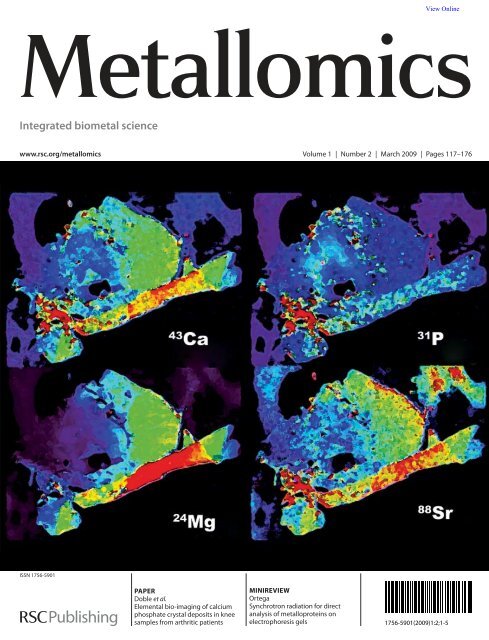
Fig. 1 shows correspond<strong>in</strong>g regions <strong>of</strong> relatively high<br />
<strong>calcium</strong> and phosphorus <strong>in</strong>tensities <strong>in</strong> the cartilage section<br />
taken from a patient with osteoarthritis. These common areas<br />
may be representative <strong>of</strong> <strong>calcium</strong> <strong>phosphate</strong>-based <strong>crystal</strong><br />
Fig. 1 <strong>Elemental</strong> distribution maps <strong>of</strong> <strong>calcium</strong> ( 43 Ca), phosphorus ( 31 P), magnesium ( 24 Mg), strontium ( 88 Sr) and z<strong>in</strong>c ( 66 Zn) <strong>in</strong> a knee cartilage<br />
section from a patient diagnosed with osteoarthritis.<br />
144 | Metallomics, 2009, 1, 142–147 This journal is c The Royal Society <strong>of</strong> Chemistry 2009
Downloaded on 19 February 2011<br />
Published on 13 February 2009 on http://pubs.rsc.org | doi:10.1039/B901310P<br />
<strong>deposits</strong>. A high frequency <strong>of</strong> CPPD <strong>crystal</strong>s <strong>in</strong> articular<br />
tissues removed from OA hips and knees has been reported<br />
<strong>in</strong> the literature. 30 Other elements <strong>in</strong>clud<strong>in</strong>g copper, iron, and<br />
selenium did not follow the same trend <strong>in</strong> distribution.<br />
The areas are significantly larger than the average <strong>crystal</strong><br />
size (up to 20 mm). 3 This may be due to aggregation <strong>of</strong> the<br />
<strong>crystal</strong>s with<strong>in</strong> the cartilage matrix. An alternative explanation<br />
was that the relatively larger laser spot size used for <strong>imag<strong>in</strong>g</strong> <strong>of</strong><br />
the smaller <strong>crystal</strong>s swamped the ICP-MS signal. That is, the<br />
<strong>crystal</strong> sizes were magnified by the use <strong>of</strong> a spot size larger than<br />
the average <strong>crystal</strong> size. This may be overcome by higher<br />
resolution <strong>imag<strong>in</strong>g</strong>.<br />
In addition, several regions <strong>of</strong> relatively high <strong>calcium</strong> and<br />
<strong>phosphate</strong> <strong>in</strong>tensities correlated with relatively high magnesium<br />
and strontium <strong>in</strong>tensities. Magnesium whitlockite, a <strong>calcium</strong><br />
ortho<strong>phosphate</strong> <strong>crystal</strong> <strong>in</strong> which <strong>calcium</strong> is partly substituted<br />
with magnesium, has been reported <strong>in</strong> both osteoarthritic and<br />
normal articular cartilage and recent data suggest it may play a<br />
pathological role <strong>in</strong> arthritis. 31–33 Our results were consistent<br />
with the association <strong>of</strong> magnesium whitlockite and BCP <strong>crystal</strong>s<br />
as areas <strong>of</strong> relatively high magnesium <strong>in</strong>tensities consistently<br />
overlapped with areas <strong>of</strong> high <strong>calcium</strong> <strong>in</strong>tensities.<br />
Fig. 1 also shows an area <strong>of</strong> relatively high strontium<br />
<strong>in</strong>tensity which corresponded to areas <strong>of</strong> high <strong>calcium</strong>,<br />
phosphorus and magnesium. To our knowledge the association<br />
<strong>of</strong> strontium with BCP <strong>crystal</strong>s has not been reported<br />
before, however it has been detected <strong>in</strong> osteoarthritic synovial<br />
fluid. 21 Possible <strong>in</strong>terferences <strong>of</strong> 88 Sr <strong>in</strong>clude double charged<br />
rare earth elements, <strong>calcium</strong> dimers and argides, and <strong>calcium</strong><br />
<strong>phosphate</strong>s. 18,34 When tun<strong>in</strong>g the ICP-MS on NIST 612, the<br />
formation <strong>of</strong> diatomic <strong>in</strong>terferences was monitored and found<br />
to be m<strong>in</strong>imal. The reaction cell was shown to further reduce<br />
this. The signal collected from m/z 88 was identified as<br />
strontium as the Sr 88 /Sr 86 isotope ratio agreed with<strong>in</strong> the<br />
accepted range <strong>of</strong> natural abundances. 35 Therefore, the<br />
strontium distribution maps appear to be genu<strong>in</strong>e.<br />
An <strong>in</strong>terest<strong>in</strong>g trend <strong>in</strong> the distribution <strong>of</strong> z<strong>in</strong>c was also<br />
observed <strong>in</strong> some samples. Areas <strong>of</strong> high <strong>calcium</strong>, phosphorus<br />
and magnesium corresponded to regions <strong>of</strong> low z<strong>in</strong>c <strong>in</strong>tensity.<br />
This method also has the potential for ratio <strong>imag<strong>in</strong>g</strong>. More<br />
objective and def<strong>in</strong>itive detection <strong>of</strong> BCP <strong>crystal</strong>s could be<br />
achieved by generat<strong>in</strong>g a 2D image <strong>of</strong> the Ca : P ratio, whereby<br />
areas with a ratio correspond<strong>in</strong>g to that <strong>of</strong> BCP or CPPD<br />
<strong>crystal</strong>s are selectively coloured aga<strong>in</strong>st the rema<strong>in</strong><strong>in</strong>g tissue.<br />
This may allow CPPD <strong>crystal</strong>s to be dist<strong>in</strong>guished from BCP<br />
<strong>crystal</strong>s.<br />
Imag<strong>in</strong>g <strong>of</strong> synovial fluids<br />
Synovial fluid is a more common sample type used by the many<br />
exist<strong>in</strong>g analytical tools for <strong>crystal</strong> detection and research as<br />
it is more accessible to the cl<strong>in</strong>ician. Therefore, <strong>imag<strong>in</strong>g</strong> by<br />
LA ICP-MS was also trialled on dried synovial drops.<br />
Synovial fluid was collected from patients suffer<strong>in</strong>g from<br />
OA and rheumatoid arthritis. A small volume (12 ml) was then<br />
dropped onto a quartz coverslip and air dried. The laser<br />
experimental parameters ensured that the centre <strong>of</strong> the drop<br />
was completely ablated. The thicker outer-rim was not always<br />
ablated completely. No sample preparation was necessary, an<br />
advantage aga<strong>in</strong>st other methods such as XRF, which requires<br />
the <strong>crystal</strong>s to be isolated first. 3<br />
BCP <strong>crystal</strong>s are uniquely associated with OA and have<br />
been reported to occur <strong>in</strong> up to 60% <strong>of</strong> patient synovial<br />
samples. 3,30 CPPD <strong>crystal</strong>s have been associated with 30%<br />
<strong>of</strong> OA synovial fluids. 30 Images consistent with the presence <strong>of</strong><br />
<strong>crystal</strong> <strong>deposits</strong> were observed for one <strong>of</strong> the OA samples and<br />
both <strong>of</strong> the RA samples. Though BCP <strong>crystal</strong>s are uniquely<br />
associated with OA, 3 the presence <strong>of</strong> CPPD <strong>crystal</strong>s <strong>in</strong> RA<br />
jo<strong>in</strong>t fluids is not uncommon as these <strong>crystal</strong>s are <strong>of</strong>ten found<br />
<strong>in</strong> many arthritides. 36<br />
Fig. 2 shows the distribution <strong>of</strong> several elements <strong>in</strong> a drop <strong>of</strong><br />
synovial fluid from an osteoarthritic jo<strong>in</strong>t. Similar trends <strong>of</strong><br />
common regions <strong>of</strong> relatively high <strong>calcium</strong>, phosphorus,<br />
magnesium and strontium were observed. An image <strong>of</strong> copper<br />
was <strong>in</strong>cluded to illustrate the different distribution <strong>of</strong> elements<br />
not associated with <strong>calcium</strong> <strong>phosphate</strong> based <strong>crystal</strong>s.<br />
Confirmation <strong>of</strong> <strong>crystal</strong> <strong>imag<strong>in</strong>g</strong> us<strong>in</strong>g synthetic synovial fluid<br />
Synovial fluid orig<strong>in</strong>ates from plasma that is filtered by a<br />
capillary net and diffuses <strong>in</strong>to the jo<strong>in</strong>t cavity, with the<br />
addition <strong>of</strong> locally synthesised hyaluronic acid, which is what<br />
gives SF its characteristic viscosity. 7 Thus, synthetic synovial<br />
fluid samples were prepared us<strong>in</strong>g human serum to mimic the<br />
components <strong>of</strong> synovial fluid. After <strong>in</strong>cubation <strong>of</strong> the solution,<br />
solid matter was observed.<br />
The solution was filtered to remove the solid matter and<br />
drops <strong>of</strong> both filtered and non-filtered solution were left to air<br />
Fig. 2 <strong>Elemental</strong> distribution maps <strong>of</strong> <strong>calcium</strong> ( 43 Ca), phosphorus ( 31 P), magnesium ( 24 Mg), strontium ( 88 Sr) and copper ( 63 Cu) <strong>in</strong> a drop <strong>of</strong><br />
osteoarthritic synovial fluid.<br />
This journal is c The Royal Society <strong>of</strong> Chemistry 2009 Metallomics, 2009, 1, 142–147 | 145
Downloaded on 19 February 2011<br />
Published on 13 February 2009 on http://pubs.rsc.org | doi:10.1039/B901310P<br />
Fig. 3 (Upper) <strong>Elemental</strong> distribution maps <strong>of</strong> <strong>calcium</strong> ( 43 Ca), phosphorus ( 31 P), magnesium ( 24 Mg) and strontium ( 88 Sr) <strong>in</strong> a drop <strong>of</strong> synthetic<br />
synovial fluid. (Lower) <strong>Elemental</strong> distribution maps <strong>of</strong> <strong>calcium</strong> ( 43 Ca), phosphorus ( 31 P), magnesium ( 24 Mg) and strontium ( 88 Sr) <strong>in</strong> a drop <strong>of</strong><br />
filtered synthetic synovial fluid.<br />
dry on a clean slide. As the fluid was not as viscous as synovial<br />
fluid, the spot was not as thick and a faster scan rate could be<br />
applied whilst still ensur<strong>in</strong>g complete ablation.<br />
Images <strong>of</strong> the elemental distribution <strong>of</strong> <strong>calcium</strong>, phosphorus,<br />
magnesium and strontium are presented <strong>in</strong> Fig. 3a. Aga<strong>in</strong>,<br />
results consistent with the presence <strong>of</strong> <strong>crystal</strong> <strong>deposits</strong> were<br />
observed <strong>in</strong> these images by common regions <strong>of</strong> relatively high<br />
<strong>in</strong>tensities <strong>of</strong> <strong>crystal</strong>-associated elements.<br />
In dist<strong>in</strong>ct contrast, the images produced from the ablation<br />
<strong>of</strong> the filtered solution do not show the small common regions<br />
that are consistent with <strong>crystal</strong> <strong>deposits</strong> (Fig. 3b). These<br />
images <strong>in</strong>stead show a more uniform distribution <strong>of</strong> the<br />
elements with <strong>in</strong>creas<strong>in</strong>g concentration radiat<strong>in</strong>g out from<br />
the centre <strong>of</strong> the spot, similar to that seen for non-<strong>crystal</strong><br />
associated elements <strong>in</strong> the synovial fluid sample. Consider<strong>in</strong>g<br />
the common size <strong>of</strong> CPPD <strong>crystal</strong>s, filtration at 0.22 mm<br />
should remove any <strong>crystal</strong>s or solid matter that formed <strong>in</strong><br />
the solution.<br />
The presence <strong>of</strong> CPPD or BCP <strong>crystal</strong>s <strong>in</strong> the cartilage or<br />
synovial fluid samples was not confirmed by other methods<br />
currently used for this purpose. However, this prelim<strong>in</strong>ary study<br />
gives a good <strong>in</strong>dication <strong>of</strong> the potential <strong>of</strong> LA ICP-MS to detect<br />
BCP and CPPD <strong>crystal</strong>s <strong>in</strong> both cartilage and synovial fluid<br />
samples. Additionally, the <strong>calcium</strong>-to-phosphorus ratio may be<br />
used to identify the <strong>crystal</strong>s present. Further studies are<br />
currently be<strong>in</strong>g pursued to confirm <strong>crystal</strong> presence by SEM<br />
prior to laser ablation. This would validate the method <strong>in</strong> terms<br />
<strong>of</strong> false positives/negatives and sensitivity limits compared to<br />
other methods.<br />
Additionally, quantification data may be obta<strong>in</strong>ed us<strong>in</strong>g<br />
standard serum solutions for spot calibration and matrix<br />
matched standardised tissue for cartilage samples.<br />
Conclusions<br />
This study has illustrated the potential <strong>of</strong> LA ICP-MS for the<br />
identification <strong>of</strong> <strong>crystal</strong> <strong>deposits</strong> <strong>in</strong> <strong>bio</strong>logical tissues and fluids<br />
through their localised elemental distributions. The distribution<br />
<strong>of</strong> BCP and CPPD <strong>crystal</strong>-associated elements, <strong>in</strong>clud<strong>in</strong>g<br />
<strong>calcium</strong>, phosphorus and magnesium, considered consistent<br />
with the presence <strong>of</strong> <strong>crystal</strong> <strong>deposits</strong>, was observed <strong>in</strong> osteoarthritic<br />
knee cartilage and synovial fluid samples by LA ICP-MS.<br />
Future studies will focus on quantification, validation <strong>of</strong><br />
<strong>crystal</strong>s by SEM and ratio <strong>imag<strong>in</strong>g</strong>.<br />
References<br />
1 P. Dieppe and P. Calvert, Crystals and Jo<strong>in</strong>t Disease, Chapman<br />
and Hall, New York, 1983.<br />
2 G. M. McCarthy, Curr. Op<strong>in</strong>. Rheumatol., 1996, 8, 255–258.<br />
3 A. Yavorskyy, A. Hernandez-Santana, G. McCarthy and<br />
G. McMahon, Analyst, 2008, 133, 302–318.<br />
4 C. W. Wu, R. Terkeltaub and K. C. Kalunian, Curr. Rheumatol.<br />
Rep., 2005, 7, 213–219.<br />
5 A. K. Rosenthal and N. Mandel, Curr. Rheumatol. Rep., 2001, 3,<br />
11–16.<br />
6 P. Dieppe and A. Swan, Ann. Rheum. Dis., 1999, 58, 261–263.<br />
7 E. Pascual and V. Jovani, B. Cl<strong>in</strong>. Rheumatol., 2005, 19, 371–386.<br />
8 J. S. Becker, M. Zoriy, J. S. Becker, J. Dobrowolska and<br />
A. Matusch, J. Anal. At. Spectrom., 2007, 22, 736–744.<br />
146 | Metallomics, 2009, 1, 142–147 This journal is c The Royal Society <strong>of</strong> Chemistry 2009
Downloaded on 19 February 2011<br />
Published on 13 February 2009 on http://pubs.rsc.org | doi:10.1039/B901310P<br />
9 S. F. Durrant and N. I. Ward, J. Anal. At. Spectrom., 2005, 20,<br />
821–829.<br />
10 R. Lob<strong>in</strong>ski, C. Moul<strong>in</strong> and R. Ortega, Biochimie, 2006, 88,<br />
1591–1604.<br />
11 M. V. Zoriy, M. Dehnhardt, A. Matusch and J. S. Becker,<br />
Spectrochim. Acta, Part B, 2008, 63, 375–382.<br />
12 R. W. Hutch<strong>in</strong>son, A. G. Cox, C. W. McLeod, P. S. Marshall,<br />
A. Harper, E. L. Dawson and D. R. Howlett, Anal. Biochem.,<br />
2005, 346, 225–233.<br />
13 M. Zoriy, A. Matusch, T. Spruss and J. S. Becker, Int. J. Mass<br />
Spectrom., 2007, 260, 102–106.<br />
14 P. M. Outridge, G. Ve<strong>in</strong>ott and R. D. Evans, Environ. Rev.<br />
(Ottawa), 1995, 3, 160–170.<br />
15 M. A. Chaudhri, J. Watl<strong>in</strong>g and F. A. Khan, J. Radioanal. Nucl.<br />
Chem., 2007, 271, 713–720.<br />
16 K. M. Lee, J. Appleton, M. Cooke, F. Keenan and K. Sawicka-<br />
Kapusta, Anal. Chim. Acta, 1999, 395, 179–185.<br />
17 C. Stadlbauer, C. Reiter, B. Patzak, G. St<strong>in</strong>geder and T. Prohaska,<br />
Anal. Bioanal. Chem., 2007, 388, 593–602.<br />
18 M. S. A. Horstwood, J. A. Evans and J. Montgomery, Geochim.<br />
Cosmochim. Acta, 2008, 72, 5659–5674.<br />
19 D. J. Bellis, K. M. Hetter, J. Jones, D. Amarasiriwardena and<br />
P. J. Parsons, J. Anal. At. Spectrom., 2006, 21, 948–954.<br />
20 T. Uryu, J. Yosh<strong>in</strong>aga, Y. Yanagisawa, M. Endo and<br />
J. Takahashi, Anal. Sci., 2003, 19, 1413–1416.<br />
21 M. Krachler, W. Domej and K. J. Irgolic, Biol. Trace Elem. Res.,<br />
2000, 75, 253–263.<br />
22 P.-T. Cheng, K. P. H. Pritzker, M. E. Adams, S. C. Nyburg and<br />
S. A. Omar, J. Rheumatol., 1980, 7, 609–616.<br />
23 P. T. Cheng and K. P. H. Pritzker, J. Rheumatol., 1983, 10,<br />
769–777.<br />
24 D. Gunther, D. Ble<strong>in</strong>er, M. Guillong, B. Hattendorf and I. Horn,<br />
Chimia, 2001, 55, 778–782.<br />
25 B. Hattendorf and D. Gunther, J. Anal. At. Spectrom., 2000, 15,<br />
1125–1131.<br />
26 P. R. D. Mason, Short Course Ser. M<strong>in</strong>eral. Assoc. Canada, 2001,<br />
29, 63–81.<br />
27 J. Fietzke, A. Eisenhauer, N. Gussone, B. Bock, V. Liebetrau,<br />
T. F. Nagler, H. J. Spero, J. Bijma and C. Dullo, Chem. Geol.,<br />
2004, 206, 11–20.<br />
28 B. Jackson, S. Harper, L. Smith and J. Fl<strong>in</strong>n, Anal. Bioanal. Chem.,<br />
2006, 384, 951–957.<br />
29 J. Feldmann, A. K<strong>in</strong>dness and P. Ek, J. Anal. At. Spectrom., 2002,<br />
17, 813–818.<br />
30 K. M. D. Jaovisidha and A. K. M. D. Rosenthal, Curr. Op<strong>in</strong>.<br />
Rheumatol., 2002, 14, 298–302.<br />
31 C. A. Scotchford and S. Y. Ali, Osteoarthritis Cartilage, 1997, 5,<br />
107–119.<br />
32 R. Lagier and C. A. Baud, Pathol. Res. Pract., 2003, 199, 329–335.<br />
33 P. B. Halverson, Curr. Op<strong>in</strong>. Rheumatol., 1996, 8, 259–261.<br />
34 P. Z. Vroon, B. Wagt, J. M. Koornneef and G. R. Davies, Anal.<br />
Bioanal. Chem., 2008, 390, 465–476.<br />
35 S. J. Fallon, M. T. McCulloch, R. van Woesik and D. J. S<strong>in</strong>clair,<br />
Earth Planet. Sci. Lett., 1999, 172, 221–238.<br />
36 Crystal-Induced Arthropathies; Gout, Pseudogout and Apatiteassociated<br />
Syndromes, ed. R. L. Wortmann, H. R. J. Schumacher,<br />
M. A. Becker and L. M. Ryan, Taylor & Francis, New York,<br />
2006.<br />
This journal is c The Royal Society <strong>of</strong> Chemistry 2009 Metallomics, 2009, 1, 142–147 | 147