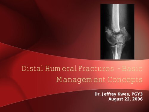
Distal Humeral Fractures - Dr. Kwee (Aug 22, 2006
Distal Humeral Fractures - Dr. Kwee (Aug 22, 2006
Distal Humeral Fractures - Dr. Kwee (Aug 22, 2006

Create successful ePaper yourself
Turn your PDF publications into a flip-book with our unique Google optimized e-Paper software.
<strong>Distal</strong> <strong>Humeral</strong> <strong>Fractures</strong> - Basic<br />
Management Concepts<br />
<strong>Dr</strong>. Jeffrey <strong>Kwee</strong>, PGY3<br />
<strong>Aug</strong>ust <strong>22</strong>, <strong>2006</strong>
• Elbow = 5% of fractures<br />
– 0.5% <strong>Distal</strong> <strong>Humeral</strong><br />
• Adults – Comminution ++<br />
– Kids – Supracondylar/condylar<br />
minimal comminution<br />
Incidence
Anatomy<br />
• <strong>Distal</strong> Humerus made up of 2 columns<br />
– Medial and lateral<br />
– Together have two articulations<br />
– Ulna humeral – Hinge joint<br />
– Radio humeral – Rotational joint<br />
– Independent but related
Anatomy 2<br />
• Very thin sections of bone at olecranon<br />
fossa, radial fossa, coranoid fossa<br />
– Provide room for ROM without impingment<br />
– Weak area
• Alignment<br />
– 4-8° of valgus (carrying<br />
angle)<br />
– 3-8°of external rotation<br />
– 40° of forward flexion<br />
Anatomy 3
• Ligaments and<br />
muscles<br />
– Medial and<br />
Lateral Ulnar<br />
collateral<br />
ligament<br />
– Flexor and<br />
extensor origins<br />
Anatomy 4
• Peri-articular - OTA Classification<br />
– A – Extra Articular<br />
Classification<br />
– B – Partial Articular<br />
– C – Total articular – No continuity shaft to<br />
articular surface
• OTA/AO<br />
Classification – Peri-articular
• Standard X-rays<br />
• Traction X-rays<br />
• CT Scan with Recon<br />
• Stress Views<br />
Diagnosis
Treatment Options - Type A <strong>Fractures</strong><br />
• As in Kids – Brachial<br />
artery injury is<br />
sometimes<br />
associated with this<br />
type<br />
• AKA transcolumnar<br />
fractures<br />
• Also classified into<br />
high, low or oblique
Management Options – Type A <strong>Fractures</strong><br />
• Closed Reduction and Casting<br />
– Now recommended only for those who are<br />
medically unfit<br />
• Closed Reduction and Traction<br />
– Neither closed technique allows for early<br />
mobilization<br />
• Percutaneous Pinning<br />
– As in kids, crossed k-wires<br />
– Must be immobilized in cast for 5-6 wks<br />
• Open Reduction Internal Fixation<br />
– The current standard of care
Management Options – Type A Freactues<br />
• Percutaneous Pinning<br />
– As in kids, first obtain closed<br />
reduction<br />
• Traction and flexion (if extension type)<br />
• Traction and posterior pressure on the<br />
forearm (if flexion type)<br />
• Then two crossed k-wires starting on the<br />
epicondyle and crossing the fracture site<br />
• Under flouro<br />
– Ulnar nerve a risk<br />
– Not stable construct – needs to<br />
be in cast/splint x 5-6 wks<br />
– Possibility of stiff elbow
Management Options – Type A <strong>Fractures</strong><br />
• Open Reduction and Internal Fixation<br />
– Method of choice<br />
– Allows for stable fixation and thus early<br />
ROM<br />
– Minimizes chance of stiff elbow<br />
– Can start ROM as soon as day 1<br />
• Technique – Same as fixation for Type C<br />
– Will go over this later
Treatment Options - Type B <strong>Fractures</strong><br />
• Milch Classification for medial or lateral<br />
column fractures<br />
– Type I – Lateral wall of trochlea attached<br />
to main mass of humerus<br />
– Type II – Lateral wall separated<br />
• High/Low<br />
Classification<br />
• OTA B1.1 or .2<br />
• OTA B2.1 or .2
Treatment Options - Type B <strong>Fractures</strong><br />
• Non-operative<br />
– Non medically fit<br />
– Low, undisplaced<br />
intraarticular fractures<br />
• Operative<br />
– Treatment of choice<br />
– Muscle Pull significant<br />
force of displacement<br />
– Cannulated screws /<br />
Lag screws
Management Options – Type B <strong>Fractures</strong><br />
• Articular <strong>Fractures</strong><br />
– Capitellum<br />
• With or without<br />
involvement of the<br />
trochlea<br />
– If undisplaced –<br />
closed treatment<br />
– If displaced<br />
• Open reduction and<br />
internal fixation<br />
• Lateral or Anterior<br />
approach and Herbert type<br />
countersunk screws
• Options<br />
Treatment Options - Type C <strong>Fractures</strong><br />
– Non-operative – Classical teaching (1960s)<br />
– At the time poor outcomes with ORIF<br />
• Traction through olecranon pin – 3 wks, then cast<br />
• Collar and Cuff - Bag of Bones Treatment – Maximal flexion<br />
initially, encouraging ROM of hands and fingers<br />
– At 6 wks begin elbow ROM<br />
– Operative – Current Choice<br />
• ORIF is now recommended because we have a better<br />
understanding of fractures, imaging and implants<br />
– Can achieve reasonably functional outcomes because of<br />
early motion<br />
• Total Elbow – A few slides from now
Treatment Options – Type C <strong>Fractures</strong><br />
• 90-90 plating<br />
– Posterior lateral plate<br />
– Medial plate<br />
• <strong>Distal</strong> end of medial plate can be bent<br />
so that it includes orthogonal screws<br />
• Compression screw can be used across<br />
the condyle<br />
– Biomechanically the strongest<br />
construct<br />
• Helfet and Hotchkiss<br />
– Pelvic reconstruction plates<br />
– Pre-contoured plates<br />
• Accumed - Locking<br />
• Zimmer - Locking
Management Options - Type C Fracture<br />
• Total Elbow<br />
• Currently only recommended in<br />
– the elderly and those with low demand<br />
– Those with very poor bone quality<br />
– Extremely comminuted fractures<br />
• Cannot be used as a bailout if olecranon<br />
osteotomy used<br />
– Must use either triceps split or osteotomy of<br />
epicondyles<br />
– Generally should be done by experienced<br />
arthroplasty surgeon
Outcomes In Type C <strong>Fractures</strong><br />
• Outcomes proportional to energy type of<br />
fracture<br />
• Stiffness<br />
– Despite ORIF and optimal early ROM still<br />
can have 20-25° flexion contracture<br />
• Pain<br />
– 25% can have exertional pain<br />
• Strength<br />
– 75% of normal side
Challenges<br />
– Avoiding articular<br />
surfaces<br />
– Avoiding impingement<br />
of fossae<br />
– Capturing multiple<br />
small fragments<br />
– Cancellous bone<br />
– Achieving stable<br />
fixation to allow early<br />
ROM<br />
– Osteoporotic bone<br />
ORIF <strong>Distal</strong> Humerus
• Posterior Approach to the<br />
elbow<br />
– Position lateral, or partial<br />
lateral<br />
– Arm across body, or with<br />
sterile bolster<br />
– Tourniquet applied high<br />
– Incision: straight posterior,<br />
or slightly medial, curving<br />
around medial side of<br />
olecranon tip up midline of<br />
arm<br />
ORIF <strong>Distal</strong> Humerus
• Posterior Approach<br />
Cont’d<br />
– Dissection consists of<br />
exposing the triceps fascia<br />
– Identifying and the ulnar<br />
nerve on medial side +/transposition<br />
depending<br />
on #<br />
– To get to the joint a<br />
triceps split or an<br />
olecranon osteotomy<br />
• Studies show no difference in<br />
muscle strength post-op<br />
ORIF <strong>Distal</strong> Humerus
• Posterior Approach<br />
Cont’d<br />
– Pre-drill and tap olecranon<br />
– Create cevron apex distal<br />
– Protect the articular<br />
surface by going through<br />
the last milimeter with an<br />
osteotome<br />
– Aim for the ‘bare patch’ at<br />
the apex of the trochlear<br />
notch<br />
ORIF <strong>Distal</strong> Humerus
ORIF <strong>Distal</strong> Humerus<br />
• Postero-lateral Approach<br />
– Used for isolated lateral column fractures<br />
– Position – Supine with arm across chest<br />
– Incision: curved from lateral epicondylar ridge to lateral<br />
border of ulnar<br />
– Internervous Plane:<br />
Anconeus and ECU<br />
supplied via radial<br />
and PIN respectively<br />
– Superficial dissection:<br />
incise deep fascia in<br />
line with skin incision<br />
to find ECU and Anconeus.<br />
They share common<br />
origin
• Deep dissection:<br />
pronate forearm<br />
to move PIN<br />
away. Find<br />
Supinator at the<br />
deep part of the<br />
incision and strip<br />
if off as<br />
necessary from<br />
epicondyle<br />
ORIF <strong>Distal</strong> Humerus