Family.- DIPLOSTOMATIDAE Poirier, Subfamily.-Diplostomatinae ...
Family.- DIPLOSTOMATIDAE Poirier, Subfamily.-Diplostomatinae ...
Family.- DIPLOSTOMATIDAE Poirier, Subfamily.-Diplostomatinae ...

Create successful ePaper yourself
Turn your PDF publications into a flip-book with our unique Google optimized e-Paper software.
Rev. Bio!. Trop., 11 (1); 75-87, 1963<br />
On two new diplostome parasites of birds, with<br />
a note on Hysteromorpha triloba (Rud., 1819> Lutz,<br />
1931 from India (Trematoda: Diplostomatidae>*<br />
by<br />
Ramesh Gupta *.,<br />
(Received fol' publication April 18, 1963 )<br />
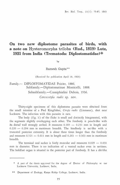
<strong>Family</strong>.- <strong>DIPLOSTOMATIDAE</strong> <strong>Poirier</strong>, 1886.<br />
<strong>Subfamily</strong>.-<strong>Diplostomatinae</strong> Monticelli, 1888.<br />
Subsubfamily.-Crassiphialini Dubois, 1936.<br />
Cercocotyla l'udis sp. nov.<br />
Thirty-eight specimens of this diplostome parasite were obtained from<br />
the small intestine of a Pied Kingfisher, Ceryle rudis (Linnaeus), shot near<br />
Lucknow. The infection with this parasite is rareo<br />
The body (fig. 1) of the fluke is small and distinetly bisegmented, with<br />
the segments slightly overlapping ea eh other. The forebody is pouch-like with<br />
its dorsal wall strongly arehed. It measures 0.185 - 0.231 mm in length and<br />
0.220 - 0.294 mm in maximum breadth. The hindbody is sac-like with a<br />
truneated posterior extremity. It is about three times longer than the forebody<br />
and measures 0.521 - 0.611 mm in length and 0.261 - 0.363 mm in maximum<br />
breadth.<br />
The terminal oral sueker is fairly muscular and measures 0.029 - 0.031<br />
mm in diameter. There is no indication of a ventral sucker even in sections.<br />
The holdfast organ is situated in the posterior part of forebody. It has a slit-like<br />
* A part of the thesis app.roved for the degree of Doctor of Philosophy ac the<br />
Lucknow University, Luc�now, India.<br />
• * Department of ZooJogy, Kanya Kubja College, Lucknow, India.<br />
75
76<br />
REVISTA DE BIOLOGIA TROPICAL<br />
opening and measures 0.058 - 0.069 mm by 0.069 - 0.076 mm. Radial<br />
musdes and minute gland cells are present in the wall of the holdfast organ.<br />
A small, but prominent adhesive gland is present immediately behind this organ.<br />
A prepharynx being absent, the mouth directly leads into a muscular<br />
pharynx which mea sures 0.026 - 0.030 mm by 0.021 mm. The pharynx leads<br />
into a short oesophagus measuring 0.019 - 0.021 mm in length. The intestinal<br />
caeca are narrow and extend back almost up to the posterior end of body.<br />
The gonads occupy the middle regíon of hindbody. The testes are tandem.<br />
They are smooth-margined and transversely elongated. The anterior testis measures<br />
0.091 - 0.112 mm by 0.154 - 0.195 mm. The posterior testis is somewhat<br />
smaller and measures 0.067 - 0.088 mm by 0.149 - 0.190 mm. The vas deferens<br />
runs posteriorly and opens into a prominent U-shaped seminal veside. A prominent<br />
ejaculatory duct continues from the seminal veside and dorsally opens into<br />
the uterus to form a very short hermaphroditic duct. A pars prostatica, as described<br />
by YAMAGUTI (8) in Cercocotyla cerylis, is not observed in the present formo<br />
The ovary is median and immediately pre-testicular. It Í's spherical and<br />
measures 0.049 - 0.054 mm in diameter. The vitellaria are confined to the<br />
hindbody and extend from behind the adhesive gland up to the posterior end.<br />
They consists of numerous large foUides which are dense in the pre-ovarian<br />
region, but sparse behind. The vitelline reservoir and Mehlis' gland are intertesticular.<br />
The Laurer's canal is presento The uterus is short and contains only<br />
a few (up to six) eggs which are pale-yeUow, operculate, and fairly large for<br />
the size of the fluke. The eggs are even larger than the ovary and measure<br />
0.0791 - 0.0912 mm by 0.0514 - 0.0625 mm. The hermaphroditic duct opens<br />
into a large copulatory bursa by the genital pore (fig. 2) which is surrounded,<br />
as stated by YAMAGUTI (8) for Cercocotyla cerylis, with a mass of radial musde<br />
fibres and a ring of several minute papilliform structures. A large sucker-like<br />
muscular structure, termed "atrial sucker" by SUDARIKOV (7) in Skrjabin's<br />
"Trematody jivotnyi i chelovekaJJ, opens into the copulatory bursa immediately<br />
ventral to the genital pore. The bursa opens to the exterior by a wide aperture at<br />
the truncate posterior extremity of the body.<br />
DISCUSSION: The presence of a so-caUed "atrial sucker" and a mass of<br />
muscular fibres, together with a ring of minute papilliform structures around<br />
the genital pore, dearly indicates that the present fluke belongs to the displostome<br />
genus Cercocotyla Yamaguti, 1939. The genus Cercocotyla was established by<br />
YAMAGUTI (8), with Cercocotyla cerylis Yamaguti, 1939 as the genotype, for<br />
a diplostome fluke obtained by him from the small intestine of Ceryle lugubris<br />
lugubris (Temm.) from Japan. No subsequent record of this genus is found in<br />
the literature. The finding of the present fluke is, therefore, the second record<br />
of this genus.<br />
The present form can be easily distinguished from the genotype by several<br />
important features. It is much smaUer with a considerably shorter and stouter<br />
hindbody. An adhesive gland is distinctly visible in the present formo The gonads<br />
are relatively large in the present form and, moreover, they are situated in the<br />
middle, and not in the posterior region of the hindbody. The vitellaria are
GUPTA: TWO NEW DIPLOSTOME PARASITES OF BIRDS 77<br />
distributed throughout the hindbody in the present form, whereas they are limited<br />
to the posterior half of the hindbody in the genotype. Furthermore, the eggs<br />
of the present form are considerably larger, even larger than the ovary of the<br />
fluke. From these differences it becomes evident that the present fluke represents<br />
a new species of the genus Cercocotyla Yamaguti, 1939, and the name Cercocotyla<br />
rudis is proposed for it.<br />
EMENDED DIAGNOSIS OF THE GENUS CERCOCOTYLA<br />
YAMAGUTI, 1939<br />
The present form necessitates an elaboration of the generic diagnosis of<br />
Cercocotyla Yamaguti, 1939 as follows:<br />
Diplostomatidae; <strong>Diplostomatinae</strong>; Crassiphialini: Body distinctly bisegmented.<br />
Forebody spoon-shaped or pouch-like. Hindbody long and slender or<br />
short and stout. Oral sucker smalI. Ventral sucker absent. Pseudosuckers<br />
absent. Holdfast organ well-developed. Prepharynx, if present, very short.<br />
Small pharynx and short oesophagus present. Intestinal caeca extend up to<br />
posterior end of body. Gonads in middle or posterior part of hindbody.<br />
Testes tandem and smooth-margined. Seminal vesicle sigmoid or U-shaped. Pars<br />
prostatica may be present. Ovary median and immediately pre-testicular. Vitellaria<br />
confined to hindbody. Ootype complex and vitelline reservoir inter-testicular.<br />
Laurer' s canal present. Ductus hermaphroditicus short, opening into a large<br />
copulatory bursa by a genital pore which is surrounded by a dense muscular<br />
tissue and a ring of minute papilliform structures. A large muscular sucker ("atrial<br />
sucker" of Sudarikov) opens into the copulatory bursa ventral to the genital<br />
pore. Copulatory bursa opens to the exterior at posterior end of body by a<br />
wide aperture. Parasites of intestine of birds.<br />
Type species :- Cercocotyla cerylis Yamaguti, 1939.<br />
A KEY TO THE SPECIES OF THE GENUS CERCOCOTYLA<br />
YAMAGUTI, 1939<br />
1. Hindbody long and slender. Gonads in posterior region of<br />
hindbody. Vitellaria in posterior half of hindbody. Eggs<br />
smaller than ovary .............................. C. cerylís Yamaguti, 1939<br />
Hindbody short and stout. Gonads in middle region of hindbody.<br />
Vitellaria extending throughout hindbody. Eggs larger<br />
than ovary ........................................................................ C. rudiJ sp. nov.<br />
<strong>Family</strong>.- <strong>DIPLOSTOMATIDAE</strong> <strong>Poirier</strong>, 1886.<br />
<strong>Subfamily</strong>.-<strong>Diplostomatinae</strong> Monticelli, 1888.<br />
Subsubfamily.-Diplostomatini Dubois, 1936.<br />
Neoharvardia pandubi gen. nov., sp. nov.<br />
Two specimens of this diplostome fluke were obtained from the small<br />
intestine of the Large Cormorant, Phalacrocorax carbo (Linnaeus). The infection<br />
with this fluke is rare.
78<br />
REVISTA DE BIOLOGIA TROPICAL<br />
The body (fig. 3) of the fluke is distinctly bisegmented. The segments<br />
are strongly retroflexed. The lateral margins of the forebody are ventrally incurved<br />
to form a deep ventral concavity. The forebody measures 1.450 - 1.617 mm<br />
in length and 1.100 � 1.289 mm in maximum breadth in the region of the<br />
holdfast organ. The hindbody is bluntly conical and measures 1.296 mm in length<br />
and 0.905 - 1.000 mm in maximum breadth in its anterior region.<br />
The oral sucker is terminal and mea sures 0.140 - 0.163 mm by 0.130 -<br />
0.182 mm. A highly mobile and muscular cup-like pseudosucker is present on<br />
each si de of the oral sucker. The ventral sucker is situated in the posterior half<br />
of the forebody. It measures 0.184 - 0.241 mm by 0.301 - 0.310mm. The<br />
holdfast organ is situated immediately behind the ventral sucker in the posterior<br />
region of forebody. It is oval and measures 0.419 mm by 0.530 - 0.725 mm.<br />
A small adhesive gland is present immediately behind the holdfast organ.<br />
The mouth leads into a short prepharynx which opens into a small pharynx<br />
measuring 0.078 - 0.097 mm by 0.098 - 0.113 mm. A short oesophagus is<br />
presento The intestinal caeca are greatly obscured by various organs.<br />
The testes are large structures, situated obliquely one behind the other<br />
in the middle region of the hindbody. The anterior testis is :sinistral and obliquely<br />
placed to the long axis of the body. It is oval and smaller than the posterior<br />
testis, measuring 0.392 - 0.507 mm by 0.310 mm. The posterior testis is large,<br />
transversely elongated, and slightly cOl1'3tricted in the middle, appearing somewhat<br />
bilobed. It measures 0.418 - 0.530 mm by 0.593 - 0.668 mm. The vas deferens<br />
is continued posteriorly into a coiled seminal veside situated just behind the<br />
posterior testis. The ductus ejaculatorius, leading from the seminal vesicle, joins<br />
the female duct to form a ductus hermaphroditicus.<br />
The ovary is submedian and situated obliquely dextral to the anterior<br />
testis. It is subspherical and measures 0.151 - 0.170 mm by 0.190 - 0.258mm.<br />
The vitellaria consists of small foUicles which are uniformly distributed from the<br />
region of the holdfast organ upto the hind end of body. A prominent vitelline<br />
reservoir and the ootype complex, with a large Mehlis' gland, are situated dextral<br />
to the anterior testis. The uterus contains several eggs (twenty-one) in the cotype,<br />
but none in the type specimen which is fully mature and seems to have<br />
voided its eggs. The eggs in the co-type measure 0.0946 - 0.1062 mm by 0.0610<br />
- 0.0725 mm. The uterus joins the male duct at the base of a large and protrusible<br />
genital con e to form the ductus hermaphroditicus which traverses the<br />
cone and opens at its tip by the genital pore. The genital cone is placed in a<br />
large copulatory bursa whose inner waU is raised to form a prominent prepucial<br />
fold around the cone. The bursa opens to the exterior by a wide terminal aperture.<br />
DISCUSSION : The fact that the present form belongs to the subsubEamily<br />
Diplostomatini Dubois, 1936 oE the subfamily <strong>Diplostomatinae</strong> Monticelli, 1888<br />
is evident Erom the distribution oE its viteUine foUides in both segments oE the<br />
body. Of all such genera oE this subsubfamily which posses lateral pseudosuckers,<br />
the present form shows a close resemblance only with the genus Harvardia Baer,<br />
1932 (1) in the retroflexion of the body segments and in the shape of the<br />
forebody. However, the presence of a large copulatory bursa, together with a
C;UPT A: TWO NEW D1PLOSTOME PARASITES OF BIRDS 79<br />
�rominent genital cone and a prepucial fold arund the cone, does not permit an<br />
inclusion of the present form in the genus Harvardia Baer, 1932. These features,<br />
in fact, indicate that the present form represents a new diplostome genus closely<br />
allied to Harvardia Baer, 1932. The name Neoharvardia, with Neoharvardia<br />
pandubi gen. nov., sp. nov. as the genotype, is here proposed for this new genus.<br />
GENERIC DIAGNOSIS OF NEOHARVARDIA GEN. NOV.<br />
Diplostomatidae; <strong>Diplostomatinae</strong>; Diplostomatini: Body distinctly bisegmented,<br />
with segments strongly retroflexed. Forebody with its lateral margins<br />
ventrally incurved to form a deep concavity. Hindbody bluntly canical. Suckers<br />
well developed. Muscular cup-like pseudosuckers present. Holdfast organ large,<br />
without gland cells in its wall. Prepharynx, pharynx, and oesophagus present.<br />
Intestinal caeca extending up to posterior end of body. Testes large, obliquely<br />
tandem. Anterior testis obliquely sinistral. Posterior testis transversely elongated<br />
and somewhat bilobed. Ovary obliquely dextral to anterior tesüs. Vitellaria<br />
consists of small follides distributed in both segments of body. Ootype complex<br />
dextral to anterior testis. The ductus hermaphroditicus traverses a large and<br />
protrusible genital cane placed in a large copulatory bursa. The inner wall of the<br />
bursa is raised to form a prominent prepucial fold around the genital cone. Eggs<br />
large. Parasites of small intestine of birds.<br />
Type species :- Neoharvardia pandubi gen. nov., sp. nov<br />
<strong>Family</strong>.- DIPLOSTOMA TIDAE <strong>Poirier</strong>, 1886.<br />
<strong>Subfamily</strong>.-<strong>Diplostomatinae</strong> Monticelli, 1888.<br />
Subsubfamily.-Diplostomatini Dubois, 1936.<br />
Hysteromorpha triloba (Rudolphi, 1819) Lutz, 1931<br />
A large number of specimens of this diplostome fluke were obtained<br />
from the small intestine oE the Large Cormorant, Phalacrocorax carbo (Linnaeus) .<br />
The infection with this fluke is quite common.<br />
This species has so Ear been recorded from different parts of the world<br />
(various countries of Europe, Japan, Canada, U.S.A., Brazil, South and Western<br />
Australia, and Russia), but never from India. The present record oE this species,<br />
therefore, extends its range of geographical distribution to India.<br />
Since the present specimens show sorne variations, a brief redescription<br />
of the species is given be1ow :<br />
The body (fig. 5) consists oE two segments-a pear-shaped anterior segment<br />
measuring 0.876 - 1.170 mm in length and 0.774 - 1.096 mm in<br />
maximum breadth in its middle region, and a bluntIy conical posterior segment<br />
measuring 0.651 - 0.755 mm in length and 0.581 - 0.866 mm in maximum<br />
breadth.
80<br />
REVISTA DE BIOLOGIA TROPICAL<br />
The terminal oral sucker measures 0.066 - 0.083 mm by 0.094 - 0.117<br />
mm. The ventral sucker measures 0.08 1 - 0.092 mm by 0.125 - 0.199 mm.<br />
The large holdfast organ, located in the middle of anterior segment, measures<br />
0.290 - 0.418 mm by 0.351 - 0.651 mm. It appears funnel-shaped with<br />
an anterior cleft, exactly as sketched by HU GGINS (6). Regarding the holdfast<br />
organ, HAWKINGS (1936), in his unpublished thesis (vide CHANDLER<br />
and RAUS CH (2» , stated, "In a contracted state the most prominent feature is<br />
the median furrow", but unfortunately the figure given by him (as reproduced<br />
by CHANDLER and RA USCH (2» do es not show the furrow at all. y AMAGUTI<br />
(8), however, described a median "incision" in the holdfast organ in Japanese<br />
specimens. In none of the specimens collected by the present writer the holdfast<br />
organ appears trilobed as stated by HU GGINS (6) and by CHANDLER and RAUSCH<br />
(2). In the present specimens a narrow circular ridge, not described by any<br />
previous worker, is present around the holdfast organ. This ridge abuts against the<br />
holdfast organ on aH si des except anteriorIy where it is quite apart from the organ.<br />
A large adhesive gland lies immediately behind the holdfast organ.<br />
A prepharynx is absent as stated by previous workers. The small pharynx<br />
measures 0.058 - 0.061 mm by 0.058 - 0.064 mm. A short oesophagus is<br />
presento The intestinal caeca extend up to a slightly behind the posterior testis,<br />
but are greatly masked by the vitellaria.<br />
The testes are asymmetrical and tandem. The anterior testis is sinistral<br />
or dextral as stated by CIUREA (3) and HUGGINS (6). It is oval, obliquely placed,<br />
and mea sures 0.246 - 0.385 mm by 0.213 - 0.250 mm. In none of the<br />
specimens in the possession of the present writer, the anterior testis is transversely<br />
elogated as HUGGINS (6) described in sorne of the specimens collected by him,<br />
nor there is, in any specimen, a ventral folding of the sides of the anterior testis<br />
as stated by DUBOIS (4,5) and by SUDARIKOV (7). The posterior testis is, however,<br />
transversely elongated and turned ventrally on one side as described by<br />
previous workers. It measures 0.190 - 0.241 mm in length and 0.432 - 0.617<br />
mm in breadth excluding the folded part. The coiled seminal vesicle is median<br />
and immediately post-testicular.<br />
The ellipsoidal ovary is pre-testicular, median or submedian, and 0.081 -<br />
0.102 mm by 0.149 - 0.215 mm. The vitellaria have the usual distribution.<br />
The maximum number of eggs in the uterus reaches thirty, and not only twenty<br />
as stated by previous workers. The eggs measure 0.0796 - 0.0962 mm by 0.0601<br />
- 0.0713 mm. The uterus joins the ejacutory duct as usual to form the ductus<br />
hermaphroditicus which traverses a genital cone placed in a small bursa. The<br />
opening of the bursa is subterminal and dorsal, not terminal as stated by<br />
YAMAGUTI (8).<br />
ACKNOWLEDGMENTS<br />
The author's thanks are due to Prot. M. B. Lal and to Dr. S. C. Baugh<br />
of Lucknow University for guidance.
GUPTA: TWO NEW DIPLOSTOMIl PARASITES OP !lIl�DS 81<br />
SUMMARY<br />
Cercocotyla mdls sp. nov., the second specí�s under the genus Cercoco/y/a<br />
Yamaguti, 1939, is described from the small intestine of the Pied Kingfisher,<br />
Ceryle rudis (Linnaeus) from India. The diagnosis of the genus Cercoco/yla<br />
Yamaguti, 1939 is emended and a key is given to differentiate the two 'Species.<br />
A new genus, Neoharvardia, with N. pandubi gen. nov., sp. nov., is<br />
proposed for diplostome parasites collected from the small intestine of the Large<br />
Cormorant, Phalacrocorax carbo (Linnaeus) in India. The new genus is closely<br />
allíed to Hctrvardia Baer, 1932.<br />
Hysteromorpha triloba (Rudolphi, 1819) Lutz, 193, 1 is reported from<br />
India for the first time, and is briefly described.<br />
RESUMEN<br />
Se describe la segunda especie del género Cercacotyla Yamaguti, 1939,<br />
C. rudis sp. nov., a partir de ejemplares encontrados en el intestino delgado del<br />
ave Ceryle rudis (Linnaeus) de la India. La descripción del género Cel
82<br />
REVISTA DE BIOLOGIA TROPICAL<br />
6. HUGGINS, E. J.<br />
1954. Life history of a strigeid trematode, Hystel'om01'pha triloba (Rud., 1819)<br />
Lutz, 1931. JI. Sporocyst throllgh adult. T,·ctns. Amer. Micro. Soc., 73: 221-236.<br />
7. SUDARIKOV, B. E.<br />
1960. Order Strigeidida (La Rue, 1926 ) Sudarikov, 1959 in Skrjabin's Tmnatody<br />
jitwnyi i che/01Jeka. OsnolJy trematodologii. XVII pp. 155 - 530 IsdteIstvo<br />
Akademii Nauk SSSR. Moskva (In Russian ).<br />
8. Y AMAGUTI, S.<br />
1939. Studies on the helminth fauna of Japan. Pt. 25 Trematodes of birds, IV.<br />
lapaneJe 101lf'. Zool., 8: 129 - 210.<br />
Figs. 1 - 2. Cercocoty/a I'lIdis sp. nov.<br />
Fig. 1. Type specimen, ventral view.<br />
Fig. 2. Posterior end of a paratype specimen enlarged to show<br />
the structures associated with the copulatory bursa.
(;UPT A: TWO NEW DIPLOSTOME PARASITES OF BIROS 83<br />
\<br />
1
84<br />
REVISTA DE BIOLOGIA TROPICAL<br />
Figs. 3 - 4. NfOhar/'ci1 dic/ pCl11duhi gen. nov., sp. nov.<br />
Fig. 3. Type specimen, lateral VleW.<br />
Fig. 4. Posterior e-nd of the w-typc specimen, enlarged to<br />
show the eggs and copulatory apparatus.
(¡UnA: TWO NEW DIPLOSTOME PAUSITES OF BIRDS 85
86<br />
REVISTA DE BIOLOGIA TROPICAL<br />
Fig. 5. Hysteromorpha /filoba (Rudolphi, 1819) Lutz, 1931.<br />
A specimen from ventral view.
GUPT A: TWO NEW DIPLOSTOME PARASITES OF BIRDS<br />
87