Electromyographic Comparison of Grand Battement Devant at the ...
Electromyographic Comparison of Grand Battement Devant at the ...
Electromyographic Comparison of Grand Battement Devant at the ...

Create successful ePaper yourself
Turn your PDF publications into a flip-book with our unique Google optimized e-Paper software.
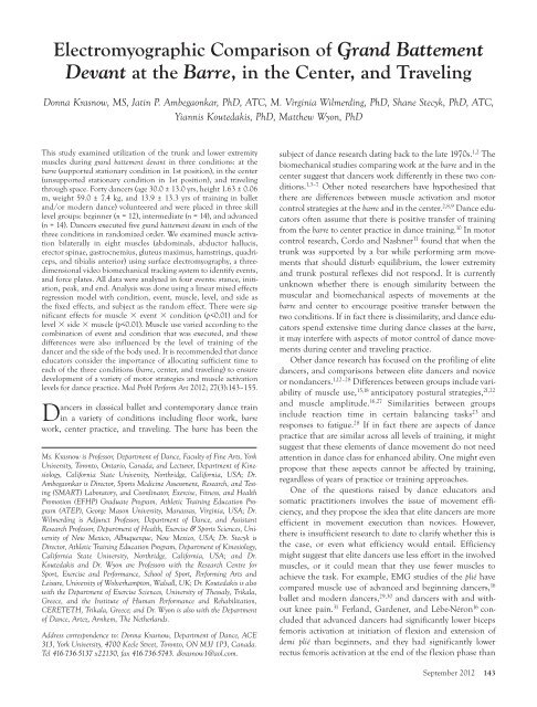
<strong>Electromyographic</strong> <strong>Comparison</strong> <strong>of</strong> <strong>Grand</strong> <strong>B<strong>at</strong>tement</strong><br />
<strong>Devant</strong> <strong>at</strong> <strong>the</strong> Barre, in <strong>the</strong> Center, and Traveling<br />
Donna Krasnow, MS, J<strong>at</strong>in P. Ambegaonkar, PhD, ATC, M. Virginia Wilmerding, PhD, Shane Stecyk, PhD, ATC,<br />
Yiannis Koutedakis, PhD, M<strong>at</strong><strong>the</strong>w Wyon, PhD<br />
This study examined utiliz<strong>at</strong>ion <strong>of</strong> <strong>the</strong> trunk and lower extremity<br />
muscles during grand b<strong>at</strong>tement devant in three conditions: <strong>at</strong> <strong>the</strong><br />
barre (supported st<strong>at</strong>ionary condition in 1st position), in <strong>the</strong> center<br />
(unsupported st<strong>at</strong>ionary condition in 1st position), and traveling<br />
through space. Forty dancers (age 30.0 ± 13.0 yrs, height 1.63 ± 0.06<br />
m, weight 59.0 ± 7.4 kg, and 13.9 ± 13.3 yrs <strong>of</strong> training in ballet<br />
and/or modern dance) volunteered and were placed in three skill<br />
level groups: beginner (n = 12), intermedi<strong>at</strong>e (n = 14), and advanced<br />
(n = 14). Dancers executed five grand b<strong>at</strong>tement devant in each <strong>of</strong> <strong>the</strong><br />
three conditions in randomized order. We examined muscle activ<strong>at</strong>ion<br />
bil<strong>at</strong>erally in eight muscles (abdominals, abductor hallucis,<br />
erector spinae, gastrocnemius, gluteus maximus, hamstrings, quadriceps,<br />
and tibialis anterior) using surface electromyography, a threedimensional<br />
video biomechanical tracking system to identify events,<br />
and force pl<strong>at</strong>es. All d<strong>at</strong>a were analyzed in four events: stance, initi<strong>at</strong>ion,<br />
peak, and end. Analysis was done using a linear mixed effects<br />
regression model with condition, event, muscle, level, and side as<br />
<strong>the</strong> fixed effects, and subject as <strong>the</strong> random effect. There were significant<br />
effects for muscle � event � condition (p
<strong>the</strong> beginners. However, an intervention study by Couillandre,<br />
Lewton-Brain, and Portero 32 revealed th<strong>at</strong> <strong>the</strong> use <strong>of</strong> <strong>the</strong><br />
biceps femoris increased post-training and was correl<strong>at</strong>ed<br />
with less “bucking” in <strong>the</strong> spine during jumps.<br />
The most recent liter<strong>at</strong>ure review <strong>of</strong> dance biomechanics<br />
studies has identified <strong>the</strong> grand b<strong>at</strong>tement as <strong>the</strong> subject <strong>of</strong> one<br />
<strong>the</strong> earliest biomechanics investig<strong>at</strong>ions in <strong>the</strong> dance liter<strong>at</strong>ure.<br />
33 Ryman and Ranney 2 collected d<strong>at</strong>a on <strong>the</strong> grand b<strong>at</strong>tement<br />
devant in <strong>the</strong> center without support, using singlecamera<br />
cinem<strong>at</strong>ography. Although <strong>the</strong>y did not collect d<strong>at</strong>a<br />
<strong>at</strong> <strong>the</strong> barre, <strong>the</strong>y claimed through anecdotal observ<strong>at</strong>ion th<strong>at</strong><br />
<strong>the</strong>re is less weight shift to <strong>the</strong> supporting leg during <strong>the</strong> b<strong>at</strong>tement<br />
<strong>at</strong> <strong>the</strong> barre than in <strong>the</strong> center. Similarly, Laws 8 proposed<br />
th<strong>at</strong> <strong>the</strong> barre allows for forward shift <strong>of</strong> <strong>the</strong> torso in<br />
arabesque and provides torso stabiliz<strong>at</strong>ion for movements<br />
such as rond de jambe th<strong>at</strong> are not possible without <strong>the</strong> barre;<br />
he questions whe<strong>the</strong>r this work is transferrable to center<br />
practice. Bronner and Oj<strong>of</strong>eitimi 13 did extensive pr<strong>of</strong>iling<br />
using kinem<strong>at</strong>ic d<strong>at</strong>a <strong>of</strong> elite dancers executing grand b<strong>at</strong>tement<br />
devant, à la seconde, and derrière, and found large pelvic<br />
movements in all three planes to accommod<strong>at</strong>e hip joint<br />
movement. However, <strong>the</strong>re is no compar<strong>at</strong>ive d<strong>at</strong>a in <strong>the</strong><br />
center or traveling, and <strong>the</strong>refore, it is not possible to know<br />
whe<strong>the</strong>r elite dancers perform <strong>the</strong>se movements with similar<br />
str<strong>at</strong>egies when unsupported.<br />
In summary, <strong>the</strong> dance biomechanics research to d<strong>at</strong>e suggests:<br />
(1) <strong>the</strong>re are important differences between some aspects<br />
<strong>of</strong> movement performed <strong>at</strong> <strong>the</strong> barre and in <strong>the</strong> center, including<br />
weight shift str<strong>at</strong>egies, muscle activ<strong>at</strong>ion, joint torque, and<br />
dynamic alignment; (2) dancers rely on <strong>the</strong> barre in some<br />
aspects <strong>of</strong> movement organiz<strong>at</strong>ion regardless <strong>of</strong> level <strong>of</strong> training;<br />
and (3) <strong>the</strong>re is high variability in muscle activ<strong>at</strong>ion when<br />
comparing barre work and center practice, and when comparing<br />
dancers <strong>of</strong> various levels <strong>of</strong> training. To d<strong>at</strong>e, no dance<br />
research has compared barre and center work to dance movement<br />
traveling in space, and determined whe<strong>the</strong>r this third<br />
condition is biomechanically different from <strong>the</strong> o<strong>the</strong>r two.<br />
If dance educ<strong>at</strong>ors are to be effective in preparing dancers<br />
for <strong>the</strong> performance <strong>of</strong> dance repertoire, it would be useful to<br />
understand which aspects <strong>of</strong> training are transferrable from<br />
barre to center and from center to travelling, and in wh<strong>at</strong> ways<br />
elite dancers differ from novice dancers. Similarly, medical<br />
practitioners working in <strong>the</strong> field <strong>of</strong> dance injury rehabilit<strong>at</strong>ion<br />
could benefit from this knowledge and improve str<strong>at</strong>egies<br />
for preparing dancers to return to full function.<br />
Therefore, <strong>the</strong> purpose <strong>of</strong> this study was to examine grand<br />
b<strong>at</strong>tement devant in three conditions: <strong>at</strong> <strong>the</strong> barre, in <strong>the</strong><br />
center, and traveling through space. The first hypo<strong>the</strong>sis was<br />
th<strong>at</strong> utiliz<strong>at</strong>ion <strong>of</strong> <strong>the</strong> trunk and lower extremity muscles in<br />
144 Medical Problems <strong>of</strong> Performing Artists<br />
TABLE 1. Subject Demographics by Training Level<br />
Level n Height (m) Weight (kg) Years Training Age (yrs)<br />
Beginners 12 1.62 ± 0.07 59.9 ± 8.4 1.5 ± 0.5 23.0 ± 4.5<br />
Intermedi<strong>at</strong>es 14 1.63 ± 0.05 59.6 ± 7.6 11.9 ± 9.6 26.0 ± 12.0<br />
Advanced 14 1.65 ± 0.07 57.6 ± 6.7 26.4 ± 11.3 40.0 ± 14.0<br />
All combined 40 1.63 ± 0.06 59.0 ± 7.4 13.9 ± 13.3 30.0 ± 13.0<br />
<strong>the</strong> grand b<strong>at</strong>tement devant would differ during <strong>the</strong> three conditions.<br />
The second hypo<strong>the</strong>sis was th<strong>at</strong> <strong>the</strong>re would be differences<br />
in muscle activ<strong>at</strong>ion levels between dancers <strong>of</strong> various<br />
training levels.<br />
Participants<br />
METHODS<br />
Dancers were recruited for <strong>the</strong> study through announcements<br />
in university dance classes and postings in pr<strong>of</strong>essional dance<br />
email listservs and local newsletters. Forty-three female<br />
dancers volunteered for <strong>the</strong> study. Inclusion criteria included<br />
enrollment in a university level dance class or in a pr<strong>of</strong>essional<br />
dance studio or training program and exposure to ballet<br />
and/or modern dance. Exclusion criteria included a history<br />
<strong>of</strong> confounding medical problems or a current injury impacting<br />
on <strong>the</strong> execution <strong>of</strong> <strong>the</strong> dance task for <strong>the</strong> study. The study<br />
was approved by <strong>the</strong> Standing Advisory Committee for <strong>the</strong><br />
Protection <strong>of</strong> Human Subjects <strong>at</strong> California St<strong>at</strong>e University,<br />
Northridge, and all participants gave informed written consent.<br />
One volunteer arrived with a recent injury and was<br />
excluded from <strong>the</strong> study. D<strong>at</strong>a for two participants had to be<br />
elimin<strong>at</strong>ed from analysis due to lost d<strong>at</strong>a during collection.<br />
The remaining 40 participants had a mean age <strong>of</strong> 30.0 ±<br />
13.0 yrs, mean height <strong>of</strong> 1.63 ± 0.06 m, mean weight <strong>of</strong> 59.0 ±<br />
7.4 kg, and an average <strong>of</strong> 13.9 ± 13.3 yrs <strong>of</strong> training in ballet<br />
and/or modern dance. The three levels for <strong>the</strong> study were<br />
defined by two dance experts as follows: (1) beginning dancers<br />
(n = 12) had less than 2 years <strong>of</strong> training, average 1.5 yrs; (2)<br />
intermedi<strong>at</strong>e dancers (n = 14) had more than 2 years <strong>of</strong> training,<br />
average 11.9 yrs, and no pr<strong>of</strong>essional (paid) dance experience;<br />
(3) advanced dancers (n = 14) had 10 or more years <strong>of</strong> training,<br />
average 25.5 yrs, and pr<strong>of</strong>essional (paid) dance experience.<br />
Dance experience included ballet, modern and contemporary<br />
dance, jazz, hip hop, break or street dance, musical <strong>the</strong><strong>at</strong>er, tap<br />
dance, and various world dance forms. Dancers from various<br />
pr<strong>of</strong>essional dance companies were included. Table 1 shows <strong>the</strong><br />
demographics <strong>of</strong> <strong>the</strong> subjects by training level.<br />
Instrument<strong>at</strong>ion<br />
Surface electrodes (DE 2.3, Myomonitor Single Differential<br />
Ag electrodes, skin contact size 10 � 1 mm, center-to-center<br />
distance <strong>of</strong> 10 mm) were applied over <strong>the</strong> skin after it was<br />
prepped with alcohol. Electrode placements were based on<br />
<strong>the</strong> SENIAM (Surface ElectroMyoGraphy for <strong>the</strong> Non-Invasive<br />
Assessment <strong>of</strong> Muscles) Project standards (http://www.<br />
seniam.org). The electrodes were placed on <strong>the</strong> body in <strong>the</strong><br />
following order:
Supine: quadriceps (QA), tibialis anterior (TA), abductor hallucis<br />
(AH);<br />
Prone: gastrocnemius (GA), biceps femoris (HAM), gluteus<br />
maximus (GM), erector spinae (ES); and<br />
Standing: rectus abdominus (ABS).<br />
This order required <strong>the</strong> least amount <strong>of</strong> participant movement,<br />
which limited <strong>the</strong> possibility <strong>of</strong> electrode disturbance<br />
during <strong>the</strong> process. The decision to collect d<strong>at</strong>a on <strong>the</strong> AH<br />
was based on previous research findings. 7<br />
All sEMG d<strong>at</strong>a were collected using a combin<strong>at</strong>ion <strong>of</strong> a 16channel<br />
Myomonitor IV wireless transmitter (Delsys Inc.,<br />
Boston, MA) with an oper<strong>at</strong>ing range <strong>of</strong> 250 m, preamplifier<br />
gain 1000 V/V with a frequency bandwidth <strong>of</strong> 20-450 Hz, a<br />
common mode rejection r<strong>at</strong>io <strong>of</strong> 92 dBmin <strong>at</strong> 60 Hz and an<br />
input impedance >1015 Ω//0.2 pF, and <strong>the</strong> Vicon Nexus<br />
1.416 system (Centennial, CO, USA). The electrode wires<br />
were wrapped around <strong>the</strong> Myomonitor belt to elimin<strong>at</strong>e<br />
excess wiring th<strong>at</strong> might interfere with movement. D<strong>at</strong>a for<br />
maximum voluntary isometric contractions (MVICs) were<br />
collected with a portable anchoring dynamometer system<br />
developed for <strong>the</strong> purposes <strong>of</strong> this study. 34 Kinetic d<strong>at</strong>a were<br />
collected with two Kistler force pl<strong>at</strong>es (9287A, 9287BA)<br />
(Kistler Instruments, Inc., Amherst, NY) <strong>at</strong> 960 Hz. Kinem<strong>at</strong>ic<br />
d<strong>at</strong>a were collected using a 7-camera Vicon MX Ultranet<br />
motion capture system (Oxford Metrics Ltd, Oxford,<br />
UK), sampled <strong>at</strong> 240 Hz.<br />
Protocol<br />
All participants wore sports bras and elastic shorts during<br />
testing and completed all trials in bare feet. After surface electrodes<br />
were placed on <strong>the</strong> body, dancers completed a selfselected<br />
warm up <strong>of</strong> 15 minutes, followed by <strong>the</strong> MVIC collection,<br />
using previously published methods. 34 Dancers were<br />
<strong>the</strong>n given a 15-minute resting interval and a second warmup<br />
period before <strong>the</strong> movement trial procedure was<br />
explained. Trials <strong>at</strong> <strong>the</strong> barre and in <strong>the</strong> center were executed<br />
in <strong>the</strong> dancer’s preferred first position (lower extremities<br />
externally rot<strong>at</strong>ed). All trials were conducted with <strong>the</strong> right<br />
leg as <strong>the</strong> gesture leg. Dancers performed five trials <strong>at</strong> <strong>the</strong><br />
barre in 1st position with <strong>the</strong> left hand <strong>at</strong> <strong>the</strong> barre, five trials<br />
in <strong>the</strong> center in 1st position, and five trials traveling. All participants<br />
followed <strong>the</strong> same randomized order and were provided<br />
with 1-minute rest periods between trials.<br />
First position was used <strong>at</strong> <strong>the</strong> barre and in <strong>the</strong> center, as it<br />
allowed for a more direct comparison between <strong>the</strong> three conditions.<br />
For barre and center trials, dancers were instructed to<br />
hold <strong>the</strong> final stance position until instructed by <strong>the</strong><br />
researchers to rest. Traveling trials included two steps (right,<br />
left) prior to <strong>the</strong> b<strong>at</strong>tement, and two steps (right, left) after <strong>the</strong><br />
b<strong>at</strong>tement. Steps were executed in plié <strong>at</strong> a depth <strong>of</strong> <strong>the</strong><br />
dancer’s choice, and dancers were instructed to take <strong>the</strong> first<br />
step onto force pl<strong>at</strong>e 1 and <strong>the</strong> second step onto force pl<strong>at</strong>e<br />
2, with <strong>the</strong> final two steps clearing <strong>the</strong> force pl<strong>at</strong>e area. While<br />
<strong>the</strong>se instructions permitted some variance due to height and<br />
leg length, <strong>the</strong> size <strong>of</strong> <strong>the</strong> force pl<strong>at</strong>es encouraged large steps.<br />
In essence, <strong>the</strong> traveling condition simul<strong>at</strong>es <strong>the</strong> prepar<strong>at</strong>ion<br />
for a grand jeté. Trials were executed in time to a recording <strong>of</strong><br />
<strong>the</strong> music titled “Dance <strong>of</strong> <strong>the</strong> Knights” from <strong>the</strong> ballet<br />
Romeo and Juliet by Sergei Prok<strong>of</strong>iev <strong>at</strong> a tempo <strong>of</strong> 104 be<strong>at</strong>s<br />
per minute. At <strong>the</strong> barre, <strong>the</strong> left hand was resting on <strong>the</strong><br />
barre and <strong>the</strong> right arm was in classical second position. For<br />
<strong>the</strong> center and traveling trials, both arms were in classical<br />
second position.<br />
Definitions <strong>of</strong> Events<br />
Reflective markers were placed on <strong>the</strong> participants’ feet (heel<br />
and toe) in order to identify biomechanical events using <strong>the</strong><br />
three-dimensional video system and force pl<strong>at</strong>es, as described<br />
in previously published methods. 35<br />
For <strong>the</strong> barre and center conditions, <strong>the</strong> events were<br />
defined as follows:<br />
1. Stance (STN) was 120 samples or frames (0.5 sec) prior to<br />
<strong>the</strong> grand b<strong>at</strong>tement initi<strong>at</strong>ion (GBI).<br />
2. <strong>Grand</strong> <strong>B<strong>at</strong>tement</strong> Initi<strong>at</strong>ion (GBI) was <strong>the</strong> point in time<br />
when <strong>the</strong> velocity <strong>of</strong> <strong>the</strong> right heel marker started moving<br />
in <strong>the</strong> forward (y-axis) direction. When <strong>the</strong> y-component<br />
<strong>of</strong> first deriv<strong>at</strong>ive (velocity) <strong>of</strong> <strong>the</strong> right heel was gre<strong>at</strong>er<br />
than 0, it indic<strong>at</strong>ed th<strong>at</strong> <strong>the</strong> right heel was moving in <strong>the</strong><br />
forward direction.<br />
3. <strong>Grand</strong> <strong>B<strong>at</strong>tement</strong> Peak (GBP) was <strong>the</strong> highest point in <strong>the</strong> zaxis<br />
for <strong>the</strong> right toe marker.<br />
4. End (END) was 120 samples or frames (0.5 sec) after <strong>the</strong><br />
point in time when <strong>the</strong> weight shifted from being entirely<br />
on <strong>the</strong> left foot back onto <strong>the</strong> right foot after <strong>the</strong> grand<br />
b<strong>at</strong>tement.<br />
For <strong>the</strong> traveling condition, <strong>the</strong> events were defined as follows:<br />
1. Stance (STN) was <strong>the</strong> point in time when all <strong>of</strong> <strong>the</strong> weight<br />
was transferred onto <strong>the</strong> left foot prior to <strong>the</strong> grand b<strong>at</strong>tement,<br />
marked by toe-<strong>of</strong>f on <strong>the</strong> back force pl<strong>at</strong>e (force pl<strong>at</strong>e<br />
1). At this point <strong>the</strong> right leg was behind <strong>the</strong> left leg but<br />
was not weight-bearing.<br />
2. <strong>Grand</strong> <strong>B<strong>at</strong>tement</strong> Initi<strong>at</strong>ion (GBI) was <strong>the</strong> point in time<br />
when <strong>the</strong> right heel passed <strong>the</strong> left heel in <strong>the</strong> y-direction,<br />
as <strong>the</strong> right leg moved forward to initi<strong>at</strong>e <strong>the</strong> b<strong>at</strong>tement.<br />
3. <strong>Grand</strong> <strong>B<strong>at</strong>tement</strong> Peak (GBP) was <strong>the</strong> highest point in <strong>the</strong> zaxis<br />
for <strong>the</strong> right toe marker.<br />
4. End (END) was 120 samples or frames (0.5 sec) after <strong>the</strong><br />
point in time when <strong>the</strong> weight shifted entirely <strong>of</strong>f <strong>the</strong> left<br />
foot onto <strong>the</strong> right foot after <strong>the</strong> grand b<strong>at</strong>tement, marked<br />
by toe-<strong>of</strong>f on <strong>the</strong> front force pl<strong>at</strong>e (force pl<strong>at</strong>e 2).<br />
St<strong>at</strong>istical Analyses<br />
D<strong>at</strong>a for <strong>the</strong> analyses were computed by dividing muscle<br />
output d<strong>at</strong>a by <strong>the</strong> MVIC (maximum voluntary isometric<br />
contraction) for each muscle for each participant. For example,<br />
0.48 indic<strong>at</strong>ed th<strong>at</strong> <strong>the</strong> participant used 48% <strong>of</strong> her maximum<br />
during th<strong>at</strong> movement.<br />
In order to identify which d<strong>at</strong>a points needed to be<br />
removed from <strong>the</strong> sample due to measurement error and/or<br />
too much influence as an outlier, <strong>the</strong> Mahalanobis distance<br />
was utilized. The Mahalanobis distance is best for non-inde-<br />
September 2012 145
TABLE 2. Muscle Activ<strong>at</strong>ion Variables for All Muscles, Events, and Conditions in All Participants*<br />
Variable Average Score SD Average Score SD<br />
Muscle L ABS 0.21 0.20 R ABS 0.27 0.43<br />
L AH 0.49 0.62 R AH 0.27 0.30<br />
L ES 0.12 0.16 R ES 0.12 0.13<br />
L GA 0.47 0.34 R GA 0.24 0.31<br />
L GM 0.35 0.50 R GM 0.10 0.10<br />
L HAM 0.26 0.25 R HAM 0.11 0.09<br />
L QA 0.27 0.23 R QA 0.34 0.32<br />
L TA 0.24 0.18 R TA 0.14 0.13<br />
Event Stance 0.15 0.17<br />
Initi<strong>at</strong>ion 0.23 0.21<br />
Peak 0.30 0.25<br />
End 0.18 0.19<br />
Condition Barre 0.16 0.18<br />
Center 0.18 0.15<br />
Travel 0.31 0.25<br />
*All muscle activ<strong>at</strong>ion d<strong>at</strong>a are expressed as a percentage <strong>of</strong> maximum voluntary isometric contractions: rectus abdominus (ABS), abductor hallucis<br />
(AH), erector spinae (ES), gastrocnemius (GA), gluteus maximus (GM), biceps femoris (HAM), quadriceps (QA), and tibialis anterior (TA);<br />
left side (L), right side (R).<br />
pendent d<strong>at</strong>a as in this study, as it takes into account <strong>the</strong><br />
covariance among <strong>the</strong> variables and measures <strong>the</strong> distance in<br />
three dimensions. 36 A chi-squared test was used to remove all<br />
d<strong>at</strong>a points with a st<strong>at</strong>istically significant result as outliers.<br />
With this criterion, 200 d<strong>at</strong>a points were removed from <strong>the</strong><br />
sample <strong>of</strong> 7680 d<strong>at</strong>a points.<br />
The hypo<strong>the</strong>ses were tested using a linear mixed effects<br />
regression model, which included muscle utiliz<strong>at</strong>ion as <strong>the</strong><br />
dependent variable. The main covari<strong>at</strong>e included in this<br />
model was condition. Variables for muscle, event and training<br />
were also included as covari<strong>at</strong>es. An indic<strong>at</strong>or variable for<br />
side was included as a control variable. Although differences<br />
between left and right sides were not one <strong>of</strong> <strong>the</strong> research<br />
questions in this study, side needed to be included as a control<br />
variable due to varying roles <strong>of</strong> <strong>the</strong> muscles tested for <strong>the</strong><br />
standing leg versus <strong>the</strong> gesture leg. Since <strong>the</strong>se measures were<br />
taken from a sample <strong>of</strong> 40 dancers, <strong>the</strong> points do not meet<br />
<strong>the</strong> assumption <strong>of</strong> independence <strong>of</strong> errors. To account for<br />
this, <strong>the</strong> d<strong>at</strong>a were analyzed using a linear mixed effects<br />
regression model. As <strong>the</strong> distribution <strong>of</strong> <strong>the</strong> dependent variable<br />
did not meet <strong>the</strong> normality assumption, <strong>the</strong> analysis was<br />
conducted using <strong>the</strong> log <strong>of</strong> <strong>the</strong> dependent variable.<br />
The linear mixed effects regression model included all <strong>the</strong><br />
covari<strong>at</strong>es as fixed effects and subject ID as <strong>the</strong> random<br />
effect. A random slope for each subject was also retained in<br />
<strong>the</strong> model. The correl<strong>at</strong>ion <strong>of</strong> <strong>the</strong> random effects was modeled<br />
using an unstructured correl<strong>at</strong>ion m<strong>at</strong>rix. The parameter<br />
estim<strong>at</strong>ion was done using <strong>the</strong> restricted maximum likelihood<br />
(REML) and <strong>the</strong> model selection process was done<br />
using <strong>the</strong> maximum likelihood. The model th<strong>at</strong> best fit <strong>the</strong><br />
d<strong>at</strong>a and answered <strong>the</strong> research question was <strong>the</strong> model th<strong>at</strong><br />
predicted <strong>the</strong> dancer’s muscle use using <strong>the</strong> fixed effects <strong>of</strong> a<br />
three-way interaction <strong>of</strong> muscle � event � condition and a<br />
three-way interaction <strong>of</strong> level � side � muscle. To test <strong>the</strong> sig-<br />
146 Medical Problems <strong>of</strong> Performing Artists<br />
nificance <strong>of</strong> <strong>the</strong> individual parameters and <strong>the</strong> effects <strong>of</strong> <strong>the</strong>ir<br />
interactions, we conducted Wald tests using a two-side t-distribution.<br />
Significance was set <strong>at</strong> p ≤0.05.<br />
Results<br />
Table 2 shows muscle activ<strong>at</strong>ion variables for all muscles,<br />
events, and conditions in all participants.<br />
Table 3 shows <strong>the</strong> muscle activ<strong>at</strong>ion for all levels <strong>of</strong> training<br />
by muscle in all events. Both Table 2 and Table 3 illustr<strong>at</strong>e<br />
th<strong>at</strong> <strong>the</strong> standard devi<strong>at</strong>ions (SD) are rel<strong>at</strong>ively large in<br />
our d<strong>at</strong>a. This is an indic<strong>at</strong>ion th<strong>at</strong> <strong>the</strong>re is a large amount <strong>of</strong><br />
vari<strong>at</strong>ion between dancers. We controlled for <strong>the</strong>se differences<br />
between individuals within our model, and it should<br />
also be noted th<strong>at</strong> our results are generaliz<strong>at</strong>ions and th<strong>at</strong><br />
individual dancers are all unique.<br />
It is clear from <strong>the</strong> model th<strong>at</strong> <strong>the</strong> way a dancer uses <strong>the</strong><br />
muscles varies according to <strong>the</strong> combin<strong>at</strong>ion <strong>of</strong> event and<br />
condition being executed. There was a significant effect for<br />
muscle � event � condition, p
TABLE 3. Muscle Activ<strong>at</strong>ion for All Levels <strong>of</strong> Training by Muscle in All Events<br />
________________________<br />
Beginner<br />
________________________<br />
Intermedi<strong>at</strong>e<br />
________________________<br />
Advanced<br />
Muscle Event Average Score SD Average Score SD Average Score SD<br />
ABS Stance 0.18 0.19 0.15 0.20 0.18 0.17<br />
Initi<strong>at</strong>ion 0.25 0.22 0.22 0.26 0.26 0.24<br />
Peak 0.25 0.19 0.23 0.25 0.26 0.20<br />
End 0.21 0.20 0.17 0.21 0.21 0.15<br />
AH Stance 0.28 0.24 0.20 0.23 0.25 0.22<br />
Initi<strong>at</strong>ion 0.43 0.30 0.26 0.24 0.33 0.27<br />
Peak 0.50 0.28 0.32 0.29 0.40 0.27<br />
End 0.36 0.26 0.20 0.20 0.30 0.24<br />
ES Stance 0.10 0.10 0.08 0.09 0.12 0.14<br />
Initi<strong>at</strong>ion 0.12 0.16 0.09 0.07 0.14 0.13<br />
Peak 0.17 0.16 0.14 0.16 0.24 0.27<br />
End 0.07 0.07 0.06 0.06 0.08 0.06<br />
GA Stance 0.14 0.21 0.15 0.21 0.20 0.24<br />
Initi<strong>at</strong>ion 0.23 0.21 0.14 0.16 0.21 0.20<br />
Peak 0.30 0.26 0.23 0.23 0.40 0.26<br />
End 0.15 0.19 0.11 0.13 0.24 0.23<br />
GM Stance 0.10 0.11 0.09 0.10 0.13 0.17<br />
Initi<strong>at</strong>ion 0.21 0.27 0.17 0.23 0.20 0.25<br />
Peak 0.23 0.21 0.23 0.23 0.30 0.28<br />
End 0.14 0.19 0.14 0.24 0.17 0.23<br />
HAM Stance 0.13 0.12 0.11 0.10 0.13 0.10<br />
Initi<strong>at</strong>ion 0.20 0.18 0.16 0.17 0.20 0.20<br />
Peak 0.27 0.21 0.26 0.22 0.31 0.26<br />
End 0.14 0.18 0.11 0.12 0.14 0.15<br />
QA Stance 0.19 0.16 0.16 0.12 0.27 0.19<br />
Initi<strong>at</strong>ion 0.20 0.18 0.18 0.12 0.27 0.18<br />
Peak 0.39 0.26 0.37 0.23 0.54 0.34<br />
End 0.23 0.25 0.24 0.24 0.35 0.27<br />
TA Stance 0.08 0.08 0.11 0.10 0.10 0.08<br />
Initi<strong>at</strong>ion 0.22 0.17 0.22 0.18 0.26 0.18<br />
Peak 0.24 0.18 0.22 0.17 0.28 0.20<br />
End 0.11 0.10 0.14 0.16 0.12 0.10<br />
<strong>of</strong> each combin<strong>at</strong>ion and its st<strong>at</strong>istical significance. Table 4<br />
has been designed to illustr<strong>at</strong>e overall differences in muscle<br />
usage by condition and event and does not show differences<br />
between sides or levels. Each event (stance, initi<strong>at</strong>ion, peak,<br />
and end) will be discussed separ<strong>at</strong>ely.<br />
Figures 1 to 8 display <strong>the</strong> graphs <strong>of</strong> each muscle (left and<br />
right sides) for <strong>the</strong> four events and three conditions. These<br />
figures have been simplified to show overall trends in muscle<br />
usage for each muscle by side and condition and do not illustr<strong>at</strong>e<br />
differences by level. Figure 9 displays <strong>the</strong> four events<br />
(stance, initi<strong>at</strong>ion, peak, and end) for <strong>the</strong> traveling condition.<br />
Stance: Muscle � Event � Condition<br />
In <strong>the</strong> stance event, most <strong>of</strong> <strong>the</strong> significant differences were<br />
found between traveling and <strong>the</strong> o<strong>the</strong>r two conditions. There<br />
were significant differences for ES, HAM, QA, and TA<br />
between barre and traveling, and between center and traveling.<br />
There were significant differences between all three conditions<br />
for AH and for GA. For ABS, <strong>the</strong>re was a significant dif-<br />
ference only between barre and traveling. For GM, <strong>the</strong>re were<br />
no significant differences between any <strong>of</strong> <strong>the</strong> three conditions.<br />
See Table 4 for p-values for all <strong>of</strong> <strong>the</strong> significance levels.<br />
Initi<strong>at</strong>ion: Muscle � Event � Condition<br />
For all muscles tested, <strong>the</strong>re were significant differences in<br />
<strong>the</strong> initi<strong>at</strong>ion event between barre and traveling, and between<br />
center and traveling. There were no significant differences for<br />
any muscles in this event for barre and center. See Table 4 for<br />
p-values for all <strong>of</strong> <strong>the</strong> significance levels.<br />
Peak: Muscle � Event � Condition<br />
As with <strong>the</strong> stance event, most <strong>of</strong> <strong>the</strong> significant differences<br />
in <strong>the</strong> peak event were found between traveling and <strong>the</strong> o<strong>the</strong>r<br />
two conditions. There were significant differences between<br />
barre and traveling, and between center and traveling for ES,<br />
GM, HAM, and TA. There were significant differences<br />
between all three conditions for AH and GA. For ABS and<br />
September 2012 147
TABLE 4. Results <strong>of</strong> Analysis <strong>of</strong> Muscle � Condition � Event in All Participants<br />
______________________________________________________________<br />
Events (p-values)<br />
Muscle Condition Stance Initi<strong>at</strong>ion Peak End<br />
ABS Barre to center 0.71 0.98 0.86 0.38<br />
Barre to traveling 0.03 0.03 0.07
and was significantly different from both barre and center.<br />
The only two muscles th<strong>at</strong> demonstr<strong>at</strong>ed differences between<br />
all three conditions were AH and GA, and activ<strong>at</strong>ion<br />
increased from barre to center and from center to traveling.<br />
It is interesting to note th<strong>at</strong> <strong>the</strong> ankle str<strong>at</strong>egy for balancing<br />
mechanisms described by Cordo and Nashner 11 starts<br />
with activ<strong>at</strong>ion <strong>of</strong> <strong>the</strong> TA and GA <strong>at</strong> <strong>the</strong> moment <strong>of</strong> loss <strong>of</strong><br />
equilibrium, and this study was done in n<strong>at</strong>ural (parallel)<br />
stance. It may be <strong>the</strong> case th<strong>at</strong> <strong>the</strong> AH takes over some <strong>of</strong> <strong>the</strong><br />
anterior postural adjustment when <strong>the</strong> legs are in external<br />
rot<strong>at</strong>ion. Ano<strong>the</strong>r noteworthy observ<strong>at</strong>ion is <strong>the</strong> lower<br />
FIGURE 1. Abdominals (left and right) by condition by event<br />
muscle activ<strong>at</strong>ion <strong>of</strong> <strong>the</strong> right GM compared to <strong>the</strong> left GM<br />
in <strong>the</strong> stance phase even though no movement initi<strong>at</strong>ion has<br />
begun. The GM is already favoring <strong>the</strong> standing (left) leg in<br />
all three conditions. Perhaps <strong>the</strong> GM is stabilizing <strong>the</strong> stance<br />
hip to accept <strong>the</strong> full body weight in single-legged balance in<br />
prepar<strong>at</strong>ion for <strong>the</strong> b<strong>at</strong>tement.<br />
Initi<strong>at</strong>ion: Muscle � Event � Condition<br />
FIGURE 2. Abductor hallucis (left and right) by condition by event<br />
In <strong>the</strong> Initi<strong>at</strong>ion event, barre and center had no significant differences<br />
for all muscles tested, but traveling was significantly<br />
September 2012 149
different from <strong>the</strong> o<strong>the</strong>r two conditions for all muscles. The<br />
muscles increased activ<strong>at</strong>ion from stance to initi<strong>at</strong>ion, and <strong>the</strong><br />
change for QA in <strong>the</strong> traveling condition mirrored <strong>the</strong> sharp<br />
decrease in this muscle in <strong>the</strong> traveling condition <strong>at</strong> stance<br />
when compared to barre and center. Clearly, differences in<br />
muscle use between <strong>the</strong> two conditions (barre and center) is<br />
not demonstr<strong>at</strong>ed <strong>at</strong> <strong>the</strong> moment <strong>of</strong> initi<strong>at</strong>ion in <strong>the</strong> grand b<strong>at</strong>-<br />
150 Medical Problems <strong>of</strong> Performing Artists<br />
FIGURE 3. Erector spinae (left and right) by condition by event<br />
FIGURE 4. Gastrocnemius (left and right) by condition by event<br />
tement even though str<strong>at</strong>egies for transferring <strong>the</strong> weight from<br />
two feet to one <strong>at</strong> <strong>the</strong> moment <strong>of</strong> initi<strong>at</strong>ion have been demonstr<strong>at</strong>ed<br />
to be significantly different for barre and center. 35 It<br />
may be <strong>the</strong> case th<strong>at</strong> upper extremity muscles are involved <strong>at</strong><br />
<strong>the</strong> barre to accommod<strong>at</strong>e weight transfer, or th<strong>at</strong> lower<br />
extremity and trunk muscles not tested, such as hip adductors,<br />
particip<strong>at</strong>e <strong>at</strong> <strong>the</strong> initi<strong>at</strong>ion <strong>of</strong> weight transfer.
Peak: Muscle � Event � Condition<br />
In <strong>the</strong> peak event, <strong>the</strong> graphs <strong>of</strong> both ABS and QA appear<br />
in <strong>the</strong> plots as fl<strong>at</strong> lines across <strong>the</strong> three conditions, meaning<br />
<strong>the</strong>re is essentially no difference across conditions in <strong>the</strong><br />
use <strong>of</strong> <strong>the</strong>se two muscles <strong>at</strong> <strong>the</strong> peak <strong>of</strong> <strong>the</strong> b<strong>at</strong>tement (see Figures<br />
1 and 7). As with stance, ES, HAM, and TA all<br />
FIGURE 5. Gluteus maximus (left and right) by condition by event<br />
FIGURE 6. Hamstrings (left and right) by condition by event<br />
increased in activ<strong>at</strong>ion from barre to traveling and from<br />
center to traveling, but did not demonstr<strong>at</strong>e significant differences<br />
between barre and center. And once again, <strong>the</strong> two<br />
muscles demonstr<strong>at</strong>ing significant differences between all<br />
three conditions are AH and GA, <strong>the</strong> lower leg muscles th<strong>at</strong><br />
may be contributing to ankle str<strong>at</strong>egy balancing mechanisms,<br />
as previously discussed.<br />
September 2012 151
End: Muscle � Event � Condition<br />
The only muscle th<strong>at</strong> had no differences between conditions<br />
in <strong>the</strong> end event was ES, appearing as a fl<strong>at</strong> line on <strong>the</strong><br />
graph (see Figure 3). For ABS, GM, HAM, QA, and TA,<br />
<strong>the</strong>re are significant differences between barre and traveling<br />
and between center and traveling. Muscle activ<strong>at</strong>ion levels<br />
increased across <strong>the</strong> three conditions (barre to center to traveling),<br />
although <strong>the</strong>re was no significant difference between<br />
152 Medical Problems <strong>of</strong> Performing Artists<br />
FIGURE 7. Quadriceps (left and right) by condition by event<br />
FIGURE 8. Tibialis anterior (left and right) by condition by event<br />
barre and center. As in both stance and peak, both AH and<br />
GA showed significant differences for all three conditions.<br />
Clearly, <strong>the</strong>se two lower leg muscles are <strong>the</strong> muscles th<strong>at</strong><br />
change activ<strong>at</strong>ion levels from barre to center to traveling,<br />
increasing with each change <strong>of</strong> difficulty level regarding balancing<br />
str<strong>at</strong>egies. The graphs <strong>of</strong> <strong>the</strong> right and left AH<br />
exhibit pronounced increases in this event, from one condition<br />
to <strong>the</strong> next, particularly for <strong>the</strong> left (standing) leg (see<br />
Figure 2).
Stance Initi<strong>at</strong>ion<br />
Peak End<br />
Overview <strong>of</strong> Each Muscle for all Conditions<br />
FIGURE 9. Four events for grand b<strong>at</strong>tement devant in <strong>the</strong> traveling condition<br />
While <strong>the</strong> ABS demonstr<strong>at</strong>ed changes primarily in <strong>the</strong> traveling<br />
condition <strong>of</strong> <strong>the</strong> initi<strong>at</strong>ion and end events, it was surprising to<br />
see how little change <strong>the</strong>re was across <strong>the</strong> three conditions for<br />
peak. One might think th<strong>at</strong> <strong>at</strong> <strong>the</strong> peak <strong>of</strong> <strong>the</strong> b<strong>at</strong>tement,<br />
abdominals would increase activity to assist in stabilizing <strong>the</strong><br />
trunk, but this was not <strong>the</strong> case. The ES appeared as an inverse<br />
image to <strong>the</strong> ABS, with more activity during traveling for stance<br />
and peak, but not for initi<strong>at</strong>ion and end (see Figures 1 and 3).<br />
It may be th<strong>at</strong> <strong>the</strong> ABS and ES act in a cooper<strong>at</strong>ive manner over<br />
<strong>the</strong> four events, with ABS increasing activ<strong>at</strong>ion across conditions<br />
for initi<strong>at</strong>ion and end, while ES has <strong>the</strong> opposite p<strong>at</strong>tern,<br />
increasing activ<strong>at</strong>ion across conditions, for stance and peak.<br />
Dance educ<strong>at</strong>ors may place such a high emphasis on abdominal<br />
use in dance training th<strong>at</strong> <strong>the</strong> motor control <strong>of</strong> multiple trunk<br />
muscles is overlooked in cuing and instruction.<br />
The o<strong>the</strong>r surprising result was <strong>the</strong> lack <strong>of</strong> GM activity on<br />
<strong>the</strong> right (gesture) leg throughout <strong>the</strong> movement, with values<br />
staying below 20% <strong>of</strong> maximum for all events and below 10%<br />
for peak (see Figure 5). While some have <strong>the</strong>orized th<strong>at</strong> <strong>the</strong><br />
GM needs to shut <strong>of</strong>f <strong>at</strong> peak to accommod<strong>at</strong>e full hip flexion,<br />
o<strong>the</strong>rs have suggested th<strong>at</strong> it remains active for external<br />
rot<strong>at</strong>ion. In <strong>the</strong> study by Bronner and Oj<strong>of</strong>eitimi, 13 external<br />
rot<strong>at</strong>ion diminished <strong>at</strong> <strong>the</strong> peak <strong>of</strong> grand b<strong>at</strong>tement devant in<br />
elite dancers. In this study, <strong>the</strong> gesture leg GM was quiet <strong>at</strong><br />
peak and, in fact, was <strong>at</strong> low levels throughout <strong>the</strong> movement.<br />
On <strong>the</strong> standing (left) side, however, we see more GM<br />
activity, particularly in traveling <strong>at</strong> initi<strong>at</strong>ion and end. The<br />
left GM demonstr<strong>at</strong>ed its highest activity <strong>at</strong> <strong>the</strong> barre and<br />
center in stance, before any movement initi<strong>at</strong>ion began. Similarly,<br />
<strong>the</strong> right (gesture) leg HAM was fairly quiet throughout<br />
<strong>the</strong> movement (below 15%), with highest levels in stance; on<br />
<strong>the</strong> left (standing) leg, activity was gre<strong>at</strong>er than <strong>the</strong> right<br />
HAM in all events and also highest in stance (see Figure 6).<br />
The QA demonstr<strong>at</strong>ed low levels <strong>of</strong> activity in stance<br />
during <strong>the</strong> traveling condition (<strong>the</strong> moment <strong>of</strong> shifting<br />
weight onto <strong>the</strong> left leg in prepar<strong>at</strong>ion <strong>of</strong> <strong>the</strong> b<strong>at</strong>tement), a<br />
September 2012 153
TABLE 5. Results (p-values) <strong>of</strong> Analysis <strong>of</strong> Muscle � Training<br />
Level<br />
Beginner Intermedi<strong>at</strong>e Beginner<br />
to to to<br />
Muscle Intermedi<strong>at</strong>e Advanced Advanced<br />
Abdominals 0.3 0.03 0.12<br />
Abductor hallucis
differences. It is clear from this study th<strong>at</strong> muscle activ<strong>at</strong>ion<br />
levels differ between barre, center, and traveling for <strong>the</strong> grand<br />
b<strong>at</strong>tement, and each condition requires sufficient <strong>at</strong>tention<br />
during training to develop <strong>the</strong> appropri<strong>at</strong>e motor str<strong>at</strong>egies.<br />
Educ<strong>at</strong>ors are encouraged to examine class structure to<br />
ensure development <strong>of</strong> a variety <strong>of</strong> motor str<strong>at</strong>egies and<br />
muscle activ<strong>at</strong>ion levels for dance practice.<br />
The authors thank He<strong>at</strong>her Krause for assistance in preparing this article.<br />
REFERENCES<br />
1. Nichols L. Structure in motion: <strong>the</strong> influence <strong>of</strong> morphology, experience,<br />
and <strong>the</strong> ballet barre on verticality <strong>of</strong> alignment in <strong>the</strong> performance<br />
<strong>of</strong> <strong>the</strong> plié. In: Taplin DT (ed): New Directions in Dance. Toronto:<br />
Pergamon Press, 1979, pp 147–157.<br />
2. Ryman R, Ranney D. A preliminary investig<strong>at</strong>ion <strong>of</strong> two vari<strong>at</strong>ions <strong>of</strong><br />
<strong>the</strong> grand b<strong>at</strong>tement devant. Dance Res J 1978/79;11(1/2):2–11.<br />
3. Kadel N, Couillandre A. Kinem<strong>at</strong>ic, kinetic, and electromyographic<br />
(EMG) analysis comparing unsupported versus supported movements<br />
in <strong>the</strong> ‘en pointe’ position [abstract]. J Dance Med Sci. 2007;11(1):23.<br />
4. Sugano A, Laws K. Horizontal and vertical forces in <strong>the</strong> use <strong>of</strong> ballet<br />
barre. Presented <strong>at</strong> <strong>the</strong> 20th Annual Symposium on Medical Problems<br />
<strong>of</strong> Musicians & Dancers, July 2002, July, Aspen, Colorado.<br />
5. Torres-Zavala C, Henriksson J, Henriksson M. The influence <strong>of</strong> <strong>the</strong><br />
barre on movement p<strong>at</strong>tern during performance <strong>of</strong> développé<br />
[abstract]. In: Solomon R, Solomon J (eds): Proceedings <strong>of</strong> <strong>the</strong> 15th<br />
Annual Meeting <strong>of</strong> <strong>the</strong> Intern<strong>at</strong>ional Associ<strong>at</strong>ion for Dance Medicine and Science.<br />
Stockholm, Sweden: IADMS, 2005, pp 147–148.<br />
6. Wieczorek N, Casebolt JB, Lambert CR, Kwon YH. Resultant joint<br />
moments during a dégagé with and without a barre. In: Solomon R,<br />
Solomon J (eds): Proceedings <strong>of</strong> <strong>the</strong> 17th Annual Meeting <strong>of</strong> <strong>the</strong> Intern<strong>at</strong>ional<br />
Associ<strong>at</strong>ion for Dance Medicine & Science. Canberra, Australia:<br />
IADMS, 2007, pp. 318–323.<br />
7. Wilmerding M, Heyward VH, King M, et al. <strong>Electromyographic</strong> comparison<br />
<strong>of</strong> <strong>the</strong> développé devant <strong>at</strong> barre and centre. J Dance Med Sci<br />
2001;5(3):69–74.<br />
8. Laws K. The biomechancis <strong>of</strong> barre use. Kinesiol Dance 1985;7(4):6-7.<br />
9. Woodruff J. Plies—some food for thought. Kinesiol Med Dance 1984;<br />
7(l):8–9.<br />
10. Wilmerding V, Krasnow, D. Dance pedagogy: myth versus reality. In<br />
Williamson A, Edwards D, Bartel L (Eds). Proceedings <strong>of</strong> <strong>the</strong> Intern<strong>at</strong>ional<br />
Symposium on Performance Science 2011 (283–289). Utrecht, The Ne<strong>the</strong>rlands:<br />
European Associ<strong>at</strong>ion <strong>of</strong> Conserv<strong>at</strong>oires. ISBN: 9789490306021.<br />
Available <strong>at</strong>: http://www.legacyweb.rcm.ac.uk/ISPS/ ISPS2011/<br />
Proceedings.<br />
11. Cordo P, Nashner L. Properties <strong>of</strong> postural adjustments associ<strong>at</strong>ed<br />
with rapid arm movements. J Neurophysiol 1982;47:287–302.<br />
12. Bronner S, Brownstein B, Wor<strong>the</strong>n L, Ames S. Skill acquisition and<br />
mastery in performance <strong>of</strong> a complex dance movement [abstract]. J<br />
Dance Med Sci 2000;4(4):138.<br />
13. Bronner S, Oj<strong>of</strong>eitimi S. Pelvis and hip three-dimensional kinem<strong>at</strong>ics<br />
in grand b<strong>at</strong>tement movements. J Dance Med Sci 2011;15(1):23–30.<br />
14. Buchman SD. A cinem<strong>at</strong>ographic analysis <strong>of</strong> <strong>the</strong> grand jeté [<strong>the</strong>sis].<br />
Texas Women’s University, Denton, 1974.<br />
15. Ch<strong>at</strong>field SJ, Krasnow DH, Herman A, Blessing G. A descriptive<br />
analysis <strong>of</strong> kinem<strong>at</strong>ic and electromyographic rel<strong>at</strong>ionships <strong>of</strong> <strong>the</strong> core<br />
during forward stepping in beginning and expert dancers. J Dance Med<br />
Sci 2007;11(3):76–84.<br />
16. Ferland G, Gardener P, Lèbe-Néron RM. Analysis <strong>of</strong> <strong>the</strong> electromyographic<br />
pr<strong>of</strong>ile <strong>of</strong> <strong>the</strong> rectus femoris and biceps femoris during <strong>the</strong><br />
demi-plié in dancers [abstract]. Med Sci Sports Exercise 1983;15:159.<br />
17. Harley YXR, Gibson AS, Harley EH, et al. Quadriceps strength and<br />
jumping efficiency in dancers. J Dance Med Sci 2002;6(3):87–94.<br />
18. Krasnow D, Ch<strong>at</strong>field SJ, Blessing G. A preliminary investig<strong>at</strong>ion <strong>of</strong><br />
<strong>the</strong> rel<strong>at</strong>ionship <strong>of</strong> alignment and abdominal activity during transfer <strong>of</strong><br />
weight through space in dancers [abstract]. J Dance Med Sci 2002;<br />
6(1):27.<br />
19. Kwon Y-H, Wilson M, Ryu J-H. Analysis <strong>of</strong> <strong>the</strong> hip joint moments in<br />
grand rond de jambe en l’air. J Dance Med Sci 2007 ;11(3):93–99.<br />
20. McNitt-Gray JL, K<strong>of</strong>f SR, Hall BL. The influence <strong>of</strong> dance training<br />
and foot position on landing mechanics. Med Probl Perform Art<br />
1992;7(3):87–91.<br />
21. Monasterio RA, Ch<strong>at</strong>field SJ, Jensen JL, Barr S. Postural adjustments<br />
for voluntary leg movements in dancers [<strong>the</strong>sis]. University <strong>of</strong> Oregon,<br />
Micr<strong>of</strong>orm Public<strong>at</strong>ions, Eugene, OR, 1994.<br />
22. Mouchnino L, Aurenty R, Massion J, Pedotti A. Coordin<strong>at</strong>ion<br />
between equilibrium and head–trunk orient<strong>at</strong>ion during leg movement:<br />
a new str<strong>at</strong>egy built up by training. J Neurophysiol 1992;<br />
67(6):1587–1598.<br />
23. Oj<strong>of</strong>eitimi S, Bronner S, Spriggs J, Brownstein B. Effect <strong>of</strong> training on<br />
postural control and center <strong>of</strong> pressure displacement during weight<br />
shift [abstract]. J Orthop Sports Phys Ther 2003;33(2):A-15.<br />
24. Ravn S, Voigt M, Simonsen EB, et al. Choice <strong>of</strong> jumping str<strong>at</strong>egy in<br />
two standard jumps, squ<strong>at</strong> and countermovement jump—effect <strong>of</strong> training<br />
background or inherited preference? Scand J Med Sci Sports 1999;<br />
9/4:201–208.<br />
25. Sandow E, Bronner S, Spriggs J, et al. A kinem<strong>at</strong>ic comparison <strong>of</strong> a<br />
dance movement in expert dancers and novices [abstract]. J Orthop<br />
Sports Phys Ther 2003;33(2):A-25.<br />
26. Spriggs J, Bronner S, Brownstein B, Oj<strong>of</strong>eitimi S. Smoothness during<br />
a multi-joint movement: 2D and 3D analysis between groups <strong>of</strong> differing<br />
skill levels [abstract]. In: Solomon R, Solomon J (eds): Proceedings <strong>of</strong><br />
<strong>the</strong> 11th Annual Meeting <strong>of</strong> <strong>the</strong> Intern<strong>at</strong>ional Associ<strong>at</strong>ion for Dance Medicine<br />
& Science. NY: IADMS, 2002.<br />
27. Wilson M, Lim B-O, Kwon Y-H. A three-dimensional kinem<strong>at</strong>ic analysis<br />
<strong>of</strong> grand rond de jambe en l’air, skilled versus novice ballet dancers.<br />
J Dance Med Sci 2004;8(4):108–115.<br />
28. Yoshida M, Kuno-Mizumura M. The changes <strong>of</strong> EMG activity with<br />
f<strong>at</strong>igue during heel-rise test in Japanese female dance students<br />
[abstract]. J Dance Med Sci 2003;7(2):66.<br />
29. Trepman E, Gellman RE, Micheli LJ, De Luca CJ. <strong>Electromyographic</strong><br />
analysis <strong>of</strong> grand-plié in ballet and modern dancers. Med Sci Sports Exerc<br />
1998;30/12:1708–1720.<br />
30. Trepman E, Gellman RE, Solomon R, et al. <strong>Electromyographic</strong> analysis<br />
standing posture and demi-plié in ballet and modern dancers. Med<br />
Sci Sports Exerc 1994;26/6:771–782.<br />
31. Clippinger-Robertson KS, Hutton RS, Miller DI, Nichols TR. Mechanical<br />
and an<strong>at</strong>omical factors rel<strong>at</strong>ing to <strong>the</strong> incidence and etiology <strong>of</strong><br />
p<strong>at</strong>ell<strong>of</strong>emoral pain in dancers. In CG Shell (ed.) The Dancer as Athlete<br />
(1984 Olympic Scientific Congress Proceedings Vol 8). Champaign,<br />
IL: Human Kinetics, 1986, pp 53–72.<br />
32. Couillandre A, Lewton-Brain P, Portero P. Exploring <strong>the</strong> effects <strong>of</strong><br />
kinesiological awareness and mental imagery on movement intention<br />
in <strong>the</strong> performance <strong>of</strong> demi-plié. J Dance Med Sci 2008;12(3):91–98.<br />
33. Krasnow D, Wilmerding MV, Stecyk S, et al. Biomechanical research<br />
in dance: a liter<strong>at</strong>ure review. Med Probl Perform Art 2011;26(1),3–23.<br />
34. Krasnow D, Ambegaonkar JP, Stecyk S, et al. Development <strong>of</strong> a<br />
portable anchored dynamometer for collection <strong>of</strong> maximal voluntary<br />
isometric contractions in biomechanics research on dancers. Med Probl<br />
Perform Art 2011;26(4):185–194.<br />
35. Krasnow D, Wilmerding MV, Stecyk S, et al. Examin<strong>at</strong>ion <strong>of</strong> weight<br />
transfer str<strong>at</strong>egies during <strong>the</strong> execution <strong>of</strong> grand b<strong>at</strong>tement devant <strong>at</strong><br />
<strong>the</strong> barre, in <strong>the</strong> center, and traveling. Med Probl Perform Art 2012;<br />
27(2):74–84.<br />
36. Shi L, Chen G. Detection <strong>of</strong> outliers in multilevel models. J St<strong>at</strong> Plan<br />
Infer 2008;138(10):3189–3199.<br />
37. Koutedakis Y. Burnout in dance: <strong>the</strong> physiological viewpoint. J Dance<br />
Med Sci 2000;4(4):122–127.<br />
38. Koutedakis Y, Owolabi E, Apostolos M. Dance biomechanics: a tool<br />
for controlling health, fitness, and training. J Dance Med Sci 2000;<br />
12(3):83–90.<br />
39. Koutedakis Y, Jamurtas AZ. The dancer as a performing <strong>at</strong>hlete: physiological<br />
consider<strong>at</strong>ions. Sports Med 2004;34(10):651–661.<br />
September 2012 155