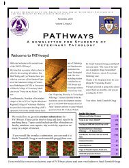
PATHways - American College of Veterinary Pathologists
PATHways - American College of Veterinary Pathologists
PATHways - American College of Veterinary Pathologists

Create successful ePaper yourself
Turn your PDF publications into a flip-book with our unique Google optimized e-Paper software.
A J O I N T N E W S L E T T E R O F T H E A M E R I C A N C O L L E G E O F V E T E R I N A R Y P A T H O L O G I S T S<br />
A N D T H E S O C I E T Y O F T O X I C O L O G I C P A T H O L O G Y<br />
Hello and welcome to the October<br />
2011 issue <strong>of</strong> <strong>PATHways</strong>.<br />
In “Discovering Discovery<br />
Path” Dr. Keiko Petrosky, a<br />
PhD scientist currently attending<br />
veterinary school at Tufts,<br />
gives a very interesting perspective<br />
on her toxicologic pathology<br />
summer internship at<br />
the biotechnology company<br />
Amgen. An overview <strong>of</strong> the student ACVP<br />
chapter at Michigan State University is given<br />
by Sara Lefman (Class <strong>of</strong> 2013). Check<br />
out all <strong>of</strong> the creative, pathology-related<br />
activities going on in Spartan-land (who<br />
knew myelomalacia could look so tasty?)!<br />
Next, Drs. Scott Fitzgerald and Jessica<br />
October, 2011<br />
Volume 3, Issue 2<br />
PAT H ways<br />
A n e w s l e t t e r f o r S t u d e n t s o f<br />
V e t e r i n a r y P a t h o l o g y<br />
We’re on the Web!<br />
www.toxpath.org<br />
www.acvp.org<br />
Did you know the STP website<br />
has a section dedicated solely<br />
to students? You can find information<br />
about membership, student<br />
opportunities and the NIH<br />
Loan Repayment Program.<br />
Welcome to <strong>PATHways</strong>!<br />
Hoane give a nice<br />
perspective on the<br />
residency training<br />
program at Michigan<br />
State University.<br />
Then Dr. Molly<br />
Boyle presents an<br />
interesting case report<br />
<strong>of</strong> parasitism in<br />
the liver <strong>of</strong> a pig.<br />
She includes some<br />
fabulous detailed photomicrographs, so<br />
brush up on your knowledge <strong>of</strong> microscopic<br />
features <strong>of</strong> a nematode! We also include a<br />
comprehensive listing <strong>of</strong> upcoming veterinary<br />
pathology related events, general announcements<br />
<strong>of</strong> interest, and student-related<br />
The STP is interested in hearing<br />
from students about how the<br />
student section <strong>of</strong> the website<br />
could be improved.<br />
Please check out the website<br />
by going to:<br />
http://www.toxpath.org and selecting<br />
“For Students”<br />
Please send any comments to<br />
Sue Pitsch at STP Headquarters<br />
(stp@toxpath.org) with<br />
“Student Website Feedback” as<br />
the subject.<br />
Inside this issue:<br />
activities at the ACVP meeting<br />
(see page 16). Page 17 gives the<br />
details on student travel funding<br />
for the ACVP meeting, and on submission<br />
<strong>of</strong> nominations for the<br />
Annual Mentor <strong>of</strong> the Year award–<br />
both <strong>of</strong> these have deadlines <strong>of</strong><br />
November 1st, so check this out<br />
first!<br />
Have fun reading the newsletter,<br />
and please don’t hesitate to let me<br />
know if you can think <strong>of</strong> a way to<br />
improve it, or if you have a contribution<br />
to make. I would love to<br />
include student contributions in the<br />
future! Your editor, Sarah<br />
Tannehill-Gregg<br />
Discovering Discovery Path 2-3<br />
Focus on Our Future… Michigan State University<br />
CVM Student ACVP Chapter<br />
Focus on Our Future… Michigan State University<br />
CVM Residency Training Program<br />
Case Report: The Case <strong>of</strong> the Milk-Spotted<br />
Liver<br />
4-5<br />
6-8<br />
9-14<br />
Announcements and Contact Information 15<br />
Additional Items <strong>of</strong> Interest 16-17<br />
Upcoming Events 18-20<br />
If you would like to receive an electronic copy <strong>of</strong> <strong>PATHways</strong>, please send your e-mail address to Sarah Tannehill-<br />
Gregg
V o l u m e 3 , I s s u e 2 P a g e 2<br />
DISCOVERING “DISCOVERY PATH”<br />
Contributed by Keiko Petrosky, PhD, Tufts Cummings School <strong>of</strong> <strong>Veterinary</strong> Medicine<br />
As veterinary students, we think <strong>of</strong> patients in terms<br />
<strong>of</strong> species: small animal v. large, exotic v. laborato-<br />
ry animals. It’s easy to forget the impact veterinari-<br />
ans can have on the human species, too. During my<br />
summer internship at Amgen, a biotech company<br />
that specializes in proteins as therapeutic agents, I<br />
met veterinarians who work to serve human pa-<br />
tients, from laboratory animal veterinarians to veter-<br />
inary pathologists, each playing an important role in<br />
the preclinical stage <strong>of</strong> the drug development pro-<br />
cess. I encountered veterinary pathologists who<br />
work in “discovery,” working as much on target<br />
validation and preclinical assay development as on<br />
efficacy and toxicity pr<strong>of</strong>iles. My particular projects<br />
were to develop a quantitative, immunoassay-<br />
based method to measure phosphosignal and to use<br />
proximity ligation to detect immune complexes<br />
from preclinical samples. I spent as much time<br />
doing bench work as reading slides! Along the<br />
way histologists taught me immunohistochemistry,<br />
multicolor immun<strong>of</strong>luorescence, and morphomet-<br />
ric analysis, the traditional tools <strong>of</strong> a veterinary<br />
pathology laboratory. With summer events like<br />
kayaking on the Charles River to the Boston Har-<br />
bor and a trip to Fenway Park, the Amgen intern-<br />
ship program coordinators fostered a sense <strong>of</strong> fun<br />
alongside a strong sense <strong>of</strong> mission to serve pa-<br />
tients.
V o l u m e 3 , I s s u e 2<br />
DISCOVERING “DISCOVERY PATH” , Cont.<br />
Assay development allowed me to flex the molecular<br />
muscles that I developed as a graduate student in bio-<br />
physics and biochemistry at UCSF. It was there that I<br />
encountered laboratory veterinarians for the first<br />
time. After completing my PhD, complete with then-<br />
governor Arnold Schwarzenegger’s signature, I spoke<br />
with every veterinary pathologist and laboratory ani-<br />
mal vet I could find to learn more about veterinary<br />
careers in biomedical research. Intrigued, I worked<br />
as a biologist at Novartis before enrolling in Tufts<br />
Cummings School <strong>of</strong> <strong>Veterinary</strong> Medicine with the<br />
express purposes <strong>of</strong> becoming a veterinary<br />
pathologist. At Tufts, I’m currently working on eval-<br />
uating grading schemes for common canine neo-<br />
plastic diseases, a unique opportunity to work with<br />
both pathologists and clinicians on applied research.<br />
<strong>Veterinary</strong> pathology is a way for me to combine my<br />
lifelong love <strong>of</strong> animals (and dead things) with re-<br />
search. From diagnostic pathology, toxicologic pa-<br />
thology, and discovery pathology, to regulatory ca-<br />
reers, veterinary pathology is a career with options<br />
for every interest. For the scientific and research-<br />
minded, and for those who want to have an impact on<br />
human as well as animal medicine, a career as a vet-<br />
erinary pathologist in industry might be for you, that<br />
is if you are willing to occasionally swap your micro-<br />
scope for a pipette, and your gross necropsies for a<br />
laboratory bench top!<br />
Dr. Keiko Petrosky<br />
“For all our conceits about<br />
being the center <strong>of</strong> the universe,<br />
we live in a routine planet <strong>of</strong> a<br />
humdrum star stuck away in an<br />
obscure corner ... on an<br />
unexceptional galaxy which is<br />
one <strong>of</strong> about 100 billion<br />
galaxies. That is the<br />
fundamental fact <strong>of</strong> the universe<br />
we inhabit, and it is very good for<br />
us to understand that”<br />
- Carl Sagan<br />
P a g e 3
V o l u m e 3 , I s s u e 2 P a g e 4<br />
Focus on our Future…. Showcasing our <strong>Veterinary</strong> Schools and Pathology<br />
Programs<br />
Michigan State University <strong>College</strong><br />
<strong>of</strong> <strong>Veterinary</strong> Medicine: Student<br />
Chapter <strong>of</strong> the ACVP<br />
Contributed by Sara Lefman (Class <strong>of</strong> 2013)<br />
The MSU CVM Pathology Club was founded in 1999<br />
by Dr. Laura Kennedy (class <strong>of</strong> 2001). With the support<br />
<strong>of</strong> faculty advisors Dr. Jon Patterson and Dr.<br />
Julia Stickle, the student executive board has organized<br />
multiple wet labs and brought in several speakers<br />
from the Michigan area. The club also works extensively<br />
with MSU’s Diagnostic Center for Population<br />
and Animal Health (DCPAH) in planning its<br />
events. On the weekends, students can partake in<br />
shadowing pathologists, residents, and clerkship students<br />
as they perform necropsies, make diagnoses<br />
and write up reports.<br />
Shadowing gives students the opportunity to apply<br />
Path Club members shadowing at DCPAH<br />
their knowledge, get their hands dirty before their<br />
clinical years and interact one-on-one with the faculty<br />
and residents. DCPAH also lends its facilities<br />
to necropsy wet labs where students can work side<br />
by side on anything from sea lions to horses. Participating<br />
in necropsy, biopsy and clinical pathology<br />
rounds <strong>of</strong>fers a welcome break from hours <strong>of</strong> lecture<br />
and helps to build confidence and problem<br />
solving skills.<br />
Other club events include the clinical pathology<br />
blood smear wet lab where participants practice<br />
preparing and interpreting slides, and a monthly<br />
“What’s Your Diagnosis?” contest on the club’s<br />
website which features a case that students must<br />
solve. The annual path club Epicurean encourages<br />
students to use their rusty right brains to come up<br />
with creative food dishes that resemble the presentation<br />
<strong>of</strong> diseases and parasites. Past winners have<br />
included melting abscess cream pie, “blood” Jello<br />
molds with gummy worm micr<strong>of</strong>ilaria, melt-inyour-mouth<br />
myelomalacia and caseous lymphadenitis<br />
chocolate puffs.
V o l u m e 3 , I s s u e 2 P a g e 5<br />
Focus on our Future…. Showcasing our <strong>Veterinary</strong> Schools and Pathology<br />
Programs, Student Chapter <strong>of</strong> the ACVP, cont.<br />
To share their knowledge and garner public support,<br />
members help out at Vet-A-Visit, an annual CVM<br />
event where the general public can visit the school<br />
and participate in club run exhibits.<br />
Kids enjoying the “Path Club Heart Worm Tunnel,” complete<br />
with mosquito and pulmonary artery filled with adult worms<br />
Path Club <strong>of</strong>ficers in the “Path Club Heart Worm Tunnel”<br />
Path Club <strong>of</strong>ficer explaining a histology slide<br />
Through all <strong>of</strong> these events, the Path Club has<br />
helped to foster interest in the subject <strong>of</strong> pathology<br />
and has churned out a number <strong>of</strong> students who have<br />
chosen to move on to residency programs. It <strong>of</strong>fers<br />
individuals the opportunity to meet like-minded students<br />
and become closer with the pathologists who<br />
will someday become their colleagues and mentors.<br />
Student chapters are an invaluable part <strong>of</strong> organized<br />
veterinary medicine that greatly improve the experiences<br />
<strong>of</strong> veterinary students and prepare them for the<br />
future.
V o l u m e 3 , I s s u e 2 P a g e 6<br />
Focus on our Future…. Showcasing our <strong>Veterinary</strong> Schools and Pathology<br />
Programs<br />
Michigan State University <strong>College</strong><br />
<strong>of</strong> <strong>Veterinary</strong> Medicine: Residen-<br />
cy Training Program<br />
Contributed by Scott Fitzgerald, DVM, PhD,<br />
DACVP (MSU Faculty) and Jessica Hoane,<br />
DVM DACVP (Charles River Pathology Associates)<br />
The mission <strong>of</strong> the Michigan State University Pathology<br />
Residency Training Program is to provide<br />
comprehensive and diverse training which leads to<br />
board certification by the <strong>American</strong> <strong>College</strong> <strong>of</strong> <strong>Veterinary</strong><br />
<strong>Pathologists</strong> (ACVP), and provides training<br />
for a successful career in any area <strong>of</strong> veterinary pathology.<br />
One <strong>of</strong> the foundations to this training is a<br />
heavy and diverse load <strong>of</strong> diagnostic cases; this is<br />
provided by the Diagnostic Center for Population &<br />
Animal Health (DCPAH) which is the only diagnostic<br />
laboratory in the state <strong>of</strong> Michigan and so<br />
receives all diagnostic specimens from the <strong>Veterinary</strong><br />
Teaching Hospital, all MSU laboratory animal<br />
specimens, as well as submissions from veterinarians,<br />
animal owners, and the four major zoos in<br />
Michigan. The DCPAH was built in 2004 and provides<br />
state-<strong>of</strong>-the-art facilities including 4 different<br />
necropsy floors and a biosafety level 3 floor.<br />
Our residency training faculty includes 11 board<br />
certified anatomic pathologists, and 4 board certified<br />
clinical pathologists, each <strong>of</strong> whom has one or<br />
more areas <strong>of</strong> specialty interest and expertise. This<br />
provides our residents with an invaluable resource<br />
for case consultations as well as mentors. The residents<br />
receive training through a series <strong>of</strong> courses<br />
and seminars in addition to their hands-on practical<br />
training. Courses are <strong>of</strong>fered over a two-year cycle.<br />
Required courses include Advanced General Pathology,<br />
Advanced Systemic Pathology, and Advanced<br />
Clinical Pathology. Elective courses include<br />
Poultry Pathology, Pathology <strong>of</strong> Neoplastic<br />
Diseases, Laboratory Animal Pathology, and Electron-microscopic<br />
Techniques. Courses are supple-<br />
mented by ongoing seminars and rounds. The training<br />
program also provides exposure to teaching, an<br />
annual mock board examination held in conjunction<br />
with the University <strong>of</strong> Guelph, Ontario, Canada,<br />
and financial support to attend various national<br />
meetings or board-preparatory short-courses.<br />
Diagnostic Center for Population and Animal Health (DCPAH) Facility, Lansing, MI
V o l u m e 3 , I s s u e 2 P a g e 7<br />
Focus on our Future…. Showcasing our <strong>Veterinary</strong> Schools and Pathology<br />
Programs, Residency Training Program, cont.<br />
Since 2000, 35 residents (26 anatomic and 9<br />
clinical) have completed their pathology training<br />
through Michigan State University with a current<br />
group <strong>of</strong> 10 residents (7 anatomic and 3 clinical). The<br />
majority <strong>of</strong> the residency positions (57%) have been<br />
funded internally, though a large number (34%) <strong>of</strong><br />
both anatomic and clinical residencies have been<br />
funded by MPI Research, Inc. a privately held contract<br />
research organization (CRO) located in Mattawan,<br />
MI. The MPI position provides a unique opportunity<br />
for anatomic and clinical pathology residents to<br />
spend 15 months in a toxicologic pathology environment<br />
during the final year <strong>of</strong> their residency. Two<br />
residents have been funded through the ACVP-STP<br />
Coalition which also provides for opportunities to<br />
spend time in a toxicologic pathology environment.<br />
Ultimately, our residents have an excellent track record<br />
at passing the ACVP board examination (over<br />
80% pass rate on the first attempt). Job placement<br />
following the residency is quite successful and diverse<br />
with 10 past residents in contract lab/<br />
pharmaceutical positions, 10 in university positions, 5<br />
in government positions, one is a private consultant,<br />
and 7 are currently in a PhD program.<br />
<strong>American</strong> Alligator Claw<br />
Photograph by Richard T Bryant<br />
http://photography.nationalgeographic.com
V o l u m e 3 , I s s u e 2 P a g e 8<br />
Focus on our Future…. Showcasing our <strong>Veterinary</strong> Schools and Pathology<br />
Programs, Residency Training Program, cont.<br />
Necropsy floor<br />
at Michigan<br />
State University<br />
CVM<br />
Friday gross pathology<br />
rounds at Michigan State<br />
University CVM with<br />
Dalen Agnew.
V o l u m e 3 , I s s u e 2 P a g e 9<br />
Case Report<br />
The case <strong>of</strong> the milk-spotted liver…<br />
The presentation…………….<br />
Signalment: 3 month old, gilt, Yorkshire Cross, Sus scr<strong>of</strong>a domesticus, porcine<br />
History: This pig was housed with 9 other gilts. She was the only pig reported to have<br />
dyspnea, cough, diarrhea, and lethargy.<br />
Necropsy/gross pathologic findings: There were multifocal to coalescing pale nodules<br />
on the serosal surface <strong>of</strong> the liver and extending into the parenchyma. These nodules<br />
extruded white pasty material on cut section. Within some <strong>of</strong> these nodules and corresponding<br />
ectatic bile ducts, there were adult ascarids measuring approximately 10-20 cm<br />
long and approximately 1 mm wide. Within the caudal lung lobes, there were locally extensive<br />
regions <strong>of</strong> mild to moderate consolidation; these foci <strong>of</strong> consolidation were pale tan<br />
to grey. Within the cranioventral lobes, there was purple mottling bilaterally.<br />
Gross appearance <strong>of</strong> the liver<br />
(Close-up view <strong>of</strong> area in box on page 10)<br />
Special thanks to Dr. Molly Boyle, Integrated Laboratory Systems, Inc., for contributing this case.
V o l u m e 3 , I s s u e 2 P a g e 10<br />
Case Report<br />
The case <strong>of</strong> the milk-spotted liver, Continued…..<br />
Gross appearance <strong>of</strong> the liver, close-up view<br />
Ascarids<br />
White pasty material<br />
Laboratory Results: Bacterial culture <strong>of</strong> the liver revealed numerous Aerococcus<br />
spp., and few coliform. Bacterial culture <strong>of</strong> the lung revealed numerous Pasteurella<br />
multocida and Mycoplasma hyorhinis. Bacterial culture <strong>of</strong> the intestine<br />
was negative. Bacterial culture <strong>of</strong> the spleen was negative. Virology (PCV-1,<br />
PCV-2, PRRS) via PCR was positive for PCV-2. Qualitative fecal revealed many<br />
Balantidium coli cysts and few Ascaris suum eggs. Parasite identification <strong>of</strong> those<br />
seen within the bile ducts <strong>of</strong> the liver during gross necropsy revealed Ascaris suum.
V o l u m e 3 , I s s u e 2 P a g e 11<br />
Case Report<br />
The case <strong>of</strong> the milk-spotted liver, Continued…….<br />
Microscopic description: Multifocally, within sections <strong>of</strong> liver, bile ducts were<br />
markedly ectatic and contained cross-sections <strong>of</strong> adult nematodes measuring approximately<br />
1 mm in diameter. These nematodes contained a thick cuticle measuring approximately<br />
15 microns and an underlying hypodermis. They had coelomyarianpolymyarian<br />
musculature and prominent lateral cords. There were variable amounts <strong>of</strong><br />
pseudocoelomic membranes connecting the intestine and sarcoplasm <strong>of</strong> the musculature<br />
and lateral cords. The large digestive tract was composed <strong>of</strong> uninucleate ciliated<br />
columnar epithelial cells. Nematodes were variably surrounded by lakes <strong>of</strong> fibrin, various<br />
combinations and concentrations <strong>of</strong> neutrophils and eosinophils, and basophilic<br />
streaming cellular (nuclear) debris. Many bile ducts were lined by markedly hypertrophic<br />
epithelium and were surrounded by a thick rim <strong>of</strong> fibrosis and lymphoplasmacytic<br />
inflammation. In these areas, there was also prominent bile duct hyperplasia<br />
and occasional lymphatic dilatation. There was no microscopic evidence <strong>of</strong> parasitic<br />
disease in other organs.<br />
Periductular<br />
inflammation<br />
Liver<br />
Bile ducts<br />
Lobule<br />
Periductular<br />
fibrosis<br />
Periparasitic cellular<br />
reaction<br />
Arteriole<br />
Nematode in cross-section.
V o l u m e 3 , I s s u e 2 P a g e 12<br />
Case Report<br />
The case <strong>of</strong> the milk-spotted liver, Continued…….<br />
Digestive tract<br />
Cuticle<br />
Ascaris suum in the liver with periparasitic cellular reaction<br />
Lateral chord<br />
Coelomyarianpolymyarian<br />
musculature
V o l u m e 3 , I s s u e 2 P a g e 13<br />
Case Report<br />
The case <strong>of</strong> the milk-spotted liver, Continued…….<br />
Nucleus<br />
Cilia<br />
Uninucleate ciliated<br />
columnar epithelial<br />
cells <strong>of</strong> the intestinal<br />
tract, A. suum
V o l u m e 3 , I s s u e 2 P a g e 14<br />
Case Report<br />
The case <strong>of</strong> the Milk-Spotted Liver, Continued…...<br />
And the answer is…………….<br />
Morphologic diagnoses: Liver: Severe chronic active cholecystitis and hepatitis with<br />
intralesional adult nematodes, marked bile duct hyperplasia and hypertrophy, and lymphatic<br />
dilatation<br />
Comments: Adults <strong>of</strong> the large roundworm, Ascaris suum, reside most commonly in the<br />
small intestine but may migrate aberrantly into the stomach or bile ducts. It’s important to<br />
note that this can occur post-mortem. The pathologist should be especially wary <strong>of</strong> this if<br />
single to small numbers <strong>of</strong> adults are seen within bile ducts. In this case, however, necropsy<br />
was performed within minutes <strong>of</strong> euthanasia, there were moderate numbers <strong>of</strong> adults<br />
within bile ducts, and severe lesions consistent with long-standing infection.<br />
In the life cycle <strong>of</strong> Ascaris suum, many eggs are produced (up to 250,000/day). They can<br />
develop to the infective stage (containing the L3 larva) in 2-3 weeks in warm conditions.<br />
For the most part, eggs are resistant to chemical agents, but low humidity, heat, or direct<br />
sunlight are detrimental to survival. After ingestion <strong>of</strong> the larvated eggs, hatching occurs<br />
in the small intestine. Within two hours, numerous small submucosal hemorrhages may be<br />
seen in the wall <strong>of</strong> the duodenum and anterior jejunum as the larvae penetrate to the hepatic<br />
portal system. Larvae arrive in the liver where they molt to the L3. The L3 leave the liver<br />
and travel to the lungs via the bloodstream. They break out <strong>of</strong> the capillaries into the<br />
alveoli where they molt; the L4 migrate up the bronchioles, bronchi, and trachea to the<br />
pharynx. The L4 are swallowed and swept to the small intestine where they grow rapidly<br />
and molt to the L5. Patency occurs at 35-60 days.<br />
The severe hepatic changes noted in this case may have contributed to clinical debilitation.<br />
Histologically, this pig also had severe bronchopneumonia (Pasteurella multocida and Mycoplasma<br />
hyorhinis), a mild lymphoplasmacytic perivascular encephalitis (nonspecific, but<br />
suggestive <strong>of</strong> sepsis), a lymphoplasmacytic interstitial nephritis, and lymphoid depletion<br />
(PCV-2). Immunohistochemistry for Leptospira spp. was negative. As such, the interstitial<br />
nephritis may have been associated with PCV-2. Intracytoplasmic botryoid inclusions<br />
were not noted in lymph node or kidney. Qualitative fecal revealed many Balantidium coli<br />
cysts and few Ascaris suum eggs.<br />
References available on page 15.
V o l u m e 3 , I s s u e 2<br />
References for Case Report<br />
References:<br />
1. M.B. Chitwood and J.R. Lichtenfels, Identification <strong>of</strong><br />
parasitic Metazoa in tissue sections, Exper. Parasitol. 32<br />
(1972), p. 472.<br />
2. Gardiner, CH, Poynton SL. An Atlas <strong>of</strong> Metazoan<br />
Parasites in Animal Tissues. Washington DC: Armed<br />
Forces Institute <strong>of</strong> Pathology, 1999, pp. 19-21.<br />
3. Leman A. D. et al. Diseases <strong>of</strong> Swine. (Ames, Iowa:<br />
Iowa State University Press, 1986.)<br />
4. Merck & Company. Merck <strong>Veterinary</strong> Manual, 9 th<br />
ed. (Whitehouse Station, NJ: Merck & Co., Inc. with<br />
Merial Limited, 2006.)<br />
A m e r i c a n C o l l e g e o f<br />
V e t e r i n a r y<br />
P a t h o l o g i s t s<br />
2810 Crossroads Drive<br />
Suite 3800<br />
Madison, Wisconsin 53718<br />
Phone: 608-443-2466<br />
Fax: 608-442-2474<br />
Email: membership@acvp.org<br />
S o c i e t y o f<br />
T o x i c o l o g i c<br />
P a t h o l o g y<br />
1821 Michael Faraday Drive<br />
Suite 300<br />
Reston, VA 20190<br />
Phone: 703-438-7508<br />
Fax: 703-438-3113<br />
Email: stp@toxpath.org<br />
Newsletter Committee Members<br />
Feel free to contact committee members with questions or for advice<br />
ACVP Recruiting Committee<br />
Student Chapter Subcommittee<br />
Newsletter Group:<br />
Krista La Perle*<br />
(la-perle.1@osu.edu)<br />
Stephanie Corn*<br />
(stephanie-corn@idexx.com)<br />
Kaori Sakamoto<br />
(kaoris@uga.edu)<br />
Brett Saladino<br />
(brett.saladino@covance.com)<br />
* Contributed to this newsletter<br />
STP Career Development and Outreach<br />
Committee<br />
Student Interactions Subcommittee<br />
Newsletter Group:<br />
Sarah Tannehill-Gregg*<br />
(stannehi@amgen.com)<br />
Jim Proctor<br />
(jprocto2@its.jnj.com)<br />
Jessica Hoane*<br />
(jessica.hoane@crl.com)<br />
Molly Boyle*<br />
(mboyle@ils-inc.coml)<br />
Gaurav Tyagi<br />
(gaurav.tyagi@roche.com)<br />
Check out the ACVP Student<br />
Chapters group on Facebook!<br />
http://www.facebook.com/#!/group.php?gid=1238795976443<br />
10<br />
This is a great site for student chapters to:<br />
� Exchange ideas about meeting topics<br />
� Get ideas for fundraising<br />
� Look for housing for externships<br />
� Find roommates for meetings<br />
http://www.r<strong>of</strong>lzoo.com/cat-the-vet.html<br />
http://www.phombo.com<br />
http://christchurch.yatbo.com/petsanimals-a-dogs-puppies-a-a3317232560-a-English-bulldog-pupp<br />
P a g e 15
V o l u m e 3 , I s s u e 2 P a g e 16<br />
Events for <strong>Veterinary</strong> Students at the ACVP/ASVCP Annual Meeting<br />
The joint annual meeting <strong>of</strong> the <strong>American</strong> <strong>College</strong> <strong>of</strong> <strong>Veterinary</strong> <strong>Pathologists</strong> and the <strong>American</strong> Society for<br />
<strong>Veterinary</strong> Clinical Pathology is rapidly approaching! Hopefully, many students will be able to attend. In<br />
addition to the scheduled scientific sessions and workshops, there are several events that focus on veterinary<br />
students. These events include:<br />
� Saturday, December 3rd<br />
� 6:00 - 7:00 PM Set up posters<br />
� 7:00 - 8:30 PM Focused Poster Session and Opening Reception (students invited to preside by<br />
their posters during this time)<br />
� Sunday, December 4 th<br />
� 7:00 - 8:00 AM <strong>Veterinary</strong> Student Breakfast<br />
� 9:30 AM - 4:30 PM Poster viewing<br />
� 9:40 - 10:15 AM Morning Refreshment Break (students encouraged to be present at their posters<br />
if available)<br />
� Monday, December 5 th<br />
� 9:30 AM - 5:00 PM Poster viewing<br />
� 1:45 - 3:00 PM Meet the <strong>Pathologists</strong>: Discussion <strong>of</strong> Careers in Pathology<br />
� 3:00 - 5:00 PM <strong>Veterinary</strong> Student Resident Forum<br />
� Tuesday, December 6 th<br />
� 9:30 AM - 3:30 PM Poster viewing (posters should be taken down after this time so you can<br />
take them back with you)<br />
� 9:40 - 10:30 AM Morning Refreshment Break (students encouraged to be present at their posters<br />
if available)<br />
� 5:15 - 7:00 PM Awards and Recognition Ceremony<br />
All students attending the meeting are encouraged to attend the <strong>Veterinary</strong> Student Breakfast, Meet the<br />
<strong>Pathologists</strong> Discussion and <strong>Veterinary</strong> Student Resident Forum. Chapters receiving ACVP Travel Awards<br />
will be required to send one or more representatives to the Breakfast during which you can mingle with<br />
members <strong>of</strong> Council and the Student Chapter Committee. We will also raffle <strong>of</strong>f the new 5 th edition <strong>of</strong><br />
Pathologic Basis <strong>of</strong> <strong>Veterinary</strong> Disease by Zachary and McGavin to one lucky student in attendance (must<br />
be present to win). Bring any questions about careers in pathology to the Meet the <strong>Pathologists</strong> Discussion.<br />
At the <strong>Veterinary</strong> Student Resident Forum, you’ll get to meet with current residents and pathology residency<br />
training coordinators from residency programs in the United States and abroad to find out which pathology<br />
residency is the best fit for you.<br />
See you in Nashville! Please check the program listing at registration for any changes to the schedule.
V o l u m e 3 , I s s u e 2 P a g e 17<br />
Travel Funds for the ACVP Annual Meeting<br />
Requests for $1000 ACVP Travel Awards to attend the ACVP Annual Meeting in Nashville should be submitted<br />
in writing to Dr. Krista La Perle (la-perle.1@osu.edu) by November 1 st . Chapters receiving Travel<br />
Awards will be required to send one or more representatives to the <strong>Veterinary</strong> Student Chapter Breakfast on<br />
Sunday December 4 th from 7-8 AM. In lieu <strong>of</strong> receipts, Chapters will also be asked to submit an Annual<br />
Meeting Activity Report which summarizes, in 500 words or less, the number <strong>of</strong> students in attendance,<br />
events that were attended, information that was learned, and all the fun that was had! Submission <strong>of</strong> photographs<br />
is also encouraged as these reports will be published on the ACVP website and/or in the March 2012<br />
issue <strong>of</strong> the ACVP Newsletter.<br />
Annual Mentor <strong>of</strong> the Year Award<br />
Want to recognize an outstanding Chapter Faculty Advisor? Is there a Pathology Resident or Graduate Student<br />
who has been actively engaged in Chapter Activities? How about a Pathologist, either within or outside<br />
your veterinary school, who mentored you during an externship? Please consider nominating him or<br />
her for the Annual Mentor <strong>of</strong> the Year Award to be announced during the Awards and Recognition Ceremony<br />
on Tuesday December 6 th from 5:15-7:00 PM. Nominations should be submitted to Dr. Krista La<br />
Perle (la-perle.1@osu.edu) by November 1 st and must include letters <strong>of</strong> nomination from (a) Student Chapter<br />
Officers/Members AND (b) a pathologist from the nominated mentor’s workplace.
V o l u m e 3 , I s s u e 2 P a g e 18<br />
Upcoming Events………………..<br />
Nov 6-9, 2011 <strong>American</strong> <strong>College</strong> <strong>of</strong> Toxicology<br />
32nd Annual Meeting<br />
Phoenix, AZ<br />
http://www.actox.org/<br />
Nov 17-18, 2011 British Society <strong>of</strong> Toxicological<br />
<strong>Pathologists</strong> 26th Annual Meeting<br />
Anti-Inflammatory Disease Therapies<br />
London, U.K.<br />
http://www.bstp.org.uk/<br />
Dec 3-7, 2011 ACVP and ASVCP Concurrent Annual<br />
Meetings<br />
Nashville, TN<br />
http://www,acvp.org/<br />
http://www,ascvp.org/<br />
Feb 1-3, 2012 British Society <strong>of</strong> Toxicological<br />
<strong>Pathologists</strong><br />
Mouse Pathology Meeting<br />
Cambridge, UK<br />
http://www.bstp.org.uk/<br />
March 11-15, 2012 Society <strong>of</strong> Toxicology Annual<br />
Meeting and ToxExpo<br />
San Francisco, CA<br />
http://toxicology.org/<br />
April 21-25, 2012 ASIP Annual Meeting at Experimental<br />
Biology 2012<br />
San Diego, CA<br />
http://www.asip.org/mtgs/eb12/<br />
June 23-28, 2012 Society <strong>of</strong> Toxicologic Pathology<br />
31st Annual Symposium<br />
Mechanisms <strong>of</strong> Toxicity<br />
Boston, MA<br />
http://www.toxpath.org/<br />
July 23-27, 2012 9th Annual Biennial Shortcourse<br />
on Industrial Toxicology and Pathology<br />
Champaign, IL<br />
http://vetmed.illinois.edu/ope/itp/<br />
July 26-27, 2012 Midwest Association <strong>of</strong> <strong>Veterinary</strong><br />
<strong>Pathologists</strong> Annual Meeting<br />
New Harmony, IN<br />
http://vetmed.illinois.edu/mavp/<br />
Sep 16-18, 2012 6th Research Triangle Park Rodent<br />
Pathology Course<br />
Cary, NC<br />
http://rtprodentpathology.org/<br />
index.html<br />
Sep 18-20, 2012 <strong>American</strong> <strong>College</strong> <strong>of</strong> <strong>Veterinary</strong><br />
<strong>Pathologists</strong> Certifying Exam<br />
Ames, IA<br />
http://www,acvp.org/<br />
Noni Nectar for Green Gecko<br />
Chandra Sherin<br />
http://ngm.nationalgeographic.com
V o l u m e 3 , I s s u e 2 P a g e 19<br />
Announcement from September 2011 Charles Louis Davis DVM Foundation newsletter
V o l u m e 3 , I s s u e 2 P a g e 20<br />
Announcement from September 2011 Charles Louis Davis DVM Foundation newsletter