Two-dimensional thin-layer chromatography of rat liver phosphatides
Two-dimensional thin-layer chromatography of rat liver phosphatides
Two-dimensional thin-layer chromatography of rat liver phosphatides

You also want an ePaper? Increase the reach of your titles
YUMPU automatically turns print PDFs into web optimized ePapers that Google loves.
J. Lipid Reeearch, October, 1962<br />
Volume 3, Number 4<br />
<strong>Two</strong>-<strong>dimensional</strong> <strong>thin</strong>-<strong>layer</strong> <strong>chromatography</strong> <strong>of</strong> <strong>rat</strong><br />
<strong>liver</strong> <strong>phosphatides</strong><br />
W. D. SKIDMORE and C. ENTENMAN<br />
U. S. Naval Radiological Defense Labo<strong>rat</strong>ory,<br />
San Francisco, California<br />
[Received for publication May 21, 19621<br />
SUMMARY<br />
A system <strong>of</strong> two-<strong>dimensional</strong> <strong>thin</strong>-<strong>layer</strong> <strong>chromatography</strong> was developed that sepa<strong>rat</strong>ed<br />
<strong>rat</strong> <strong>liver</strong> <strong>phosphatides</strong> into several phosphate-positive spots in about 2 hr developing<br />
time. Characteristic hydrolysis products derived from phosphatidyl serine, phosphatidyl<br />
ethanolamine, phosphatidyl inositol, phosphatidyl choline, sphingomyelin, and lysophosphatidyl<br />
choline were identified. The hydrolytic products <strong>of</strong> “phosphatidic acid” were not<br />
definitely characterized. The application <strong>of</strong> <strong>thin</strong>-<strong>layer</strong> <strong>chromatography</strong> as described for<br />
<strong>rat</strong> <strong>liver</strong> <strong>phosphatides</strong> can be extended to phosphatide extracts <strong>of</strong> other tissues.<br />
T h e trend toward widespread use <strong>of</strong> <strong>thin</strong>-<strong>layer</strong><br />
<strong>chromatography</strong> (TLC) was accele<strong>rat</strong>ed by the work <strong>of</strong><br />
Stahl (1-3) who demonst<strong>rat</strong>ed its potential usefulness in<br />
lipid research. In an excellent review, Mangold (4)<br />
presented a detailed description <strong>of</strong> commercially available<br />
appa<strong>rat</strong>us and several techniques used in TLC as<br />
well as applications <strong>of</strong> the method to lipids in general.<br />
One-<strong>dimensional</strong> TLC has been applied to the sepa<strong>rat</strong>ion<br />
<strong>of</strong> <strong>phosphatides</strong> from brain (5, 6), serum (7-g), and<br />
other sources (10) with varying degrees <strong>of</strong> success. It<br />
was observed in this labo<strong>rat</strong>ory that one-<strong>dimensional</strong><br />
TLC did not sepa<strong>rat</strong>e all <strong>of</strong> the <strong>phosphatides</strong> found in a<br />
<strong>rat</strong> <strong>liver</strong> extract with the solvent systems used. Therefore,<br />
a system <strong>of</strong> two-<strong>dimensional</strong> TLC was applied.<br />
Tentative identification <strong>of</strong> the <strong>phosphatides</strong> was made<br />
possible by the application <strong>of</strong> different color-developing<br />
reagents. Further identification was accomplished by<br />
eluting the <strong>phosphatides</strong>, hydrolyzing them, and identifying<br />
the hydrolytic cleavage products with standards<br />
by TLC. In this way, the seven phosphatide spots<br />
were identified as “phosphatidic acid” (PM), phosphatidyl<br />
serine (PhS) , phosphatidyl ethanolamine<br />
(PhE), phosphatidyl inositol (PhI), phosphatidyl choline<br />
(PhC) , sphingomyelin (Sph), and lysophosphatidyl<br />
choline (L-phc).<br />
MATERIALS<br />
1. Detection reagents for <strong>phosphatides</strong> and their hydrolytic<br />
products.<br />
a. Iodine vapor (12) to detect lipids nonspecifically. Dry<br />
plates were expmed for about 1 min in a plastic box containing<br />
iodine vapor produced from iodine crystals (4).<br />
471<br />
b. Ninhydrin (Nin) to detect amino <strong>phosphatides</strong>. Dry<br />
plates were sprayed with a solution <strong>of</strong> 0.3 g ninhydrin in 5 m1<br />
lutidine and 95 m1 wbutanol satu<strong>rat</strong>ed with water. As the<br />
plates were dried at room tempe<strong>rat</strong>ure, red-violet spots appeared<br />
on a white background.<br />
c. Molybdic acid (MO) to detect <strong>phosphatides</strong>. Dry plates<br />
were sprayed with a solution <strong>of</strong> 5 m1 60% w/v perchloric acid,<br />
10 m1 N HC1, and 25 m1 4% w/v ammonium molybdate (11).<br />
Blue spots appeared on a white background as the plates were<br />
dried at room tempe<strong>rat</strong>ure.<br />
d. Ferric chloride-sulfosalicylic acid (Fe) to detect phosphate<br />
groups. Dry plates were sprayed with a solution <strong>of</strong><br />
7.0 g sulfosalicylic acid, 0.1 g FeC13.6H20, and 25 m1 water<br />
diluted to 100 m1 with 95% ethanol (12). White fluorescent<br />
spots appeared on a purple background as the plates were<br />
dried at room tempe<strong>rat</strong>ure.<br />
e. Ammoniacal silver nit<strong>rat</strong>e (Ag) to detect glycerol and<br />
inositol. Dry plates were sprayed with a mixture <strong>of</strong> equal<br />
volumes <strong>of</strong> 0.1 N AgN03 and 7 N ammonium hydroxide (13).<br />
The plates were then heated at llOo until dark brown spots<br />
appeared on a white background.<br />
f. Dragendorf reagent (Bi) to detect choline. Dry plates<br />
were sprayed with a mixture <strong>of</strong> 4 m1 solution I, l m1 solution<br />
11, and 20 m1 distilled water. Solution I contained 1.7 g<br />
Bi(N03)3.5Hz0 diluted to 100 m1 with 20% v/v acetic acid.<br />
Solution I1 contained 40 g K1 in 100 m1 water. As the plates<br />
were dried at room tempe<strong>rat</strong>ure, free choline produced a purple<br />
spot and choline-containing compounds produced orange spots<br />
(6).<br />
g. Fuchsin-sulfurous acid (Schiff’s reagent) to detect aldehyde<br />
groups. Dry plates were sprayed with a solution <strong>of</strong> 1 m1<br />
Fuchsin-sulfite reagent (14), 1 m1 0.05 M HgCl,, and 10 m1<br />
0.05 M HzSOa diluted to 100 m1 with distilled water. Violet<br />
spots appeared rapidly on a pale violet background.<br />
h. Hydroxylamine-ferric chloride to detect esterified fatty<br />
acids (EFA). Dry plates were sprayed with alkaline hydroxylamine<br />
reagent, dried briefly, and then sprayed with<br />
ethereal acid ferric chloride reagent. Purple spots appeared<br />
quickly on a yellow background (15).<br />
The alkaline hydroxylamine reagent was prepared by dissolving<br />
10 g HONH2 ‘HC1 in 25 m1 water and diluting it to 100<br />
m1 with ethanol and mixing this reagent with 26 m1 <strong>of</strong> satu<strong>rat</strong>ed<br />
aqueous NaOH diluted to 200 m1 with ethanol. The<br />
Downloaded from<br />
www.jlr.org by guest, on January 14, 2013
472 SKIDMORE ASD ENTEKSMAW<br />
I<br />
* I<br />
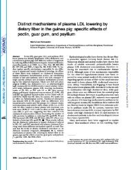
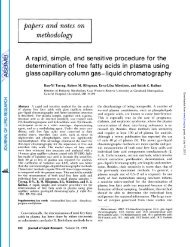
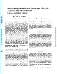
FIG. 1. Twcdimenaionnl TIE chromntogrnm <strong>of</strong> mixed phosphntidea.<br />
A portion (10 pl, 45 pg P) <strong>of</strong> rnt <strong>liver</strong> phogphnt.ide<br />
extrnct was npplicd to the origin <strong>of</strong> n TLC plate. The first<br />
dimension was developed for 1 hr with Solvent I, nnd the second<br />
dimension was developd for 1 hr with Solvent 11. The phosphntides<br />
were datcctcd with iodine vapor. The following<br />
nhbrrvintions are used: LPhC (lysophoephntidyl choline), Sph<br />
(sphingomyelin), PhC (phosphatidyl choline), PhS (phosphntdyl<br />
serine), I’hI (phosphntidyl int~itol), PhlC (phosphntidyl csthnnolnnline),<br />
and PhA (“phcwphnticlic acid”).<br />
sodium chloride precipitnte was removed by filt<strong>rat</strong>ion. The<br />
ethereal mid ferric chloride reagent waa prepnred by first<br />
grinding together n mixture <strong>of</strong> 10 g FeClr.GH20 nnd ‘LO ml HCI<br />
(:37yo w/v) with n mortnr and pestle nnd then shnkinq the r e<br />
sulting solution with 3 00 ml ether.<br />
2. Developing aolvenL~jor TI.
THIN-LAYER CHROMATOGRAPHY OF PHOSPHATIDES 4'73<br />
in an enclosed plastic box. After dark yellow spots on a<br />
white background were observed, the plate was removed<br />
and the iodine on the plate was then allowed to evap-<br />
o<strong>rat</strong>e and the spots allowed to disappear. The plate<br />
then could be sprayed with a variety <strong>of</strong> different detec-<br />
tion reagents. A permanent record <strong>of</strong> the chromato-<br />
grams was obtained by photographing them with a<br />
Polaroid camera.<br />
Only chromatograms on which the lipid had been<br />
revealed with iodine were used for elution <strong>of</strong> lipid from<br />
the spot. After the iodine had evapo<strong>rat</strong>ed, the spots<br />
were scraped <strong>of</strong>f the chromatogram, collected sepa<strong>rat</strong>ely<br />
on weighing papers, and transferred to centrifuge tubes.<br />
Solvent I was added to each tube. The mixture was<br />
then agitated, centrifuged, and the supernatant fluid<br />
decanted into another tube. The <strong>thin</strong>-<strong>layer</strong> residue<br />
was extracted two more times with methanol. All<br />
supernatants were combined. Portions were taken for<br />
phosphorus and esterified fatty acid determinations, for<br />
hydrolysis studies, and for re<strong>chromatography</strong> on TLC.<br />
Prepa<strong>rat</strong>ion <strong>of</strong> Rat Liver Phosphatide Solution.<br />
Phosphatides were extracted from fresh <strong>rat</strong> <strong>liver</strong> by the<br />
method <strong>of</strong> Folch et al. (16) and finally precipitated with<br />
acetone (17).<br />
Hydrolysis <strong>of</strong> Phosphatides. Portions <strong>of</strong> each eluted<br />
phosphatide were heated in sealed ampules with 1.7 N<br />
methanolic HCl (anhydrous) for 4 hr at 1OOO (18). The<br />
hydrolysates were taken to dryness under reduced pres-<br />
sure at 75'. Distilled water was added to each ampule,<br />
and a portion <strong>of</strong> each sample was applied to TLC.<br />
Samples <strong>of</strong> each <strong>of</strong> the expected characteristic prod-<br />
ucts <strong>of</strong> hydrolysis were carried through this same pro-<br />
cedure to check for possible further degradation.<br />
Chemical Methods. Phosphorus (P) was determined<br />
by the method <strong>of</strong> Bartlett (19) and esterified fatty acids<br />
(EFA) by the method <strong>of</strong> Skidmore and Entenman (20).<br />
RESULTS<br />
The phosphatide mixture prepared from <strong>rat</strong> <strong>liver</strong> was<br />
dissolved in chlor<strong>of</strong>orm, and a portion was applied to a<br />
TLC plate and developed with Solvent I in the first<br />
dimension and Solvent I1 in the second. Seven iodinepositive<br />
spots were observed between the origin and the<br />
solvent fronts. The. platk was immediately photographed<br />
(Fig. 1). The <strong>phosphatides</strong> were tentatively<br />
identified by observing the color reactions <strong>of</strong> the spots<br />
(Table 1).<br />
In order to identify the seven phosphate-positive<br />
spots, it was necessary to obtain a sufficient quantity<br />
<strong>of</strong> each phosphatide to characterize it by hydrolysis.<br />
Partial sepa<strong>rat</strong>ion <strong>of</strong> the <strong>phosphatides</strong> was accomplished<br />
with a silicic acid column (17). The seven <strong>phosphatides</strong><br />
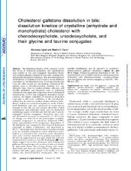
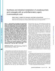
FIG. 2. Rechromntogr:lphy <strong>of</strong> phtrspht~tidrs c4utrtl Iron1 a TLC<br />
chromatogram. The chromatogr:~m was dcvrloprd in one<br />
dimension with Solvent I for 10 cm. The spots were detected<br />
with iodine vapor and circled. The dotted circles indicate<br />
weakly stained spots. The iodine was allowed to evapo<strong>rat</strong>e and<br />
the plate then sprayed for <strong>phosphatides</strong> with the molybdic acid<br />
reagent. The photograph shows the phosphate-positive spots.<br />
The phosphatide and the number <strong>of</strong> micrograms <strong>of</strong> P applied tv<br />
the origin were as follows: (1) PhA, 2.ti; (2) PhS, 1.4; (3)<br />
PhE, 2.7; (4) PhI, 23; (5) PhC, 2.5; (6) Sph, 2.1; (7) L-<br />
PhC, 2.7.<br />
werc finally isolated by selecting partially sepa<strong>rat</strong>ed<br />
column fractions, which contained one or two different<br />
<strong>phosphatides</strong> as major components, and applying large<br />
portions to TLC plates. The plates were developed<br />
with Solvent I in one dimension. The individual phos-<br />
phatides were eluted as described in the Methods sec-<br />
tion, and a portion <strong>of</strong> each was applied to TLC and<br />
developed in one dimension with Solvent I (Fig. 2). It<br />
was evident that some <strong>of</strong> the <strong>phosphatides</strong> had deteri-<br />
o<strong>rat</strong>ed. The P/EFA <strong>rat</strong>io and the Rr values <strong>of</strong> the re-<br />
chromatographed <strong>phosphatides</strong> are listed in Table 2.<br />
Another portion <strong>of</strong> each isolated phosphatide was hy-<br />
drolyzed, and a portion <strong>of</strong> each hydrolyzed sample was<br />
TABLE 2. Rf VALUES AND PHOSPHORUS/FATIT<br />
ACID RATIOS OF<br />
PURIFIED PHOSPHATIDES<br />
Rf Values P/EFA<br />
with Molar<br />
Phosohatide Eluted Solvcnt I* Ratio<br />
"Phosphatidic acid" 0.70 1:1.4<br />
Phosphatidyl serine 0.23 1:l.S<br />
Phosphatidyl ethanolamine 0.52 1:2.1<br />
Phosphatidyl inositol 0.33 1:1.5<br />
Phosphatidyl choline 0.32 1:lA<br />
Sphingomyelin 0.17 1:0.05<br />
Lysophosphatidyl choline 0.12 1:o.g<br />
* The Rf values were determined from Fig. 2.<br />
Downloaded from<br />
www.jlr.org<br />
by guest, on January 14, 2013
""I<br />
SKIDMORE AND ENTENMAN<br />
.- - , '~ ".<br />
SOLVENT FRONT<br />
SOLVENT- FRONT<br />
h I<br />
i<br />
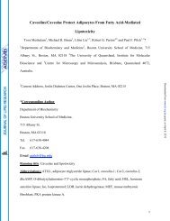
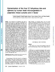
FIG. 3. TLC chromatogram <strong>of</strong> standards and <strong>of</strong> hydrolysis<br />
products <strong>of</strong> <strong>phosphatides</strong>. The chromatogram waa developed<br />
with Solvent 111 for 10 cm. The left half <strong>of</strong> the plate was<br />
sprayed with ninhydrin and the right half with Dragendorf<br />
reagent. The substances hydrolysed and the amounts applied<br />
to TLC were aa follows: (1) PhS, 8 pg P; (2) serine, 50 pg;<br />
(3) PhE, 3 pg P; (4) ethanolamine, 50 pg; (5) PhC, 8 pg P:<br />
(6) phosphoryl choline, 150 pg; (7) choline, 50 pg; (8) Sph,<br />
3 pg P; (9) L-PhC, 8pg P.<br />
chromatographed by TLC (Figs. 3 and 4). PhS was<br />
shown to contain serine and no ethanolamine. PhE<br />
was shown to contain ethanolamine and no serine. PhC<br />
and LPhC produced choline and no phosphoryl choline.<br />
Sph gave phosphoryl choline and no choline. PhI<br />
produced inositol and 1-phosphoryl inositol. PhA was<br />
not definitely characterized. Rr values and color reac-<br />
tions <strong>of</strong> the hydrolysis products <strong>of</strong> <strong>phosphatides</strong> and<br />
standards are presented in Table 3.<br />
Preliminary studies <strong>of</strong> the recovery <strong>of</strong> P indicated<br />
that essentially all <strong>of</strong> the P that moved in the two-<br />
<strong>dimensional</strong> TLC chromatogram could be recovered<br />
from the phosphatide spots. About 25% <strong>of</strong> the P<br />
applied to the plates, however, could not be recovered<br />
from the Silica Gel G by this method <strong>of</strong> elution. Ap-<br />
parently there is a certain amount <strong>of</strong> P-containing<br />
material that does not move with either Solvent I or<br />
Solvent I1 and remains at the origin. It is under-<br />
standable, then, that this same material resists elution<br />
with a similar eluting solvent.<br />
Since breakdown <strong>of</strong> <strong>phosphatides</strong> occurs on storage,<br />
especially after silicic acid <strong>chromatography</strong>, information<br />
on TLC behavior <strong>of</strong> the breakdown products, including<br />
the lyso<strong>phosphatides</strong>, is <strong>of</strong> interest. The movement <strong>of</strong><br />
lysophosphatide spots has been observed on twdimen-<br />
sional TLC chromatograms. Lysophosphatidyl eth-<br />
anolamine moves with the PhI spot on TLC and has<br />
been noted to be present as a component on old ex-<br />
tracts <strong>of</strong> PhE by using the ninhydrin spray. Lysophos-<br />
phatidyl serine moves closely behind PhS and appears<br />
as a spot between PhS and the origin. The eluted<br />
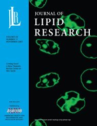
FIG. 4. TLC chromatogram <strong>of</strong> standards and <strong>of</strong> hydrolysis<br />
products <strong>of</strong> <strong>phosphatides</strong>. The chromatogram waa developed<br />
in one dimension with Solvent IV for 5 cm. The SpOk were<br />
detected with ammoniacal silver nit<strong>rat</strong>e. The substances<br />
hydrolysed and the amounts applied to TLC plate were aa<br />
follows: (10) PhE, 4 pg P; (11) inositol-l-POc, 30 pg; (12)<br />
inositol, 20 fig; (13) PhA, 8 pg P; (14) glycerolpho phosphate, 100<br />
pg; (15) glycerol, 50 rg.<br />
<strong>phosphatides</strong> were apparently unstable when stored<br />
in the eluting solvent at 4'. Traces <strong>of</strong> iodine-posi-<br />
tive substances were observed on TLC with Rf values<br />
similar to the corresponding lysocompounds and free<br />
fatty acids (Fig. 2).<br />
TABLE 3. COLOR REACTIONS AND Rf VALUES<br />
AND OF HYDROLYSIS<br />
OF STANDARDS<br />
PRODUCTS OF PHOSPHATIDES USING<br />
THIN-LAYER CHROMATOGRAPHY<br />
Rr Values <strong>of</strong><br />
Characteristic Color<br />
No. * Substance<br />
1 Phosphatidyl serine<br />
2 Serine<br />
Products Reactiont<br />
Solvent 111 Solvent IV Bi Nin Aa<br />
0.74 + -<br />
0.72 + -<br />
3<br />
4<br />
5<br />
G<br />
7<br />
8<br />
9<br />
Phosphatidyl<br />
ethanolamine<br />
Ethanolamine<br />
Phosphatidyl choline<br />
Phosphoryl choline<br />
Choline<br />
Sphingomyelin<br />
Lysophosphatidyl choline<br />
0.34<br />
0.36<br />
0.08<br />
0. a0<br />
0.09<br />
0.27<br />
0.08<br />
+ -<br />
+ -<br />
- +<br />
- +<br />
- +<br />
- +<br />
- +<br />
10 Phosphatidyl inositol 0.40, 0.64.<br />
and 0.95 +<br />
11 Inositol-1-PO4 0.40. 0.64 +<br />
12 Inositol 0.64 +<br />
13 "Phosphatidic acid" 0.95 +<br />
14 a-Glycerol-PO4 0.55 +<br />
15 Glycerol 0.7B +<br />
* The numbers are given to identify the substance applied to the plates for<br />
<strong>chromatography</strong> as shown in Figures 3 and 4.<br />
t See section on Materials for abbreviations used for color reagents.<br />
Downloaded from<br />
www.jlr.org<br />
by guest, on January 14, 2013
THIN-LAYER CHROMATOGRAPHY OF PHOSPHATIDES 475<br />
REFERENCES<br />
1. Stahl, E. Phurmuzie 11: 633, 1956.<br />
2. Stahl, E. Agnew. Chem. 73 : 646, 1961.<br />
3. Stahl, E. Z. Anal. Chem. 181: 303, 1961.<br />
4. Mangold, H. K. J. Am. Oil Chemists’ Soc. 38 : 708,1961.<br />
5. Jatzkewitz, H., and E. Mehl. Z. physiol. Chem. Hoppe-<br />
Seyler’s 320 : 251, 1960.<br />
6. Wagner, H., L. Horhammer, and P. Wolff. Biochem. 2.<br />
334: 175, 1961.<br />
7. Habermann, E., G. Bandflow, and B. Krusche. KIin.<br />
Wochschr. 39: 816, 1961.<br />
8. Weicker, H. Klin. Wochschr. 37: 763, 1959.<br />
9. Vogel, W. C., W. M. Doizaki, and L. Zieve. J. Lipid<br />
Research 3 : 138, 1962.<br />
10. Shlemmer, W. Bell. Soc. Ital. Bwl. Sper. 37: 134, 1961<br />
(in Italian).<br />
11. Hanes, C. S., and F. A. Isherwood. Nature 164 : 1107,<br />
1949.<br />
12. Wade, H. E., and D. M. Morgan. Biochm. J. 60:<br />
264, 1955.<br />
13. Partridge, S. M. Biochem. J. 42: 238, 1948.<br />
14. Block, R. J., E. L. Durrum, and G. Zweig. A Munual<br />
<strong>of</strong> Paper Chromatography and Paper Electrophoresis. New<br />
York, Academic Press Inc., 1955, p. 409.<br />
15. Whittaker, V. P., and S. Wijesundera. Biochem. J. 51:<br />
348, 1952.<br />
16. Folch, J., M. M. Lees, and G. H. Sloane Stanley. J. Biol.<br />
Chem. 226: 497, 1957.<br />
17. Hanahan, D. J., J. C. Dittmer, and E. Warashina. J.<br />
Biol. Chem. 228: 685, 1957.<br />
18. Dawson, R. M. C. Biochem. J. 75: 45, 1960.<br />
19. Bartlett, G. R. J. Bid. Chem. 234: 466, 1959.<br />
20. Skidmore, W. D., and C. Entenman. J. Lipid Research<br />
3 : 356, 1962.<br />
Downloaded from<br />
www.jlr.org<br />
by guest, on January 14, 2013