endothelium specular microscope em-3000
endothelium specular microscope em-3000
endothelium specular microscope em-3000

You also want an ePaper? Increase the reach of your titles
YUMPU automatically turns print PDFs into web optimized ePapers that Google loves.
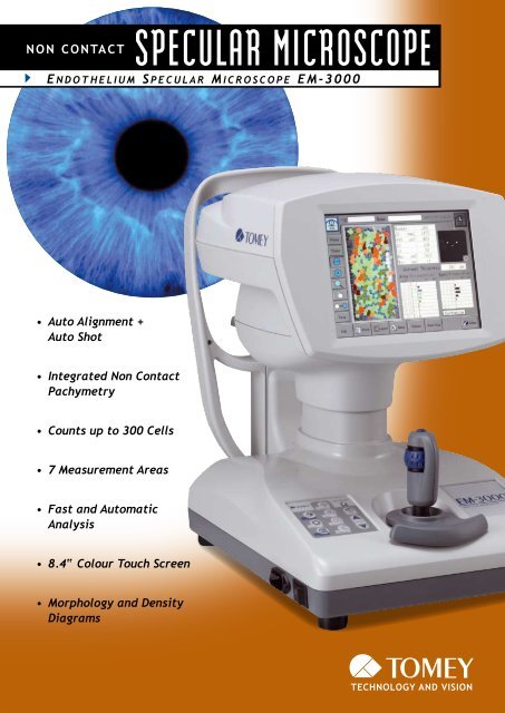
SPECULAR MICROSCOPE<br />
NON CONTACT<br />
e n d o T h e l i u m S p e c u l a r m i c r o S c o p e e m - 3 0 0 0<br />
• Auto Alignment +<br />
Auto Shot<br />
• Integrated Non Contact<br />
Pachymetry<br />
• Counts up to 300 Cells<br />
• 7 Measur<strong>em</strong>ent Areas<br />
• Fast and Automatic<br />
Analysis<br />
• 8.4“ Colour Touch Screen<br />
• Morphology and Density<br />
Diagrams
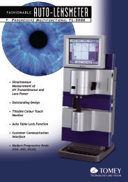
ALL-In-OnE SPECULAR MIC ROSCOPE<br />
wIth CORnEAL EndOthELIUM PhOtOgRAPhIng And AUtOMAtIC AnALySIS<br />
Non-contact examination, auto<br />
alignment and automatic analysis of<br />
the <strong>endothelium</strong> layer makes working<br />
with the EM-<strong>3000</strong> professional<br />
and quick. Due to the low intensity<br />
source of light needed for evaluation,<br />
the Specular Microscope EM-<br />
<strong>3000</strong> assures maximum patient comfort.<br />
The integrated non contact<br />
Step 1: Step 2:<br />
Simply touch the<br />
center of the pupil<br />
Auto Alignment + Auto Shot<br />
pachymetry will be automatically<br />
measured with every examination.<br />
The 8.4 inch colour touch screen is<br />
used as an operating monitor as well<br />
as for displaying all measured values.<br />
You can even move the unit in<br />
all directions by simply touching the<br />
screen. All commands can be given<br />
via touch screen.<br />
The handling of the EM-<strong>3000</strong> is very easy - it does almost everything by itself.<br />
Alignment and measur<strong>em</strong>ent are done automatically. You just roughly align the<br />
syst<strong>em</strong> towards the patient eye and the rest is taken care of by the instrument.<br />
With a tip on the screen the syst<strong>em</strong> automatically moves to the left or right eye.<br />
Of course you also can do the examination in the manual mode.<br />
Image is taken<br />
automatically<br />
Automated Capturing of 15 Images<br />
The EM-<strong>3000</strong> takes 15 Images with every examination. The best image out of these<br />
15 shots is automatically selected and displayed on the screen.<br />
Best Image
7 Measur<strong>em</strong>ent Areas + Automatic<br />
Pachymetry<br />
The EM-<strong>3000</strong> has a very large measur<strong>em</strong>ent area. With up to<br />
300 counted cells the syst<strong>em</strong> assures a representative cell<br />
density analysis of your patients cornea. Images can be taken<br />
at 7 positions: the center and 6 peripheral points (2,4,6,8,10<br />
and 12° clock position). Additional to that the thickness of the<br />
cornea will be automatically measured with every exam – of<br />
course in non contact method.<br />
Fast and Automatic Analysis<br />
of Corneal Endothelium Cells<br />
The software evaluates all relevant data respective to<br />
the <strong>endothelium</strong>, such as number and density of cells as<br />
well as their form and size. High-quality images enable<br />
discovering irregularities or possible degeneration of the<br />
<strong>endothelium</strong>. Also a manual adjustment of the evaluated<br />
area within the <strong>endothelium</strong> image is possible.<br />
Various Display Functions<br />
The image of the corneals <strong>endothelium</strong> can be<br />
displayed with the cell shapes traced, as well<br />
as with different areas and structural forms of<br />
cells displayed in different colours. This provides<br />
a visual understanding of the condition of the<br />
corneal <strong>endothelium</strong>.<br />
Analysis results screen<br />
Photography<br />
of <strong>endothelium</strong><br />
An easy to use colour touch screen shows<br />
even the tiniest detail. The EM-<strong>3000</strong> does<br />
not need a seperate monitor or computer.<br />
Traced image Image showing<br />
different areas<br />
Image showing<br />
different polygonal<br />
shapes
03.07 8-) Subject to change without notice<br />
T o m e y<br />
<strong>em</strong>-<strong>3000</strong><br />
E n d o t h E l i u m SpEcular microScopE<br />
S p e c i f i c aT i o n S<br />
Resolution<br />
Data Manag<strong>em</strong>ent<br />
Pixels Used for Picture<br />
Print Out Via PictBridge Printer<br />
Taking 480 (V) x 180 (H) Pixels<br />
Data Export Via Data Transfer SW<br />
Capturing Scope 0.25 × 0.54 mm<br />
1 Center + 6 Peripheral<br />
Operating Environment<br />
Measur<strong>em</strong>ents 7 x Fixation Points<br />
(center; 2; 4; 6; 8; 10;<br />
12 o´clock)<br />
Min. Cell Resolution 1.14 μm (V) x 1.45 μm (H)<br />
T<strong>em</strong>perature<br />
Humidity<br />
Atmospheric Pressure<br />
+10° to +40°<br />
30 to 75 %<br />
700 to 1060 hPa<br />
Optical Magnification<br />
Display<br />
x 190<br />
8.4“ LCD Colour<br />
Communication Ports<br />
Display Resolution 1.14 μm<br />
USB for PictBridge Printer<br />
LAN Data Transfer SW<br />
Measur<strong>em</strong>ent<br />
Auto Alignment Yes<br />
Auto Shot Yes<br />
Manual Mode (1 & 2) Yes<br />
Measur<strong>em</strong>ent Function<br />
Automated Captured<br />
Examina 15 Pictures for Analysis<br />
Up to 300 Cells<br />
Cell Density<br />
CV / SD<br />
Cell Size<br />
Cell Morphology<br />
None Contact Pachymetry<br />
Stroke of Moving<br />
Section X: 88 mm; Y: 40 mm;<br />
Z: 50 mm<br />
Stroke of Electrical<br />
Chin Rest 70 mm<br />
Measuring Accuracy<br />
Pachymetrie ± 10 μm<br />
Dimensions & Electric Requir<strong>em</strong>ents<br />
Dimensions WDH 453 x 308 x 493 mm<br />
Weight Approx. 18.0 kg<br />
Power Supply AC 100 to 240 V<br />
Frequency 50/60 Hz<br />
Power Consumption 100 to 130 VA<br />
TOMEY EUROPE<br />
TOMEY ASIA-PACIFIC<br />
TOMEY TOMEY EUROPE GmbH<br />
TOMEY TOMEY ASIA-PACIFIC CORPORATION JAPAN<br />
TOMEY Am Weichselgarten GmbH 19a TOMEY 2-11-33 CORPORATION Noritakeshinmachi JAPAN<br />
Am 91058 Weichselgarten Erlangen 19a 2-11-33 Nishi-ku, Noritakeshinmachi<br />
Nagoya 451-0051<br />
91058 Germany Erlangen<br />
Nishi-ku, JapanNagoya<br />
451-0051<br />
Germany Phone (+49) - 9131 - 77710Japan<br />
Phone (+81) - 52 - 581- 5327<br />
Phone Fax (+49) (+49) - 9131 - 9131 - 77710 - 777120Phone<br />
Fax (+81) (+81) - 52 - - 52 581- - 561- 5327 4735<br />
Fax eMail: (+49) info@tomey.de<br />
- 9131 - 777120 Fax eMail: (+81) intl@tomey.co.jp<br />
- 52 - 561- 4735<br />
eMail: info@tomey.de eMail: intl@tomey.co.jp<br />
EM-<strong>3000</strong>