Computed dental radiography used to reproduce ... - Library
Computed dental radiography used to reproduce ... - Library
Computed dental radiography used to reproduce ... - Library

You also want an ePaper? Increase the reach of your titles
YUMPU automatically turns print PDFs into web optimized ePapers that Google loves.
402 JOURNAL OF FORENSIC SCIENCES<br />
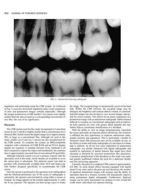
FIG. 1—Antemortem bitewing radiograph.<br />
angulation and positioning using the CDR system. As evidenced the image. The accepted image is au<strong>to</strong>matically saved on the hard<br />
in Fig. 3, excessive horizontal angulation make visual comparison disk. Within the CDR software, the accepted image may be<br />
of ante- and postmortem images virtually impossible. Although enlarged, the brightness and contrast altered, rotated, or colorized.<br />
the images produced on a CDR number 2 size sensor were slightly Selected images can also be tiled <strong>to</strong> view several images side-by-<br />
smaller than the area covered on a corresponding conventional #2 side for closer scrutiny. This allows for an easier comparison of a<br />
size film, this was of no significance.<br />
postmortem image with an antemortem radiograph. Subtle features<br />
difficult <strong>to</strong> visualize on conventional radiographs such as trabecu-<br />
Discussion<br />
lar bone patterns are seen with greater detail digitally and may<br />
The CDR system <strong>used</strong> for this study incorporated a 5-mm-thick<br />
sensor in size 2 which is slightly smaller than a conventional size 2<br />
intraoral film. Schick claims the digital image size is approximately<br />
90% as large as a conventional film. Although not <strong>used</strong> in this<br />
study, smaller size 0 and size 1 sensors are also manufactured for<br />
the CDR system. To complete the system, an IBM-compatible<br />
computer with a minimum of 8 MB RAM and an SVGA display<br />
adapter are required. A standard intraoral X-ray tubehead is all<br />
that is required <strong>to</strong> expose the subject and inci<strong>dental</strong>ly, the exposure<br />
time is approximately one-tenth that of conventional D-speed film.<br />
Although infection control was not a primary concern with the<br />
specimens <strong>used</strong> in this study, sterile sheaths are available <strong>to</strong> cover<br />
the sensor prior <strong>to</strong> placement. The intraoral sensor was held in<br />
position with commercially available Rinn XCP and Snap-a-ray<br />
further help <strong>to</strong> corroborate identification of a decedent.<br />
With the ability <strong>to</strong> view an image instantaneously, reposition<br />
the sensor and retake an exposure almost effortlessly, the examiner<br />
is afforded the best opportunity <strong>to</strong> replicate antemortem radiographic<br />
position and angulation. This is particularly valuable in<br />
situations where film processors may not be available on-site, or<br />
the ability <strong>to</strong> return and retake radiographs of a decedent at a future<br />
date is unlikely. In all ten test cases replication of antemortem<br />
radiographs was greatly enhanced with digital <strong>radiography</strong> and<br />
resulted in replication of <strong>dental</strong> features that might have been<br />
obscured or dis<strong>to</strong>rted by incorrect postmortem film placement.<br />
Conclusive identification of the decedents in this study was easily<br />
and quickly reaffirmed without the need for a darkroom facility<br />
and film processing apparatus.<br />
Currently, the price of a complete CDR system is approximately<br />
film holders designed specifically <strong>to</strong> accommodate the CDR $10 000. As more <strong>dental</strong> offices become equipped with digital<br />
sensor. <strong>dental</strong> X-ray units, and as the price per unit decreases, the number<br />
Once the sensor is positioned in the specimen <strong>to</strong> be radiographed of digitized antemortem images will increase and the ability <strong>to</strong><br />
and the tubehead positioned, one of the series of radiographs is <strong>reproduce</strong> them by a forensic scientist will dramatically improve<br />
selected by the opera<strong>to</strong>r and activated by using either a mouse or using postmortem digital <strong>radiography</strong>. Furthermore, digitized<br />
footpad. An image appears on the moni<strong>to</strong>r approximately 5 s after images can be easily transmitted via modems <strong>to</strong> sites around the<br />
the X-ray exposure. The opera<strong>to</strong>r can then accept, reject or retake<br />
world where individuals are reported missing.