Varicocelectomy: microsurgical subinguinal technique - Canadian ...
Varicocelectomy: microsurgical subinguinal technique - Canadian ...
Varicocelectomy: microsurgical subinguinal technique - Canadian ...

You also want an ePaper? Increase the reach of your titles
YUMPU automatically turns print PDFs into web optimized ePapers that Google loves.
Point / Counterpoint<br />
Pathophysiology of varicocele<br />
The adverse effect of varicocele on male fertility<br />
is most clearly manifested by the testicular atrophy<br />
generally associated with this condition. 4 Using<br />
scrotal ultrasound, we objectively demonstrated<br />
that left testicular volume is less than right testicular<br />
volume in men with a left varicocele. 30<br />
A varicocele is associated with bilateral spermatogenic<br />
abnormalities and Leydig cell dysfunction.<br />
31–35 The testicular histology in infertile men<br />
with varicocele is variable, but most studies report<br />
reduced spermatogenesis (hypospermatogenesis).<br />
The observed increase in germ cell apoptosis is<br />
thought to occur as a result of hyperthermia and<br />
low testosterone levels in the testicle. 26 Testosterone<br />
concentration (testosterone is secreted by Leydig<br />
cells) is lower in older (> 30 yr) compared with<br />
younger men with varicocele, which is a trend not<br />
seen in men without varicocele and suggests a progressive,<br />
adverse effect of varicocele on Leydig<br />
cell function. 4<br />
MacLeod (1965) and other investigators<br />
observed that most semen samples from infertile<br />
men with varicocele have poorer sperm parameters<br />
(lower sperm counts, increased number of spermatozoa<br />
with abnormal forms and decreased sperm<br />
motility) than fertile men. 4,23,36 However, this “stress<br />
pattern” is not a specific marker for varicocele and<br />
therefore is not diagnostic of this condition. 37<br />
Surprisingly, few studies have been conducted over<br />
the past 40 years to better define the pathophysiology<br />
of varicocele, in particular, the effect of this<br />
prevalent condition on human sperm function. This<br />
is especially critical in light of the inherent limitations<br />
(e.g., high biological variability) and modest<br />
predictive value of the standard sperm parameters<br />
in terms of reproductive outcomes. 38,39<br />
<strong>Varicocelectomy</strong>: approaches<br />
There are several approaches for varicocelectomy.<br />
These include retroperitoneal and conventional<br />
inguinal open <strong>technique</strong>s, <strong>microsurgical</strong> inguinal<br />
and <strong>subinguinal</strong> approaches, laparoscopic repairs<br />
and radiographic embolization. 40–44 The <strong>microsurgical</strong><br />
varicocelectomy is considered the “gold standard”<br />
because it is associated with the lowest risk<br />
of complications (varicocele recurrence, hydrocele<br />
formation [fluid collection around the testicle]<br />
and testicular atrophy). 41,45–47<br />
274 CUAJ • September 2007 • Volume 1, Issue 3<br />
We have favoured the <strong>microsurgical</strong> <strong>subinguinal</strong><br />
approach because it is associated with a higher<br />
success rate (disappearance of varicocele) and a<br />
lower complication rate (recurrence rate and<br />
hydrocele formation), compared with non<strong>microsurgical</strong><br />
<strong>technique</strong>s. 46,48 The <strong>subinguinal</strong><br />
approach is also associated with less operative and<br />
postoperative pain than inguinal approaches. 49,50<br />
However, the <strong>subinguinal</strong> approach is more challenging<br />
owing to the greater number of vessels<br />
(arteries and veins) encountered at this level, compared<br />
with the inguinal canal. 51<br />
Microsurgical sub-inguinal varicocelectomy<br />
We start with a 2–3-cm oblique skin incision centred<br />
over the external inguinal ring, as previously<br />
described. 52 The incision is deepened through<br />
Camper’s and Scarpa’s fascias and the spermatic<br />
cord is then grasped with a Babcock clamp, delivered<br />
and placed over a large (1-inch) Penrose<br />
drain. The testicle is then delivered and the gubernacular<br />
veins and external spermatic perforators<br />
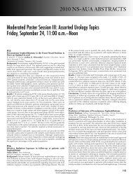
are isolated and divided (Fig. 1). The testicle is<br />
returned to the scrotum and the spermatic cord<br />
is elevated on a large Penrose drain. The microscope<br />
is then brought into the operating field and<br />
the cord examined under 8–15 power magnification.<br />
The internal and external spermatic fascias<br />
are incised and the cord structures are again examined<br />
(Fig. 2).<br />
To simplify the procedure and protect the vas<br />
deferens and its vessels from potential injury dur-<br />
Fig. 1. Testicle delivered through the <strong>subinguinal</strong> incision depicting<br />
the spermatic cord (held by Penrose drain; bottom left) and<br />
the gubernaculum (held by Penrose drain; right).