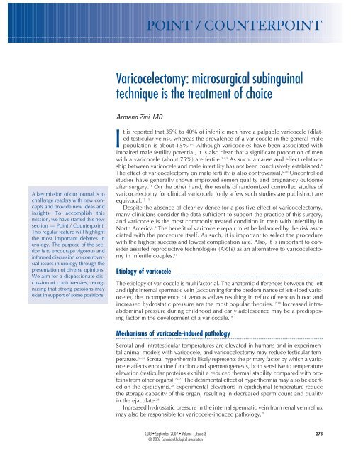
Varicocelectomy: microsurgical subinguinal technique - Canadian ...
Varicocelectomy: microsurgical subinguinal technique - Canadian ...
Varicocelectomy: microsurgical subinguinal technique - Canadian ...

You also want an ePaper? Increase the reach of your titles
YUMPU automatically turns print PDFs into web optimized ePapers that Google loves.
A key mission of our journal is to<br />
challenge readers with new concepts<br />
and provide new ideas and<br />
insights. To accomplish this<br />
mission, we have started this new<br />
section — Point / Counterpoint.<br />
This regular feature will highlight<br />
the most important debates in<br />
urology. The purpose of the section<br />
is to encourage vigorous and<br />
informed discussion on controversial<br />
issues in urology through the<br />
presentation of diverse opinions.<br />
We aim for a dispassionate discussion<br />
of controversies, recognizing<br />
that strong passions may<br />
exist in support of some positions.<br />
POINT / COUNTERPOINT<br />
<strong>Varicocelectomy</strong>: <strong>microsurgical</strong> <strong>subinguinal</strong><br />
<strong>technique</strong> is the treatment of choice<br />
Armand Zini, MD<br />
It is reported that 35% to 40% of infertile men have a palpable varicocele (dilated<br />
testicular veins), whereas the prevalence of a varicocele in the general male<br />
population is about 15%. 1–3 Although varicoceles have been associated with<br />
impaired male fertility potential, it is also clear that a significant proportion of men<br />
with a varicocele (about 75%) are fertile. 2,4,5 As such, a cause and effect relationship<br />
between varicocele and male infertility has not been conclusively established. 6<br />
The effect of varicocelectomy on male fertility is also controversial. 6–10 Uncontrolled<br />
studies have generally shown improved semen quality and pregnancy outcome<br />
after surgery. 11 On the other hand, the results of randomized controlled studies of<br />
varicocelectomy for clinical varicocele (only a few such studies are published) are<br />
equivocal. 12–15<br />
Despite the absence of clear evidence for a positive effect of varicocelectomy,<br />
many clinicians consider the data sufficient to support the practice of this surgery,<br />
and varicocele is the most commonly treated condition in men with infertility in<br />
North America. 8 The benefit of varicocele repair must be balanced by the risk associated<br />
with the procedure itself. As such, it is important to select the procedure<br />
with the highest success and lowest complication rate. Also, it is important to consider<br />
assisted reproductive technologies (ARTs) as an alternative to varicocelectomy<br />
in infertile couples. 16<br />
Etiology of varicocele<br />
The etiology of varicocele is multifactorial. The anatomic differences between the left<br />
and right internal spermatic vein (accounting for the predominance of left-sided varicocele),<br />
the incompetence of venous valves resulting in reflux of venous blood and<br />
increased hydrostatic pressure are the most popular theories. 17,18 Increased intraabdominal<br />
pressure during childhood and early adolescence may be a predisposing<br />
factor in the development of a varicocele. 19<br />
Mechanisms of varicocele-induced pathology<br />
Scrotal and intratesticular temperatures are elevated in humans and in experimental<br />
animal models with varicocele, and varicocelectomy may reduce testicular temperature.<br />
20–24 Scrotal hyperthermia likely represents the primary factor by which a varicocele<br />
affects endocrine function and spermatogenesis, both sensitive to temperature<br />
elevation (testicular proteins exhibit a reduced thermal stability compared with proteins<br />
from other organs). 25–27 The detrimental effect of hyperthermia may also be exerted<br />
on the epididymis. 28 Experimental elevations in epididymal temperature reduce<br />
the storage capacity of this organ, resulting in decreased sperm count and quality<br />
in the ejaculate. 28<br />
Increased hydrostatic pressure in the internal spermatic vein from renal vein reflux<br />
may also be responsible for varicocele-induced pathology. 29<br />
CUAJ • September 2007 • Volume 1, Issue 3<br />
© 2007 <strong>Canadian</strong> Urological Association<br />
273
Point / Counterpoint<br />
Pathophysiology of varicocele<br />
The adverse effect of varicocele on male fertility<br />
is most clearly manifested by the testicular atrophy<br />
generally associated with this condition. 4 Using<br />
scrotal ultrasound, we objectively demonstrated<br />
that left testicular volume is less than right testicular<br />
volume in men with a left varicocele. 30<br />
A varicocele is associated with bilateral spermatogenic<br />
abnormalities and Leydig cell dysfunction.<br />
31–35 The testicular histology in infertile men<br />
with varicocele is variable, but most studies report<br />
reduced spermatogenesis (hypospermatogenesis).<br />
The observed increase in germ cell apoptosis is<br />
thought to occur as a result of hyperthermia and<br />
low testosterone levels in the testicle. 26 Testosterone<br />
concentration (testosterone is secreted by Leydig<br />
cells) is lower in older (> 30 yr) compared with<br />
younger men with varicocele, which is a trend not<br />
seen in men without varicocele and suggests a progressive,<br />
adverse effect of varicocele on Leydig<br />
cell function. 4<br />
MacLeod (1965) and other investigators<br />
observed that most semen samples from infertile<br />
men with varicocele have poorer sperm parameters<br />
(lower sperm counts, increased number of spermatozoa<br />
with abnormal forms and decreased sperm<br />
motility) than fertile men. 4,23,36 However, this “stress<br />
pattern” is not a specific marker for varicocele and<br />
therefore is not diagnostic of this condition. 37<br />
Surprisingly, few studies have been conducted over<br />
the past 40 years to better define the pathophysiology<br />
of varicocele, in particular, the effect of this<br />
prevalent condition on human sperm function. This<br />
is especially critical in light of the inherent limitations<br />
(e.g., high biological variability) and modest<br />
predictive value of the standard sperm parameters<br />
in terms of reproductive outcomes. 38,39<br />
<strong>Varicocelectomy</strong>: approaches<br />
There are several approaches for varicocelectomy.<br />
These include retroperitoneal and conventional<br />
inguinal open <strong>technique</strong>s, <strong>microsurgical</strong> inguinal<br />
and <strong>subinguinal</strong> approaches, laparoscopic repairs<br />
and radiographic embolization. 40–44 The <strong>microsurgical</strong><br />
varicocelectomy is considered the “gold standard”<br />
because it is associated with the lowest risk<br />
of complications (varicocele recurrence, hydrocele<br />
formation [fluid collection around the testicle]<br />
and testicular atrophy). 41,45–47<br />
274 CUAJ • September 2007 • Volume 1, Issue 3<br />
We have favoured the <strong>microsurgical</strong> <strong>subinguinal</strong><br />
approach because it is associated with a higher<br />
success rate (disappearance of varicocele) and a<br />
lower complication rate (recurrence rate and<br />
hydrocele formation), compared with non<strong>microsurgical</strong><br />
<strong>technique</strong>s. 46,48 The <strong>subinguinal</strong><br />
approach is also associated with less operative and<br />
postoperative pain than inguinal approaches. 49,50<br />
However, the <strong>subinguinal</strong> approach is more challenging<br />
owing to the greater number of vessels<br />
(arteries and veins) encountered at this level, compared<br />
with the inguinal canal. 51<br />
Microsurgical sub-inguinal varicocelectomy<br />
We start with a 2–3-cm oblique skin incision centred<br />
over the external inguinal ring, as previously<br />
described. 52 The incision is deepened through<br />
Camper’s and Scarpa’s fascias and the spermatic<br />
cord is then grasped with a Babcock clamp, delivered<br />
and placed over a large (1-inch) Penrose<br />
drain. The testicle is then delivered and the gubernacular<br />
veins and external spermatic perforators<br />
are isolated and divided (Fig. 1). The testicle is<br />
returned to the scrotum and the spermatic cord<br />
is elevated on a large Penrose drain. The microscope<br />
is then brought into the operating field and<br />
the cord examined under 8–15 power magnification.<br />
The internal and external spermatic fascias<br />
are incised and the cord structures are again examined<br />
(Fig. 2).<br />
To simplify the procedure and protect the vas<br />
deferens and its vessels from potential injury dur-<br />
Fig. 1. Testicle delivered through the <strong>subinguinal</strong> incision depicting<br />
the spermatic cord (held by Penrose drain; bottom left) and<br />
the gubernaculum (held by Penrose drain; right).
ing subsequent cord dissection, we first create a<br />
window between the internal spermatic vessels<br />
and the external spermatic fascia so that the internal<br />
spermatic vessels are separate from the external<br />
spermatic fascia and its associated structures<br />
(cremasteric fibres, external spermatic vessels, vas<br />
deferens and its vessels). 52 A second Penrose drain<br />
is then introduced between the internal spermatic<br />
vessels and the external spermatic fascia and its<br />
associated structures.<br />
We first dissect the contents of the internal spermatic<br />
fascia (lying on top of the most superficial<br />
Penrose drain). Subtle pulsations will usually reveal<br />
the location of the underlying internal spermatic<br />
artery (or arteries). Once identified, the artery is<br />
dissected free of all surrounding veins and encircled<br />
with a 2-0 silk ligature for identification. Care<br />
is taken to identify a number of lymphatics (usually<br />
2–5 channels) and these are also encircled<br />
with a 2-0 silk ligature. All internal spermatic veins<br />
are clipped or ligated (with 4-0 silk) and divided.<br />
At the end of the first dissection, the cord is<br />
skeletonized so that only the identified artery (or<br />
arteries) and lymphatics are preserved.<br />
We then elevate and dissect the contents of the<br />
external spermatic fascia (lying between the 2<br />
Penrose drains). The vas deferens and its associated<br />
vessels are readily identified and preserved.<br />
Any cremasteric artery is also preserved. The<br />
remaining cremasteric fibres and veins are ligated<br />
and cut thus skeletonizing the cord. At the completion<br />
of varicocelectomy, the cord should contain<br />
only the testicular artery or arteries, vas deferens,<br />
and associated vessels and spermatic cord<br />
lymphatics. The wound is irrigated with 1%<br />
Neomycin irrigation, and Scarpa’s and Camper’s<br />
Fig. 2. Spermatic cord with Penrose drain beneth it (the internal<br />
and external spermatic fascias have been opened).<br />
fascia are closed with a single 3-0 chromic catgut<br />
suture. The incision is infiltrated with 0.5%<br />
Marcaine solution with epinephrine, and the skin<br />
is closed with a running 4-0 Vicryl subcuticular<br />
closure reinforced with Steri-Strips. A dry sterile<br />
dressing is applied.<br />
Summary<br />
A variety of approaches have been advocated for<br />
management of varicoceles but recent evidence<br />
supports the premise that the <strong>microsurgical</strong> <strong>technique</strong><br />
is the “gold standard.” 46,48 In a number of<br />
studies, it has been shown that <strong>microsurgical</strong> varicocelectomy<br />
(inguinal or <strong>subinguinal</strong>) is superior<br />
to non-<strong>microsurgical</strong> procedures with respect to<br />
the development of postoperative complications<br />
such as hydrocele or recurrence. 41,46,47 Hydrocele<br />
formation is believed to be due to ligation of lymphatic<br />
channels and recurrence generally results<br />
from incomplete ligation of collateral venous channels.<br />
53,54 Magnification of the spermatic cord with<br />
the use of the operating microscope reduces the<br />
potential for development of such complications.<br />
41,46,47 However, <strong>microsurgical</strong> varicocelectomy,<br />
particularly the <strong>subinguinal</strong> approach, remains<br />
a technically challenging procedure that requires<br />
<strong>microsurgical</strong> expertise.<br />
From the Division of Urology, Department of Surgery, Royal Victoria Hospital, McGill<br />
University, Montréal, Que.<br />
This article has been peer reviewed.<br />
Competing interests: None declared.<br />
References<br />
1. Clarke BG. Incidence of varicocele in normal men and among men of different ages.<br />
JAMA 1966;198:1121-2.<br />
2. Lipshultz LI, Corriere JN. Progressive testicular atrophy in the varicocele patient.<br />
J Urol 1977;117:175.<br />
3. Akbay E, Cayan S, Doruk E, et al. The prevalence of varicocele and varicocele-related<br />
testicular atrophy in Turkish children and adolescents. BJU Int 2000;86:490-3.<br />
4. World Health Organization. The influence of varicocele on parameters of fertility in a<br />
large group of men presenting to infertility clinics. Fertil Steril 1992;57:1289-92.<br />
5. Pinto KJ, Kroovand RL, Jarow JP. Varicocele related testicular atrophy and its predictive<br />
effect upon fertility. J Urol 1994;152:788-90.<br />
6. Jarow JP. Effects of varicocele on male fertility. Hum Reprod Update 2001;7:59-64.<br />
7. Kamischke A, Nieschlag E. Varicocele treatment in the light of evidence-based andrology.<br />
Hum Reprod Update 2001;7:65-9.<br />
8. Sharlip ID, Jarow JP, Belker AM, et al. Best practice policies for male infertility. Fertil<br />
Steril 2002;77:873-82.<br />
9. Evers JL, Collins JA. Assessment of efficacy of varicocele repair for male subfertility:<br />
a systematic review. Lancet 2003;361:1849-52.<br />
CUAJ • September 2007 • Volume 1, Issue 3<br />
The treatment of varicocele<br />
275
Point / Counterpoint<br />
276<br />
10. Ficarra V, Cerruto MA, Liguori G, et al. Treatment of varicocele in subfertile men: The<br />
Cochrane Review–a contrary opinion. Eur Urol 2006;49:258-63.<br />
11. Schlesinger MH, Wilets IF, Nagler HM. Treatment outcome after varicocelectomy: A critical<br />
analysis. Urol Clin North Am 1994;21:517-29.<br />
12. Nilsson S, Edvinsson A, Nilsson B. Improvement of semen and pregnancy rate after ligation<br />
and division of internal spermatic vein: fact or fiction? Br J Urol 1979;51:591-6.<br />
13. Madgar I, Weissenberg R, Lunenfeld B, et al. Controlled trial of high spermatic vein<br />
ligation for varicocele in infertile men. Fertil Steril 1995;63:120-4.<br />
14. Nieschlag E, Hertle L, Fischedick A, et al. Update on treatment of varicocele: counselling<br />
as effective as occlusion of the vena spermatica. Hum Reprod 1998;13:2147-50.<br />
15. Krause W, Muller HH, Schafer H, et al. Does treatment of varicocele improve male<br />
fertility? Results of the ‘Deutsche Varikozelenstudie’, a multicentre study of 14 collaborating<br />
centres. Andrologia 2002;34:164-71.<br />
16. Kamal KM, Jarvi K, Zini A. Microsurgical varicocelectomy in the era of assisted reproductive<br />
technology: influence of initial semen quality on pregnancy rates. Fertil Steril<br />
2001;75:1013-6.<br />
17. Buschi AJ, Harrison RB, Brenbridge AN, et al. Distended left renal vein: CT/sonographic<br />
normal variant. Am J Radiol 1980;135:339-42.<br />
18. Braedel HU, Steffens J, Ziegler M, et al. A possible ontogenic etiology for idiopathic left<br />
varicocele. J Urol 1994;151:62-6.<br />
19. Scaramuzza A, Tavana R, Marchi A. Varicoceles in young soccer players. Lancet<br />
1996;348:1180-1.<br />
20. Zorgniotti AW, MacLeod J. Studies in temperature, human semen quality, and varicocele.<br />
Fertil Steril 1973;24:854-63.<br />
21. Saypol DC, Howards SS, Turner TT. Influence of surgically induced varicocele on testicular<br />
blood flow, temperature, and histology in adult rats and dogs. J Clin Invest<br />
1981;68:39.<br />
22. Goldstein M, Eid JF. Elevation of intratesticular and scrotal skin surface temperature<br />
in men with varicocele. J Urol 1989;142:743-5.<br />
23. Ali JI, Weaver DJ, Weinstein SH, et al. Scrotal temperature and semen quality in<br />
men with and without varicocele. Arch Androl 1990;24:215-9.<br />
24. Wright EJ, Young GP, Goldstein M. Reduction in testicular temperature after varicocelectomy<br />
in infertile men. Urology 1997;50:257-9.<br />
25. Mieusset R, Bujan L, Plantavid M, et al. Increased levels of serum follicle-stimulating<br />
hormone and luteinizing hormone associated with intrinsic testicular hyperthermia in<br />
oligospermic infertile men. J Clin Endocrinol Metab 1989;68:419-25.<br />
26. Lue YH, Lasley BL, Laughlin LS, et al. Mild testicular hyperthermia induces profound<br />
transitional spermatogenic suppression through increased germ cell apoptosis in adult<br />
cynomolgus monkeys (Macaca fascicularis). J Androl 2002;23:799-805.<br />
27. Sarge KD, Bray AE, Goodson ML. Altered stress response in testis. Nature 1995;374:126.<br />
28. Bedford JM. Effects of elevated temperature on the epididymis and testis: experimental<br />
studies. Adv Exp Med Biol 1991;286:19-32.<br />
29. Shafik A, Bedeir GA. Venous tension patterns in cord veins in normal and varicocele<br />
individuals. J Urol 1980;123:383-5.<br />
30. Zini A, Buckspan M, Berardinucci D, et al. The influence of clinical and subclinical<br />
varicocele on testicular volume. Fertil Steril 1997;68:671-4.<br />
31. Dubin L, Hotchkiss RS. Testis biopsy in subfertile men with varicocele. Fertil Steril<br />
1969;20:50-7.<br />
32. Comhaire F, Vermeulen A. Plasma testosterone in patients with varicocele and sexual<br />
inadequacy. J Clin Endocrin Metab 1975;40:824-9.<br />
33. Johnsen SG, Agger P. Quantitative evaluation of testicular biopsies in varicocele.<br />
CUAJ • September 2007 • Volume 1, Issue 3<br />
Fertil Steril 1978a;29:52-7.<br />
34. Hudson RW. The endocrinology of varicoceles. Fertil Steril 1988;49:199-208.<br />
35. Su LM, Goldstein M, Schlegel PN. The effect of varicocelectomy on serum testosterone<br />
levels in infertile men with varicoceles. J Urol 1995;154:1752-5.<br />
36. Macleod J. Seminal cytology in the presence of varicocele. Fertil Steril 1965;16:735-57.<br />
37. Ayodeji O, Baker HW. Is there a specific abnormality of sperm morphology in men with<br />
varicoceles? Fertil Steril 1986;45:839-42.<br />
38. Guzick DS, Overstreet JW, Factor-Litvak P, et al; National Cooperative Reproductive<br />
Medicine Network. Sperm morphology, motility, and concentration in fertile and<br />
infertile men. N Engl J Med 2001;345:1388-93.<br />
39. Menkveld R, Wong WY, Lombard CJ, et al. Semen parameters, including WHO and<br />
strict criteria morphology, in a fertile and subfertile population: an effort towards<br />
standardization of in-vivo thresholds. Hum Reprod 2001;16:1165-71.<br />
40. Walsh PC, White RI. Balloon occlusion of the internal spermatic vein for the treatment<br />
of varicoceles. JAMA 1981;246:1701.<br />
41. Goldstein M, Gilbert BR, Dicker AP, et al. Microsurgical inguinal varicocelectomy with delivery<br />
of the testis: An artery and lymphatic sparing <strong>technique</strong>. J Urol 1992;148:1808-11.<br />
42. Donovan JF, Winfield HN. Laparoscopic varix ligation. J Urol 1992;147:77.<br />
43. Jarow JP, Assimos DJ, Pittaway DE. Effectiveness of laparoscopic varicocelectomy.<br />
Urology 1993;42:544-7.<br />
44. Enquist E, Stein BS, Sigman M. Laparoscopic versus <strong>subinguinal</strong> varicocelectomy:<br />
a comparative study. Fertil Steril 1994;61:1092-6.<br />
45. Murray RR, Mitchell SE, Kadir S, et al. Comparison of recurrent varicocele anatomy<br />
following surgery and percutaneous balloon occlusion. J Urol 1986;135:286-9.<br />
46. Cayan S, Kadioglu TC, Tefekli A, et al. Comparison of results and complications of<br />
high ligation surgery and <strong>microsurgical</strong> high inguinal varicocelectomy in the treatment<br />
of varicocele. Urology 2000;55:750-4.<br />
47. Grober ED. O’brien J, Jarvi KA, Zini A. Preservation of testicular arteries during <strong>subinguinal</strong><br />
<strong>microsurgical</strong> varicocelectomy: clinical considerations. J Androl 2004;25:740-3.<br />
48. Ghanem H, Anis T, El-Nashar A, et al. Subinguinal microvaricocelectomy versus retroperitoneal<br />
varicocelectomy: comparative study of complications and surgical outcome.<br />
Urology 2004;64:1005-9.<br />
49. Gontero P, Pretti G, Fontana F, et al. Inguinal versus <strong>subinguinal</strong> varicocele vein ligation<br />
using magnifying loupe under local anesthesia: which <strong>technique</strong> is preferable in<br />
clinical practice? Urology 2005;66:1075-9.<br />
50. Al-Kandari AM, Shabaan H, Ibrahim HM, et al. Comparison of outcomes of different<br />
varicocelectomy <strong>technique</strong>s: open inguinal, laparoscopic, and <strong>subinguinal</strong> microscopic<br />
varicocelectomy: a randomized clinical trial. Urology 2007;69:417-20.<br />
51. Hopps CV, Lemer ML, Schlegel PN, et al. Intraoperative varicocele anatomy: a microscopic<br />
study of the inguinal versus <strong>subinguinal</strong> approach. J Urol 2003;170:2366-70.<br />
52. Zini A, Fischer MA, Bellack D, et al. Technical modification of <strong>microsurgical</strong> varicocelectomy<br />
can reduce operating time. Urology 2006;67:803-6.<br />
53. Kaufman SL, Kadir S, Barth KH, et al. Mechanisms of recurrent varicocele after balloon<br />
occlusion or surgical ligation of the internal spermatic vein. Radiology<br />
1983;147:435-40.<br />
54. Szabo R, Kessler R. Hydrocele following internal spermatic vein ligation: A retrospective<br />
study and review of the literature. J Urol 1984;132:924-5.<br />
Correspondence: Dr. Armand Zini, St. Mary’s Hospital, 3830 Lacombe Ave., Montréal<br />
QC H3T 1M5; ziniarmand@yahoo.com
<strong>Varicocelectomy</strong>: <strong>microsurgical</strong> inguinal varicocelectomy<br />
is the treatment of choice<br />
Saleh Binsaleh, MD; Kirk C. Lo, MD<br />
Introduction<br />
<strong>Varicocelectomy</strong> is by far the most commonly performed<br />
operation for the treatment of male infertility.<br />
The goal of treatment of the varicocele is<br />
to obstruct the refluxing venous drainage to the<br />
testis while maintaining arterial inflow and lymphatic<br />
drainage.<br />
In principle, repair of varicocele should halt any<br />
further damage to testicular function, 1 and in a<br />
large percentage of men, results in improved spermatogenesis<br />
2 as well as enhanced Leydig cell function.<br />
3 Urologists, therefore, have a potentially<br />
important role in preventing future infertility, which<br />
underscores the importance of using a varicocelectomy<br />
<strong>technique</strong> that minimizes the risk of complications<br />
and recurrence. 4<br />
A variety of surgical and nonsurgical approaches<br />
have been advocated for varicocelectomy. They<br />
include minimally invasive procedures, such as<br />
laparoscopic varicocelectomy and transvenous<br />
percutaneous embolization, and the traditional<br />
open surgical approach (retroperitoneal, inguinal<br />
and <strong>subinguinal</strong>). The current standard of care is<br />
to perform open surgical varicocele repair with<br />
microscopic assistance to minimize possible complications.<br />
In this section, we discuss the <strong>microsurgical</strong><br />
inguinal approach as the treatment of choice for<br />
varicocele ligation.<br />
Inguinal <strong>microsurgical</strong> approach<br />
Inguinal approach is the modification of the <strong>technique</strong><br />
described by Ivanissevich and Gregorini in<br />
1918. The inguinal approach involves a 3–5-cm<br />
incision over the inguinal canal, the opening of<br />
the external oblique aponeurosis and the delivery<br />
of the spermatic cord. The cord is elevated<br />
and any external spermatic veins that are running<br />
parallel to the spermatic cord or perforating the<br />
floor of the inguinal canal are identified and ligated.<br />
All internal spermatic veins are identified<br />
and dissected under microscopy and then ligated<br />
with sutures or surgical clips. The vas deferens,<br />
vasal vessels, testicular artery (or arteries) and<br />
as many lymphatic channels as possible are preserved.<br />
Postoperatively, testicular venous return<br />
is via the vasal veins, which drain into the internal<br />
pudendal system and usually have competent<br />
valves. 4<br />
Compared with inguinal approach, the use<br />
of <strong>subinguinal</strong> approach is associated with a<br />
greater number of internal spermatic veins and<br />
arteries requiring attention. Hopps and colleagues<br />
5 confirmed this microanatomy variation<br />
and its impact on the surgical <strong>technique</strong>. The primary<br />
branch point for the testicular artery<br />
occurred most commonly during its course<br />
through the inguinal canal. Internal spermatic<br />
arteries at the <strong>subinguinal</strong> level were more than<br />
3 times as likely to be surrounded by a dense network<br />
of adherent veins than when they were identified<br />
at the inguinal level. Taken together, these<br />
data suggest that microscopic dissection is more<br />
difficult with a <strong>subinguinal</strong> incision. Similarly,<br />
identification of testicular artery pulsation can be<br />
difficult in the <strong>subinguinal</strong> approach owing to<br />
arterial compression by the edge of the external<br />
ring against elevated cord, compared with<br />
when the external oblique aponeurosis is<br />
opened. 6 Artery identification is crucial in every<br />
patient, and even more so in children or prepubertal<br />
adolescents in whom the artery is very<br />
small and systemic blood pressure is low. Inguinal<br />
approach should also be the first choice in men<br />
with a solitary testis in whom preservation of the<br />
artery is critical. 6<br />
Delivery of the testis for direct visual access<br />
to all possible avenues of testicular venous<br />
drainage (scrotal or gubernacular collaterals) or<br />
for concomitant diagnostic testicular biopsy is possible<br />
through a small inguinal incision 4 ; however,<br />
others found that varicocelectomy without testicular<br />
delivery has equal or greater beneficial<br />
effects on semen parameters without affecting<br />
The treatment of varicocele<br />
CUAJ • September 2007 • Volume 1, Issue 3 277
Point / Counterpoint<br />
278<br />
varicocele recurrence rates or pregnancy rates after<br />
varicocelectomy. 7<br />
There are few studies that compare head to head<br />
the 2 open approaches using <strong>microsurgical</strong> <strong>technique</strong>.<br />
A group of researchers from Italy 8 compared<br />
the intraoperative results of inguinal versus <strong>subinguinal</strong><br />
varicocelectomy using a magnifying loupe<br />
under local anesthesia. The inguinal approach<br />
to the spermatic cord showed a trend toward an<br />
easier preservation of the artery and a reduced incidence<br />
of recurrence, while postoperative pain was<br />
significantly lower in the <strong>subinguinal</strong> group. Orhan<br />
and colleagues 9 retrospectively evaluated 82<br />
<strong>microsurgical</strong> inguinal varicocelectomies and 65<br />
<strong>subinguinal</strong> cases. There was no significant difference<br />
between the 2 groups in operative time,<br />
semen improvement or pregnancy rate, although<br />
the number of veins and arteries was higher in the<br />
<strong>subinguinal</strong> group.<br />
Finally, open <strong>microsurgical</strong> varicocelectomy<br />
has a lower recurrence rate and fewer complications,<br />
compared with laparoscopic varicocelectomy<br />
or retroperitoneal high-open ligation. 10–12<br />
In conclusion, inguinal varicocelectomy is the<br />
original approach and the standard against which<br />
others should be compared. Except in limited situations,<br />
such as previous inguinal surgery or<br />
marked obesity that hinders dissection, <strong>microsurgical</strong><br />
inguinal varicocelectomy should be the<br />
treatment of choice.<br />
From the Division of Urology, Department of Surgery, Murray Koffler Urologic Wellness<br />
Centre, Mount Sinai Hospital, University of Toronto, Toronto, Ont.<br />
Introduction<br />
The treatment of varicocele by percutaneous<br />
embolization of the internal spermatic vein is a<br />
safe and effective minimally invasive procedure.<br />
Its very low morbidity and complication rates, high<br />
long-term success rates and demonstrated cost<br />
effectiveness relative to surgery have led some<br />
authors to argue that percutaneous embolic tech-<br />
CUAJ • September 2007 • Volume 1, Issue 3<br />
This article has been peer reviewed.<br />
Competing interests: None declared.<br />
References<br />
Percutaneous varicocele embolization<br />
J. Robert D. Beecroft, BSc, MD, FRCPC<br />
1. Kass EJ, Chandra RS, Belman AB. Testicular histology in the adolescent with a varicocele.<br />
Pediatrics 1987;79:996-8.<br />
2. Dubin L, Amelar R. <strong>Varicocelectomy</strong>: 986 cases in a 12 year study. Urology 1977;<br />
10:446-9.<br />
3. Su L-M, Goldstein M, Schlegel PN. The effect of varicocelectomy on serum testosterone<br />
levels in infertile men with varicoceles. J Urol 1995;154:1752-5.<br />
4. Goldstein M, Gilbert BR, Dicker AP, et al. Microsurgical inguinal varicocelectomy with delivery<br />
of the testis: an artery and lymphatic sparing <strong>technique</strong>. J Urol 1992;148:1808-11.<br />
5. Hopps CV, Lemer ML, Schlegel PN, et al. Intraoperative varicocele anatomy: a microscopic<br />
study of the inguinal versus <strong>subinguinal</strong> approach. J Urol 2003;170:2366-70.<br />
6. Goldstein M. Surgical management of male infertility and other scrotal disorders. In<br />
Campbell’s Urology, 8th edition. Walsh PC, Retik AB, Vaughan ED, Wein AJ, eds.<br />
Philadelphia: W.B. Saunders; 2002. p 1573-4.<br />
7. Ramasamy R, Schlegel PN. Microsurgical inguinal varicocelectomy with and without<br />
testicular delivery. Urology 2006;68:1323-6.<br />
8. Gontero P, Pretti G, Fontana F, et al. Inguinal versus <strong>subinguinal</strong> varicocele vein ligation<br />
using magnifying loupe under local anesthesia: Which <strong>technique</strong> is preferable in<br />
clinical practice? Urology 2005;66:1075-9.<br />
9. Orhan I, Onur R, Semercioz A, et al. Comparison of two different <strong>microsurgical</strong> methods<br />
in the treatment of varicocele. Arch Androl 2005;51:213-20.<br />
10. Al-Kandari AM, Shabaan H, Ibrahim HM, et al. Comparison of outcomes of different<br />
varicocelectomy <strong>technique</strong>s: open inguinal, laparoscopic, and <strong>subinguinal</strong> microscopic<br />
varicocelectomy: a randomized clinical trial. Urology 2007;69:417-20.<br />
11. Watanabe M, Nagai A, Kusumi N, et al. Minimal invasiveness and effectivity of <strong>subinguinal</strong><br />
microscopic varicocelectomy: a comparative study with retroperitoneal high and<br />
laparoscopic approaches. Int J Urol 2005;12:892-8.<br />
12. Hirsch IH, Abdel-Meguid TA, Gomella LG. Postsurgical outcomes assessment following varicocele<br />
ligation: laparoscopic versus <strong>subinguinal</strong> approach. Urology 1998;51:810-5.<br />
Correspondence: Dr. Kirk C. Lo, Division of Urology, Department of Surgery, Murray<br />
Koffler Urologic Wellness Centre, 60 Murray St., 6th Fl., Toronto ON M5G 1X5;<br />
klo@mtsinai.on.ca<br />
niques should be the primary therapy to treat varicoceles,<br />
or at least a viable and valuable alternative<br />
to surgical options. 1–5<br />
Technique<br />
Percutaneous embolization of varicocele requires<br />
selective catheterization of the internal spermatic<br />
vein(s) followed by its occlusion with either a
sclerosant or solid embolic devices. 6 Although<br />
many devices and agents have been described for<br />
this purpose, current <strong>technique</strong>s use predominantly<br />
coils (stainless steel or platinum) as the solid<br />
embolic agent, sodium tetradecyl sulfate as the<br />
sclerosant or a combination of the 2.<br />
The procedure is performed on an outpatient<br />
basis under local anesthesia. Conscious sedation<br />
with titrated doses of intravenous midazolam and<br />
fentanyl can be used if required. The patient is<br />
placed supine on the angiography table, and<br />
gonads shielded from irradiation. Aseptic conditions<br />
are used. The procedure is performed from<br />
internal jugular or common femoral venous<br />
approaches; the selected access vein is punctured<br />
under ultrasound guidance. Using the Seldinger<br />
<strong>technique</strong>, an appropriate catheter (typically 5–7 Fr<br />
in size) is used to select the left renal vein. Some<br />
interventional radiologists advocate initial left renal<br />
venography to demonstrate reflux of contrast into<br />
left internal spermatic vein due to incompetent<br />
valves and to delineate potential collateral pathways,<br />
while others proceed to selection of the left<br />
or right internal spermatic vein and internal spermatic<br />
venography. The catheter is advanced retrogradely<br />
down the internal spermatic vein to just<br />
above the inguinal ligament level. Venography<br />
is performed to document the position of the<br />
catheter before commencing embolization, as well<br />
as assess the size of the internal spermatic vein<br />
and the presence of any collateral circulation.<br />
If coils are being used, embolization is commenced<br />
at this level, with additional coils deployed<br />
in the more cephalad internal spermatic vein extending<br />
to near its junction with the left renal vein or<br />
inferior vena cava (for right internal spermatic vein)<br />
so that the coils occlude the main branch and all<br />
accessible collaterals. To minimize the risk of recurrence,<br />
it is necessary to isolate the most distal (caudal)<br />
segment of the internal spermatic vein from any<br />
potential collateral supply. In some patients, collateral<br />
parallel channels must be selectively catheterized<br />
and occluded.<br />
When sclerosants are used, the <strong>technique</strong> is<br />
similar, with care taken to apply external pressure<br />
at the inguinal crease when injecting the sclerosant<br />
to prevent reflux into the pampiniform plexus. The<br />
Trendelenburg position can also be used to<br />
decrease the risk of reflux into the pampiniform<br />
venous plexus.<br />
If a combination of coils and sclerosant are<br />
being used (referred to, by some, as the “sandwich”<br />
<strong>technique</strong>), coils are placed in the distal<br />
internal spermatic vein just above the inguinal ligament<br />
level. The purpose of the coils is to prevent<br />
reflux of sclerosant into the pampiniform plexus,<br />
and is in addition to the previously described<br />
maneuouvres. Sclerosant is then injected slowly<br />
along the length of the internal spermatic vein<br />
while withdrawing the catheter, followed by placing<br />
coils in the cephalad internal spermatic vein.<br />
Postprocedure hemostasis is achieved at the<br />
puncture site with manual compression. The<br />
patient is observed for approximately 2–3 hours<br />
post procedure before being discharged home.<br />
Patients are typically able to return to work the following<br />
day, but are advised to avoid heavy lifting<br />
and contact sports for 5–7 days.<br />
Results<br />
In recently published studies, technical success<br />
rates are 92.4% 7 –96%. 8 Recurrence rates are<br />
< 2% 8 –4% 9 among those referred for infertility.<br />
In the pediatric and adolescent population, longterm<br />
recurrence rates in those for whom the procedure<br />
was initially technically successful are as<br />
low as 7% 5 and 11%. 10 Most of the patients in<br />
the quoted studies have unilateral left-sided varicoceles,<br />
though right-sided varicoceles are included<br />
in the results. The rates of technical success and<br />
recurrence rates in the recent literature have<br />
improved, compared with previously published<br />
studies in the 1980s and early 1990s. This is owing<br />
to improvements in <strong>technique</strong>s, increasing expertise<br />
in the area and improved equipment including<br />
catheters, coils and contrast media. 8<br />
With regard to outcomes in the treatment of<br />
varicoceles in the infertile or subfertile population,<br />
the improvements in seminal parameters and pregnancy<br />
outcomes are equivalent in patients who<br />
have undergone percutaneous embolization versus<br />
surgical ligation. 3,9,11 Reyes and colleagues<br />
found the long-term success and complication rates<br />
of percutaneous embolization of adolescent varicocele<br />
comparable to those with surgical ligation. 5<br />
The complications of percutaneous therapy are<br />
infrequent and typically mild. 12 Complication rates<br />
in recent literature have been reported from 0%, 8<br />
to 5% 5 and 11%. 9 Thrombophlebitis of the<br />
pampiniform plexus is a potential complication<br />
when sclerosants are used; Wunsch and colleagues<br />
The treatment of varicocele<br />
CUAJ • September 2007 • Volume 1, Issue 3 279
Point / Counterpoint<br />
280<br />
report its occurrence in 0.5% of cases, 1 and it<br />
requires treatment with anti-inflammatories and<br />
antibiotics. It is prevented by compression at the<br />
inguinal crease or by using coils at the outset. Coil<br />
migration is a rare complication that is always<br />
linked to excessively distal release. Reported cases<br />
to date have been asymptomatic. 6 Hydrocele and<br />
testicular atrophy are not potential complications<br />
with embolization <strong>technique</strong>s.<br />
Exposure to ionizing radiation during image<br />
guided percutaneous therapy is a potential concern<br />
given the procedure is typically performed on<br />
healthy young males with normal life expectancy<br />
and the future potential to reproduce. 13 Studies have<br />
shown that if proper <strong>technique</strong>s are used (shielding<br />
the gonads, avoiding exposure of the scrotum<br />
to the primary beam, collimation of beam to smallest<br />
practical area, and using pulsed fluoroscopy<br />
and image capture to minimize angiographic runs<br />
and spot images), doses are within the range of<br />
other diagnostic procedures such as CT scan, and<br />
gonadal dose values are low enough to exclude<br />
induction of deterministic and hereditary effects. 7<br />
Benefits<br />
The benefits of percutaneous embolic therapy for<br />
varicocele extend beyond its high technical and<br />
clinical success rates, equivalency to surgical therapies<br />
in terms of outcomes and very low complication<br />
rates. It is a minimally invasive, outpatient<br />
procedure that allows quick patient recovery, minimal<br />
discomfort compared with surgery, and shorter<br />
time to return to work (typically within<br />
1–2 d) and full activities. 3 It is cost effective relative<br />
to surgery in that the procedural costs are less<br />
or similar, but embolization has the financial<br />
advantage in that shorter recovery time minimizes<br />
inconvenience and loss of potential working days. 3<br />
Feneley and colleagues showed that patients who<br />
underwent both embolization and surgical ligation<br />
expressed a strong preference for embolization.<br />
2 Additional advantages to the embolization<br />
approach are that bilateral varicoceles can be treated<br />
at a single setting via the same venous access,<br />
and that it has a high technical success rate in treating<br />
recurrent varicoceles post surgical ligation. 5<br />
Conclusion<br />
Percutaneous embolization of the internal sper-<br />
CUAJ • September 2007 • Volume 1, Issue 3<br />
matic vein to treat varicoceles is a minimally invasive<br />
outpatient procedure that, when performed<br />
by experienced interventional radiologists, has<br />
high technical success rates, low recurrence rates,<br />
very low morbidity and minimal radiation. It has<br />
been demonstrated to be equal to surgical ligation<br />
in clinical results and as or more cost effective. Its<br />
minimally invasive nature allows it be well tolerated<br />
with shorter recovery times and less discomfort<br />
relative to surgery. When skilled and experienced<br />
vascular and interventional radiology<br />
services are available, embolization is an effective<br />
alternative to surgery and should be offered as such<br />
or as primary therapy for varicocele treatment.<br />
From the Division of Vascular and Interventional Radiology, Department of Medical<br />
Imaging, University Health Network, Mount Sinai Hospital, Toronto, Ont.<br />
This article has been peer reviewed.<br />
Competing interests: None declared.<br />
References<br />
1. Wunsch R, Efinger K. The interventional therapy of varicoceles amongst children,<br />
adolescents, and young men. Eur J Radiol 2005;53:46-56.<br />
2. Feneley MR, Pal MK, Nockler IB, et al. Retrograde embolization and causes of failure<br />
in the primary treatment of varicocele. Br J Urol 1997;80:642-6.<br />
3. Dewire DM, Thomas AJ, Falk RM, et al. Clinical outcome and cost comparison of percutaneous<br />
embolization and surgical ligation of varicocele. J Androl 1994;15:38S-42S.<br />
4. Lenk S, Fahlenkamp D, Gliech V, et al. Comparison of different methods of treating<br />
varicocele. J Androl 1994;15:34S-7S.<br />
5. Reyes BL, Trerotola SO, Venbrux AC, et al. Percutaneous embolotherapy of adolescent<br />
varicocele: results and long term follow-up. J Vasc Interv Radiol 1995;5:131-4.<br />
6. Cornud F, Belin X, Amar E, et al. Varicocele: strategies in diagnosis and treatment.<br />
Eur Radiol 1999;9:536-45.<br />
7. Gazzera C, Rampado O, Savio L, et al. Radiological treatment of male varicocele: technical,<br />
clinical, seminal, and dosimetric aspects. Radiol Med (Torino) 2006;111:449-58.<br />
8. Nabi G, Asterlings S, Greene DR, et al. Percutaneous embolization of varicoceles: outcomes<br />
and correlation of semen improvement with pregnancy. Urology 2004;63:359-63.<br />
9. Shlansky-Goldberg RD, VanArsdalen KN, Rutter CM, et al. Percutaneous varicocele<br />
embolization versus surgical ligation for the treatment on infertility: changes in seminal<br />
parameters and pregnancy outcomes. J Vasc Interv Radiol 1997;8:759-67.<br />
10. Alqahtani A, Yazbeck S, Dubois J, et al. Percutaneous embolization of varicocele in children:<br />
a <strong>Canadian</strong> experience. J Pediatr Surg 2002;37:783-5.<br />
11. Nieschlag E, Behre HM, Schlingheider A, et al. Surgical ligation vs. angiographic embolization<br />
of the vena spermatica: a prospective randomized study for the treatment of<br />
varicocele-related infertility. Andrologia 1993;25:233-7.<br />
12. Practice committee of the American Society for reproductive Medicine. Report on<br />
varicoceles and infertility. Fertil Steril 2006;86:S93-5.<br />
13. Chalmers N, Hufton AP, Jackson AW, et al. Radiation risk estimation in varicocele<br />
embolization. Br J Radiol 2000;73:293-7.<br />
Correspondence: Dr. J.R. Beecroft, Rm. 575, Department of Medical Imaging,<br />
Mount Sinai Hospital, 600 University Ave., Toronto ON M5G 1X5;<br />
Rob.Beecroft@uhn.on.ca
Dr. Zini’s rebuttal<br />
Varicocele repair is indicated for the management<br />
of clinical varicocele associated<br />
with male infertility, testicular pain or testicular<br />
atrophy (in the child or adolescent). There<br />
are several approaches for the management of clinical<br />
varicoceles (retroperitoneal and conventional<br />
inguinal <strong>technique</strong>s, <strong>microsurgical</strong> inguinal and<br />
<strong>subinguinal</strong> approaches, laparoscopic repairs and<br />
radiographic embolization), each associated with<br />
variable success and complication rates. The cure<br />
and complication rate of varicocele repair depends<br />
on the specific <strong>technique</strong> as well as on the expertise<br />
of the clinician or surgeon performing the procedure.<br />
Therefore, before proceeding to varicocele<br />
repair, the clinician should discuss with the patient<br />
the various <strong>technique</strong>s, the availability of these<br />
<strong>technique</strong>s (locally and abroad), and the cure and<br />
complication rates associated with the <strong>technique</strong>s<br />
(locally and abroad). The patient can then make an<br />
informed decision regarding varicocele repair.<br />
The <strong>microsurgical</strong> varicocelectomy is considered<br />
the “gold standard” because it is associated<br />
with the lowest risk (< 1% risk) of complications<br />
(varicocele recurrence, hydrocele formation [fluid<br />
collection around the testicle] and testicular atrophy).<br />
1–4 Hydrocele formation is believed to be due<br />
to ligation of lymphatic channels, and recurrence<br />
generally results form incomplete ligation of collateral<br />
venous channels. 5,6 Magnification of the spermatic<br />
cord with the use of the operating microscope<br />
reduces the potential for development of<br />
such complications. 1–4 However, it is clear that this<br />
technically challenging procedure requires substantial<br />
<strong>microsurgical</strong> expertise. We have favoured<br />
the <strong>microsurgical</strong> <strong>subinguinal</strong> approach because it<br />
is also associated with less operative and postoperative<br />
pain than inguinal approaches. 7,8 However,<br />
the <strong>subinguinal</strong> approach is even more challenging<br />
owing to the greater number of vessels (arteries<br />
and veins) encountered at this level, compared<br />
with that encountered at the level of the inguinal<br />
canal. 9 As such, we recognize that the more novice<br />
microsurgeon should perhaps initially adopt the<br />
inguinal approach and only with substantial expertise<br />
switch to the <strong>subinguinal</strong> approach.<br />
This article has been peer reviewed.<br />
Competing interests: None declared.<br />
References<br />
1. Goldstein M, Gilbert BR, Dicker AP, et al. Microsurgical inguinal varicocelectomy with delivery<br />
of the testis: An artery and lymphatic sparing <strong>technique</strong>. J Urol 1992;148:1808-11.<br />
2. Murray RR, Mitchell SE, Kadir S, et al. Comparison of recurrent varicocele anatomy<br />
following surgery and percutaneous balloon occlusion. J Urol 1986;135:286-9.<br />
3. Cayan S, Kadioglu TC, Tefekli A, et al. Comparison of results and complications of<br />
high ligation surgery and <strong>microsurgical</strong> high inguinal varicocelectomy in the treatment<br />
of varicocele. Urology 2000;55:750-4.<br />
4. Grober ED. O’brien J, Jarvi KA, Zini A. Preservation of testicular arteries during <strong>subinguinal</strong><br />
<strong>microsurgical</strong> varicocelectomy: clinical considerations. J Androl 2004;25:740-3.<br />
5. Kaufman SL, Kadir S, Barth KH, et al. Mechanisms of recurrent varicocele after balloon<br />
occlusion or surgical ligation of the internal spermatic vein. Radiology<br />
1983;147:435-40.<br />
6. Szabo R, Kessler R. Hydrocele following internal spermatic vein ligation: a retrospective<br />
study and review of the literature. J Urol 1984;132:924-5.<br />
7. Gontero P, Pretti G, Fontana F, et al. Inguinal versus <strong>subinguinal</strong> varicocele vein ligation<br />
using magnifying loupe under local anesthesia: which <strong>technique</strong> is preferable in<br />
clinical practice? Urology 2005;66:1075-9.<br />
8. Al-Kandari AM, Shabaan H, Ibrahim HM, et al. Comparison of outcomes of different<br />
varicocelectomy <strong>technique</strong>s: open inguinal, laparoscopic, and <strong>subinguinal</strong> microscopic<br />
varicocelectomy: a randomized clinical trial. Urology 2007;69:417-20.<br />
9. Hopps CV, Lemer ML, Schlegel PN, et al. Intraoperative varicocele anatomy: a microscopic<br />
study of the inguinal versus <strong>subinguinal</strong> approach. J Urol 2003;170:2366-70.<br />
The treatment of varicocele<br />
CUAJ • September 2007 • Volume 1, Issue 3 281
Point / Counterpoint<br />
282<br />
Dr. Lo’s rebuttal<br />
Dr. Zini described the <strong>microsurgical</strong> <strong>subinguinal</strong><br />
<strong>technique</strong> in the treatment of<br />
varicocele and Dr. Beecroft highlighted<br />
the advantage of percutaneous varicocele<br />
embolization comparing it with the surgical<br />
approaches. The arguments from both articles are<br />
sound based on their selected references.<br />
However, there are a few issues, such as the learning<br />
curve for the challenging <strong>subinguinal</strong> <strong>technique</strong>s<br />
and the long-term effect of radiation exposure<br />
during embolization, that should not be<br />
overlooked.<br />
With the aid of the surgical microscope, both<br />
inguinal and <strong>subinguinal</strong> approaches achieve similar<br />
results in terms of improvement in semen<br />
parameters and low complication and recurrence<br />
rates. However, the complexity of the spermatic<br />
arteries and veins at the <strong>subinguinal</strong> level lends<br />
itself to potential arterial injury resulting in permanent<br />
damage to the testis. Even in expert hands,<br />
the risk of accidental arterial ligation using the <strong>subinguinal</strong><br />
method is about 1%. 1 Extensive training<br />
in this <strong>technique</strong> is required to ensure safe and<br />
proper treatment for the patient undergoing this<br />
elective procedure.<br />
Percutaneous embolization of the varicocele<br />
appears to be a promising alternative with its<br />
“non-invasive” nature and quicker recovery posttreatment.<br />
The recurrence rate has also been<br />
decreasing with modern equipment, catheters<br />
and coils. Nevertheless, the major criticism of<br />
this <strong>technique</strong> has been the relatively high rate<br />
of unperformable procedures ranging from 8%<br />
to 15%. 2–4 It is particularly difficult to gain access<br />
CUAJ • September 2007 • Volume 1, Issue 3<br />
to right-sided varicoceles owing to their anatomic<br />
variations. The discrepancy in the literature<br />
concerning the recurrence and failure rate may<br />
be operator dependent as well. Dr. Beecroft<br />
briefly mentioned the radiation exposure during<br />
the procedure. Although care can be taken<br />
to minimize radiation dosage, the long-term effect<br />
is still not well established.<br />
Overall, there are pros and cons for each <strong>technique</strong><br />
described in this series of debates. Patients<br />
should be informed of the options and their potential<br />
benefits and risks. In my opinion, the <strong>microsurgical</strong><br />
inguinal <strong>technique</strong> has the advantage of<br />
easier identifiable anatomic vasculature, hence<br />
a shorter learning curve and lower risk of arterial<br />
damage. Surgical varicocele repair also offers a<br />
more direct approach with respect to isolation and<br />
ligation of the spermatic veins, especially in<br />
patients with bilateral varicocele.<br />
This article has been peer reviewed.<br />
Competing interests: None declared.<br />
References<br />
1. Chan PT, Wright EJ, Goldstein M. Incidence and postoperative outcomes of accidental<br />
ligation of the testicular artery during <strong>microsurgical</strong> varicocelectomy. J Urol<br />
2005;173:482-4.<br />
2. Lenz M, Hof N, Kersting-Sommerhoff B, et al. Anatomic variants of the spermatic<br />
vein: importance for percutaneous sclerotherapy of idiopathic varicocele. Radiology<br />
1996;198:425-31.<br />
3. Porst H, Bahren W, Lenz M, et al. Percutaneous sclerotherapy of varicoceles —<br />
an alternative to conventional surgical methods. Br J Urol 1984;56:73-8.<br />
4. Punekar SV, Prem AR, Ridhorkar VR, et al. Post-surgical recurrent varicocele: efficacy of<br />
internal spermatic venography and steel-coil embolization. Br J Urol 1996;77:124-8.
BROCHURES<br />
INFO-PATIENTS DE L’AUC<br />
Offerts gratuitement aux membres<br />
actifs, seniors et honoraires en<br />
règle avec l’AUC<br />
Pour obtenir plus de renseignements,<br />
contacter l’AUC à :<br />
Tél. : 514 395-0376<br />
Téléc. : 514 875-0205<br />
Courriel : brochures@cua.org<br />
Les bons de commande sont disponibles à :<br />
www.cua.org<br />
Choisir de 50 sujets – ci-dessous :<br />
Général<br />
Cathétérismes intermittents propres pour les hommes<br />
Cathétérismes intermittents propres pour les femmes<br />
Cystoscopie<br />
Cystoscopie - Instructions après l’intervention<br />
Hématurie<br />
La chirurgie laparoscopique et l’urologie<br />
Les soins palliatifs pour une vie de qualité<br />
Soins du cathéter urétral<br />
Bilan dynamique<br />
Calendrier mictionnel<br />
Pédiatrique<br />
L’incontinence urinaire nocturne<br />
Circoncision chez l’enfant<br />
Circoncision chez l’enfant - Instructions après l’intervention<br />
Troubles de l’élimination chez les enfants<br />
Soins de la prépuce chez les garçons<br />
Testicule non descendu<br />
Le reflux vésico-urétéral<br />
La vessie<br />
Traitement de BCG pour le cancer de la vessie<br />
Les infections de la vessie chez la femme<br />
Tumeur de la vessie<br />
Résection de tumeur vésicale<br />
Cystite interstitielle<br />
Vessie hyperactive<br />
L’incontinence urinaire à l’effort<br />
La prostate<br />
Thérapie hormonale pour cancer de la prostate<br />
Échographie et biopsies de la prostate<br />
Prostatectomie radicale - Instructions après l’intervention<br />
Prostatectomie radicale rétropubienne<br />
Radiothérapie pour le cancer de la prostate<br />
Résection trans-urétrale de la prostate<br />
Résection trans-urétrale de la prostate - Instructions après l’intervention<br />
Infertilité/sexualité<br />
Dysfonction érectile - options de traitement<br />
Traitement androgénique<br />
La maladie de la Peyronie<br />
Le rein/l’uretère<br />
Calculs rénaux<br />
Les tumeurs rénales<br />
Néphrolithotomie percutanée<br />
Colique néphrétique<br />
Lithotritie par ondes de choc pour pierres aux reins<br />
Lithotritie par ondes de choc - Instructions après l’intervention<br />
Mise en place de sonde urétérale<br />
Obstruction de la jonction urétéro-pyélique<br />
L’urétéroscopie<br />
Génital<br />
La douleur scrotale<br />
Chirurgie scrotale - Instructions après l’intervention<br />
Tuméfactions scrotales<br />
Les stenoses urétrales chez les hommes<br />
Vasectomie<br />
Vasectomie - Instructions après l’intervention<br />
Vasovasostomie
ASSOCIATION DES UROLOGUES DU CANADA<br />
BON DE COMMANDE - Brochures info-patients<br />
Veuillez cocher (√) le titre et le nombre de brochures de l’AUC que vous désirer<br />
recevoir. Pour télécharger ce bon de commande, rendez-vous au site www.cua.org<br />
et cliquez sur l’icône « information au patients ».<br />
Urologue(s) : ________________________________________<br />
Numéro(s) de membre AUC : __________________________<br />
Adresse :____________________________________________<br />
Ville : ___________________________ Prov : ____________<br />
Code Postal : ___________________<br />
Rein / Uretère<br />
Génital<br />
1<br />
2<br />
Cathétérismes intermittents propres<br />
pour les hommes<br />
Cathétérismes intermittents propres<br />
pour les femmes<br />
3 Cystoscopie<br />
4 Cystoscopie - instr.après interv.<br />
5 Hématurie<br />
6 La chuirurgie laparoscopique et l’urologie<br />
7 Les soins palliatifs pour une vie de qualité<br />
8 Soins du cathéter urétral<br />
9 Bilan urodynamique<br />
10 Calendrier mictionnel<br />
11 L’incontinence urinaire nocturne<br />
12 Circoncision chez l’enfant<br />
13 Circoncision chez l’enfant - instr. après interv.<br />
14 Troubles de l’élimination chez les enfants<br />
15 Soins du prépuce chez les garçons<br />
Pédiatrique Général Brochures info-patients de l’AUC<br />
16 Testicule non descendu<br />
17 Le reflux vésico-urétéral<br />
35 Calculs rénaux<br />
36 Les tumeurs rénales<br />
37 Néphrolithotomie percutanée<br />
38 Colique néphrétique<br />
39 Lithotritie par ondes de choc pour pierres aux reins<br />
40 Lithotritie par ondes de choc - instr. après interv.<br />
41 Mise en place de sonde urétérale<br />
42 Obstruction de la jonction urétéro-pyélique<br />
43 L’urétéroscopie<br />
44 La douleur scrotale<br />
45 Chirurgie scrotale - instr. après interv.<br />
46 Tuméfactions scrotales<br />
47 Les sténoses urétrales chez les hommes<br />
48 Vasectomie<br />
49 Vasectomie - instr. après interv.<br />
50 Renversement de vasectomie<br />
Anglais Français<br />
50 100 50 100<br />
Anglais Français<br />
50 100 50 100<br />
Prostate Vessie<br />
Sexualité Infertilité<br />
18 Traitement de BCG pour le cancer de la vessie<br />
19 Les infections de la vessie chez la femme<br />
20 Tumeur de la vessie<br />
21 Résection de tumeur vésicale<br />
22 Cystite interstitielle<br />
23 Vessie hyperactive<br />
24 L’incontinence urinaire à l’effort<br />
25<br />
Thérapie hormonale pour cancer<br />
de la prostate<br />
26 Échographie et biopsies de la prostate<br />
27 Radiothérapie pour le cancer de la prostate<br />
28 Prostatectomie radicale rétropubienne<br />
29 Prostatectomie radicale - instr. après interv.<br />
30 Résection trans-urétrale de la prostate<br />
31<br />
Résection trans-urétrale de la prostate -<br />
instr. après interv.<br />
32 Dysfunction érectile - options de traitement<br />
33<br />
Supplément hormonal chez<br />
l’homme vieillissant<br />
34 Maladie de la Peyronie<br />
Conseil canadien sur les maladies de la prostate<br />
Brochures info-patients<br />
Titres disponibles<br />
51 Hypertrophie bénigne de la prostate<br />
52 Prévention du cancer de la prostate<br />
53 Antigène prostatique spécifique<br />
54 Cancer de la prostate<br />
55 Prostatite<br />
Anglais Français<br />
50 100 50 100<br />
Anglais Français<br />
25 50 25 50<br />
Notes supplémentaires :<br />
______________________________________<br />
______________________________________<br />
______________________________________<br />
______________________________________<br />
Pour tout renseignement, veuillez communiquer avec nous par:<br />
Téléphone : 514 395-0376<br />
Télécopieur : 514 875-0205<br />
Courriel : brochures@cua.org<br />
✁