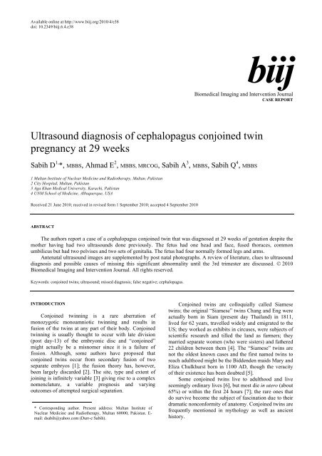
Ultrasound diagnosis of cephalopagus conjoined twin pregnancy at ...
Ultrasound diagnosis of cephalopagus conjoined twin pregnancy at ...
Ultrasound diagnosis of cephalopagus conjoined twin pregnancy at ...

You also want an ePaper? Increase the reach of your titles
YUMPU automatically turns print PDFs into web optimized ePapers that Google loves.
Available online <strong>at</strong> http://www.biij.org/2010/4/e38<br />
doi: 10.2349/biij.6.4.e38<br />
biij<br />
Biomedical Imaging and Intervention Journal<br />
CASE REPORT<br />
<strong>Ultrasound</strong> <strong>diagnosis</strong> <strong>of</strong> <strong>cephalopagus</strong> <strong>conjoined</strong> <strong>twin</strong><br />
<strong>pregnancy</strong> <strong>at</strong> 29 weeks<br />
Sabih D 1, *, MBBS, Ahmad E 2 , MBBS, MRCOG, Sabih A 3 , MBBS, Sabih Q 4 , MBBS<br />
1 Multan Institute <strong>of</strong> Nuclear Medicine and Radiotherapy, Multan, Pakistan<br />
2 City Hospital, Multan, Pakistan<br />
3 Aga Khan Medical University, Karachi, Pakistan<br />
4 UNM School <strong>of</strong> Medicine, Albuquerque, USA<br />
Received 21 June 2010; received in revised form 1 September 2010; accepted 4 September 2010<br />
ABSTRACT<br />
The authors report a case <strong>of</strong> a <strong>cephalopagus</strong> <strong>conjoined</strong> <strong>twin</strong> th<strong>at</strong> was diagnosed <strong>at</strong> 29 weeks <strong>of</strong> gest<strong>at</strong>ion despite the<br />
mother having had two ultrasounds done previously. The fetus had one head and face, fused thoraces, common<br />
umbilicus but had two pelvises and two sets <strong>of</strong> genitalia. The fetus had four normally formed legs and arms.<br />
Anten<strong>at</strong>al ultrasound images are supplemented by post n<strong>at</strong>al photographs. A review <strong>of</strong> liter<strong>at</strong>ure, clues to ultrasound<br />
<strong>diagnosis</strong> and possible causes <strong>of</strong> missing this significant abnormality until the 3rd trimester are discussed. © 2010<br />
Biomedical Imaging and Intervention Journal. All rights reserved.<br />
Keywords: <strong>conjoined</strong> <strong>twin</strong>s; ultrasound; missed <strong>diagnosis</strong>; false neg<strong>at</strong>ive; <strong>cephalopagus</strong>.<br />
INTRODUCTION<br />
Conjoined <strong>twin</strong>ning is a rare aberr<strong>at</strong>ion <strong>of</strong><br />
monozygotic monoamniotic <strong>twin</strong>ning and results in<br />
fusion <strong>of</strong> the <strong>twin</strong>s <strong>at</strong> any part <strong>of</strong> their body. Conjoined<br />
<strong>twin</strong>ning is usually thought to occur with l<strong>at</strong>e division<br />
(post day-13) <strong>of</strong> the embryonic disc and “<strong>conjoined</strong>”<br />
might actually be a misnomer since it is a failure <strong>of</strong><br />
fission. Although, some authors have proposed th<strong>at</strong><br />
<strong>conjoined</strong> <strong>twin</strong>s occur from secondary fusion <strong>of</strong> two<br />
separ<strong>at</strong>e embryos [1]; the fusion theory has, however,<br />
been largely discarded [2]. The site, type and extent <strong>of</strong><br />
joining is infinitely variable [3] giving rise to a complex<br />
nomencl<strong>at</strong>ure, a variable prognosis and varying<br />
outcomes <strong>of</strong> <strong>at</strong>tempted surgical separ<strong>at</strong>ion.<br />
* Corresponding author. Present address: Multan Institute <strong>of</strong><br />
Nuclear Medicine and Radiotherapy, Multan 60000, Pakistan. Email:<br />
dsabih@yahoo.com (Durr-e Sabih).<br />
Conjoined <strong>twin</strong>s are colloquially called Siamese<br />
<strong>twin</strong>s; the original “Siamese” <strong>twin</strong>s Chang and Eng were<br />
actually born in Siam (present day Thailand) in 1811,<br />
lived for 62 years, travelled widely and emigr<strong>at</strong>ed to the<br />
US; they worked as exhibits in circuses, were subjects <strong>of</strong><br />
scientific research and tilled the land as farmers; they<br />
married separ<strong>at</strong>e women (who were sisters) and f<strong>at</strong>hered<br />
22 children between them [4]. The “Siamese” <strong>twin</strong>s are<br />
not the oldest known cases and the first named <strong>twin</strong>s to<br />
reach adulthood might be the Biddenden maids Mary and<br />
Eliza Chulkhurst born in 1100 AD, though the veracity<br />
<strong>of</strong> their existence has been doubted [5].<br />
Some <strong>conjoined</strong> <strong>twin</strong>s live to adulthood and live<br />
seemingly ordinary lives [6], but most die in utero (about<br />
65%) or within the first 24 hours [7]; the rare ones th<strong>at</strong><br />
do survive become the subject <strong>of</strong> fascin<strong>at</strong>ion due to their<br />
dram<strong>at</strong>ic nonconformity <strong>of</strong> an<strong>at</strong>omy. Conjoined <strong>twin</strong>s are<br />
frequently mentioned in mythology as well as ancient<br />
history.
Sabih et al. Biomed Imaging Interv J 2010; 6(4):e38 2<br />
This page number is not<br />
for cit<strong>at</strong>ion purposes<br />
(a)<br />
(c)<br />
(e)<br />
Figure 1 a) Transverse ultrasound sections <strong>of</strong> the fetal head; note delta shape (left) and absence <strong>of</strong> cerebellum and posterior fossa (right); b)<br />
transverse section <strong>of</strong> the fetal thorax, note two spines on opposite sides <strong>of</strong> a fuse thorax, arrows mark vertebrae; c) transverse<br />
section <strong>of</strong> the fetal thorax, showing two unfused hearts (arrows); d) transverse section <strong>of</strong> the upper abdomen, four kidneys are seen<br />
(arrows); e) an oblique transverse section <strong>of</strong> the fetal abdomen, note the two bladders, the way the fetal lower abdomens angul<strong>at</strong>e<br />
and the obliquity <strong>of</strong> the section, this is a rel<strong>at</strong>ively coronal section and the fused liver and unfused bladder are seen in both fetuses;<br />
f) the two pelvises and external genitalia and four thighs are seen as the lower unfused abdomens diverge; a cross section <strong>of</strong> the<br />
cord shows multiple vessels.<br />
(b)<br />
(d)<br />
(f)
Sabih et al. Biomed Imaging Interv J 2010; 6(4):e38 3<br />
This page number is not<br />
for cit<strong>at</strong>ion purposes<br />
CASE REPORT<br />
A 28-year-old healthy woman, gravida 2, para 1,<br />
came for a routine ultrasound <strong>at</strong> 29 weeks <strong>of</strong> gest<strong>at</strong>ion;<br />
her previous <strong>pregnancy</strong> had been uneventful and the<br />
child was alive and healthy. There was no consanguinity<br />
and no history <strong>of</strong> <strong>twin</strong>ning in the family. <strong>Ultrasound</strong> was<br />
done with a digital scanner using a broad-band convex<br />
transducer (Toshiba, SSA-550A; with PVM375-AT<br />
probe; Toshiba Medical Systems, Tustin CA).<br />
The ultrasound showed a fetus with an unusual<br />
delta-shaped head in transverse section, the cerebellum<br />
could not be seen, there was a fused thorax showing two<br />
spines, two separ<strong>at</strong>ely be<strong>at</strong>ing hearts, two rib cages with<br />
sharing <strong>of</strong> the liver across the fetuses, there were four<br />
kidneys and two bladders; the lower abdomens were<br />
separ<strong>at</strong>e with two male external genitalia and four legs;<br />
and the cord had multiple vessels (Figure 1).<br />
The parents opted for a termin<strong>at</strong>ion and a<br />
cephalopagic fetus with the abnormalities noted on<br />
ultrasound was delivered. The head had an unusual broad<br />
outline but the vertex seemed normal and the face<br />
seemed normal too. There were two ears. A small<br />
complex irregular mass with multiple ridges was seen<br />
over the occipital midline.<br />
The parents did not give permission for an autopsy<br />
so the internal an<strong>at</strong>omy could not be confirmed.<br />
DISCUSSION<br />
Conjoined <strong>twin</strong>s are rare and are reported to occur<br />
with a prevalence <strong>of</strong> 1:50,000 in utero to 1:250,000 live<br />
births [8]. The actual r<strong>at</strong>es might be higher because <strong>of</strong><br />
unreported termin<strong>at</strong>ion. Conjoined <strong>twin</strong>s are generally<br />
incomp<strong>at</strong>ible with life i.e. 65% <strong>of</strong> cases are stillborn<br />
while <strong>of</strong> those th<strong>at</strong> are born alive, 35% die within the<br />
first 24 hours [7]. Only 25% survive to an age where<br />
surgical separ<strong>at</strong>ion can be considered [9]. The incidence<br />
<strong>of</strong> monozygotic <strong>twin</strong>ning is independent <strong>of</strong> race,<br />
m<strong>at</strong>ernal age and geography. There is a predominance <strong>of</strong><br />
females in <strong>conjoined</strong> <strong>twin</strong>s on the order <strong>of</strong> 3:1 and no<br />
increase in risk is known with parity, race, m<strong>at</strong>ernal age<br />
or heredity [10]. Over 1,000 descendants <strong>of</strong> the original<br />
“Siamese” <strong>twin</strong>s were studied; the next <strong>twin</strong>ning took<br />
place after the 4th gener<strong>at</strong>ion, these were normal<br />
monozygotic healthy <strong>twin</strong>s [11], this supports the<br />
absence <strong>of</strong> hereditary influence on <strong>conjoined</strong> <strong>twin</strong>ning.<br />
Conjoined <strong>twin</strong>ning can occur <strong>at</strong> infinite sites <strong>of</strong><br />
junction [3] giving rise to a complex and sometimes<br />
confusing classific<strong>at</strong>ion [12]. Usually, the same sites fuse<br />
but parasitic <strong>twin</strong>s are a subset where “asymmetric”<br />
joining occurs [2]. Parasitic <strong>twin</strong>s have their own<br />
variants and independent classific<strong>at</strong>ion [13]. The<br />
<strong>conjoined</strong> <strong>twin</strong>s, though monozygotic, are less<br />
“identical” than the usual, separ<strong>at</strong>e identical <strong>twin</strong>s; one<br />
<strong>twin</strong> might have more internal disarray <strong>of</strong> an<strong>at</strong>omy than<br />
the other [14, 15].<br />
Conjoined <strong>twin</strong>s are classified according to the site<br />
<strong>of</strong> fusion. When there is complete duplic<strong>at</strong>ion, the region<br />
<strong>of</strong> duplic<strong>at</strong>ion (usually with ventral fusion) is used to<br />
name the class, followed by the Greek term for fastened<br />
“pagus” (e.g. thoracopagus: joined <strong>at</strong> the thorax,<br />
craniopagus: joined <strong>at</strong> the head). When the duplic<strong>at</strong>ion is<br />
incomplete, the fusion is more extensive and usually<br />
l<strong>at</strong>eral; the part th<strong>at</strong> remains duplic<strong>at</strong>e and separ<strong>at</strong>e<br />
denotes the type <strong>of</strong> conjoining e.g. dicephalus: two heads,<br />
dipygus: two pelvises, etc; dorsal union (rachipagus) is<br />
extremely rare [3, 12, 16, 17]. The case reported here has<br />
been called cephalothoracopagus [2] or <strong>cephalopagus</strong> [12].<br />
(a)<br />
(b)<br />
Figure 2 a) Anterior photograph <strong>of</strong> the delivered fetus, the face<br />
and head look almost normal; b) posterior photograph <strong>of</strong><br />
the delivered fetus, arrow points to a small ridged<br />
structure over the occiput which is probably the<br />
otocephalic face on the other side <strong>of</strong> the “normal” face.
Sabih et al. Biomed Imaging Interv J 2010; 6(4):e38 4<br />
This page number is not<br />
for cit<strong>at</strong>ion purposes<br />
Conjoined <strong>twin</strong>s with sharing <strong>of</strong> the neural<br />
structures are very rare (11%) [2] and include the<br />
following types: craniopagus, with various grades <strong>of</strong><br />
skull, meningial and dural venous sinus sharing; there is<br />
no sharing <strong>of</strong> the face, foramen magnum or brain.<br />
Although the brain commonly remains separ<strong>at</strong>e, it may<br />
be connected by a bridge <strong>of</strong> neural tissue and the trunks<br />
are not joined [14]. Classific<strong>at</strong>ions concerned with<br />
surgical separ<strong>at</strong>ion divides craniopagus <strong>twin</strong>s into partial<br />
and complete forms depending upon the degree <strong>of</strong> dural<br />
venous sinus sharing [18] or the region <strong>of</strong> conjunction<br />
(frontal, parietal, temporoparietal and occipital) [19].<br />
Syncephalus, involves fusion <strong>of</strong> the brain and varying<br />
degree <strong>of</strong> facial fusion also. Cephalothoracopagus or<br />
<strong>cephalopagus</strong> involves the brain, face, thorax and upper<br />
abdomen; this could either be the Janiceps (Greek god<br />
with two faces) type, with two faces on either side <strong>of</strong> the<br />
head; or more rarely, one side <strong>of</strong> the head is rel<strong>at</strong>ively<br />
normal with a small otocephalic face on the other side<br />
(Figure 2b). Klinkosch [20] made beautiful engravings <strong>of</strong><br />
this type <strong>of</strong> <strong>twin</strong>ning in his book published in 1767, these<br />
have been reproduced by Kaufman [2]. The engraving <strong>of</strong><br />
the base <strong>of</strong> the brain shows one cerebrum but two<br />
obliquely oriented diverging cerebella and brain-stems;<br />
these would explain the inability <strong>of</strong> cerebellar<br />
visualiz<strong>at</strong>ion in the expected loc<strong>at</strong>ion within the posterior<br />
fossa on ultrasound in the authors' case. The heart and<br />
liver are <strong>of</strong>ten <strong>conjoined</strong> in cephalopagi, but in their case<br />
there were two separ<strong>at</strong>e hearts, although the liver was<br />
shared by the <strong>twin</strong>s.<br />
Table 1 <strong>Ultrasound</strong> criteria for <strong>diagnosis</strong> <strong>of</strong> <strong>conjoined</strong> <strong>twin</strong>ning (adapted from [3, 7, 19, 24, 25, 26])<br />
Clues Pitfalls<br />
Bifid appearance <strong>of</strong> the 1 st trimester fetal pole with a “V” or<br />
“Y” shaped embryo<br />
Continuous skin contour <strong>at</strong> the same an<strong>at</strong>omic level<br />
Single amniotic cavity with no dividing membrane<br />
Single placenta<br />
Fetal anomalies like omphaloceles, complex cardiac<br />
malform<strong>at</strong>ions etc<br />
Abnormal number <strong>of</strong> vessels (more than 3) in the cord<br />
Unusual extension <strong>of</strong> the spines and both fetuses facing each<br />
other<br />
Bi-breech or bi-cephalic present<strong>at</strong>ion<br />
Before the widespread use <strong>of</strong> ultrasound, most<br />
<strong>conjoined</strong> <strong>twin</strong>s could not be identified in utero. Even<br />
with radiographs, only about 25% <strong>of</strong> cases could be<br />
diagnosed in one series [3]. Plain X-rays were <strong>of</strong>ten not<br />
helpful because <strong>diagnosis</strong> could only be made if there<br />
was bony fusion. Contrast radiography could be used<br />
with the contrast m<strong>at</strong>erial instilled in the amniotic cavity<br />
in the hope <strong>of</strong> outlining the skin surface enabling<br />
continuity to be seen. However, the presence <strong>of</strong><br />
polyhydramnios usually diluted the contrast and the<br />
procedure’s inform<strong>at</strong>ion content. The first case<br />
diagnosed primarily by ultrasound was reported in 1977<br />
[21] <strong>at</strong> 28 weeks <strong>of</strong> gest<strong>at</strong>ion. Since then, criteria for<br />
ultrasound <strong>diagnosis</strong> have been well established<br />
(Table 1). There are also known pitfalls, which if<br />
avoided, will make the <strong>diagnosis</strong> feasible in most cases.<br />
First trimester ultrasound <strong>diagnosis</strong> is possible [22, 23]<br />
and the earliest <strong>diagnosis</strong> was reported after about seven<br />
weeks <strong>of</strong> <strong>pregnancy</strong> [24]. However, first trimester<br />
ultrasound <strong>diagnosis</strong> requires care and follow-ups to<br />
confirm the initial impression [25, 26].<br />
The authors’ case was diagnosed <strong>at</strong> 29 weeks <strong>of</strong><br />
gest<strong>at</strong>ion after the mother had undergone two ultrasounds<br />
<strong>at</strong> 22 weeks and <strong>at</strong> 28 weeks during which no<br />
abnormality was reported. Then she was referred to us<br />
for evalu<strong>at</strong>ion <strong>of</strong> mild polyhydramnios. <strong>Ultrasound</strong><br />
<strong>diagnosis</strong> <strong>of</strong> such an obvious and gross anomaly should<br />
be straightforward and it seemed difficult to understand<br />
how such a dram<strong>at</strong>ic abnormality could be missed on<br />
repe<strong>at</strong>ed ultrasound examin<strong>at</strong>ions, but such a case is not<br />
In some diamniotic <strong>twin</strong>s the dividing membrane can be very<br />
thin and difficult to identify<br />
The conjoining might be so severe as to mimic a single fetus<br />
with multiple anomalies<br />
All <strong>conjoined</strong> <strong>twin</strong>s do not present as mirror image fetuses and<br />
can be miss-diagnosed as a single fetus with malform<strong>at</strong>ion.<br />
Discordant present<strong>at</strong>ion is possible in omphalopagus <strong>twin</strong>s<br />
where the bridge is pliable and allows rot<strong>at</strong>ion [35].<br />
Single heart with gross and complex anomalies Both fetuses might have their own hearts<br />
Fixed position <strong>of</strong> the fetus rel<strong>at</strong>ive to each other on multiple<br />
exams<br />
Heads <strong>at</strong> the same level<br />
During first trimester, before amnio-chorionic separ<strong>at</strong>ion and<br />
with a rel<strong>at</strong>ively small amniotic cavity the <strong>twin</strong>s might be<br />
apposed to each other, appear to be fixed and having skin<br />
continuity[24, 27]<br />
In bi-cephalic <strong>pregnancy</strong>, one head might be fixed and the other<br />
higher up
Sabih et al. Biomed Imaging Interv J 2010; 6(4):e38 5<br />
This page number is not<br />
for cit<strong>at</strong>ion purposes<br />
an isol<strong>at</strong>ed case as we reported another dicephalic fetus<br />
th<strong>at</strong> went unreported until 28 weeks [27]. Others have<br />
reported a false neg<strong>at</strong>ive <strong>diagnosis</strong> <strong>of</strong> a pyopagus<br />
tetrapus parasitic <strong>twin</strong> on three scans, being confirmed<br />
only <strong>at</strong> 28 weeks on the fourth exam [28], several other<br />
3rd trimester diagnoses were also reported [29, 30]. It is<br />
possible th<strong>at</strong> if this condition is not thought <strong>of</strong> it will be<br />
missed, similar to missing a <strong>twin</strong> on a l<strong>at</strong>e ultrasound or<br />
a triplet when a <strong>twin</strong> is diagnosed just because the<br />
sonologist is not thinking <strong>of</strong> the possibility [31, 32].<br />
<strong>Ultrasound</strong> (non-panoramic) in the third trimester can<br />
only show a part <strong>of</strong> the fetal an<strong>at</strong>omy and it is possible<br />
th<strong>at</strong> the structure <strong>of</strong> one fetus is ascribed to the other<br />
fetus causing a mistaken impression <strong>of</strong> normal an<strong>at</strong>omy.<br />
This case highlights the fe<strong>at</strong>ures <strong>of</strong> an extremely<br />
rare fetal malform<strong>at</strong>ion and concludes with the<br />
suggestion th<strong>at</strong> everyone's mind should be kept open to<br />
all possibilities when making a <strong>diagnosis</strong>, as rare causes<br />
are rare but do occur in everyday practice <strong>of</strong> medicine.<br />
REFERENCES<br />
1. Spencer R., Theoretical and analytical embryology <strong>of</strong> <strong>conjoined</strong><br />
<strong>twin</strong>s: part I: embryogenesis. Clin An<strong>at</strong> 2000; 13(1):36–53.<br />
2. Kaufman MH. The embryology <strong>of</strong> <strong>conjoined</strong> <strong>twin</strong>s. Childs Nerv<br />
Syst 2004; 20(8-9):508–25.<br />
3. Barth RA, Filly RA, Goldberg JD, Moore P and Silverman NH.<br />
Conjoined <strong>twin</strong>s: pren<strong>at</strong>al <strong>diagnosis</strong> and assessment <strong>of</strong> associ<strong>at</strong>ed<br />
malform<strong>at</strong>ions. Radiology 1990;177(1):201–7.<br />
4. Daniels WB. The Siamese <strong>twin</strong>s: some observ<strong>at</strong>ions on their life,<br />
last illness and autopsy. Med Ann Dist Columbia, 1963; 32:273–7.<br />
5. Bondeson J. The Biddenden Maids: a curious chapter in the history<br />
<strong>of</strong> <strong>conjoined</strong> <strong>twin</strong>s. J R Soc Med, 1992;85(4):217–21.<br />
6. Dreger A. One <strong>of</strong> Us: Conjoined Twins and the Future <strong>of</strong> Normal.<br />
2004, Cambridge (Massachusetts): Harvard University Press.<br />
7. Winkler N, Kennedy A, Byrne J and Woodward P. The imaging<br />
spectrum <strong>of</strong> <strong>conjoined</strong> <strong>twin</strong>s. <strong>Ultrasound</strong> Q 2008; 24(4):249–55.<br />
8. Spitz L. Conjoined <strong>twin</strong>s. Pren<strong>at</strong> Diagn 2005; 25(9):814–9.<br />
9. Filler RM. Conjoined <strong>twin</strong>s and their separ<strong>at</strong>ion. Semin Perin<strong>at</strong>ol<br />
1986; 10(1):82–91.<br />
10. Edmonds LD, Layde PM. Conjoined <strong>twin</strong>s in the united st<strong>at</strong>es,<br />
1970-1977. Ter<strong>at</strong>ology 1982; 25(3):301–8.<br />
11. Aird I. Conjoined <strong>twin</strong>s; further observ<strong>at</strong>ions. Br Med J 1959;<br />
1(5133):1313–5.<br />
12. Spencer R. An<strong>at</strong>omic description <strong>of</strong> <strong>conjoined</strong> <strong>twin</strong>s: a plea for<br />
standardized terminology. J Pedi<strong>at</strong>r Surg 1996; 31(7):941–4.<br />
13. Spencer R. Parasitic <strong>conjoined</strong> <strong>twin</strong>s: external, internal (fetuses in<br />
fetu and ter<strong>at</strong>omas), and detached (acardiacs). Clin An<strong>at</strong> 2001;<br />
14(6):428–44.<br />
14. Kingston CA, McHugh K, Kumaradevan J, Kiely EM and Spitz L.<br />
Imaging in the preoper<strong>at</strong>ive assessment <strong>of</strong> <strong>conjoined</strong> <strong>twin</strong>s.<br />
Radiographics 2001; 21(5):1187–208.<br />
15. Weingast GR, Johnson ML, Pretorius DH, Porreco RP, Waldstein<br />
G, Foley C and Appareti K. Difficulty in sonographic <strong>diagnosis</strong> <strong>of</strong><br />
cephalothoracopagus. J <strong>Ultrasound</strong> Med 1984;3(9):421–3.<br />
16. Guttmacher AF and Nichols BL. Ter<strong>at</strong>ology <strong>of</strong> <strong>conjoined</strong> <strong>twin</strong>s.<br />
Birth Defects Orig Art Ser 1967; 3(1):3–9.<br />
17. McHugh K, Kiely EM and Spitz L. Imaging <strong>of</strong> <strong>conjoined</strong> <strong>twin</strong>s.<br />
Pedi<strong>at</strong>r Radiol 2006; 36(9):899–910; quiz 1002–3.<br />
18. Stone JL and Goodrich JT. The craniopagus malform<strong>at</strong>ion:<br />
classific<strong>at</strong>ion and implic<strong>at</strong>ions for surgical separ<strong>at</strong>ion. Brain 2006;<br />
129(Pt 5):1084–95.<br />
19. Bucholz RD, Yoon KW and Shively RE. Temporoparietal<br />
craniopagus. Case report and review <strong>of</strong> the liter<strong>at</strong>ure. J Neurosurg<br />
1987; 66(1):72–9.<br />
20. Klinkosch J. Programma quo An<strong>at</strong>omicam Monstri Bicorporei<br />
Monocaphali Descriptionen Proponit. 1767, Vetero-Pragae:<br />
Clauser.<br />
21. Fagan CJ. Antepartum <strong>diagnosis</strong> <strong>of</strong> <strong>conjoined</strong> <strong>twin</strong>s by<br />
ultrasonography. AJR Am J Roentgenol 1977; 129(5):921–2.<br />
22. Pajkrt E and Jauniaux E. First-trimester <strong>diagnosis</strong> <strong>of</strong> <strong>conjoined</strong><br />
<strong>twin</strong>s. Pren<strong>at</strong> Diagn 2005; 25(9):820–6.<br />
23. Lam YH, Sin SY, Lam C, Lee CP, Tang MH and Tse HY. Pren<strong>at</strong>al<br />
sonographic <strong>diagnosis</strong> <strong>of</strong> <strong>conjoined</strong> <strong>twin</strong>s in the first trimester: two<br />
case reports. <strong>Ultrasound</strong> Obstet Gynecol 1998; 11(4):289–91.<br />
24. Taner MZ, Kurdoglu M, Taskiran C, Kurdoglu Z, Himmetoglu O<br />
and Balci S. Early pren<strong>at</strong>al <strong>diagnosis</strong> <strong>of</strong> <strong>conjoined</strong> <strong>twin</strong>s <strong>at</strong> 7 weeks<br />
and 6 days' gest<strong>at</strong>ion with two-dimensional Doppler ultrasound: a<br />
case report. Cases J 2009;2:8330.<br />
25. Usta IM and Awwad JT. A false positive <strong>diagnosis</strong> <strong>of</strong> <strong>conjoined</strong><br />
<strong>twin</strong>s in a triplet <strong>pregnancy</strong>: pitfalls <strong>of</strong> first trimester<br />
ultrasonographic pren<strong>at</strong>al <strong>diagnosis</strong>. Pren<strong>at</strong> Diagn 2000;<br />
20(2):169–70.<br />
26. Weiss JL and Devine PC. False positive <strong>diagnosis</strong> <strong>of</strong> <strong>conjoined</strong><br />
<strong>twin</strong>s in the first trimester. <strong>Ultrasound</strong> Obstet Gynecol 2002;<br />
20(5):516–8.<br />
27. Durr-e-Sabih, Sabih Z, Worrall JA Jr and Khan AN. <strong>Ultrasound</strong><br />
<strong>diagnosis</strong> <strong>of</strong> dicephalic <strong>conjoined</strong> <strong>twin</strong>s <strong>at</strong> 24 weeks <strong>of</strong> gest<strong>at</strong>ion. J<br />
Clin <strong>Ultrasound</strong> 2009; 38(6):328–31.<br />
28. Gul A, Aslan H and Ceylan Y. Pren<strong>at</strong>al <strong>diagnosis</strong> <strong>of</strong> pygopagus<br />
tetrapus parasitic <strong>twin</strong>: case report. BMC Pregnancy Childbirth<br />
2004; 4(1):13.<br />
29. Kalchbrenner M, Weiner S, Templeton J and Losure TA. Pren<strong>at</strong>al<br />
ultrasound <strong>diagnosis</strong> <strong>of</strong> thoracopagus <strong>conjoined</strong> <strong>twin</strong>s. J Clin<br />
<strong>Ultrasound</strong> 1987;15(1):59–63.<br />
30. Karsdorp VH, van der Linden JC, Sobotka-Plojhar MA, Prins H,<br />
van der Harten JJ and van Vugt JM. Ultrasonographic pren<strong>at</strong>al<br />
<strong>diagnosis</strong> <strong>of</strong> <strong>conjoined</strong> thoracopagus <strong>twin</strong>s: a case report. Eur J<br />
Obstet Gynecol Reprod Biol 1991; 39(2):157–61.<br />
31. Divers WA Jr. and Hemsell DL. The use <strong>of</strong> ultrasound in multiple<br />
gest<strong>at</strong>ions. Obstet Gynecol 1979; 53(4):500–4.<br />
32. Eik-Nes SH, Salvesen KA, Okland O and V<strong>at</strong>ten LJ. Routine<br />
ultrasound fetal examin<strong>at</strong>ion in <strong>pregnancy</strong>: the 'Alesund'<br />
randomized controlled trial. <strong>Ultrasound</strong> Obstet Gynecol 2000;<br />
15(6):473–8.