PROSTHESES CONTROL BASED ON TMR a case study Hans Dietl ...
PROSTHESES CONTROL BASED ON TMR a case study Hans Dietl ...
PROSTHESES CONTROL BASED ON TMR a case study Hans Dietl ...

Create successful ePaper yourself
Turn your PDF publications into a flip-book with our unique Google optimized e-Paper software.
From “MEC '08 Measuring Success in Upper Limb Prosthetics,” Proceedings of the 2008 MyoElectric Controls/Powered Prosthetics<br />
Symposium, held in Fredericton, New Brunswick, Canada, August 13–15, 2008.<br />
<strong>PROSTHESES</strong> <strong>C<strong>ON</strong>TROL</strong> <strong>BASED</strong> <strong>ON</strong> <strong>TMR</strong><br />
a <strong>case</strong> <strong>study</strong><br />
<strong>Hans</strong> <strong>Dietl</strong>, PhD<br />
Otto Bock Healthcare Products GmbH<br />
Introduction<br />
The surgical treatment by targeted muscle reinnervation has been applied to<br />
more than 25 patients after traumatic loss of upper extremities in North America.<br />
In November 2006 this procedure was applied for the first time to a patient in<br />
Europe. The 18- year-old male patient suffered from bilateral limb loss after a high<br />
voltage accident, which happened in October 2006. The patient ended up with a<br />
medium long transhumeral residual limb on the right side and a shoulder<br />
disarticulation on the left side.<br />
He did undergo a primary rehabilitation at the rehabilitation center "Weisser Hof"<br />
and left rehabilitation after three months. The right side was fitted with a<br />
DynamicArm®, a wrist rotator and a SensorHand Speed. The system was controlled<br />
by the EMG signals of the biceps and the triceps. The switching between levels was<br />
achieved by co-contraction. The left side was fitted by a cosmetically arm with<br />
minimal functionality since trial fittings with functional arms failed.<br />
Assessment Phase<br />
The initial rehabilitation outcome was not in accordance with the specific needs of<br />
the patient: he did not reach a sufficient level of independence for everyday activities<br />
and it was not possible for him to return to work. For an improved functional outcome<br />
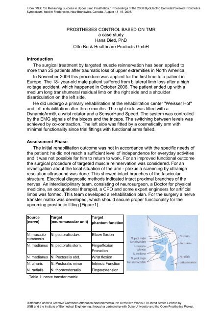
the surgical procedure of targeted muscle reinnervation was considered. For an<br />
investigation about the local situation of the arm - plexus a screening by ultrahigh<br />
resolution ultrasound was done. This showed intact branches of the fascicular<br />
structure. Electrical diagnostic methods indicated intact proximal branches of the<br />
nerves. An interdisciplinary team, consisting of neurosurgeon, a Doctor for physical<br />
medicine, an occupational therapist, a CPO and some expert engineers for artificial<br />
limbs was formed. This team developed a rehabilitation plan. For the surgery a nerve<br />
transfer matrix was developed, which should secure proper functionality for the<br />
upcoming prosthetic fitting [Figure1].<br />
Source<br />
(nerve)<br />
N. musculocutaneous<br />
Target<br />
(neuromuscular unit)<br />
Table 1: nerve transfer matrix<br />
Target<br />
phantom function<br />
N. pectoralis clav. Elbow flexion<br />
N. medianus N. pectoralis stern. Fingerflexion<br />
Pronation<br />
N. medianus N. Pectoralis abd. Wrist flexion<br />
N. ulnaris N. Pectoralis minor Intrinsic Function<br />
N. radialis N. thoracodorsalis Fingerextension<br />
Distributed under a Creative Commons Attribution-Noncommercial-No Derivative Works 3.0 United States License by<br />
UNB and the Institute of Biomedical Engineering, through a partnership with Duke University and the Open Prosthetics Project.
From “MEC '08 Measuring Success in Upper Limb Prosthetics,” Proceedings of the 2008 MyoElectric Controls/Powered Prosthetics<br />
Symposium, held in Fredericton, New Brunswick, Canada, August 13–15, 2008.<br />
Surgical intervention<br />
The goal of the intervention was to unhinge the greater branches of the arm<br />
plexus and to connect them to existing neuromuscular units. This rerouting was done<br />
according to figure1.<br />
The nerve transfers were done by end to end nerve-cooptation. For better signal<br />
separation the musculus pectoralis minor and the M pectoralis major pars<br />
clavicularis were moved.<br />
In addition the trunctus medius C7 was connected to the nervus supraclavicularis,<br />
which was cut at the punctum nervosum. By that means a sensible skin area should<br />
be generated, which can represent the sensory areas of the hand. The whole region<br />
was defatted for better signal quality.<br />
Postoperative phase<br />
Six weeks after the intervention the patient did undergo a training plan. He was<br />
supervised by an occupational therapist. The primary goal was to enhance the<br />
overall fitness and endurance and to correct the body posture and symmetry. As<br />
soon as measurable EMGs were detectable a specific training program was started.<br />
The patient visited the clinics every six weeks for follow-up. After three months<br />
voluntary muscle contractions at the M. pectoralis minor et major could be identified.<br />
Sensibility at the Trigonum colli laterale recovered. There the patient could clearly<br />
differentiate different regions of his hand. (Figure 2).<br />
Figure 2: isobaric lines of sensation and hand regions<br />
After six months there were sufficient signals at the segments of the M pectoralis,<br />
which allowed starting the fitting process (Table 2)<br />
Source<br />
(nerve)<br />
Target (neuromuscular unit)Result Target<br />
phantom function<br />
N. musculo-cutaneous N. pectoralis clav. excellent Elbow Flexion<br />
N. medianus N. pectoralis med. excellent Close fist<br />
N. medianus N. Pectoralis inf. good Wrist Flexion<br />
N. ulnaris N. Pectoralis minor ?? Intrinsic Function<br />
N. radialis N. thoracodorsalis ?? Finger Extension<br />
Table 2 nerve transfer matrix postoperative after six months<br />
Distributed under a Creative Commons Attribution-Noncommercial-No Derivative Works 3.0 United States License by<br />
UNB and the Institute of Biomedical Engineering, through a partnership with Duke University and the Open Prosthetics Project.
From “MEC '08 Measuring Success in Upper Limb Prosthetics,” Proceedings of the 2008 MyoElectric Controls/Powered Prosthetics<br />
Symposium, held in Fredericton, New Brunswick, Canada, August 13–15, 2008.<br />
After 17 months the nerve growth process seemed to be completed. All targets<br />
provide excellent signals. On the target units a further differentiation of phantom arm<br />
movements was possible (Table 3)<br />
Source (nerve) Target<br />
(neuromuscular<br />
unit)<br />
N. musculocutaneous<br />
Result Target<br />
phantom function<br />
N. pectoralis clav. excellent Ellbow Flexion<br />
N. medianus N. pectoralis med. excellent Close fist<br />
N. medianus N. Pectoralis inf. excellent Wrist Flexion,<br />
Pronation<br />
N. ulnaris N. Pectoralis minor excellent Intrinsic Function<br />
N. radialis N. thoracodorsalis excellent Finger Extension,<br />
Supination<br />
Table 3 nerve transfer matrix postoperative after 17 months<br />
Prosthetic fitting<br />
The patient was fitted with two different arm systems, the �Take Home Arm� and<br />
the �6 Degrees of Freedom Arm�.<br />
The �Take Home Arm�<br />
This system has a passive shoulder joint with free swing and a preflected<br />
position, a modified DynamicArm®, a wrist rotation unit and a Sensorhand Speed.<br />
For direct control of the 3 Degrees Of Motion a sixth EMG signal was needed.<br />
Therefore the musculus deltoideus was integrated into the control scheme. Standard<br />
EMG electrodes were used, however because of the skin movement a modified<br />
suspension of the electrodes was necessary (Figure 3).<br />
Figure 3: modified suspension of electrodes<br />
This system is used by the patient daily as his standard rehabilitation device.<br />
Distributed under a Creative Commons Attribution-Noncommercial-No Derivative Works 3.0 United States License by<br />
UNB and the Institute of Biomedical Engineering, through a partnership with Duke University and the Open Prosthetics Project.
From “MEC '08 Measuring Success in Upper Limb Prosthetics,” Proceedings of the 2008 MyoElectric Controls/Powered Prosthetics<br />
Symposium, held in Fredericton, New Brunswick, Canada, August 13–15, 2008.<br />
The �7 Degrees of Motion Arm�<br />
This arm is an outcome of the DARPA �Revolutionizing Prosthetics� program. It<br />
provides 7 degrees of motion and is controlled by pattern recognition techniques<br />
(Figure 4).<br />
.<br />
Pattern recognition<br />
Figure 4: 7 Degrees of Freedom Arm<br />
7 myoelectric controlled degrees of freedom<br />
1. Hand: open/close<br />
2. Thumb: flexion/extension<br />
3. Wrist: palmar flexion/dorsal flexion<br />
4. Wrist: pronation/supination<br />
5. Ellbow: flexion/extension<br />
6. Humeral rotation<br />
7. Shoulder flexion/extension<br />
Up to 24 adhesive electrodes were used for control. This system was used in the<br />
laboratory only<br />
Patient outcome:<br />
The patient is using the �Take Home Arm� daily as his standard rehabilitation<br />
device. He achieved a sufficient degree of independence and was able to return to<br />
work in the logistics and warehouse of a car dealer. The �7 Degrees of Motion Arm�<br />
was tested on a regular basis in a laboratory environment. The patient was able to<br />
intuitively control all degrees of motion. No additional input devices were necessary.<br />
Outlook<br />
The functionality and the degrees of motion of the �Take Home Arm� will be<br />
extended step by step by using the findings of the laboratory tests.<br />
References<br />
1. Aszmann OC, Muse V, Dellon AL: Evidence of collateral sprouting after sensory nerve<br />
resection. Ann Plast Surg 1996; 37: 520-525<br />
2. Aszmann OC, Rab M, Kamolz PM, Frey M: The anatomy of the pectoral nerves and their<br />
significance in brachial plexus reconstruction. J Hand Surg [Am] 2000; 25: 942-947<br />
3. Hijjawi JB, Kuiken TA, Lipschutz RD, Miller LA, Stubblefield KA, Dumanian GA: Improved<br />
myoelectric prosthesis control accomplished using multiple nerve transfers. Plast Reconstr<br />
Surg 2006; 118: 1573-1578<br />
4. Kuiken TA, Lowery MM, Stoykov NS: The effect of subcutaneous fat on myoelectric signal<br />
amplitude and cross talk. Prosthet Orthot Int 2003; 27: 48-54<br />
Distributed under a Creative Commons Attribution-Noncommercial-No Derivative Works 3.0 United States License by<br />
UNB and the Institute of Biomedical Engineering, through a partnership with Duke University and the Open Prosthetics Project.
From “MEC '08 Measuring Success in Upper Limb Prosthetics,” Proceedings of the 2008 MyoElectric Controls/Powered Prosthetics<br />
Symposium, held in Fredericton, New Brunswick, Canada, August 13–15, 2008.<br />
5. Kuiken TA, Dumanian GA, Lipschutz RD, Miller LA, Stubblefield KA: The use of targeted<br />
muscle reinnervation for improved myoelectric prosthesis control in a bilateral shoulder<br />
disarticulation amputee. Prosthet Orthot Int 2004; 28: 245-253<br />
6. Aszmann OC, <strong>Dietl</strong> H, Frey M: Selective Nerve Transfers to Improve the Control of<br />
Myoelectrical Arm Prostheses<br />
7. Miller LA, Lipschutz RD, Stubblefield KA, Lock BA, Huang H, Williams W, Weir RF, Kuiken<br />
TA: Control of a Six Degree-of-freedom Prosthetic Arm after Targeted Muscle Reinnervation<br />
Surgery<br />
8. Zhou P, Lowery M, Englehart KB, Huang H, Li G, Hargrove L, Dewald J, Kuiken TA:<br />
Decoding a New Neural Machine Interface for Control of Artificial Limbs<br />
J Neuophysiol Nov 2007; 5: 2974 � 2982<br />
Distributed under a Creative Commons Attribution-Noncommercial-No Derivative Works 3.0 United States License by<br />
UNB and the Institute of Biomedical Engineering, through a partnership with Duke University and the Open Prosthetics Project.