Synthesis of Iron Oxide Nanoparticles with Biological Coatings
Synthesis of Iron Oxide Nanoparticles with Biological Coatings
Synthesis of Iron Oxide Nanoparticles with Biological Coatings

You also want an ePaper? Increase the reach of your titles
YUMPU automatically turns print PDFs into web optimized ePapers that Google loves.
RESEARCH IRON OXIDE NANOPARTICLES<br />
<strong>Synthesis</strong> <strong>of</strong> <strong>Iron</strong> <strong>Oxide</strong> <strong>Nanoparticles</strong> <strong>with</strong> <strong>Biological</strong> <strong>Coatings</strong><br />
David Dozier, Soubantika Palchoudhury, *Yuping Bao, Ph.D.<br />
*Faculty Sponsor<br />
Department <strong>of</strong> Chemical and <strong>Biological</strong> Engineering, The University <strong>of</strong> Alabama, Tuscaloosa, AL 35487, USA<br />
For biological and biomedical applications, it is required to produce nanoparticles that are water soluble and<br />
biocompatible. Here, we report the synthesis <strong>of</strong> iron oxide nanoparticles coated <strong>with</strong> biological molecules (e.g.,<br />
gluconic acid, lactobionic acid, or polyacrylic acid) via a co‐precipitation method. These nanoparticles have<br />
narrow size distribution and are highly water soluble. Because <strong>of</strong> the biological coatings, they will have great<br />
potential in numerous biomedical applications such as tissue engineering.<br />
Introduction<br />
Magnetic nanoparticles have shown great<br />
potential in many biological and biomedical<br />
applications such as targeted drug delivery, magnetic<br />
fluid hyperthermia, magnetic resonance imaging, and<br />
tissue engineering [1, 3, 5, 6]. All these applications<br />
require magnetic nanoparticles to be water soluble<br />
and biocompatible.<br />
For biological and biomedical applications,<br />
magnetic iron oxide nanoparticles are the primary<br />
choice because <strong>of</strong> their biocompatibility and chemical<br />
stability. Many synthesis methods have been explored<br />
for magnetic iron oxide nanoparticles. These include<br />
organic solvent heating method, polyol method, and<br />
co‐precipitation method [2, 4, 7]. The co‐precipitation<br />
method is the most effective technique for preparing<br />
aqueous dispersions <strong>of</strong> iron oxide nanoparticles<br />
because the synthesis is conducted in water. For this<br />
report, we studied several biological molecules as<br />
surface coatings to achieve biocompatibility such as<br />
gluconic acid (GA), lactobionic acid (LBA), and<br />
polyacrylic acid (PAA). These molecules were used to<br />
control the particle size, to prevent the nanoparticles<br />
from aggregation, and to achieve biocompatibility.<br />
Experimental<br />
<strong>Synthesis</strong><br />
<strong>Iron</strong> oxide nanoparticles were synthesized by a<br />
modified co‐precipitation method. Ferric chloride<br />
(FeCl3, 0.074 g) and ferrous chloride (FeCl2, 0.190 g)<br />
at a ratio <strong>of</strong> 2 to 1 were dissolved in 20 mL deionized<br />
water, which was then stirred and heated to 60 °C.<br />
The solution was bubbled <strong>with</strong> Argon gas to prevent<br />
unwanted oxidation. Subsequently, 10 mL <strong>of</strong> 2.5 M<br />
NaOH solution was injected at 60 °C and the reaction<br />
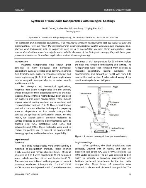
continued at that temperature for 20 minutes before<br />
the flask was removed from heating and stirring. The<br />
nanoparticles were then removed from solution by<br />
magnetic separation. During synthesis, the<br />
concentration and amount <strong>of</strong> NaOH was varied to<br />
control the particle size. A schematic drawing <strong>of</strong> the<br />
reaction set‐up is shown in Figure 1.<br />
Figure 1. Schematic drawing <strong>of</strong> the experimental set‐up.<br />
Surface coatings<br />
After synthesis, the black precipitates were<br />
collected, washed <strong>with</strong> DI water, and then re‐<br />
dispersed into 10 mL GA, LBA, or PAA solutions (100<br />
mM) under sonication. The pH was adjusted to 8 in<br />
order to simulate a biological environment and<br />
facilitate surfactant attachment to the iron oxide<br />
nanoparticles. Three hours <strong>of</strong> sonication were<br />
required to obtain well dispersed nanoparticles. The<br />
BAMA.UA.EDU/~JOSHUA 16
IRON OXIDE NANOPARTICLES RESEARCH<br />
nanoparticle solutions were then left at room<br />
temperature.<br />
Characterization<br />
The size and morphology <strong>of</strong> nanoparticles were<br />
studied by transmission electron microscopy (TEM);<br />
the crystal structure was verified using x‐ray<br />
diffraction (XRD); the hydrodynamic size <strong>of</strong> the<br />
nanoparticles in solution was studied using dynamic<br />
light scattering (DLS). The solutions were also checked<br />
for precipitation. Solutions <strong>with</strong> a high level <strong>of</strong><br />
precipitate indicated aggregation <strong>of</strong> nanoparticles.<br />
These aggregates are not suitable for biological<br />
applications.<br />
Results and Discussion<br />
Figure 2 shows the representative TEM images<br />
<strong>of</strong> iron oxide nanoparticles coated <strong>with</strong> GA and LBA.<br />
Although the sizes <strong>of</strong> both types were roughly 10 nm,<br />
the GA coated nanoparticles were more dispersed<br />
than the LBA coated nanoparticles, which showed a<br />
certain degree <strong>of</strong> aggregation.<br />
Figure 2. TEM images <strong>of</strong> iron oxide nanoparticles coated<br />
<strong>with</strong> (A) GA and (B) LBA.<br />
The TEM image was not produced for the PAA<br />
coated iron oxide nanoparticles due to the large<br />
amount <strong>of</strong> the polymers present on the TEM grid,<br />
which interfered <strong>with</strong> the electron beam. However,<br />
the nanoparticle solution was well dispersed <strong>with</strong> no<br />
evident precipitate. Normally, precipitation is an<br />
indication <strong>of</strong> aggregation <strong>of</strong> nanoparticles.<br />
The crystal structures <strong>of</strong> these nanoparticles<br />
were studied on GA coated nanoparticles. It was<br />
confirmed that these iron oxide nanoparticles are<br />
maghemite (Fe2O3), as shown in Figure 3, instead <strong>of</strong><br />
the commonly formed magnetite nanoparticles<br />
(Fe3O4). This study suggests that the nanoparticles<br />
were fully oxidized either during or after the<br />
synthesis.<br />
Intensity (a.u.)<br />
(200)<br />
(311)<br />
(400)<br />
(440)<br />
20 30 40 50 60 70 80<br />
2 theta (degree)<br />
Figure 3. XRD scan <strong>of</strong> iron oxide nanoparticles.<br />
For biological applications, nanoparticles are<br />
normally used in solution form. Therefore, it is<br />
important to study their hydrodynamic size in<br />
solution. DLS was used to measure the size <strong>of</strong> the<br />
synthesized nanoparticles. GA produced the smallest<br />
particles, showing a peak around 90 nm. LBA and PAA<br />
both had peaks around 140 nm as shown in Figure 4.<br />
The hydrodynamic sizes <strong>of</strong> the synthesized<br />
nanoparticles were significantly larger than those<br />
indicated by their TEM images. This is possibly due to<br />
the hydrogen bond formation between the carboxyl<br />
groups on adjacent surfaces, which can cause cross‐<br />
linking between particles and result in a large<br />
hydrodynamic size. The size from DLS did not reveal a<br />
large difference between the MNPs synthesized <strong>with</strong><br />
2.5 M NaOH and 5 M NaOH.<br />
Intensity (%)<br />
15<br />
10<br />
5<br />
PAA<br />
LBA<br />
GA<br />
0<br />
0 100 200 300<br />
Diameter (nm)<br />
400 500<br />
Figure 4. DLS plots <strong>of</strong> iron oxide nanoparticles coated <strong>with</strong><br />
GA (cross), LBA (triangle), and PAA (circle).<br />
17 JOSHUA | May 2010 | Vol 7
RESEARCH IRON OXIDE NANOPARTICLES<br />
Conclusion<br />
In conclusion, water soluble iron oxide<br />
nanoparticles were synthesized using a co‐<br />
precipitation method. These nanoparticles were<br />
subsequently coated <strong>with</strong> GA, LBA, or PAA. Both GA<br />
and LBA coatings produced well dispersed<br />
nanoparticles. They were in fully oxidized oxide form<br />
as confirmed by XRD. In solution, these nanoparticles<br />
showed a much larger hydrodynamic size, possibly<br />
due to hydrogen bond formation.<br />
References<br />
[1] Babes L, Denizot B, Tanguy G, Le Jeune JJ & Jallet P.<br />
(1999). <strong>Synthesis</strong> <strong>of</strong> iron oxide nanoparticles used as<br />
MRI contrast agents: A parametric study. Journal <strong>of</strong><br />
Colloid and Interface Science, 212(2):474‐482.<br />
[2] Ge JP, Hu YX, Biasini M, Dong CL, Guo JH, Beyermann<br />
WP & Yin YD. (2007). One‐step synthesis <strong>of</strong> highly<br />
water‐soluble magnetite colloidal nanocrystals.<br />
Chemistry‐a European Journal, 13(25):7153‐7161.<br />
[3] Gu FX, Karnik R, Wang AZ, Alexis F, Levy‐Nissenbaum<br />
E, Hong S, Langer RS & Farokhzad OC. (2007).<br />
Targeted nanoparticles for cancer therapy. Nano<br />
Today, 2(3):14‐21.<br />
[4] Hyeon T. Chemical synthesis <strong>of</strong> magnetic<br />
nanoparticles. (2002). Chemical Commun., 8:927‐<br />
934.<br />
[5] Shimizu K, Ito A, Arinobe M, Murase Y, Iwata Y,<br />
Narita Y, Kagami H, Ueda M & Honda H. (2007).<br />
Effective cell‐seeding technqiue using magnetite<br />
nanoparticles and magnetic force onto<br />
decellularized blood vessels for vascular tissue<br />
engineering. J. <strong>of</strong> Bioscience and Bioenginer.,<br />
103(5):472‐478.<br />
[6] Thiesen B & Jordan A. (2008). Clinical applications <strong>of</strong><br />
magnetic nanoparticles for hyperthermia.<br />
International J. Hyperthermia, 24(6):467‐474.<br />
[7] Wenguang Y. Tonglai Z, Jianguo Z, Jinyu G & Ruifeng<br />
W. (2007). The preparation methods <strong>of</strong> magnetite<br />
nanoparticles and their morphology. Progress in<br />
Chemistry, 19(6):884‐892.<br />
David Dozier is a junior from Thomasville, Alabama,<br />
majoring in chemical engineering <strong>with</strong> a minor in<br />
biology. He currently works in the laboratory <strong>of</strong> Dr.<br />
Yuping Bao in the Department <strong>of</strong> Chemical and<br />
<strong>Biological</strong> Engineering. David also has an interest in<br />
going to medical school after graduation. He is a<br />
Presidential Scholar and a member <strong>of</strong> the Golden<br />
Key Honor Society.<br />
BAMA.UA.EDU/~JOSHUA 18