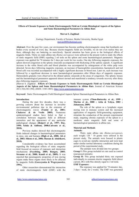
Effects of Chronic Exposure - The Journal of American Science
Effects of Chronic Exposure - The Journal of American Science
Effects of Chronic Exposure - The Journal of American Science

Create successful ePaper yourself
Turn your PDF publications into a flip-book with our unique Google optimized e-Paper software.
<strong>Journal</strong> <strong>of</strong> <strong>American</strong> <strong>Science</strong>, 2011;7(8) http://www.americanscience.org<br />
<strong>Effects</strong> <strong>of</strong> <strong>Chronic</strong> <strong>Exposure</strong> to Static Electromagnetic Field on Certain Histological Aspects <strong>of</strong> the Spleen<br />
and Some Haematological Parameters in Albino Rats<br />
Mervat S. Zaghloul<br />
Zoology Department, Faculty <strong>of</strong> <strong>Science</strong>, Benha University, Benha Egypt<br />
mervat2009@hotmail.com<br />
Abstract: Over the past few years, our environment has become seething electromagnetic smog that bombards our<br />
bodies every second <strong>of</strong> every day. Because electro-magnetic fields are invisible, we do not even realize they are<br />
there, although they are battering us mercilessly. Special attention has been given to the biological effects <strong>of</strong><br />
magnetic fields. Thirty six male albino rats (Rattus norvegicus) were utilized in the present work to study the effects<br />
<strong>of</strong> static magnetic field (SMF) equaling 2 ml tesla on the spleen and some haematological parameters. Magnetic<br />
exposure was applied for ٦0 minutes for 3 days per week for two weeks. One day following magnetic exposure, the<br />
spleen showed congestion in the splenic sinusoids accompanied with thickening <strong>of</strong> the splenic capsule. A significant<br />
increase in the white blood cells and blood platelets was accompanied by enlargement <strong>of</strong> the white pulp were<br />
detected. Seven days following magnetic exposure, an increase <strong>of</strong> haemoglobin concentration; haematocrit and red<br />
blood cells was recorded accompanied with a highly significant decrease in blood iron. Later on, such increase was<br />
followed by a significant decrease in most haematological parameters after fifteen days <strong>of</strong> magnetic exposure.<br />
Hemosiderin granules were observed in the dilated splenic sinusoids at the areas <strong>of</strong> congestion. <strong>The</strong> splenic tissues<br />
and the haematological parameters appeared almost normal and manifested a tendency towards recovery after thirty<br />
days following magnetic exposure.<br />
[Mervat S. Zaghloul . <strong>Effects</strong> <strong>of</strong> <strong>Chronic</strong> <strong>Exposure</strong> to Static Electromagnetic Field on Certain Histological<br />
Aspects <strong>of</strong> the Spleen and Some Haematological Parameters in Albino Rats. <strong>Journal</strong> <strong>of</strong> <strong>American</strong> <strong>Science</strong><br />
2011;7(8):383-394]. (ISSN: 1545-1003). http://www.americanscience.org.<br />
Keyword: Static Electromagnetic Field Histological Aspects Spleen Haematological Parameters in Albino Rats<br />
Introduction:<br />
During the past few decades, there was a<br />
growing concern about the increase in invisible<br />
environmental pollution due to the emission <strong>of</strong><br />
electromagnetic waves (Tonini et al., 2001;<br />
Chakeres & de Vocht 2005). Numerous<br />
epidemiological studies have failed to find a<br />
correlation between magnetic field at different<br />
intensities and the appearance <strong>of</strong> any particular<br />
pathological changes (Reipert et al., 1997; Day,<br />
1999; Schüz & Ahlbom, 2008;Calvente et al.,<br />
2010).<br />
Previous studies showed that electromagnetic<br />
fields induced changes in haematological parameters<br />
in mice, rats and humans (High et al., 2000; Ali et<br />
al., 2003; Sihem et al., 2006; Hassan & Abdelkawi,<br />
2010).<br />
Considerable evidence has been accumulated<br />
regarding the biological effects <strong>of</strong> static magnetic<br />
field focused on sources <strong>of</strong> exposure and interaction<br />
mechanisms (Feychting, 2005; Rongen, 2005;<br />
Straume et al., 2008; Kundi et al., 2009). It was<br />
reported that paramagnetic properties <strong>of</strong> iron storing<br />
organs make these organs more likely to be affacted<br />
by magnetic fields (Gorczynska & Wegrzynowicz,<br />
1991).<br />
Other researchers demonstrated the interaction<br />
<strong>of</strong> static electromagnetic field (EMF) with the<br />
immune system (Thun-Battersby et al., 1999 ;<br />
Marino et al., 2000 ; Attia & Yehya, 2002 ;<br />
Johansson, 2009)<br />
<strong>The</strong> role <strong>of</strong> the spleen as a lymphatic organ<br />
storing iron in immune system and the increased<br />
application <strong>of</strong> magnetic field-generating equipment,<br />
stimulate the conduction <strong>of</strong> the present experimental<br />
study, targeting chronic exposure <strong>of</strong> the spleen to a<br />
moderate static magnetic field and some <strong>of</strong><br />
haematological parameters in albino rats.<br />
Material and Methods<br />
Animals:<br />
Thirty male albino rats (Rattus norvegicus),<br />
each weighs 120 + 10 grams were utilized in the<br />
present study. <strong>The</strong> animals were housed in plastic<br />
cages to avoid any metallic interaction and were kept<br />
under similar normal laboratory conditions during the<br />
period <strong>of</strong> the experimental study.<br />
<strong>The</strong> animals were divided into two groups:<br />
Group I : This group included six rats used as control<br />
(unexposed animals).<br />
Group II: This group included thirty rats exposed<br />
individually to constant electric magnetic field (direct<br />
current, DC) with flux density equal to 2 ml tesla.<br />
EMF <strong>Exposure</strong>:<br />
<strong>The</strong> exposure was applied for ٦0 minutes/ day,<br />
3 days / week, for 2 weeks. <strong>The</strong> EMF was generated<br />
http://www.americanscience.org 383<br />
editor@americanscience.org
<strong>Journal</strong> <strong>of</strong> <strong>American</strong> <strong>Science</strong>, 2011;7(8) http://www.americanscience.org<br />
by applying an electric current to the coil <strong>of</strong> artificial<br />
EMF apparatus constructed in the Department <strong>of</strong><br />
Physics, Jubail Faculty <strong>of</strong> Education for Girls,<br />
Kingdom <strong>of</strong> Saudi Arabia. <strong>The</strong> animals were kept in<br />
a perforated plastic box, placed in the coil chamber <strong>of</strong><br />
the apparatus. <strong>The</strong>n, a horizontal magnetic induction<br />
was applied to the whole animal body. <strong>The</strong> field<br />
strength was monitored with a gauss meter.<br />
At the end <strong>of</strong> the experiment, all the animals<br />
were dissected and specimens <strong>of</strong> spleen were taken<br />
on 1, 7, 15, and 30 days post-irradiation. All the<br />
control animals and six animals <strong>of</strong> the second group<br />
were sacrificed after 1, 3,6,15 and 30 days following<br />
the exposure.<br />
Histological methods:<br />
Small spleen specimens were cut into small<br />
blocks, fixed in 10% neutral buffered formalin and<br />
processed up to paraffin pieces. Semi-serial sections<br />
<strong>of</strong> 6 µm were prepared and stained with Harris'<br />
hematoxylin and eosin (Drury & Wallington 1980)<br />
to illustrate the histological structure. Spleen<br />
specimens also stained for haemosiderin by Perl’s<br />
Prussian blue reaction technique (1867) for<br />
detection <strong>of</strong> haemosiderin iron.<br />
Haematological and Blood chemical methods:<br />
At the end <strong>of</strong> the experiment the blood<br />
samples were collected (0.5 ml approximately/<br />
sample) from the supra-orbital venous plexus <strong>of</strong> rats<br />
using heparinized syringes into two tubes. <strong>The</strong> first<br />
tube contained heparin as anticoagulant. <strong>The</strong><br />
heparinized blood was used for RBC, haematocrit,<br />
haemoglobin, WBC and platelets analysis, using<br />
standard methods (Feldman et al., 2000).<br />
<strong>The</strong> blood sample in the other tube was left for<br />
a short time to allow clotting. Clear serum samples<br />
were obtained by centrifugation at 3000 r.p.m. for 20<br />
min and then kept at -20°C prior to biochemical<br />
analysis.<br />
A serum level <strong>of</strong> iron was measured using<br />
Stanbio serum iron liquicolour commercial Kits,<br />
(Procepdure No. 0370) according to the method by<br />
Weissman &Leggi (1974).<br />
Statistical Analysis:<br />
<strong>The</strong> data was present as the mean ± SD for<br />
each correlation was calculated using Micros<strong>of</strong>t<br />
Excel.<br />
Results<br />
I. <strong>The</strong> control animals:<br />
<strong>The</strong> spleen is surrounded by a fibrous<br />
connective tissue capsule interspersed with<br />
smooth muscle fibres . Irregular spaced trabeculae<br />
<strong>of</strong> smooth muscle and fibroelastic tissue emanate<br />
from the splenic capsule into the splenic<br />
parenchyma containing blood and lymph vessels.<br />
<strong>The</strong> parenchyma <strong>of</strong> the spleen is termed the<br />
pulp. Most <strong>of</strong> the pulp is s<strong>of</strong>t red. It consists <strong>of</strong><br />
large, irregular, thin-walled blood vessels, splenic<br />
sinusoids, interposed between sheets <strong>of</strong> thin<br />
connective tissues and splenic cords. <strong>The</strong> splenic<br />
cords are the masses <strong>of</strong> cells in between the<br />
sinusoids. <strong>The</strong>y contain a lot <strong>of</strong> erythrocytes<br />
(RBCs) and some other cell types as macrophages<br />
and megakaryocytes.<br />
Within the red pulp, small oval or/and<br />
rounded grayish blue stained areas represent the<br />
white pulps. <strong>The</strong>y consist almost entirely <strong>of</strong><br />
lymphocytes, in a peculiar association with the<br />
arterial blood supply (Fig .1).<br />
<strong>The</strong> splenic tissue reveals a scanty amount<br />
<strong>of</strong> blue stained haemosiderin granules that are<br />
diffused and distributed throughout the red pulp<br />
cords (Fig. 2).<br />
II. <strong>The</strong> SMF-exposed animals:<br />
1. One day post exposure to SMF:<br />
<strong>The</strong> spleen had a thick connective tissue<br />
capsule, variable in its thickness at different areas.<br />
Numerous thick trabeculae extended from the<br />
splenic capsule inwards, branched and divided the<br />
spleen into numerous parts. <strong>The</strong>re were multiple<br />
clear areas in the subcapsular spaces and few<br />
haemorrhagic areas as directed inwards. Within<br />
the red pulps, small foci <strong>of</strong> cellular necrosis were<br />
observed and the splenic sinusoids were slightly<br />
dilated and congested with blood cells (Fig.3).<br />
Most <strong>of</strong> the white pulps were large and<br />
irregular in shape revealing cellular proliferation<br />
<strong>of</strong> homogeneous population <strong>of</strong> immunoplastic<br />
cells, and macrophages. Small foci <strong>of</strong> cellular<br />
lesions were observed scattering throughout the<br />
white pulps (Fig .4).<br />
2. Seven days post exposure to SMF:<br />
Numerous subcapsular clear spaces were<br />
found between the capsule and trabeculae . Few<br />
other ones containing haemolysed RBCs were<br />
observed between the splenic parenchyma (Fig.<br />
5).<br />
<strong>The</strong> cells <strong>of</strong> the splenic cords revealed<br />
necrotic changes scattering throughout the red<br />
pulp. <strong>The</strong> splenic sinusoids were dilated contained<br />
some <strong>of</strong> erythrocytes or hemolysed blood.<br />
Numerous inflammatory cells and<br />
megakaryocytes were noted in the splenic<br />
parenchyma and sinusoids.<br />
<strong>The</strong> white pulps were numerous, fragmented<br />
and appeared diffused in an irregular manner in<br />
the red pulp (Fig. 6).<br />
http://www.americanscience.org 384<br />
editor@americanscience.org
<strong>Journal</strong> <strong>of</strong> <strong>American</strong> <strong>Science</strong>, 2011;7(8) http://www.americanscience.org<br />
3. Fifteen days post exposure to SMF:<br />
<strong>The</strong> splenic vasculature revealed<br />
progressive changes represented by congestion <strong>of</strong><br />
the splenic sinuses and diffusion <strong>of</strong> haemorrhagic<br />
and hemolysed areas throughout the red pulps.<br />
Numerous inflammatory cells, macrophage and<br />
megakaryocytes were observed in the splenic<br />
parenchyma and sinusoids (Fig.7).<br />
Some splenic white pulps suffered from<br />
lymphoid depletion and the others became<br />
hyperplastic.<br />
Heavy iron blue pigments <strong>of</strong> hemosiderin<br />
granules were observed in the dilated red pulp<br />
spaces which were filled with hemolysed red<br />
blood cells (Fig.8).<br />
Fig. 1: A photomicrograph <strong>of</strong> a spleen section <strong>of</strong><br />
control rat showing a fibrous connective tissue<br />
capsule surrounding the splenic pulp (arrow).<br />
Most <strong>of</strong> the pulp is a s<strong>of</strong>t red pulp (RP) and some<br />
oval grayish blue-stained areas represent the white<br />
pulp (WP). (H&E stain X250)<br />
Fig. 3: A photomicrograph <strong>of</strong> a spleen section <strong>of</strong> rat<br />
after one day following the end <strong>of</strong> SMF exposure<br />
showing a thick connective tissue capsule<br />
(arrowhead) , Multiple clear sinusoids in the<br />
subcapsular space(black arrow) and few<br />
haemorrhagic ( white arrow). (H&E stain<br />
X400<br />
4. Thirty days post the end <strong>of</strong> exposure to SMF:<br />
<strong>The</strong> splenic tissues appeared almost normal<br />
and manifested a tendency towards recovery.<br />
Some significant signs towards complete<br />
vasculature and tissues recovery were observed in<br />
both red and white pulp as compared with the<br />
previous examined groups, where no areas <strong>of</strong><br />
blood haemorrhage or haemolysis were observed.<br />
Brown hemosiderin granules were seen in<br />
the cytoplasm <strong>of</strong> macrophages (Fig .9).<br />
Fig. 2: A photomicrograph <strong>of</strong> a spleen section <strong>of</strong><br />
control rat showing a splenic tissue revealing a<br />
scanty amount <strong>of</strong> blue-stained haemosiderin<br />
granules diffusely distributed throughout the red<br />
pulp cords. (Perl’s Prussian blue stain X400)<br />
Fig. 4: A photomicrograph <strong>of</strong> a spleen section <strong>of</strong> rat<br />
after one day following the end <strong>of</strong> SMF exposure<br />
showing large and irregular white pulps (WP)<br />
revealing cellular proliferation <strong>of</strong> homogeneous<br />
population <strong>of</strong> immunoplastic cells, and macrophages<br />
(arrowhead) in addition to small foci <strong>of</strong> cellular<br />
lesions (arrow). ( H&E stain X400)<br />
http://www.americanscience.org 385<br />
editor@americanscience.org
<strong>Journal</strong> <strong>of</strong> <strong>American</strong> <strong>Science</strong>, 2011;7(8) http://www.americanscience.org<br />
Fig. 5: A photomicrograph <strong>of</strong> a spleen section <strong>of</strong> rat<br />
after seven days following the end <strong>of</strong> SMF exposure<br />
showing numerous subcapsular clear spaces (**) and<br />
few other ones containing haemolysed RBCs<br />
(arrowhead) between splenic parenchyma.<br />
(H&E stain X250)<br />
Fig. 7: A photomicrograph <strong>of</strong> a spleen section <strong>of</strong> rat<br />
after fifteen days following the end <strong>of</strong> SMF<br />
exposure showing haemorrhagic and haemolysed<br />
sinusoids diffused throughout the red pulps (*),<br />
some inflammatory cells, macrophage (arrow) and<br />
megakaryocytes (arrowheads) are observed in the<br />
splenic parenchyma. (H&E Stain X400)<br />
Fig. 6: A photomicrograph <strong>of</strong> a spleen section <strong>of</strong> rat<br />
after seven days following the end <strong>of</strong> SMF exposure<br />
showing dilated splenic sinusoids containing some <strong>of</strong><br />
blood cells (arrowhead), or hemolysed blood. <strong>The</strong><br />
white pulps (WP) (WP) are numerous, fragmented<br />
and appear diffused in an irregular manner throughout<br />
the red pulp. (H&E Stain X400)<br />
Fig. 8: A photomicrograph <strong>of</strong> a spleen section <strong>of</strong> rat<br />
after fifteen days following the end <strong>of</strong> SMF<br />
exposure showing heavy iron blue pigments <strong>of</strong><br />
haemosiderin granules in the dilated red pulp<br />
spaces. (Perl’s Prussian blue method X400)<br />
Fig. 9: A photomicrograph <strong>of</strong> a spleen section <strong>of</strong> rat after thirty days following the end <strong>of</strong> SMF exposure showing brown<br />
haemosiderin granules (arrowhead) in the cytoplasm <strong>of</strong> macrophages. (H&E StainX600)<br />
http://www.americanscience.org 386<br />
editor@americanscience.org
<strong>Journal</strong> <strong>of</strong> <strong>American</strong> <strong>Science</strong>, 2011;7(8) http://www.americanscience.org<br />
Haematological results;<br />
Table (1): Effect <strong>of</strong> EMF on serum iron levels (µg/dl) <strong>of</strong> adult male rats<br />
GROUP Serum iron µg/dl (M + SE)<br />
Control 153.44 + 9. 56<br />
One day 133. 49 + 7.46 *<br />
7 days 118.49 + 11.56 **<br />
15 days 173. 54 + 17.46 *<br />
30 days 149. 34 + 5.37<br />
All data are Mean value + Standard error. N= 6 animals <strong>of</strong> each group.<br />
* Significant at (p < 0.05). ** Highly significant at (< 0.01).<br />
200<br />
180<br />
160<br />
140<br />
120<br />
100<br />
80<br />
60<br />
40<br />
20<br />
0<br />
control 1 day 7 days 15 days 30 days<br />
Table (2): Effect <strong>of</strong> EMF on red blood cells (RBCs) count (10 6 /mm 3 ) <strong>of</strong> adult male rats<br />
GROUP Red blood cells (RBCs) 10 6 /mm 3 (M + SE)<br />
Control 7.25 + 0. 13<br />
One day 7. 16 + 0. 42<br />
7 days 7. 89 + 0. 29*<br />
15 days 6.67 + 0. 36*<br />
30 days 7. 88 + 0. 51<br />
All data are Mean value + Standard error. N= 6 animals <strong>of</strong> each group.<br />
* Significant at (p < 0.05). ** Highly significant at (< 0.01).<br />
8<br />
7.8<br />
7.6<br />
7.4<br />
7.2<br />
7<br />
6.8<br />
6.6<br />
6.4<br />
6.2<br />
6<br />
control 1 day 7 days 15 days 30 days<br />
http://www.americanscience.org 387<br />
editor@americanscience.org
<strong>Journal</strong> <strong>of</strong> <strong>American</strong> <strong>Science</strong>, 2011;7(8) http://www.americanscience.org<br />
Table (3): Effect <strong>of</strong> EMF on haemoglobin levels (g/dl) <strong>of</strong> adult male rats Haemoglobin levels (g/dl) <strong>of</strong> adult<br />
male rats<br />
GROUP Haemoglobin levels (g/dl) (M + SE)<br />
Control 11. 98 + 0.25<br />
One day 9. 34 + 0.21 *<br />
7 days 13. 54 + 0.46 *<br />
15 days 8. 95 + 0. 39 *<br />
30 days 10. 55 + 0. 43<br />
All data are Mean value + Standard error. N= 6 animals <strong>of</strong> each group.<br />
* Significant at (p < 0.05). ** Highly significant at (< 0.01).<br />
16<br />
14<br />
12<br />
10<br />
8<br />
6<br />
4<br />
2<br />
0<br />
Table (4): Effect <strong>of</strong> EMF on haematocrit value % <strong>of</strong> adult male rats haematocrit value %<br />
GROUP Ht (%)<br />
Control 36. 21 + 0. 45<br />
One day 31. 1 9 + 0.26 *<br />
7 days 41. 54 + 0. 76 *<br />
15 days 29. 54 + 0.46 **<br />
30 days 37. 34 + 0. 76<br />
All data are Mean value + Standard error N= 6 animals <strong>of</strong> each group.<br />
* Significant at (p < 0.05). ** Highly significant at (p < 0.01).<br />
45<br />
40<br />
35<br />
30<br />
25<br />
20<br />
15<br />
10<br />
5<br />
0<br />
control 1 day 7 days 15 days 30 days<br />
Control 1 Day 7 Days 15 Days 30 Days<br />
http://www.americanscience.org 388<br />
editor@americanscience.org
<strong>Journal</strong> <strong>of</strong> <strong>American</strong> <strong>Science</strong>, 2011;7(8) http://www.americanscience.org<br />
Table (5): Effect <strong>of</strong> EMF on white blood cells (WBCs) count (10 3 /mm 3 ) <strong>of</strong> adult male rats<br />
All data are Mean value + Standard error N= 6 animals <strong>of</strong> each group.<br />
* Significant at (p < 0.05). ** Highly significant at (p < 0.01).<br />
Table (6): Effect <strong>of</strong> EMF on blood platelets count (10 3 /mm 3 ) <strong>of</strong> adult male rats blood platelets count<br />
(10 3 /mm 3 )<br />
All data<br />
are Mean<br />
value + GROUP White blood cells (WBCs) 10<br />
Standard error N= 6 animals <strong>of</strong> each group.<br />
* Significant at (p < 0.05). ** Highly significant at (p < 0.01).<br />
3 /mm 3 (M + SE)<br />
Control 11.35 + 0. 13<br />
One day 13.65 + 0. 27*<br />
7 days 15.68 + 1. 27**<br />
15 days 14. 89 + 0. 76**<br />
30 days 12. 23 + 0. 39<br />
Discussion<br />
GROUP Blood platelets 103/mm 3 (M+SE)<br />
Control 536.23 + 14.8<br />
One day 628.17 +15.45*<br />
7 days 694.45 + 35.65**<br />
15 days 634.16 +12.73*<br />
30 days 602.33+14.04<br />
18<br />
16<br />
14<br />
12<br />
10<br />
8<br />
6<br />
4<br />
2<br />
0<br />
800<br />
700<br />
600<br />
500<br />
400<br />
300<br />
200<br />
100<br />
0<br />
Control 1 Day 7 Days 15 Days 30 Days<br />
C ontrol 1 Day 7 Days 15 Days 30 Days<br />
http://www.americanscience.org 389<br />
editor@americanscience.org
<strong>Journal</strong> <strong>of</strong> <strong>American</strong> <strong>Science</strong>, 2011;7(8) http://www.americanscience.org<br />
<strong>The</strong> present experimental study showed that<br />
the chronic exposure to SMF induced different<br />
splenic histological and haematological disruption in<br />
albino rats. <strong>The</strong> earliest haematological responses<br />
reported one day after the end <strong>of</strong> exposure to SMF<br />
revealed a significant decrease in haemoglobin,<br />
serum iron and hematocrit values. However, no<br />
significant change in RBCs was detected.<br />
This decrease could be attributed to the<br />
interaction between heme (iron) and SMF where the<br />
magnetic field penetrates the body and acts on ions in<br />
all organs, altering the cell membrane potential and<br />
distribution <strong>of</strong> ions (Kula, 1996; Berg, 1993).<br />
Regular exposure to SMF leads to an increase <strong>of</strong><br />
plasma volume. <strong>The</strong>refore, haemoglobin<br />
concentration was slightly below normal values in the<br />
presence <strong>of</strong> low serum iron levels (Amara et al.,<br />
2006 ; Chater et al., 2006). In addition, the static<br />
magnetic field may cause cardiovascular stress<br />
accompanied with a slow development <strong>of</strong> mild<br />
cardiac decompensation during the exposure period,<br />
hence developing heart failure with subsequent<br />
passive congestion and stagnant hypoxia (Walter &<br />
Israel 1987; Snover 1989 ; Grawford 1994). Thus,<br />
it can be concluded that as the body adapts to the<br />
higher oxygen needs, more fluid would be in the<br />
blood. In turn, measured haemoglobin in such cases<br />
would be apparently low. This is because it was<br />
diluted out by a larger plasma volume. Moreover, the<br />
red blood cells looked normal on the blood smear<br />
although haemoglobin, serum iron and haematocrit<br />
values were significantly decreased, hence resulted in<br />
anemia. Usually, this type <strong>of</strong> anemia is mild and<br />
appears like the pseudo-anemia caused by sports. In<br />
this respect, regular physical activity, especially<br />
extensive running and exercises increase iron loss<br />
causing mild iron deficiency (Bärtsch et al., 1998).<br />
True iron deficiency can even occur especially when<br />
nutritional iron intake is insufficient and iron demand<br />
is increased.<br />
From the histopathological perspectives, the<br />
spleen which showed congestion or storage <strong>of</strong> RBCs<br />
in the splenic sinusoids was accompanied with a<br />
conspicuous thickening in the splenic capsule and<br />
trabeculae. In this context Cesta (2006) described<br />
that the splenic capsule is composed <strong>of</strong> dense fibrous<br />
tissue, elastic fibres, and smooth muscles.<br />
However, animals that depend on running for<br />
their survival tend to have rather muscular splenic<br />
capsules; they can store erythrocytes and release<br />
them into the general circulation when needed for<br />
extra oxygen carrying capacity (Bacha & Linda,<br />
2000).<br />
<strong>The</strong> capsule and trabeculae <strong>of</strong> dogs, horses<br />
and cats contained more smooth muscle than that <strong>of</strong><br />
mice and rats. So, the spleen <strong>of</strong> rodents do not<br />
contracts as rapidly and tends to be varying in their<br />
gross appearance (Valli et al., 2002).<br />
Thus, the thickness <strong>of</strong> the capsule, trabeculae<br />
and concentration <strong>of</strong> smooth muscles are very<br />
important agents to make strong contraction when the<br />
body needs the blood and the smooth muscle<br />
concentration may play a role in the immune<br />
reactions. Indeed, the thickened splenic capsule and<br />
trabeculae after chronic exposure to SMF allow the<br />
spleen to contract and eject stored extra erythrocytes<br />
from the splenic sinusoids when needed for extra<br />
oxygen. This is due to the resultant stagnant hypoxia<br />
like status. This view supports the opinion <strong>of</strong> Bacha<br />
and Linda (2000), who reported that animals<br />
evolved to live at low altitudes <strong>of</strong>ten have oxygen<br />
loading curves that depend on a high partial pressure<br />
<strong>of</strong> atmospheric oxygen. When moved to high<br />
elevations, they <strong>of</strong>ten generate extra erythrocytes to<br />
compensate for the thin air. <strong>The</strong>se extra erythrocytes<br />
are stored in the spleen. <strong>The</strong>n they would be released<br />
when needed to deal with exertion. This was also<br />
supported by the work <strong>of</strong> Pinkus et al. (1986) in their<br />
study on human spleen.<br />
A significant increase in white blood cells<br />
and blood platelets count that were accompanied by<br />
an enlargement <strong>of</strong> the white pulp masses was<br />
detected one day after the end <strong>of</strong> exposure to SMF.<br />
It is clear that a splenic white pulp represents<br />
an active site <strong>of</strong> lymphocytic cell proliferation. This<br />
is supported by the most recent studies by Kaszuba<br />
et al.(2008) and Mohammadnejad et al. (2010),who<br />
reported that actively lymphocytic proliferaton are<br />
more sensitive to environmental factors including<br />
magnetic fields.<br />
<strong>The</strong> increase in haemoglobin and<br />
haematocrit seven days after sub-acute exposure to<br />
SMF may be explained by the installation <strong>of</strong> hypoxialike<br />
status which is probably resulting from the<br />
oxygen binding impairment <strong>of</strong> haemoglobin or iron<br />
metabolism disruption. Thus, exposure to SMF<br />
decreased the serum iron level. This is in accordance<br />
with previous studies showing that exposure to<br />
electromagnetic field induced a decrease in blood<br />
iron (Stashkov & Gorokhov, 1998;<br />
Nourmohammadi et al., 2001). Similarly,<br />
Hachulla( 2000) reported that iron was decreased in<br />
plasma <strong>of</strong> French population living near riverside<br />
high-voltage transmission lines.<br />
It is well documented that transferrin<br />
controlled transit <strong>of</strong> iron since intestinal enterocytes<br />
increase medullar erythroblasts and allowed recovery<br />
<strong>of</strong> iron after destruction <strong>of</strong> erythrocytes by<br />
macrophagic system (Wagner, 2000).<br />
<strong>The</strong> increase <strong>of</strong> haemoglobin and red blood,<br />
seven days after exposure to SMF may be explained<br />
by the hypoxia-like status. However, the precise way<br />
http://www.americanscience.org 390<br />
editor@americanscience.org
<strong>Journal</strong> <strong>of</strong> <strong>American</strong> <strong>Science</strong>, 2011;7(8) http://www.americanscience.org<br />
in which SMF induced hypoxia-like status has not yet<br />
been fully clarified. <strong>The</strong> hypothesis <strong>of</strong> an action <strong>of</strong><br />
SMF on the geometrical conformation <strong>of</strong><br />
haemoglobin was reinforced by the fact that SMF<br />
induced a prominent effect on the haemoglobin<br />
structure as previously demonstrated by Amara et al.<br />
(2006).<br />
Recent studies by Hassan &Abdelkawi<br />
(2010) showed that exposure <strong>of</strong> the animals to<br />
moderate and strong static magnetic fields induced<br />
change in the absorption spectra and conductivity<br />
measurements <strong>of</strong> hemoglobin molecules.<br />
Furthermore, they found different degrees <strong>of</strong> globin<br />
unfolding. <strong>The</strong>y regarded them as a sign <strong>of</strong><br />
molecular destabilization. This reflects the function<br />
<strong>of</strong> haemoglobin which would be converted from oxyhaemoglobin<br />
to non functional met haemoglobin with<br />
decreasing oxygen affinity.<br />
After seven days following the end <strong>of</strong><br />
exposure to SMF, the histopathology <strong>of</strong> splenic<br />
tissues revealed numerous dilated sinusoids<br />
containing some <strong>of</strong> erythrocytes and/or haemolysed<br />
blood. Faine et al. (1999) reported large zones <strong>of</strong><br />
haemorrhage and some features <strong>of</strong> vascular<br />
congestion in the red pulps <strong>of</strong> infected spleens. In<br />
such cases, damage <strong>of</strong> the vascular walls may be a<br />
reason for altering the permeability <strong>of</strong> sinusoidal<br />
capillaries, allowing the leakage <strong>of</strong> red blood cells,<br />
their progressing to haemorrhagic areas and<br />
scattering throughout the red pulp. Thus congestion is<br />
very likely resulted from disruption <strong>of</strong> splenic<br />
vasculature.<br />
In addition, the white pulps were numerous,<br />
fragmented and appeared diffused in an irregular<br />
manner throughout the red pulp, such pathologic<br />
consequences were confirmed haematologicaly by<br />
the significant increase <strong>of</strong> the number <strong>of</strong> white blood<br />
cells. Thus, it is clear that magnetic field affects the<br />
population <strong>of</strong> lymphatic cells and it has a suppressive<br />
effect on immune system.<br />
<strong>The</strong>se findings are in accordance with those <strong>of</strong><br />
Attia &Yehia (2002) who reported a progressive<br />
depletion <strong>of</strong> splenocytes in the white pulp areas in<br />
addition to the fragmentation <strong>of</strong> the tissues.<br />
Numerous inflammatory cells, macrophages and<br />
megakaryocytes were also noted in the splenic<br />
parenchyma and sinusoids, seven and fifteen days<br />
after the end <strong>of</strong> exposure to SMF.<br />
Regarding the increase in the number <strong>of</strong><br />
macrophages, demonstrated in this study, it was<br />
clear that they are defensive and resistant cells.<br />
<strong>The</strong>re were several reports showing that under<br />
stimulatory conditions and tissue damage,<br />
macrophages become more active (Zidek et al.,<br />
1998; Cui & Benowitz 2009) and their number<br />
increase (Lissbrant el al., 2000). On the other<br />
hand Simko et al. (2001) have shown that<br />
magnetic field results in a significant increase <strong>of</strong><br />
the phagocytic activity <strong>of</strong> macrophages.<br />
Brown haemosiderin granules were seen in<br />
the cytoplasm <strong>of</strong> macrophages after thirty days<br />
following the end <strong>of</strong> exposure to SMF. <strong>The</strong>se<br />
granules represent the result <strong>of</strong> haemolysis <strong>of</strong> red<br />
blood cells.<br />
Haemosiderin was found in all cases that<br />
showed large zones <strong>of</strong> haemorrhage which<br />
resulted from disruption <strong>of</strong> splenic vasculature.<br />
Haemolysis <strong>of</strong> red blood cells results in the<br />
release <strong>of</strong> haemoglobin, which is then<br />
phagocytosed by macrophages and stored in the<br />
cytoplasm in the form <strong>of</strong> hemosiderin<br />
(Damjanov, 1996; Faine et al., 1999). Moreover,<br />
the ultrastructural appearance <strong>of</strong> haemosiderin<br />
pigments within sidrosomes in splenic<br />
macrophages suggests that erythrocytes<br />
degeneration is contributed to be pigment<br />
production (Ward & Reznik-Schüller, 1980).<br />
A significant decrease in most<br />
hematological parameters was reported after<br />
fifteen days following the end <strong>of</strong> exposure to<br />
SMF. Our data demonstrated that SMF exposure<br />
was associated with significant low levels <strong>of</strong><br />
RBCs numbers and the haemoglobin was<br />
associated with a significant increase in serum<br />
iron level. <strong>The</strong>se findings were further confirmed<br />
histopathologically in the spleen which showed<br />
splenic sinusoids with hemorrhagic or/and<br />
hemolysed blood and an increase <strong>of</strong> hemosiderin<br />
granules.<br />
Based on Perl’s Prussian blue reaction <strong>of</strong><br />
iron, the haemosiderin granules observed in the<br />
dilated splenic sinusoids which were filled with<br />
haemolysed red blood cells. <strong>The</strong> accumulation <strong>of</strong><br />
haemosiderin pigments in the spleen as iron storage<br />
organ is mainly due to the rapid and continuous<br />
destruction <strong>of</strong> erythrocytes with erythrophagia and<br />
breakdown <strong>of</strong> haemoglobin and its conversation to<br />
haemosiderin.<br />
Histopathological features <strong>of</strong> the white<br />
pulps that revealed lymphoid depletion and others<br />
which were hyperplastic with proliferation <strong>of</strong><br />
megakaryocytes. <strong>The</strong>se features observed in this<br />
context were confirmed haematologically with<br />
significant increase in the blood platelets and<br />
WBCs after 15 days following the end <strong>of</strong><br />
exposure to SMF.<br />
Henrykowska et al. (2009) indicated that<br />
exposure to magnetic field induced oxidative stress<br />
and free radicals generation in human blood platelets,<br />
producing a number <strong>of</strong> adverse effects and thus may<br />
lead to systemic disturbances in the human body.<br />
http://www.americanscience.org 391<br />
editor@americanscience.org
<strong>Journal</strong> <strong>of</strong> <strong>American</strong> <strong>Science</strong>, 2011;7(8) http://www.americanscience.org<br />
Thirty days following magnetic exposure,<br />
the splenic tissues appeared almost normal and<br />
manifested a tendency towards recovery.<br />
Conclusion,<br />
Several experiments are still necessary to<br />
elucidate which frequency, intensity, exposure time<br />
and other parameters involved with SMF in order to<br />
be safe, especially, these concurrent with the<br />
environmental pollutants. In turn, we should protect<br />
ourselves against this pollutant.<br />
Corresponding author<br />
Mervat S. Zaghloul<br />
Zoology Department, Faculty <strong>of</strong> <strong>Science</strong>, Benha<br />
University, Benha Egypt<br />
mervat2009@hotmail.com<br />
References<br />
Ali, F.M.; Mohamed, W.S. and Mohamed, M. R.<br />
(2003): Effect <strong>of</strong> 50 Hz, 0.2 mT magnetic field on<br />
RBC properties and heart functions <strong>of</strong> albino rats.<br />
Bioelectromagnetics; 24: 535- 545.<br />
Amara, S. ; Abdelmelek, H. ; Ben Salem, M.<br />
Abidi, R. and Sakly, M. (2006): <strong>Effects</strong> <strong>of</strong> static<br />
magnetic field exposure on hematological and<br />
biochemical parameters in rats. Brazilian<br />
Archives <strong>of</strong> Biology and Technology; 49:889-<br />
895.<br />
Attia, A. A. and Yehia, M. A. (2002): Histological,<br />
ultrastructural and immunohistochemical studies<br />
<strong>of</strong> the low frequency electromagnetic field effect<br />
on thymus, spleen and liver <strong>of</strong> albino swiss mice.<br />
Pakistan <strong>Journal</strong> <strong>of</strong> Biological <strong>Science</strong>s, 5(9):<br />
931-937.<br />
Bacha, W. J. and Linda, M. B. (2000): Color Atlas<br />
<strong>of</strong> Veterinary Histology. 2nd Ed. Lippincott<br />
Williams & Wilkins; Amazon com. Australia.<br />
Bärtsch P; Mairbäurl, H. and Friedmann, B.<br />
(1998): Pseudo-anemia caused by sports. <strong>The</strong>r<br />
Umsch.;55(4):251-255.<br />
Berg, H. (1993): Electrostimulation <strong>of</strong> cell<br />
metabolism by low frequency electric and<br />
electromagnetic fields, Bioelectrochem.<br />
Bioenerg., 31, 1–25.<br />
Calvente, I.; Fernandez, M.F.; Villalba, J.; Olea,<br />
N. and Nuñez, M.I. (2010): <strong>Exposure</strong> to<br />
electromagnetic fields (non-ionizing radiation)<br />
and its relationship with childhood leukemia.<br />
<strong>Science</strong> <strong>of</strong> the Total Environment, 408(16):3062–<br />
3069.<br />
Cesta, M. F. (2006): Normal structure, function, and<br />
histology <strong>of</strong> spleen. Toxicol. Pathol. 34:455–465.<br />
Chakeres, D.W. and de Vocht, F. (2005): Static<br />
magnetic field effect on human subjects related to<br />
magnetic resonance imaging systems. Phys. Mol.<br />
Biol.; 87 (2-3): 255.<br />
Chater, S.; Abdelmelek, H; Pequignot, J.M. ;<br />
Sakly, M. and Rhouma, K.B. (2006): <strong>Effects</strong> <strong>of</strong><br />
sub-acute exposure to static magnetic field on<br />
hematologic and biochemical parameters in<br />
pregnant rats. Electromagn Biol Med.; 25(3):135-<br />
44.<br />
Cotran, R.S.; Kumar, V. and Robbins, S.L.<br />
(1994): Robbins Pathological Basis <strong>of</strong> Disease.<br />
5 th Ed. Philadelphia, London, Saunders<br />
Company. pp. 1-34.<br />
Cui, Q.; Yin, Y. and Benowitz, L. I. (2009): <strong>The</strong><br />
role <strong>of</strong> macrophages in optic nerve regeneration.<br />
Neuroscience, 158(3):1039-1048.<br />
Day, N. (1999): <strong>Exposure</strong> to power-frequency<br />
magnetic fields and the risk <strong>of</strong> childhood cancer.<br />
Lancet (3) 54:1925-1931.<br />
Damjanov, I. (1996): Histopathology: a color atlas<br />
and textbook 1 st ed. Baltimore, USA: William<br />
&Wilkins.<br />
Drury, R.A. and Wallington, E.A. (1980):<br />
Carleton's Histological Techniques.5 th edition.<br />
Oxford University Press. London. pp.362.<br />
Faine, S.; Adler, B.; Bolin, C. and Perolat, P.<br />
(1999): Leptospira and Leptospirosis, 2nd edn.<br />
Melbourne: Medi. Sci.<br />
Feldman, B.F.; Zinkl, J.G. and Jain, N.C.<br />
(2000): Schalm’s Veterinary Hematology. 5th<br />
Ed., Lippincott Williams and Wilkins. A Wolters<br />
Company. Philadelphia, Baltimore, New York,<br />
London.<br />
Feychting, M. (2005): Health effects <strong>of</strong> static<br />
magnetic fields –a review <strong>of</strong> the epidemiological<br />
evidence. Prog. Bioph. Mol. Biol.; 87: 241.<br />
Gorczynska, E. and Wegrzynowicz, R. (1991):<br />
Glucose homeostasis in rats exposed to magnetic<br />
fields. Invest Radiol., 26, 1095-1100.<br />
Grawford, J.M. (1994): <strong>The</strong> liver and the biliary<br />
tract. Quoted from: Cotran, et al. (1994).<br />
Hassan, N. S. and Abdelkawi, S.A. (2010): Changes<br />
in Molecular Structure <strong>of</strong> Hemoglobin in<br />
<strong>Exposure</strong> to 50 Hz Magnetic Fields. Nature and<br />
<strong>Science</strong>, 8(8):236-243.<br />
Hachulla, E. (2000): Pseudo-iron deficiency in a<br />
French population living near high-voltage<br />
transmission lines: a dilemma for clinicians. Eur.<br />
J. Int. Med.;11:351-352.<br />
Henrykowska, G.; Janskowsk, W.; Pacholsk, K.;<br />
Lewicka, M.; Smigielsk, J.; Dziedziczak-<br />
Buczynska, M. and Buczynsk, A. (2009): <strong>The</strong><br />
effect <strong>of</strong> 50 Hz magnetic field <strong>of</strong> different shape<br />
on oxygen metabolism in blood platelets: in vitro<br />
studies .International <strong>Journal</strong> <strong>of</strong> Occupational<br />
Medicine and Environmental Health; 22(3): 269 –<br />
276.<br />
http://www.americanscience.org 392<br />
editor@americanscience.org
<strong>Journal</strong> <strong>of</strong> <strong>American</strong> <strong>Science</strong>, 2011;7(8) http://www.americanscience.org<br />
High, W. B.; Sikora, J.; Ugurbil, K. and Garwood,<br />
M. (2000): Subchronic in vivo effects <strong>of</strong> a high<br />
static magnetic field (9.4 T) in rats. <strong>Journal</strong> <strong>of</strong><br />
Magnetic Resonance lmaging,12: 122-139.<br />
Johansson, O. (2009): Disturbance <strong>of</strong> the immune<br />
system by electromagnetic fields-A potentially<br />
underlying cause for cellular damage and tissue<br />
repair reduction which could lead to disease and<br />
impairment. Pathophysiology, 16(2-3):157-177.<br />
Kaszuba-Zwoińska, J.; Ciećko-Michalska, I.;<br />
Madroszkiewicz, D.; Mach, T.; Słodowska-<br />
Hajduk, Z.; Rokita, E.; Zaraska, W. and<br />
Thor, P. (2008): Magnetic field antiinflammatory<br />
effects in Crohn's disease depends<br />
upon viability and cytokine pr<strong>of</strong>ile <strong>of</strong> the immune<br />
competent cells. . J. Physiol. Pharmacol., 59: 177-<br />
187.<br />
Kula, B. and Drozdz, M. (1996): A study <strong>of</strong><br />
magnetic field effects on fibroblasts cultures, Part<br />
2: <strong>The</strong> evaluation <strong>of</strong> effects <strong>of</strong> static and<br />
extremely low frequency (ELF) magnetic fields<br />
on free radical processes in fibroblasts cultures,<br />
Bioelectrochem. Bioenerg., 39, 27–30.<br />
Kundi, M.; Hardell, L.; Sage, C. and Sobel, E.<br />
(2009): Electromagnetic fields and the<br />
precautionary principle. Environ Health Perspect ;<br />
117(11): 484–485.<br />
Lissbrant, I.F.; Stattin, P.;Wikstrom, P.; Damber,<br />
J.E.; Egevad, L. and Bergh, A. (2000): Tumor<br />
associated macrophages in human prostate<br />
cancer: relation to clinicopathological variables<br />
and survival. Int. J. Oncol. , 17(3):445-451.<br />
Marino, A.A.; Wolcott, R.M.; Chervenak, R.;<br />
Jourd'heuil, F.; Nilsen, E. and Frilot, C.<br />
(2000): Nonlinear response <strong>of</strong> the immune system<br />
to power-frequency magnetic fields. Am. J.<br />
Physio. Regul. .Integr.Comp. Physiol., 279: 761-<br />
768.<br />
Mohammadnejad, D.; Soleimani rad, J. ;Azami,<br />
A.; Khojasteh, B.; Rajaei, F.; Tayefei nasr<br />
abadei, H.; Hematei, A. ; Valilou, M. and Lotfi,<br />
A. (2010): Protective effect <strong>of</strong> vitamin E in<br />
electromagnetic field induced damages in spleen:<br />
an ultrastructure and light microscopic studies.<br />
Global Veterinary, 4(4):416-421.<br />
Nourmohammadi, I.; Ahmadvand, H. and<br />
Taghikhani, M. (2001): Evaluation <strong>of</strong> levels <strong>of</strong><br />
macro- and micro-nutrients in workers exposed to<br />
electromagnetic fields and comparison with levels<br />
<strong>of</strong> patients with leukemia. Iran. Biomed. J.; 5:79-<br />
85.<br />
Perls, N. (1867): Virchows Arch. Path. Anat.,<br />
39: 42. (Cited in: Drury, R.A.B., and Wallington,<br />
E.A. (1980): Carlot's histological techniques. 5 th<br />
Ed. Oxford. Press. New york Toranto.<br />
Pinkus, G. S.; Warhol, M. J.; O'Connor, E. M.;<br />
Etheridge, C. L and Fujiwara, K. (1986):<br />
Immunohistochemical localization <strong>of</strong> smooth<br />
muscle myosin in human spleen, lymph node, and<br />
other lymphoid tissues. Unique staining patterns<br />
in splenic white pulp and sinuses, lymphoid<br />
follicles, and certain vasculature, with<br />
ultrastructural correlations. <strong>American</strong> J. Pathol.,<br />
123:440-453.<br />
Rongen, E.V. (2005): <strong>Effects</strong> <strong>of</strong> static magnetic<br />
fields relevant to human health. Rapporteurs<br />
report: dosimetry and volunteer studies .Prog.<br />
Bioph. Mol. Biol.; 87: 329-333.<br />
Reipert, B.M.; Allan, D.; Reipert, S., and Dexter,<br />
T.M. (1997): Apoptosis in haemopoietic<br />
progenitor cells exposed to extremely lowfrequency<br />
fields. Life Sci., 61:1571-1582.<br />
Schüz,J.and Ahlbom, A. (2008): <strong>Exposure</strong> to<br />
electromagnetic fields and the risk <strong>of</strong> childhood<br />
leukaemia. Radiation Protection Dosimetry,<br />
132(2): 202-211.<br />
Sihem, C.; Hafedh, A.; Mohsen, S.; Mar, P.J. and<br />
Khmais, B.R. (2006): <strong>Effects</strong> <strong>of</strong> sub-acute<br />
exposure to magnetic field on blood<br />
hematological and biochemical parameters in<br />
female rats, Turk. J. Hematol., 23, 182–187.<br />
Simkó, M.; Droste, S.; Kriehuber, R. and Weiss,<br />
D.G. (2001): Stimulation <strong>of</strong> phagocytosis and<br />
free radical production in murine macrophages by<br />
50 Hz electromagnetic fields. Eur. J. Cell Biol.,<br />
80(8):562-566.<br />
Snover, D.C. (1989): Non neoplastic liver disease. In<br />
: Strenberg, S.S.(ed.), Diagnostic Surgical<br />
Pathology. Ravan Press, New York: pp., 1096 -<br />
1101. Stenger, R.J. (1970). Organelle pathology<br />
<strong>of</strong> the liver. Gastroenterology, 58: 554-574.<br />
Stashkov, A.M. and Gorokhov, I.E. (1998):<br />
Hypoxic and antioxidant biological effect <strong>of</strong><br />
multi-day application <strong>of</strong> a weak variable superlow<br />
frequency magnetic field. Bi<strong>of</strong>izika., 43:807-<br />
810.<br />
Straume, A.; Johnsson, A., and Oftedal, G. (2008):<br />
ELF-magnetic flux densities measured in a city<br />
environment in summer and winter.<br />
Bioelectromagnetics; 29: 20–28.<br />
Thun-Battersby, S.; Westermann, J. and Löscher,<br />
W. (1999): Lymphocyte subset analyses in blood,<br />
spleen and lymph nodes <strong>of</strong> female Sprague-<br />
Dawley rats after short or prolonged exposure to a<br />
50 Hz 100-microT magnetic field. Radiat.<br />
Res.;152(4):436-443.<br />
Tonini, R.; Baroni, M.D.; Masala, E.; Micheleti,<br />
M.; Ferroni, A. and Mazzanti, M. (2001):<br />
Calcium protects differentiating neuroblastoma<br />
cells during 50 Hz electromagnetic radiation.<br />
Biophys J., 81:2580-2589.<br />
http://www.americanscience.org 393<br />
editor@americanscience.org
<strong>Journal</strong> <strong>of</strong> <strong>American</strong> <strong>Science</strong>, 2011;7(8) http://www.americanscience.org<br />
Valli, V. E.;. McGrath, J. P and Chu, I. (2002):<br />
Hematopoietic System. In: Handbook <strong>of</strong><br />
toxicologic Pathology (W. M. Haschek, C. G.<br />
Rousseaux and M. A. Wallig, Eds.), Vol. 2, pp.<br />
647–679. Academic Press, SanDiego.<br />
Wagner, A. (2000): Le rôle du laboratoire dans<br />
l’exploration du métabolisme du fer. Revue de<br />
l’Acomen;6:23-7.<br />
Walter, J.B. and Israel, M.S. (1987): General<br />
Pathology, 6th ed. Livingstone Churchill<br />
Edinburgh, London, New York, pp. 57-67, 897-<br />
899.<br />
Ward, J.M. and Reznik-Schüller, H. (1980):<br />
Morphological and histochemical characteristics<br />
7/19/2011<br />
<strong>of</strong> pigments in aging F344 rats. Vet. Pathol.;<br />
17(6):678-85.<br />
Weissman, N. P., and Leggi, V.J. (1974): In clinical<br />
chemistry principals and techniques, 2 nd Ed., RJ.<br />
Henry, DC. Canon, JW. Winkelman, Eds,<br />
Harper& Row, 1974, pp 692- 693.<br />
Zidek, Z.L. ; Tucková, M.; Mára R.; Barot-<br />
Ciorbaru, L.; Proke, ová , and Tlaskalová-<br />
Hogenová. (1998): Stimulation <strong>of</strong> macrophages<br />
by Bacillus firmus : production <strong>of</strong> nitric oxide and<br />
cytokines. International <strong>Journal</strong> <strong>of</strong><br />
Immunopharmacology, 20: 359-368.<br />
http://www.americanscience.org 394<br />
editor@americanscience.org