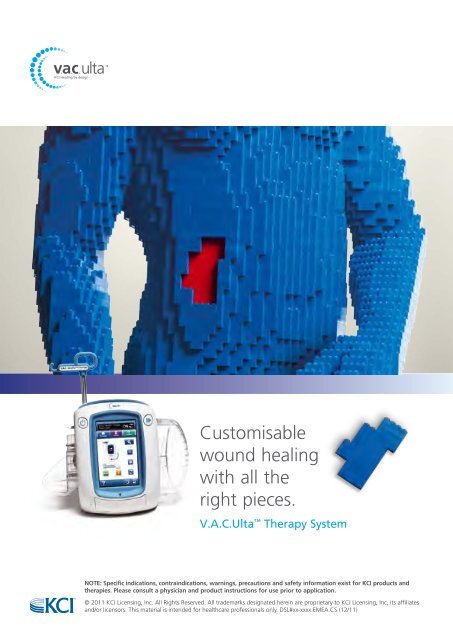
Customisable wound healing with all the right pieces. V.A.C.Ulta Therapy System NOTE: Specific indications, contraindications, warnings, precautions and safety information exist for KCI products and therapies. Please consult a physician and product instructions for use prior to application. © 2011 KCI Licensing, Inc. All Rights Reserved. All trademarks designated herein are proprietary to KCI Licensing, Inc, its affiliates and/or licensors. This material is intended for healthcare professionals only. DSL#xx-xxxx.EMEA.CS (12/11)
The skin’s own bacteria may aggravate inflammatory and occlusive changes in atherosclerotic arteries of lower limbs Key words: atherosclerosis, femoral artery, inflammation, bacteria, bacterial DNA ABSTRACT Lower limb ischemia is not infrequently accompanied by toe necrosis. The question arises whether this is only the consequence of lack of arterial perfusion or in addition infection of ischemic tissues. So far, not much attention has been attached to the patient’s own limb skin and deep tissue’s bacterial flora, activated in ischemic tissues. This flora may enhance the inflammatory and thrombotic process in the atherosclerotic arteries. Aim: To identify microbial cells and their DNA in perivascular tissues and arterial walls of ischemic lower limbs. Methods: Bacterial cultures and PCR method for detection of 16s RNA and immunohistopathological staining for identification of immune cells infiltrating vascular bundles. Results: a) specimens of atherosclerotic calf and femoral arteries contained bacterial isolates and/ or their DNA, whereas, in control normal cadaveric organ donors’ limb arteries or patients’ carotid arteries and aorta bacteria they were detected only sporadically, b) lower limb lymphatics contained bacterial cells in 76% of specimens, whereas controls only in 10%, c) isolates from limb arteries and lymphatics belonged in majority to the coagulase-negative staphylococci and S.aureus, however, other highly pathogenic strains were also detected and d) immunohistopathological evaluation arterial walls and periarterial tissue showed dense focal infiltrates of granulocytes and macrophages. Conclusions: Own bacterial isolates can be responsible for dense neutrophil and macrophage inflitrates of atherosclerotic walls and periarterial tissue in lower limbs and this aggravates ischemic changes. INTRODUCTI<strong>ON</strong> Multiple studies were carried out on Chlamydia, Helicobacter, Herpes and dental bacteria’s’ role in atherosclerosis (1-5) . So far, not much attention has been attached to patients’ own bacterial foot and calf bacteria activated in ischemic tissues. They may enhance the inflammatory and thrombotic processes in the atherosclerotic arteries. This especially applies to the arteries of lower limbs with a dramatically higher exposure to environmental bacterial stress than arteries of other body regions (6) . Foot and leg skin is inhabited by a number of bacterial strains not encountered in other body regions (7, 8) . They belong to the skin saprophytic flora and in particular to the coagulase-negative Staphylococci (9) . There are also highly pathogenic bacteria originating from the anal and perineal regions floating down on epidermal scales and colonising foot skin. Microbes may penetrate epidermis upon even minor injuries infecting limb vascular bundles either directly or through the lymphatics draining foot skin accompanying large blood vessels (10, 11) . The question arises whether infection of lower limb arteries by foot and calf bacteria may aggravate the inflammatory and occlusive atheromatous changes and act as a secondary destructive factor. Moreover, the question is whether these bacterial strains may be the causative factor of infective complications after arterial grafting (12, 13, 14) . We investigated the atherosclerotic tibial, popliteal and femoral arterial walls harvested from the amputated ischemic limbs for presence of (i) bacterial cells and (ii) their DNA, with use of broad- range PCR targeting conserved region 16s RNA of nosocomial microbes. The findings were compared with those obtained from normal arteries harvested from organ donors. The control material consisted also of carotid plaques and punch fragments of thoracic aorta obtained during CABG operation. Moreover, bacteriological � Science, Practice and Education Waldemar L. Olszewski 1,3 Piotr Andziak 1,2 M. Moscicka-Wesolowska 1 Bozenna Interewicz 1 Ewa Swoboda 4 Ewa Stelmach 1,4 1 Department of Surgical Research and Transplantology, Medical Research Center, Polish Academy of Sciences, Warsaw, Poland 2 Department of General and Vascular Surgery, Central Clinical Hospital, Ministry of Internal Affairs, Warsaw, Poland 3 Department of Gastroenterological Surgery and Transplantation, Central Clinical Hospital, Ministry of Internal Affairs, Warsaw, Poland 4 Department of Clinical Microbiology, Warsaw Medical University, Warsaw, Poland Correspondence: wlo@cmdik.pan.pl <strong>EWMA</strong> Journal 2012 vol 12 no 1 27