of Muhlenbergia (Poaceae) - Fungal diversity
of Muhlenbergia (Poaceae) - Fungal diversity
of Muhlenbergia (Poaceae) - Fungal diversity

Create successful ePaper yourself
Turn your PDF publications into a flip-book with our unique Google optimized e-Paper software.
<strong>Fungal</strong> Diversity<br />
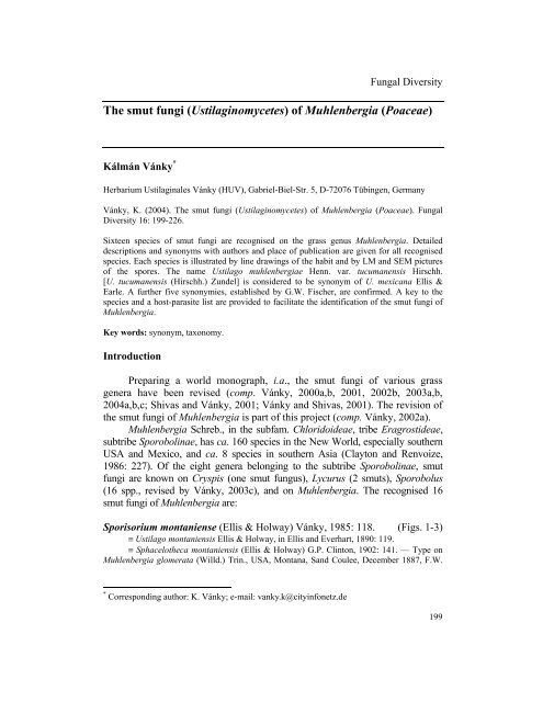
The smut fungi (Ustilaginomycetes) <strong>of</strong> <strong>Muhlenbergia</strong> (<strong>Poaceae</strong>)<br />
Kálmán Vánky *<br />
Herbarium Ustilaginales Vánky (HUV), Gabriel-Biel-Str. 5, D-72076 Tübingen, Germany<br />
Vánky, K. (2004). The smut fungi (Ustilaginomycetes) <strong>of</strong> <strong>Muhlenbergia</strong> (<strong>Poaceae</strong>). <strong>Fungal</strong><br />
Diversity 16: 199-226.<br />
Sixteen species <strong>of</strong> smut fungi are recognised on the grass genus <strong>Muhlenbergia</strong>. Detailed<br />
descriptions and synonyms with authors and place <strong>of</strong> publication are given for all recognised<br />
species. Each species is illustrated by line drawings <strong>of</strong> the habit and by LM and SEM pictures<br />
<strong>of</strong> the spores. The name Ustilago muhlenbergiae Henn. var. tucumanensis Hirschh.<br />
[U. tucumanensis (Hirschh.) Zundel] is considered to be synonym <strong>of</strong> U. mexicana Ellis &<br />
Earle. A further five synonymies, established by G.W. Fischer, are confirmed. A key to the<br />
species and a host-parasite list are provided to facilitate the identification <strong>of</strong> the smut fungi <strong>of</strong><br />
<strong>Muhlenbergia</strong>.<br />
Key words: synonym, taxonomy.<br />
Introduction<br />
Preparing a world monograph, i.a., the smut fungi <strong>of</strong> various grass<br />
genera have been revised (comp. Vánky, 2000a,b, 2001, 2002b, 2003a,b,<br />
2004a,b,c; Shivas and Vánky, 2001; Vánky and Shivas, 2001). The revision <strong>of</strong><br />
the smut fungi <strong>of</strong> <strong>Muhlenbergia</strong> is part <strong>of</strong> this project (comp. Vánky, 2002a).<br />
<strong>Muhlenbergia</strong> Schreb., in the subfam. Chloridoideae, tribe Eragrostideae,<br />
subtribe Sporobolinae, has ca. 160 species in the New World, especially southern<br />
USA and Mexico, and ca. 8 species in southern Asia (Clayton and Renvoize,<br />
1986: 227). Of the eight genera belonging to the subtribe Sporobolinae, smut<br />
fungi are known on Cryspis (one smut fungus), Lycurus (2 smuts), Sporobolus<br />
(16 spp., revised by Vánky, 2003c), and on <strong>Muhlenbergia</strong>. The recognised 16<br />
smut fungi <strong>of</strong> <strong>Muhlenbergia</strong> are:<br />
Sporisorium montaniense (Ellis & Holway) Vánky, 1985: 118. (Figs. 1-3)<br />
≡ Ustilago montaniensis Ellis & Holway, in Ellis and Everhart, 1890: 119.<br />
≡ Sphacelotheca montaniensis (Ellis & Holway) G.P. Clinton, 1902: 141. — Type on<br />
<strong>Muhlenbergia</strong> glomerata (Willd.) Trin., USA, Montana, Sand Coulee, December 1887, F.W.<br />
* Corresponding author: K. Vánky; e-mail: vanky.k@cityinfonetz.de<br />
199
Anderson. Topotypes, collected in July 1888, BPI 163401!, 163402!, 195684!, and also<br />
distributed as "n. sp." in Ellis & Ev., N. Amer. fgi. no. 2263, HUV 1964!<br />
= Ustilago strangulans Issatchenko, 1896: 225.<br />
≡ Sphacelotheca strangulans (Iss.) G.P. Clinton, 1904: 392.<br />
≡ Sphacelotheca strangulans (Iss.) Moesz, 1921: 62 (comb. superfl.). — Lectotype<br />
(design. by Vánky, 1985: 119) on Eragrostis poaeoides P. Beauv. (= E. minor Host), Russia,<br />
Cherson Prov., Nikolaiev, 2 July 1894. Topotype, collected in August 1897 by S. Fedossejew;<br />
200<br />
Fig. 1. Sori <strong>of</strong> Sporisorium<br />
montaniense in the<br />
inflorescences <strong>of</strong><br />
<strong>Muhlenbergia</strong> glomerata.<br />
Bar = 1 cm.
<strong>Fungal</strong> Diversity<br />
isotopotypes in Jacz., Kom., Tranz., Fgi. Ross. exs. no. 153, HUV 1965! (syn. by Fischer,<br />
1953: 143, confirmed).<br />
Sori (Fig. 1) on top <strong>of</strong> the shoots, usually destroying the whole<br />
inflorescence (although some healthy or aborted distal spikelets may be<br />
present), surrounding distal internodes and including also the basal part <strong>of</strong> the<br />
uppermost leaves. Sori irregularly cylindrical or ovoid, 1-4 mm wide, from a<br />
few mm to 2-3 cm long, partly protected by leaf-sheaths and covered by a<br />
whitish peridium composed <strong>of</strong> fungal elements and host tissue. At maturity, the<br />
peridium ruptures irregularly to expose the dark brown, powdery spore mass<br />
surrounding a stout, narrowing, central columella, <strong>of</strong>ten with short, lateral<br />
branches. Rarely, sori restricted to individual spikelets only. Spores (Figs. 2, 3)<br />
globose, ovoid to slightly irregular, 10-14.5 × 12.5-15 µm, yellowish-brown to<br />
reddish-brown; wall even, 0.8-1 µm thick, moderately densely echinulate,<br />
spore pr<strong>of</strong>ile finely serrulate, in SEM sparsely to moderately densely, minutely<br />
verruculose between the spines. Sterile cells, if present among the spores, in<br />
easily separable, irregular groups, subglobose to irregular, smaller than the<br />
spores, hyaline, thick-walled, smooth.<br />
Hosts: numerous Eragrostis species, cosmopolitan. Also on<br />
<strong>Muhlenbergia</strong> asperifolia (Nees & Meyen) Parodi, M. cuspidata (Torr.) Rydb.,<br />
M. glomerata (Willd.) Trin., M. pulcherrima Scribner, M. racemosa (Michaux)<br />
B.S.P.<br />
Known distribution: N. America (USA, Mexico).<br />
Sporisorium parodii (Hirschh.) Vánky, 2003b: 59. (Figs. 4-5)<br />
≡ Ustilago parodii Hirschhorn, 1939: 404. — Type on <strong>Muhlenbergia</strong> diffusa Willd.<br />
(= M. schreberi Gmel.), Argentina, near La Plata, Bosque, October 1935, L.R. Parodi, Herb.<br />
Hirschhorn 35; isotype LPS 3055! Topotype LPS 3054.<br />
Sori comprising the distal part <strong>of</strong> the shoots (inflorescence? and some<br />
terminal leaves), long cylindrical, 3-4 × 10-12 mm, first covered by a whitishbrown<br />
peridium with the tips <strong>of</strong> 1-2 leaves. The peridium ruptures<br />
longitudinally disclosing the brown, granular to powdery mass <strong>of</strong> spores<br />
surrounding 8-12 filiform columellae <strong>of</strong> the length <strong>of</strong> the sorus. Spores (Figs.<br />
4, 5) single when mature, globose, subglobose, ellipsoidal to slightly irregular,<br />
8-12 × 8-15 µm, globose spores 8-12 µm in diameter, yellowish-brown; wall<br />
even, 1-2 µm thick, finely, densely verruculose, spore pr<strong>of</strong>ile wavy to finely<br />
serrulate, in SEM warts in irregular, small groups or short, irregular,<br />
labyrinthiform rows. Sterile cells not seen. Spore germination results in<br />
4-celled basidia developing mycelia (Hirschhorn, 1939: 405 + fig. 4C).<br />
Host: <strong>Muhlenbergia</strong> schreberi Gmelin (M. diffusa Willd.).<br />
Known distribution: S. America (Argentina).<br />
201
Figs. 2, 3. Spores and sterile cells <strong>of</strong> Sporisorium montaniense on <strong>Muhlenbergia</strong> glomerata, in<br />
LM and in SEM (topotype). Figs. 4, 5. Spores <strong>of</strong> Sporisorium parodii on <strong>Muhlenbergia</strong><br />
schreberi, in LM and in SEM (type). Figs. 7, 8. Spores and sterile cells <strong>of</strong> Tilletia asperifolia<br />
on <strong>Muhlenbergia</strong> asperifolia, in LM and in SEM (Fischer, Gramin. smuts N. Amer. no. 2;<br />
HUV 9966). Bars = 10 µm.<br />
202
<strong>Fungal</strong> Diversity<br />
Fig. 6. Sori <strong>of</strong> Tilletia asperifolia in the ovaries <strong>of</strong> <strong>Muhlenbergia</strong> asperifolia. Enlarged a<br />
spikelet with two sori. Bars = 1 cm for habit, 1 mm for the detail drawing.<br />
Tilletia asperifolia Ellis & Everhart, 1887: 55. (Figs. 6-8)<br />
Type on Sporobolus asperifolius Nees & Meyen (= <strong>Muhlenbergia</strong> asperifolia (Nees &<br />
Meyen) Parodi), USA, Rocky Mountains, comm. F.L. Scribner.<br />
= Tilletia eremophila Spegazzini, 1909: 291.<br />
≡ Tolyposporella eremophila (Speg.) Ciferri, 1938: 224. — Lectotype (design. by<br />
Vánky, 2003b: 36) on Sporobolus asperifolius Nees & Meyen, Argentina, Mendoza,<br />
5 December 1902, C. Spegazzini, LPS 3676; syntype LPS 3675. (syn. by Durán and Fischer,<br />
1961: 30, confirmed).<br />
203
Sori (Fig. 6) in all ovaries <strong>of</strong> an inflorescence, ovoid or broadly<br />
ellipsoidal, 0.5-1 × 0.5-1.5 mm, evident between the spreading floral<br />
envelopes, first covered by the greyish-brown, delicate pericarp which ruptures<br />
irregularly, disclosing the light to dark reddish-brown, powdery mass <strong>of</strong> spores<br />
and sterile cells. Spores (Figs. 7, 8) globose, subglobose, ovoid, ellipsoidal to<br />
slightly irregular, 17-22.5 × 20-25 µm, pale to medium dark yellowish-brown;<br />
wall reticulate, meshes polyangular, <strong>of</strong> very variable diameter, rarely<br />
incomplete, 6-8 per spore diameter, muri 1.5-2.5 µm high, in optical median<br />
view acute, spiniform, rarely subacute, embedded in a hyaline or yellowishbrown<br />
tinted sheath extending just beyond the muri. Sterile cells (Figs. 7, 8)<br />
subglobose, ovoid, elongated, <strong>of</strong>ten subpolyhedrally irregular, <strong>of</strong> variable size,<br />
11-24 × 13-35 µm, subhyaline, content pale yellowish-brown tinted, granular;<br />
wall 2-7 µm thick, smooth.<br />
Hosts: <strong>Muhlenbergia</strong> arenacea Buckl. (Sporobolus auriculatus Vasey),<br />
M. asperifolia (Nees & Meyen) Parodi (Sporobolus asperifolius Nees & Meyen).<br />
Known distribution: N. and S. America (Canada, USA, Mexico,<br />
Argentina), common.<br />
Other host plants, given in the literature, such as M. arenacea (Buckl.)<br />
Hitchc. (Sporobolus auriculatus Vasey), M. filiformis (Thurb.) Rydb., M.<br />
minutissima (Steudel) Swallen, Sporobolus confusus Vasey, have to be<br />
checked. At least M. minutissima (Sporobolus microspermus (Lag.) Hitchc.)<br />
from USA, Colorado, 31 August 1899, coll. Bartholomew 2582 (as on S.<br />
confusus Vasey), BPI 172516, 172517, 172474, represents <strong>Muhlenbergia</strong><br />
asperifolia (teste K. Vánky).<br />
Tilletia asperifolioides G.W. Fischer, 1952: 6. (Figs. 9-11)<br />
Type on <strong>Muhlenbergia</strong> cuspidata (Torr.) Rydb., USA, Colorado, Sand Creek, 10<br />
August 1948, G.W. Fischer, R. Sprague & J.P. Meiners; isotypes in Fischer, Gramin. smuts N.<br />
Amer. no. 237, HUV 9972! Paratype on <strong>Muhlenbergia</strong> filiformis (Thurb.) Rydb. (= misnamed<br />
M. cuspidata, teste K. Vánky), USA, Oregon, Silvies Creek, 28 July 1950, G.W. Fischer &<br />
R. Sprague; isoparatypes in Fischer, Gramin. smuts no. 253, HUV 9973!<br />
Sori (Fig. 9) in all ovaries <strong>of</strong> an inflorescence, narrow ellipsoidal to<br />
ovoid, with a short acute tip and remnant <strong>of</strong> the style, 0.5-1 × 1-2 mm, more or<br />
less hidden by the floral envelopes, first covered by the greenish- to greyishbrown,<br />
delicate pericarp which ruptures irregularly, disclosing the blackishbrown,<br />
powdery mass <strong>of</strong> spores and sterile cells. Spores (Figs. 10, 11) globose,<br />
subglobose, broadly ellipsoidal to slightly irregular, 23-29(-32) × 24-30(-34)<br />
µm, medium to dark reddish-brown; wall reticulate, meshes polyangular, <strong>of</strong>ten<br />
incomplete, (4-)5-6 per spore diameter, muri 1.5-2.5 µm high, in optical<br />
median view subacute or acute, spiniform (especially in young spores). Meshes<br />
embedded in a hyaline or yellowish-brown tinted sheath, which in young, pale<br />
204
<strong>Fungal</strong> Diversity<br />
Fig. 9. Sori <strong>of</strong> Tilletia asperifolioides<br />
in the ovaries <strong>of</strong> <strong>Muhlenbergia</strong><br />
cuspidata. Enlarged a sorus. To the<br />
left a healthy inflorescence. Bars =<br />
1 cm for habit, 1 mm for the detail<br />
drawing.<br />
coloured spores exceed the muri by 1-2 µm, in dark spores sheath is apparently<br />
lacking. Sterile cells (Figs. 10, 11) variable in shape and size, subglobose,<br />
ellipsoidal, usually subpolyhedrally irregular, 15-28 × 16-38 µm, subhyaline to<br />
pale yellowish-brown tinted; wall 2-7 µm thick, smooth; the large, thick-walled<br />
cells are <strong>of</strong>ten irregularly verrucose or with trace <strong>of</strong> a reticulum ("immature<br />
spores", "intermediate forms").<br />
Host: <strong>Muhlenbergia</strong> cuspidata (Torr.) Rydb.<br />
Known distribution: N. America (USA).<br />
T. asperifolioides differs from T. asperifolia especially by the larger,<br />
darker spores with fewer meshes per spore diameter and higher muri.<br />
The host plants <strong>of</strong> the HUV sample <strong>of</strong> the paratype, and those <strong>of</strong> a further<br />
collection (HUV 6878 ex WSP 62023) are <strong>Muhlenbergia</strong> cuspidata (Torr.)<br />
205
Figs. 10, 11. Spores and sterile cells <strong>of</strong> Tilletia asperifolioides on <strong>Muhlenbergia</strong> cuspidata, in<br />
LM and in SEM (Fischer, Gramin. smuts N. Amer. no. 257; HUV 9972). Figs. 13, 14. Spores<br />
and sterile cells <strong>of</strong> Tilletia macrotuberculata on <strong>Muhlenbergia</strong> pulcherrima, in LM and in SEM<br />
(type). Figs. 16, 17. Spores and sterile cells <strong>of</strong> Tilletia montana on <strong>Muhlenbergia</strong> filiformis, in<br />
LM and in SEM (type). Bars = 10 µm.<br />
206
<strong>Fungal</strong> Diversity<br />
Rydb., possessing acute glumes, not the closely related M. filiformis (Thurb.)<br />
Rydb., which has ovate, obtuse glumes.<br />
Tilletia macrotuberculata Durán, 1987: 156. (Figs. 12-14)<br />
Type on <strong>Muhlenbergia</strong> pulcherrima Scribner, Mexico, Durango, 55.3 km W <strong>of</strong><br />
Durango, <strong>of</strong>f Hwy. 40, alt. 2621 m, 27 October 1976, R. Durán, WSP 67746; isotype HUV<br />
14464! Paratypes on <strong>Muhlenbergia</strong> wolfii (Vasey) Rydb., Mexico, Durango, 2.6 km W <strong>of</strong> the<br />
turn<strong>of</strong>f to Lecheria, <strong>of</strong>f Hwy 40, alt. 2682 m, 12 & 21 October 1978, R. Durán & P.M. Gray,<br />
WSP 68617 & 68675; isoparatypes HUV 14465 & 14466.<br />
Sori (Fig. 12) in all ovaries <strong>of</strong> an inflorescence, spherical to ovoid, 0.5-1<br />
× 0.5-1 mm, showing between the spreading floral envelopes, first covered by<br />
the yellowish- to dark brown, fragile pericarp which ruptures irregularly,<br />
disclosing the dark reddish-brown, powdery mass <strong>of</strong> spores and sterile cells.<br />
Spores (Figs. 13, 14) globose, subglobose to broadly ellipsoidal, 19-23(-25)<br />
× 21-24(-28) µm, pale to dark yellowish-brown, provided with 2.5-4 µm high,<br />
blunt, subcylindrical or subpyramidal warts or tubercles with subacute or<br />
flattened tip, embedded in a subhyaline or amber tinted sheath. In surface view,<br />
the warts appear as darker, irregular, polyangular spots, (4-)5-7(-8) per spore<br />
diameter. Sterile cells (Figs. 13, 14) subglobose, ovoid, ellipsoidal, to slightly<br />
irregular, smaller than the spores, 10-18.5 × 12-24 µm, subhyaline with a pale<br />
yellow, granular content; wall 1.5-3(-4) µm thick, smooth, occasionally finely<br />
laminated. Spore germination results in holobasidia bearing a terminal whorl <strong>of</strong><br />
ca. 16, long, fusiform, first mononucleate, later septate and binucleate<br />
basidiospores which do not fuse (Durán, 1987: 157, pl. 79, Figs. G, H).<br />
Fig. 12. Sori <strong>of</strong> Tilletia macrotuberculata<br />
in the ovaries <strong>of</strong><br />
<strong>Muhlenbergia</strong> pulcherrima.<br />
Enlarged a sorus and a healthy<br />
spikelet. Bars = 1 cm for habit,<br />
1 mm for the detail drawing.<br />
207
208<br />
Hosts: <strong>Muhlenbergia</strong> pulcherrima Scribner, M. wolfii (Vasey) Rydb.<br />
Known distribution: N. America (Mexico).<br />
Tilletia montana Ellis & Everhart, 1887: 55. (Figs. 15-17)<br />
Lectotype on Sporobolus gracillimus Vasey (= <strong>Muhlenbergia</strong> filiformis (Thurb.)<br />
Rydb.), USA, Rocky Mountains, 1886, F.L. Scribner, (design. here) BPI 173611; isolectotype<br />
BPI 173610.<br />
Sori (Fig. 15) in all ovaries <strong>of</strong> an inflorescence, ovoid or ellipsoidal,<br />
0.5-1 × 0.5-1.5 mm, more or less hidden by the floral envelopes, first covered<br />
by the pale or greyish-brown, delicate pericarp which ruptures irregularly,<br />
disclosing the reddish-brown, powdery mass <strong>of</strong> spores and sterile cells. Spores<br />
(Figs. 16, 17) globose, subglobose, ellipsoidal to slightly irregular, 18-20 ×<br />
19-24 µm, pale to medium dark honey-yellow; wall basically reticulate but<br />
mostly incompletely and irregularly, giving the spore surface a peculiar,<br />
irregular aspect, muri 1-1.5 µm high, in optical median view acute, spiniform,<br />
embedded in a hyaline sheath, which usually exceeds the muri by 0.5-1 µm.<br />
Spores occasionally lacrymiform, with an acute tip, more <strong>of</strong>ten only with a<br />
hyaline papilla or a narrow appendage, the remnant <strong>of</strong> the sporogenous hypha.<br />
In SEM muri appear thick (due to the dried sheath), irregular, cerebriform.<br />
Sterile cells (Fig. 16) few, rounded irregular, <strong>of</strong>ten with flattened sides, smaller<br />
than the spores, 6.5-12 × 8-16 µm, subhyaline; wall 0.5-4 µm thick, smooth.<br />
Host: <strong>Muhlenbergia</strong> filiformis (Thurb.) Rydb. (Sporobolus gracillimus<br />
Vasey; S. simplex Scribner).<br />
Known distribution: N. America (USA, Mexico).<br />
The HUV sample (no. 14736) <strong>of</strong> "Tilletia asperifolia" on Sporobolus<br />
simplex, in Griffiths, W. Amer. fgi. no. 226, represents T. montana.<br />
Fig. 15. Sori <strong>of</strong> Tilletia montana<br />
in the ovaries <strong>of</strong> <strong>Muhlenbergia</strong><br />
filiformis. Enlarged a sorus.<br />
Bars = 1 cm for habit, 1 mm for<br />
the detail drawing.
<strong>Fungal</strong> Diversity<br />
Tilletia muhlenbergiae G.P. Clinton, 1906: 49. (Figs. 18-20)<br />
Type on <strong>Muhlenbergia</strong> schaffneri Fourn. var. elongata Scribner, Mexico, north-eastern<br />
Durango, October 1905, C.G. Pringle, BPI 173618; isotype BPI173617.<br />
Sori (Fig. 18) in all ovaries <strong>of</strong> an inflorescence, fusiform, ellipsoidal or<br />
ovoid, <strong>of</strong>ten with a short, acute tip, 0.5-1 × 1-3 mm, more or less hidden by the<br />
floral envelopes, first covered by the greyish-brown pericarp which ruptures at<br />
maturity, disclosing the dark reddish- to blackish-brown, powdery mass <strong>of</strong><br />
spores and sterile cells. Spores (Figs. 19, 20) globose, subglobose, ovoid,<br />
ellipsoidal to slightly irregular, 28-33 × 29-36(-38) µm, yellowish- to dark<br />
reddish- or smoke-brown; wall coarsely reticulate, meshes polyangular, regular<br />
to irregular, rarely incomplete, 4-6 per spore diameter, muri 2-4 µm high, in<br />
optical median view acute, interspaces rough to finely verruculose, sheath<br />
lacking. Sterile cells (Figs. 19, 20) subglobose, ovoid, ellipsoidal to slightly<br />
irregular, smaller than the spores, 12-25 × 16-30 µm, subhyaline or yellowish<br />
tinted; wall 1.5-4 µm thick, smooth. Spore germination: on top <strong>of</strong> holobasidia<br />
fusiform, multinucleate, aseptate basidiospores are produced which do not fuse<br />
(Durán, 1987: 158, pl. 81, fig. B).<br />
Hosts: <strong>Muhlenbergia</strong> depauperata Scribner, M. microsperma (DC.)<br />
Kunth, M. quadridentata Kunth, M. schaffneri Fourn. var. elongata Scribner,<br />
M. tenella (H.B.K.) Trin.<br />
Known distribution: N. America (Mexico).<br />
Tilletia pachyderma G.W. Fischer, 1952: 7. (Figs. 21-22)<br />
Type on <strong>Muhlenbergia</strong> utilis (Torr.) Hitchc., Mexico, Mexico State, Temascaltepec<br />
Distr., ca. 50 km SW <strong>of</strong> Toluca, Tequexquiapan, 28 October 1932, G.B. Hinton 2319, WSP<br />
34683 (lacking sori); isotype BPI 173661 (poor material).<br />
Sori in all ovaries <strong>of</strong> an inflorescence, 2-3 mm long, showing through the<br />
lemma and palea, first covered by the rather durable, olive-green to greyishgreen<br />
pericarp which upon rupturing releases the light golden- to honey-brown,<br />
semiagglutinated to powdery mass <strong>of</strong> spores and sterile cells. Spores (Figs. 21,<br />
22) globose, subglobose to broadly ellipsoidal, 18.5-22.5 × 18.5-24 µm<br />
(excluding the sheath); wall reticulate, 3-6(-7) meshes per spore diameter, muri<br />
in optical median view acute, 1.5-3(-4) µm high, embedded in a thick, hyaline<br />
sheath, usually considerably exceeding the muri. In SEM, due to the dried<br />
sheath, the spores appear only with shallow meshes. Sterile cells (Figs. 21, 22)<br />
few, subglobose, ovoid to irregular, 16-25 µm long, hyaline; wall two-layered,<br />
4-7 µm thick, smooth, content yellowish-tinted, granular.<br />
Host: <strong>Muhlenbergia</strong> utilis (Torr.) Hitchc.<br />
Known distribution: N. America (Mexico).<br />
209
Fig. 18. Sori <strong>of</strong> Tilletia muhlenbergiae in the ovaries <strong>of</strong> <strong>Muhlenbergia</strong> microsperma. Enlarged<br />
a sorus and a healthy spikelet. To the left a healthy inflorescence. Bars = 1 cm for habit, 1 mm<br />
for the detail drawing.<br />
210
<strong>Fungal</strong> Diversity<br />
Figs. 19, 20. Spores and sterile cells <strong>of</strong> Tilletia muhlenbergiae on <strong>Muhlenbergia</strong> microsperma,<br />
in LM and in SEM (Mexico, Oaxaca State, Las Animas, 16 November, 1978, R. Durán & P.M.<br />
Gray, HUV 14470). Figs. 21, 22. Spores and sterile cells <strong>of</strong> Tilletia pachyderma on<br />
<strong>Muhlenbergia</strong> utilis, in LM and in SEM (type). Figs. 24, 25. Spores and sterile cells <strong>of</strong> Tilletia<br />
tuberculata on <strong>Muhlenbergia</strong> depauperata, in LM and in SEM (type). Bars = 10 µm.<br />
211
Tilletia tuberculata Durán, 1987: 164. (Figs. 23-25)<br />
Type on <strong>Muhlenbergia</strong> depauperata Scribner, Mexico, Durango, 8 km W <strong>of</strong> Durango,<br />
<strong>of</strong>f Hwy. 40, alt. 2072 m, 27 October 1976, R. Durán, WSP 67749; isotype HUV 14049! (For<br />
paratypes see Durán, 1987: 165).<br />
Sori (Fig. 23) in some ovaries <strong>of</strong> an inflorescence, long fusiform, <strong>of</strong>ten<br />
with widened basal part, 0.5-1.5 × 1.5-4(-5) mm, showing between the floral<br />
envelopes, first covered by the thin, olivaceous- to dark brown pericarp, which<br />
ruptures irregularly at maturity, disclosing the dark brown, semiagglutinated to<br />
powdery mass <strong>of</strong> spores and sterile cells. Spores (Figs. 24, 25) globose or<br />
subglobose, 15-20(-21.5) × 15-21(-23) µm, from pale yellowish- to dark<br />
reddish-brown, provided with subacute or blunt, conical warts (tubercles),<br />
1-2.5 µm high, in surface view appearing as rounded, darker spots, (3-)4-6(-8)<br />
per spore diameter. The warts are connected with each other by thin, darker<br />
lines, evident especially in LM, on dark spores. Sterile cells (Figs. 24, 25)<br />
212<br />
Fig. 23. Sori <strong>of</strong> Tilletia<br />
tuberculata in the ovaries <strong>of</strong><br />
<strong>Muhlenbergia</strong> depauperata.<br />
Enlarged a spikelet with a sorus<br />
and a healthy spikelet. Bars =<br />
1 cm for habit, 1 mm for the<br />
detail drawing.
<strong>Fungal</strong> Diversity<br />
subglobose, broadly ellipsoidal to slightly irregular, extremely variable in size,<br />
subglobose cells 8-28 µm in diameter, subhyaline to pale yellowish-brown;<br />
wall 1-3 µm thick, smooth, sometimes concentrically slightly laminated. Spore<br />
germination results in multinucleate holobasidia producing a terminal whorl <strong>of</strong><br />
ca. 16-20, long, fusiform, first mononucleate, later one-septate and binucleate<br />
basidiospores which do not fuse (Durán, 1987: 165, pl. 92, Figs. D, E).<br />
Hosts: <strong>Muhlenbergia</strong> depauperata Scribner, M. pectinata C.O.<br />
Goodding.<br />
Known distribution: N. America (Mexico).<br />
Tilletia zonata Brefeld, 1895: 161.<br />
Type on <strong>Muhlenbergia</strong> ligularis (Hack.) Hitchc., Ecuador, Quito, G. Lagerheim.<br />
Sori in the ovaries, filled by a black, powdery mass <strong>of</strong> spores. Spores<br />
chiefly globose, ca. 19-24 µm in diameter, including the 2-3 µm thick, hyaline<br />
sheath (spores without sheath 15-18 µm), yellowish-brown, reticulate. Judged<br />
from the illustrations, meshes polyangular, rather regular, sometimes<br />
incomplete, 3-5 per spore diameter. Spore germination results in a<br />
holobasidium apically bearing 4-8 basidiospores which fuse in pairs, giving<br />
rise to hyphae on which sickle shaped, secondary ballistoconidia are produced<br />
on short sterigmata (Brefeld, 1895: 161, pl. X, Figs. 3-7).<br />
Host: <strong>Muhlenbergia</strong> ligularis (Hack.) Hitchc.<br />
Known distribution: S. America (Ecuador). Known only from the type<br />
locality.<br />
The type material probably no longer exists. Recollection is desired.<br />
Brefeld's original description is very poor, except for that <strong>of</strong> the spore<br />
germination. The description above is based on Brefeld's description and on his<br />
illustrations.<br />
Ustilago bethelii Zundel, 1933: 350. (Figs. 26-28)<br />
Type on <strong>Muhlenbergia</strong> montana (Nutt.) Hitchc., USA, Colorado, Idaho Springs,<br />
3 September 1923, E. Bethel, BPI 157974!; isotypes BPI 157973, 157978.<br />
Sori (Fig. 26) forming slightly swollen, lead-coloured striae on the leaves<br />
between the veins, 0.2-0.5 × 0.5-40 mm or longer, first covered by the<br />
epidermis which ruptures longitudinally disclosing the blackish,<br />
semiagglutinated to powdery mass <strong>of</strong> spores which is scattered, leaving behind<br />
perforated or shredded leaves. Occasionally, sori may appear on the stems,<br />
exceptionally in the spikelets. Spores (Figs. 27, 28) subglobose, ellipsoidal,<br />
ovoid to slightly irregular, 11-16 × 12-18(-21) µm, yellowish- to reddishbrown,<br />
wall even or slightly uneven, 0.8-2(-2.5) µm thick, finely, rather<br />
densely verrucose-echinulate, spore pr<strong>of</strong>ile wavy to finely serrulate. Spore<br />
213
germination results in mononucleate mycelia or in basidia with branches<br />
producing mononucleate sporidia (Durán, 1987: 225, pl. 108, fig. E).<br />
Host: <strong>Muhlenbergia</strong> montana (Nutt.) Hitchc. (M. gracilis auctt. Amer.).<br />
Known distribution: N. America (USA, Mexico).<br />
Ustilago buchloës Ellis & Tracy, 1890: 77, s. lat.<br />
Lectotype on Buchloë dactyloides (Nutt.) Engelm., USA, New Mexico, Coolidge, 20<br />
June 1887, S.M. Tracy, (design. by Piepenbring, 2003: 158) BPI 159165!; isolectotypes BPI<br />
159164!, 159166!<br />
214<br />
Fig. 26. Sori <strong>of</strong> Ustilago<br />
bethelii forming striae on the<br />
leaves <strong>of</strong> <strong>Muhlenbergia</strong><br />
montana. To the left a healthy<br />
inflorescence. Bar = 1 cm.
<strong>Fungal</strong> Diversity<br />
Figs. 27, 28. Spores <strong>of</strong> Ustilago bethelii on <strong>Muhlenbergia</strong> montana, in LM and in SEM<br />
(Fischer, Gramin. smuts N. Amer. no. 157; HUV 10042). Figs. 30, 31. Spores <strong>of</strong> Ustilago<br />
hyalinobipolaris on <strong>Muhlenbergia</strong> porteri, in LM and in SEM (USA, Arizona, Silver Bell<br />
Mts., 12 February 1981, G.B. Cummins; HUV 10424). Figs. 33, 34. Spores <strong>of</strong> Ustilago<br />
mexicana on <strong>Muhlenbergia</strong> sp., in LM and in SEM (type). Bars = 10 µm.<br />
215
= Ustilago pseudohieronymi Zundel, 1933: 351. — Lectotype on <strong>Muhlenbergia</strong><br />
squarrosa (Trin.) Rydb., USA, Colorado, San Luis Valley, 27 June 1921, E. Bethel, (design. by<br />
Vánky, 2004c: 182) BPI 165475; isotypes BPI 165476-165478. (syn. by Fischer, 1953: 246,<br />
confirmed).<br />
= Ustilago coloradensis Zundel, 1933: 351. — Type on <strong>Muhlenbergia</strong> gracillima Torr.,<br />
USA, Colorado, Manitou, 4 July 1924, E. Bethel, BPI 159658!; isotype BPI 159659! (syn. by<br />
Fischer, 1953: 246, confirmed).<br />
For further synonyms, description, illustrations and host plant range see<br />
Vánky, 2004c: 182.<br />
Hosts: Bouteloua, Buchloë, Cathestecum, Tridens species, but also on<br />
<strong>Muhlenbergia</strong> richardsonis (Trin.) Rydb. (M. squarrosa (Trin.) Rydb.),<br />
M. torreyi (Kunth) Hitchc. ex Bush (M. gracillima Torr.).<br />
Known distribution: N. and S. America (USA, Mexico, Argentina),<br />
Antilles (Dominican Rep.).<br />
Ustilago hyalinobipolaris G.W. Fischer & Hirschhorn, 1945: 324. (Figs. 29-31)<br />
Replacing ≡ Ustilago muhlenbergiae G.P. Clinton, October 1902: 133 (later homonym,<br />
not Hennings, April 1902). — Type on <strong>Muhlenbergia</strong> texana Turb. (= M. porteri Scribner),<br />
USA, southern Arizona, August 1884, C.G. Pringle, FH!; isotypes BPI 163423-26, 163428.<br />
Sori (Fig. 29) on the top and along sterile shoots at the base <strong>of</strong> leaves,<br />
globoid to ovoid, 1-2.5 × 1-4 mm, with long, acute leaf remnants on their top,<br />
covered by a thin, greyish peridium which ruptures irregularly at maturity,<br />
disclosing the black, semiagglutinated to powdery mass <strong>of</strong> spores and some<br />
host tissue remnants, like a short, ramifying columella. Spores (Figs. 30, 31)<br />
globose, subglobose, laterally slightly compressed, 5-5.5 × 5-6.5 µm, dark<br />
brown; wall uneven, ca. 0.4-0.8 µm, thinner on the flattened sides, evidently,<br />
sparsely verrucose-echinulate, spore pr<strong>of</strong>ile wavy to sparsely, finely serrulate<br />
on the flattened sides. In excessively rehydrated spores the flattened, thinwalled<br />
sides protrude as paler, bipolar areas.<br />
216<br />
Fig. 29. Sori <strong>of</strong> Ustilago<br />
hyalinobipolaris in the inflorescence<br />
<strong>of</strong> <strong>Muhlenbergia</strong> porteri. Enlarged<br />
two sori. Bars = 1 cm for habit,<br />
2 mm for the detail drawing.
<strong>Fungal</strong> Diversity<br />
Hosts: <strong>Muhlenbergia</strong> microsperma (DC.) Kunth, M. pauciflora Buckl.,<br />
M. porteri Scribner (M. texana Turb.).<br />
Known distribution: N. America (USA).<br />
Ustilago hyalinobipolaris is close to U. muhlenbergiae, both on<br />
<strong>Muhlenbergia</strong> species. Not only are the sori identical but also the shape, size<br />
and wall thickness <strong>of</strong> the spores. However, they differ in the colour and<br />
ornamentation <strong>of</strong> the spores. It is questionable if U. hyalinobipolaris merits<br />
separate specific rank or should be considered as an extreme variation within<br />
U. muhlenbergiae. Clinton (1904: 347) considered his U. muhlenbergiae to be<br />
"in all probability" a synonym <strong>of</strong> U. muhlenbergiae Henn., published a few<br />
months earlier. For the present, I accept U. hyalinobipolaris as a separate<br />
species until intermediate forms are seen.<br />
Ustilago mexicana Ellis & Everhart, 1887: 56. (Figs. 32-34)<br />
Lectotype on <strong>Muhlenbergia</strong> sp., Mexico, Chihuahua, Mts. near Batopilas, alt. ca. 1859<br />
m, 1885, E. Palmer, (design. by Piepenbring, 2003: 175) BPI 163303!; isolectotypes BPI<br />
163300-163302, 163305, and in Ellis & Ev., N. Amer. fgi., Ser. 2, no. 1891, HUV 4116!<br />
= Ustilago muhlenbergiae Henn. var. tucumanensis Hirschhorn, 1939: 386.<br />
≡ Ustilago tucumanensis (Hirschh.) Zundel, 1953: 182.<br />
≡ Ustilago tucumanensis (Hirschh.) Hirschhorn, 1986: 327 (comb. superfl.) — Type on<br />
<strong>Muhlenbergia</strong> caerulea (Gris.) Mez. (= M. angustata (Presl) Kunth), Argentina, Tucuman<br />
Prov., San José, 2200 m, 26 January 1933, L.R. Parodi 10709, BPI 163406! (syn. nov.)<br />
= Ustilago epicampida Zundel, 1942: 123. — Type on Epicampes emersleyi (Vasey)<br />
Hitchc. (= <strong>Muhlenbergia</strong> emersleyi Vasey), Mexico, Michoacan State, Cerro Tancitaro, alt. 355<br />
m, 19 August 1940, W.C. Leavenworth 718, BPI 160368!; isotype BPI 160369! (syn. by<br />
Fischer, 1953: 281, confirmed).<br />
Sori (Fig. 32) in all spikelets <strong>of</strong> an inflorescence, globoid, 0.5-1.5 mm in<br />
diameter, showing between the spreading glumes, first covered by a peridium<br />
<strong>of</strong> host origin, <strong>of</strong>ten with 1-2 acute remnants <strong>of</strong> inner floral organs and even<br />
awns. The peridium ruptures irregularly at maturity disclosing the blackishbrown,<br />
semiagglutinated to powdery mass <strong>of</strong> spores. Spores (Figs. 33, 34)<br />
globose, subglobose, ellipsoidal to slightly irregular with a more or less<br />
flattened side, 5-8 × 5.5-9 µm, yellowish-brown, paler on one side; wall<br />
uneven, 0.5-0.8 µm thick, thinner on the paler, flattened side, in LM finely,<br />
densely punctate, spore pr<strong>of</strong>ile smooth to finely wavy on the flattened side, in<br />
SEM finely, very densely verruculose.<br />
Hosts: <strong>Muhlenbergia</strong> angustata (Presl) Kunth (M. caerulea (Gris.) Mez.),<br />
M. distichophylla (Presl) Kunth, M. emersleyi Vasey (Epicampes emersleyi<br />
(Vasey) Hitchc.), <strong>Muhlenbergia</strong> sp.<br />
Known distribution: N. and S. America (Mexico, Argentina).<br />
No material <strong>of</strong> Ustilago tucumanensis was seen, but according to the<br />
original description it appears identical with U. mexicana. Hirschhorn (1939:<br />
217
Fig. 32. Sori <strong>of</strong> Ustilago mexicana in the ovaries <strong>of</strong> <strong>Muhlenbergia</strong> sp. Enlarged a sorus.<br />
Bars = 1 cm for habit, 1 mm for the detail drawing.<br />
218
<strong>Fungal</strong> Diversity<br />
386) mentioned the presence <strong>of</strong> spore balls, but later (Hirschhorn, 1986: 327)<br />
she suspected that these could be artefacts.<br />
The spores <strong>of</strong> the type <strong>of</strong> Ustilago epicampida are slightly larger (5.5-8 ×<br />
6.5-9 µm), paler, more evidently ornamented, and the spore wall is more<br />
evenly thick than in the spores <strong>of</strong> the type <strong>of</strong> U. mexicana which measure 5-7 ×<br />
5.5-8 µm. These differences are considered variations within the same species.<br />
Ustilago muhlenbergiae Hennings, April 1902: (61). (not Clinton, October<br />
1902, q.e. Ustilago hyalinobipolaris). (Figs. 35-37)<br />
Type on <strong>Muhlenbergia</strong> pringlei Scribner (= M. pauciflora Buckl.), USA, New Mexico,<br />
Hot Springs, 15 September 1896, E.W.D. Holway (not Mexico, as stated in the original<br />
description); isotypes BPI 163410, and in Seymour & Earle, Econ. fgi., Suppl. C, no. 142,<br />
HUV 9730!<br />
Sori (Fig. 35) on top <strong>of</strong> the shoots, destroying the inflorescence,<br />
comprising also the basal part <strong>of</strong> some congested, distal leaf sheaths,<br />
subspherical, ovoid or fusiform, first hard, somewhat gall-like, 1.5-3 × 2.5-6<br />
mm, partly hidden by leaf sheaths, covered by a yellowish-grey, rather thick<br />
peridium, <strong>of</strong>ten with floral remnants on it and also with long, setaceous leaf<br />
remnants on its top. At maturity the peridium becomes thinner, ruptures<br />
irregularly, disclosing the blackish-brown, agglutinated, later powdery mass <strong>of</strong><br />
spores. Spores (Figs. 36, 37) globose to subglobose, laterally slightly<br />
compressed, 5-5.5 × 5.5-6(-6.5) µm, dark yellowish-brown; wall uneven,<br />
0.4-0.8 µm, thinner on the flattened sides which, in excessively rehydrated<br />
spores may protrude and even rupture, reminescent <strong>of</strong> spores <strong>of</strong> Tranzscheliella<br />
williamsii (see Fischer, 1953: 284, fig. 111 B). Spore surface in LM finely,<br />
moderately densely punctate, spore pr<strong>of</strong>ile smooth, on the flattened sides finely<br />
wavy, in SEM finely, densely verruculose. Sterile cells absent.<br />
Hosts: <strong>Muhlenbergia</strong> pauciflora Buckl. (M. pringlei Scribner), M. porteri<br />
Scribner (M. texana Turb.).<br />
Known distribution: N. America (USA).<br />
Ustilago sonoriana Zundel, in Fischer, 1953: 295. (Figs. 38-40)<br />
Type on <strong>Muhlenbergia</strong> dumosa Scribner, Mexico, Sonora, near Bavispe, Santa Rosa<br />
Canyon, 19 July 1938, S.S. White, BPI 166514!<br />
Sori (Fig. 38) destroying the inner floral organs, globoid to ovoid, 0.5-1 ×<br />
1-1.5 mm, with a short acute tip, partly hidden by leaf sheaths and outer floral<br />
envelopes, at first covered by a thin, greyish peridium <strong>of</strong> host origin,<br />
sometimes with the tips <strong>of</strong> destroyed inner floral envelopes. At maturity, the<br />
peridium ruptures irregularly disclosing the blackish-brown, powdery mass <strong>of</strong><br />
spores. Spores (Figs. 39, 40) globose, subglobose, ellipsoidal or like a rugby<br />
ball, 5.5-7 × 5.5-8 µm, yellowish-brown, <strong>of</strong>ten with a wide, slightly darker,<br />
219
Fig. 35. Sori <strong>of</strong> Ustilago muhlenbergiae in the inflorescence <strong>of</strong> <strong>Muhlenbergia</strong> pauciflora<br />
Buckl. To the left a healthy inflorescence. Bar = 1 cm.<br />
longitudinal band; wall uneven, 0.5-8 µm thick, thicker at the longitudinal end<br />
<strong>of</strong> the spores, surface apparently smooth to indistinctly punctate.<br />
Host: <strong>Muhlenbergia</strong> dumosa Scribner.<br />
Known distribution: N. America (Mexico). Known only from the type<br />
collection.<br />
Two additional smut fungi on <strong>Muhlenbergia</strong> from Argentina have been<br />
published invalidly (no Latin diagnosis, ICBN 36.1, and no type indicated,<br />
ICBN 37.1), under the names <strong>of</strong> Tilletia atacamensis Hirschhorn (1986: 148),<br />
220
<strong>Fungal</strong> Diversity<br />
Figs. 36, 37. Spores <strong>of</strong> Ustilago muhlenbergiae on <strong>Muhlenbergia</strong> pauciflora, in LM and in<br />
SEM (type). Figs. 39, 40. Spores <strong>of</strong> Ustilago sonoriana on <strong>Muhlenbergia</strong> dumosa, in LM and<br />
in SEM (type). Bars = 10 µm.<br />
and T. georfischeri Hirschhorn (1986: 177). No specimens were available for<br />
study to validate the names or to establish synonymy.<br />
HOST — PARASITE LIST<br />
(M. = <strong>Muhlenbergia</strong>, T. = Tilletia, U. = Ustilago)<br />
Bouteloua spp. — U. buchloës<br />
Buchloë spp. — U. buchloës<br />
Cathestecum spp. — U. buchloës<br />
Epicampes emersleyi = M. emersleyi<br />
221
Fig. 38. Sori <strong>of</strong> Ustilago sonoriana in the flowers <strong>of</strong> <strong>Muhlenbergia</strong> dumosa. Enlarged three<br />
sori. Bars = 1 cm for habit, 1 mm for the detail drawing.<br />
Eragrostis spp. — Sporisorium montaniense<br />
M. angustata — U. mexicana<br />
M. arenacea — T. asperifolia<br />
M. asperifolia — Sporisorium montaniense, T. asperifolia<br />
M. caerulea = M. angustata<br />
M. cuspidata — Sporisorium montaniense, T. asperifolioides<br />
M. depauperata — T. muhlenbergiae, T. tuberculata<br />
M. diffusa = M. schreberi<br />
M. distichophylla — U. mexicana<br />
M. dumosa — U. sonoriana<br />
222
M. emersleyi — U. mexicana<br />
M. filiformis — T. asperifolia, T. montana<br />
M. glomerata — Sporisorium montaniense<br />
M. gracillima = M. torreyi<br />
M. ligularis — T. zonata<br />
M. microsperma — T. muhlenbergiae<br />
M. minutissima — T. asperifolia<br />
M. montana — U. bethelii<br />
M. pauciflora — U. hyalinobipolaris, U. muhlenbergiae Henn.<br />
M. pectinata — T. tuberculata<br />
M. porteri — U. hyalinobipolaris, U. muhlenbergiae Henn.<br />
M. pringlei = M. pauciflora<br />
M. pulcherrima — Sporisorium montaniense, T. macrotuberculata<br />
M. quadridentata — T. muhlenbergiae<br />
M. racemosa — Sporisorium montaniense<br />
M. richardsonis — U. buchloës<br />
M. schaffneri var. elongata — T. muhlenbergiae<br />
M. schreberi — Sporisorium parodii<br />
M. squarrosa = M. richardsonis<br />
M. tenella — T. muhlenbergiae<br />
M. texana = M. porteri<br />
M. torreyi — U. buchloës<br />
M. utilis — T. pachyderma<br />
M. wolfii — T. macrotuberculata<br />
M. sp. — U. mexicana<br />
Sporobolus asperifolius = M. asperifolia<br />
Sporobolus auriculatus = M. arenacea<br />
Sporobolus confusus — T. asperifolia<br />
Sporobolus gracillimus = M. filiformis<br />
Sporobolus simplex = M. filiformis<br />
Tridens spp. — U. buchloës<br />
FUNGUS NAMES<br />
(valid names in bold face)<br />
asperifolia Tilletia<br />
asperifolioides Tilletia<br />
bethelii Ustilago<br />
buchloës Ustilago<br />
coloradensis Ustilago = Ustilago buchloës<br />
epicampida Ustilago = Ustilago mexicana<br />
eremophila Tilletia = Tilletia asperifolia<br />
eremophila Tolyposporella = Tilletia asperifolia<br />
hyalinobipolaris Ustilago<br />
macrotuberculata Tilletia<br />
mexicana Ustilago<br />
montana Tilletia<br />
montaniense Sporisorium<br />
montaniensis Sphacelotheca = Sporisorium montaniense<br />
<strong>Fungal</strong> Diversity<br />
223
montaniensis Ustilago = Sporisorium montaniense<br />
muhlenbergiae G.P. Clinton, Ustilago = Ustilago hyalinobipolaris<br />
muhlenbergiae Henn., Ustilago<br />
muhlenbergiae Tilletia<br />
muhlenbergiae Ustilago var. tucumanensis = Ustilago mexicana<br />
pachyderma Tilletia<br />
parodii Sporisorium<br />
parodii Ustilago = Sporisorium parodii<br />
pseudohieronymi Ustilago = Ustilago buchloës<br />
sonoriana Ustilago<br />
strangulans (Iss.) G.P. Clinton, Sphacelotheca = Sporisorium montaniense<br />
strangulans (Iss.) Moesz, Sphacelotheca = Sporisorium montaniense<br />
strangulans Ustilago = Sporisorium montaniense<br />
tuberculata Tilletia<br />
tucumanensis (Hirschh.) Hirschh., Ustilago = Ustilago mexicana<br />
tucumanensis (Hirschh.) Zundel, Ustilago = Ustilago mexicana<br />
zonata Tilletia<br />
Key to the smut fungi <strong>of</strong> <strong>Muhlenbergia</strong> (T. = Tilletia, U. = Ustilago)<br />
1. Sori in the ovaries .............................................................................................................2<br />
1. Sori elsewhere...................................................................................................................9<br />
2. Spores verrucose-tuberculate............................................................................................3<br />
2. Spores reticulate................................................................................................................4<br />
3. Spores 15-21(-23) µm long. Tubercles 1-2.5 µm high ............................... T. tuberculata<br />
3. Spores 21-24(-28) µm long. Tubercles 2.5-4 µm high .....................T. macrotuberculata<br />
4. Spores 28-53 µm long, including the 3-14 µm thick sheath. Meshes 3-5 per spore diam. .<br />
...................................................................................................................T. pachyderma<br />
4. Spores shorter. Sheath thinner or absent. Meshes more per spore diam. ..........................5<br />
5. Spores 29-36(-38) µm long. Muri 2-4 µm high .................................... T. muhlenbergiae<br />
5. Spores shorter. Muri shorter .............................................................................................6<br />
6. Spores 24-30(-34) µm long. Muri 1.5-2.5 µm high ............................... T. asperifolioides<br />
6. Spores 19-25 µm long.......................................................................................................7<br />
7. Spores basically reticulate but mostly incompletely and irregularly. Muri 1-1.5 µm high.<br />
........................................................................................................................ T. montana<br />
7. Spores clearly reticulate. Muri higher...............................................................................8<br />
8. Meshes 6-8 per spore diam. Sheath extending just beyond the 1.5-2.5 µm high muri .......<br />
......................................................................................................................T. asperifolia<br />
8. Meshes ca. 3-5 per spore diam. Sheath 2-3 µm thick ......................................... T. zonata<br />
9(1). Sori predominantly on leaves or also on leaf sheaths forming pustules or striae............10<br />
9. Sori not so.......................................................................................................................11<br />
224
<strong>Fungal</strong> Diversity<br />
10. Sori on leaves forming slightly swollen striae ...................................................U. bethelii<br />
10. Sori on leaves and leaf sheaths forming bullate pustules or sausage-shaped striae ............<br />
........................................................................................................................ U. buchloës<br />
11. Sori in spikelets. Spores paler and flattened on one side ...............................U. mexicana<br />
11. Sori in the whole inflorescence and distal leaf sheaths. Spores not so ...........................12<br />
12. Columella present. Spores up to 15 µm long, without paler polar areas.........................13<br />
12. Columella absent. Spores up to 9(-10) µm long, with paler polar areas .........................14<br />
13. Columella one, stout. Spores echinulate .................................. Sporisorium montaniense<br />
13. Columella 8-12, filiform. Spores verruculose................................... Sporisorium parodii<br />
14. Spores 6-9(-10) µm long............................................................................... U. sonoriana<br />
14. Spores 5.5-6.5 µm long...................................................................................................15<br />
15. Spores finely punctate. Spore pr<strong>of</strong>ile smooth to wavy .........................U. muhlenbergiae<br />
15. Spores evidently verrucose-echinulate. Spore pr<strong>of</strong>ile wavy to finely serrulate ..................<br />
............................................................................................................U. hyalinobipolaris<br />
Acknowledgements<br />
I am grateful to Dr. E.H.C. McKenzie (Auckland, New Zealand) for reading the manuscript<br />
and checking my English. Thanks are also due to the Directors and Curators <strong>of</strong> the Herbaria<br />
BPI, FH, LPS and WSP for loans and/or exchange <strong>of</strong> specimens.<br />
References<br />
Brefeld, O. (1895). Untersuchungen aus dem Gesammtgebiete der Mykologie. XII.<br />
Hemibasidii. Brandpilze III. Münster i. W., Commissions-Verlag v. H. Schöningh. IV +<br />
99-236p. + Pls. VI-XII.<br />
Ciferri, R. (1938). Ustilaginales. Flora Italica Cryptogama, Pars I. Fungi, Fascicolo 17: 1-443.<br />
Clayton, W.D. and Renvoize, S.A. (1986). Genera graminum. Grasses <strong>of</strong> the world. Kew<br />
Bulletin Additional Series XIII. London, UK, 389p.<br />
Clinton, G.P. (1902). North American Ustilagineae. Journal <strong>of</strong> Mycology 8: 128-156.<br />
Clinton, G.P. (1904). North American Ustilagineae. Proceedings <strong>of</strong> the Boston Society <strong>of</strong><br />
Natural History 31: 329-529.<br />
Clinton, G.P. (1906). Order Ustilaginales. North American Flora 7: 1-82.<br />
Durán, R. (1987). Ustilaginales <strong>of</strong> Mexico. Taxonomy, symptomatology, spore germination,<br />
and basidial cytology. Washington State University, Pullman, USA, 331p.<br />
Durán, R. and Fischer, G.W. (1961). The genus Tilletia. Washington State University, 138p.<br />
Ellis, J.B. and Everhart, B.M. (1887). New species <strong>of</strong> Ustilagineae and Uredineae. Journal <strong>of</strong><br />
Mycology 3: 55-57.<br />
Ellis, J.B. and Everhart, B.M. (1890). New species <strong>of</strong> Uredineae and Ustilagineae. Journal <strong>of</strong><br />
Mycology 6: 118-121.<br />
Ellis, J.B. and Tracy, S.M. (1890). A few new fungi. Journal <strong>of</strong> Mycology 6: 76-77.<br />
Fischer, G.W. (1952). Some new species <strong>of</strong> Ustilaginales from North America. Research<br />
Studies <strong>of</strong> the State College <strong>of</strong> Washington 20: 3-10.<br />
225
Fischer, G.W. (1953). Manual <strong>of</strong> the North American Smut Fungi. New York, Ronald Press<br />
Co., 343p.<br />
Fischer, G.W. and Hirschhorn, E. (1945). Observations on certain species <strong>of</strong> Ustilago on<br />
Hilaria, Stenotaphrum, and <strong>Muhlenbergia</strong>. Mycologia 37: 318-325.<br />
Hennings, P. (1902). Fungi nonnulli novi ex regionibus variis. Hedwigia, Beiblatt 41: 61-66.<br />
Hirschhorn, E. (1939). Las especies del género Ustilago en la Argentina. Darwiniana 3: 347-<br />
418 + Pls. I-VI.<br />
Hirschhorn, E. (1986). Las Ustilaginales de la flora Argentina. La Plata, CIC, 530p.<br />
Issatchenko, V. (1896). (Ueber die parasitischen Pilze des Gouvernements Kherson).<br />
Botanicheskije Zapiski. 12: 219-244.<br />
Moesz, G. (1921). Mykologiai közlemények. IV. (Mycological notes. IV; in Hungarian).<br />
Botanikai Közlemények 19: 44-66.<br />
Piepenbring, M. (2003). Smut fungi (Ustilaginomycetes p.p. and Microbotryales,<br />
Basidiomycota). Flora Neotropica. Monograph 86. New York Botanical Garden Press,<br />
New York.<br />
Shivas, R.G. and Vánky, K. (2001). The smut fungi on Cynodon, including Sporosorium<br />
normanensis sp. nov. from Australia. <strong>Fungal</strong> Diversity 8: 149-154.<br />
Spegazzini, C. (1909). Mycetes argentinenses (Series IV). Anales del Museo Nacional de<br />
Buenos Aires, Ser. 3, 12: 257-458.<br />
Vánky, K. (1985). Carpathian Ustilaginales. Symbolae Botanicae Upsalienses 24: 1-309.<br />
Vánky, K. (2000a). Taxonomical studies on Ustilaginales. XX. Mycotaxon 74: 161-215.<br />
Vánky, K. (2000b). The smut fungi on Saccharum and related grasses. Australasian Plant<br />
Pathology 29: 155-163.<br />
Vánky, K. (2001). Taxonomical studies on Ustilaginales. XXI. Mycotaxon 78: 265-326.<br />
Vánky, K. (2002a). The smut fungi <strong>of</strong> the world. A survey. Acta Microbiologica et<br />
Immunologica Hungarica 49: 163-175.<br />
Vánky, K. (2002b). Taxonomical studies on Ustilaginales. XXII. Mycotaxon 81: 367-430.<br />
Vánky, K. (2003a). The smut fungi (Ustilaginomycetes) <strong>of</strong> Hyparrhenia (<strong>Poaceae</strong>). <strong>Fungal</strong><br />
Diversity 12: 179-205.<br />
Vánky, K. (2003b). Taxonomical studies on Ustilaginales. XXIII. Mycotaxon 85: 1-65.<br />
Vánky, K. (2003c). Smut fungi (Ustilaginomycetes) <strong>of</strong> Sporobolus (<strong>Poaceae</strong>). <strong>Fungal</strong><br />
Diversity 14: 205-241.<br />
Vánky, K. (2004a). The smut fungi (Ustilaginomycetes) <strong>of</strong> Bothriochloa, Capillipedium and<br />
Dichanthium (<strong>Poaceae</strong>). <strong>Fungal</strong> Diversity 15: 219-244.<br />
Vánky, K. (2004b). Taxonomic studies on Ustilaginomycetes - 24. Mycotaxon 89: 55-118.<br />
Vánky, K. (2004c). The smut fungi (Ustilaginomycetes) <strong>of</strong> Boutelouae (<strong>Poaceae</strong>). <strong>Fungal</strong><br />
Diversity 16: 167-198.<br />
Vánky, K. & Shivas, R.G. (2001). Smut fungi (Ustilaginomycetes) <strong>of</strong> Sorghum (Gramineae)<br />
with special regard to Australasia. Mycotaxon 80: 339-353.<br />
Zundel, G.L. (1933). New and rare North and South American Ustilaginales. Mycologia 25:<br />
349-355.<br />
Zundel, G.L. (1942). Studies on the Ustilaginales <strong>of</strong> the world. II. Mycologia 34: 123-127.<br />
Zundel, G.L. (1953). The Ustilaginales <strong>of</strong> the World. Pennsylvania State College, School <strong>of</strong><br />
Agriculture, Department <strong>of</strong> Botany. Contributions 176: XI + 1-410.<br />
226<br />
(Received 1 January 2004; accepted 4 March 2004)