Tadcaster Castle Osteological Report PDF - Towton Battlefield Society
Tadcaster Castle Osteological Report PDF - Towton Battlefield Society
Tadcaster Castle Osteological Report PDF - Towton Battlefield Society

Create successful ePaper yourself
Turn your PDF publications into a flip-book with our unique Google optimized e-Paper software.
Registered in England No 4982542<br />
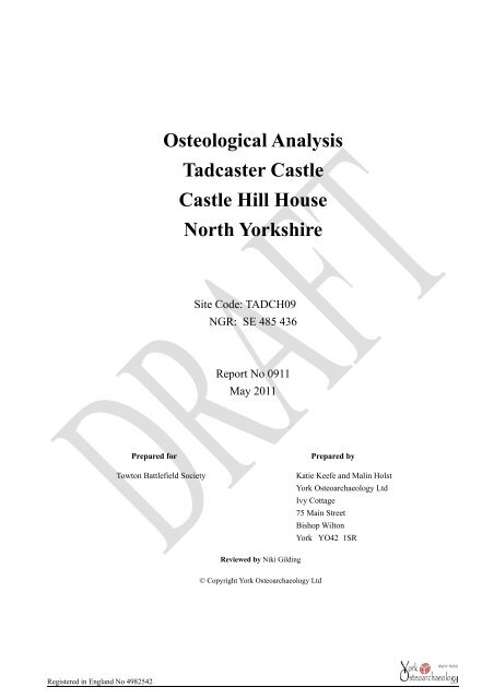
<strong>Osteological</strong> Analysis<br />
<strong>Tadcaster</strong> <strong>Castle</strong><br />
<strong>Castle</strong> Hill House<br />
North Yorkshire<br />
Site Code: TADCH09<br />
NGR: SE 485 436<br />
<strong>Report</strong> No 0911<br />
May 2011<br />
Prepared for Prepared by<br />
<strong>Towton</strong> <strong>Battlefield</strong> <strong>Society</strong> Katie Keefe and Malin Holst<br />
Reviewed by Niki Gilding<br />
York Osteoarchaeology Ltd<br />
Ivy Cottage<br />
75 Main Street<br />
Bishop Wilton<br />
© Copyright York Osteoarchaeology Ltd<br />
York YO42 1SR
<strong>Tadcaster</strong> <strong>Osteological</strong> <strong>Report</strong><br />
TABLE OF CONTENTS<br />
CONTENTS Page<br />
Summary iii<br />
Acknowledgements iii<br />
1.0 INTRODUCTION 1<br />
1.1 AIMS AND OBJECTIVES 1<br />
1.2 METHODOLOGY 1<br />
2.0 OSTEOLOGICAL ANALYSIS 1<br />
2.1 PRESERVATION 1<br />
2.2 MINIMUM NUMBER OF INDIVIDUALS 2<br />
2.3 ASSESSMENT OF AGE 2<br />
2.4 SEX DETERMINATION 3<br />
2.5 METRIC ANALYSIS 3<br />
2.6 NON-METRIC TRAITS<br />
4<br />
2.7<br />
CONCLUSION<br />
3.0 PATHOLOGICAL ANALYSIS<br />
3.1 JOINT DISEASE<br />
3.1.2 Degenerative Joint Disease<br />
3.1.3 Osteoarthritis<br />
3.1.4 Schmorl’s Nodes<br />
3.2 Transitional or Supernumerary Vertebra<br />
3.3<br />
Activity Related Change<br />
3.4 CONCLUSION<br />
4.0 DENTAL HEALTH<br />
5.0 DISCUSSION AND SUMMARY<br />
References<br />
Tables<br />
1 Summary of osteological and palaeopathological results<br />
1<br />
2<br />
Plates<br />
Female pelvis with wide sciatic notch<br />
OA of vertebral inferior articular facet<br />
3 Six sacral vertebrae<br />
4<br />
4<br />
5<br />
5<br />
5<br />
5<br />
6<br />
6<br />
7<br />
7<br />
8<br />
10<br />
2<br />
3<br />
5<br />
6<br />
i
<strong>Tadcaster</strong> <strong>Osteological</strong> <strong>Report</strong><br />
4 Advanced tooth wear<br />
5 Carious lesion and supernumerary tooth<br />
APPENDICES<br />
A <strong>Osteological</strong> And Palaeopathological Catalogue<br />
7<br />
8<br />
A<br />
ii
<strong>Tadcaster</strong> <strong>Osteological</strong> <strong>Report</strong><br />
1Summary<br />
In 2010 York Osteoarchaeology was commissioned by the <strong>Towton</strong> <strong>Battlefield</strong> <strong>Society</strong> to complete the full<br />
osteological analysis of a single inhumation recovered during excavations at <strong>Tadcaster</strong> <strong>Castle</strong>, <strong>Tadcaster</strong>,<br />
North Yorkshire (NGR SE 485 436).<br />
<strong>Osteological</strong> analysis revealed that the skeleton was a mature adult female, aged at least 46 years old when she<br />
died. The woman was around 173.5cm tall, making her above the average female height for all periods in<br />
history. She appears to have suffered from moderate osteoarthritis in her upper and mid spine, with mild<br />
degenerative joint disease affecting her left shoulder. The skeleton also exhibited a mild congenital defect, an<br />
additional sacral (tail bone) vertebra.<br />
The woman had suffered from numerous large dental cavities, possibly brought on as a result of a high sugar<br />
diet as attested by the moderate calculus deposits on her teeth. Two externally draining dental abscesses would<br />
have caused her discomfort. Advanced stages of wear on the chewing surfaces of most teeth may be evidence of<br />
her mature years, habitual tooth grinding or repetitive use of the teeth as tools.<br />
Acknowledgements<br />
York Osteoarchaeology Ltd would like to thank Tim Sutherland and the <strong>Towton</strong> <strong>Battlefield</strong> <strong>Society</strong> for their<br />
help and support.<br />
iii
1.0 INTRODUCTION<br />
In 2010 York Osteoarchaeology Ltd was commissioned by the <strong>Towton</strong> <strong>Battlefield</strong> <strong>Society</strong> to carry out the<br />
osteological analysis of a fully articulated skeleton discovered during archaeological excavations at <strong>Tadcaster</strong><br />
<strong>Castle</strong>, <strong>Tadcaster</strong>, North Yorkshire (SE 485 436). The excavation was undertaken in order to ascertain the<br />
existence of a possible mass grave thought to have been discovered during a previous excavation by<br />
C.V.Bellamy in 1972 (T.Sutherland pers comm. April 2011).<br />
1.1 AIMS AND OBJECTIVES<br />
The aim of the skeletal analysis was to determine the age, sex and stature of the skeleton, as well as to record<br />
and diagnose any skeletal manifestations of disease and trauma.<br />
1.2 METHODOLOGY<br />
The skeleton were analysed in detail, assessing the preservation and completeness, calculating the minimum<br />
number of individuals present as well as determining the age, sex and the stature of the individual (Appendix<br />
A). All pathological lesions were recorded and described.<br />
2.0 OSTEOLOGICAL ANALYSIS<br />
<strong>Osteological</strong> analysis is concerned with the determination of the identity of a skeleton, by estimating its age, sex<br />
and stature. Robusticity and non-metric traits can provide further information on the appearance and familial<br />
affinities of the individual studied. This information is essential in order to determine the prevalence of disease<br />
types and age-related changes. It is crucial for identifying gender dimorphism in occupation, lifestyle and diet,<br />
as well as the role of different age groups in society.<br />
2.1 PRESERVATION<br />
Skeletal preservation depends upon a number of factors, including the age and sex of the individual as well as<br />
the size, shape and robusticity of the bone. Burial environment, post-depositional disturbance and treatment<br />
following excavation can also have a considerable impact on bone condition. Preservation of human skeletal<br />
remains is assessed subjectively, depending upon the severity of bone surface erosion and post-mortem breaks,<br />
but disregarding completeness.<br />
Preservation was assessed using a grading system of five categories: very poor, poor, moderate, good and<br />
excellent. Excellent preservation implied no bone surface erosion and very few or no breaks, whereas very poor<br />
preservation indicated complete or almost complete loss of the bone surface due to erosion and severe<br />
fragmentation.<br />
The preservation of the skeleton was good (Table 1). The proximal and distal bones of the forearms had been<br />
damaged, as had the proximal ends of the femora. The long bones had also suffered from bone surface erosion,<br />
which was most apparent on the radii and ulnae.
Table 1 Summary of osteological and palaeopathological results<br />
Skeleton<br />
Preservation Completeness Age Sex Stature Pathology<br />
No<br />
1 Good 90% Mature<br />
Adult,<br />
46+<br />
years<br />
Female 173.5cm Osteoarthritis in the cervical and thoracic spine; DJD in<br />
cervical, thoracic and lumbar spine and the left<br />
shoulder, possible supernumerary sacral vertebra, or<br />
border shifting at coccygo-sacral border and sacro-<br />
lumbar border, Schmorl’s node on L1, muscle<br />
excavation on right clavicle for costoclavicular<br />
ligament, enthesopathies bilaterally on clavicles for<br />
conoid ligament. Caries, moderate calculus and dental<br />
abscesses<br />
The skeleton was missing all of the carpals (wrist bones), many of the metacarpals and hand phalanges (palm<br />
and finger bones), along with all foot phalanges (toe bones); these small bones have a tendency to move in the<br />
burial environment due to ploughing and animal and plant activity which can damage them as well as making<br />
them more elusive for excavators.<br />
2.2 MINIMUM NUMBER OF INDIVIDUALS<br />
A count of the ‘minimum number of individuals’ (MNI) recovered from a cemetery is carried out as standard<br />
procedure in osteological reports on inhumations in order to establish how many individuals are represented by<br />
the articulated and disarticulated human bones (without taking the archaeologically defined graves into<br />
account). The MNI is calculated by counting all long bone ends, as well as other larger skeletal elements<br />
recovered. The largest number of these is then taken as the MNI. The MNI is likely to be lower than the actual<br />
number of skeletons which would have been interred on the site, but represents the minimum number of<br />
individuals which can be scientifically proven to be present.<br />
No skeletal elements were duplicated suggesting a minimum number of individuals of one.<br />
2.3 ASSESSMENT OF AGE<br />
Age was determined using standard ageing techniques, as specified in Scheuer and Black (2000a; 2000b) and<br />
Cox (2000). Age estimation relies on the presence of the pelvis and uses different stages of bone development<br />
and degeneration in order to calculate the age of an individual. Age is split into a number of categories, from<br />
foetus (up to 40 weeks in utero), neonate (around the time of birth), infant (newborn to one year), juvenile (1-12<br />
years), adolescent (13-17 years), young adult (ya; 18-25 years), young middle adult (yma; 26-35 years), old<br />
middle adult (oma; 36-45 years), mature adult (ma; 46+) to adult (an individual whose age could not be<br />
determined more accurately as over the age of seventeen). However, it is important to note that several studies<br />
(for example Molleson and Cox 1993; Molleson 1995; Miles, et al. 2008) have highlighted the difficulty of<br />
accurately determining the age-at-death of adults from their skeletal remains, with age-at-death frequently<br />
underestimated for older individuals. The categories defined here should perhaps be taken as a general guide to<br />
the relative physiological age of the adult, rather than being an accurate portrayal of the real chronological age;<br />
no doubt many of those aged ‘46+’ would in actuality have been in their sixties, seventies or eighties when they
died.<br />
Due to the poor survival rate of many of the skeletal elements used for the determination of age, the estimation<br />
of the skeletons age was based solely on the degeneration of the auricular surface (the joint between the hip and<br />
tail bone). Analysis revealed that the individual was at least 46 years old when they died.<br />
2.4 SEX DETERMINATION<br />
Sex determination was carried out<br />
using standard osteological<br />
techniques, such as those described by<br />
Mays and Cox (2000). Assessment of<br />
sex in both males and females relies on<br />
the preservation of the skull and<br />
the pelvis and can only be carried out<br />
once sexual characteristics have<br />
The sexually dimorphic elements of<br />
Skeleton 1 included the cranium and the<br />
pelvis; it was also possible to perform<br />
metric analysis on a small number of<br />
Plate 1 Female pelvis with wide sciatic notch<br />
bones in order to determine sex.<br />
The skull exhibited ostensibly male characteristics; the anterior mandible, mastoid process and supra-orbital<br />
ridge all exhibited male morphological traits (rugged and robust), while the orbital rims were female (sharp).<br />
The morphology of the pelvis was clearly female (Plate 1), with the majority of traits suggesting Skeleton 1 was<br />
a female. It is not unusual for older females to develop male cranial characteristics.<br />
Measurements of the post-cranial elements revealed mixed results, with bones falling within both the male and<br />
female ranges, suggesting that she was probably a robust or well-built individual.<br />
2.5 METRIC ANALYSIS<br />
Stature depends on two main factors, heredity and environment. However, stature can also fluctuate between<br />
chronological periods. It is only possible to calculate in skeletons if at least one complete and fully fused long<br />
bone is present. The bone is measured on an osteometric board, and stature is then calculated using a regression<br />
formula developed upon individuals of known stature<br />
Metrical analysis of the tibia from Skeleton 1 revealed that the individual was 173.5 ±3.4cm, which is above the<br />
average range of height for females during all periods of history (Roberts and Cox 2003). Considering the<br />
location of the burial, it is feasible that the skeleton dates to the early medieval period, who tend to be tall and/or<br />
that the individual may have been an occupant from the castle. Individuals of high status would have had the<br />
greatest access to a nutritionally rounded diet and as such would have achieved their biologically determined<br />
stature. Caffell (1997) notes five previously analysed populations, dating to the early medieval period that had a
female range of heights that exceeded 175cm.<br />
The meric index is a method of calculating the shape and robusticity of the adult femoral shaft. Calculations<br />
revealed that both femora fell into the platymeric range (broad and flat). The cnemic index of the tibiae was<br />
calculated in order to establish the degree of tibial shaft flatness. Calculations revealed that both tibiae were<br />
eurycnemic (broad).<br />
The cranial index describes the shape of the cranium; measurement indicated that the individual was<br />
dolichocranic (long or narrow headed). The cranial breadth-height index expresses the relationship between the<br />
breadth and height of a skull as a percentage. The results revealed that the individual had an average or medium<br />
shaped skull (metriocranic). Further cranial indices pertaining to the facial skeleton revealed that the individual<br />
had a broad or wide nasal aperture (platrrhiny), wide orbits (chamaeconchy) and an average shaped palate<br />
(mesostaphylinic).<br />
2.6 NON-METRIC TRAITS<br />
Non-metric traits are additional sutures, facets, bony processes, canals and foramina, which occur in a minority<br />
of skeletons and are believed to suggest hereditary affiliation between skeletons (Saunders 1989). The origins of<br />
non-metric traits have been extensively discussed in the osteological literature and it is now thought that while<br />
most non-metric traits have genetic origins, some can be produced by factors such as mechanical stress<br />
(Kennedy 1989) or environment (Trinkhaus 1978).<br />
A total of thirty cranial (skull) and thirty post-cranial (bones of the body and limbs) non-metric traits were<br />
selected from the osteological literature (Buikstra and Ubelaker 1994, Finnegan 1978, Berry and Berry 1967)<br />
and recorded.<br />
Skeleton 1 exhibited six cranial non-metric traits. These consisted of an ossicle at lambda (extra bone at the<br />
back of the skull), ossicle in lambdoid, ossicle at asterion, ossicle in parietal notch (extra bones in the sutures at<br />
the back of the skull) open foramen ovale, posterior condylar canal open (small holes at the base of the skull).<br />
Bennett (1965) has suggested that the formation of ossicles within the sutures may be related to stresses placed<br />
on the growing cranium during foetal life and early infancy.<br />
Further non-metric traits were observed in the post-cranial skeleton; these included exostosis in the trochanteric<br />
fossa (spicules of bone on the proximal femur), a third trochanter (bony nodule on the back of the proximal<br />
femur) and a peroneal tubercle (small nodule of bone on the ankle).<br />
2.7 CONCLUSION<br />
Skeleton 1 was largely complete, with only some of the bones of the hands, feet and wrists missing. The<br />
skeleton was in good condition with moderate surface erosion evident on the long bones. The individual<br />
appeared to be a female, although the individual expressed a mixture of sexual characteristics, making the<br />
assessment tentative. She would have been between 170cm and 177cm tall, which was above average for<br />
females of all periods in history (Roberts and Cox 2003). A variety of non-metric traits were observed, which<br />
may have been caused by both genetic and mechanical factors. None of these traits would have caused any
symptoms.<br />
3.0 PATHOLOGICAL ANALYSIS<br />
Pathological conditions (disease) can manifest themselves on the skeleton, especially when these are chronic<br />
conditions or the result of trauma to the bone. The bone elements to which muscles attach can also provide<br />
information on muscle trauma and excessive use of muscles. All bones were examined macroscopically for<br />
evidence of pathological changes.<br />
3.1 JOINT DISEASE<br />
The term joint disease encompasses a large number of conditions with different causes, which all affect the<br />
articular joints of the skeleton. Factors influencing joint disease include physical activity, occupation, workload<br />
and advancing age, which manifest as degenerative joint disease and osteoarthritis. Alternatively, joint changes<br />
may have inflammatory causes in the spondyloarthropathies, such as sceptic or rheumatoid arthritis. Different<br />
joint diseases affect the articular joints in a different way, and it is the type of lesion, together with the<br />
distribution of skeletal manifestations, which determines the diagnosis (Rogers 2000, Roberts and Manchester<br />
2005).<br />
3.1.1 Degenerative Joint Disease<br />
The most common type of joint disease observed tends to be degenerative joint disease (DJD). DJD is<br />
characterised by both bone formation (osteophytes) and bone resorption (porosity) at and around the articular<br />
surfaces of the joints, which can cause great discomfort and disability (Rogers 2000).<br />
Skeleton 1 revealed signs of spinal DJD, with mild to moderate osteophytic lipping observed on the margins of<br />
many cervical, thoracic and lumbar articular facets. Further DJD was seen on the articular margins of the left<br />
scapula and proximal humerus<br />
3.1.2 Osteoarthritis<br />
Osteoarthritis is a form of degenerative joint disease characterised by the deterioration of the joint<br />
cartilage, leading to exposure of the underlying bony joint surface. The resulting bone-to-bone<br />
contact can produce a polished appearance on the joint surface termed ‘eburnation’, which is the<br />
most obvious expression of osteoarthritis. Osteoarthritis can be the result of mechanical stress and<br />
other factors, including lifestyle, food acquisition and preparation, social status, sex and general<br />
health (Larsen 1997). Osteoarthritis was recorded on the articular facets of cervical vertebrae two,<br />
three and four (central part of the neck) and the articular facet of the fifth thoracic vertebra (Plate<br />
2).<br />
Plate 2 OA of inferior articular facet
3.1.3 Schmorl’s Nodes<br />
Schmorl’s nodes are a condition which affect the spine. They manifest as indentations in the upper and lower<br />
surfaces of the vertebral bodies caused by the pressure of herniated vertebral discs (Aufderheide and Rodríguez-<br />
Martín 1998). Discs may rupture due to trauma, but vertebrae weakened by infection, osteoporosis or neoplastic<br />
disease may be more vulnerable (Roberts and Manchester 2005). Schmorl’s nodes are often associated with<br />
degenerative changes to the vertebral bodies (Aufderheide and Rodríguez-Martín 1998, Hilton et al. 1976), and<br />
are most commonly seen in the lower thoracic vertebrae (Hilton et al. 1976).<br />
A Schmorl’s node was present on the lower body of the first lumbar vertebra (lower spine). Poor preservation of<br />
the vertebral bodies inhibited the observation of many thoracic and lumbar vertebral bodies, and as a result may<br />
have masked a greater prevalence. The presence of such lesions may indicate that the individual was involved in<br />
physically arduous activities.<br />
3.2 CONGENITAL ANOMALIES<br />
The normal human spine consists of seven cervical (neck), twelve thoracic (chest) and five lumbar (lower back)<br />
vertebrae, making a total of 24 independent segments. The sacrum (at the base of the spine, forming the back of<br />
the pelvis) is usually composed of five fused vertebral segments, and the coccyx (vestigial tail) is normally<br />
made up of four fused vertebral segments. The overall total of vertebral segments is therefore 33.<br />
Transitional vertebrae can occur at the borders between different types of vertebra, when a vertebra from one<br />
group takes on some or all of the characteristics of an adjacent group, for example the first lumbar vertebra (in<br />
the lower back) may develop vestigial ribs (Barnes 1994, 79-116). The process by which this happens is known<br />
as ‘border shifting’. The end result is to increase the number of segments in one part of the spine at the expense<br />
of the adjoining part (e.g. increasing the number of thoracic vertebrae to 13 through incorporating the first<br />
lumbar vertebra, but decreasing the number of lumbar vertebrae to four). Transitional vertebrae are reasonably<br />
common, particularly at the lumbo-sacral border (between the fifth lumbar vertebra and the sacrum, at the base<br />
of the spine), but the consequences of the border shift become more severe the higher up the spine it occurs<br />
(ibid). Supernumerary vertebrae, according to Barnes (1994), are the result<br />
of an abnormal number of somites (precursors to vertebrae found in the<br />
human embryo). However, unless the full complement of cervical to<br />
coccygeal vertebrae are present, it is impossible to distinguish between<br />
border shifts and supernumerary vertebrae.<br />
The sacrum of Skeleton 1 consisted of six vertebrae (Plate 3), instead of the<br />
usual five. The coccyx (tip of the tail bone) belonging to the individual had<br />
not survived and as a result it was not possible to distinguish whether<br />
Skeleton 1 had experienced a caudal border shift at the sacro-caudal border,<br />
so called because the interface between the two sets of vertebrae has been<br />
moved away from the skull, due to the lower vertebrae assimilating the<br />
features of the upper (Barnes 1994, 114). According to Barnes such shifts<br />
are common in the osteological record (ibid). Sacralisation or lumbarisation<br />
occurs in 3-5% of the population (Aufderheide and Rodríguez-Martín<br />
Plate 3 Six sacral vertebrae
1998) and has been associated with lower back pain in modern studies (Sture 2001).<br />
3.3 ACTIVITY RELATED CHANGE<br />
Bone is a dynamic material which can change its morphology, size and robustness in response to prolonged<br />
activity (Knüsel 2000, 383). As a result, greater activity and mechanical stress causes the bone to become<br />
shapelier, with ridges and depressions caused by muscle action. It is possible to infer trauma to the soft tissue<br />
on the bones, in the form of ligamentous or muscular lesions. Enthesopathies (bony processes) or cortical bone<br />
defects at the site of muscle or ligament attachments can develop (Hawkey and Merbs 1995, 329). Bone defects<br />
may also be observed at the site of muscle insertions, which may be the result of constant micro-trauma.<br />
Pronounced muscle insertions were noted on both clavicles for the conoid ligament of Skeleton 1. The conoid<br />
ligament attaches to the shoulder, and, in combination with other ligaments, holds the shoulder position<br />
preventing superior dislocation of the shoulder (Abrahams et al.2003, 122). The individual also exhibited a bone<br />
excavation at the insertion of the right costoclavicular ligament on the individual’s clavicle.<br />
3.4 CONCLUSION<br />
The osteological examination of the human remains from <strong>Tadcaster</strong> <strong>Castle</strong> has revealed a number of interesting<br />
details about the individual. The woman appears to have suffered from osteoarthritis of the neck and central<br />
back which would have caused reduced flexibility and stiffness. She also had a mild congenital defect which<br />
manifested at the base of her spine; such a defect may indicate a disruption to her early foetal development and<br />
may have been associated with lower back pain. A lesion present on one of the vertebral bodies, combined with<br />
a small number of pronounced muscle attachments, may also suggest that the individual carried out physically<br />
arduous activities.<br />
4.0 DENTAL HEALTH<br />
Of the usual 32 tooth positions which comprise the adult dentition, it<br />
was possible to observe 31 in Skeleton 1. Of the observable positions,<br />
three teeth had been lost post-mortem.<br />
Dental wear tends to be more common and severe in archaeological<br />
populations than in modern teeth. Severity of the dental wear was<br />
assessed using a chart developed by Smith (1984). Each tooth was scored<br />
using a grading system ranging from 1 (no wear) to 8 (severe attrition of<br />
the whole tooth crown). Skeleton 1 exhibited advanced tooth wear<br />
on all of her teeth. The pattern of wear was flat and even across all of her<br />
teeth (Plate 4); such a degree of tooth wear may simply reflect the<br />
individual’s mature age, or alternatively she may have had bruxism<br />
Calculus (dental plaque) is commonly Plate observed 4 Advanced in archaeological<br />
tooth wear<br />
populations whose dental hygiene was not a rigorous as it is today.
Calculus mineralises and forms concretions on the tooth crowns, along the line of the gums. Calculus was<br />
observed on 72.4% (21/ 29) of the teeth belonging to Skeleton1. Periodontitis (receding gums) was slight<br />
around the anterior teeth of the mandible but more moderate on the maxilla and may have been caused by the<br />
widespread calculus deposits.<br />
Caries (tooth decay) are multi-factoral in origin, but develop as<br />
a result of aggressive bacterial attack in the presence of sucrose<br />
(Hillson 1996, 282) and fermentable carbohydrates (Roberts<br />
and Manchester 1995, 47). As the bacteria metabolise the sugar,<br />
acid is produced which can erode the hard mineral surface of<br />
the crown. Simple sugars can be found in fruits, vegetables,<br />
dried fruits and honey, as well as processed, refined sugar; since<br />
the latter three contain the most sucrose they are most likely to<br />
cause tooth decay. Skeleton 1 exhibited six caries on five teeth,<br />
affecting 20.7% of teeth (6/29), many cavities were large and<br />
affected the crown root junction (Plate 5).<br />
Two externally draining abscesses were present at the root apex of the left mandibular first molar and the right<br />
maxillary third molar. Dental abscesses occur when bacteria enters the pulp cavity of a tooth causing<br />
inflammation and a build-up of pus at the tip of the root. Eventually, a hole forms in the surrounding bone<br />
allowing the pus to drain out and relieve the pressure. They can form as a result of cavities, heavy dental wear,<br />
damage to the teeth, or periodontal disease (Roberts and Manchester 1995). The right maxillary third molar was<br />
affected by a large cavity, and it is possible that the abscess was a result of the bacteria which caused the cavity.<br />
The individual may also have had a supernumerary tooth. The root of a tooth largely destroyed by caries was<br />
located on the left mandible, anterior to the third molar. The tooth cannot have been a retained deciduous tooth,<br />
due to the location (see Plate 5).<br />
The dental health of the individual was poor. Large caries may indicate that the individual had a high sugar diet<br />
and calculus deposits point to a diet also rich in protein. Abscesses suffered by the individual may have been<br />
caused by the numerous carious lesions.<br />
5.0 DISCUSSION AND SUMMARY<br />
The skeleton recovered from <strong>Tadcaster</strong> <strong>Castle</strong> is currently of unknown date. The individual was laid out in an<br />
extended supine position, orientated west to east, with the hands on the pelvis, in what appears to be a typically<br />
Christian manner, however, radiocarbon dates are awaited. Skeleton 1 was well preserved and almost complete.<br />
The remains were those of a mature adult female of above average stature for females, aged at least 46 years<br />
when she died.<br />
Plate 5 Caries and supernumerary tooth<br />
The female probably suffered from disturbances during her early foetal development, causing minor anomalies<br />
in her lower spine that may have led to lower back pain. The individual appears to have suffered from<br />
osteoarthritis of the neck and central spine in older age, which would have caused stiffness and reduced
flexibility. Evidence of an active and strenuous life is attested to through lesions evident on the individual’s<br />
vertebrae and at the insertions of numerous muscles.<br />
The dental health of the individual was poor; cavities and mineralised plaque were common in the mature adult<br />
female’s dentition and are likely to have resulted from a diet high in sugar and protein. A number of abscesses<br />
would also have caused the individual constant discomfort, making eating and talking challenging.
References<br />
Abrahams. P. H., Marks Jr. S. C. and Hutchings. R. T. 2003. McMinn’s Color Atlas of Human Anatomy.<br />
Edinburgh, London, New York, Oxford, Philadelphia, St Louis, Sidney, Toronto<br />
Aufderheide, A.C. and Rodríguez-Martín, C. 1998. The Cambridge Encyclopedia of Human Paleopathology<br />
(Cambridge)<br />
Barnes, E. 1994. Developmental Defects of the Axial Skeleton in Paleopathology (Niwot, Colorado)<br />
Bennett, K. A. 1965. ‘The etiology and genetics of wormian bones’, American Journal of Physical<br />
Anthropology 23: 255-260<br />
Berry, A.C. and Berry, R.J. 1967. ‘Epigenetic variation in the human cranium’, Journal of Anatomy 101 (2):<br />
361-379<br />
Buikstra, J.E. and Ubelaker D.H. (eds) 1994. Standards for Data Collection from Human Skeletal Remains<br />
(Fayetteville)<br />
Caffell, A. 1997. A Comparison of Stature between British Skeletal Populations, Bradford University,<br />
Unpublished Undergraduate Dissertation<br />
Cox, M. 2000. ‘Ageing adults from the skeleton’, in M. Cox and S. Mays (eds), Human Osteology in<br />
Archaeology and Forensic Science (London): 61-82<br />
Finnegan, M. 1978. ‘Non-metric variation of the infracranial skeleton’, Journal of Anatomy 125: 23-37<br />
Hawkey, D.E. and Merbs, C.F. 1995. ‘Activity-induced musculoskeletal stress markers (MSM) and subsistence<br />
strategy changes among ancient Hudson Bay Eskimos’, International Journal of Osteoarchaeology 5: 324-<br />
338<br />
Hillson, S. 1996. Dental Anthropology (Cambridge)<br />
Hilton, R.C., Ball, J. and Benn R.T. 1976. ‘Vertebral end-plate lesions (Schmorl’s nodes) in the dorsolumbar<br />
spine’, Ann Rheum. Dis. 35: 127-132<br />
Kennedy, K.A.R. 1989. ‘Skeletal markers of occupational stress’, in M.Y. Işcan. and K.A.R. Kennedy (eds),<br />
Reconstruction of Life from the Skeleton (New York):129-160<br />
Knüsel, C. 2000. ‘Bone adaptation and its relationship to physical activity in the past’, in M. Cox and S. Mays<br />
(eds), Human Osteology in Archaeology and Forensic Science (London): 381-402<br />
Larsen, C S, 1997 Bioarchaeology: Interpreting Behaviour from the Human Skeleton (Cambridge)<br />
Mays, S. and Cox, M. 2000. ‘Sex determination in skeletal remains’, in M. Cox and S. Mays (eds), Human<br />
Osteology in Archaeology and Forensic Science (London): 117-130<br />
Miles, A., Powers, N., Wroe-Brown, R. and Walker, D. 2008, St Marylebone Church and Burial Ground in the<br />
18th to 19th Centuries: Excavations at St Marylebone School, 1992 and 2004-6, MoLAS Monograph 46<br />
(London)<br />
Molleson, T. 1995. 'Rates of ageing in the eighteenth century" in S.R. Saunders and A. Herring (eds) Grave<br />
Reflections: Portraying the Past through Cemetery Studies (Toronto): 199-222<br />
Molleson, T. and Cox, M. 1993. The Spitalfields Project. Vol. 2. The Anthropology: the Middling Sort, CBA<br />
Research <strong>Report</strong>, 86 (York)<br />
Roberts, C.A. and Cox, M. 2003. Health and Disease in Britain (Stroud)<br />
Roberts, C.A. and Manchester, K. 1995. The Archaeology of Disease (Stroud)<br />
Roberts, C.A. and Manchester, K. 2005. The Archaeology of Disease (Stroud)<br />
Rogers, J. 2000. ‘The palaeopathology of joint disease’, in M. Cox and S. Mays (eds), Human Osteology in<br />
Archaeology and Forensic Science (London): 163-182<br />
Saunders, S.R. 1989. ‘Non-metric variation’, in M.Y. Işcan and K.A.R. Kennedy (eds) Reconstruction of Life<br />
from the Skeleton (New York): 95-108
Scheuer, L. and Black, S. 2000a. ‘Development and ageing of the juvenile skeleton’, in M. Cox and S. Mays<br />
(eds), Human Osteology in Archaeology and Forensic Science (London): 9-22<br />
Scheuer, L. and Black, S. 2000b. Developmental Juvenile Osteology (San Diego)<br />
Smith, B.H. 1984. ‘Patterns of molar wear in hunter-gatherers and agriculturalists’, American Journal of<br />
Physical Anthropology 63: 39-56<br />
Sture, J. F. 2001. Biocultural Perspectives on Birth Defects in Medieval Urban and Rural English Populations.<br />
(Durham) Unpublished PhD Thesis<br />
Trinkhaus, E. 1978. ‘Bilateral asymmetry of human skeletal non-metric traits’, American Journal of Physical<br />
Anthropology 49: 315-318
<strong>Tadcaster</strong>_<strong>Castle</strong>_<strong>Osteological</strong>_<strong>Report</strong>.doc A<br />
APPENDIX A: OSTEOLOGICAL AND PALAEOPATHOLOGICAL CATALOGUE<br />
1Skeleton Number 1<br />
Preservation Good, ribs and scapulae well preserved, epiphyses of radii and ulnae missing, cortical surface of long<br />
bones heavily eroded<br />
Completeness 90% bones of hands and wrists missing as are pedal phalanges<br />
Age 46 + years old, Mature Adult<br />
Sex Female?<br />
Stature 175.9cm<br />
Non-Metric Traits Cranial traits; ossicle at lambda, ossicle at lambdoid (left), ossicle at pterion (left), ossicle at asterion<br />
(left), posterior condylar canal open (bilateral) open foramen spinosum (right)<br />
Post cranial traits; exostosis in trochanteric fossa (left), third trochanter (left), peroneal tubercle (left), os<br />
trigonum (left)<br />
Pathology Osteoarthritis in cervical spine; DJD in cervical, thoracic and lumbar spine, and on the left shoulder,<br />
possible supernumerary sacral vertebra, or border shifting at coccygo-sacral border and sacro-lumbar<br />
border, Schmorl’s node on L1, muscle excavation on right clavicle for costoclavicular ligament,<br />
enthesopathies bilaterally on clavicles for conoid ligament<br />
Dental Health 31 positions present, three teeth lost post mortem, moderate calculus on the lingual and buccal surfaces<br />
of the lower right dentition whereas only flecks were present on the buccal surface of the lower left and<br />
upper left anterior dentition, more moderate deposits were recorded on the buccal surface of all upper<br />
left maxillary molars, six caries were present on five teeth many were large and affected the crown root<br />
junction, slight periodontal disease evident on the right mandible and moderate on the left maxilla, two<br />
externally draining abscesses were present at the root apex of the left mandibular first molar and the<br />
right maxillary third molar, on the left mandible anterior to the third molar was a root of a tooth which<br />
had been largely destroyed by a caries, cannot have been a deciduous tooth due to the location, possibly<br />
Right Dentition<br />
supernumerary, advanced tooth wear on all teeth<br />
Left Dentition<br />
Present P - P P P P P PM P P P P P P P P<br />
Calculus - - Sl - - - - - Sl Fb Fb Sb - Mb Mb Mb<br />
DEH - - - - - - - - - - - - - - - -<br />
Caries Lo - - - - - - - - - - - - - Md -<br />
Wear - - 8 7 7 5 5 - 6 5 5 6 6 7<br />
crj<br />
6 6<br />
Maxilla 8 7 6 5 4 3 2 1 1 2 3 4 5 6 7 8<br />
Mandible 8 7 6 5 4 3 2 1 1 2 3 4 5 6 7 8<br />
Present P P P P P P P P P P P P P PM PM P<br />
Calculus Mdl Mlb Mlb Mb Mb Fb Fb Sb Mbl Sb Fb b - - - Sd<br />
b<br />
Sl Sl Sl Sl Ml<br />
DEH - - - - - - L - - - - - - - - -<br />
Caries - - Ld - - - - - - - - - Lc - - Lm<br />
crj<br />
Mm<br />
Wear<br />
1KEY:<br />
4 -<br />
crj<br />
- - 2 3 4 - 4 4 4 3 - - - 3<br />
Present - Tooth presence; am - ante-mortem tooth loss; pm - post-mortem tooth loss; p - tooth present; p (u) – tooth present but unerupted<br />
- - jaw not present<br />
Caries - Calculus; F - flecks of calculus; S - slight calculus; M - moderate calculus; H - heavy calculus; a - all surfaces; b - buccal<br />
surface; d - distal surface; m - mesial surface; l - lingual surface; o - occlusal surface; crj – crown root junction<br />
DEH - dental enamel hypoplasia; l - lines; g - grooves; p - pits<br />
Caries - caries; s - small lesions; m - moderate lesions; l - large lesions<br />
Wear - dental wear; numbers from 1-8 - slight to severe wear<br />
crj