

Genotypic characterisation of Echinococcus granulosus isolated ...
Genotypic characterisation of Echinococcus granulosus isolated ...
Genotypic characterisation of Echinococcus granulosus isolated ...

Create successful ePaper yourself
Turn your PDF publications into a flip-book with our unique Google optimized e-Paper software.
African Journal <strong>of</strong> Microbiology Research Vol. 4 (7), pp. 551-555, 4 Aprıl, 2010<br />
Available online http://www.academicjournals.org/ajmr<br />
ISSN 1996-0808 © 2010 Academic Journals<br />
Full Length Research Paper<br />
<strong>Genotypic</strong> <strong>characterisation</strong> <strong>of</strong> <strong>Echinococcus</strong><br />
<strong>granulosus</strong> <strong>isolated</strong> from human in Turkey<br />
Sevgi Ergin 1 , Suat Saribas 1 , Pelin Yuksel 1 , Kaan Zengin 2 , Kenan Midilli 1 , Gokhan Adas 3 ,<br />
Soykan Arikan 4 , Mustafa Aslan 1 , Hayriye Uysal 5 , Reyhan Caliskan 1 , Ali Oner 5 , Omer<br />
Kucukbasmaci 1 , Arif Kaygusuz 1 , Muzeyyen Mamal Torun 1 and Bekir Kocazeybek 1*<br />
1 Istanbul University, Cerrahpa a Faculty <strong>of</strong> Medicine, Microbology and Clinical Microbiology Department, Istanbul,<br />
Turkey.<br />
2 Istanbul University, Cerrahpa a Faculty <strong>of</strong> Medicine, General Surgery Department, Istanbul, Turkey.<br />
3 Health Minister Okmeydanı Education and Training Hospital, General Surgery Department, Istanbul, Turkey.<br />
4 Health Minister Istanbul Education and Training Hospital, General Surgery Department, Istanbul, Turkey.<br />
5 Istanbul University, Istanbul Faculty <strong>of</strong> Medicine, Parasitology Department, Istanbul, Turkey.<br />
Accepted 15 March, 2010<br />
Cystic echinococcosis is one <strong>of</strong> the most important zoonotic diseases in Turkey. The aim <strong>of</strong> this study<br />
was for the molecular analysis <strong>of</strong> <strong>Echinococcus</strong> <strong>granulosus</strong> isolates from different regions <strong>of</strong> Turkey.<br />
For this purpose, 46 hydatid cyst samples collected from humans during the surgery were included.<br />
The partial sequences <strong>of</strong> mitochondrial cytochrome c oxidase subunit 1 (CO1) gene was used for<br />
identification and molecular analysis <strong>of</strong> E. <strong>granulosus</strong> strains. Molecular analysis showed that all <strong>of</strong> the<br />
human cysts belonged to the G1 genotype (common sheep strain) <strong>of</strong> E. <strong>granulosus</strong>. According to the<br />
results <strong>of</strong> our study, the sheep strain (G1) is the predominant genotype <strong>of</strong> E. <strong>granulosus</strong> in humans in<br />
our country.<br />
Key words: <strong>Echinococcus</strong> <strong>granulosus</strong>, human, hydatid cyst, genotype, Turkey.<br />
INTRODUCTION<br />
<strong>Echinococcus</strong> <strong>granulosus</strong> is the causative agent <strong>of</strong> cystic<br />
echinococcosis (CE) which has serious impacts on<br />
human and/or animal health resulting with significant economic<br />
losses (Eckert et al., 2001; Sarıözkan and Yalçın,<br />
2009; Snabel et al., 2009). The disease is endemic in the<br />
Middle East, Central Asia and Nothern and Eastern Africa<br />
(Eckert et al., 2001; Sadjjadi, 2006; Azlaf and Dakkak,<br />
2006).<br />
A number <strong>of</strong> genetic variants <strong>of</strong> E. <strong>granulosus</strong> have<br />
been described. This genetic variations may determine<br />
phenotypic characteristics, host specificity, antigenicity,<br />
transmission dynamics, infection route, pathology, control,<br />
antimicrobial susceptibility and vaccine development<br />
strategies (Thampson and Mc Manus, 2001). So far,<br />
based on the partial sequences <strong>of</strong> mitochondrial cytochrome<br />
coxidase subunit 1 (CO1) and NADH dehydroge-<br />
*Corresponding author. E-mail: bzeybek@istanbul.edu.tr. Tel:<br />
00-90-532-6168150. Fax: 00-90-212-6322122.<br />
nase 1 (ND1) genes, ten distinct genetic types (G1-G10)<br />
<strong>of</strong> E. <strong>granulosus</strong> have been identified (Bowles and Mc<br />
Manus, 1993a; Bowles et al., 1994; Snabel et al., 2000).<br />
Recently, E. <strong>granulosus</strong> was divided following groups; E.<br />
<strong>granulosus</strong> sensu stricto (G1; sheep strain, G2; Tasmanian<br />
sheep strain, G3; buffalo strain), E. equinis (G4; horse<br />
strain), E. ortleppi (G5; cattle strain) ve E. canadensis (G6;<br />
camel strain, G7; pig strain, G8; cervid strain, G9; human<br />
strain, G10; Fennoscandian cervid strain) (Nakao et al.,<br />
2007). Among the genotypes in the E. <strong>granulosus</strong> sensu<br />
strico group, the sheep strain (G1 genotype) has the<br />
most wide geographic distribution around the world. The<br />
sheep strain is also dominant in the Mediterranean area<br />
(Euzeby, 1991; Breyer et al., 2004; Eckert and Thampson,<br />
1997; Mc Manus and Thampson, 2003; Romig et al., 2006;<br />
Varcasia et al., 2007; Busi et al., 2007). Some <strong>of</strong> the<br />
other genotypes <strong>of</strong> E. <strong>granulosus</strong> have been reported to<br />
cause occasionally infections in human, but some<br />
genotypes are not involved in human infections (Bowles<br />
and Mc Manus, 1993b; Dinkel et al., 2004). Although<br />
cystic hydatid disease continues to be endemic in our
552 Afr. J. Microbiol. Res.<br />
Table 1. The distribution genotypes <strong>of</strong> E. <strong>granulosus</strong> according to<br />
the origin <strong>of</strong> the patients and the localisation <strong>of</strong> the cysts.<br />
Origin <strong>of</strong> the patients Genotype Liver Lung<br />
Black sea region G1 2 -<br />
Marmara region G1 3 1<br />
East Anatolia region G1 14 -<br />
Southeast Anatolia region G1 13 -<br />
Mediterranean region G1 2 1<br />
Central Anatolia region G1 10 -<br />
country, large molecular epidemiologic data regarding the<br />
genotypes <strong>of</strong> E. <strong>granulosus</strong> strains infecting humans are<br />
still limited. The aim <strong>of</strong> this study was for the molecular<br />
<strong>characterisation</strong> <strong>of</strong> E. <strong>granulosus</strong> strains obtained from<br />
human originating from different geographic regions <strong>of</strong><br />
Turkey.<br />
MATERIALS AND METHODS<br />
Collection <strong>of</strong> cysts<br />
Between March 2008 - December 2009, cyst contents <strong>of</strong> 46<br />
patients (28 males and 18 women) were collected during the<br />
surgical procedures performed in different hospitals in Istanbul. Demographic,<br />
epidemiologic and clinical data were recorded (Table 1).<br />
The ages <strong>of</strong> the patients were between 16-74 years old. Hydatid<br />
cyst diseases diagnosis was based on clinical, radiological and<br />
serological findings. The samples were checked for the presence <strong>of</strong><br />
protoscoleces microscopically for the assessment <strong>of</strong> fertility states.<br />
Protoscoleces and cyst walls (germinal and laminar layers) were<br />
preserved in 70% ethanol until DNA purification. The samples were<br />
rinsed several times with sterile distilled water to remove the<br />
ethanol prior to DNA purification.<br />
DNA purification<br />
The hydatid cyst walls and protoscoleces were minced using surgical<br />
scalpales under sterile conditions. Genomic DNA was purified<br />
from 25-30 mg <strong>of</strong> minced tissue using a commercial DNA<br />
purification kit (Nucleospin tissue kit, Macherey Nagel, Germany) in<br />
accordance with the manufacturer’s instructions and were stored at<br />
-80°C until use.<br />
PCR amplification<br />
Primers choosen from the mitochondrial cytochrome c oxidase<br />
subunit 1 (CO1) gene <strong>of</strong> E. <strong>granulosus</strong> were used for the PCR<br />
amplification as previously described (Bowles et al., 1992) (Table<br />
2). The PCR mixture contained 0.5 µM each primer, 10 mM Tris-<br />
HCl, 1.5 mM MgCl2, 50 mM KCl(pH 8.3), 0.2 mM each deoxynucleoside<br />
triphosphate (Fermentase®, Lithuania), and 1.25 U <strong>of</strong><br />
Taq DNA polymerase (Fermentase®, Lithuania). The final reaction<br />
volume was 50 µl containing 10 µl <strong>of</strong> DNA sample. The thermal<br />
cycling conditions were as follow: the first denaturation cycle was<br />
performed at 95°C for 2 min followed by 30 cycles dena-turation at<br />
94°C for 30 s, annealing at 55°C for 30 s and extension at 72°C for<br />
30 s. The final extension step consisted <strong>of</strong> 5 min at 72°C. The amplification<br />
products were run on a 1.5% agarose gel electrophoresis<br />
and visualised on an UV transilluminator. The expected size <strong>of</strong> the<br />
PCR product was 446 bp. (Figure 1). For the exclusion <strong>of</strong> the PCR<br />
inhibitors, all samples were tested in a separated run using betaglobulin<br />
primers.<br />
DNA sequencing<br />
The PCR products were purified by using a commercial PCR product<br />
purification kit (High Pure PCR Product Purification kit, Roche<br />
Diagnostic, GmbH, Germany), and subjected to the cycle<br />
sequencing using big-dye terminator kit (ABI®, USA). Following the<br />
cleaning-up procedure through sephadex G-50 fine columns the<br />
cycle sequencing products were run on an automated DNA<br />
sequencer (ABI®, 310). The obtained sequences were edited and<br />
aligned, using the Bioedit s<strong>of</strong>tware (Hall, 1999) then compared<br />
against the data available in GenBank.<br />
RESULTS<br />
All samples were positive for E. <strong>granulosus</strong> DNA and<br />
none <strong>of</strong> them contained PCR inhibitors. The patients<br />
originating from different geographical regions <strong>of</strong> Turkey<br />
had either history <strong>of</strong> previous rural life or were still living<br />
in rural aereas. Microscopically all human cysts were<br />
found to be fertile. According to the results <strong>of</strong> the<br />
comparison <strong>of</strong> the obtained the partial mitochondrial<br />
cytochrome c oxidase subunit 1 (CO1) gene sequences<br />
with the similar sequences deposited at the GenBank<br />
using BLAST<br />
(http://www.ncbi.nlm.nih.gov./BLAST/BLASTdatabases.html)<br />
s<strong>of</strong>tware program, all sequences were<br />
G1 genotype (sheep strain). The alignment <strong>of</strong> the partial<br />
mitochondrial cytochrome c oxidase subunit 1 (CO1)<br />
gene sequences and prototype strains is given in Figure<br />
2. The sheep strain (G1 genotype) <strong>of</strong> E. <strong>granulosus</strong> was<br />
predominant genotype in humans in the study.<br />
DISCUSSION<br />
The sheep strain (G1 genotype ) <strong>of</strong> E. <strong>granulosus</strong> is the<br />
most widely distributed strain around the world. It has<br />
been found to be dominant strain both in human and<br />
animals (Thampson and Mc Manus, 2001; Ahmadi and<br />
Dalimi, 2006; Varcasia et al., 2006; Bart et al., 2006b; Li<br />
et al., 2008).<br />
Despite geographically, Turkey is localised in a high<br />
endemicity region, data about the prevalence <strong>of</strong> CE are<br />
still limited. The prevalence rates among cattle and sheep<br />
are 39.7 and 58.6% respectively (Altınta , 2003; Uluta et<br />
al., 2007). Although, there is no community based large<br />
screening studies in Turkey, based on hospital records<br />
the prevalence rate has been estimated to be between<br />
0.8-2.0/100 000 (Yolası maz et al., 2006; Altınta ,<br />
2008). The major risk groups for CE are people living in<br />
rural areas especially stockbreeders and farmers and<br />
abottoir workers. One <strong>of</strong> the major hinderances in the<br />
prevention <strong>of</strong> CE in Turkey is in house slaughtering
Table 2. The primer set used in this study.<br />
Primer set Direction Sequence (5’-3’) Position Product size<br />
JB 3 Sense CO1 ttt ttt ggg cat cct gag gtt tat 2575<br />
JB 4.5 Antisense CO1 taa aga aag aac ata atg aaa atg 3021 446 bp.<br />
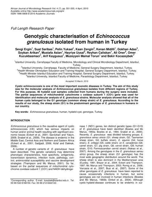
Figure 1. PCR products <strong>of</strong> some positive cases.<br />
SM: Size marker<br />
Figure 2. Some <strong>of</strong> the study strains and prototype strains (CO1 partial sequences).<br />
Ergin et al. 553
554 Afr. J. Microbiol. Res.<br />
practises and adequacy <strong>of</strong> the slaughtering facilities.<br />
Although, it has been tougth that CE considerable<br />
impacts on both human and animal health in Turkey, and<br />
any nation wide preventive programs have not been<br />
implemented yet (Vural et al., 2008). The production and<br />
<strong>of</strong>fal losses in animals have been calculated as 12-13%<br />
and 1.5-3.2% respectively (Umur, 2003; Sarıözkan and<br />
Yalçın, 2009).<br />
Genotype <strong>of</strong> infecting strain affects the fertility rate <strong>of</strong><br />
the cysts in the intermediate hosts and thereby the<br />
infectivity <strong>of</strong> strain for the subsequent hosts. In addition,<br />
the size and localisation <strong>of</strong> the cysts may be different<br />
among the genotypes which may be relevant regarding<br />
examination <strong>of</strong> the organs with naked eye for the cysts.<br />
Also the prepatent periods may vary between genotypes<br />
which can be important for the programs based on<br />
regular antiparasitic treatment <strong>of</strong> the terminal host, before<br />
the parasite produces eggs (Thompson and Lymbery,<br />
1988). The genotypes are also important regarding the<br />
host specificity and life cycle <strong>of</strong> the E. <strong>granulosus</strong><br />
(Bowles and Mc Manus, 1993b; Dinkel et al., 2004).<br />
The occurrance and host preference <strong>of</strong> the sheep strain<br />
(G1 genotype) <strong>of</strong> E. <strong>granulosus</strong> in different countries<br />
have been shown by previous molecular epidemiologic<br />
studies based on mitochondrial gene sequences (Bowles<br />
et al., 1992). Although, G1 genotype strains may also<br />
infect other intermediate host such as cattle, goats, dog,<br />
fertility rate <strong>of</strong> the cysts in these animals low is or it<br />
dosen’t produce fertile cysts at all (Eckert and Thompson,<br />
1997; Mwambate et al., 2004). To our knowledge this is<br />
the most comprehensive genotyping study in humans<br />
performed in Turkey and involve 46 cases originating<br />
from all geographic regions, except Aegean region. In<br />
this study, all <strong>of</strong> 46 strains were G1 genotype (sheep<br />
strain) based on the partial sequences <strong>of</strong> mitochondrial<br />
cytochrome c oxidase subunit 1 (CO1).<br />
In a study conducted by Snabel et al. (2009) in Aegean<br />
region which is not included in our study, five <strong>of</strong> ten<br />
human hydatid cyst samples were G1 genotype and only<br />
one was G7 genotype. In the same study also sheep cyst<br />
samples were genotyped as G1, G3 and G7 Utuk et al.<br />
(2008) investigated 179 sheep, 19 cattle, 7 goat, 1 camel,<br />
1 dog and 1 human cyst and including the human strain<br />
all were G1 genotype. Among animal cysts investigated<br />
Vural et al. (2008) again G1 genotype was dominant<br />
genotype.<br />
From many countries in the Mediterranean area<br />
including our country, G1 genotype has been reported as<br />
the most prevalent genotype (Mwambete et al., 2004;<br />
M’rad et al., 2005; Bart et al., 2006a; Schneider et al.,<br />
2008) in both <strong>of</strong> human and animals. Based on the<br />
results <strong>of</strong> this study and other studies reported from our<br />
country and Mediterranean area it can not be suggested<br />
that G1 genotype is predominant genotype both in human<br />
and animals. This finding should be taken into account<br />
during the planing and implementing E. <strong>granulosus</strong><br />
control programs.<br />
ACKNOWLEDGMENT<br />
This study was supported by the Research Fund <strong>of</strong> the<br />
Istanbul University with the Project No. 2893.<br />
REFERENCES<br />
Ahmadi N, Dalimi A (2006). Characterization <strong>of</strong> <strong>Echinococcus</strong><br />
<strong>granulosus</strong> isolates from human, sheep and camel in Iran. Inf. Gene.<br />
Evol. 6: 85-90.<br />
Altınta N (2003). Past to present: echinococcosis in Turkey. Acta.<br />
Trop. 85: 105-112.<br />
Altınta N (2008). Zoonotic parasitic diseases in Turkey. Vet. Ital. 44:<br />
633-646.<br />
Azlaf A, Dakkak A (2006). Epidemiological study <strong>of</strong> the cystic<br />
echinococcosis in Marocco. Vet. Parasitol. 137: 83-93.<br />
Bart JM, Morariu S, Knapp J, Ilie MS, Pitulescu M, Anghel A,<br />
Cosoroaba I, Piarroux R (2006a). Genetic typing <strong>of</strong> <strong>Echinococcus</strong><br />
<strong>granulosus</strong> in Romania. Parasitol. Res. 98: 130-137.<br />
Bart JM, Abdukader M, Zhang YL, Lın RY, Wang YH, Nakao M, Ito A,<br />
Craig S, Piarroux R, Vuitton DA, Wen H (2006b). Genotyping <strong>of</strong><br />
human cystic echinococcosis in Xinjiang, PR China. Parasitology<br />
133: 571-579.<br />
Bowles J, Blair D, Mc Manus DP (1992). Genetic variants within the<br />
genus <strong>Echinococcus</strong> identified by mitochondrial DNA sequencing.<br />
Mole. Biochem. Parasitol. 54 : 165-174.<br />
Bowles J, Mc Manus DP (1993a). NADH dehydrogenase I gene<br />
sequences compared for species and strains <strong>of</strong> the genus<br />
<strong>Echinococcus</strong>. Int. J. Parasitol. 23: 969-972.<br />
Bowles J, Mc Manus DP (1993b). Molecular variation in <strong>Echinococcus</strong>.<br />
Acta. Trop. 53: 291-305.<br />
Bowles J, Blair D, Mc Manus DP (1994). Molecular genetic<br />
<strong>characterisation</strong> <strong>of</strong> the cervid strain (‘nothern form’) <strong>of</strong> <strong>Echinococcus</strong><br />
<strong>granulosus</strong>. Parasitology. 109: 215-221.<br />
Breyer I, Georgieva D, Kurdova R, Gottstein R (2004). <strong>Echinococcus</strong><br />
<strong>granulosus</strong> strain typing in Bulgaria: the G1 genotype is predominant<br />
in intermediate and definitive wild hosts. Parasitol. Res. 93: 127-130.<br />
Busi M, Snabel V, Vercasia A, Garippa G, Perrone V, De Liberato C,<br />
D’Amelio S (2007). Genetic variation within and between G1 and G3<br />
genotypes <strong>of</strong> <strong>Echinococcus</strong> <strong>granulosus</strong> in Italy revealed by multilocus<br />
DNA sequencing. Vet. Parasitol. 150: 75-83.<br />
Dinkel A, Njorage EM, Zimmermann A, Walz M, Zeyhle E, Elhamdi IE,<br />
Mackenstedt U, Roming T (2004). A PCR systems for detection <strong>of</strong><br />
species and genotypes <strong>of</strong> the <strong>Echinococcus</strong> <strong>granulosus</strong> complex,<br />
with reference to the epidemiological situation in eastern Africa. Int. J.<br />
Parasitol. 34(5): 645-653.<br />
Eckert J, Thompson RC (1997). Intraspesific variation <strong>of</strong> <strong>Echinococcus</strong><br />
<strong>granulosus</strong> and related species with emphasis on their infectivity to<br />
humans. Acta. Trop. 64: 19-34.<br />
Eckert J, Schantz PM, Gasser RB, Torgerson PR, Bessonov AS,<br />
Movsessian SO (2001). Geographic distribution and prevalence. In:<br />
Eckert J, Gemmell MA, Meslin FX, Pawlowski ZS (eds) WHO/OIE<br />
Manual on Echinococcosis in Human and animals: A public health<br />
problem <strong>of</strong> global concern, Paris: World Organisation for Animal<br />
Health; pp. 100-142.<br />
Euzeby J (1991). The epidemiology <strong>of</strong> hydatidosis with special<br />
reference to Mediterranean area. Parassitologia 33: 25-39.<br />
Hall TA (1999). Bioedit: a user-friendly biological sequence alignment<br />
editor and analysis program for Windows and 95/98/NT, Nucleic<br />
Acids Symposium, pp. 95-8.<br />
Li T, Ito A, Nakaya K, Qiu J, Nakao M, Zhen R, Xiao N, Chen X,<br />
Giraudoux P, Craig S (2008). Species identification <strong>of</strong> human<br />
echinococcosis using histopathology and genotyping in northwestern<br />
China. Trans. R. Soc. Trop. Med. Hyg. 102(6): 585-590.<br />
Mc Manus DP, Thompson RC (2003). Molecular epidemiology <strong>of</strong> cystic<br />
echinococcosis. Parasitology 127: 37-51.<br />
M’rad S, Filisetti D, Oudni M, Mekki M, Belguith M, Nouri A, Sayadi T,<br />
Lahmar S, Candolfi E, Azaiez R, Mezhoud H, Babba H (2005). Molecular<br />
evidence <strong>of</strong> ovine (G1) and camel (G6) strains <strong>of</strong> <strong>Echinococcus</strong>
<strong>granulosus</strong> in Tunisia and putative role <strong>of</strong> cattle in human<br />
contamination. Vet. Parasitol. 129: 267-272.<br />
Mwambete KD, Ponce-Gordo F, Cuesta-Bandera C (2004). Genetic<br />
identification and host range <strong>of</strong> the Spanish strains <strong>of</strong> <strong>Echinococcus</strong><br />
<strong>granulosus</strong>. Acta. Trop. 91: 87-93.<br />
Nakao M, Mc Manus DP, Schantz PM, Craig PS, Ito A (2007). A<br />
molecular phylogeny <strong>of</strong> the genus <strong>Echinococcus</strong> inferred from<br />
complete mitochondrial genomes. Parasitology 134: 713-722.<br />
Romig T, Dinkel A, Mackenstedt U (2006). The present situation <strong>of</strong><br />
echinococcosis in Europe. Parasitol. Int. 55: 187-191.<br />
Sadjjadi SM (2006). Present situation <strong>of</strong> echinococcosis in the Middle<br />
East and Arabic North Africa. Parasitol. Int. 55: 197-202.<br />
Sarıözkan S, Yalçın C (2009). Estimating the production losses due to<br />
cystic echinococcosis in ruminants in Turkey. Vet. Parasitol. 163:<br />
330-334.<br />
Schneider R, Gollackner B, Edel B, Schmid K, Wrba F, Tucek G,<br />
Walochnik J, Auer H (2008). Development <strong>of</strong> a new PCR protocol for<br />
the detection <strong>of</strong> species and genotypes (strains) <strong>of</strong> <strong>Echinococcus</strong> in<br />
formalin-fixed, parafin-embedded tissues. Int. J. Parasitol. 38: 1065-<br />
1071.<br />
Snabel V, D’Amelio S, Mathiopoulos K, Turcekova L, Dubinsky P<br />
(2000). Molecular evidence for the presence <strong>of</strong> a G7 genotype <strong>of</strong><br />
<strong>Echinococcus</strong> <strong>granulosus</strong> in Slovakia. J. Helminthol. 74: 177-181.<br />
Snabel V, Altınta N, D’Amelio S, Nakao M, Romig T, Yolası maz A,<br />
Güne K, Türk M, Busi M, Hüttner M, Sevcova D, Ito A, Dubinsky P<br />
(2009). Cystic echinococcosis in Turkey: genetic variability and first<br />
record <strong>of</strong> the pig strain (G7) in the country. Parasitol. Res. 105: 145-<br />
154.<br />
Thompson RCA, Lymbery AJ (1988). The nature, extent and<br />
significance <strong>of</strong> variation the genus <strong>Echinococcus</strong>. Adv. Parasitol. 27:<br />
209-258.<br />
Thompson RCA, Mc Manus DP (2001). Aetiology: parasites and life<br />
cycles. In: Eckert J, Gemmell MA, Meslin FX, Pawlowski ZS (eds)<br />
WHO/OIE Manual on Echinococcosis in Human and animals: A<br />
public health problem <strong>of</strong> global concern, Paris: World Organisation for<br />
Animal Health; pp. 1-19.<br />
Ergin et al. 555<br />
Uluta Esatgil M, Tüzer E (2007). Prevalence <strong>of</strong> hydatidosis in<br />
slaughtered animals in Thrace Turkey. Acta. Parasitol. Turcica. 31:<br />
41-45.<br />
Umur (2003). Prevalence and economic importance <strong>of</strong> cystic<br />
echinococcosis in slaughtered ruminants in Burdur, Turkey. J. Vet.<br />
Med. 50: 247-252.<br />
Utuk AE, Simsek S, Koroglu E, Mc Manus DP (2008). Molecular genetic<br />
characterization <strong>of</strong> different isolates <strong>of</strong> <strong>Echinococcus</strong> <strong>granulosus</strong> in<br />
east and southeast regions <strong>of</strong> Turkey. Acta. Trop. 107: 192-194.<br />
Varcasia A, Canu S, Lightowlers MW, Scala A, Garippa G (2006).<br />
Molecular characterization <strong>of</strong> <strong>Echinococcus</strong> <strong>granulosus</strong> strains in<br />
Sardinia. Parasitol. Res. 98: 273-277.<br />
Varcasia A, Canu S, Kogkos A, Pipia AP, Scala A, Garippa G, Seimenis<br />
A (2007). Preliminary data on diffusion and molecular<br />
characterization <strong>of</strong> cystic echinococcosis in small ruminants in<br />
Peloponnesus, Greece. Parasitol. Res. 101: 1135-1139.<br />
Vural G, Unsal Baca A, Gauci CG, Bagcı O, Gicik Y, Lightowlers MW<br />
(2008). Variability in the <strong>Echinococcus</strong> <strong>granulosus</strong> cytochrome C<br />
oxidase 1 mitochondrial gene sequnce from livestok in Turkey and a<br />
re-appraisal <strong>of</strong> the G1-3 genotype cluster. Vet. Parasitol. 154: 347-<br />
350.<br />
Yolası maz A, Reiterova K, Türk M, Reyhan E, Bozda AD, Karabab<br />
AO, Altınta N (2006). Comparison <strong>of</strong> serological and clinical findings<br />
in Turkish patients with cystic echinococcosis. Helminthol. 43: 220-<br />
225.


