
Create successful ePaper yourself
Turn your PDF publications into a flip-book with our unique Google optimized e-Paper software.
Ryuta Nakae, MD*<br />
Hiroyuki Yokota, MD, PhD*<br />
Daizo Yoshida, MD, PhD‡<br />
Akira Teramoto, MD, PhD‡<br />
Departments of *Emergency and Critical<br />
Care Medic<strong>in</strong>e and ‡Neurosurgery, Nippon<br />
Medical School, Tokyo, Japan<br />
Correspondence:<br />
Ryuta Nakae, MD,<br />
Department of Emergency and Critical<br />
Care Medic<strong>in</strong>e,<br />
Nippon Medical School,<br />
1-1-5, Sendagi, Bunkyo-ku,<br />
Tokyo 113-8603, Japan.<br />
E-mail: nakae@nms.ac.jp<br />
Received, August 28, 2010.<br />
Accepted, March 31, 2011.<br />
Published Onl<strong>in</strong>e, May 6, 2011.<br />
Copyright ª 2011 by the<br />
Congress of Neurological Surgeons<br />
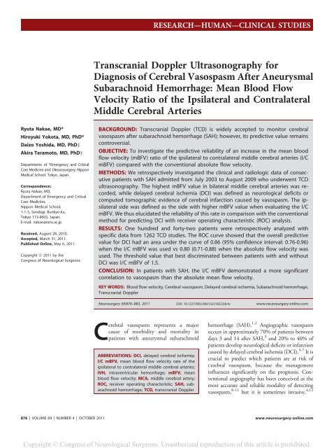
Transcranial Doppler Ultrasonography for<br />
Diagnosis of Cerebral Vasospasm After Aneurysmal<br />
Subarachnoid Hemorrhage: Mean Blood Flow<br />
Velocity Ratio of the Ipsilateral and Contralateral<br />
Middle Cerebral Arteries<br />
BACKGROUND: Transcranial Doppler (<strong>TCD</strong>) is widely accepted to monitor cerebral<br />
vasospasm after subarachnoid hemorrhage (<strong>SAH</strong>); however, its predictive value rema<strong>in</strong>s<br />
controversial.<br />
OBJECTIVE: To <strong>in</strong>vestigate the predictive reliability of an <strong>in</strong>crease <strong>in</strong> the mean blood<br />
flow velocity (mBFV) ratio of the ipsilateral to contralateral middle cerebral arteries (I/C<br />
mBFV) compared with the conventional absolute flow velocity.<br />
METHODS: We retrospectively <strong>in</strong>vestigated the cl<strong>in</strong>ical and radiologic data of consecutive<br />
patients with <strong>SAH</strong> admitted from July 2003 to August 2009 who underwent <strong>TCD</strong><br />
ultrasonography. The highest mBFV value <strong>in</strong> bilateral middle cerebral arteries was recorded,<br />
while delayed cerebral ischemia (DCI) was def<strong>in</strong>ed as neurological deficits or<br />
computed tomographic evidence of cerebral <strong>in</strong>farction caused by vasospasm. The ipsilateral<br />
side was def<strong>in</strong>ed as the side with higher mBFV value when evaluat<strong>in</strong>g the I/C<br />
mBFV. We thus elucidated the reliability of this rate <strong>in</strong> comparison with the conventional<br />
method for predict<strong>in</strong>g DCI with receiver operat<strong>in</strong>g characteristic (ROC) analysis.<br />
RESULTS: One hundred and forty-two patients were retrospectively analyzed with<br />
specific data from 1262 <strong>TCD</strong> studies. The ROC curve showed that the overall predictive<br />
value for DCI had an area under the curve of 0.86 (95% confidence <strong>in</strong>terval: 0.76-0.96)<br />
when the I/C mBFV was used vs 0.80 (0.71-0.88) when the absolute flow velocity was<br />
used. The threshold value that best discrim<strong>in</strong>ated between patients with and without<br />
DCI was I/C mBFV of 1.5.<br />
CONCLUSION: In patients with <strong>SAH</strong>, the I/C mBFV demonstrated a more significant<br />
correlation to vasospasm than the absolute mean flow velocity.<br />
KEY WORDS: Blood flow velocity, Cerebral vasospasm, Delayed cerebral ischemia, Subarachnoid hemorrhage,<br />
Transcranial Doppler<br />
Neurosurgery 69:876–883, 2011 DOI: 10.1227/NEU.0b013e318222dc4c www.neurosurgery-onl<strong>in</strong>e.com<br />
Cerebral vasospasm represents a major<br />
cause of morbidity and mortality <strong>in</strong><br />
patients with aneurysmal subarachnoid<br />
ABBREVIATIONS: DCI, delayed cerebral ischemia;<br />
I/C mBFV, mean blood flow velocity rate of the<br />
ipsilateral to contralateral middle cerebral arteries;<br />
IVH, <strong>in</strong>traventricular hemorrhage; mBFV, mean<br />
blood flow velocity; MCA, middle cerebral artery;<br />
ROC, receiver operat<strong>in</strong>g characteristic; <strong>SAH</strong>, subarachnoid<br />
hemorrhage; <strong>TCD</strong>, transcranial Doppler<br />
RESEARCH—HUMAN—CLINICAL STUDIES<br />
TOPIC RESEARCH—HUMAN—CLINICAL STUDIES<br />
hemorrhage (<strong>SAH</strong>). 1,2 Angiographic vasospasm<br />
occurs <strong>in</strong> approximately 70% of patients between<br />
days 3 and 14 after <strong>SAH</strong>, 3 and 20% to 40% of<br />
patients develop neurological deficits or <strong>in</strong>farction<br />
caused by delayed cerebral ischemia (DCI). 4-7 It is<br />
crucial to predict which patients are at risk of<br />
Cerebral vasospasm, because the management<br />
<strong>in</strong>fluences significantly on the prognosis. Conventional<br />
angiography has been conceived as the<br />
most accurate and reliable modality of detect<strong>in</strong>g<br />
vasospasm, 8-11 but it is sometimes <strong>in</strong>vasive. 4,12<br />
876 | VOLUME 69 | NUMBER 4 | OCTOBER 2011 www.neurosurgery-onl<strong>in</strong>e.com<br />
Copyright © Congress of Neurological Surgeons. Unauthorized reproduction of this article is prohibited.
Induction of transcranial Doppler (<strong>TCD</strong>) ultrasonography <strong>in</strong><br />
1982 by Aaslid et al 13 allowed non<strong>in</strong>vasive detection of cerebral<br />
vasospasm, monitor<strong>in</strong>g of the onset, and resolution of this condition.<br />
14-17 Several studies have shown that the severity of cerebral<br />
vasospasm and the cl<strong>in</strong>ical onset of delayed ischemic deficits cannot<br />
be accurately detected only by measur<strong>in</strong>g the absolute flow velocities<br />
with <strong>TCD</strong>. A mean blood flow velocity (mBFV) of more<br />
than 120 cm/s on <strong>TCD</strong> shows approximately 80% sensitivity and<br />
specificity for the presence of angiographic vasospasm <strong>in</strong> the<br />
proximal middle cerebral artery (MCA). 18 Meanwhile, <strong>TCD</strong> is<br />
usually not beneficial to detect spasm aris<strong>in</strong>g <strong>in</strong> the distal vessels.<br />
Previous studies have discussed the limited reliability of <strong>TCD</strong> to<br />
predict symptomatic vasospasm. An elevated <strong>TCD</strong> flow velocity<br />
<strong>in</strong>duces only a small <strong>in</strong>crease of the risk of DCI after <strong>SAH</strong>. 19<br />
This is the case depend<strong>in</strong>g on which methodology, such as specific<br />
blood flow velocity thresholds, 11,20-22 the absolute <strong>in</strong>crease of<br />
the mBFV, 23-25 or an <strong>in</strong>creased L<strong>in</strong>degaard ratio 21,26 (mBFV of the<br />
MCA divided by that of the cervical <strong>in</strong>ternal carotid artery) is<br />
employed to identify patients at risk.<br />
<strong>TCD</strong>, however, rema<strong>in</strong>s the most widely exam<strong>in</strong>ed imag<strong>in</strong>g<br />
modality for diagnos<strong>in</strong>g cerebral vasospasm. The sensitivity and<br />
predictive value of <strong>TCD</strong> are limited, and improved methods for<br />
identify<strong>in</strong>g patients at high risk for DCI after <strong>SAH</strong> are still<br />
needed. The purpose of the present study was to improve the<br />
accuracy of <strong>TCD</strong> for diagnosis of cerebral vasospasm after <strong>SAH</strong><br />
by elucidat<strong>in</strong>g the prognostic value of the mBFV of the ipsilateral<br />
to contralateral MCA (I/C mBFV). Additionally, we also<br />
analyzed cl<strong>in</strong>ical variables that could play critical roles <strong>in</strong> the onset<br />
of cerebral vasospasm.<br />
METHODS<br />
Patients’ Eligibility<br />
We reviewed the cl<strong>in</strong>ical and radiological <strong>in</strong>formation of all patients<br />
admitted to the Department of Emergency and Critical Care Medic<strong>in</strong>e at<br />
Nippon Medical School (Tokyo, Japan) with spontaneous <strong>SAH</strong> from<br />
July 2003 to August 2009. An <strong>in</strong>formed consent for this study was<br />
obta<strong>in</strong>ed from the patient or a surrogate, and it was approved by the local<br />
<strong>in</strong>stitutional review board. The diagnosis of <strong>SAH</strong> was established from<br />
the f<strong>in</strong>d<strong>in</strong>gs on admission computed tomography (CT) scans or magnetic<br />
resonance images, or by xanthochromia of the cerebrosp<strong>in</strong>al fluid<br />
while CT was nondiagnostic.<br />
Patients eligible <strong>in</strong> the present study were treated by microsurgical<br />
clipp<strong>in</strong>g or endovascular coil<strong>in</strong>g. Their first <strong>TCD</strong> exam<strong>in</strong>ations were<br />
performed on or before day 3 (day 0 <strong>in</strong>dicates the calendar day of the<br />
bleed<strong>in</strong>g event). Exclusion criteria <strong>in</strong>cluded nonaneurysmal <strong>SAH</strong> (eg,<br />
due to trauma, ruptured arteriovenous malformation, ruptured mycotic<br />
aneurysm, vasculitis, or cryptogenic causes), cardiopulmonary arrest<br />
before or on arrival at hospital, death by day 14, treatment by trapp<strong>in</strong>g<br />
and/or bypass, #7 <strong>TCD</strong> studies up to <strong>SAH</strong> day 14, lack of adequate<br />
cranial <strong>TCD</strong> w<strong>in</strong>dows, and <strong>in</strong>complete medical or radiological records.<br />
F<strong>in</strong>ally, of 397 patients with aneurysmal <strong>SAH</strong> who were managed from<br />
July 2003 to August 2009, 54 were excluded because of cardiopulmonary<br />
arrest on or before arrival at hospital, 83 were excluded because of<br />
death by day 14, and 5 were excluded because they received trapp<strong>in</strong>g<br />
TRANSCRANIAL DOPPLER IN SUBARACHNOID HEMORRHAGE<br />
and/or bypass. Subsequently, 82 were elim<strong>in</strong>ated because <strong>in</strong>itial <strong>TCD</strong><br />
was performed after <strong>SAH</strong> day 3 and/or <strong>TCD</strong> was done ,8 times up to<br />
<strong>SAH</strong> day 14, and 21 were excluded because of <strong>in</strong>adequate w<strong>in</strong>dows. We<br />
excluded 10 by <strong>in</strong>complete medical or radiological records. One hundred<br />
forty-two patients were <strong>in</strong>cluded: 57 men (40.1%) and 85 women<br />
(59.9%) with a mean age of 62.1 6 12.2 years. Aneurysms were treated<br />
by surgical clipp<strong>in</strong>g, except <strong>in</strong> 5 patients who underwent embolization<br />
with coils. All of the available medical or radiological records were<br />
reviewed and <strong>in</strong>formation was exclusively collected about factors that are<br />
known or thought to be important key factors <strong>in</strong> vasospasm, the age, sex,<br />
history of hypertension, smok<strong>in</strong>g status, admission Glasgow Coma Scale<br />
score, 27 Hunt and Hess grade, 28 systolic and diastolic blood pressures on<br />
admission, and body temperature. The <strong>in</strong>itial laboratory workup was<br />
performed <strong>in</strong> all patients after admission, <strong>in</strong>tegrat<strong>in</strong>g blood cell count,<br />
hemoglob<strong>in</strong>, hematocrit, sodium, potassium, blood glucose, and arterial<br />
blood gases. Admission head CT scans were <strong>in</strong>dependently evaluated by<br />
study neuro<strong>in</strong>tensivists, and the amount of blood clot <strong>in</strong> the subarachnoid<br />
space was classified accord<strong>in</strong>g to the modified Fisher scale. 29<br />
They also determ<strong>in</strong>ed the <strong>SAH</strong> sum score 30 based on the amount and<br />
location of subarachnoid blood, the <strong>in</strong>traventricular hemorrhage (IVH)<br />
sum score 30 based on the amount and location of <strong>in</strong>traventricular blood,<br />
the presence of <strong>in</strong>tracerebral hematoma, and the presence of hydrocephalus.<br />
The <strong>SAH</strong> sum score was calculated as the total of scores from<br />
0 to 3 assigned for each of 10 cisterns or fissures. A score of 0 <strong>in</strong>dicated<br />
no blood, 1 meant that blood was barely visible, 2 meant an <strong>in</strong>termediate<br />
amount of blood, and 3 was assigned when the cistern/fissure was<br />
completely filled with blood. Judgment was based on the extent and<br />
<strong>in</strong>tensity of density changes. The 10 cisterns/fissures <strong>in</strong>vestigated were<br />
the frontal <strong>in</strong>terhemispheric fissure, the quadrigem<strong>in</strong>al cistern, both<br />
suprasellar cisterns, both ambient cisterns, both basal Sylvian fissures,<br />
and both lateral Sylvian fissures. The IVH sum score was calculated as<br />
the total of the separate scores for each of the 4 ventricles, ie, both lateral<br />
ventricles, the third ventricle, and the fourth ventricle. Scores were<br />
assigned as: 0, no blood; 1, sedimentation of erythrocytes <strong>in</strong> the posterior<br />
part; 2, ventricle partly filled with blood; and 3, ventricle completely<br />
filled with blood. Angiography or 3-dimensional CT angiography was<br />
performed on admission to locate the aneurysm. We recorded the<br />
location and size of each aneurysm, as well as the treatment (microsurgical<br />
clipp<strong>in</strong>g or endovascular coil<strong>in</strong>g). Cl<strong>in</strong>ical evaluation was done serially<br />
throughout the day (at least every 2 hours) by the staff of the <strong>in</strong>tensive care<br />
unit. Additional CT or MRI was performed more than once a week and for<br />
every major event. Patient outcome was scored accord<strong>in</strong>g to Glasgow<br />
Outcome Scale score 31 at the end of their admission.<br />
Cl<strong>in</strong>ical Management<br />
Patients received neuro<strong>in</strong>tensive care to stabilize and regulate their<br />
cardiopulmonary function, fluid balance, arterial blood pressure, <strong>in</strong>tracranial<br />
pressure, serum glucose, and arterial blood gases. Before the<br />
aneurysm had been treated, the systolic blood pressure was ma<strong>in</strong>ta<strong>in</strong>ed at<br />
#160 mm Hg <strong>in</strong> most cases. If necessary, the patient was sedated and<br />
ventilated. Triple-H therapy (hypertension, hypervolemia, and hemodilution<br />
therapy) 32 was rout<strong>in</strong>ely performed after treatment of the<br />
aneurysm, <strong>in</strong>volv<strong>in</strong>g a target central venous pressure of more than 8 mm<br />
Hg, <strong>in</strong>duction of hypertension with dopam<strong>in</strong>e to ma<strong>in</strong>ta<strong>in</strong> a systolic<br />
blood pressure of 140 to 160 mm Hg, and ma<strong>in</strong>tenance of the cardiac<br />
<strong>in</strong>dex at 3.5 L/m<strong>in</strong>/m 2 or more by <strong>in</strong>fusion of dopam<strong>in</strong>e or dobutam<strong>in</strong>e<br />
as needed. After April 2009, oral stat<strong>in</strong> therapy (fluvastat<strong>in</strong> sodium: 30<br />
mg/d) was rout<strong>in</strong>ely started with<strong>in</strong> 72 hours of the event. Intracranial<br />
hypertension and acute symptomatic <strong>in</strong>tracranial mass effect were treated<br />
NEUROSURGERY VOLUME 69 | NUMBER 4 | OCTOBER 2011 | 877<br />
Copyright © Congress of Neurological Surgeons. Unauthorized reproduction of this article is prohibited.
NAKAE ET AL<br />
with repeated boluses of 10% glycerol (0.4-0.6 g/kg). Persistent fever<br />
(temperature exceed<strong>in</strong>g 38.5°C) was treated with nonsteroidal anti-<strong>in</strong>flammatory<br />
drugs and surface cool<strong>in</strong>g. To ma<strong>in</strong>ta<strong>in</strong> the hemoglob<strong>in</strong> at<br />
$8 mg/dL we gave blood transfusions to such patients. Angiography was<br />
rout<strong>in</strong>ely performed <strong>in</strong> patients with DCI, and the location of angiographic<br />
vasospasm was recorded. Endovascular treatment of vasospasm<br />
entailed either <strong>in</strong>tra-arterial fasudil hydrochloride hydrate or angioplasty.<br />
The treat<strong>in</strong>g consultants made decisions for each endovascular<br />
<strong>in</strong>tervention.<br />
Def<strong>in</strong>ition of DCI<br />
DCI was def<strong>in</strong>ed as a new hypodensity on CT scans located <strong>in</strong><br />
a vascular territory and/or associated symptoms, <strong>in</strong>clud<strong>in</strong>g a decrease of<br />
consciousness and focal deficits, due to cerebral vasospasm and not<br />
expla<strong>in</strong>ed by other causes (eg, rebleed<strong>in</strong>g, hydrocephalus, cardioembolic<br />
sources of emboli, hypoxia, electrolyte disturbances, or seizures). Patients<br />
who had cerebral <strong>in</strong>farction that was possibly related to complications of<br />
surgery or angiography were excluded.<br />
<strong>TCD</strong> Studies<br />
<strong>TCD</strong> ultrasonography of the left and right MCAs was performed daily<br />
or every other day between <strong>SAH</strong> days 1 and 14 by 2 experienced<br />
technicians. The mBFV was measured through transtemporal w<strong>in</strong>dows<br />
with a 2-MHz hand-held transducer (Intra-View; Rimed, Ltd, Park<br />
Raanana, Israel). The bilateral maximal mBFV and the ratio of the<br />
mBFV <strong>in</strong> the ipsilateral MCA to that <strong>in</strong> the contralateral MCA (I/C<br />
mBFV) were recorded for each <strong>TCD</strong> study. We def<strong>in</strong>ed the ipsilateral<br />
side as the side with a higher mBFV.<br />
Statistical Analysis<br />
Analysis of data was performed with standard statistical software<br />
(Version 16.0; SPSS, Inc, Chicago, Ill<strong>in</strong>ois). To determ<strong>in</strong>e whether there<br />
was a correlation between DCI and categorical variables, the x 2 test or<br />
Fisher exact test was applied, whereas cont<strong>in</strong>uous variables were assessed<br />
with an <strong>in</strong>dependent two-tailed Student t test. For nonnormally distributed<br />
cont<strong>in</strong>uous variables, the Mann-Whitney U test was exam<strong>in</strong>ed.<br />
Receiver operat<strong>in</strong>g characteristic (ROC) curves were drawn, and the area<br />
under the curve (C-statistic) was calculated to assess the overall predictive<br />
value for DCI of the I/C mBFV and the absolute flow velocity. To<br />
evaluate <strong>in</strong>dependent predictors of DCI, significant univariate variables<br />
(P , .05) were <strong>in</strong>cluded <strong>in</strong> a multivariate analysis.<br />
RESULTS<br />
A total of 1262 <strong>TCD</strong> exam<strong>in</strong>ations performed up to <strong>SAH</strong> day<br />
14 were analyzed (a mean of 8.9 per patient). DCI occurred <strong>in</strong> 28<br />
of the 142 patients (19.7%), and the cl<strong>in</strong>ical characteristics of the<br />
groups with or without DCI are compared <strong>in</strong> Tables 1 and 2. The<br />
mean age of the patients with DCI was 57.0 6 10.0 years, which<br />
was significantly younger than the mean age of the patients<br />
without DCI (63.4 6 12.4 y, P = .012). Meanwhile, BFVs on<br />
<strong>TCD</strong> were significantly higher <strong>in</strong> patients who developed DCI vs<br />
those without DCI, except on day 1 and day 3. The mBFV of<br />
patients with DCI <strong>in</strong>creased progressively between <strong>SAH</strong> days 3<br />
and 5 before subsequently show<strong>in</strong>g a decrease, and the condition<br />
did not happen <strong>in</strong> patients without DCI (Figure 1). The mean<br />
mBFV between <strong>SAH</strong> days 1 and 14 of patients with DCI was<br />
TABLE 1. Demographic, Cl<strong>in</strong>ical, and Laboratory Characteristics of<br />
Patients With or Without Delayed Cerebral Ischemia a<br />
Characteristic<br />
Demographic data<br />
No DCI DCI P Value<br />
No. of patients 114 28<br />
Age, y 63.4 6 12.4 57.0 6 10.0 .012<br />
Female, % 70 (61.4) 15 (53.6) .45<br />
History of<br />
hypertension, %<br />
73 (64.0) 15 (53.6) .31<br />
Smok<strong>in</strong>g status, %<br />
Cl<strong>in</strong>ical features<br />
42 (36.8) 11 (39.3) .81<br />
Glasgow Coma Scale 9.8 6 4.4 8.2 6 4.1 .09<br />
Eye component 2.5 6 1.3 2.0 6 1.2 .09<br />
Verbal component 2.8 6 1.7 2.3 6 1.6 .22<br />
Motor component 4.6 6 1.8 3.9 6 1.8 .016<br />
Hunt and Hess grade .44<br />
1-2, % 25 (21.9) 4 (14.3)<br />
3-5, % 89 (78.1) 24 (85.7)<br />
Systolic blood<br />
pressure, mm Hg<br />
171.8 6 36.3 185.6 6 44.5 .19<br />
Diastolic blood<br />
pressure, mm Hg<br />
96.6 6 19.5 108.5 6 18.1 .004<br />
Body temperature, °C<br />
Laboratory f<strong>in</strong>d<strong>in</strong>gs<br />
35.8 6 1.0 36.0 6 1.1 .39<br />
WBC, 310 9 /L 11.5 6 4.2 11.3 6 4.9 .85<br />
Hb, mg/dL 12.9 6 1.8 13.3 6 1.4 .24<br />
Ht, % 38.8 6 5.0 40.0 6 4.1 .27<br />
Sodium, mEq/L 140.3 6 3.2 139.3 6 3.5 .14<br />
Potassium, mEq/L 3.4 6 0.5 3.2 6 0.4 .10<br />
Blood glucose, mg/dL<br />
Arterial blood gases<br />
175.0 6 55.1 187.8 6 40.0 .08<br />
pH 7.39 6 0.06 7.38 6 0.05 .15<br />
pCO2, mm Hg 40.2 6 8.3 37.7 6 6.6 .13<br />
pO2, mm Hg 270.5 6 159.2 273.6 6 142.9 .73<br />
2<br />
HCO3 , mmol/L 23.7 6 2.7 22.9 6 2.2 .19<br />
Base excess, mmol/L 20.6 6 2.5 21.5 6 2.2 .09<br />
Lactate, mg/dL 27.0 6 16.4 28.5 6 15.2 .41<br />
a<br />
DCI, delayed cerebral ischemia; Hb, hemoglob<strong>in</strong>; Ht, hematocrit; WBC, white blood<br />
cell.<br />
103.7 6 22.4 cm/s, which was significantly higher than the mean<br />
mBFV of patients without DCI (77.9 6 25.7 cm/s, P , .001).<br />
Three other cl<strong>in</strong>ical variables were also significantly associated<br />
with DCI, <strong>in</strong>clud<strong>in</strong>g high diastolic blood pressure on admission<br />
(P , .01), a large amount of blood <strong>in</strong> the suprasellar cisterns<br />
(Table 3, P , .05), and performance of decompressive craniectomy<br />
(P , .01). The <strong>SAH</strong> sum score and IVH sum score were not<br />
significantly different between patients with and without DCI<br />
(Tables 3 and 4). Although the associations were not specifically<br />
significant, the presence of <strong>in</strong>tracerebral hematoma (P =.054)was<br />
highly associated with an <strong>in</strong>creased risk of DCI.<br />
The I/C mBFV of 121 patients was available. Accord<strong>in</strong>g to<br />
ROC analysis, the I/C mBFV had a higher detection rate for<br />
patients with DCI compared with the conventional absolute<br />
mBFV (Figure 2). When ROC curves display<strong>in</strong>g the overall<br />
878 | VOLUME 69 | NUMBER 4 | OCTOBER 2011 www.neurosurgery-onl<strong>in</strong>e.com<br />
Copyright © Congress of Neurological Surgeons. Unauthorized reproduction of this article is prohibited.
TABLE 2. Radiological Characteristics, Treatment, and<br />
Transcranial Doppler F<strong>in</strong>d<strong>in</strong>gs of Patients With or Without Delayed<br />
Cerebral Ischemia a<br />
Characteristic<br />
Radiological f<strong>in</strong>d<strong>in</strong>gs<br />
No DCI DCI<br />
P<br />
Value<br />
Modified Fisher grade .61<br />
1 21 (18.4) 3 (10.7)<br />
2 38 (33.3) 9 (32.2)<br />
3 15 (13.2) 6 (21.4)<br />
4 40 (35.1) 10 (35.7)<br />
ICH, % 28 (24.6) 12 (42.9) .054<br />
Hydrocephalus, % 38 (33.3) 9 (32.1) .90<br />
Location of ruptured aneurysm, % .75<br />
ACA 40 (35.1) 11 (39.3)<br />
ICA 29 (25.4) 9 (32.2)<br />
MCA 34 (29.8) 6 (21.4)<br />
Posterior circulation 11 (9.7) 2 (7.1)<br />
Aneurysm size, mm .08<br />
#12 94 (82.5) 25 (89.3)<br />
13–24 18 (15.8) 1 (3.6)<br />
$25<br />
Treatment<br />
2 (1.7) 2 (7.1)<br />
Operation .58<br />
Microsurgical clipp<strong>in</strong>g, % 109 (95.6) 28 (100.0)<br />
Endovascular coil<strong>in</strong>g, % 5 (4.4) 0 (0.0)<br />
Decompressive<br />
craniectomy, %<br />
42 (35.6) 18 (64.3) .008<br />
Stat<strong>in</strong>, %<br />
<strong>TCD</strong> f<strong>in</strong>d<strong>in</strong>gs<br />
23 (20.2) 7 (25.0) .58<br />
mBFV, cm/s 77.9 6 25.7103.7<br />
22.4<br />
6 ,.001<br />
a ACA, anterior cerebral artery; DCI, delayed cerebral ischemia; ICA, <strong>in</strong>ternal carotid<br />
artery; ICH, <strong>in</strong>tracerebral hematoma; mBFV, mean blood flow velocity; MCA, middle<br />
cerebral artery; <strong>TCD</strong>, transcranial Doppler.<br />
diagnostic utility for DCI were drawn, the area under the curve<br />
was 0.86 (95% confidence <strong>in</strong>terval: 0.76-0.96) us<strong>in</strong>g the I/C<br />
mBFV and 0.80 (0.71-0.88) by the conventional method. The<br />
mBFV threshold value that best discrim<strong>in</strong>ated between patients<br />
with and without DCI was 1.5 for the I/C mBFV and 125 cm/s<br />
for the absolute velocity. With the use of these threshold values,<br />
the sensitivity, specificity, and positive predictive of the I/C<br />
mBFV and absolute velocity for predict<strong>in</strong>g DCI was 77.0%,<br />
80.0%, and 51.3% vs 67.9%, 71.9%, and 37.3%, respectively.<br />
Angiography data of 26 patients with DCI <strong>in</strong> whom the I/C<br />
mBFV could be calculated were available (Table 5). In our<br />
analysis, the angiographic vasospasm <strong>in</strong> patients with the I/C<br />
mBFV $1.5 found no significant difference between anatomical<br />
locations. However, most angiographic vasospasm <strong>in</strong> patients<br />
with the I/C mBFV ,1.5 occurred on the anterior cerebral artery<br />
and the artery of the posterior circulation (P = .008).<br />
The prognosis is significantly worse <strong>in</strong> patients with the I/C<br />
mBFV $1.5 compared with those ,1.5 (Table 6, P = .031). In<br />
patients with the I/C mBFV $1.5, the rate of good outcome or<br />
moderate disability was low, but the mortality rate was extremely<br />
high, because 6 of the 39 (15.4%) patients died. In DCI patients,<br />
there was no significant difference <strong>in</strong> Glasgow Outcome Scale<br />
score between those treated with <strong>in</strong>tra-arterial fasudil hydrochloride<br />
hydrate and with angioplasty (Table 7, P = .88).<br />
DISCUSSION<br />
TRANSCRANIAL DOPPLER IN SUBARACHNOID HEMORRHAGE<br />
The present study showed that: (1) DCI occurred <strong>in</strong> 28 of 142<br />
patients with <strong>SAH</strong> (19.7%); (2) predictors of DCI were younger<br />
age, high diastolic blood pressure on admission, large amount of<br />
blood <strong>in</strong> the suprasellar cisterns, and decompressive craniectomy;<br />
and (3) the I/C mBFV was more predictable than the conventional<br />
absolute mBFV for identification of vasospasm accord<strong>in</strong>g<br />
to ROC analysis, and the cutoff value for predict<strong>in</strong>g DCI was an<br />
I/C mBFV of 1.5. The prognosis is significantly worse <strong>in</strong> patients<br />
with I/C mBFV $1.5 compared with those ,1.5.<br />
The most accurate and reliable method of detect<strong>in</strong>g vasospasm<br />
is conventional angiography. 8-11 A meta-analysis was recently<br />
elucidated <strong>in</strong> 26 trials compar<strong>in</strong>g <strong>TCD</strong> with cerebral angiography<br />
<strong>in</strong> patients with <strong>SAH</strong> and concluded that <strong>TCD</strong> of the MCA has<br />
a high specificity (99%) and a high positive predictive value<br />
(97%), but a low sensitivity (67%). This meta-analysis also found<br />
FIGURE 1. Maximal mean blood flow velocity after <strong>SAH</strong> <strong>in</strong> patients with or<br />
without DCI. Velocities were significantly higher <strong>in</strong> patients who developed DCI,<br />
except on days 1 and 3. The mBFV of patients with DCI <strong>in</strong>creased progressively<br />
between days 3 and 5. Values are the mean 6 SE. *P , .05, †P , .01,<br />
‡P , .001. mBFV, mean blood flow velocity; DCI, delayed cerebral ischemia;<br />
<strong>SAH</strong>, subarachnoid hemorrhage.<br />
NEUROSURGERY VOLUME 69 | NUMBER 4 | OCTOBER 2011 | 879<br />
Copyright © Congress of Neurological Surgeons. Unauthorized reproduction of this article is prohibited.
NAKAE ET AL<br />
TABLE 3. Subarachnoid Hemorrhage Score of 10 Cisterns and<br />
Subarachnoid Hemorrhage Sum Score a<br />
No DCI DCI P Value<br />
Interhemispheric fissure 1.32 6 0.74 1.46 6 0.84 .38<br />
Quadrigem<strong>in</strong>al cistern 1.36 6 0.83 1.36 6 0.49 .97<br />
Rt. suprasellar cistern 1.67 6 0.72 2.00 6 0.47 .024<br />
Lt. suprasellar cistern 1.68 6 0.73 2.04 6 0.51 .016<br />
Rt. ambient cistern 1.40 6 0.76 1.43 6 0.50 .96<br />
Lt. ambient cistern 1.40 6 0.77 1.46 6 0.51 .78<br />
Rt. basal sylvian fissure 1.55 6 0.71 1.71 6 0.85 .37<br />
Lt. basal sylvian fissure 1.68 6 0.80 1.64 6 0.83 .84<br />
Rt. lateral sylvian fissure 1.54 6 0.79 1.75 6 0.89 .18<br />
Lt. lateral sylvian fissure 1.69 6 0.80 1.61 6 0.92 .78<br />
<strong>SAH</strong> sum score 15.28 6 5.28 16.46 6 3.56 .40<br />
a DCI, delayed cerebral ischemia; lt., left; rt., right; <strong>SAH</strong>, subarachnoid hemorrhage.<br />
that <strong>TCD</strong> of other arteries showed no evidence of accuracy to<br />
detect vasospasm. 18 In addition, cerebral <strong>in</strong>farction may occur <strong>in</strong><br />
some patients without apparent vasospasm on angiography. The<br />
sensitivity and predictive value of <strong>TCD</strong> are limited, and sophisticated<br />
methods for identify<strong>in</strong>g patients with a high risk for<br />
cerebral vasospasm and DCI after <strong>SAH</strong> are further necessitated.<br />
In the present study, analysis with the I/C mBFV achieved<br />
a higher detection rate of patients who developed DCI after <strong>SAH</strong><br />
than the absolute mBFV accord<strong>in</strong>g to ROC analysis. Lee et al 33<br />
reported that use of <strong>TCD</strong> to predict cerebral <strong>in</strong>farction after <strong>SAH</strong><br />
had a sensitivity of 69.6% and a specificity of 77.1%, while<br />
Suarez et al 6 reported values of 70% and 73%, respectively. In the<br />
current study, <strong>TCD</strong> had a higher sensitivity (77.0%) and specificity<br />
(80.0%) when the I/C mBFV was used, and this rate was<br />
more closely related to cl<strong>in</strong>ically significant vasospasm <strong>in</strong> patients<br />
with aneurysmal <strong>SAH</strong> than absolute flow velocity <strong>in</strong>dices. That is,<br />
to say, we can improve the low sensitivity of <strong>TCD</strong> for DCI.<br />
To exam<strong>in</strong>e a further correlation between <strong>TCD</strong> and angiogram<br />
data, we analyzed correlation between the I/C mBFV and the<br />
location of angiographic vasospasm (Table 5). Our f<strong>in</strong>d<strong>in</strong>gs<br />
highly implicated that, for the MCA and the <strong>in</strong>ternal carotid<br />
TABLE 4. Intraventricular Hemorrhage Score of 4 Ventricles and<br />
Intraventricular Hemorrhage Sum Score a<br />
No DCI DCI P Value<br />
Rt. lateral ventricle 0.86 6 0.86 0.93 6 0.98 .84<br />
Lt. lateral ventricle 0.84 6 0.86 0.93 6 0.81 .51<br />
Third ventricle 0.75 6 1.04 0.79 6 1.07 .86<br />
Fourth ventricle 0.79 6 1.17 0.75 6 1.08 .92<br />
IVH sum score 3.25 6 3.37 3.39 6 3.38 .84<br />
a DCI, delayed cerebral ischemia; IVH, <strong>in</strong>traventricular hemorrhage; lt., left; rt., right.<br />
TABLE 5. Locations of Angiographic Vasospasm <strong>in</strong> Delayed<br />
Cerebral Ischemia Patients a<br />
I/C mBFV<br />
$1.5<br />
I/C mBFV<br />
,1.5 P Value<br />
Locations of vasospasm .008<br />
ICA 2 (10.0) 0 (0)<br />
ACA 1 (5.0) 3 (50.0)<br />
MCA 4 (20.0) 0 (0)<br />
ICA + MCA 2 (10.0) 0 (0)<br />
ACA + MCA 6 (30.0) 0 (0)<br />
CA + ACA + MCA 4 (20.0) 0 (0)<br />
Bilateral ACA + MCA 1 (5.0) 1 (16.7)<br />
Posterior circulation 0 (0) 2 (33.3)<br />
Total 20 6<br />
a ACA, anterior cerebral artery; ICA, <strong>in</strong>ternal carotid artery; I/C mBFV, mean blood<br />
flow velocity rate of the ipsilateral to contralateral middle cerebral arteries; MCA,<br />
middle cerebral artery.<br />
artery, the I/C mBFV tended to show 1.5 and more when<br />
angiography shows cerebral vasospasm. For the other arteries such<br />
as the anterior cerebral artery and the artery of the posterior<br />
circulation, there seemed to be no <strong>in</strong>dication to use the I/C<br />
mBFV to detect vasospasm.<br />
FIGURE 2. Receiver operat<strong>in</strong>g characteristic curves compar<strong>in</strong>g the absolute<br />
mean mBFV and the I/C mBFV for prediction of DCI. The area under the ROC<br />
curve for the I/C mBFV was 0.86 (95% confidence <strong>in</strong>terval: 0.76-0.96), while<br />
that for the absolute mBFV was 0.80 (0.71-0.88). mBFV, mean blood flow<br />
velocity; I/C mBFV, mBFV of the ipsilateral to contralateral middle cerebral<br />
artery; ROC, receiver operat<strong>in</strong>g characteristic; DCI, delayed cerebral ischemia.<br />
880 | VOLUME 69 | NUMBER 4 | OCTOBER 2011 www.neurosurgery-onl<strong>in</strong>e.com<br />
Copyright © Congress of Neurological Surgeons. Unauthorized reproduction of this article is prohibited.
TABLE 6. Glasgow Outcome Scale Scores of the Patients Who Had<br />
the I/C mBFV Not Less Than or Less Than 1.5 a<br />
I/C mBFV<br />
$1.5<br />
I/C mBFV<br />
,1.5 P Value<br />
GOS scores .031<br />
4–5 8 (20.5) 35 (42.7)<br />
2–3 25 (64.1) 42 (51.2)<br />
1 6 (15.4) 5 (6.1)<br />
Total 39 82<br />
a GOS, Glasgow Outcome Scale; GOS score of 1 <strong>in</strong>dicates death; 2, persistent<br />
vegetative state; 3, severe disability; 4, moderate disability; and 5, good recovery;<br />
I/C mBFV, mean blood flow velocity rate of the ipsilateral to contralateral middle<br />
cerebral arteries.<br />
The crucial risk factors to predict DCI after <strong>SAH</strong> were the<br />
presence of thick clots <strong>in</strong> the basal cisterns or blood <strong>in</strong> the<br />
ventricular system. 34 Other possible risk factors for DCI <strong>in</strong>volved<br />
younger age, 35 female sex, 19 cigarette smok<strong>in</strong>g, 36,37 poor cl<strong>in</strong>ical<br />
grade, 19,38 high systolic blood pressure, 19 fever, 38 high glucose<br />
level, 19 poor modified Fisher scale score, 19 large amount of<br />
subarachnoid blood, 37 <strong>in</strong>tracerebral hematoma, 19 and <strong>in</strong>travascular<br />
volume depletion. 39 In the present study, we identified<br />
4 predictors of DCI: young age, high diastolic blood pressure on<br />
admission, large amount of subarachnoid blood at suprasellar<br />
cisterns, and decompressive craniectomy.<br />
A younger age was associated with a significantly higher<br />
<strong>in</strong>cidence of DCI (P = .012). Magge et al 35 <strong>in</strong>vestigated that<br />
<strong>in</strong>creas<strong>in</strong>g stiffness of the cerebral vasculature associated with<br />
advanc<strong>in</strong>g age may expla<strong>in</strong> the lower <strong>in</strong>cidence of angiographic<br />
vasospasm <strong>in</strong> the elderly, and they suggested that older patients<br />
may require less aggressive prophylactic treatment for vasospasm.<br />
A relationship between vasospasm and the modified Fisher grade,<br />
<strong>SAH</strong> sum score, or IVH sum score has been reported. 29,30 Our<br />
study showed that the volume of <strong>SAH</strong> or IVH was not a significant<br />
TABLE 7. Glasgow Outcome Scale Scores of the Patients With<br />
Delayed Cerebral Ischemia Who Were Treated With Intra-arterial<br />
Fasudil Hydrochloride Hydrate or With Angioplasty a<br />
Intra-arterial Fasudil<br />
Hydrochloride<br />
Hydrate Angioplasty P Value<br />
GOS scores .88<br />
4–5 5 (20.8) 1 (25.0)<br />
2–3 15 (62.5) 2 (50.0)<br />
1 4 (16.7) 1 (25.0)<br />
Total 24 4<br />
a GOS, Glasgow Outcome Scale; GOS score of 1 <strong>in</strong>dicates death; 2, persistent<br />
vegetative state; 3, severe disability; 4, moderate disability; and 5, good recovery.<br />
TRANSCRANIAL DOPPLER IN SUBARACHNOID HEMORRHAGE<br />
predictor of vasospasm; meanwhile, a large amount of subarachnoid<br />
blood <strong>in</strong> the suprasellar cisterns was significantly related to DCI<br />
(P , .05). Moreover, we found that the diastolic blood pressure on<br />
admission was significantly higher <strong>in</strong> patients who developed DCI<br />
than <strong>in</strong> those who did not (P , .01). Rosen et al 40 also reported<br />
that the admission diastolic blood pressure is one of the risk factors<br />
for a large <strong>SAH</strong> volume. Our f<strong>in</strong>d<strong>in</strong>gs were consistent with this<br />
report (Figure 3). Furthermore, we found that hematoma <strong>in</strong> the<br />
suprasellar cisterns is most closely related to vasospasm.<br />
Decompressive craniectomy has been shown to improve outcomes<br />
<strong>in</strong> patients experienc<strong>in</strong>g massive ischemic <strong>in</strong>farction and<br />
severe head trauma; however, the role of craniectomy <strong>in</strong> aneurysmal<br />
<strong>SAH</strong> is less well def<strong>in</strong>ed. 41 We performed decompressive<br />
craniectomy <strong>in</strong> <strong>SAH</strong> patients who had large <strong>in</strong>tracerebral hematomas<br />
and/or Sylvian fissure hematomas to reduce the <strong>in</strong>tracranial<br />
pressure or to elevate the perfusion pressure. In our series,<br />
18 (64.3%) of 28 patients with DCI underwent decompressive<br />
craniectomy, and this percentage was significantly higher than<br />
for patients without DCI (P , .01). In addition, although the<br />
correlation was not significant, an <strong>in</strong>crease of DCI tended to be<br />
along with <strong>in</strong>tracerebral hematoma (P =.054).Wespeculatethat<br />
elevated <strong>in</strong>tracranial pressure may also be a risk factor of DCI.<br />
We did not f<strong>in</strong>d any correlation between the neurological<br />
status (Hunt and Hess grade or Glasgow Coma Scale) and the<br />
development of DCI, contrary to a previous report. Among the<br />
142 patients who met all of the <strong>in</strong>clusion criteria, 29 patients<br />
(20.4%) were <strong>in</strong> Hunt and Hess grade 1 or 2 and 113 patients<br />
(79.6%) were <strong>in</strong> grades 3-5 (Table 1). We consider that this<br />
f<strong>in</strong>d<strong>in</strong>g may depend on the higher proportion of poor grade<br />
patients than <strong>in</strong> previous reports. 19,38 This difference presumably<br />
can arise simply because our Department of Emergency and<br />
Critical Care Medic<strong>in</strong>e particularly handles severe cases.<br />
FIGURE 3. Relation between the diastolic blood pressure on admission and the<br />
<strong>SAH</strong> sum score. <strong>SAH</strong>, subarachnoid hemorrhage.<br />
NEUROSURGERY VOLUME 69 | NUMBER 4 | OCTOBER 2011 | 881<br />
Copyright © Congress of Neurological Surgeons. Unauthorized reproduction of this article is prohibited.
NAKAE ET AL<br />
Limitations<br />
There were 21 patients <strong>in</strong> whom <strong>TCD</strong> was only performed on<br />
one side, so that the I/C mBFV could not be evaluated. If craniotomy<br />
has been performed, <strong>TCD</strong> is easy to do through the burr<br />
hole. However, the contralateral side does not have a burr hole<br />
and <strong>TCD</strong> exam<strong>in</strong>ation becomes difficult.<br />
CONCLUSION<br />
Our study demonstrated that the I/C mBFV can be a reliable<br />
predictor of cl<strong>in</strong>ically significant/symptomatic vasospasm <strong>in</strong><br />
patients with aneurysmal <strong>SAH</strong> compared with the absolute flow<br />
velocity. Evaluation of the I/C mBFV and the comb<strong>in</strong>ation of<br />
the I/C mBFV and the absolute flow velocity can improve the<br />
diagnostic accuracy of <strong>TCD</strong> to detect vasospasm, but further<br />
studies should be conducted to expand our f<strong>in</strong>d<strong>in</strong>gs.<br />
Disclosure<br />
The authors have no personal f<strong>in</strong>ancial or <strong>in</strong>stitutional <strong>in</strong>terest <strong>in</strong> any of the<br />
drugs, materials, or devices described <strong>in</strong> this article.<br />
REFERENCES<br />
1. Dorsch NW. Therapeutic approaches to vasospasm <strong>in</strong> subarachnoid hemorrhage.<br />
Curr Op<strong>in</strong> Crit Care. 2002;8(2):128-133.<br />
2. Kassell NF, Torner JC, Jane JA, Haley EC Jr, Adams HP. The International<br />
Cooperative Study on the Tim<strong>in</strong>g of Aneurysm Surgery. Part 2: Surgical results.<br />
J Neurosurg. 1990;73(1):37-47.<br />
3. Sarrafzadeh A, Haux D, Sakowitz O, et al. Acute focal neurological deficits <strong>in</strong> aneurysmal<br />
subarachnoid hemorrhage: relation of cl<strong>in</strong>ical course, CT f<strong>in</strong>d<strong>in</strong>gs, and metabolite<br />
abnormalities monitored with bedside microdialysis. Stroke. 2003;34(6):1382-1388.<br />
4. Charpentier C, Audibert G, Guillem<strong>in</strong> F, et al. Multivariate analysis of predictors<br />
of cerebral vasospasm occurrence after aneurysmal subarachnoid hemorrhage.<br />
Stroke. 1999;30(7):1402-1408.<br />
5. Rab<strong>in</strong>ste<strong>in</strong> AA, Friedman JA, Weigand SD, et al. Predictors of cerebral <strong>in</strong>farction<br />
<strong>in</strong> aneurysmal subarachnoid hemorrhage. Stroke. 2004;35(8):1862-1866.<br />
6. Suarez JI, Qureshi AI, Yahia AB, et al. Symptomatic vasospasm diagnosis after<br />
subarachnoid hemorrhage: evaluation of transcranial Doppler ultrasound and<br />
cerebral angiography as related to compromised vascular distribution. Crit Care<br />
Med. 2002;30(6):1348-1355.<br />
7. van Gijn J, Kerr RS, R<strong>in</strong>kel GJ. Subarachnoid haemorrhage. Lancet. 2007;<br />
369(9558):306-318.<br />
8. Burch CM, Wozniak MA, Sloan MA, et al. Detection of <strong>in</strong>tracranial <strong>in</strong>ternal<br />
carotid artery and middle cerebral artery vasospasm follow<strong>in</strong>g subarachnoid<br />
hemorrhage. J Neuroimag<strong>in</strong>g. 1996;6(1):8-15.<br />
9. Sloan MA, Haley EC Jr, Kassell NF, et al. Sensitivity and specificity of transcranial<br />
Doppler ultrasonography <strong>in</strong> the diagnosis of vasospasm follow<strong>in</strong>g subarachnoid<br />
hemorrhage. Neurology. 1989;39(11):1514-1518.<br />
10. Creissard P, Proust F, Langlois O. Vasospasm diagnosis: theoretical and real<br />
transcranial Doppler sensitivity. Acta Neurochir (Wien). 1995;136(3-4):181-185.<br />
11. Vora YY, Suarez-Almazor M, Ste<strong>in</strong>ke DE, Mart<strong>in</strong> ML, F<strong>in</strong>dlay JM. Role of<br />
transcranial Doppler monitor<strong>in</strong>g <strong>in</strong> the diagnosis of cerebral vasospasm after subarachnoid<br />
hemorrhage. Neurosurgery. 1999;44(6):1237-1247; discussion 1247-1248.<br />
12. Boncoeur MP, Turjman F. [Imag<strong>in</strong>g of vasospasm]. J Neuroradiol. 1999;<br />
26(1 suppl):S17-S21.<br />
13. Aaslid R, Markwalder TM, Nornes H. Non<strong>in</strong>vasive transcranial Doppler ultrasound<br />
record<strong>in</strong>g of flow velocity <strong>in</strong> basal cerebral arteries. J Neurosurg.<br />
1982;57(6):769-774.<br />
14. Aaslid R, Huber P, Nornes H. A transcranial Doppler method <strong>in</strong> the evaluation of<br />
cerebrovascular spasm. Neuroradiology. 1986;28(1):11-16.<br />
15. Aaslid R, Huber P, Nornes H. Evaluation of cerebrovascular spasm with transcranial<br />
Doppler ultrasound. J Neurosurg. 1984;60(1):37-41.<br />
16. Grosset DG, Straiton J, McDonald I, Bullock R. Angiographic and Doppler<br />
diagnosis of cerebral artery vasospasm follow<strong>in</strong>g subarachnoid haemorrhage. Br J<br />
Neurosurg. 1993;7(3):291-298.<br />
17. Seiler RW, Grolimund P, Aaslid R, Huber P, Nornes H. Cerebral vasospasm<br />
evaluated by transcranial ultrasound correlated with cl<strong>in</strong>ical grade and CT-visualized<br />
subarachnoid hemorrhage. J Neurosurg. 1986;64(4):594-600.<br />
18. Lysakowski C, Walder B, Costanza MC, Tramer MR. Transcranial Doppler versus<br />
angiography <strong>in</strong> patients with vasospasm due to a ruptured cerebral aneurysm: A<br />
systematic review. Stroke. 2001;32(10):2292-2298.<br />
19. Carrera E, Schmidt JM, Oddo M, et al. Transcranial Doppler for predict<strong>in</strong>g<br />
delayed cerebral ischemia after subarachnoid hemorrhage. Neurosurgery.<br />
2009;65(2):316-323; discussion 323-324.<br />
20. Harders AG, Gilsbach JM. Time course of blood velocity changes related to<br />
vasospasm <strong>in</strong> the circle of Willis measured by transcranial Doppler ultrasound.<br />
J Neurosurg. 1987;66(5):718-728.<br />
21. L<strong>in</strong>degaard KF, Nornes H, Bakke SJ, Sorteberg W, Nakstad P. Cerebral vasospasm<br />
diagnosis by means of angiography and blood velocity measurements. Acta<br />
Neurochir (Wien). 1989;100(1-2):12-24.<br />
22. Sekhar LN, Wechsler LR, Yonas H, Luyckx K, Obrist W. Value of transcranial<br />
Doppler exam<strong>in</strong>ation <strong>in</strong> the diagnosis of cerebral vasospasm after subarachnoid<br />
hemorrhage. Neurosurgery. 1988;22(5):813-821.<br />
23. Grosset DG, Straiton J, du Trevou M, Bullock R. Prediction of symptomatic<br />
vasospasm after subarachnoid hemorrhage by rapidly <strong>in</strong>creas<strong>in</strong>g transcranial<br />
Doppler velocity and cerebral blood flow changes. Stroke. 1992;23(5):674-679.<br />
24. Grosset DG, Straiton J, McDonald I, Cockburn M, Bullock R. Use of transcranial<br />
Doppler sonography to predict development of a delayed ischemic deficit after<br />
subarachnoid hemorrhage. J Neurosurg. 1993;78(2):183-187.<br />
25. Muttaq<strong>in</strong> Z, Uozumi T, Kuwabara S, et al. Hyperaemia prior to acute cerebral<br />
swell<strong>in</strong>g <strong>in</strong> severe head <strong>in</strong>juries: the role of transcranial Doppler monitor<strong>in</strong>g. Acta<br />
Neurochir (Wien). 1993;123(1-2):76-81.<br />
26. Krejza J, Szydlik P, Liebesk<strong>in</strong>d DS, et al. Age and sex variability and normal<br />
reference values for the V(MCA)/V(ICA) <strong>in</strong>dex. AJNR Am J Neuroradiol.<br />
2005;26(4):730-735.<br />
27. Teasdale G, Jennett B. Assessment of coma and impaired consciousness. A<br />
practical scale. Lancet. 1974;2(7872):81-84.<br />
28. Hunt WE, Hess RM. Surgical risk as related to time of <strong>in</strong>tervention <strong>in</strong> the repair of<br />
<strong>in</strong>tracranial aneurysms. J Neurosurg. 1968;28(1):14-20.<br />
29. Frontera JA, Claassen J, Schmidt JM, et al. Prediction of symptomatic vasospasm<br />
after subarachnoid hemorrhage: the modified fisher scale. Neurosurgery.<br />
2006;59(1):21-27; discussion 21-27.<br />
30. Hijdra A, van Gijn J, Nagelkerke NJ, Vermeulen M, van Crevel H. Prediction of<br />
delayed cerebral ischemia, rebleed<strong>in</strong>g, and outcome after aneurysmal subarachnoid<br />
hemorrhage. Stroke. 1988;19(10):1250-1256.<br />
31. Jennett B, Snoek J, Bond MR, Brooks N. Disability after severe head <strong>in</strong>jury:<br />
observations on the use of the Glasgow Outcome Scale. J Neurol Neurosurg<br />
Psychiatry. 1981;44(4):285-293.<br />
32. Origitano TC, Wascher TM, Reichman OH, Anderson DE. Susta<strong>in</strong>ed <strong>in</strong>creased<br />
cerebral blood flow with prophylactic hypertensive hypervolemic hemodilution<br />
(‘‘triple-H’’ therapy) after subarachnoid hemorrhage. Neurosurgery. 1990;27(5):<br />
729-739; discussion 739-740.<br />
33. Lee JY, Lee MS, Whang K, Lee JM, Kim SH, Lee SS. Accuracy of transcranial<br />
Doppler sonography for predict<strong>in</strong>g cerebral <strong>in</strong>farction <strong>in</strong> aneurysmal subarachnoid<br />
hemorrhage. J Cl<strong>in</strong> Ultrasound. 2006;34(8):380-384.<br />
34. Fisher CM, Kistler JP, Davis JM. Relation of cerebral vasospasm to subarachnoid<br />
hemorrhage visualized by computerized tomographic scann<strong>in</strong>g. Neurosurgery.<br />
1980;6(1):1-9.<br />
35. Magge SN, Chen HI, Ramakrishna R, et al. Association of a younger age with an<br />
<strong>in</strong>creased risk of angiographic and symptomatic vasospasms follow<strong>in</strong>g subarachnoid<br />
hemorrhage. J Neurosurg. 2010;112(6):1208-1215.<br />
36. Lasner TM, Weil RJ, Ri<strong>in</strong>a HA, et al. Cigarette smok<strong>in</strong>g-<strong>in</strong>duced <strong>in</strong>crease <strong>in</strong> the<br />
risk of symptomatic vasospasm after aneurysmal subarachnoid hemorrhage.<br />
J Neurosurg. 1997;87(3):381-384.<br />
37. Dupont SA, Wijdicks EF, Manno EM, Lanz<strong>in</strong>o G, Rab<strong>in</strong>ste<strong>in</strong> AA. Prediction of<br />
angiographic vasospasm after aneurysmal subarachnoid hemorrhage: value of the<br />
Hijdra sum scor<strong>in</strong>g system. Neurocrit Care. 2009;11(2):172-176.<br />
38. Fergusen S, Macdonald RL. Predictors of cerebral <strong>in</strong>farction <strong>in</strong> patients<br />
with aneurysmal subarachnoid hemorrhage. Neurosurgery. 2007;60(4):658-667;<br />
discussion 667.<br />
882 | VOLUME 69 | NUMBER 4 | OCTOBER 2011 www.neurosurgery-onl<strong>in</strong>e.com<br />
Copyright © Congress of Neurological Surgeons. Unauthorized reproduction of this article is prohibited.
39. McGirt MJ, Bless<strong>in</strong>g R, Nimjee SM, et al. Correlation of serum bra<strong>in</strong> natriuretic<br />
peptide with hyponatremia and delayed ischemic neurological deficits after subarachnoid<br />
hemorrhage. Neurosurgery. 2004;54(6):1369-1373; discussion 1373-1364.<br />
40. Rosen DS, Amidei C, Tolent<strong>in</strong>o J, Reilly C, Macdonald RL. Subarachnoid clot<br />
volume correlates with age, neurological grade, and blood pressure. Neurosurgery.<br />
2007;60(2):259-266; discussion 266-257.<br />
41. D’Ambrosio AL, Sughrue ME, Yorgason JG, et al. Decompressive hemicraniectomy<br />
for poor-grade aneurysmal subarachnoid hemorrhage patients with<br />
associated <strong>in</strong>tracerebral hemorrhage: cl<strong>in</strong>ical outcome and quality of life assessment.<br />
Neurosurgery. 2005;56(1):12-19; dicussion 19-20.<br />
COMMENT<br />
The authors present an analysis of the <strong>TCD</strong> data from patients admitted<br />
to their center between July 2003 and August 2009. Specifically,<br />
the authors analyzed mean blood flow velocities (mBFVs) <strong>in</strong><br />
both middle cerebral arteries (MCAs). They calculated the ratio of ipsilateral<br />
MCA mBFV (ie, MCA with the highest mBFV) to contralateral<br />
MCA mBFV (I/C mBFV). They built ROC curves to compare reliability<br />
of the proposed <strong>in</strong>dex compared with standard absolute mBFV. They<br />
also studied delayed cerebral ischemia (DCI) def<strong>in</strong>ed as neurological<br />
deficits or CT evidence of cerebral <strong>in</strong>farction caused by vasospasm. The<br />
authors analyzed data from 142 patients and 1262 <strong>TCD</strong> studies. The<br />
study was retrospective. The authors report that the area under the ROC<br />
curve was 0.86 (95%CI 0.76-0.96) for I/C mBFV compared with 0.8<br />
(95%CI 0.71-0.88) for absolute mBFV. The threshold that best discrim<strong>in</strong>ated<br />
was 1.5. The authors also report that the variables associated<br />
TRANSCRANIAL DOPPLER IN SUBARACHNOID HEMORRHAGE<br />
with DCI were younger age, high diastolic blood pressure record<strong>in</strong>gs,<br />
large amount of blood <strong>in</strong> the suprasellar cisterns, and performance of<br />
decompressive craniectomy. The authors concluded that ‘‘<strong>in</strong> patients<br />
with aneurysmal <strong>SAH</strong>, the I/C mBFV shows a closer association with<br />
cl<strong>in</strong>ically significant vasospasm than the absolute mean flow velocity.’’<br />
This report is <strong>in</strong>terest<strong>in</strong>g. I agree with the authors that better methods or<br />
calculations to predict DCI are needed to hopefully improve patient<br />
outcome. However, the study has some shortcom<strong>in</strong>gs. First, the authors<br />
performed a comparison between the proposed I/C mBFV ratio and the<br />
absolute mBFV values but did not compared it with other ratios such as<br />
the L<strong>in</strong>degaard ratio. The latter would be <strong>in</strong>terest<strong>in</strong>g. If their proposed<br />
ratio is better than what is be<strong>in</strong>g currently used then practitioners will be<br />
more <strong>in</strong>cl<strong>in</strong>ed to use the proposed measurements. Second, the authors<br />
performed analyses of predictors of DCI. However, it is not clear as to<br />
whether all patients who met the criteria for DCI underwent cerebral<br />
angiography and complete studies to rule out other cl<strong>in</strong>ical conditions<br />
such as electrolyte imbalances, cardioembolic sources of emboli, and<br />
seizures. In addition, the authors report that their patients did not receive<br />
calcium-channel blockers but stat<strong>in</strong>s. Nimodip<strong>in</strong>e has become the<br />
standard the care for patients with <strong>SAH</strong>. The authors did not expla<strong>in</strong> the<br />
reasons for their patients not receiv<strong>in</strong>g nimodip<strong>in</strong>e but some receiv<strong>in</strong>g<br />
stat<strong>in</strong>s. The latter has not been clearly demonstrated to be beneficial.<br />
Overall the article suggests that calculation of the I/C mBFV may be<br />
more useful than the absolute mBFV value.<br />
Jose I. Suarez<br />
Houston, Texas<br />
NEUROSURGERY VOLUME 69 | NUMBER 4 | OCTOBER 2011 | 883<br />
Copyright © Congress of Neurological Surgeons. Unauthorized reproduction of this article is prohibited.