Pseudo ECG-gating in fetal cardiac MRI
Pseudo ECG-gating in fetal cardiac MRI
Pseudo ECG-gating in fetal cardiac MRI
- TAGS
- pseudo
- fetal
- cardiac
- www.ohsu.edu

You also want an ePaper? Increase the reach of your titles
YUMPU automatically turns print PDFs into web optimized ePapers that Google loves.
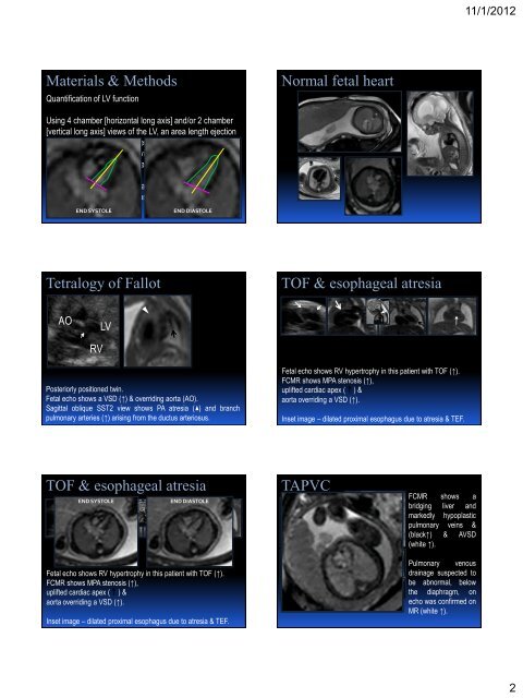
Materials & Methods<br />
Quantification of LV function<br />
Us<strong>in</strong>g 4 chamber [horizontal long axis] and/or 2 chamber<br />
[vertical long axis] views of the LV, an area length ejection<br />
fraction measurement was performed on FCMR images<br />
and qualitative function was visually assessed and graded<br />
as decreased, normal or <strong>in</strong>creased<br />
Echo – LV and RV ejection fraction was visually assessed<br />
and graded as decreased, normal or <strong>in</strong>creased<br />
END SYSTOLE END DIASTOLE<br />
Normal <strong>fetal</strong> heart<br />
Tetralogy of Fallot TOF & esophageal atresia<br />
AO<br />
RV<br />
LV<br />
Posteriorly positioned tw<strong>in</strong>.<br />
Fetal echo shows a VSD (↑) & overrid<strong>in</strong>g aorta (AO).<br />
Sagittal oblique SST2 view shows PA atresia ( ) and branch<br />
pulmonary arteries (↑) aris<strong>in</strong>g from the ductus arteriosus.<br />
TOF & esophageal atresia<br />
END SYSTOLE END DIASTOLE<br />
Fetal echo shows RV hypertrophy <strong>in</strong> this patient with TOF (↑).<br />
FCMR shows MPA stenosis (↑),<br />
uplifted <strong>cardiac</strong> apex ( ) &<br />
aorta overrid<strong>in</strong>g a VSD (↑).<br />
Inset image – dilated proximal esophagus due to atresia & TEF.<br />
Fetal echo shows RV hypertrophy <strong>in</strong> this patient with TOF (↑).<br />
FCMR shows MPA stenosis (↑),<br />
uplifted <strong>cardiac</strong> apex ( ) &<br />
aorta overrid<strong>in</strong>g a VSD (↑).<br />
Inset image – dilated proximal esophagus due to atresia & TEF.<br />
TAPVC<br />
FCMR shows a<br />
bridg<strong>in</strong>g liver and<br />
markedly hypoplastic<br />
pulmonary ve<strong>in</strong>s &<br />
(black↑) & AVSD<br />
(white ↑).<br />
Pulmonary venous<br />
dra<strong>in</strong>age suspected to<br />
be abnormal, below<br />
the diaphragm, on<br />
echo was confirmed on<br />
MR (white ↑).<br />
11/1/2012<br />
2