protonema - Bryophyte Ecology
protonema - Bryophyte Ecology
protonema - Bryophyte Ecology

You also want an ePaper? Increase the reach of your titles
YUMPU automatically turns print PDFs into web optimized ePapers that Google loves.
CHAPTER 5-3<br />
ECOPHYSIOLOGY OF DEVELOPMENT:<br />
PROTONEMA<br />
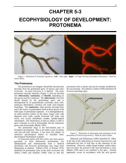
Figure 1. Protonema of Fontinalis squamosa. Left: white light. Right: UV light showing chlorophyll fluorescence. Photo by<br />
Janice Glime.<br />
The Protonema<br />
The <strong>protonema</strong> is an elongate, thread-like structure that<br />
develops from the germinated spore of mosses and some<br />
liverworts. In most liverworts it is thalloid. The moss<br />
<strong>protonema</strong> typically branches (Figure 1) and can develop<br />
into chloronema, caulonema, or rhizoids, depending on<br />
the species and the conditions. The chloronema is the first<br />
thread formed by the germinating spore and is<br />
distinguished by its perpendicular crosswalls, short cells,<br />
numerous chloroplasts, colorless cell walls, and irregular<br />
branching. The caulonema, when present, develops later<br />
and is the source of gametophore buds in those species with<br />
both types of <strong>protonema</strong>l segments. It is distinguished by<br />
its distal position relative to the spore, longer cells with<br />
diagonal cross walls, usually brownish cell walls, and<br />
fewer, less evenly distributed, smaller, spindle-shaped<br />
chloroplasts. The chloronema, at least in culture, is able to<br />
grow vertically as well as horizontally, but the caulonema<br />
grows only horizontally (Bhatla 1994).<br />
The <strong>protonema</strong>l stage is the best-studied part of<br />
bryophyte development. Due to its relative ease of culture<br />
and one-cell-wide structure, it has been the subject of<br />
numerous physiological studies to elucidate basic<br />
physiological mechanisms in plants.<br />
As discussed earlier with life cycles, true moss spores<br />
germinate to form filamentous <strong>protonema</strong>ta, but<br />
Sphagnopsida and Andreaeopsida form a thalloid<br />
<strong>protonema</strong>, and liverwort <strong>protonema</strong>ta may range from<br />
filamentous to thalloid. Fulford (1956, in Watson 1974)<br />
identified 10 <strong>protonema</strong>l types in the leafy liverworts, but<br />
Nehira (1966) and Schuster (1966) warn us that the<br />
<strong>protonema</strong> form is plastic and can be strongly modified by<br />
the environment. Nevertheless, Nehira (1966) identified 24<br />
liverwort sporeling types.<br />
Figure 2. Distinction of chloronema and caulonema on the<br />
<strong>protonema</strong> of Funaria hygrometrica. Photo by Janice Glime.<br />
Bittisnich and Williamson (1989) identified H + efflux<br />
at the tips of the chloronema in Funaria hygrometrica and<br />
elaborated the role of acid flux in the extension of the<br />
<strong>protonema</strong>. However, unlike fungal hyphae, pollen tubes,<br />
and root hairs, the growth of the moss <strong>protonema</strong> is slow<br />
(Bhatla 1994) and is not confined to the apex. Growth<br />
apparently occurs in accordance with the acid growth<br />
35
36 Chapter 5-3: Ecophysiology of Development: Protonema<br />
mechanism, in which H + ions, induced by light and IAA,<br />
loosen the cell wall. In Funaria hygrometrica,<br />
acidification of the medium to pH 5.5 increases the<br />
extension of the tip cells, whereas buffering to a pH of 6.8<br />
prevents it. Calcium seems necessary for the acquisition of<br />
new materials to the wall and the ability to extend the wall.<br />
When the leafy liverwort Leucolejeunea clypeata was<br />
grown on a Ca-free medium, the spores became distended,<br />
but the <strong>protonema</strong> failed to develop during the next five<br />
months of culture, whereas in the normal medium young<br />
plants had developed (Geldreich 1948).<br />
The changes from distended spore to filamentous<br />
<strong>protonema</strong> growth to gametophore buds can require<br />
increasingly more specialized conditions. For example,<br />
Forman (1964) found that spore germination in Tetraphis<br />
pellucida requires a pH of 3.0-7.3 whereas growth of the<br />
leafy shoot occurs in the much narrower pH range of 5.1 to<br />
5.8. Temperature requirements, on the other hand, are<br />
broader for the leafy shoot, but as the humidity drops, the<br />
viable temperature range narrows. Furthermore, the change<br />
from chloronema to caulonema can be delayed by<br />
inappropriate environmental conditions. Bopp (1961)<br />
found that the caulonema stage, and thus the bud stage, can<br />
be delayed by low temperature, submersion, or low light.<br />
There seems to be controversy over the degree of<br />
difference between chloronema and caulonema, with Bopp<br />
(1959) contending that they are distinct stages, and Kofler<br />
(1958) and others finding no consistent distinction, even in<br />
Funaria hygrometrica, for which Bopp first made his<br />
claim. Several factors appear to lead to these<br />
disagreements (Watson 1974). The plasticity of the<br />
<strong>protonema</strong> permits it to respond differently to the varying<br />
environmental conditions. The distinction is exhibited<br />
more strongly in some species than others, and in some<br />
species, apparently no distinction exists. And, Kofler<br />
contended that genetic differences are more likely to be<br />
expressed in the <strong>protonema</strong> than in the gametophore or<br />
sporophyte because the environment has less time to exert<br />
selective pressure on the <strong>protonema</strong>. Hmmm...<br />
Even in mosses such as Funaria hygrometrica that<br />
have well-developed caulonema, culture in liquid media<br />
can inhibit the formation of caulonema, resulting in<br />
reduced bud formation – suggesting that very wet<br />
conditions would be detrimental to the development of<br />
gametophores in these taxa (Johri & Desai 1973).<br />
Furthermore, high cell densities cause failure of caulonema<br />
differentiation, suggesting some sort of self-inhibition.<br />
This might be another adaptive mechanism that prevents<br />
the gametophores from competing with each other and that<br />
permits the <strong>protonema</strong> time to revert to chloronema, spread<br />
to a wider area, or partially die off before putting forth<br />
upright plants.<br />
Application of IAA induces the switch from<br />
chloronema to caulonema side branches (Johri & Desai<br />
1973; Christianson 2000) and inhibits the further growth<br />
and initiation of chloronema branches (Johri & Desai<br />
1973). Application of ABA to chloronema instead results<br />
in cell division and the formation of asexual reproductive<br />
cells, but not in caulonemata (Christianson 2000).<br />
Inadequate calcium causes the chloronema cells to divide<br />
unevenly and to form tmema (abscission cell that ruptures<br />
to release moss gemmae), but not in caulonemata.<br />
Cytokinin stimulates the formation of gametophore buds in<br />
the caulonema, but not in the chloronema. Perhaps even<br />
more surprising, chloronemata exhibit positive<br />
phototropism, whereas caulonemata exhibit negative<br />
phototropism.<br />
But are these hormone responses initiated by moss<br />
hormone productions? In the well-studied Physcomitrella<br />
patens, we do know that transition from chloronema to<br />
caulonema cells is under control of auxin (Gonneau et al<br />
2001). Since IAA concentrations seem to be under<br />
environmental influence, variability and inconsistencies<br />
may be explained in the near future as we unravel the<br />
cryptochrome/IAA complex of reactions in this moss, and<br />
plants in general, using gene knockout techniques.<br />
Water Relations<br />
We have often assumed that the <strong>protonema</strong> stage is the<br />
most susceptible to desiccation damage. However, this is<br />
not always true. During (pers. comm.) found that<br />
unsuccessful cultures of xerophytes such as Grimmia<br />
produced gametophores only after being put aside and<br />
forgotten, i.e., after desiccation. But it is surprising that<br />
Glime and Knoop (1986) found that after cultures of the<br />
aquatic moss Fontinalis squamosa had dried out, added<br />
water caused the <strong>protonema</strong>ta to swell and again become<br />
active. This is further supported by observations on<br />
<strong>protonema</strong>ta that dried overnight on a microscope slide.<br />
When I added water to observe them for fluorescence, they<br />
produced vivid red chlorophyll fluorescence and regained<br />
their normal shape. It appears that <strong>protonema</strong>ta may have<br />
considerable desiccation tolerance.<br />
Further evidence that the <strong>protonema</strong> is desiccation<br />
tolerant can be gleaned from their dispersal period. As<br />
seen in the chapter on phenology, dispersal in spring is<br />
commonplace. It would seem, therefore, that the<br />
<strong>protonema</strong> must be growing in summer, when desiccation<br />
is most likely. The other period of high spore dispersal is<br />
fall, again preceding the dry season of winter. Although<br />
we have insufficient evidence to show that the <strong>protonema</strong>ta<br />
are present during these two relatively dry seasons, it<br />
appears likely that they are in at least some, if not many,<br />
species. Figure 3 shows a hydrated <strong>protonema</strong> in the field.<br />
Figure 3. Protonema of Plagiomnium in the field. Photo by<br />
Janice Glime.<br />
Seasonal Light/Temperature Changes<br />
It is hard to talk about light without also considering<br />
temperature, since brighter light generally means greater<br />
exposure and higher temperatures. Higher temperatures<br />
and brighter light are also usually coupled with a longer
photoperiod. Knowledge of their effects on <strong>protonema</strong>l<br />
growth and development is based on laboratory cultures.<br />
Light, coupled with temperature, seems to play a role<br />
in the pattern of development of <strong>protonema</strong>ta in the aquatic<br />
moss Fontinalis. Fontinalis squamosa spores germinated<br />
throughout the range of 40 to 3000 lux, and cultures<br />
exhibited unipolar, bipolar, tripolar, and one tetrapolar<br />
germination (Glime & Knoop 1986). The number of germ<br />
tubes was generally consistent within a single plate, despite<br />
having bands of spores from three different capsules. At<br />
3ºC and 120 lux, germination required four weeks, and<br />
only distended spores with a single protrusion were present<br />
(Figure 4). At 14ºC, 1200 lux, two plates of spores had<br />
single threads (Figure 5), one had double threads, and one<br />
had short single and double threads. At 20ºC, 2100 lux,<br />
two plates had only single germ threads that formed weak<br />
spirals and two had many spores with two or three germ<br />
threads and no spiral growth (Figure 6); branching was<br />
much more extensive than at 14ºC and 1200 lux. Although<br />
effects of temperature cannot be separated from those of<br />
light intensity, they mimic environmental conditions as<br />
they change from winter to summer. Such environmental<br />
controls can prevent spores from germinating or<br />
<strong>protonema</strong>ta from developing too early in the season. The<br />
high degree of branching at higher light and temperatures<br />
could afford more self-protection from desiccation by<br />
providing overlapping threads. Bipolar and tripolar<br />
germination is also likely to be a response to the greater<br />
ability to photosynthesize with more light and provide<br />
energy for the developing germ tube.<br />
Figure 4. Distended spore of Fontinalis squamosa as one<br />
might find at 3ºC and 120 lux. Photo by Janice Glime.<br />
Figure 5. Single-thread <strong>protonema</strong>ta of Fontinalis squamosa<br />
formed at 14ºC and 1200 lux. Photo by Janice Glime.<br />
Chapter 5-3: Ecophysiology of Development: Protonema 37<br />
High temperatures required for the <strong>protonema</strong>ta can<br />
force a species into a narrow geographic range despite the<br />
ability of the spores to germinate at cooler temperatures.<br />
For example, Anisothecium molliculum has an optimum<br />
temperature of 25°C, not only for <strong>protonema</strong>l growth, but<br />
also bud formation (Kumra & Chopra 1985), preventing it<br />
from living in polar regions.<br />
At least for Fontinalis squamosa, higher light<br />
intensity and temperatures result in more germ tubes<br />
arising from the spore, suggesting that more sugars might<br />
be available, both for energy and for creating a high<br />
osmotic potential. The increased number of<br />
<strong>protonema</strong>tal branches at higher light intensities and<br />
temperatures could provide a thicker mat to decrease<br />
evaporative losses and to increase self-shading against<br />
UV light damage.<br />
Light Intensity<br />
High light intensity can promote <strong>protonema</strong>l growth,<br />
as in Microdus, Hymenostylium, and Campylopus (Mehta<br />
1988). In the ephemeral Physcomitrella patens, high light<br />
intensities promote branching of the caulonema, thus<br />
proliferating the potential bud sites (Cove et al. 1978,<br />
1979). Continued high light promotes secondary<br />
caulonemata instead of bud formation. Is this adaptive by<br />
extending the plant to a darker location? Or is it merely a<br />
way of measuring all the available illuminated space for<br />
successful gametophores? Sood (1975) also observed an<br />
effect of light intensity on the number of germ tubes arising<br />
from the spore in Pogonatum aloides. At 1000 lux<br />
germination was unipolar, increasing at 3000 lux. At 6-<br />
8000 lux some spores swelled but failed to germinate. In<br />
germinating spores of Polytrichum commune and P.<br />
juniperinum, there was a lag in synthesis of chlorophyll,<br />
being longer in P. commune (Karunen 1973). The<br />
chlorophyll a/b ratio at that time in P. commune was 1.4-<br />
1.8, thus providing little antenna effect by chlorophyll b.<br />
The low concentration of chlorophyll in general and the<br />
reduced relative amount of light-gathering chlorophyll b<br />
would force the gametophyte to require food reserves<br />
during early development.<br />
Figure 6. Protonemata of Fontinalis squamosa showing<br />
unipolar, bipolar, tripolar germination typical at 20ºC and 2100<br />
lux. Photo by Janice Glime.
38 Chapter 5-3: Ecophysiology of Development: Protonema<br />
Although light generally seems to be necessary for<br />
spore distension, in some cases the <strong>protonema</strong> can even<br />
grow in the dark. In Ceratodon purpureus darkness first<br />
induces an increase of starch grains in the chloroplast<br />
(Valanne 1971). This is followed by disappearance of<br />
starch and an increase in the number of grana lamellae.<br />
Light Quality<br />
It is clear that light quality affects the growth and<br />
development of at least some <strong>protonema</strong>ta. Light quality<br />
shift from white light to green and far red, as found in the<br />
forest, resulted in reduced <strong>protonema</strong>l growth in Pohlia<br />
nutans, with the least growth occurring in green light<br />
(Mitra et al. 1959). Giles and von Maltzahn (1967) found<br />
that red light stimulates mature leaf cells of Plagiomnium<br />
affine to regenerate by <strong>protonema</strong>ta, and they suggested<br />
that phytochrome was most likely involved. Although<br />
liverworts seem to lack any consistent kind of<br />
photoregulation (Hartmann & Weber 1990), mosses<br />
respond differently to different wavelengths. Their best<br />
chloronema growth seems to be in white light (Bhatla<br />
1994), but we must question whether this is true for all<br />
species that grow only under a canopy of green. In<br />
Funaria hygrometrica, the red range stimulates normal<br />
growth, whereas the blue range leads to the development of<br />
caulonema-like cells. It is possible that these shifts in light<br />
quality response could help to signal the time to develop<br />
gametophores as the <strong>protonema</strong>l mat thickens from<br />
extensive growth, changing the light quality of underlying<br />
strands.<br />
Light quality could also serve to signal that it is time to<br />
break dormancy. Both blue and red light will permit<br />
maintenance of normal chloroplasts in Ceratodon<br />
purpureus <strong>protonema</strong>ta, but blue light results in richer<br />
starch, denser stromata (colorless matrix of chloroplast in<br />
which packets of chlorophyll are embedded), and more<br />
mitochondria, whereas red results in a more effective use of<br />
lipids (Valanne 1971). Is there any adaptive value in this?<br />
Is the moss able to sense the decreasing cover by snow, as<br />
voles do, based on light quality and intensity?<br />
Photoperiod<br />
In Ceratodon purpureus, long days stimulate<br />
elongation of the <strong>protonema</strong>, whereas short days result in<br />
<strong>protonema</strong>l branching (Larpent-Gourgaud & Aumaitre<br />
1980). The two systems are antagonists. This relationship<br />
suggests that an IAA/cytokinin balance may be the<br />
important controlling factor, with long days promoting<br />
IAA, probably through phytochrome mediation.<br />
In Bryum pseudotriquetrum a day length of ten or<br />
more hours is required for germination and <strong>protonema</strong><br />
growth (Kinugawa & Nakao 1965, Figure 7). However<br />
two minutes of light during a 16-hr dark period is sufficient<br />
to remove the inhibitory effect developed during the dark<br />
period and will likewise stimulate germination and growth.<br />
This further supports the hypothesis of a phytochrome<br />
response and is much like the photoperiodic control of<br />
flowering.<br />
Figure 7. Effect of photoperiod on spore germination after 5<br />
days (left) and <strong>protonema</strong> growth after 3 days (right) of Bryum<br />
pseudo-triquetrum. Redrawn from Kinugawa & Nakao (1965).<br />
Hormonal Response<br />
The complexity of these light responses and the<br />
implications of involvement by phytochrome is<br />
undoubtedly under the control of hormones. In the<br />
ephemeral Physcomitrella patens, light and hormonal<br />
combinations coordinate development (Cove et al. 1978,<br />
1979). Cytokinin in the presence of auxin promotes buds<br />
(Gorton & Eakin 1957), and high concentrations inhibit<br />
caulonemata (Cove et al. 1978, 1979). This combination<br />
would therefore promote caulonema growth while the<br />
caulonemata are sparse, ensuring sufficient plants for a<br />
viable population and providing a sufficiently dense<br />
<strong>protonema</strong>tal mat to help maintain moisture at the soil<br />
surface. When this mat becomes very dense, self-shading<br />
could stimulate the production of auxin and cytokinin and<br />
shift the development to bud formation. Once these selfshaded<br />
<strong>protonema</strong>ta have shifted to bud development, they<br />
are likely to communicate this signal to the surface<br />
<strong>protonema</strong>ta and induce buds throughout the mat. Figure 8<br />
shows a developmental scheme modified from Cove et al.<br />
(1979) to include these environmental stimuli.<br />
Figure 8. Effects of auxin and cytokinin on Physcomitrella<br />
patens. Redrawn from Cove et al. (1979).<br />
Bierfreund et al. (2003) used Physcomitrella patens to<br />
determine the distribution of auxin in the <strong>protonema</strong>. As in<br />
higher plants, the highest concentrations were in the<br />
dividing and young cells. Concentrations declined from the
tip cells back to the basal cells of the <strong>protonema</strong>,<br />
supporting earlier work of Bopp and Atzorn (1992).<br />
Auxin (IAA) is important in the transition of<br />
chloronema to caulonema (Johri & Desai 1973; Figure 8)<br />
and the appropriate concentration maintains the caulonema<br />
state (Bopp 2000). Although we generally think that<br />
endogenous hormones from one plant cannot affect<br />
another, in Funaria hygrometrica the minute quantity of<br />
10 -16 mol IAA/mg fw seems to be responsible for the<br />
change from chloronema to caulonema (Bhatla & Dhingra-<br />
Babbar 1990). Such a small quantity could surely leak<br />
from other members of the same species or from a different<br />
species to help coordinate behavior among individuals. In<br />
fact, as the <strong>protonema</strong> matures, the <strong>protonema</strong> can excrete<br />
most of its auxin to its substrate, as shown in<br />
Physcomitrella patens (Reutter et al. 1998).<br />
Figure 9. Culture of Funaria hygrometrica showing distinct<br />
colonies resulting most likely from hormonal interaction between<br />
clones at the <strong>protonema</strong>l stage. Photo by Janice Glime.<br />
We already know that uptake of IAA by the <strong>protonema</strong><br />
occurs; in the lab, uptake of IAA by <strong>protonema</strong>tal cells is<br />
both passive and active. The passive component is pHdependent,<br />
with the greatest increase in uptake occurring at<br />
pH 4.5-4.7, indicating a dissociation of the IAA molecule<br />
(pK = 4.7; pK is pH at which equal concentrations of<br />
acidic and basic forms of substance are present). The<br />
potential for an exogenous developmental regulator has<br />
enormous implications not only for development, but for<br />
systematics and ecology as well.<br />
Tropisms<br />
Tropisms, the bending of a plant in response to a<br />
stimulus, are adaptive in orienting the plant into its most<br />
beneficial position. In bryophytes, <strong>protonema</strong>ta are<br />
positively phototropic (bend toward light), whereas<br />
rhizoids are photonegative (bend away from light) (Heitz<br />
1942). Although Kofler and coworkers investigated the<br />
effects of the environment on bryophyte tropisms as early<br />
as 1958 (Kofler 1958, 1971; Kofler et al. 1963), bryophyte<br />
tropisms have remained largely unstudied until recently.<br />
However, because of their simple <strong>protonema</strong>l structure,<br />
much of our current understanding of tropisms in plants has<br />
been learned from using bryophytes as model systems. The<br />
one-cell-thick <strong>protonema</strong> makes it easy to observe the<br />
statoliths that are special amyloplasts (colorless plastids<br />
containing starch) that respond to gravity. These statoliths<br />
are involved in gravitropism (directional growth in<br />
Chapter 5-3: Ecophysiology of Development: Protonema 39<br />
response to gravity). The ability to knock out or add genes<br />
that are easily expressed in the 1n plants has made the<br />
necessary manipulation much easier than in tracheophytes.<br />
Yet bryophytes have different phototropic responses<br />
(directional growth in response to light) from those of<br />
tracheophytes. Rather than responding to blue light, as do<br />
the tracheophytes, most bryophytes seem to respond to red<br />
light, using phytochromes instead of cryptochromes as their<br />
sensory pigments (Wada & Kadota 1989). Jaffe and Etzold<br />
(1965) demonstrated that even spores in Funaria respond<br />
to red light, resulting in chloronema growth in the opposite<br />
direction from that of rhizoids. And even more intriguing<br />
is the ability of bryophytes to store a phototropic stimulus<br />
(Hartmann & Weber 1988), further suggesting the use of<br />
phytochromes. However, the expected dark reversal does<br />
not occur, indicating something else is involved<br />
(Christianson 2000).<br />
Schwuchow and Sack (1990) reported for the first time<br />
an effect of gravity on microtubule (essential protein<br />
filament of cell structural skeleton) distribution in plants,<br />
based on studies in <strong>protonema</strong>ta of Ceratodon purpureus.<br />
In fact, this moss served as the model organism to<br />
demonstrate that microtubules help organelles to maintain<br />
their positions within the cell (Schwuchow & Sack 1994).<br />
Nevertheless, our understanding of gravitropism in<br />
<strong>protonema</strong>ta is still in its early stages. We don't even have<br />
a very long list yet of mosses with demonstrated<br />
<strong>protonema</strong>l gravitropism, and we seem to know even less<br />
about liverworts. Schwuchow et al. (2002) have only<br />
recently found tropisms in Barbula unguiculata, Fissidens<br />
adianthoides, Fissidens cristatus, and Physcomitrium<br />
pyriforme, despite the report of positive phototropism in<br />
Funaria <strong>protonema</strong>ta in 1942 by Heitz.<br />
Repp et al. (2004) used genetically modified<br />
Physcomitrella patens to demonstrate the role of cytokinin<br />
signalling for gravitropism. When a knockout mutant lost<br />
its sensitivity to cytokinin, it had greatly reduced ability to<br />
respond gravitropically in the dark.<br />
Wagner and Sack (1998) reported that the gravitropic<br />
response occurs within 1-2 cell divisions in the <strong>protonema</strong>l<br />
tip cells of Ceratodon purpureus, which grow upward in<br />
the dark (Wagner et al. 1997). These five mosses and four<br />
other species, representing five orders, support the<br />
hypothesis that amyloplast sedimentation probably serves<br />
in gravity sensing in moss <strong>protonema</strong>ta. It appears that<br />
these amyloplasts tug on the cytoskeleton (structural<br />
support within cell), pulling down on it, much like trapped<br />
insects on a spider web. One theory is that this causes the<br />
cytoskeleton to pull on the cell membrane, creating larger<br />
holes in the membrane that facilitate the entry of Ca ++ .<br />
This creates a higher concentration of Ca ++ on the upper<br />
side of the cell, possibly causing it to inhibit the IAA on<br />
that side of the cell. Auxin inhibitors block this gravitropic<br />
response in Ceratodon purpureus (Schwuchow et al. 2001),<br />
suggesting the role of IAA in the process. Nevertheless,<br />
high levels of exogenous IAA do not block the apical<br />
tropism of the <strong>protonema</strong>, suggesting the mechanism<br />
differs from that in higher plants.<br />
What little we thought we knew about gravitropisms in<br />
moss <strong>protonema</strong>ta was further confused when growing<br />
<strong>protonema</strong>ta of the moss Ceratodon purpureus took a twoweek<br />
trip in space on the space shuttle Columbia (Miller &<br />
Phillips 2003; Kern et al. 2005). On 16 July 2002, plant
40 Chapter 5-3: Ecophysiology of Development: Protonema<br />
physiologist Fred Sack carefully opened a Petri dish that<br />
had spent the two weeks without gravity and without light.<br />
To his surprise, the <strong>protonema</strong>ta had grown in a spiral<br />
pattern (Figure 10). This is quite different from the normal<br />
tangle of <strong>protonema</strong>ta grown on Earth.<br />
According to Fred Sack (Miller & Phillips 2003),<br />
"These odd spirals mark the first time in space that a plant<br />
normally oriented by gravity has grown in a non-random<br />
Figure 10. Spiral growth of the <strong>protonema</strong>ta of Ceratodon<br />
purpureus aboard the space shuttle Columbia. Photo courtesy of<br />
Fred Sack.<br />
Another piece of this gravitropic puzzle is that a highgradient<br />
magnetic field can substitute for gravity, causing<br />
curvature of tip cells in Ceratodon purpureus (Kuznetsov<br />
et al. 1999). Genetically modified <strong>protonema</strong>ta with larger<br />
plastids responded more strongly, supporting the<br />
hypothesis that plastids are involved in gravity sensing.<br />
It appears, based on our observations with<br />
<strong>protonema</strong>ta, that the statoliths (amyloplasts) settle<br />
downward within the cell in response to gravity. This<br />
pulls on the cytoskeleton. The cytoskeleton is attached<br />
to the cell membrane, so this downward pull tugs on the<br />
membrane in the upper portion of the cell (Figure 11). A<br />
plausible theory is that this stretches the membrane,<br />
making it more permeable. This in turn permits more<br />
Ca ++ to enter the upper side of the cell, where it inhibits<br />
the action of IAA, permitting the lower side of the cell to<br />
grow more.<br />
Figure 11. Schematic model of hypothetical relationship of<br />
amyloplasts (statoliths) of a <strong>protonema</strong> in response to gravity.<br />
Arrows denote pull of cytoskeleton on cell membrane. Drawing<br />
by Janice Glime.<br />
pattern." The puzzle begins with the amyloplasts. These<br />
starch bodies experience sedimentation in gravity and seem<br />
to tug on the cell skeleton. However, on the shuttle, with<br />
no gravity, this should not happen. Rather, they should<br />
float at random within the cell. Instead, they bunched<br />
together. This indicates a natural propensity for growing in<br />
a spiral that is overridden by the gravity of Earth. Perhaps<br />
Seifritz was right – all life does have a twist in it.<br />
Nutation<br />
Under some circumstances, the <strong>protonema</strong> will exhibit<br />
nutation – a spiral or circular growth pattern that is<br />
displayed in time-lapse photography by apparent<br />
movements of the stem (or <strong>protonema</strong>) in a circle. In<br />
Funaria hygrometrica, red light causes the <strong>protonema</strong> to<br />
grow into a ring (Simon & Naef 1981). I have observed the<br />
same nutation in contaminated cultures of Fontinalis<br />
squamosa and in air-grown rhizoids of that species.<br />
Nutation appears to facilitate a kind of seeking – altering<br />
growth directions until a more favorable condition is<br />
located. It is beneficial when no directional stimulus is<br />
present, such as spiral growth of rhizoids until they contact<br />
a substrate, as observed in Fontinalis squamosa. Although<br />
nutation is an IAA/ethylene response in higher plants<br />
(Morgan & Powell 1970), its occurrence as a response to<br />
red light suggests it results from a somewhat different<br />
mechanism here since red light is known to inhibit ethylene<br />
production. Could this be the same spiraling mechanism<br />
seen in the space-travelling Ceratodon purpureus<br />
<strong>protonema</strong>ta? The curiosity there is that the entire<br />
population of <strong>protonema</strong>ta grew in a spiral.<br />
Interactions<br />
We have already implied that exogenous growth<br />
regulators could determine events in the development of<br />
the moss <strong>protonema</strong>. Protonemata grow on substrata that<br />
are not sterile. Rather, they are teaming with fungi,<br />
bacteria, algae, and exudates of other plants. One might<br />
then predict that at least some of the <strong>protonema</strong>ta respond<br />
in positive or negative ways to these companions.<br />
One possible outcome of cohabitation is that fungi or<br />
other organisms may provide the growth substances needed<br />
to stimulate the next phase of development. Fungi<br />
commonly produce gibberellic acid that escapes into the<br />
environment. Vaarama and Tarén (1059) found that not<br />
only did 0.01% GA promote both spore germination and<br />
<strong>protonema</strong> growth in several mosses (Dicranum scoparium,<br />
D. undulatum, Dicranoweisia crispula, and Pogonatum<br />
urnigerum), but also inoculation with several fungi<br />
(Aspergillus flavus, Penicillium martensii, Mucor<br />
racemosus, Fusarium scirpi, & Rhodotorula ad<br />
mucilaginosa)had even more effect than did the gibberellic<br />
acid.<br />
In contaminated cultures of Fontinalis squamosa, most<br />
of the <strong>protonema</strong>ta formed mature caulonemata in less than<br />
four weeks, whereas in uncontaminated cultures the<br />
chloronema state predominated (Glime & Knoop 1986;<br />
Glime, unpub data). And only the contaminated cultures<br />
ever produced buds. This suggests that at least some<br />
microbes might alter the developmental state of the moss.<br />
Based on these observations and those of Spiess et al.<br />
(1971) on the influence of bacteria on the development of<br />
Pylaisiella, it appears that we should focus some of our
attention to possible developmental interaction with the<br />
microbial community.<br />
Fungi have effects on other bryophyte <strong>protonema</strong>ta as<br />
well. Hildebrand and coworkers (1978) found that fungal<br />
exudates promoted the growth of Atrichum, Funaria, and<br />
Brachythecium <strong>protonema</strong>ta at low pH. As suggested<br />
above for spore germination, Splachnum ampullaceum<br />
(Figure 12) <strong>protonema</strong>tal growth is promoted by several<br />
species of fungi (von Maltzahn & MacQuarrie 1958).<br />
Certainly growth hormones exuded by the fungi could be of<br />
importance here (see Bopp 1980).<br />
Figure 12. Splachnum ampullaceum growing among<br />
Sphagnum on dung, where changing dung conditions and fungal<br />
exudates influence development. Photo by Janice Glime.<br />
In addition, contributions of vitamins from algae or<br />
amino acids or other organic compounds from bacteria<br />
might either be essential or promote a growth rate that is<br />
compatible with the seasons. Gibberellic acid, produced by<br />
many fungi, has a variety of effects, depending on the<br />
species of moss. It increases the number and length of<br />
<strong>protonema</strong>l cells in Dicranum and Dicranoweisia, but it has<br />
no effect on Racomitrium fasciculare (Vaarama & Tarén<br />
1959). Since R. fasciculare grows on rocks where fungi<br />
are unlikely to occur, and fungi are a natural source of GA,<br />
these differences in responses are consistent with habitat<br />
differences.<br />
We know that the induction Factor H (an adenine<br />
derivative discussed earlier) is present in Funaria. It will<br />
induce not only other <strong>protonema</strong>ta of Funaria, but it can be<br />
induced by other species (e.g. Leptobryum pyriforme) as<br />
well (Klein 1967; Bopp 1976). Such a factor is adaptive in<br />
insuring a sufficient breeding population, but perhaps more<br />
importantly it insures a community organization that offers<br />
resistance against desiccation, where middle plants are<br />
protected by outer ones in the population. In submerged<br />
mosses such as Fontinalis, on the other hand, moisture<br />
conservation is not so critical, and multiple gametophores<br />
would only offer competition for the limited substrate<br />
available for anchorage.<br />
Whereas some interactions can enhance growth of<br />
moss <strong>protonema</strong>ta, others inhibit it, preventing the<br />
colonization of that substrate. Shrimal (1975) showed that<br />
bark extracts of several trees inhibited mitosis in onion root<br />
tips and caused non-separation of chromosomes. If these<br />
substances have the same effects on mosses, it could<br />
explain why some trees lack bryophytic epiphytes.<br />
Inhibition can also occur within a species, as already<br />
suggested for Funaria. In this species, <strong>protonema</strong>ta from<br />
Chapter 5-3: Ecophysiology of Development: Protonema 41<br />
several spores in one culture will not intersect (Watson<br />
1981). The mat attains the same density when the<br />
<strong>protonema</strong>ta are derived from many spores as when they<br />
are derived from only one. Watson also suggests that one<br />
species may inhibit another, thus making time an important<br />
factor in access to a habitat. And Funaria is not the only<br />
moss where some exudate of the <strong>protonema</strong> retards<br />
development of competing <strong>protonema</strong>ta of the same<br />
species. This has been observed in culture in<br />
Physcomitrella patens as well (Schween et al. 2003). It is<br />
perhaps a widespread phenomenon.<br />
In Funaria, this factor of inhibition seems to break<br />
down in mature cultures. When I placed disks of agar from<br />
a mature culture onto fresh plates and inoculated the plates<br />
with spores, some of the <strong>protonema</strong>ta grew on the disks<br />
from the mature cultures. In no case did I find a zone of<br />
inhibition around the agar disk. This suggests to me that<br />
the substance preventing live <strong>protonema</strong>ta from<br />
intersecting might be a gas produced by the growing<br />
<strong>protonema</strong>ta. Gases are instrumental in maintaining<br />
maximum distance among sporangia of some slime molds,<br />
and one gas that could accomplish this in mosses is<br />
ethylene. Since ethylene is known from Funaria<br />
<strong>protonema</strong>ta (Rohwer & Bopp 1985) and it is a known<br />
inhibitor of cell division (Abeles 1973), small<br />
concentrations produced by the tips could easily signal<br />
their presence to neighbors. Ethylene production is<br />
stimulated by the action of IAA on S-adenosylmethionine<br />
(SAM), so we might expect the tip (where there is the most<br />
IAA) to have the highest ethylene concentration. The<br />
longest branches will interact first, and these are the ones<br />
most likely to be IAA-rich and apically dominant.<br />
Hormones produced by other organisms in the<br />
environment can affect the development of <strong>protonema</strong>ta,<br />
and in some cases these may be required to take the<br />
bryophyte to its next developmental stage. Among these,<br />
GA (gibberellic acid) is a likely candidate. It is produced<br />
by many fungi and readily enters the environment. It is<br />
known to increase the number and length of<br />
<strong>protonema</strong>tal cells in some soil-inhabiting species, but<br />
may have no effect on rock-dwelling taxa that normally<br />
would have much less contact with soil fungi. Bark<br />
exudates may also inhibit growth of some bryophyte<br />
<strong>protonema</strong>ta, and some bryophytes may inhibit each<br />
other, both of different species and of other clones of<br />
their own species.<br />
Nutrients<br />
In some mosses, the form of the <strong>protonema</strong> is<br />
dependent on available nutrients. For example, in nature<br />
Sphagnum normally has a thalloid <strong>protonema</strong> (Figure 13).<br />
However, in a medium with high potassium, the <strong>protonema</strong><br />
becomes filamentous (Schofield 1985). Since Sphagnum<br />
normally grows in habitats very low in potassium, this<br />
filamentous growth form is not observed in nature.<br />
Sundberg and Rydin (2000) showed that Sphagnum<br />
establishment from spores was limited by amount of<br />
phosphate released by underlying litter. Added moose<br />
dung likewise promoted establishment. They concluded<br />
that cover of other plants and nutrient release from litter<br />
provided safe sites where Sphagnum spores could<br />
germinate and establish new plants.
42 Chapter 5-3: Ecophysiology of Development: Protonema<br />
Figure 13. Thalloid <strong>protonema</strong>ta of Sphagnum papillosum.<br />
Photo by Yenhung Li.<br />
Various heavy metals seem to alter <strong>protonema</strong>tal form.<br />
Kapur and Chopra (1989) found that in the moss Timmiella<br />
anomala, when grown aseptically (conditions free of<br />
microorganisms), aluminum causes <strong>protonema</strong>l cells to<br />
become rounded and packed with chloroplasts and starch<br />
grains; the filaments themselves form bunches. Zinc and<br />
arsenic likewise cause rounded cells, with zinc-damaged<br />
cells becoming reddish; most arsenic effects are seen at the<br />
terminal and intercalary positions. Mercury causes cells to<br />
become broad with dense particles, whereas nickel results<br />
in long, thin <strong>protonema</strong>ta with little branching. At 10 -6 M,<br />
nickel increases <strong>protonema</strong>l growth slightly, but at 10 -5 M<br />
it drastically decreases the number of gametophore buds.<br />
Cobalt inhibits <strong>protonema</strong>l growth but seems to have no<br />
effect on bud formation. What do these effects mean to<br />
development of the moss, and are they likely to occur in<br />
nature where soil chelators (organic compounds that bind<br />
metal by forming ring structure around it) may inhibit<br />
uptake, or concentrations never reach these levels? Could<br />
they actually affect appearance of mature gametophytes<br />
resulting from these anomalous forms and hence confound<br />
our understanding of the taxonomy?<br />
Calcium seems important to <strong>protonema</strong> development<br />
in some species and may be the actual factor where pH<br />
affects viability. For Funaria hygrometrica, Reiss and<br />
Herth (1979) suggest that a calcium gradient is responsible<br />
for <strong>protonema</strong>l tip growth. The calcium concentration is<br />
highest at the tip where fluorescence is strongest. It is<br />
likely that calcium is involved in transport of substances<br />
across cell membranes.<br />
Nutrient availability is affected by pH. Thus pH could<br />
affect success of <strong>protonema</strong>ta. In Physcomitrella patens<br />
(Figure 14, Figure 15), changes in pH in the range of 4.5 to<br />
7.0 influenced differentiation of <strong>protonema</strong>ta but did not<br />
have any negative impact on growth rate (Hohe et al.<br />
2002). In another example, Anisothecium molliculum has<br />
an optimum pH of 5.5 for not only <strong>protonema</strong>l growth, but<br />
also for bud formation (Kumra & Chopra 1985). The pH<br />
may not only alter the ability of bryophyte <strong>protonema</strong>ta to<br />
obtain nutrients, but also affect their susceptibility to<br />
exudates from other plants and fungi. Following fire,<br />
invasion by bryophytes onto the charred substrate seems to<br />
be likewise influenced by both pH and residual chemicals<br />
(Thomas et al. 1994). Germination success in the moss<br />
Campylopus pyriformis is positively influenced by<br />
increases in the pH in the range of 3.5-6.4.<br />
Figure 14. Physcomitrella patens in its natural habitat where<br />
pH and moisture can change considerably as spring flooding<br />
recedes. Photo by Michael Lüth.<br />
Figure 15. Physcomitrella patens plants with <strong>protonema</strong>ta<br />
on the wet soil. Photos by Michael Lüth.<br />
Our knowledge of nutrient requirements is based<br />
mostly on cultures of bryophytes and we know little of<br />
the generalities that might be important. For example,<br />
elevated potassium causes Sphagnum <strong>protonema</strong>ta to<br />
become filamentous instead of thalloid, but in nature we<br />
have not observed <strong>protonema</strong>ta in habitats where this<br />
condition exists. The level of phosphorus is often<br />
limiting and we can assume this plays a role in nature as<br />
well. An important observation is that heavy metals such<br />
as aluminum, zinc, mercury, and arsenic can cause<br />
abnormal <strong>protonema</strong>ta with such symptoms as rounded<br />
cells with dense chloroplasts and starch. Elevated nickel,<br />
on the other hand, causes the <strong>protonema</strong>ta to be thin.<br />
Calcium is undoubtedly important and its function may<br />
relate to membrane transport of other ions into the cell.<br />
All of these nutrient effects are likely to be affected by<br />
the pH because a lower (acidic) pH generally makes<br />
most nutrient ions more soluble.<br />
Rhizoids<br />
Rhizoids form on the <strong>protonema</strong> at different stages,<br />
depending on the species and the growing conditions. On<br />
nutrient-free agar and in distilled water the first filaments to<br />
emerge from the spore are rhizoidal (Bhatla 1994). They<br />
are distinguished by their pigmented (usually brown) cell<br />
walls, oblique crosswalls, and discoid or cylindrical<br />
plastids. The rhizoids seem to depend on forced calcium<br />
entry (active uptake requiring energy) for growth and at<br />
least in those tested, respond positively to a calcium<br />
gradient (Bhatla 1994).<br />
Rhizoids usually exhibit strong positive gravitropism<br />
(grow toward the center of gravity), negative<br />
phototropism (grow away from light), and thigmotropism
(alter their growth upon contact), with the latter overriding<br />
the effects of the former once a substrate is contacted<br />
(Bhatla 1994). When growing in air, they often exhibit a<br />
spiral growth (nutation) until a substrate is contacted<br />
(Glime 1987). Upon contact, they may branch into short,<br />
fingerlike tips (Odu 1988), as noted in Lophocolea<br />
cuspidata (Odu & Richards 1976) and Fontinalis squamosa<br />
(Glime 1987). Among the liverworts, apical branching<br />
seems to be in part phylogenetically constrained, appearing<br />
commonly in the Jungermanniales but only in the<br />
Metzgeriineae of the Metzgeriales and not at all in the<br />
Marchantiopsida (Pocock & Duckett 1985). Those<br />
liverworts with swollen rhizoids grow exclusively on peat<br />
and rotten wood associated with fungal hyphae.<br />
Pleurocarpous moss rhizoids become flattened near the<br />
tips, but in acrocarpous mosses these flattenings extend<br />
well behind the tips of the rhizoids (Odu 1988).<br />
Adhesion of rhizoids seems to be stimulated by the<br />
substrate itself (Odu 1988). Upon contact, rhizoids<br />
produce such extra-wall materials as sulfated<br />
mucopolysaccharides. These are highly viscous substances<br />
that serve as a sticky adhesive, also known in algae and<br />
other microorganisms.<br />
But what controls the production of these rhizoids?<br />
Goode et al. (1992) were unable to get Tetraphis pellucida<br />
to produce any <strong>protonema</strong>l rhizoids in culture, yet these<br />
occurred routinely in nature. They ascribed this difference<br />
to the limited nutrients and different irradiance in the wild.<br />
But hormones available from surrounding vegetation,<br />
bacteria, and fungi could play a role as well, as they<br />
apparently do for the <strong>protonema</strong>ta.<br />
Tmema<br />
Tmema cells are rounded cells that rupture, setting<br />
free a <strong>protonema</strong>l gemma. These cells result from a very<br />
unequal division of the cell near the proximal cross wall<br />
and divide the chloronema filaments into fragments of only<br />
a few cells. The tmema cells have few chloroplasts which<br />
soon become reduced in size, but the cell elongates in its<br />
proximal direction by expanding its newly formed wall,<br />
progressing in the opposite direction from normal cells.<br />
The new tmema wall forms inside the old lateral wall and<br />
the subsequent loosening of the old wall results in<br />
fragmentation of the <strong>protonema</strong>. This separation also<br />
occurs in older, untreated cultures of F. hygrometrica (>25<br />
days) (Bhatla & Dhingra-Babbar 1990).<br />
Bopp and coworkers (1991) have demonstrated the<br />
importance of IAA in the development of tmema cells in<br />
<strong>protonema</strong>ta of Funaria hygrometrica. They found that<br />
addition of only 10 µM IAA would suppress the production<br />
of tmema cells, indicating IAA deficiency. But these are<br />
laboratory results. Does the tmema occur in nature? Is it<br />
adaptive? Could it permit small fragments of the<br />
<strong>protonema</strong> to have one more chance at dispersal before<br />
producing its upright gametophore, hence possibly<br />
allowing it to arrive at a place where it could indeed<br />
produce enough of its own IAA in a more favorable<br />
setting? How remarkable a survival mechanism if indeed it<br />
permits another chance at dispersal.<br />
Tmema are one means of providing vegetative<br />
reproductive structures on the <strong>protonema</strong>. Various<br />
<strong>protonema</strong>tal asexual reproductive structures will be<br />
Chapter 5-3: Ecophysiology of Development: Protonema 43<br />
discussed in a later development chapter on asexual<br />
reproduction.<br />
Summary<br />
The filamentous <strong>protonema</strong> of Bryophyta can<br />
differentiate into two types: chloronema and<br />
caulonema, distinguished by short cells with<br />
perpendicular crosswalls, numerous chloroplasts,<br />
colorless cell walls, and irregular branching in the<br />
former and longer cells, diagonal crosswalls, brownish<br />
cell walls, and fewer, scattered, small chloroplasts in<br />
the latter. IAA induces the switch to caulonema;<br />
cytokinins promote branching. Protonemata of<br />
Sphagnopsida, Anthocerotophyta, and most<br />
Marchantiophyta are thalloid.<br />
Protonemata can produce a variety of brood cells,<br />
possibly stimulated by ABA, and sometimes<br />
disarticulated from the <strong>protonema</strong> by tmema cells.<br />
Light quantity, quality, photoperiod, and temperature<br />
influence both the rate of development and the form of<br />
the <strong>protonema</strong>. Their direction of growth is influenced<br />
by both gravity and light, causing negative<br />
gravitropism in the dark and positive phototropism in<br />
the light.<br />
Other organisms may supply IAA and GA that<br />
influence development, and Factor H (a likely<br />
cytokinin) may be supplied both endogenously and<br />
exogenously to control population size. Nutrients can<br />
affect the development, with heavy metals generally<br />
causing abnormalities or arrested development.<br />
Rhizoids exhibit positive gravitropism and<br />
negative phototropism, but also possess<br />
thigmotropism, typically expanding, branching, or<br />
flattening upon contact with a substrate.<br />
Acknowledgments<br />
Inspiration for this chapter evolved from discussions<br />
with Dr. Martin Bopp and especially with Dr. Gert Steen<br />
Mogensen. Several of the experiments were conducted at<br />
the Botanisches Institut, Universitat Heidelberg, Germany.<br />
I appreciate the many suggestions from a student's<br />
perspective by Medora Burke-Scoll. KT McConnell<br />
helped with the glossary and suggested the minisummaries<br />
after some of the topics.<br />
Literature Cited<br />
Abeles, F. B. 1973. Ethylene in Plant Biology. Academic Press,<br />
New York.<br />
Bhatla, S. C. 1994. Moss Protonema Differentiation. Research<br />
Studies Press Ltd., Somerset, England, 296 pp.<br />
Bhatla, S. C. and Dhingra-Babbar, S. 1990. Growth regulating<br />
substances in mosses. In: Chopra, R. N. and Bhatla, S. C.<br />
(eds.). <strong>Bryophyte</strong> Development: Physiology and<br />
Biochemistry, CRC Press, Ann Arbor, pp. 79-101.<br />
Bierfreund, N. M., Reski, R., and Decker, E. L. 2003. Use of an<br />
inducible reporter gene system for the analysis of auxin<br />
distribution in the moss Physcomitrella patens. Plant Cell<br />
Rept. 21: 1143-1152.<br />
Bittisnich, D. J. and Williamson, R. E. 1989. Tip-localised H +<br />
fluxes and the applicability of the acid-growth hypothesis to<br />
tip-growing cells: Control of chloronemal extension in<br />
Funaria hygrometrica by auxin and light. Planta 178: 96-<br />
102.
44 Chapter 5-3: Ecophysiology of Development: Protonema<br />
Bopp, M. 1959. Neue Gesichtspunkte zum Problem der<br />
Protonemadifferenzierung. Rev. Bryol. Lichenol. 28: 319-<br />
325.<br />
Bopp, M. 1961. Morphogenese der Laubmoose. Biol. Rev. 36:<br />
237-280.<br />
Bopp, M. 1976. External and internal regulation of the<br />
differentiation of the moss <strong>protonema</strong>. J. Hattori Bot. Lab.<br />
41: 167-177.<br />
Bopp, M. 1980. The hormonal regulation of morphogenesis in<br />
mosses. In: Skoog, F. (ed.). Plant Growth Substances.<br />
Springer-Verlag, Berlin, pp. 351-361.<br />
Bopp, M. 2000. 50 years of the moss story. In: Progress in<br />
Botany, Vol. 61. Springer, Berlin, Heidelberg, New York,<br />
pp. 1-34.<br />
Bopp, M. and Atzorn, R. 1992. The morphogenetic system of the<br />
moss <strong>protonema</strong>. Cryptog. Bot. 3: 3-10.<br />
Christianson, M. L. 2000. Control of morphogenesis in<br />
bryophytes. In: Shaw, J. A. and Goffinet, B. <strong>Bryophyte</strong><br />
Biology. Cambridge University Press, Cambridge, UK, pp.<br />
199-224.<br />
Cove, D. J., Ashton, N. W., Featherstone, D. R., and Wang, T. L.<br />
1979. The use of mutant strains in the study of hormone<br />
action and metabolism in the moss Physcomitrella patens.<br />
Proceedings of the Fourth John Innes Symposium, pp. 231-<br />
241.<br />
Cove, D. J., Schild, A., Ashton, N. W., and Hartmann, E. 1978.<br />
Genetic and physiological studies of the effect of light on the<br />
development of the moss, Physcomitrella patens.<br />
Photochem. Photobiol. 27: 249-254.<br />
Forman, R. T. T. 1964. Growth under controlled conditions to<br />
explain the hierarchical distributions of a moss, Tetraphis<br />
pellucida. Ecol. Monogr. 34:1-25.<br />
Fulford, M. 1956. Sporelings and regenerants of Ptilidium<br />
pulcherrimum (Web.) Hampe. Rev. Bryol. Lichénol. 25:<br />
247-253.<br />
Geldreich, E. E. Jr. 1948. The effects of a calcium-deficient<br />
medium on sporelings of Leucolejeunea clypeata.<br />
Bryologist 51: 229-235.<br />
Giles, K. L. and Maltzahn, K. E. von. 1967. Interaction of red,<br />
far-red, and blue light in cellular regeneration of leaves of<br />
Mnium affine. Bryologist 70: 312-315.<br />
Glime, J. M. 1987. The role of tropisms in rhizoid attachment<br />
and branch orientation in Fontinalis. Lindbergia 13: 85-90.<br />
Glime, J. M. and Knoop, B. C. 1986. Spore germination and<br />
<strong>protonema</strong>l development of Fontinalis squamosa. J. Hattori<br />
Bot. Lab. 61: 487-497.<br />
Gonneau, M., Pagant, S., Brun, F., and Laloue, M. 2001.<br />
Photoaffinity labelling with the cytokinin agonist azido-<br />
CPPU of a 34 kDa peptide of the intracellular pathogenesisrelated<br />
protein family in the moss Physcomitrella patens.<br />
Plant Molec. Biol. 46: 539-548.<br />
Goode, J. A., Duckett, J. G., and Stead, A. D. 1992. Protonemal<br />
morphogenesis of the moss Tetraphis pellucida Hedw. in<br />
culture and in the wild. Ann. Bot. 70: 519-530.<br />
Gorton, B. S. and Eakin, R. E. 1957. Development of the<br />
gametophyte in the moss Tortella caespitosa. Bot. Gaz. 119:<br />
31-38.<br />
Hartmann, E. and Weber, M. 1988. Storage of the phytochromemediated<br />
phototrophic stimulus of moss <strong>protonema</strong>l tip cells.<br />
Planta 175: 39-49.<br />
Hartmann, E. and Weber, M. 1990. Photomodulation of<br />
<strong>protonema</strong> development. In: Chopra, R. N. and Bhatla, S.<br />
C. (eds.). <strong>Bryophyte</strong> Development: Physiology and<br />
Biochemistry, CRC Press, Ann Arbor, pp. 33-54.<br />
Heitz, E. 1942. Die keimende Funaria-Spore als Physiologisches<br />
Versuchesobjekt. Ber. Deutsch. Bot. Ges. 60: 17-27.<br />
Hildebrand, V. R., Kottke, I., and Winkler, S. 1978.<br />
Untersuchung über den Einfluss von Pilzen auf die pH-<br />
Abhangigkeit von Laubmoosen. Beitr. Biol. Pflanzen 54: 1-<br />
12.<br />
Hohe, A., Decker, E. L., Gorr, G., Schween, G., and Reski, R.<br />
2002. Tight control of growth and cell differentiation in<br />
photoautotrophically growing moss (Physcomitrella patens)<br />
bioreactor cultures. Plant Cell Rept. 20: 1135-1140.<br />
Jaffe, L. and Etzold, H. 1965. Tropic responses of Funaria<br />
spores to red light. Biophys. J. 5: 715-742.<br />
Johri, M. M. and Desai, S. 1973. Auxin regulation of caulonema<br />
formation in moss <strong>protonema</strong>. Nature New Biol. 245: 223-<br />
224.<br />
Kapur, A. and Chopra, R. N. 1989. Effects of some metal ions<br />
on <strong>protonema</strong>l growth and bud formation in the moss<br />
Timmiella anomala grown in aseptic cultures. J. Hattori Bot.<br />
Lab. 66: 283-298.<br />
Karunen, P. 1973. Studies on moss spores II. Production of<br />
chlorophylls in germinating Polytrichum commune Hedw.<br />
spores. J. Exper. Bot. 24: 1183-1188.<br />
Kern, V. D., Schwuchow, J. M., Reed, D. W., Nadeau1, J. A.,<br />
Lucas, J., Skripnikov, A., and Sack, F. D. 2005. Gravitropic<br />
moss cells default to spiral growth on the clinostat and in<br />
microgravity during spaceflight. Planta 221: 149 – 157.<br />
Kinugawa, K. and Nakao, S. 1965. Note on spore germination<br />
and <strong>protonema</strong>l growth controlled by day length in Bryum<br />
pseudo-triquetrum. Bot. Mag. (Tokyo) 78: 194-197.<br />
Klein, B. 1967. Versuche zur Analyse der<br />
Protonemaentwicklung der Laubmoose. IV. Der Endogene<br />
Faktor H und seine Rolle bei der Morphogenese von Funaria<br />
hygrometrica. Planta 73: 12-27.<br />
Kofler, L. 1958. Contribution a l'etude biologique des mousses<br />
cultivees in vitro: Germination des spores, croissance et<br />
developpement du <strong>protonema</strong> chez Funaria hygrometrica.<br />
Rev. Bryol. Lichenol. 28(1-2): 1-202.<br />
Kofler, L. 1971. Influence de la temperature sur le geotropisme<br />
des spores de funaire. C. R. Acad. Sci. (Paris) 272: 1244-<br />
1247.<br />
Kofler, L., Dutel, J., and Nurit, F. 1963. Variations in the<br />
geotropic sensitivity of germinating Funaria spores in<br />
response to some external influences. J. Linn. Soc. London<br />
Bot. 58: 311-319.<br />
Kumra, S. and Chopra, R. N. 1985. In vitro studies on spore<br />
germination, <strong>protonema</strong>l differentiation and bud formation in<br />
the moss, Anisothecium molliculum (Mitt.) Broth.<br />
Phytomorphology 35: 223-231.<br />
Kuznetsov, O. A., Schwuchow, J., Sack, F. D., and Hasenstein, K.<br />
H. 1999. Curvature induced by amyloplast magnetophoresis<br />
in <strong>protonema</strong>ta of the moss Ceratodon purpureus. Plant<br />
Physiol. 119: 645-650.<br />
Larpent-Gourgaud, M. and Aumaitre, M. P. 1980.<br />
Photoperiodism and morphogenesis of the <strong>protonema</strong> of<br />
Ceratodon purpureus (Hedw.) Brid. Experientia 36: 1366-<br />
1367.<br />
Maltzahn, K. E. von and MacQuarrie, I. G. 1958. Effect of<br />
gibberellic acid on the growth of <strong>protonema</strong>ta in Splachnum<br />
ampullaceum (L.) Hedw. Nature (London) 181: 1139-1140.<br />
Mehta, P. 1988. In vitro studies on spore germination,<br />
<strong>protonema</strong>l differentiation and bud formation in three mosses<br />
grown in vitro. J. Hattori Bot. Lab. 64: 401-410.<br />
Miller, K. and Phillips, T. 2003. Mossy space spirals. National<br />
Aeronautics and Space Administration. Exploration Systems
Articles. Accessed 22 June 2006 at<br />
<br />
Mitra, G. C., Allsopp, A., and Wareing, P. F. 1959. The effects<br />
of light of various qualities on the development of the<br />
<strong>protonema</strong> and bud formation in Pohlia nutans (Hedw.)<br />
Lindb. Phytomorphology 9: 47-55.<br />
Morgan, P. W. and Powell, R. D. 1970. Involvement of ethylene<br />
in responses of etiolated bean hypocotyl hook to coumarin.<br />
Plant Physiol. 45: 553-557.<br />
Nehira, K. 1966. Sporelings in the Jungermanniales. J. Sci.<br />
Hiroshima Univ. B. Div. 2, 11(1): 1-49.<br />
Odu, E. A. 1988. Extracellular adhesive substances on bryophyte<br />
rhizoids. Acta Bot. Hung. 35(1-4): 273-277.<br />
Odu, E. and Richards, P. W. 1976. The stimulus to branching of<br />
the rhizoid tip in Lophocdloea cuspidata (Nees) Limpr. J.<br />
Bryol. 9: 93-95.<br />
Pocock, K. and Duckett, J. G. 1985. On the occurrence of<br />
branched and swollen rhizoids in British hepatics: Their<br />
relationships with the substratum and associations with<br />
fungi. New Phytol. 99: 281-304.<br />
Reiss, H. D. and Herth, W. 1979. Calcium gradients in tip<br />
growing plant cells visualized by chlorotetracycline<br />
fluorescsence. Planta 146: 615-621.<br />
Remezov, N. P. and Pogrebnyak, P. S. 1965. Forest Soil Science.<br />
Moskva, 261 pp.<br />
Repp, A., Mikami, K., Mittmann, F., and Hartmann, E. 2004.<br />
Phosphoinositide-specific phospholipase C is involved in<br />
cytokinin and gravity responses in the moss Physcomitrella<br />
patens. Plant J. 40: 250-259.<br />
Reutter, K., Atzorn, R., Hadeler, B., Schmülling, T., and Reski, R.<br />
1998. Expression of the bacterial ipt gene in Physcomitrella<br />
rescues mutations in budding and in plastid division. Planta<br />
206: 196-203.<br />
Rohwer, F. and Bopp, M. 1985. Ethylene synthesis in moss<br />
<strong>protonema</strong>. J. Plant Physiol. 117:331-338.<br />
Schofield, W. B. 1985. Introduction to Bryology. Macmillan<br />
Publishing Co., New York, 431 pp.<br />
Schuster, R. M. 1966. The Hepaticae and Anthocerotae of North<br />
America. Vol. I. Columbia Univ. Press, N. Y. 1344 pp.<br />
Schween, G., Hohe, A., Koprivova, A., and Reski, R. 2003.<br />
Effects of nutrients, cell density and culture techniques on<br />
protoplast regeneration and early <strong>protonema</strong> development in<br />
a moss, Physcomitrella patens. J. Plant Physiol. 160: 209-<br />
212.<br />
Schwuchow, J. and Sack, F. D. 1990. Microtubule distribution in<br />
gravitropic <strong>protonema</strong>ta of the moss Ceratodon.<br />
Protoplasma 159:60-69.<br />
Schwuchow, J. and Sack, F. D. 1994. Microtubules restrict<br />
plastid sedimentation in <strong>protonema</strong>ta of the moss Ceratodon.<br />
Cell Motility Cytoskeleton 29: 366-374.<br />
Chapter 5-3: Ecophysiology of Development: Protonema 45<br />
Schwuchow, J. M., Kern, V. D., White, N. J., and Sack, F. D.<br />
2002. Conservation of the plastid sedimentation zone in all<br />
moss genera with known gravitropic <strong>protonema</strong>ta. J. Plant<br />
Growth Reg. 21: 146-155.<br />
Schwuchow, J., Michalke, W., and Hertel, R. 2001. An auxin<br />
transport inhibitor interferes with unicellular gravitropism in<br />
<strong>protonema</strong>ta of the moss Ceratodon purpureus. Plant Biol.<br />
3: 357-363.<br />
Shrimal, S. K. 1975. Antimitotic activity of certain bark extracts.<br />
Beitr. Biol. Pflanzen 51: 77-80.<br />
Simon, P. E. and Naef, J. B. 1981. Light dependency of the<br />
cytokinin- induced bud initiation in <strong>protonema</strong>ta of the moss<br />
Funaria hygrometrica. Physiol. Plant. 53: 13-18.<br />
Sood, S. 1975. Morphogenetic studies on Pogonatum aloides.<br />
Beitr. Biol. Pflanzen 51: 99-110.<br />
Spiess, L. D., Lippincott, B. B., and Lippincott, J. A. 1971.<br />
Development and gametophore initiation in the moss<br />
Pylaisella selwynii as influenced by Agrobacterium<br />
tumefaciens. Amer. J. Bot. 5l8: 726-731.<br />
Sundberg, S. and Rydin, H. 2000. Experimental evidence for a<br />
persistent spore bank in Sphagnum. New Phytol. 148: 105-<br />
116.<br />
Thomas, P. A., Proctor, M. C. F., and Maltby, E. 1994. The<br />
ecology of severe moorland fire on the North York Moors:<br />
Chemical and physical constraints on moss establishment<br />
from spores. J. Ecol. 82: 457-474.<br />
Vaarama, A. and Tarén, N. 1959. The effect of gibberellic acid<br />
and fungi on spore germination and <strong>protonema</strong> growth in<br />
mosses. Bot. Not. 112: 481-488.<br />
Valanne, N. 1971. The effects of prolonged darkness and light<br />
on the fine structure of Ceratodon purpureus. Can. J. Bot.<br />
49: 547-554.<br />
Wada, M. and Kadota, A. 1989. Photomorphogenesis in lower<br />
green plants. Ann. Rev. Plant Physiol. Plant Molec. Biol. 40:<br />
169-191.<br />
Wagner, T. A. and Sack, F. D. 1998. Gravitropism and<br />
gravimorphism during regeneration from protoplasts of the<br />
moss Ceratodon purpureus (Hedw.) Brid. Planta 205: 352-<br />
358.<br />
Wagner, T. A., Cove, D. J., and Sack, F. D. 1997. A positively<br />
gravitropic mutant mirrors the wild-type <strong>protonema</strong>l<br />
response in the moss Ceratodon purpureus. Planta 202: 149-<br />
154.<br />
Watson, E. V. 1974. The Structure and Life of <strong>Bryophyte</strong>s.<br />
Hutchinson University Library, London, 211 pp.<br />
Watson, M. A. 1981. Chemically mediated interactions among<br />
juvenile mosses as possible determinants of their community<br />
structure. J. Chem. Ecol. 7: 367-376.
46 Chapter 5-3: Ecophysiology of Development: Protonema