IWC Annual Report 2008 - Institut für Wasserchemie und chemische ...
IWC Annual Report 2008 - Institut für Wasserchemie und chemische ...
IWC Annual Report 2008 - Institut für Wasserchemie und chemische ...

You also want an ePaper? Increase the reach of your titles
YUMPU automatically turns print PDFs into web optimized ePapers that Google loves.
1.3.3 Analysis of biofilm matrix by Raman Microscopy and Surface Enhanced<br />
Raman Scattering (SERS)<br />
F<strong>und</strong>ing: DFG<br />
Cooperation: Prof. Horn, TUM<br />
Biofilms present a ubiquitous form of microbial life in natural environment and<br />
can occur at solid–liquid, solid–air, liquid–liquid, and liquid–air interfaces. They are<br />
structured communities of microorganisms, which are embedded in a matrix formed<br />
by extracellular polymeric substances (EPS, such as polysaccharides, proteins, nucleic<br />
acids and lipids). Detailed information about chemical composition and structure of<br />
the EPS matrix is relevant e.g. for the optimization of biocides, of antifouling strategies<br />
and for biological wastewater treatment. Raman Microscopy (RM) is a nondestructive<br />
spectroscopic analytical technique which is based on the effect of inelastic light<br />
scattering by molecules. RM provides “whole-organism fingerprints” for the characterization<br />
and identification of different biological systems with spatial resolution of<br />
optical microscope. RM requires no or limited sample preparation. Raman spectra<br />
are characterized by a high specificity, generally revealing sharper and clearer bands<br />
than IR spectra, and low water backgro<strong>und</strong>.<br />
The study of multispecies biofilms showed that RM provides<br />
detailed chemical information about different constituents<br />
of a complex biofilm matrix, and can correlate<br />
variations of the chemical composition to different structural<br />
appearances within the EPS matrix. The results of<br />
the RM analysis of biofilms are in good agreement with<br />
data obtained by Confocal Laser Scanning Microscopy<br />
(CLSM) that was applied to study the distribution of microorganisms<br />
and EPS glycoconjugates in biofilm matrix.<br />
However, the sensitivity of RM is limited. Thus, collection<br />
times of minutes are needed to record spectra with a<br />
high signal-to-noise ratio, even when applying high laser<br />
power.<br />
The Raman effect can be dramatically enhanced if a<br />
molecule is attached or in the immediate proximity to<br />
metallic substrate (usually Ag or Au). This technique is<br />
known as SERS. We obtained reproducible SERS spectra<br />
from different constituents (aggregates and protozoa) of<br />
��������������������������<br />
�����<br />
�����<br />
�����<br />
�����<br />
�����<br />
�����<br />
�����<br />
�����<br />
�����<br />
�����<br />
�����<br />
����<br />
����<br />
����<br />
����<br />
�<br />
����<br />
����<br />
����<br />
����<br />
����<br />
����<br />
����<br />
����<br />
����<br />
����<br />
����<br />
���<br />
����<br />
���<br />
���<br />
���<br />
���<br />
���������������� ��<br />
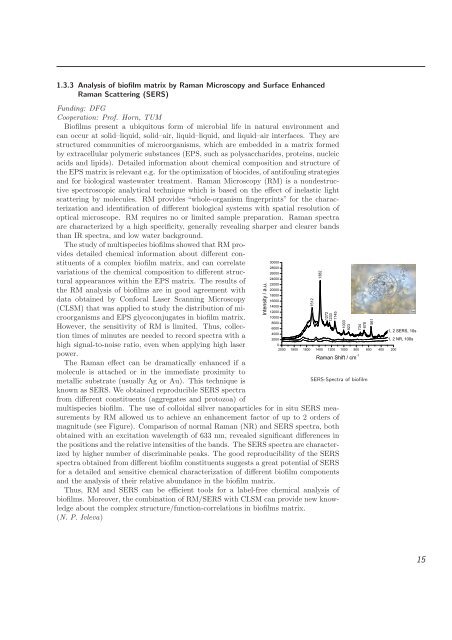
SERS-Spectra of biofilm<br />
multispecies biofilm. The use of colloidal silver nanoparticles for in situ SERS measurements<br />
by RM allowed us to achieve an enhancement factor of up to 2 orders of<br />
magnitude (see Figure). Comparison of normal Raman (NR) and SERS spectra, both<br />
obtained with an excitation wavelength of 633 nm, revealed significant differences in<br />
the positions and the relative intensities of the bands. The SERS spectra are characterized<br />
by higher number of discriminable peaks. The good reproducibility of the SERS<br />
spectra obtained from different biofilm constituents suggests a great potential of SERS<br />
for a detailed and sensitive chemical characterization of different biofilm components<br />
and the analysis of their relative ab<strong>und</strong>ance in the biofilm matrix.<br />
Thus, RM and SERS can be efficient tools for a label-free chemical analysis of<br />
biofilms. Moreover, the combination of RM/SERS with CLSM can provide new knowledge<br />
about the complex structure/function-correlations in biofilms matrix.<br />
(N. P. Ivleva)<br />
���<br />
���<br />
��������������<br />
�������������<br />
���<br />
15