ImageMaster 2D Platinum v6.0 - GeneBio
ImageMaster 2D Platinum v6.0 - GeneBio
ImageMaster 2D Platinum v6.0 - GeneBio

Create successful ePaper yourself
Turn your PDF publications into a flip-book with our unique Google optimized e-Paper software.
Part of GE Healthcare<br />
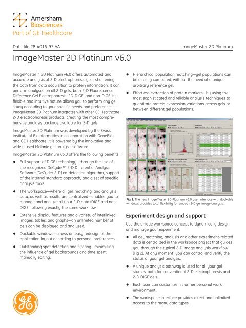
Data file 28-4016-97 AA <strong>ImageMaster</strong> <strong>2D</strong> <strong>Platinum</strong><br />
<strong>ImageMaster</strong> <strong>2D</strong> <strong>Platinum</strong> <strong>v6.0</strong><br />
<strong>ImageMaster</strong> <strong>2D</strong> <strong>Platinum</strong> <strong>v6.0</strong> offers automated and<br />
accurate analysis of 2-D electrophoresis gels, shortening<br />
the path from data acquisition to protein information. It can<br />
perform analyses on all 2-D gels, both 2-D Fluorescence<br />
Difference Gel Electrophoresis (<strong>2D</strong>-DIGE) and non-DIGE. Its<br />
flexible and intuitive nature allows you to perform any gel<br />
study according to your specific needs and preferences.<br />
<strong>ImageMaster</strong> <strong>2D</strong> <strong>Platinum</strong> integrates with other GE Healthcare<br />
2-D electrophoresis products, creating the most comprehensive<br />
analysis package available for 2-D gels.<br />
<strong>ImageMaster</strong> <strong>2D</strong> <strong>Platinum</strong> was developed by the Swiss<br />
Institute of Bioinformatics in collaboration with <strong>GeneBio</strong><br />
and GE Healthcare. It is powered by the innovative and<br />
widely used Melanie gel analysis software.<br />
<strong>ImageMaster</strong> <strong>2D</strong> <strong>Platinum</strong> <strong>v6.0</strong> offers the following benefits:<br />
• Full support of DIGE technology—through the use of<br />
the recognized DeCyder 2-D Differential Analysis<br />
Software (DeCyder 2-D) co-detection algorithm, support<br />
of the internal standard approach, and a set of specific<br />
analysis tools.<br />
• The workspace—where all gel, matching, and analysis<br />
data, as well as results are centralized—enables you to<br />
manage and analyze all your 2-D data (DIGE and non-<br />
DIGE) following exactly the same workflow.<br />
• Extensive display features and a variety of interlinked<br />
images, tables, and graphs—an unlimited number of<br />
gels can be displayed and analyzed.<br />
• Dockable windows—allows an easy redesign of the<br />
application layout according to personal preferences.<br />
• Outstanding spot detection and filtering—minimizing<br />
the influence of gel backgrounds and time spent<br />
manually editing.<br />
• Hierarchical population matching—gel populations can<br />
be directly compared, without the need of a unique<br />
arbitrary reference gel.<br />
• Effortless extraction of protein markers—by using the<br />
most sophisticated and reliable analysis techniques to<br />
quantitate protein expression variations across gels or<br />
between different gel populations.<br />
Fig 1. The new <strong>ImageMaster</strong> <strong>2D</strong> <strong>Platinum</strong> <strong>v6.0</strong> user interface with dockable<br />
windows provides total flexibility for smooth 2-D gel image analysis.<br />
Experiment design and support<br />
Use the unique workspace concept to dynamically design<br />
and manage your experiment:<br />
• All gel, matching, analysis and other experiment-related<br />
data is centralized in the workspace project that guides<br />
you through the typical 2-D image analysis workflow<br />
(Fig 2). At any moment, you can control and verify the<br />
status of your gel analysis.<br />
• A unique analysis pathway is used for all your gel<br />
studies, both for conventional 2-D electrophoresis and<br />
2-D DIGE gels.<br />
• Each user can customize his or her personal work<br />
environment.<br />
• The workspace interface provides direct and unlimited<br />
access to the many data types.
Interface<br />
The software provides a powerful and highly flexible work<br />
environment:<br />
• Analyze a virtually unlimited number of gels. Whether<br />
you work with 10, 50, or 500 images, you can display,<br />
manipulate, and process your gel data with unmatched<br />
flexibility.<br />
• The intelligent multiworksheet display guarantees a<br />
structured and consistent presentation of your gels,<br />
where related images always remain grouped and you<br />
can instantly view the desired gel subsets (Fig 3).<br />
• The application layout and gel images can be freely<br />
reorganized to optimize space and visibility in accordance<br />
with personal preferences.<br />
Image Visualization<br />
Extensive display features allow you to interact with the<br />
results and target the analyses. Comprehensive feedback<br />
through multiple images, tables, and graphs ensures that<br />
the user is fully informed of all analytical procedures, information<br />
that is critical to objective scientific investigation.<br />
Customize the display for your needs with:<br />
• Contrast mapping and pseudo colors that can be<br />
adjusted instantly.<br />
• Color palettes, spot profile, or 3-D view to inspect signal<br />
intensity (Fig 4).<br />
• Various zooming modes and easy moving of gels.<br />
• Gel overview to navigate extensively through gels.<br />
• Full control over how and what information is<br />
displayed (e.g. spot shape and color, visibility of<br />
annotations).<br />
Benefit from the comprehensive and intuitive visual<br />
comparison tools:<br />
• Tile mode instantly shows how the proteins are<br />
expressed through a series of gels.<br />
• Stack mode gives you the possibility to track protein<br />
variations by rapidly flipping between the gels.<br />
• Easily check your matching results by displaying<br />
pair vectors or superimposing spots with the<br />
overlapped mode.<br />
• Automatic warping aligns gel images, eliminating<br />
differences in spot position. Aligned images are then<br />
overlaid to produce dual channel images with clearly<br />
visible differences in protein expression.<br />
• Synchronized and simultaneous 3-D views of multiple<br />
gels can be displayed.<br />
Take advantage of the dynamic display:<br />
• Easily navigate any data, simply by clicking reports<br />
and gels.<br />
• Two-dimensional calibration grids use pI and MW<br />
values of known standards to automatically calculate<br />
values for all other spots and propagate these<br />
through all matched gels.<br />
• Raw image data is automatically loaded and<br />
unloaded when necessary to free memory and speed<br />
up the display-refreshing process.<br />
Workspace<br />
project folders<br />
Detecting<br />
Matching<br />
Analyzing<br />
Data file 28-4016-97 AA 2<br />
Gels<br />
MatchSets<br />
Classes<br />
Reports<br />
Documents<br />
Operation<br />
Exporting<br />
Fig 2. Image analysis from start to finish in the workspace.
Image Processing<br />
The cornerstones of a successful 2-D gel analysis are efficient<br />
spot detection, accurate spot quantitation and robust<br />
gel matching. Our software offers outstanding algorithms<br />
to successfully perform these steps:<br />
• Automatic, sensitive and robust spot detection requiring<br />
minimum user intervention.<br />
• The <strong>ImageMaster</strong> <strong>2D</strong> <strong>Platinum</strong> algorithm for conventional<br />
2-D gels—provides only a few easily adjustable<br />
spot detection parameters, for distinguishing real spots<br />
from noise.<br />
• The powerful co-detection algorithm from DeCyder 2-D<br />
for DIGE images—simultaneously process one, two, or<br />
three images derived from a single gel.<br />
• Optional spot editing including merging, splitting, and<br />
growing of spots.<br />
• Highly accurate spot quantitation.<br />
• Powerful parameter-free matching algorithm using spot<br />
coordinates, spot abundance, and surrounding.<br />
• Fast landmarking using special annotations for easy<br />
definition of tie points in multiple gel studies.<br />
• No more matching against a unique arbitrary reference<br />
gel. Gel populations, defined as match sets represented<br />
by a master image, can be directly compared. Matches<br />
are automatically propagated at each level of the<br />
unrestrained hierarchical match set structure.<br />
• Merging several gels from the same sample into a<br />
composite gel with fine control over the included proteins.<br />
Calibration and normalization<br />
The reproducibility of the 2-D separation process can be<br />
affected by a number of factors, including differences in<br />
sample preparation and loading, staining and image<br />
acquisition. To accurately compare the abundance of a<br />
spot across gels, it is essential to compensate for these<br />
variations using calibration or normalization methods:<br />
• Removal of image scanning variations, by<br />
intensity calibration.<br />
• Removal of variations due to staining and sample<br />
loading, by relative spot quantitation.<br />
• Compensation for varying staining absorption across<br />
proteins, by normalizing protein expression change.<br />
• Compensation for gel distortions attributable to protein<br />
migration variations, by warping gels.<br />
• Removal of interference and outliers, by using<br />
robust statistics.<br />
Fig 3. Two gel subsets side by side for visual comparison.<br />
Fig 4. 3-D view for several gel regions.<br />
Fig 5. Sophisticated analysis techniques identify and quantitate<br />
protein expression variations.<br />
Data file 28-4016-97 AA 3
Statistical analysis<br />
Extract protein markers effortlessly by using the most<br />
sophisticated and reliable analysis techniques to quantitate<br />
protein expression variations across gels or between different<br />
gel populations (Fig 5):<br />
• Scatter plots, to analyze gel similarities or experimental<br />
variations.<br />
• Descriptive statistics of central tendency and dispersion<br />
calculated and displayed in various reports and histograms.<br />
• Histograms to visualize expression profiles.<br />
• Overlapping measures that summarize each gel class<br />
by an interval and compute the overlap between these<br />
intervals.<br />
• Statistical tests (two-sample Student t-test, Mann-<br />
Whitney/Wilcoxon, Kolmogorov-Smirnov).<br />
• Factor analysis, enabling the identification of similar<br />
gels, and of spots that are characteristic for a particular<br />
population of gels.<br />
• Automatic classification by artificial intelligence<br />
techniques.<br />
• Comparative tables and graphical reports to browse<br />
through, for easy pinpointing of relevant proteins in gels.<br />
Automation<br />
• Use customizable scripts to automate repetitive tasks—<br />
by running and saving a typical analysis on a set of<br />
gels, and subsequently applying it to another set,<br />
possibly after some adjustments.<br />
• Handle large numbers of 2-D gels simultaneously.<br />
Integration<br />
The proteomics scientist works with data from many<br />
sources. Our software covers this need by providing<br />
comprehensive and unlimited annotation capabilities to<br />
link gel objects to external query engines or data sources<br />
of any format:<br />
• Default or user definable annotation categories can<br />
contain any type of information, such as pI/MW, landmark,<br />
comments, and database accession number<br />
(Fig 6).<br />
• Each annotation can be linked with Internet query<br />
engines (databases such as UniProt, SWISS-<strong>2D</strong>PAGE,<br />
and PDB) or data sources of any format (e.g. mass<br />
spectrometry profiles, text, HTML, spreadsheet, and<br />
multimedia).<br />
• A simple click on a linked annotation displays the<br />
associated data.<br />
Numerous other features enable seamless integration<br />
of our software into your laboratory workflow:<br />
• Direct analysis of image files acquired with GE<br />
Healthcare’s Ettan DIGE Imager, a scanning CCD<br />
camera designed especially for creating high quality<br />
images of 2-D DIGE gels.<br />
• Integration with GE Healthcare's ImageScanner II<br />
imager via the LabScan software (developed by the<br />
<strong>ImageMaster</strong> software team) .<br />
• Direct image acquisition from Twain-compatible<br />
scanners.<br />
• Support of all 2-D gel image formats, including GEL,<br />
IMG, TIFF, and PNG.<br />
• Open architecture based on the XML standard for<br />
import, export, and reports.<br />
• Fully automated integration with spot-picking robots.<br />
• Clipboard support to copy gel images, graphics, and<br />
data tables to other programs (e.g. tables to spreadsheet<br />
software such as Microsoft Excel, or graphics to<br />
programs like Microsoft Word or Adobe Photoshop).<br />
Fig 6. Annotations contain labels from default or user-definable categories.<br />
Data file 28-4016-97 AA 4
Reporting<br />
The software’s versatile search engine allows you to formulate<br />
complex queries without difficulty. These not only<br />
allow you to select and filter data to compose data subsets<br />
for further analysis or reporting, but will also help you to<br />
answer biological questions based on:<br />
• Sophisticated textual requests.<br />
• Qualitative and quantitative information.<br />
• Distribution of the values for a specific quantitation<br />
measure.<br />
• Protein expression changes, for example by ratio or<br />
similarity.<br />
You can visualize and analyze your data using more than<br />
20 graphical and tabular reports, which together contain<br />
over 50 data types.<br />
• All reports can be saved for further use and easily<br />
retrieved from the workspace.<br />
• Reports are customizable and editable (Fig 7).<br />
• Easy and rapid navigation between gels and associated<br />
reports and graphics, including 3D views.<br />
• Extended support allows you to import data into publications:<br />
WYSIWYP (What You See Is What You Publish).<br />
• Graphics, tables, and gel images can be printed directly<br />
as they appear or can be freely copied to any other<br />
programs.<br />
Quality and data integrity<br />
It is important to protect all your gel data, not only by<br />
ensuring data integrity and consistency, but also by allowing<br />
you to reverse undesired manipulations. Our application<br />
therefore offers:<br />
• Unique identifiers for each gel, report, and spot to<br />
assure data consistency, reliable identification across<br />
networks, and database integration.<br />
• Sophisticated multiple undo/redo function.<br />
• Experiment backup and restore function.<br />
Quality control is an important aspect of the analysis:<br />
• High quality standards are reached using margins<br />
of error and validation tools that help to distinguish<br />
between real protein expression changes and differences<br />
induced by inconsistencies in the detection or<br />
matching processes.<br />
• The history function keeps track of all operations carried<br />
out during a work session, for control and quality<br />
assurance.<br />
Fig 7. Reports are customizable and editable.<br />
Ordering information<br />
<strong>ImageMaster</strong> <strong>2D</strong> <strong>Platinum</strong> <strong>v6.0</strong> 11-0034-25<br />
DIGE Enabled, single-seat license<br />
<strong>ImageMaster</strong> <strong>2D</strong> <strong>Platinum</strong> <strong>v6.0</strong> 11-0034-29<br />
DIGE Enabled, single-seat<br />
network license<br />
<strong>ImageMaster</strong> <strong>2D</strong> <strong>Platinum</strong> <strong>v6.0</strong> 11-0034-30<br />
DIGE Enabled, additional five-seat<br />
network license<br />
<strong>ImageMaster</strong> <strong>2D</strong> <strong>Platinum</strong> <strong>v6.0</strong>, 11-0034-27<br />
single-seat license<br />
<strong>ImageMaster</strong> <strong>2D</strong> <strong>Platinum</strong> <strong>v6.0</strong>, 11-0034-31<br />
single-seat network license<br />
<strong>ImageMaster</strong> <strong>2D</strong> <strong>Platinum</strong> <strong>v6.0</strong>, 11-0034-32<br />
additional five-seat network license<br />
Other packages are available. Note: An e-license is required for access<br />
to <strong>ImageMaster</strong> <strong>2D</strong> <strong>Platinum</strong> <strong>v6.0</strong>.<br />
Related products<br />
ImageScanner II, 100–240 V 18-1170-84<br />
Ettan DIGE Imager 63-0056-42<br />
Data file 28-4016-97 AA 5
Asia Pacific Tel: +852 2811 8693 Fax: +852 2811 5251<br />
Australasia Tel: + 61 2 9899 0999 Fax: +61 2 9899 7511<br />
Austria Tel: 01/57606-1619 Fax: 01/57606-1627<br />
Belgium Tel: 0800 73 888 Fax: 03 272 1637<br />
Canada Tel: 1 800 463 5800 Fax: 1 800 567 1008<br />
Central, East, & Tel: +43 1 982 3826 Fax: +43 1 985 8327<br />
South East Europe<br />
Denmark Tel: 45 16 2400 Fax: 45 16 2424<br />
Finland & Baltics Tel: +358-(0)9-512 39 40 Fax: +358 (0)9 512 39 439<br />
France Tel: 01 69 35 67 00 Fax: 01 69 41 96 77<br />
Germany Tel: 0761/4903-490 Fax: 0761/4903-405<br />
Italy Tel: 02 27322 1 Fax: 02 27302 212<br />
Japan Tel: +81 3 5331 9336 Fax: +81 3 5331 9370<br />
www.2d-gel-analysis.com<br />
GE Healthcare<br />
Amersham Place<br />
Little Chalfont<br />
Buckinghamshire<br />
HP7 9NA<br />
UK<br />
imagination at work<br />
Latin America Tel: +55 11 3933 7300 Fax: +55 11 3933 7304<br />
Middle East & Africa Tel: +30 210 9600 687 Fax: +30 210 9600 693<br />
Netherlands Tel: 0165 580 410 Fax: 0165 580 401<br />
Norway Tel: 815 65 555 Fax: 815 65 666<br />
Portugal Tel: 21 417 7035 Fax: 21 417 3184<br />
Russia & other Tel: +7 (095) 232 0250, 956 1137 Fax: +7 (095) 230 6377<br />
C.I.S. & N.I.S<br />
South East Asia Tel: 60 3 8024 2080 Fax: 60 3 8024 2090<br />
Spain Tel: 93 594 49 50 Fax: 93 594 49 55<br />
Sweden Tel: 018 612 1900 Fax: 018 612 1910<br />
Switzerland Tel: 0848 8028 12 Fax: 0848 8028 13<br />
UK Tel: 0800 616928 Fax: 0800 616927<br />
USA Tel: +1 800 526 3593 Fax: +1 877 295 8102<br />
General Electric Company reserves the right, subject to any<br />
regulatory approval if required, to make changes in specifications<br />
and features shown herein, or discontinue the product<br />
described at any time without notice or obligation. Contact<br />
your GE Representative for the most current information.<br />
© 2005 General Electric Company—All rights reserved. GE and<br />
GE Monogram are trademarks of General Electric Company.<br />
Amersham Biosciences, DeCyder, Ettan, <strong>ImageMaster</strong>,<br />
ImageScanner, and Labscan are trademarks of GE Healthcare<br />
companies. Excel, Windows, and Word are trademarks<br />
of Microsoft Corporation. Photoshop is a trademark of<br />
Adobe Corporation.<br />
Application note 28-4016-97 AA 6