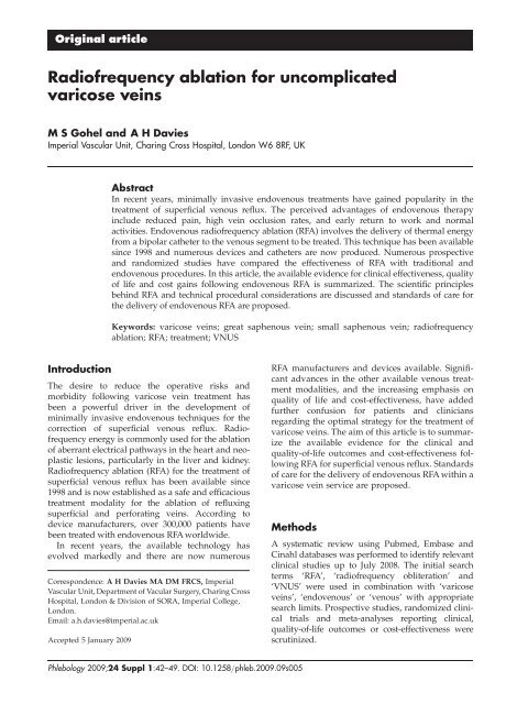
Radiofrequency ablation for uncomplicated varicose veins
Radiofrequency ablation for uncomplicated varicose veins
Radiofrequency ablation for uncomplicated varicose veins

You also want an ePaper? Increase the reach of your titles
YUMPU automatically turns print PDFs into web optimized ePapers that Google loves.
Original article<br />
<strong>Radiofrequency</strong> <strong>ablation</strong> <strong>for</strong> <strong>uncomplicated</strong><br />
<strong>varicose</strong> <strong>veins</strong><br />
M S Gohel and A H Davies<br />
Imperial Vascular Unit, Charing Cross Hospital, London W6 8RF, UK<br />
Introduction<br />
Abstract<br />
In recent years, minimally invasive endovenous treatments have gained popularity in the<br />
treatment of superficial venous reflux. The perceived advantages of endovenous therapy<br />
include reduced pain, high vein occlusion rates, and early return to work and normal<br />
activities. Endovenous radiofrequency <strong>ablation</strong> (RFA) involves the delivery of thermal energy<br />
from a bipolar catheter to the venous segment to be treated. This technique has been available<br />
since 1998 and numerous devices and catheters are now produced. Numerous prospective<br />
and randomized studies have compared the effectiveness of RFA with traditional and<br />
endovenous procedures. In this article, the available evidence <strong>for</strong> clinical effectiveness, quality<br />
of life and cost gains following endovenous RFA is summarized. The scientific principles<br />
behind RFA and technical procedural considerations are discussed and standards of care <strong>for</strong><br />
the delivery of endovenous RFA are proposed.<br />
Keywords: <strong>varicose</strong> <strong>veins</strong>; great saphenous vein; small saphenous vein; radiofrequency<br />
<strong>ablation</strong>; RFA; treatment; VNUS<br />
The desire to reduce the operative risks and<br />
morbidity following <strong>varicose</strong> vein treatment has<br />
been a powerful driver in the development of<br />
minimally invasive endovenous techniques <strong>for</strong> the<br />
correction of superficial venous reflux. <strong>Radiofrequency</strong><br />
energy is commonly used <strong>for</strong> the <strong>ablation</strong><br />
of aberrant electrical pathways in the heart and neoplastic<br />
lesions, particularly in the liver and kidney.<br />
<strong>Radiofrequency</strong> <strong>ablation</strong> (RFA) <strong>for</strong> the treatment of<br />
superficial venous reflux has been available since<br />
1998 and is now established as a safe and efficacious<br />
treatment modality <strong>for</strong> the <strong>ablation</strong> of refluxing<br />
superficial and per<strong>for</strong>ating <strong>veins</strong>. According to<br />
device manufacturers, over 300,000 patients have<br />
been treated with endovenous RFA worldwide.<br />
In recent years, the available technology has<br />
evolved markedly and there are now numerous<br />
Correspondence: A H Davies MA DM FRCS, Imperial<br />
Vascular Unit, Department of Vacular Surgery, Charing Cross<br />
Hospital, London & Division of SORA, Imperial College,<br />
London.<br />
Email: a.h.davies@imperial.ac.uk<br />
Accepted 5 January 2009<br />
RFA manufacturers and devices available. Significant<br />
advances in the other available venous treatment<br />
modalities, and the increasing emphasis on<br />
quality of life and cost-effectiveness, have added<br />
further confusion <strong>for</strong> patients and clinicians<br />
regarding the optimal strategy <strong>for</strong> the treatment of<br />
<strong>varicose</strong> <strong>veins</strong>. The aim of this article is to summarize<br />
the available evidence <strong>for</strong> the clinical and<br />
quality-of-life outcomes and cost-effectiveness following<br />
RFA <strong>for</strong> superficial venous reflux. Standards<br />
of care <strong>for</strong> the delivery of endovenous RFA within a<br />
<strong>varicose</strong> vein service are proposed.<br />
Methods<br />
Phlebology 2009;24 Suppl 1:42–49. DOI: 10.1258/phleb.2009.09s005<br />
A systematic review using Pubmed, Embase and<br />
Cinahl databases was per<strong>for</strong>med to identify relevant<br />
clinical studies up to July 2008. The initial search<br />
terms ‘RFA’, ‘radiofrequency obliteration’ and<br />
‘VNUS’ were used in combination with ‘<strong>varicose</strong><br />
<strong>veins</strong>’, ‘endovenous’ or ‘venous’ with appropriate<br />
search limits. Prospective studies, randomized clinical<br />
trials and meta-analyses reporting clinical,<br />
quality-of-life outcomes or cost-effectiveness were<br />
scrutinized.
M S Gohel and A H Davies. RFA <strong>for</strong> <strong>uncomplicated</strong> <strong>varicose</strong> <strong>veins</strong> Original article<br />
Principles of radiofrequency <strong>ablation</strong><br />
Scientific principles<br />
The underlying principle of RFA involves the delivery<br />
of thermal energy derived from an electric<br />
current to the venous segment to be treated. This<br />
is achieved using a bipolar endovenous catheter<br />
with a typical power of 2–4 W, which is used to<br />
generate temperatures of 85–1208C. As the procedure<br />
relies on direct contact between the RFA<br />
catheter and the vein wall, it is essential that the<br />
vein is emptied of blood during <strong>ablation</strong> (achieved<br />
using Trendelenberg position, use of tumescent<br />
anaesthesia and extrinsic compression). There is<br />
an in-built feedback mechanism, which evaluates<br />
the vein wall impedance and can adjust the<br />
energy delivery accordingly to ensure that the<br />
fibre temperature remains consistent.<br />
Ex vivo histological studies of venous segments<br />
treated with RFA demonstrated homogeneous<br />
intimal and medial thermal <strong>ablation</strong> and disintegration.<br />
This differed from <strong>veins</strong> treated with<br />
endovenous laser <strong>ablation</strong>, where major perivenous<br />
tissue <strong>ablation</strong> and vein wall per<strong>for</strong>ations were<br />
present. 1<br />
Available devices<br />
The current market leader <strong>for</strong> endovenous RFA<br />
devices is VNUS w Medical Technologies Inc. (San<br />
José, CA, USA). The VNUS Closure TM catheter has<br />
been available <strong>for</strong> nearly a decade and requires a<br />
continuous pullback technique. This was com-<br />
monly used with a tight compressive rubber<br />
bandage (Esmark w , Hygenic Corp., Akron, OH,<br />
USA) to facilitate venous emptying during treatment.<br />
In 2006, VNUS launched the ClosureFast TM<br />
segmental <strong>ablation</strong> catheter, which allows RFA of<br />
superficial <strong>veins</strong> in 7 cm segments, thus eliminating<br />
the continuous pull-back technique (Figure 1). The<br />
segmental <strong>ablation</strong> has the theoretical advantages<br />
of greater consistency in the vein treatment and<br />
increased speed of <strong>ablation</strong> as each 7 cm segment<br />
can be treated in 20 seconds. It should be noted<br />
that VNUS also market a specific stylet device<br />
(VNUS RFS TM stylet) <strong>for</strong> the <strong>ablation</strong> of incompetent<br />
per<strong>for</strong>ating <strong>veins</strong>. All VNUS catheters may be<br />
used with the VNUS RFG Plus TM generator<br />
(Figure 2).<br />
The Olympus Celon RFITT TM (Olympus Medical<br />
Systems, Hamburg, Germany) is an alternative<br />
RFA system which has been upgraded recently<br />
(Figure 3). The system uses the continuous pullback<br />
technique and claims to treat venous segments<br />
at a pull-back speed of 1 cm/s, although clinical<br />
studies using this new device are scarce.<br />
Description of technique<br />
Although there are broad similarities between the<br />
various endovenous therapies, the location of treatment,<br />
type of anaesthetic and numerous other<br />
factors will be dictated by the personal preference of<br />
clinicians (see Delivering a radiofrequency <strong>ablation</strong><br />
<strong>varicose</strong> vein service below). The generic technique<br />
<strong>for</strong> RFA can be summarized in the following steps:<br />
Figure 1 Illustration of the segmental <strong>ablation</strong> technique using the VNUS ClosureFast TM catheter.<br />
(Reproduced with permission from VNUS)<br />
Phlebology 2009;24 Suppl 1:42–49 43
Original article M S Gohel and A H Davies. RFA <strong>for</strong> <strong>uncomplicated</strong> <strong>varicose</strong> <strong>veins</strong><br />
(1) Preoperative planning and consent<br />
As occlusion of the treated venous segment is<br />
widely accepted as a measure of treatment<br />
success, preoperative duplex imaging should<br />
be per<strong>for</strong>med prior to any endovenous intervention.<br />
Preoperative marking of the incompetent<br />
venous segment and junction with the<br />
deep vein should also be considered, as this<br />
may aid intra-operative catheter placement.<br />
Patients should be warned about the potential<br />
risks of recurrence, nerve damage, skin burns,<br />
deep venous thermal injury and deep vein<br />
thrombosis (DVT) (see Adverse events and<br />
potential hazards below).<br />
(2) Patient positioning<br />
The patient should be positioned supine <strong>for</strong><br />
great saphenous or anterior thigh vein <strong>ablation</strong><br />
and in the prone position <strong>for</strong> <strong>ablation</strong> of small<br />
saphenous or Giacomini <strong>veins</strong>. The operating<br />
table or trolley should allow Trendelenberg<br />
and reverse Trendelenberg positioning.<br />
(3) Cannulation of vein to be treated<br />
In the reverse Trendelenberg position, the<br />
refluxing superficial vein should be cannulated<br />
with a 7F sheath using the Seldinger technique<br />
under ultrasound guidance. Ideally, the<br />
cannulation site should be at the most distal<br />
point of venous reflux and cannulation may<br />
be facilitated using proximal venous compression<br />
(to promote venous distension) or<br />
surgical cut down.<br />
(4) Positioning of radiofrequency catheter using<br />
ultrasound<br />
For the treatment of GSV or SSV reflux, device<br />
manufacturers recommend that the catheter<br />
tip should be placed no closer than 2 cm<br />
from the saphenofemoral or saphenopopliteal<br />
junctions, respectively.<br />
(5) Tumescent anaesthesia<br />
The aim of tumescent anaesthesia is to provide<br />
a thermal buffer around the vein to be treated<br />
and also offer perioperative analgesia. A<br />
dilute mixture of lidocaine in normal saline<br />
may be used. Fifty millilitres of 1% lidocaine<br />
with 1:200,000 adrenaline is used in 500 mL<br />
0.9% saline <strong>for</strong> unilateral procedures, or in<br />
1000 mL 0.9% saline <strong>for</strong> bilateral procedures.<br />
The same preparations are used <strong>for</strong> procedures<br />
per<strong>for</strong>med using local or general anaesthesia.<br />
The anaesthetic should be delivered under<br />
ultrasound guidance either by hand using a<br />
needle and syringe, or using a pump device.<br />
Volumes of tumescence administered may<br />
vary, but are commonly in the region of 75–<br />
100 mL per 10 cm of vein. Care should be<br />
taken to ensure adequate tumescent infiltration<br />
between the proximal GSV and deep vein near<br />
the saphenofemoral junction. With procedures<br />
per<strong>for</strong>med using tumescent/local anaesthesia<br />
alone, the use of local anaesthetic cream<br />
along the length of the vein to be treated may<br />
aid the administration of tumescence.<br />
(6) <strong>Radiofrequency</strong> <strong>ablation</strong><br />
For continuous pull-back systems, the catheter<br />
should be gradually withdrawn according to<br />
the recommendations of the specific catheter<br />
manufacturer. Using the VNUS Closure TM<br />
catheter, a slow infusion of heparinized saline<br />
via the catheter is recommended during pullback.<br />
With the VNUS ClosureFast TM segmental<br />
Figure 2 VNUS RFG Plus TM radiofrequency generator with<br />
ClosureFast TM segmental <strong>ablation</strong> catheter. (Reproduced with<br />
permission from VNUS) Figure 3 Olympus RFITT TM RFA generator<br />
44 Phlebology 2009;24 Suppl 1:42–49
M S Gohel and A H Davies. RFA <strong>for</strong> <strong>uncomplicated</strong> <strong>varicose</strong> <strong>veins</strong> Original article<br />
<strong>ablation</strong> catheter, the vein is ablated in 7 cm<br />
segments with extrinsic compression applied<br />
using a roll of crêpe bandage. The manufacturers<br />
recommend double treatment of the<br />
most proximal venous segment (near the SFJ)<br />
and single treatment of subsequent venous<br />
segments. Immediately following the <strong>ablation</strong>,<br />
the patency and compressibility of the femoral<br />
vein (<strong>for</strong> GSV <strong>ablation</strong>) should be verified with<br />
ultrasound.<br />
(7) Postoperative management, follow-up and outcome<br />
surveillance<br />
There is no consensus on optimal type or duration<br />
of compression following RFA, but TEDS,<br />
or class I or II above knee compression stockings<br />
worn <strong>for</strong> one to two weeks are common regimens.<br />
Recent guidelines from the American<br />
Venous Forum <strong>for</strong> the reporting of studies of<br />
endovenous treatment recommended early<br />
(,1 month) and late postoperative duplex<br />
scans. Clearly, local resource availability and<br />
clinician preference will dictate local follow-up<br />
and imaging protocols. It should be noted that<br />
the most recent NICE guidance <strong>for</strong> RFA was<br />
published in 2003 and this concluded that the<br />
procedure was safe and efficacious, but commented<br />
on the lack of long-term studies and<br />
recommended audit of outcomes.<br />
Results following radiofrequency<br />
<strong>ablation</strong><br />
When interpreting these results, it should be noted<br />
that the vast majority of published studies report<br />
outcomes using the VNUS Closure TM continuous<br />
pull-back catheter, whereas the VNUS ClosureFast TM<br />
segmental <strong>ablation</strong> catheter is the device primarily<br />
marketed by VNUS and growing in popularity.<br />
A total of 23 published reports, consisting of three randomized<br />
trials, two meta-analyses and 15 prospective<br />
observational studies were included in this review.<br />
Only one prospective case series using the VNUS<br />
ClosureFast TM catheter was identified. Details of the<br />
studies included are presented in Table 1.<br />
Technical success and clinical outcomes<br />
Although there is significant heterogeneity between<br />
clinical studies and variations in reported outcomes,<br />
recanalization of the treated venous segment is commonly<br />
reported as a marker of technical success.<br />
VNUS closure<br />
Reported vein closure rates vary between 67% and<br />
100% (Table 1). In a recent meta-analysis, the early<br />
technical success following RFA was found to be<br />
89% (3 months), reducing to 80% after five years. 2<br />
These figures compared favourably to traditional<br />
surgery and foam sclerotherapy, but were lower<br />
than endovenous laser <strong>ablation</strong>. By far, the largest<br />
patient series treated by VNUS Closure TM was published<br />
in 2005 and reported five-year clinical and anatomical<br />
outcomes following RFA in 1006 patients<br />
(1222 legs). 3 Data were collected from a prospective<br />
international registry and occlusion rates of 87.2%<br />
were seen at five years. Significant improvements in<br />
pain, fatigue and oedema were seen up to five years<br />
and interestingly, these improvements were present<br />
despite recurrent truncal reflux. Recurrent varicosities<br />
were seen in 27% of patients at five years and anatomical<br />
failure of RFAwas an independent risk factor<br />
<strong>for</strong> varicosity recurrence. 3 Within randomized<br />
studies, recurrent varicosities were seen in 48/217<br />
(22%) of patients at four years. 4<br />
VNUS ClosureFast TM<br />
The only published study reporting outcomes<br />
following VNUS ClosureFast TM was published in<br />
early 2008. The occlusion rate following segmental<br />
RFA was 99.6% at two years and 70% of treated<br />
patients did not require any analgesia postprocedure.<br />
5 A direct comparison of postoperative<br />
pain scores between RFA and laser <strong>ablation</strong> has<br />
been conducted in the Recovery trial sponsored by<br />
VNUS (unpublished data), but preliminary results<br />
showed significant lower pain scores following<br />
VNUS ClosureFast TM . 6<br />
Quality-of-life changes<br />
Studies of quality of life are scarce, but significant<br />
improvements in disease-specific quality-of-life following<br />
RFA were reported in the EVOLVeS study,<br />
using the CIVIQ2 questionnaire. 7,8 Moreover, these<br />
quality-of-life gains were greater than patients<br />
treated with traditional venous surgery.<br />
Cost-effectiveness of treatment<br />
To date, there have been few studies assessing the<br />
cost-effectiveness of RFA <strong>for</strong> the treatment of <strong>varicose</strong><br />
<strong>veins</strong>. In a small randomized study of 28<br />
patients, a basic cost-analysis demonstrated that<br />
VNUS Closure was more expensive than conventional<br />
surgery in terms of direct costs, but the<br />
Phlebology 2009;24 Suppl 1:42–49 45
Original article M S Gohel and A H Davies. RFA <strong>for</strong> <strong>uncomplicated</strong> <strong>varicose</strong> <strong>veins</strong><br />
Table 1 Published prospective clinical studies of endovenous radiofrequency <strong>ablation</strong> (RFA)<br />
Author Year Study design<br />
Luebke and Brunkwall 4<br />
van den Bos et al. 2<br />
Rautio et al. 9 / Perala et al. 12<br />
Lurie et al. 7 / Lurie et al. 8<br />
Hinchliffe et al. 13<br />
Chandler et al. 14<br />
Manfrini et al. 15<br />
Goldman and Amiry 16<br />
Merchant et al. 17 / Merchant<br />
and Pichot 3<br />
Sybrandy and Wittens 18<br />
Weiss and Weiss 19<br />
Fassiadis et al. 20<br />
Hingorani et al. 11<br />
Pichot et al. 21<br />
Ogawa et al. 22<br />
Nicolini 23<br />
Kianifard et al. 24<br />
Dunn et al. 25<br />
Zan et al. 26<br />
Proebstle et al. 5<br />
costs to society were significantly lower in view of<br />
the rapid return to work in the RFA group. 9<br />
A subsequent decision-tree analysis also suggested<br />
that RFA per<strong>for</strong>med in an office setting may be<br />
cheaper than traditional surgery. 10 Clearly, the<br />
cost of procedures will be heavily influenced by<br />
clinician preferences on type of anaesthetic, the<br />
use of concomitant ambulatory phlebectomy and<br />
whether to per<strong>for</strong>m bilateral procedures in a<br />
single sitting, thus saving the cost of an additional<br />
RFA catheter. For the treatment of unilateral great<br />
saphenous vein reflux, office-based RFA using<br />
only tumescent anaesthesia is likely to be cheaper<br />
than traditional GSV-stripping under general anaesthesia,<br />
and this has been the main component of the<br />
NHS business case produced by VNUS to support<br />
the use of their catheters. However, further welldesigned<br />
studies of cost-effectiveness are urgently<br />
needed to guide those having to make decisions<br />
about treatment commissioning and rationing.<br />
Potential hazards and adverse events<br />
As with all endovenous interventions, RFA may be<br />
associated with technical difficulties in cannulation,<br />
guide wire and catheter advancement. These issues<br />
are likely to resolve with increasing experience and<br />
familiarity with the equipment and technique.<br />
Specific complications including DVT, skin burns,<br />
superficial thrombophlebitis, neuralgia and bruising<br />
have been reported following RFA, but the<br />
RFA sample<br />
size (limbs)<br />
Follow-up<br />
(months)<br />
incidence of these problems appears low. In one<br />
study, 12/73 limbs (16%) developed DVT within<br />
30 days of RFA, 11 but the incidence of DVT in the<br />
majority of studies is ,1%. Of the 1006 patients<br />
recorded in the Closure international registry, the<br />
reported complications were DVT (0.9%), phlebitis<br />
(2.9%) and skin burn (1.2%), although the majority<br />
of burns occurred in patients not given tumescence.<br />
3 The influence of the volume of tumescence<br />
and post-procedure compression on the incidence<br />
of these complications is poorly understood and<br />
warrants further investigation.<br />
Training implications<br />
Occlusion<br />
rate (%)<br />
Adverse events<br />
There is currently no structured pathway <strong>for</strong> the<br />
training of clinicians to per<strong>for</strong>m endovenous <strong>ablation</strong><br />
procedures. Those planning to per<strong>for</strong>m RFA<br />
should ensure familiarity with the energy generator<br />
and treatment catheters and be able to demonstrate<br />
competence in the use of intra-operative colour<br />
duplex ultrasound. Depending on local resources<br />
and knowledge, attendance at clinical workshops<br />
and courses or clinical mentorship programmes<br />
may help to achieve competence.<br />
Delivering a radiofrequency <strong>ablation</strong><br />
<strong>varicose</strong> vein service<br />
DVT Skin burn<br />
2008 Meta-analysis 315 36 81 – –<br />
2008 Meta-analysis n/a 60 80 – –<br />
2002/2005 RCT 15 36 67 0/15 0/15<br />
2003/2005 RCT 65 24 86 0/65 –<br />
2006 RCT 16 1 81 0/16 –<br />
2000 Prospective 120 12 90 0/120 –<br />
2000 Prospective 151 6 96 0/151 –<br />
2002 Prospective 50 24 68 0/50 –<br />
2002/2005 Prospective 1222 60 87 11/1222 15/1222<br />
2002 Prospective 26 12 88 – 1/26<br />
2002 Prospective 140 24 90 0/21 0/21<br />
2003 Prospective 59 12 98 – –<br />
2004 Prospective 73 1 96 12/73 –<br />
2004 Prospective 63 25 90 – –<br />
2005 Prospective 25 1 100 0/25 –<br />
2005 Prospective 330 36 75 0/330 –<br />
2006 Prospective 51 12 100 – –<br />
2006 Prospective 85 6 90 0/85 0/85<br />
2007 Prospective 24 24 96 – –<br />
2008 Prospective 252 6 99.6 0/252 0/252<br />
RCT, randomized clinical trial. Study of segmental RFA using VNUS ClosureFast<br />
46 Phlebology 2009;24 Suppl 1:42–49<br />
The precise <strong>for</strong>mat of a RFA <strong>varicose</strong> vein service<br />
will depend heavily on the personal preference of
M S Gohel and A H Davies. RFA <strong>for</strong> <strong>uncomplicated</strong> <strong>varicose</strong> <strong>veins</strong> Original article<br />
clinicians. Specific factors dependent on clinician<br />
preference include:<br />
† Type of anaesthetic;<br />
† Treatment strategy <strong>for</strong> varicosities;<br />
† Approach to patients requiring bilateral interventions;<br />
† Postprocedure compression type and duration;<br />
† Follow-up and surveillance protocol.<br />
Nevertheless, a number of general key issues<br />
should be considered and addressed when planning,<br />
starting and delivering a RFA <strong>varicose</strong> vein<br />
service. These include:<br />
Training and assessment of competence<br />
The responsible clinician should ensure appropriate<br />
theoretical and practical training <strong>for</strong> core team<br />
members prior to starting a service. Local vascular<br />
scientists may be an excellent resource <strong>for</strong> the acquisition<br />
of duplex ultrasound skills and it may be<br />
appropriate to per<strong>for</strong>m initial cases with a vascular<br />
scientist per<strong>for</strong>ming the imaging. Mentorship<br />
arrangements involving experienced practitioners<br />
and departments with a large throughput of RFA<br />
procedures may be useful strategies to acquire<br />
and rein<strong>for</strong>ce new endovenous skills.<br />
Trust approval and funding<br />
The clinical team may be required to undertake a<br />
risk assessment and produce a business case to<br />
justify the introduction of a new technique. Proposals<br />
should be discussed with Clinical Governance<br />
and Clinical Safety departments. Device manufacturers<br />
usually have considerable experience of this<br />
process and may be of assistance.<br />
Multidisciplinary team approach<br />
Vascular surgeons, vascular scientists, theatre and<br />
outpatient nursing staff are likely to be key<br />
members of the multidisciplinary team. They<br />
should be consulted and involved at each stage of<br />
the planning, training and service implementation<br />
processes.<br />
Patient selection and consent<br />
The process and risks of RFA should be openly discussed<br />
with all patients as well as the level of<br />
experience of the clinical team. Clinical in<strong>for</strong>mation<br />
leaflets should be produced (using local in<strong>for</strong>mation<br />
guidelines) and given to prospective patients.<br />
Patients could be directed to the wealth of<br />
online in<strong>for</strong>mation available on endovenous<br />
procedures.<br />
Location of treatment<br />
Treatment may be offered in an operating theatre or<br />
in an office-based or adapted clinic room setting.<br />
The treatment of patients outside the expensive<br />
operating theatre setting may seem appealing, but<br />
it is essential to ensure that any area considered<br />
<strong>for</strong> RFA treatment has adequate space, lighting,<br />
access to equipment and other essential facilities.<br />
In particular, the availability of an appropriate operating<br />
table or bed is essential.<br />
Treatment algorithm<br />
To ensure that the provision of RFA is consistent,<br />
treatment algorithms <strong>for</strong> the management of<br />
patients with <strong>varicose</strong> <strong>veins</strong> should be locally<br />
agreed and disseminated.<br />
Clinical follow-up, outcome evaluation and audit<br />
Specific follow-up protocols will be dictated by<br />
individual clinician preference, but ef<strong>for</strong>ts should<br />
be made to audit technical and clinical outcomes<br />
as with all new interventions.<br />
Proposed standards <strong>for</strong> endovenous<br />
radiofrequency <strong>ablation</strong><br />
There are currently no clinical guidelines specifying<br />
basic standards in the provision of RFA <strong>for</strong> superficial<br />
venous reflux. In addition to the principles<br />
of Clinical Governance, the following benchmarks<br />
are proposed as minimum standards of clinical<br />
practice:<br />
† Intra-operative colour duplex imaging should<br />
be per<strong>for</strong>med by an individual able to demonstrate<br />
technical competence in vascular duplex<br />
imaging;<br />
† Pre-operative duplex imaging should be per<strong>for</strong>med<br />
by an accredited vascular scientist (or<br />
appropriately trained clinician) in all cases;<br />
† Procedural documentation should include:<br />
† Length of vein ablated;<br />
† The vein treated and precise site of cannulation;<br />
† Duration of treatment;<br />
Phlebology 2009;24 Suppl 1:42–49 47
Original article M S Gohel and A H Davies. RFA <strong>for</strong> <strong>uncomplicated</strong> <strong>varicose</strong> <strong>veins</strong><br />
† Details of energy delivered, power settings<br />
and other variables.<br />
Post-procedural follow-up is controversial, but<br />
should ideally include clinical assessment and<br />
colour duplex imaging to evaluate technical success.<br />
This is particularly important during the learning<br />
phase as an objective assessment of quality control.<br />
Clearly, this policy will be strongly influenced by<br />
local resource availability and a more pragmatic<br />
approach of scanning only symptomatic patients<br />
may be preferred. However, as RFA <strong>for</strong> the treatment<br />
of <strong>varicose</strong> <strong>veins</strong> is a novel technique, objective evaluation<br />
of technical success may be desirable. Reasons<br />
<strong>for</strong> not implementing a policy of routine postoperative<br />
duplex imaging include cost and also the fact<br />
that that main determinant of subsequent therapy is<br />
likely to be the presence of symptoms, rather than<br />
the presence of venous reflux.<br />
Clinicians should prospectively collect audit data<br />
including procedural details, adverse events, clinical<br />
and technical outcomes. This may be best<br />
achieved by using an international registry.<br />
Conclusion<br />
RFA is established as an acceptable and efficacious<br />
endovenous treatment modality <strong>for</strong> the treatment of<br />
<strong>varicose</strong> <strong>veins</strong>. However, further studies are needed<br />
to clarify the advantages of the recently introduced<br />
segmental <strong>ablation</strong> VNUS ClosureFast TM catheter,<br />
particularly in terms of postoperative pain and bruising.<br />
Although many treatment variables are highly<br />
dependent on clinician preference, principles <strong>for</strong> the<br />
safe introduction of a RFA service and standards of<br />
care are proposed in this document. Clinicians per<strong>for</strong>ming<br />
RFA <strong>for</strong> the treatment of <strong>varicose</strong> <strong>veins</strong><br />
should ensure accurate audit of interventions and<br />
outcomes. Further consensus is needed on the<br />
optimal post-procedural treatment regimen and<br />
follow-up surveillance in this patient group.<br />
Conflict of interest<br />
The authors hereby declare no conflict of interests.<br />
References<br />
1 Schmedt CG, Sroka R, Steckmeier S, et al. Investigation<br />
on radiofrequency and laser (980 nm) effects after<br />
endoluminal treatment of saphenous vein insufficiency<br />
in an ex-vivo model. Eur J Vasc Endovasc Surg 2006;<br />
32:318–25<br />
48 Phlebology 2009;24 Suppl 1:42–49<br />
2 van den Bos R, Arends L, Kockaert M, Neumann M,<br />
Nijsten T. Endovenous therapies of lower extremity varicosities<br />
are at least as effective as surgical stripping or<br />
foam sclerotherapy: meta-analysis and meta-regression<br />
of case series and randomized clinical trials. J Vasc<br />
Surg 2008 [published ahead of print]<br />
3 Merchant RF, Pichot O. Long-term outcomes of endovenous<br />
radiofrequency obliteration of saphenous reflux<br />
as a treatment <strong>for</strong> superficial venous insufficiency.<br />
J Vasc Surg 2005;42:502–9, discussion 9<br />
4 Luebke T, Brunkwall J. Systematic review and<br />
meta-analysis of endovenous radiofrequency obliteration,<br />
endovenous laser therapy, and foam sclerotherapy<br />
<strong>for</strong> primary varicosis. J Cardiovasc Surg (Torino) 2008;49:<br />
213–33<br />
5 Proebstle TM, Vago B, Alm J, Gockeritz O, Lebard C,<br />
Pichot O. Treatment of the incompetent great saphenous<br />
vein by endovenous radiofrequency powered segmental<br />
thermal <strong>ablation</strong>: first clinical experience. J Vasc Surg<br />
2008;47:151–6<br />
6 Almeida J, Raines J, Kaufman J, et al. Results from the Recovery<br />
Trial. 2008. See http://www.vnus.com/navigation/<br />
physician_recovery.htm (last accessed 15 January 2009)<br />
7 Lurie F, Creton D, Eklof B, et al. Prospective randomized<br />
study of endovenous radiofrequency obliteration<br />
(closure procedure) versus ligation and stripping in a<br />
selected patient population (EVOLVeS Study). J Vasc<br />
Surg 2003;38:207–14<br />
8 Lurie F, Creton D, Eklof B, et al. Prospective randomised<br />
study of endovenous radiofrequency obliteration<br />
(closure) versus ligation and vein stripping (EVOLVeS):<br />
two-year follow-up. Eur J Vasc Endovasc Surg 2005;29:<br />
67–73<br />
9 Rautio T, Ohinmaa A, Perala J, et al. Endovenous obliteration<br />
versus conventional stripping operation in the<br />
treatment of primary <strong>varicose</strong> <strong>veins</strong>: a randomized<br />
controlled trial with comparison of the costs. J Vasc<br />
Surg 2002;35:958–65<br />
10 Gohel MS, Hamish M, Davies AH. The cost of traditional<br />
and endovenous treatments <strong>for</strong> <strong>varicose</strong> <strong>veins</strong>:<br />
a decision tree economic analysis. Phlebology 2008;23:<br />
196–202<br />
11 Hingorani AP, Ascher E, Markevich N, et al. Deep<br />
venous thrombosis after radiofrequency <strong>ablation</strong> of<br />
greater saphenous vein: a word of caution. J Vasc Surg<br />
2004;40:500–4<br />
12 Perala J, Rautio T, Biancari F, et al. <strong>Radiofrequency</strong> endovenous<br />
obliteration versus stripping of the long saphenous<br />
vein in the management of primary <strong>varicose</strong><br />
<strong>veins</strong>: 3-year outcome of a randomized study. Ann Vasc<br />
Surg 2005;19:669–72<br />
13 Hinchliffe RJ, Ubhi J, Beech A, Ellison J, Braithwaite BD.<br />
A prospective randomised controlled trial of VNUS<br />
closure versus surgery <strong>for</strong> the treatment of recurrent<br />
long saphenous <strong>varicose</strong> <strong>veins</strong>. Eur J Vasc Endovasc<br />
Surg 2006;31:212–8<br />
14 Chandler JG, Pichot O, Sessa C, Schuller-Petrovic S, Osse<br />
FJ, Bergan JJ. Defining the role of extended saphenofemoral<br />
junction ligation: a prospective comparative<br />
study. J Vasc Surg 2000;32:941–53<br />
15 Manfrini S, Gasbarro V, Danielsson G, et al. Endovenous<br />
management of saphenous vein reflux. Endovenous<br />
Reflux Management Study Group. J Vasc Surg 2000;32:<br />
330–42
M S Gohel and A H Davies. RFA <strong>for</strong> <strong>uncomplicated</strong> <strong>varicose</strong> <strong>veins</strong> Original article<br />
16 Goldman MP, Amiry S. Closure of the greater saphenous<br />
vein with endoluminal radiofrequency thermal heating<br />
of the vein wall in combination with ambulatory<br />
phlebectomy: 50 patients with more than 6-month<br />
follow-up. Dermatol Surg 2002;28:29–31<br />
17 Merchant RF, DePalma RG, Kabnick LS. Endovascular<br />
obliteration of saphenous reflux: a multicenter study.<br />
J Vasc Surg 2002;35:1190–6<br />
18 Sybrandy JE, Wittens CH. Initial experiences in endovenous<br />
treatment of saphenous vein reflux. J Vasc Surg<br />
2002;36:1207–12<br />
19 Weiss RA, Weiss MA. Controlled radiofrequency endovenous<br />
occlusion using a unique radiofrequency catheter<br />
under duplex guidance to eliminate saphenous<br />
<strong>varicose</strong> vein reflux: a 2-year follow-up. Dermatol Surg<br />
2002;28:38–42<br />
20 Fassiadis, Holdstock, Whiteley. Endoluminal radiofrequency<br />
<strong>ablation</strong> of the long saphenous vein (VNUS<br />
closure) – a minimally invasive management of <strong>varicose</strong><br />
<strong>veins</strong>. Minim Invasive Ther Allied Technol 2003;12:91–4<br />
21 Pichot O, Kabnick LS, Creton D, Merchant RF,<br />
Schuller-Petroviae S, Chandler JG. Duplex ultrasound<br />
scan findings two years after great saphenous vein<br />
radiofrequency endovenous obliteration. J Vasc Surg.<br />
2004;39:189–95<br />
22 Ogawa T, Hoshino S, Midorikawa H, Sato K. Clinical<br />
results of radiofrequency endovenous obliteration <strong>for</strong><br />
<strong>varicose</strong> <strong>veins</strong>. Surg Today 2005;35:47–51<br />
23 Nicolini P. Treatment of primary <strong>varicose</strong> <strong>veins</strong> by endovenous<br />
obliteration with the VNUS closure system:<br />
results of a prospective multicentre study. Eur J Vasc<br />
Endovasc Surg 2005;29:433–9<br />
24 Kianifard B, Holdstock JM, Whiteley MS. <strong>Radiofrequency</strong><br />
<strong>ablation</strong> (VNUS closure) does not cause neovascularisation<br />
at the groin at one year: results of a<br />
case controlled study. Surgeon 2006;4:71–4<br />
25 Dunn CW, Kabnick LS, Merchant RF, Owens R, Weiss<br />
RA. Endovascular radiofrequency obliteration using<br />
90 degrees C <strong>for</strong> treatment of great saphenous vein.<br />
Ann Vasc Surg 2006;20:625–9<br />
26 Zan S, Contessa L, Varetto G, et al. <strong>Radiofrequency</strong> minimally<br />
invasive endovascular treatment of lower limbs<br />
<strong>varicose</strong> <strong>veins</strong>: clinical experience and literature<br />
review. Minerva Cardioangiol 2007;55:443–58<br />
Phlebology 2009;24 Suppl 1:42–49 49