Parasite-host relationship: a lesson from a professional killer
Parasite-host relationship: a lesson from a professional killer
Parasite-host relationship: a lesson from a professional killer

Create successful ePaper yourself
Turn your PDF publications into a flip-book with our unique Google optimized e-Paper software.
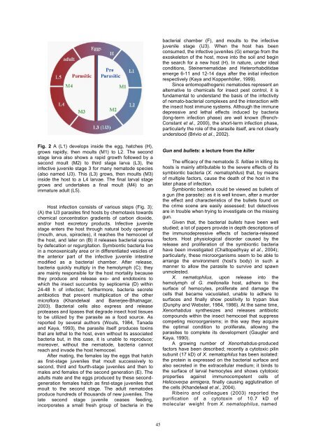
Fig. 2 A (L1) develops inside the egg, hatches (H),<br />
grows rapidly, then moults (M1) to L2. The second<br />
stage larva also shows a rapid growth followed by a<br />
second moult (M2) to third stage larva (L3), the<br />
infective juvenile stage 3 for many nematode species<br />
(also named IJ3). This (L3) grows, then moults (M3)<br />
inside the <strong>host</strong> to a L4 larvae. The final larval stage<br />
grows and undertakes a final moult (M4) to an<br />
immature adult (L5).<br />
Host infection consists of various steps (Fig. 3);<br />
(A) the IJ3 parasites find <strong>host</strong>s by chemotaxis towards<br />
chemical concentration gradients of carbon dioxide,<br />
and/or <strong>host</strong> excretory products. Infective juvenile<br />
stage enters the <strong>host</strong> through natural body openings<br />
(mouth, anus, spiracles), it reaches the hemocoel of<br />
the <strong>host</strong>, and later on (B) it releases bacterial spores<br />
by defecation or regurgitation. Symbiontic bacteria live<br />
in a monoxenically area or in differentiated vesicles of<br />
the anterior part of the infective juvenile intestine<br />
modified as a bacterial chamber. After release,<br />
bacteria quickly multiply in the hemolymph (C); they<br />
are mainly responsible for the <strong>host</strong> mortality because<br />
they produce and release exo- and endotoxins to<br />
which the insect succumbs by septicemia (D) within<br />
24-48 h of infection; furthermore, bacteria secrete<br />
antibiotics that prevent multiplication of the other<br />
microflora (Khandelwal and Banerjee-Bhatnagar,<br />
2003). Bacterial cells also express and release<br />
proteases and lipases that degrade insect <strong>host</strong> tissues<br />
to be utilized by the parasite as a food source. As<br />
reported by several authors (Wouts, 1984, Tanada<br />
and Kaya, 1993), the parasite itself produces toxins<br />
that are lethal to the <strong>host</strong>, even without its associated<br />
bacteria but, in this case, it is unable to reproduce;<br />
moreover, without the nematode, bacteria cannot<br />
reach and invade the <strong>host</strong> hemocoel.<br />
After mating, the females lay the eggs that hatch<br />
as first-stage juveniles that moult successively to<br />
second, third and fourth-stage juveniles and then to<br />
males and females of the second generation (E). The<br />
adults mate and the eggs produced by these secondgeneration<br />
females hatch as first-stage juveniles that<br />
moult to the second stage. The adult nematodes<br />
produce hundreds of thousands of new juveniles. The<br />
late second stage juvenile ceases feeding,<br />
incorporates a small fresh group of bacteria in the<br />
45<br />
bacterial chamber (F), and moults to the infective<br />
juvenile stage (IJ3). When the <strong>host</strong> has been<br />
consumed, the infective juveniles (G) emerge <strong>from</strong> the<br />
exoskeleton of the <strong>host</strong>, move into the soil and begin<br />
the search for a new <strong>host</strong> (H). In nature, under ideal<br />
conditions, Steinernematidae and Heterorhabditidae<br />
emerge 6-11 and 12-14 days after the initial infection<br />
respectively (Kaya and Koppenhöfer, 1999).<br />
Since entomopathogenic nematodes represent an<br />
alternative to chemicals for insect pest control, it is<br />
fundamental to understand the basis of the infectivity<br />
of nemato-bacterial complexes and the interaction with<br />
the insect <strong>host</strong> immune systems. Although the immune<br />
depressive and lethal effects induced by bacteria<br />
(long-term infection phase) are well known (ffrench-<br />
Constant et al., 2000), the short-term infection phase,<br />
particularly the role of the parasite itself, are not clearly<br />
understood (Brivio et al., 2002).<br />
Gun and bullets: a lecture <strong>from</strong> the <strong>killer</strong><br />
The efficacy of the nematode S. feltiae in killing its<br />
<strong>host</strong>s is mainly attributable to the severe effects of its<br />
symbiontic bacteria (X. nematophilus) that, by means<br />
of multiple factors, cause the death of the <strong>host</strong> in the<br />
later phase of infection.<br />
Symbiontic bacteria could be viewed as bullets of<br />
a gun (the parasite): as it is well known, after a murder<br />
the effect and characteristics of the bullets found on<br />
the crime scene are easily assessed; but detectives<br />
are in trouble when trying to investigate on the missing<br />
gun.<br />
Given that, the bacterial bullets have been well<br />
studied; a lot of papers provide in depth descriptions of<br />
the immunodepressive effects of bacteria-released<br />
factors. Host physiological disorder caused by the<br />
release and proliferation of the symbiontic bacteria<br />
have been investigated (Chattopadhyay et al., 2004);<br />
particularly, these microorganisms seem to be able to<br />
arrange the environment (<strong>host</strong>’s body) in such a<br />
manner to allow the parasite to survive and spawn<br />
unmolested.<br />
X. nematophilus, upon release into the<br />
hemolymph of G. mellonella <strong>host</strong>, adhere to the<br />
surface of hemocytes, proliferate and damage the<br />
cells that became vacuolated, unable to adhere to<br />
surfaces and finally show positivity to trypan blue<br />
(Dunphy and Webster, 1984, 1986). At the same time,<br />
Xenorhabdus synthesizes and releases antibiotic<br />
compounds within the insect hemocoel that suppress<br />
competing microorganisms; in this way they acquire<br />
the optimal condition to proliferate, allowing the<br />
parasites to complete its development (Gaugler and<br />
Kaya, 1990).<br />
A growing number of Xenorhabdus-produced<br />
factors have been described; recently a cytotoxic pilin<br />
subunit (17 kD) of X. nematophilus has been isolated:<br />
the protein is expressed on the bacterial surface and<br />
also secreted in the extracellular medium; it binds to<br />
the surface of larval hemocytes and shows cytotoxic<br />
properties against immunocompetent cells of<br />
Helicoverpa armigera, finally causing agglutination of<br />
the cells (Khandelwal et al., 2004).<br />
Ribeiro and colleagues (2003) reported the<br />
purification of a cytotoxin of 10.7 kD of<br />
molecular weight <strong>from</strong> X. nematophilus, named