

The Frenular Delta - NORM
The Frenular Delta - NORM
The Frenular Delta - NORM

You also want an ePaper? Increase the reach of your titles
YUMPU automatically turns print PDFs into web optimized ePapers that Google loves.
From: Understanding Circumcision: A Multi-Disciplinary Approach to a Multi-Dimensional Problem<br />
Denniston GC, Hodges FM, Milos MF eds.<br />
Kluwer Academic/Plenum Publishers, New York, 2001.<br />
Chapter 11<br />
<strong>The</strong> <strong>Frenular</strong> <strong>Delta</strong><br />
A New Preputial Structure<br />
Ken McGrath<br />
Senior Lecturer in Pathology, Faculty of Health Studies, Auckland University of Technology<br />
Abstract: Textbooks and papers referring to penile function state that the source of penile sensation<br />
is solely the glans and often justify the existence of the prepuce by stating it protects the<br />
'sensitive' glans. <strong>The</strong>se statements are contrary to the neuro-anatomical and physiological<br />
facts accumulated over more than a century. This study reviews the findings of Taylor, et<br />
al., that the prepuce is the primary sensory platform of the penis, and describes a new<br />
preputial structure.<br />
INTRODUCTION<br />
Looking back, it is astounding that the complex nature of the prepuce (or the fact that it is not a simple<br />
fold of skin) was not described until 1991 1 and that this did not appear in the mainstream medical media<br />
until 1996 2 When Dr. John Taylor faced family enquiries, he asked himself: 'What exactly is removed by<br />
circumcision?' A total absence of an answer to that question from the medical literature was the<br />
motivation for his investigation. <strong>The</strong> reason why this had never been asked, during a century of extensive<br />
study of the male genitalia and increasing application of circumcision, remains a mystery. No special<br />
techniques, or dissections, or equipment were initially required — just an observant eye and scientific<br />
curiosity.<br />
1. REVIEW OF PREPUTIAL ANATOMY<br />
When Taylor and his colleagues examined 22 prepuces in males aged 22 to 58 years, 2 they discovered<br />
that the classical description, which simply divided the prepuce into outer and inner (or mucosal) areas, 3<br />
was incomplete. <strong>The</strong> outer area is a continuation of the shaft skin, up to the rim of junction with the<br />
mucosa at the opening of the prepuce at rest, with all its characteristics of form and mild sensitivity. On<br />
the other hand, the mucosa, that inner preputial skin from the junctional or transitional zone to the sulcus<br />
where it continues as the mucosal covering of the glans, he found to be more sophisticated than had<br />
been previously described. Just inside the junctional rim of the prepuce at the dorsal aspect is a<br />
transverse band of ridges 10-15 mm wide. Beyond this ridged band, the remainder of the mucosa is<br />
smooth (15-30 mm wide dorsally) — the smooth zone. <strong>The</strong> ridged band is continuous with the frenulum,<br />
being a radial band over about 60% of the penile diameter — the dorsal and lateral aspects — then<br />
turning distally to form a 'V’ shape in the ventral aspect whilst smoothly reducing its width to merge with<br />
the frenulum. This continuity is the reason why the ridged band is also known as the frenular band.<br />
Under microscopic examination, the entire mucosa is invested with Meissner's corpuscles (the nerve<br />
endings which mediate low-threshold and fine-touch sensation) which are heavily concentrated in the<br />
projections of the dermis that rise into the ridged band and in the frenulum. <strong>The</strong>se corpuscles are<br />
innervated by thick myelinated axons of the epicritical sensory system. A recent quantitative study has<br />
shown mean axon bundle counts in the prepuce of 17.9 bundles/mm ventrally, 8.6 bundles/mm laterally<br />
and 6.2 bundles/mm dorsally.4 As each bundle contains tens of axons, the nerve supply to the prepuce<br />
is obviously very substantial.
Another feature of the inner prepuce not previously described prior to Taylor's reports is the extensive<br />
plexus of venules and capillaries that are collected into the large superficial veins of the outer prepuce<br />
and penile shaft skin. <strong>The</strong>se superficial veins begin to divide as they approach the prepuce and have<br />
frequent anastomoses especially in the outer prepuce and distal shaft skin. <strong>The</strong> anastomoses in the<br />
prepuce have sometimes been termed 'radial veins' because, in some individuals, they are<br />
circumferential. From the junctional rim, the many smaller veins divide frequently as they run towards the<br />
sulcus to form a dense plexus of venules and capillaries. This vascularity was noted by Taylor as being<br />
pronounced in the ridged band and often seen as a bright flush. 2<br />
In most genitally intact males, some core branches of the superficial veins do not divide further on<br />
crossing the inner prepuce. <strong>The</strong>se medium- sized veins continue unchanged to run under the corona to<br />
form at least part of the drainage of the glans surface. This is contrary to a report by Breza, et al., that<br />
describes the superficial veins as draining the shaft skin and prepuce only. 5 This otherwise excellent<br />
and extensive paper is flawed by not reporting the state (intact or circumcised) of the cadaver penises<br />
examined. It is reasonable to suppose that, in California, all, or the majority, were circumcised and thus<br />
could not present the normal anatomy of the superficial veins. Clearly, the volume of blood gathered by<br />
these veins, and their consequent prominence, if greater than could be involved solely in drainage of this<br />
skin. Furthermore, the superficial veins become more prominent with erection, suggesting that they may<br />
be carrying extra blood from the glans portion of the corpus spongiosum. It may be postulated that (1)<br />
as circumcision interrupts this venous circuit and (2) as superficial veins are not restored in the great<br />
majority of preputial remnants, the change in the color of the glans from a venous purple in genitally<br />
intact males to the pink tint of the penile shaft in circumcised males is due to a dramatic change in<br />
circumcised males due to a dramatic change in the circulation in the circumcised penis.<br />
<strong>The</strong> other major feature of the prepuce noted by Taylor, which it shares with the shaft skin, is a layer of<br />
smooth muscle bundles ..2 This peripenic muscle has been known since at least 1907 and is the<br />
continuation of the dartos muscle of the scrotal skin. 6 In the shaft skin, these muscle bundles are<br />
oriented in mixed direction but they become specifically oriented in the prepuce to add to its complexity.<br />
In the outer prepuce, the bundles are aligned longitudinally to mediate the length of that portion of skin.<br />
At the tip of the prepuce (i.e., in the junctional zone), the muscle bundles are radially oriented to form a<br />
kind of sphincter which tends to close the orifice when the penis is flaccid. 6 Together with abundant<br />
elastic fibers, this muscle layer give the prepuce a very snug fit over the glans. In the absence of<br />
construction of the preputial orifice (a preputial stenosis or true pathological — as opposed to<br />
physiological — ‘phimosis’, which can cause back-flow of the urinary stream), all extraneous matter<br />
(including urine, sand, etc.) Is prevented from gaining access to the preputial space.<br />
Finally, a comment on what is probably the longest enduring myth in anatomy. To the present day,<br />
textbooks still asset the presence of glands in the mucosa of the sulcus, especially on both sides of the<br />
frenulum, despite the existence of these so-called Tyson’s glands being rejected by many European<br />
investigators as far back as 1884. 8 <strong>The</strong> problem has arisen because of the finding of Tyson’s paper of<br />
1699 on the orangutan was interpreted as applying to humans, which it does not. 9 No actual description<br />
of them in humans exists and a number of modern investigations have failed to find any glands in the<br />
inner prepuce or sulcus. 10-13 My colleague, Christopher Cold, and I have also failed to find any glands in<br />
ths region. <strong>The</strong> 1904 comment of Keith and Shillitoe best sums up this myth: ‘It shows the strength of<br />
the faith of anatomists that they have for nearly two centuries described glands which have not, and<br />
never had, an existence.’ 8 It remains to observe that smegma is epithelial debris shed from the mucosa<br />
suspended in a proteinaceous fluid; 12,14 Taylor and I believe this fluid to be a transudate — extracellular<br />
fluid that leaks out from betwen cells that have no tight junctions. 15<br />
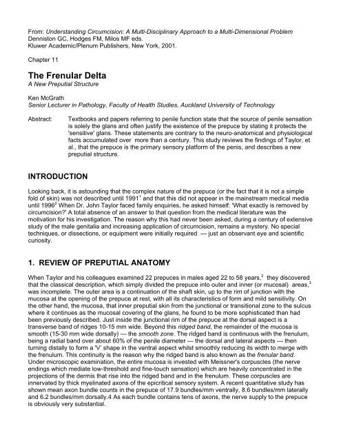
2. THE FRENULAR DELTA
Some months ago, whilst rereading the report of Taylor, et al., I realized that their description and<br />
illustrations of the inner prepuce and ridged band were confined to the dorsal aspect. 2 <strong>The</strong> frenular<br />
attachment of the ridged band was described, but there was no other mention or an illustration of the<br />
ventral aspect.<br />
<strong>The</strong> ridged band is not circumferential over the ventral aspect, since it curves distally to merge with the<br />
frenulum, as noted above. This means that the smooth zone is progressively reduced as the median<br />
raphe is approached until it disappears at the mid-line. As the smooth zone is reduced, a corresponding<br />
zone of the inner prepuce appears proximally between the ridged band and the circumferential junctional<br />
rim. Because the length of the inner prepuce actually increases as the median raphe is approached due<br />
to the curving of the corona towards the meatus to form the ventral cleft in the glans, the mucosal area of<br />
the prepuce is also greater in the ventral aspect. Thus, in the ventral prepuce, there is an undescribed<br />
zone that occupied about 20-40% of the mucosa.<br />
I propose the name frenular delta for this zone, because of its shape and its association with the<br />
frenulum. It may be described as that preputial mucosa of triangular shape proximal to the ridged<br />
band in the ventral aspect, having the frenulum at its point, its sides defined by the ridged band and as<br />
Figure 1. Ventral and lateral views of the<br />
human penis showing the frenular delta<br />
(outlined in grey tone) and frenular veins,<br />
together with other landmarks.<br />
its base the junctional rim (Figure 1). It presents as the greater area of the mucosa ventrally and does<br />
not include the ridged band which is a zone in its own right. <strong>The</strong> median raphe runs longitudinally<br />
through its mid-line to become the frenulum and bisects the delta into two equal halves. In males with a<br />
short prepuce, the ridged band lies immediately inside the junctional rim at rest and against the glans in<br />
a variable position according to the preputial length. <strong>The</strong> frenular delta in these individuals does not<br />
extend beyond the lateral aspects. On the other hand, males with a long prepuce forming an<br />
akroposthion (the tubular and tapered part of the prepuce that extends beyond the glans penis) have<br />
their ridged band lying over the glans tip at rest. In this situation, the frenular delta base extends beyond<br />
the lateral aspects in a ribbon-like band as the extra dorsal strip of mucosa between the junctional rim<br />
and the ridged band. <strong>The</strong> frenular delta is noted by men as the most sensitive area of their penis,<br />
especially in the mid-line nearest the frenulum, and the frenulum itself.<br />
Two of the medium ‘veins fo passage’ described above are of relevance here: regardless of the pattern<br />
and numbers of superficial veins, two medium-sized branches curve from each side of the outer prepuce<br />
to course through the frenular delta ever closer to the median raphe, eventually to lie on either side of the<br />
frenulum before disappearing into its depths. In this way, they appear eventually to flank the frenular
artery to form a triplet analogous to the two dorsal arteries and deep dorsal vein of the penile shaft. For<br />
these two veins, I propose the name frenular veins. After careful observation of a large number of intact<br />
penises, I have not found any that lack these veins and would also observe that they are almost never<br />
seen in circumcised penises. <strong>The</strong>re is no observable division of these veins into smaller venules before<br />
they disappear into the sides of the frenulum, so we may assume that they drain the area of the glans<br />
supplied by the frenular artery (i.e., the immediate surroundings of the cleft and meatus).<br />
3. DISCUSSION<br />
Our knowledge of penile anatomy is far from complete. Some of the early findings on gross anatomy,<br />
such as the peripenic muscle mentioned in the review, are in need of rehabilitation, as they do not<br />
appear in modem literature. Another example is the presence of hair on the underside of the penis — a<br />
source of concern for some men; this is explained by the area scroti of Klatsch, which does not seem to<br />
have been reported since 1916. 6 This ventral extension of hair-bearing skin from the scrotum (with most<br />
of its characteristics) is a triangular region with its base at the penile-scrotal junction, having its point of<br />
disappearance at a variable distance up the shaft (but not beyond the beginning of the outer prepuce),<br />
and is equally distributed on either side of the median raphe. Even the recent 'detailed anatomy' study<br />
of neurovascular structures leaves questions unanswered: 5 the patterns of arterial supply and<br />
innervation of the prepuce remain unknown.<br />
<strong>The</strong> nerve supply to the prepuce has been reported as coming from the dorsal nerves (to the dorsal and<br />
lateral aspects) and from the perineal nerve (to the ventral aspect and frenulum) with some overlap in<br />
the lateral aspect. 16 This recent report explains the frequent failure to provide total skin anesthesia by<br />
the dorsal nerve block that is widely recommended for and used in circumcision. 17 How these nerves<br />
reach the prepuce from their deeper sites remains unanswered and leaves us with a physiological<br />
conundrum: because the whole skin of the penis is uniquely mobile, any direct branches of nerves to the<br />
surface would be broken by this extensive longitudinal and radial movement. Nerve bundles must,<br />
therefore, enter from either end of the tubular skin, as do the superficial blood vessels. Ironically,<br />
circumcision provides an important clue, since nerves running down the skin to the prepuce from the<br />
proximal end at the penile base would be severed by that excision of a large circumferential segment of<br />
skin, leaving the preputial stump and frenulum denervated. As circumcised men often describe the<br />
preputial and frenular remnants as highly sensitive, it seems apparent that innervation of the prepuce<br />
must be retrograde, i.e., entering the skin from the distal end and progressing back towards the base.<br />
Moreover, nerves regenerating after surgery seldom, if ever, cross the scar, as it is an impenetrable<br />
barrier to the delicate growth cones sprouting from the proximal nerve stump which often form neuromata<br />
at the scar boundary. Neuromata have been seen recently by Cold in microscope sections of<br />
circumcision scars. 18<br />
Given the ventral nerve bundle counts and the universal accounts of its sensitivity, which it shares with<br />
the frenulum, the frenular delta must have impressive sensory resources. Tracing of preputial nerve<br />
pathways and quantitative studies of nerve endings should provide evidence to confirm these<br />
observations. <strong>The</strong> neurological differences from the female equivalents also need to be more thoroughly<br />
investigated. 19 That the so-called 'G-spot' of males could have been ignored by neuro-anatomical studies<br />
to the present day is extraordinary.<br />
ACKNOWLEDGEMENTS<br />
<strong>The</strong> author is grateful to Martin Novoa and Christopher Price for helpful discussions and<br />
suggestions.<br />
REFERENCES
1. Taylor J. <strong>The</strong> prepuce: what, exactly, is removed by circumcision — a preliminary report.<br />
Second International Symposium on Circumcision. 30 April-3 May 1991, San Francisco,<br />
California.<br />
2. Taylor JR, Lockwood AP, Taylor AJ. <strong>The</strong> prepuce: specialized mucosa of the penis and its loss<br />
to circumcision. Br J Urol 1996:77:291-5.<br />
3. Williams PL, Warwick R, Dyson M, Bannister L, editors. Gray's Anatomy. 37th edition.<br />
Edinburgh: Churchill Livingstone, 1989; p. 1432.<br />
4. Moldwin RM, Valderrama E. Immunohistochemical analysis of nerve distribution patterns within<br />
preputial tissue. J Urol 1989; 141:499A.<br />
5. Breza J, Aboseif SR, Orvis BR, Lue TF, Tanagho EA. Detailed anatomy of penile neurovascular<br />
structures: surgical significance. J Urol 1989;141:437-43.<br />
6. Jefferson G. <strong>The</strong> peripenic muscle; some observations on the anatomy of phimosis. Surg<br />
Gynecol Obstet (Chicago) 1916;23:177-81.<br />
7. Lakshmanan S, Prakash S. Human prepuce — some aspects of structure and function. Indian J<br />
Surg 1980:42:134-7.<br />
8. Keith A, Shillitoe A. <strong>The</strong> preputial or odoriferous glands of man. Lancet<br />
1904;1(4194):146-8. [16 January 1904].<br />
9. Tyson E. Orang-Outang, Sive Homo Sylvestris: Or, <strong>The</strong> Anatomy of a Pygmie Compared with<br />
that of a Monkey, an Ape, and a Man. To which is added, A Philological Essay Concerning the<br />
Pygmies, the Cynocephali, the Satyrs, and Sphinges of the Ancients. London: s.n; 1699. Cited in:<br />
Saalfeld E. Uber die Tyson'schen driisen. Arch MikrAnat 1899:53:212-8.<br />
10. Dickinson RL. Human Sex Anatomy: A Topographical Hand Atlas. Baltimore: Williams & Wilkins,<br />
1949:82.<br />
11. Hyman AB, Brownstein MH. Tyson's "Glands." Ectopic sebaceous glands and papillomatosis<br />
penis. Arch Dermatol 1969:99:31-6.<br />
12. Parkash S, Jeyakumar S, Subramanyan K, Chaudhuri S. Human subpreputial collection: its<br />
nature and formation. J Urol 1973:110:211-2.<br />
13. Barreto J, Caballero C, Cubilla A. Penis. In: Stemberg S, editor. Histology for Pathologists. New<br />
York: Raven Press; 1992. p. 725.<br />
14. Prakash [sic, Parkash] S, Rao R, Venkatesan K, Ramakrishnan S. Sub-preputial wetness: it [sic]<br />
nature. Ann Nati Med Sci (India) 1982;18:109-12.<br />
15. Taylor JR. Personal communication.<br />
16. Yang CC, Bradley WE. Innervation of the human glans penis. J Urol 1999;161:97-102.<br />
17. Van Howe RS. Anaesthesia for circumcision: a review of the literature. In: Denniston GC,<br />
Hodges FM, Milos MF, editors. Male and Female Circumcision: Medical, Legal, and Ethical<br />
Considerations in Pediatric Practice. New York: Kluwer/Plenum- 1999 pp 67-97.<br />
18. Cold CJ. Personal communication. (Confirmed by me during a visit to his laboratory in<br />
1998.)<br />
19. Cold CJ, McGrath KA. Anatomy and histology of the penile and clitoral prepuce in primates:<br />
evolutionary perspective of specialised sensory tissue of the external genitalia. In: Denniston GC,<br />
Hodges FM, Milos MF, editors. Male and Female Circumcision: Medical, Legal, and Ethical<br />
Considerations in Pediatric Practice. New York: Kluwer/Plenum, 1999; pp. 19-29.