Nuclear localization of the Arabidopsis blue light receptor ...
Nuclear localization of the Arabidopsis blue light receptor ...
Nuclear localization of the Arabidopsis blue light receptor ...

Create successful ePaper yourself
Turn your PDF publications into a flip-book with our unique Google optimized e-Paper software.
290 Oliver Kleiner et al.<br />
found in <strong>the</strong> mitochondria, whereas most <strong>of</strong> <strong>the</strong> mCry2<br />
was located in <strong>the</strong> nucleus (Kobayashi et al., 1998). In line<br />
with <strong>the</strong> immuno-<strong>localization</strong> <strong>of</strong> mCry2, an hCry2±GFP<br />
fusion protein was targeted to <strong>the</strong> nucleus (Thresher et al.,<br />
1998). <strong>Arabidopsis</strong> CRY1 was found to be a soluble protein<br />
(Lin et al., 1996b), but was also found to be enriched in <strong>the</strong><br />
membrane fraction (Ahmad et al., 1998a). Very recently it<br />
was shown that a fusion protein consisting <strong>of</strong> <strong>Arabidopsis</strong><br />
CRY1 and GFP localizes to <strong>the</strong> nucleus on transient<br />
expression in onion epidermal cells, indicating that<br />
<strong>Arabidopsis</strong> CRY1 is a nuclear protein (Cashmore et al.,<br />
1999).<br />
Here we demonstrate, by <strong>the</strong> use <strong>of</strong> antibodies speci®c<br />
for CRY2, that <strong>Arabidopsis</strong> CRY2 is localized in <strong>the</strong> nucleus,<br />
as is mCry2 and hCry2. Thus, nuclear <strong>localization</strong> <strong>of</strong> CRY2<br />
is evolutionarily conserved in plants and animals. In order<br />
to identify <strong>the</strong> domain responsible for nuclear targeting,<br />
GFP was fused with de®ned regions <strong>of</strong> CRY2. The Cterminal<br />
extension <strong>of</strong> CRY2, which contains one bipartite<br />
NLS, is suf®cient for nuclear targeting.<br />
Results<br />
Antibodies speci®c for CRY2<br />
<strong>Arabidopsis</strong> CRY1 and CRY2 are well conserved within<br />
<strong>the</strong> N-terminal region (amino acid positions 1±500),<br />
sharing 54% identity. In contrast, <strong>the</strong> C-terminal regions<br />
<strong>of</strong> CRY1 and CRY2 differ in length and in sequence,<br />
except for a short serine-rich stretch (H<strong>of</strong>fman et al.,<br />
1996). In Escherichia coli cells we expressed <strong>the</strong> Cterminal<br />
region (amino acid position 501±612) <strong>of</strong> CRY2<br />
as a His±Taq fusion protein and puri®ed <strong>the</strong> protein by<br />
Ni-NTA chromatography. The puri®ed protein was used<br />
to raise antibodies in rabbits (see Experimental procedures).<br />
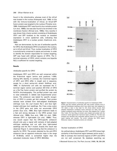
The antiserum was tested for speci®c recognition<br />
<strong>of</strong> CRY2 in protein gel blot analysis. Total protein<br />
extracts were isolated from dark-adapted <strong>Arabidopsis</strong><br />
wild-type (Ler), <strong>the</strong> cry2 mutant fha-1, and <strong>blue</strong> <strong>light</strong>treated<br />
wild-type (Ler) seedlings. fha-1 has a stop codon<br />
in <strong>the</strong> CRY2 gene and does not accumulate CRY2<br />
protein (Guo et al., 1998). Blue <strong>light</strong> treatment leads to<br />
a fast and strong decrease in <strong>the</strong> CRY2 protein level<br />
(Ahmad et al., 1998b; Guo et al., 1998; Lin et al., 1998),<br />
whereas CRY1 is <strong>light</strong>-stable (Lin et al., 1996b). Thus,<br />
one would expect that <strong>the</strong> CRY2-speci®c antiserum<br />
should only give a signal with extracts <strong>of</strong> dark-adapted<br />
wild-type seedlings and not with <strong>blue</strong> <strong>light</strong>-treated<br />
seedlings and <strong>the</strong> fha-1 mutant. Exactly this result was<br />
observed (Figure 1), demonstrating that <strong>the</strong> antiserum is<br />
speci®c for CRY2. The protein detected by <strong>the</strong> anti-CRY2<br />
antiserum has an apparent molecular mass in <strong>the</strong> range<br />
<strong>of</strong> 75±80 kDa, which is somewhat higher than <strong>the</strong><br />
calculated molecular mass <strong>of</strong> CRY2 (69.9 kDa).<br />
Figure 1. Characterization <strong>of</strong> antibodies speci®c for <strong>Arabidopsis</strong> CRY2.<br />
Protein gel blot analysis performed with total protein extracts from 6days-old<br />
<strong>Arabidopsis</strong> seedlings which have ei<strong>the</strong>r been dark adapted for<br />
72 h (dark) or kept in <strong>blue</strong> <strong>light</strong> for 24 h (<strong>blue</strong>) before harvest. Dark<br />
adapted <strong>Arabidopsis</strong> Landsberg erecta wild-type (Ler dark), <strong>blue</strong> <strong>light</strong>treated<br />
Landsberg erecta wild-type (Ler <strong>blue</strong>), dark adapted fha-1 mutant<br />
(fha-1 dark). Per lane 50 mg <strong>of</strong> protein were loaded and probed with <strong>the</strong><br />
antiserum raised against <strong>the</strong> C-terminal region <strong>of</strong> CRY2 (a). The blot was<br />
stripped and reprobed with antibodies against DnaK (b) as a control for<br />
equal loading and transfer. The arrow indicates <strong>the</strong> CRY2 signal.<br />
Molecular masses <strong>of</strong> standard proteins are indicated.<br />
<strong>Nuclear</strong> <strong>localization</strong> <strong>of</strong> CRY2<br />
As outlined above, <strong>Arabidopsis</strong> CRY1 and CRY2 share high<br />
similarity in <strong>the</strong> N-terminal region between amino acids 1±<br />
500. In contrast, <strong>the</strong> C-terminal regions <strong>of</strong> CRY1 and CRY2<br />
are not well conserved. By database searching we<br />
ã Blackwell Science Ltd, The Plant Journal, (1999), 19, 289±296