Link to Letter in Nature - Monash University
Link to Letter in Nature - Monash University
Link to Letter in Nature - Monash University

Create successful ePaper yourself
Turn your PDF publications into a flip-book with our unique Google optimized e-Paper software.
LETTER doi:10.1038/nature09448<br />
; The structural basis for au<strong>to</strong>nomous dimerization of<br />
the pre-T-cell antigen recep<strong>to</strong>r<br />
Siew Siew Pang 1 , Richard Berry 1 , Zhenjun Chen 2 , Lars Kjer-Nielsen 2 , Matthew A. Perug<strong>in</strong>i 3 , Glenn F. K<strong>in</strong>g 4 , Christ<strong>in</strong>a Wang 1 ,<br />
Sock Hui Chew 1 , Nicole L. La Gruta 2 , Neal K. Williams 1 , Travis Beddoe 1 , Tony Tiganis 1 , Nathan P. Cowieson 1 {, Dale I. Godfrey 2 ,<br />
Anthony W. Purcell 3 , Matthew C. J. Wilce 1 , James McCluskey 2 & Jamie Rossjohn 1<br />
The pre-T-cell antigen recep<strong>to</strong>r (pre-TCR), expressed by immature<br />
thymocytes, has a pivotal role <strong>in</strong> early T-cell development, <strong>in</strong>clud<strong>in</strong>g<br />
TCR b-selection, survival and proliferation of CD4 2 CD8 2<br />
double-negative thymocytes, and subsequent ab T-cell l<strong>in</strong>eage differentiation<br />
1–3 . Whereas abTCR ligation by the peptide-loaded<br />
major his<strong>to</strong>compatibility complex <strong>in</strong>itiates T-cell signall<strong>in</strong>g 4 , pre-<br />
TCR-<strong>in</strong>duced signall<strong>in</strong>g occurs by means of a ligand-<strong>in</strong>dependent<br />
dimerization event 5 . The pre-TCR comprises an <strong>in</strong>variant a-cha<strong>in</strong><br />
(pre-Ta) that pairs with any TCR b-cha<strong>in</strong> (TCRb) follow<strong>in</strong>g<br />
successful TCR b-gene rearrangement 6 . Here we provide the basis<br />
of pre-Ta–TCRb assembly and pre-TCR dimerization. The pre-Ta<br />
cha<strong>in</strong> comprised a s<strong>in</strong>gle immunoglobul<strong>in</strong>-like doma<strong>in</strong> that is<br />
structurally dist<strong>in</strong>ct from the constant (C) doma<strong>in</strong> of the TCR<br />
a-cha<strong>in</strong> 7 ; nevertheless, the mode of association between pre-Ta<br />
and TCRb mirrored that mediated by the Ca–Cb doma<strong>in</strong>s of the<br />
abTCR. The pre-TCR had a propensity <strong>to</strong> dimerize <strong>in</strong> solution, and<br />
the molecular envelope of the pre-TCR dimer correlated well with<br />
the observed head-<strong>to</strong>-tail pre-TCR dimer. This mode of pre-TCR<br />
dimerization enabled the pre-Ta doma<strong>in</strong> <strong>to</strong> <strong>in</strong>teract with the<br />
variable (V) b doma<strong>in</strong> through residues that are highly conserved<br />
across the Vb and jo<strong>in</strong><strong>in</strong>g (J) b gene families, thus mimick<strong>in</strong>g the<br />
<strong>in</strong>teractions at the core of the abTCR’s Va–Vb <strong>in</strong>terface.<br />
Disruption of this pre-Ta–Vb dimer <strong>in</strong>terface abrogated pre-<br />
TCR dimerization <strong>in</strong> solution and impaired pre-TCR expression<br />
on the cell surface. Accord<strong>in</strong>gly, we provide a mechanism of pre-<br />
TCR self-association that allows the pre-Ta cha<strong>in</strong> <strong>to</strong> simultaneously<br />
‘sample’ the correct fold<strong>in</strong>g of both the V and C doma<strong>in</strong>s<br />
of any TCR b-cha<strong>in</strong>, regardless of its ultimate specificity, which<br />
represents a critical checkpo<strong>in</strong>t <strong>in</strong> T-cell development. This<br />
unusual dual-chaperone-like sens<strong>in</strong>g function of pre-Ta represents<br />
a unique mechanism <strong>in</strong> nature whereby developmental quality<br />
control regulates the expression and signall<strong>in</strong>g of an <strong>in</strong>tegral membrane<br />
recep<strong>to</strong>r complex.<br />
How the extracellular doma<strong>in</strong> of the pre-Ta cha<strong>in</strong> is able <strong>to</strong> ‘report’<br />
productive TCR b-gene rearrangement and proper b-cha<strong>in</strong> fold<strong>in</strong>g<br />
through pair<strong>in</strong>g with any successfully rearranged TCR b-cha<strong>in</strong>, compris<strong>in</strong>g<br />
variable V, D and J segments, was unclear. Therefore, we<br />
expressed and purified the extracellular doma<strong>in</strong>s of pre-Ta–TCRb and<br />
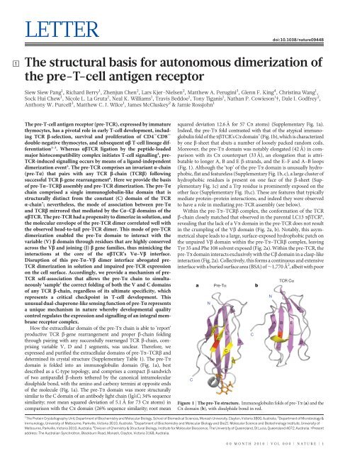
determ<strong>in</strong>ed its crystal structure (Supplementary Table 1). The pre-Ta<br />
doma<strong>in</strong> is folded <strong>in</strong><strong>to</strong> an immunoglobul<strong>in</strong> doma<strong>in</strong> (Fig. 1a), best<br />
described as a C-type <strong>to</strong>pology, and comprises a compact b-sandwich<br />
of two antiparallel b-sheets tethered by the canonical <strong>in</strong>tramolecular<br />
disulphide bond, with the am<strong>in</strong>o and carboxy term<strong>in</strong>i at opposite ends<br />
of the molecule (Fig. 1a). The pre-Ta doma<strong>in</strong> was more structurally<br />
similar <strong>to</strong> the C doma<strong>in</strong> of an antibody light cha<strong>in</strong> (IglC; 34% sequence<br />
similarity; root mean squared deviation of 5.1 A ˚ for 73 Ca a<strong>to</strong>ms) <strong>in</strong><br />
comparison with the Ca doma<strong>in</strong> (26% sequence similarity; root mean<br />
squared deviation 12.6 A ˚ for 57 Ca a<strong>to</strong>ms) (Supplementary Fig. 1a).<br />
Indeed, the pre-Ta fold contrasted with that of the atypical immunoglobul<strong>in</strong><br />
fold of the abTCR’s Ca doma<strong>in</strong> 7 (Fig. 1b), which is characterized<br />
by one b-sheet that abuts a number of loosely packed random coils.<br />
Moreover, the pre-Ta doma<strong>in</strong> was notably elongated (42 A ˚ ) <strong>in</strong> comparison<br />
with its Ca counterpart (33 A ˚ ), an elongation that is attributable<br />
<strong>to</strong> longer A, B and E b-strands, and the E–F and A–B loops<br />
(Fig. 1). Although the ‘<strong>to</strong>p’ of the pre-Ta doma<strong>in</strong> is unusually hydrophobic,<br />
flat and featureless (Supplementary Fig. 1b, c), a large cluster of<br />
hydrophobic residues is present on one face of the b-sheet (Supplementary<br />
Fig. 1c) and a Trp residue is prom<strong>in</strong>ently exposed on the<br />
other face (Supplementary Fig. 1b,c). These are features that typically<br />
mediate prote<strong>in</strong>–prote<strong>in</strong> <strong>in</strong>teractions, and <strong>in</strong>deed they were observed<br />
<strong>to</strong> have a role <strong>in</strong> mediat<strong>in</strong>g pre-TCR assembly (see below).<br />
With<strong>in</strong> the pre-Ta–TCRb complex, the conformation of the TCR<br />
b-cha<strong>in</strong> closely matched that observed <strong>in</strong> the parental LC13 abTCR 8 ,<br />
reveal<strong>in</strong>g that the lack of a Va doma<strong>in</strong> <strong>in</strong> the pre-TCR does not result<br />
<strong>in</strong> the crumpl<strong>in</strong>g of the Vb doma<strong>in</strong> (Fig. 2a, b). Notably, this asymmetrical<br />
shape leads <strong>to</strong> a large, surface-exposed hydrophobic patch on<br />
the unpaired Vb doma<strong>in</strong> with<strong>in</strong> the pre-Ta–TCRb complex, leav<strong>in</strong>g<br />
Tyr 35 and Phe 108 solvent exposed (Fig. 2a). With<strong>in</strong> the pre-TCR, the<br />
pre-Ta doma<strong>in</strong> <strong>in</strong>teracts exclusively with the Cb doma<strong>in</strong> <strong>in</strong> a clasp-like<br />
<strong>in</strong>teraction (Fig. 2a). Collectively, this forms a cont<strong>in</strong>uous and extensive<br />
<strong>in</strong>terface with a buried surface area (BSA) of ,1,770 A ˚ 2 , albeit with poor<br />
1 The Prote<strong>in</strong> Crystallography Unit, Department of Biochemistry and Molecular Biology, School of Biomedical Sciences, <strong>Monash</strong> <strong>University</strong>, Clay<strong>to</strong>n, Vic<strong>to</strong>ria 3800, Australia. 2 Department of Microbiology &<br />
Immunology, <strong>University</strong> of Melbourne, Parkville, Vic<strong>to</strong>ria 3010, Australia. 3 Department of Biochemistry and Molecular Biology and Bio21 Molecular Science and Biotechnology Institute, <strong>University</strong> of<br />
Melbourne, Parkville, Vic<strong>to</strong>ria 3010, Australia. 4 Division of Chemistry & Structural Biology, Institute for Molecular Bioscience, The <strong>University</strong> of Queensland, St Lucia, Queensland 4072, Australia. {Present<br />
address: The Australian Synchrotron, Blackburn Road, <strong>Monash</strong>, Clay<strong>to</strong>n, Vic<strong>to</strong>ria 3168, Australia.<br />
C<br />
a<br />
N<br />
A B<br />
G<br />
A<br />
Pre-Tα<br />
F<br />
E<br />
C<br />
D<br />
C′<br />
D<br />
TCR Cα<br />
A B E<br />
Figure 1 | The pre-Ta structure. Immunoglobul<strong>in</strong> folds of pre-Ta (a) and the<br />
Ca doma<strong>in</strong> (b), with disulphide bond <strong>in</strong> red.<br />
C<br />
b<br />
N<br />
00 MONTH 2010 | VOL 000 | NATURE | 1<br />
D<br />
D
RESEARCH LETTER<br />
a b<br />
c<br />
Y35<br />
P43<br />
F108<br />
Pre-Tα<br />
N<br />
C<br />
A192 S194 L149V147T145<br />
F58<br />
Y60<br />
Pre-Tα<br />
T71<br />
L73<br />
L32<br />
H75<br />
F131<br />
V28<br />
V30<br />
Cβ<br />
shape complementarity, of 0.57 (Fig. 2c and Supplementary Table 2).<br />
The core of the pre-Ta–Cb <strong>in</strong>terface is dom<strong>in</strong>ated by van der Waals<br />
contacts, <strong>in</strong>clud<strong>in</strong>g <strong>in</strong>teractions mediated by a number of small polar or<br />
aliphatic residues along the surface of the b-sheets and a cluster of<br />
aromatic residues (Fig. 2c and Supplementary Table 2). The location<br />
of the pre-Ta–Cb <strong>in</strong>terface co<strong>in</strong>cides with that of the Ca–Cb <strong>in</strong>terface,<br />
although the latter <strong>in</strong>terface is much more extensive 9 (BSA < 2,500 A ˚ 2 ;<br />
Fig. 2d). The larger Ca–Cb <strong>in</strong>terface arises ma<strong>in</strong>ly from the longer D–E<br />
loop of the Ca doma<strong>in</strong>. Nevertheless, core <strong>in</strong>teractions at the Ca–Cb<br />
<strong>in</strong>terface are mimicked at the pre-Ta–Cb <strong>in</strong>terface, <strong>in</strong>volv<strong>in</strong>g 17 common<br />
Cb contact residues that <strong>in</strong>clude a series of <strong>in</strong>terlock<strong>in</strong>g small<br />
hydrophobic residues between the b-sheets and a small aromatic cluster<br />
(Fig. 2d and Supplementary Table 2), which suggests that hydrophobic<br />
forces drive pre-Ta–TCRb assembly 9 . The larger BSA and greater shape<br />
complementarity (0.67) at the Ca–Cb <strong>in</strong>terface is consistent with the<br />
ability of the TCR a-cha<strong>in</strong> <strong>to</strong> compete off the pre-Ta doma<strong>in</strong> effectively<br />
dur<strong>in</strong>g abTCR development.<br />
The pre-TCR is known <strong>to</strong> self-associate on the cell surface of<br />
CD4 2 CD8 2 double-negative thymocytes, and spontaneously dimerizes<br />
<strong>in</strong> vitro and <strong>in</strong> vivo, with the dimerization be<strong>in</strong>g mediated by residues<br />
with<strong>in</strong> the pre-Ta doma<strong>in</strong> 5,10 . In the absence of any structural data, this<br />
has led <strong>to</strong> speculative models of the pre-TCR dimeric assembly. With<strong>in</strong><br />
the crystal lattice, two potential pre-TCR dimers were observed. The<br />
first pre-TCR dimer <strong>in</strong>volved a side-by-side arrangement <strong>in</strong> which the<br />
<strong>to</strong>tal BSA at the <strong>in</strong>terface was small (BSA < 1,380 A ˚ 2 ), and <strong>in</strong>volved<br />
contacts that were mediated ma<strong>in</strong>ly through polar residues (Supplementary<br />
Fig. 2). This side-by-side pre-TCR dimer would sterically<br />
block any <strong>in</strong>teractions the Cb doma<strong>in</strong> is reported <strong>to</strong> make with the CD3<br />
complex 11 . Furthermore, the shape of this side-by-side pre-TCR dimer<br />
did not match what was observed <strong>in</strong> solution (see below). Accord<strong>in</strong>gly,<br />
the side-by-side pre-TCR dimer was considered <strong>to</strong> be attributable solely<br />
<strong>to</strong> the crystal contacts and was discounted from further analyses. The<br />
2 | NATURE | VOL 000 | 00 MONTH 2010<br />
<strong>Nature</strong> nature09448.3d 21/9/10 12:38:56<br />
Vβ<br />
Cβ<br />
Vα<br />
Cα<br />
A192 S194 L149V147T145<br />
F131<br />
Y159 W181<br />
S177<br />
L140<br />
V179<br />
V138 L128<br />
Figure 2 | Comparison between the pre-TCR and abTCR structures. a, Pre-<br />
Ta (magenta) <strong>in</strong>teracts with the Cb doma<strong>in</strong>. The hydrophobic patch is shown<br />
as a blue stick model (also see <strong>in</strong>set); Trp 46, red. Dotted l<strong>in</strong>es represent the N<br />
and C term<strong>in</strong>i, which are not visible <strong>in</strong> the crystal structure. b, abTCR (Prote<strong>in</strong><br />
Data Bank ID, 1KGC 9 ) conta<strong>in</strong>s two cha<strong>in</strong>s (a, green; b, cyan), with Va<br />
<strong>in</strong>teract<strong>in</strong>g with Vb and Ca <strong>in</strong>teract<strong>in</strong>g with Cb. c, d, The contact <strong>in</strong>terfaces<br />
between pre-Ta (magenta) and Cb (cyan) of pre-TCR (c) and between Ca<br />
(green) and Cb (cyan) of abTCR (d). The contact residues are shown as stick<br />
models.<br />
d<br />
Cα<br />
Vβ<br />
Cβ<br />
Cβ<br />
a b<br />
c<br />
e<br />
Cβ<br />
Vβ<br />
Vβ<br />
Pre-Tα<br />
Pre-Tα<br />
Y35<br />
P43<br />
Cell membrane<br />
Vβ<br />
Cβ<br />
F108<br />
W46<br />
Cβ<br />
Pre-Tα<br />
Vβ<br />
Pre-Tα<br />
Pre-Tα<br />
secondpre-TCRdimer(Fig.3)observed<strong>in</strong>thecrystallatticewasmarkedly<br />
more biophysically and biologically conv<strong>in</strong>c<strong>in</strong>g, and <strong>in</strong>volved a head<strong>to</strong>-tail<br />
dimeric arrangement of the pre-TCR heterodimers. Here the<br />
pre-Ta doma<strong>in</strong> of one pro<strong>to</strong>mer was sandwiched between the Cb<br />
and Vb doma<strong>in</strong>s (Fig. 3a, b). The hydrophobic and featureless ‘<strong>to</strong>p’<br />
of the pre-Ta doma<strong>in</strong> was critical for this head-<strong>to</strong>-tail dimeric arrangement,<br />
as large, bulky and charged residues would have prevented this<br />
mode of dimerization. The pre-TCR head-<strong>to</strong>-tail dimerization creates<br />
two new <strong>in</strong>terfaces, each <strong>in</strong>volv<strong>in</strong>g the C9CGF b-sheet of pre-Ta from<br />
one recep<strong>to</strong>r <strong>in</strong>teract<strong>in</strong>g with the b-sheet of the unpaired Vb doma<strong>in</strong><br />
from the other (Fig. 3b and Supplementary Table 3). This arrangement<br />
occludes the otherwise surface-exposed hydrophobic patch on the Vb<br />
doma<strong>in</strong> (Fig. 2a) and provides immediate <strong>in</strong>sight <strong>in</strong><strong>to</strong> how and why the<br />
d<br />
Vβ<br />
Y35<br />
P43<br />
F108<br />
F106<br />
Figure 3 | The pre-TCR dimer. a, Pre-TCR (pre-Ta, magenta; b-cha<strong>in</strong>, cyan)<br />
<strong>in</strong>teracts with a second recep<strong>to</strong>r (pre-Ta, yellow; b-cha<strong>in</strong>, white) <strong>in</strong> a head-<strong>to</strong>tail<br />
arrangement. b, Dimerization is mediated ma<strong>in</strong>ly between the pre-Ta and<br />
the unpaired Vb doma<strong>in</strong>s. Some of the residues <strong>in</strong>volved at these <strong>in</strong>terfaces are<br />
shown as space-fill<strong>in</strong>g models. c, d, Contact <strong>in</strong>terface between pre-Ta (yellow)<br />
and Vb (cyan) of pre-TCR (c) and between Va (green) and Vb (cyan) of<br />
abTCR (d). The highly conserved <strong>in</strong>teract<strong>in</strong>g residues are labelled and shown<br />
as stick models. e, Car<strong>to</strong>on show<strong>in</strong>g orientation of the TCRs on the cell surface:<br />
left, mature abTCR; right, pre-TCR.<br />
Vβ<br />
Cβ<br />
Vα
pre-TCR au<strong>to</strong>nomously dimerizes <strong>in</strong> a cis-configuration on the cell<br />
surface (Fig. 3e). By pair<strong>in</strong>g with the Vb doma<strong>in</strong>, the location of the<br />
pre-Ta doma<strong>in</strong> mimics the position<strong>in</strong>g of the Va doma<strong>in</strong> at the Va-Vb<br />
<strong>in</strong>terface, although different faces of the doma<strong>in</strong>s are used (Fig. 2b). The<br />
<strong>to</strong>tal BSA at the head-<strong>to</strong>-tail pre-TCR dimer <strong>in</strong>terface was large<br />
(BSA < 3,000 A˚ 2 ) and comprised a large number of van der Waals<br />
<strong>in</strong>teractions (Supplementary Table 3), as is typically observed with<strong>in</strong><br />
oligomeric prote<strong>in</strong>–prote<strong>in</strong> complexes. Unlike <strong>in</strong> previous theories of<br />
pre-TCR dimerization 5 , charged residues with<strong>in</strong> the pre-Ta doma<strong>in</strong> did<br />
not have a role <strong>in</strong> mediat<strong>in</strong>g pre-TCR dimerization (Supplementary Fig.<br />
3), and thus may be <strong>in</strong>volved <strong>in</strong> other ways, such as <strong>in</strong> pre-TCR–CD3<br />
assembly. Consistent with this, some of the mutations known <strong>to</strong> affect<br />
pre-TCR function reside with<strong>in</strong> the A–B loop of the pre-Ta doma<strong>in</strong><br />
(Asp 22 and Lys 24), a loop that is implicated <strong>in</strong> abTCR–CD3 <strong>in</strong>teractions<br />
12 . Importantly, the pre-Ta–Vb <strong>in</strong>terface pr<strong>in</strong>cipally <strong>in</strong>volved<br />
residues located on the C9CGF-sheet of the pre-Ta doma<strong>in</strong> (Fig. 3c),<br />
a region that is structurally dissimilar from the Ca doma<strong>in</strong>, thereby<br />
provid<strong>in</strong>g an explanation of why the correspond<strong>in</strong>g Ca doma<strong>in</strong> cannot<br />
act as a suitable surrogate for the pre-Ta doma<strong>in</strong> <strong>in</strong> mediat<strong>in</strong>g this head<strong>to</strong>-tail<br />
dimeric assembly and, hence, pre-TCR signall<strong>in</strong>g. With<strong>in</strong> this<br />
<strong>in</strong>terface, which covers ,1,260 A ˚ 2 , the <strong>in</strong>variant pre-Ta Trp 46 is<br />
positioned at the core (Fig. 3c and Supplementary Table 3). This balland-socket<br />
<strong>in</strong>teraction mirrored what was observed at the LC13 Va–Vb<br />
<strong>in</strong>terface (BSA < 1,490 A˚ 2 ), <strong>in</strong> which Phe 106 from Va is the correspond<strong>in</strong>g<br />
central residue; furthermore, there were 12 common Vb<br />
contact po<strong>in</strong>ts between pre-Ta–Vb and Va–Vb (Fig. 3d and Supplementary<br />
Table 3). Notably, pre-Ta makes contact with residues<br />
encoded by the Vb doma<strong>in</strong> (Tyr 35 and Pro 43) and the Jb region<br />
(Phe 108), thus provid<strong>in</strong>g a mechanism that enables the pre-Ta doma<strong>in</strong><br />
<strong>to</strong> ‘sense’ the correct fold<strong>in</strong>g of the various components derived from<br />
disparate gene segments that create the TCR b-cha<strong>in</strong>. With<strong>in</strong> the TCR<br />
b-locus, there are 42 Vb gene segments, but of these two Vb contact<br />
po<strong>in</strong>ts, position 43 is <strong>in</strong>variably occupied by a short hydrophobic<br />
residue (Pro, Leu, Ile or Val), and Tyr 35 is <strong>in</strong>variant. Furthermore,<br />
Phe 108 is almost <strong>in</strong>variant with<strong>in</strong> the 12 Jb gene segments (Supplementary<br />
Fig. 4). This conserved constellation of <strong>in</strong>teract<strong>in</strong>g<br />
residues provides a framework for understand<strong>in</strong>g how pre-Ta can<br />
<strong>in</strong>discrim<strong>in</strong>ately pair with any successfully rearranged TCR b-cha<strong>in</strong>.<br />
Next, <strong>to</strong> further verify that the pre-TCR could dimerize <strong>in</strong> vitro,we<br />
<strong>in</strong>vestigated the behaviour of the pre-TCR <strong>in</strong> solution. As judged by<br />
sedimentation velocity analytical ultracentrifugation (SV-AUC) and<br />
small-angle X-ray scatter<strong>in</strong>g (SAXS), the pre-TCR displayed the hallmarks<br />
of a weak self-associat<strong>in</strong>g system, typical of many immunological<br />
recep<strong>to</strong>rs 13 . Specifically, us<strong>in</strong>g SAXS analysis, we observed the<br />
pre-TCR <strong>to</strong> undergo a concentration-dependent <strong>in</strong>crease <strong>in</strong> molecular<br />
mass, radius of gyration (Rg) and maximal particle dimension (Supplementary<br />
Table 4). These effects cannot be accounted for by the<br />
presence of non-specific prote<strong>in</strong> aggregation, as judged by the l<strong>in</strong>earity<br />
of SAXS Gu<strong>in</strong>ier plots (Supplementary Fig. 5). Furthermore, us<strong>in</strong>g<br />
SV-AUC, we see two dist<strong>in</strong>ct species (Fig. 4a). On the basis of hydrodynamic<br />
calculations, these species correspond <strong>to</strong> pre-Ta–TCRb<br />
(apparent sedimentation coefficient, sapp 5 3.1 S; frictional ratio,<br />
f/f0 5 1.44) and the pre-TCR dimer (sapp 5 5.0 S, f/f0 5 1.37) (Fig. 4b<br />
and Supplementary Table 5). Consistent with the AUC results, the<br />
SAXS measurements show an <strong>in</strong>crease <strong>in</strong> average particle size with<br />
<strong>in</strong>creas<strong>in</strong>g prote<strong>in</strong> concentration, consistent with a monomer–dimer<br />
equilibrium (Supplementary Fig. 6a). To ga<strong>in</strong> an <strong>in</strong>sight <strong>in</strong><strong>to</strong> the shape<br />
of the pre-TCR <strong>in</strong> solution, low-resolution models were res<strong>to</strong>red<br />
from the SAXS scatter<strong>in</strong>g curves ab <strong>in</strong>itio us<strong>in</strong>g the DAMMIF 14 and<br />
GASBOR 15 programs (Supplementary Fig. 6a). As the molecular<br />
weight determ<strong>in</strong>ed by SAXS suggests that the pre-TCR is fully dimeric<br />
at 15 mg ml 21 , we fitted the pre-TCR dimer <strong>in</strong><strong>to</strong> this SAXS model. In<br />
contrast <strong>to</strong> the side-by-side pre-TCR dimer (Supplementary Fig. 6b),<br />
the head-<strong>to</strong>-tail pre-TCR dimer conv<strong>in</strong>c<strong>in</strong>gly fitted the central<br />
globular region of the SAXS model (Fig. 4f and Supplementary Fig.<br />
6c). Furthermore, <strong>in</strong> the head-<strong>to</strong>-tail pre-TCR dimer, the regions that<br />
c(s) (S –1 )<br />
a<br />
1.20<br />
0.90<br />
0.60<br />
0.30<br />
0.00<br />
b<br />
1.60<br />
1.20<br />
0.80<br />
0.40<br />
0.00<br />
c<br />
0.60<br />
0.45<br />
0.30<br />
0.15<br />
0.00<br />
d<br />
1.25<br />
1.00<br />
0.75<br />
0.50<br />
0.25<br />
0.00<br />
0.0 1.0 2.0 3.0 4.0 5.0 6.0<br />
Sedimentation coefficient (S)<br />
are miss<strong>in</strong>g from the crystal structure (pr<strong>in</strong>cipally the C-term<strong>in</strong>al<br />
stalks of both pre-Ta and the TCR b-cha<strong>in</strong>s) are present at oppos<strong>in</strong>g<br />
poles, where they are well positioned <strong>to</strong> occupy the areas left vacant by<br />
the crystal structure. Thus, the head-<strong>to</strong>-tail pre-TCR dimer observed <strong>in</strong><br />
the crystal structure correlated well with the differ<strong>in</strong>g solution-based<br />
measurements. This cis-dimeric arrangement suggests that the pre-<br />
TCR sits ‘flat’, almost parallel <strong>to</strong> the membrane surface, <strong>in</strong> contrast<br />
<strong>to</strong> the abTCR, which is usually depicted <strong>in</strong> an upright orientation on<br />
the membrane (Fig. 3e). Consistent with this, the relative dependency<br />
of the various CD3 subunits with<strong>in</strong> the pre-TCR complex may differ<br />
from that with<strong>in</strong> the abTCR–CD3 complex 16 .<br />
Next we used three dist<strong>in</strong>ct approaches <strong>to</strong> further validate the pre-<br />
TCR dimer observed <strong>in</strong> the crystal structure and <strong>in</strong> solution. First, we<br />
used crossl<strong>in</strong>k<strong>in</strong>g/mass spectrometry studies of recomb<strong>in</strong>ant pre-TCR<br />
<strong>to</strong> directly assess the configuration of the dimer (Supplementary Fig. 7<br />
and Supplementary Table 6; see Methods). Notably, the only crossl<strong>in</strong>ked<br />
species between the pre-Ta cha<strong>in</strong> and the TCR b-cha<strong>in</strong> that we<br />
observed was between the N-term<strong>in</strong>al sequence (head) of the Vb<br />
doma<strong>in</strong> and the C-term<strong>in</strong>al sequence (tail) of the pre-Ta cha<strong>in</strong> (Supplementary<br />
Fig. 7 and Supplementary Table 6). This crossl<strong>in</strong>ked species<br />
is <strong>in</strong>compatible with the pre-Ta–TCRb and the side-by-side pre-TCR<br />
dimer, but is fully consistent with a head-<strong>to</strong>-tail pre-TCR dimer configuration.<br />
Second, we generated a W46aRmutant<strong>in</strong>thepre-Ta cha<strong>in</strong><br />
and assessed its impact on pre-TCR dimerization as judged by SV-<br />
AUC. In contrast <strong>to</strong> the wild-type (WT) pre-TCR, sedimentation velocity<br />
analysis of the W46aR mutant shows that it is monomeric <strong>in</strong><br />
solution (sapp 5 3.1 S) with no evidence of dimerization (Fig. 4).<br />
Third, <strong>to</strong> assess the pre-TCR dimer conformation on the cell surface,<br />
we transfected WT and mutant pre-TCRs <strong>in</strong><strong>to</strong> the TCR ab-negative,<br />
CD3-positive SKW3 T-cell l<strong>in</strong>e and visualized cell surface and <strong>in</strong>tracellular<br />
pre-TCR localization (Fig. 5). As a control, we also transfected<br />
e<br />
Pre-TCR monomer<br />
f<br />
LETTER RESEARCH<br />
Pre-TCR dimer<br />
Figure 4 | Pre-TCR dimerization <strong>in</strong> solution. a–d, Cont<strong>in</strong>uous size<br />
distributions, determ<strong>in</strong>ed by SV-AUC, of pre-TCR at 1 mg ml 21 (a), 3 mg ml 21<br />
(b) and 8.7 mg ml 21 (c), and of W46aR pre-TCR mutant at 1.4 mg ml 21<br />
(d). Insets show the correspond<strong>in</strong>g residuals. e, Bead models generated from the<br />
pre-TCR structure for the monomer (left) and the head-<strong>to</strong>-tail dimer (right).<br />
f, Overlay of the pre-TCR head-<strong>to</strong>-tail dimer (red ribbon) and the DAMMIF<br />
SAXS model (cyan spheres), shown <strong>in</strong> two orientations. Red dots <strong>in</strong>dicate<br />
C-term<strong>in</strong>al residues of the pre-Ta and LC13 b-cha<strong>in</strong>s, orange dots <strong>in</strong>dicate<br />
modelled C-term<strong>in</strong>al tails and yellow dots <strong>in</strong>dicate N-l<strong>in</strong>ked glycosylation sites.<br />
Scale bar, 100 nm.<br />
90°<br />
00 MONTH 2010 | VOL 000 | NATURE | 3
RESEARCH LETTER<br />
a<br />
b<br />
c Pre-Tα–TCRβ-F108A Pre-Tα-W46R–TCRβ Pre-Tα–TCRβ-Y35A<br />
α-TCRβ–Hoechst<br />
α-CD3ε–Hoechst α-TCRβ–Hoechst<br />
TCRα–TCRβ<br />
Unstimulated<br />
TCRα–TCRβ<br />
d F108βA W46αR Y35βA<br />
Pre-Tα–TCRβ<br />
+ α-CD3ε–IgG<br />
the parental LC13 abTCR <strong>in</strong><strong>to</strong> the SKW3 cell l<strong>in</strong>e. We demonstrated,<br />
us<strong>in</strong>g immunofluorescence, that transfection of the WT pre-TCR<br />
resulted <strong>in</strong> a punctate expression pattern, typical of pre-TCR cluster<strong>in</strong>g<br />
on the cell surface and consequent endocy<strong>to</strong>sis, which is <strong>in</strong> direct contrast<br />
<strong>to</strong> the l<strong>in</strong>ear cell surface expression pattern of the mature abTCR <strong>in</strong><br />
unstimulated cells (Fig. 5a) and the large puncta result<strong>in</strong>g from crossl<strong>in</strong>k<strong>in</strong>g<br />
the abTCR on the cell surface with a-CD3e (Fig. 5b). Next we<br />
exam<strong>in</strong>ed the impact of three pre-TCR mutants, Y35bA, F108bAand<br />
W46aR, on cell surface expression and <strong>in</strong>tracellular localization us<strong>in</strong>g<br />
flow cy<strong>to</strong>metric analysis and immunofluorescence microscopy. The<br />
F108bA and W46aR pre-TCR mutants were expressed at lower levels<br />
on the cell surface and led <strong>to</strong> a greater retention of these mutants <strong>in</strong> an<br />
<strong>in</strong>tracellular compartment typical of the endoplasmic reticulum<br />
(Fig. 5c, d). Furthermore, the Y35bA TCR–Vb mutant completely<br />
4 | NATURE | VOL 000 | 00 MONTH 2010<br />
<strong>Nature</strong> nature09448.3d 21/9/10 12:38:59<br />
Pre-Tα or TCRβ only<br />
Pre-TCR<br />
Mutant pre-TCR<br />
TCRβ-Y35A vs TCRβ<br />
Pre-Tα or TCRα only<br />
Pre-TCR TCR<br />
Pre-Tα–TCRβ-Y35A or<br />
TCRα–TCRβ-Y35A<br />
Pre-Tα–TCRβ or<br />
TCRα–TCR<br />
CD3<br />
Figure 5 | Expression of pre-TCR mutants. a,TCRbsta<strong>in</strong><strong>in</strong>g of SKW3 T cells<br />
express<strong>in</strong>g TCRa–TCRb or pre-Ta–TCRb. Hoechst, Hoechst sta<strong>in</strong>. b, CD3e<br />
sta<strong>in</strong><strong>in</strong>g of unstimulated and activated SKW3 T cells express<strong>in</strong>g TCRa–TCRb.<br />
IgG, immunoglobul<strong>in</strong> G. c, TCRb sta<strong>in</strong><strong>in</strong>g of T cells express<strong>in</strong>g the <strong>in</strong>dicated<br />
pre-Ta and TCRb mutations. d, Flow sta<strong>in</strong><strong>in</strong>g (CD3e) of T cells express<strong>in</strong>g pre-<br />
Ta and TCRVb mutations. The <strong>to</strong>p row shows negative-control T cells (either<br />
pre-Ta or LC13b alone; th<strong>in</strong> dashed l<strong>in</strong>es), positive-control pre-Ta–LC13b<br />
transfectants (solid l<strong>in</strong>es) and SKW3.pre-Ta1LC13b mutants (grey shad<strong>in</strong>g).<br />
The bot<strong>to</strong>m row shows unsorted T cells express<strong>in</strong>g either pre-Ta–LC13b-<br />
Y35A, pre-Ta alone or pre-Ta–LC13b (left), and unsorted SKW3 cells<br />
express<strong>in</strong>g LC13a–LC13b-Y35A, LC13a–LC13b or TCRa alone (negative<br />
control; right). See also Supplementary Fig. 8.<br />
abrogated the cell surface expression of the pre-TCR (Fig. 5d) and<br />
resulted <strong>in</strong> accumulation of the expressed pre-TCR Y35bA mutant<strong>in</strong><br />
per<strong>in</strong>uclear puncta rem<strong>in</strong>iscent of the Golgi complex and the endoplasmic<br />
reticulum, consistent with defective traffick<strong>in</strong>g <strong>to</strong> the cell surface<br />
(Fig. 5c). However, the Y35bA mutant did not affect LC13 abTCR cell<br />
surface expression (Fig. 5d), <strong>in</strong>dicat<strong>in</strong>g that this mutant does not affect<br />
the fold of the TCR b-cha<strong>in</strong> per se. This f<strong>in</strong>d<strong>in</strong>g highlights the fact that<br />
pre-TCR cell surface expression is f<strong>in</strong>ely tuned <strong>to</strong> pre-Ta–Vb <strong>in</strong>teractions.<br />
Collectively, the crossl<strong>in</strong>k<strong>in</strong>g experiments <strong>in</strong> solution, coupled<br />
with the effect of the mutants on pre-TCR dimerization <strong>in</strong> solution and<br />
on the cell surface, are fully consistent with the head-<strong>to</strong>-tail pre-TCR<br />
dimer configuration.<br />
In vertebrates there are three classes of TCR, namely abTCR,<br />
cdTCR and the pre-TCR. Although structural data exists on the architecture<br />
of abTCR 7 and cdTCRs 17 , <strong>in</strong>formation is lack<strong>in</strong>g on the pre-<br />
TCR. Furthermore, whereas abTCR and cdTCR <strong>in</strong>teract with def<strong>in</strong>ed<br />
ligands, the pre-TCR is unique <strong>in</strong> that a ligand-<strong>in</strong>dependent mechanism<br />
underp<strong>in</strong>s its role <strong>in</strong> T-cell development. Our f<strong>in</strong>d<strong>in</strong>gs formally<br />
demonstrate that the pre-Ta cha<strong>in</strong> functions much more than a simple<br />
surrogate TCR a-cha<strong>in</strong>, whereby the pre-Ta doma<strong>in</strong> simultaneously<br />
b<strong>in</strong>ds <strong>to</strong> the Cb and Vb doma<strong>in</strong>s with<strong>in</strong> the head-<strong>to</strong>-tail pre-TCR<br />
dimer. The role of the pre-Ta doma<strong>in</strong> is <strong>to</strong> act as a checkpo<strong>in</strong>t that<br />
assesses correct rearrangement and expression of the TCR b-cha<strong>in</strong> on<br />
the cell surface. The observed mode of dimerization can only occur<br />
when both the variable and constant doma<strong>in</strong>s of the TCR b-cha<strong>in</strong> are<br />
correctly folded; this is a previously unrecognized determ<strong>in</strong>ant for<br />
b-selection. Thus, the pre-Ta has a dual chaperone-sens<strong>in</strong>g role that<br />
is synchronously l<strong>in</strong>ked <strong>to</strong> dimerization and pre-TCR signal transduction.<br />
Many prote<strong>in</strong>s require chaperones for correct fold<strong>in</strong>g, and subsequent<br />
function, with <strong>in</strong>tricate cellular mach<strong>in</strong>es typically be<strong>in</strong>g<br />
required <strong>to</strong> aid <strong>in</strong> this process. The pre-B-cell recep<strong>to</strong>r 18 has a number<br />
of components that allow productive expression of the B-cell recep<strong>to</strong>r<br />
<strong>in</strong> a manner that is quite dist<strong>in</strong>ct from the <strong>in</strong>dividual pre-TCR. The<br />
evolutionarily conserved pre-Ta has achieved its role by adopt<strong>in</strong>g dual<br />
functionality and, as such, represents a previously undescribed mechanism<br />
<strong>in</strong> nature that ensures the <strong>in</strong>tegrity of prote<strong>in</strong> assembly and<br />
function.<br />
METHODS SUMMARY<br />
Pre-TCR clon<strong>in</strong>g, expression and purification. We cloned the ec<strong>to</strong>doma<strong>in</strong>s of<br />
the human pre-Ta and TCR LC13b <strong>in</strong><strong>to</strong> the pFastBac Dual vec<strong>to</strong>r (Invitrogen).<br />
The pre-TCR was expressed, purified and crystallized, and its structure determ<strong>in</strong>ed.<br />
Further details are provided <strong>in</strong> Methods.<br />
SAXS. SAXS data were collected at the Australian Synchrotron, Melbourne, and<br />
the Advanced Light Source, Berkeley. We assessed data quality us<strong>in</strong>g Gu<strong>in</strong>ier<br />
analysis, and determ<strong>in</strong>ed R g and the maximal particle dimension, D max, us<strong>in</strong>g<br />
GNOM 19 . Ab <strong>in</strong>itio models were generated us<strong>in</strong>g DAMMIF and GASBOR and<br />
were overlaid with high-resolution models us<strong>in</strong>g PYMOL (http://www.pymol.org/).<br />
Mass spectrometry and crossl<strong>in</strong>k<strong>in</strong>g studies. These studies were performed on<br />
recomb<strong>in</strong>ant pre-TCR as described <strong>in</strong> Methods.<br />
Functional studies. We assessed pre-TCR and mutants of pre-TCR, as well as<br />
abTCR and mutants of abTCR, for cell surface expression as described <strong>in</strong><br />
Methods. T cells were processed for epifluorescence microscopy as described <strong>in</strong><br />
Methods.<br />
AUC. AUC experiments on WT and mutant pre-TCR were conducted us<strong>in</strong>g a<br />
Beckman model XL-I analytical ultracentrifuge at a temperature of 20 uC us<strong>in</strong>g<br />
standard pro<strong>to</strong>cols and analyses. Bead models represent<strong>in</strong>g pre-TCR were generated<br />
us<strong>in</strong>g SOMO 20 . See Methods for more details.<br />
Full Methods and any associated references are available <strong>in</strong> the onl<strong>in</strong>e version of<br />
the paper at www.nature.com/nature.<br />
Received 20 April; accepted 23 August 2010.<br />
Published onl<strong>in</strong>e XX 2010.<br />
1. von Boehmer, H. Unique features of the pre-T-cell recep<strong>to</strong>r a-cha<strong>in</strong>: not just a<br />
surrogate. <strong>Nature</strong> Rev. Immunol. 5, 571–577 (2005).<br />
2. Sa<strong>in</strong>t-Ruf, C. et al. Different <strong>in</strong>itiation of pre-TCR and cdTCR signall<strong>in</strong>g. <strong>Nature</strong> 406,<br />
524–527 (2000).<br />
3. Borowski, C., Li, X., Aifantis, I., Gounari, F. & von Boehmer, H. Pre-TCRa and TCRa<br />
are not <strong>in</strong>terchangeable partners of TCRb dur<strong>in</strong>g T lymphocyte development.<br />
J. Exp. Med. 199, 607–615 (2004).<br />
4. Z<strong>in</strong>kernagel, R. M. & Doherty, P. C. Restriction of <strong>in</strong> vitro T cell-mediated cy<strong>to</strong><strong>to</strong>xicity<br />
<strong>in</strong> lymphocytic choriomen<strong>in</strong>gitis with<strong>in</strong> a syngeneic or semiallogeneic system.<br />
<strong>Nature</strong> 248, 701–702 (1974).<br />
5. Yamasaki, S. et al. Mechanistic basis of pre-T cell recep<strong>to</strong>r-mediated au<strong>to</strong>nomous<br />
signal<strong>in</strong>g critical for thymocyte development. <strong>Nature</strong> Immunol. 7, 67–75 (2005).<br />
6. Sa<strong>in</strong>t-Ruf, C. Analysis and expression of a cloned pre-T cell recep<strong>to</strong>r gene. Science<br />
266, 1208–1212 (1994).<br />
7. Garcia, K. C. et al. An ab T cell recep<strong>to</strong>r structure at 2.5 A ˚ and its orientation <strong>in</strong> the<br />
TCR-MHC complex. Science 274, 209–219 (1996).<br />
8. Kjer-Nielsen, L. et al. A structural basis for the selection of dom<strong>in</strong>ant ab T cell<br />
recep<strong>to</strong>rs <strong>in</strong> antiviral immunity. Immunity 18, 53–64 (2003).<br />
9. Kjer-Nielsen, L. et al. The 1.5 A ˚ crystal structure of a highly selected antiviral T cell<br />
recep<strong>to</strong>r provides evidence for a structural basis of immunodom<strong>in</strong>ance. Structure<br />
10, 1521–1532 (2002).<br />
10. Yamasaki, S. & Sai<strong>to</strong>, T. Molecular basis for pre-TCR-mediated au<strong>to</strong>nomous<br />
signal<strong>in</strong>g. Trends Immunol. 28, 39–43 (2007).<br />
11. Kuhns, M. S. & Davis, M. M. Disruption of extracellular <strong>in</strong>teractions impairs T cell<br />
recep<strong>to</strong>r-CD3 complex stability and signal<strong>in</strong>g. Immunity 26, 357–369 (2007).<br />
12. Beddoe, T. et al. Antigen ligation triggers a conformational change with<strong>in</strong> the<br />
constant doma<strong>in</strong> of the ab T cell recep<strong>to</strong>r. Immunity 30, 777–788 (2009).<br />
13. van der Merwe, P. A. & Davis, S. J. Molecular <strong>in</strong>teractions mediat<strong>in</strong>g T cell antigen<br />
recognition. Annu. Rev. Immunol. 21, 659–684 (2003).<br />
14. Franke, D. & Svergun, D. I. DAMMIF, a program for rapid ab-<strong>in</strong>itio shape<br />
determ<strong>in</strong>ation <strong>in</strong> small-angle scatter<strong>in</strong>g. J. Appl. Crystallogr. 42, 342–346 (2009).<br />
15. Svergun, D. I., Pe<strong>to</strong>ukhov, M. V. & Koch, M. H. Determ<strong>in</strong>ation of doma<strong>in</strong> structure of<br />
prote<strong>in</strong>s from X-ray solution scatter<strong>in</strong>g. Biophys. J. 80, 2946–2953 (2001).<br />
16. Dave, V. P. Hierarchical role of CD3 cha<strong>in</strong>s <strong>in</strong> thymocyte development. Immunol.<br />
Rev. 232, 22–33 (2009).<br />
17. Allison, T. J., W<strong>in</strong>ter, C. C., Fournie, J. J., Bonneville, M. & Garboczi, D. N. Structure of<br />
a human cd T-cell antigen recep<strong>to</strong>r. <strong>Nature</strong> 411, 820–824 (2001).<br />
18. Melchers, F. The pre-B-cell recep<strong>to</strong>r: selec<strong>to</strong>r of fitt<strong>in</strong>g immunoglobul<strong>in</strong> heavy<br />
cha<strong>in</strong>s for the B-cell reper<strong>to</strong>ire. <strong>Nature</strong> Rev. Immunol. 5, 578–584 (2005).<br />
LETTER RESEARCH<br />
19. Semenyuk, A. V. & Svergun, D. I. GNOM - a program package for small-angle<br />
scatter<strong>in</strong>g data process<strong>in</strong>g. J. Appl. Crystallogr. 24, 537–540 (1991).<br />
20. Brookes, E., Demeler, B., Rosano, C. & Rocco, M. The implementation of SOMO<br />
(SOlution MOdeller) <strong>in</strong> the UltraScan analytical ultracentrifugation data analysis<br />
suite: enhanced capabilities allow the reliable hydrodynamic model<strong>in</strong>g of virtually<br />
any k<strong>in</strong>d of biomacromolecule. Eur. Biophys. J. 39, 423–435 (2010).<br />
Supplementary Information is l<strong>in</strong>ked <strong>to</strong> the onl<strong>in</strong>e version of the paper at<br />
www.nature.com/nature.<br />
Acknowledgements We thank S. Turner for discussions; S. Ramarath<strong>in</strong>am for<br />
technical assistance; the staff at the Australian synchrotron (MX and SAXS/WAXS<br />
beaml<strong>in</strong>es) and Berkeley Advanced Light Source (SIBYLS beaml<strong>in</strong>e) for assistance with<br />
data collection; and T. Caradoc-Davies for advice on process<strong>in</strong>g of merohedral tw<strong>in</strong>ned<br />
X-ray data. This research was supported by grants from the Australian Research<br />
Council and the National Health and Medical Research Council of Australia. A.W.P., T.T.<br />
and M.C.J.W. are supported by NHMRC Senior Research Fellowships, and D.I.G. is<br />
supported by an NHMRC Pr<strong>in</strong>cipal Research Fellowship. M.A.P. is supported by an ARC<br />
Future Fellowship and J.R. is supported by an ARC Federation Fellowship.<br />
Author Contributions S.S.P. solved the structure, under<strong>to</strong>ok analysis, performed<br />
experiments and contributed <strong>to</strong> manuscript preparation. J.M., D.I.G. and J.R.<br />
contributed <strong>to</strong> the design and <strong>in</strong>terpretation of experiments, project management and<br />
the writ<strong>in</strong>g of the manuscript. R.B., Z.C., L.K.-N., M.A.P., G.F.K., N.L.L.G., T.T. and A.W.P.<br />
performed experiments and contributed <strong>to</strong> the writ<strong>in</strong>g of the manuscript; C.W., S.H.C.,<br />
N.K.W., T.B., N.P.C. and M.C.J.W. performed experiments. J.M. and J.R. were the jo<strong>in</strong>t<br />
senior authors—they co-led the <strong>in</strong>vestigation, devised the project, analysed the data<br />
and wrote the manuscript.<br />
Author Information The a<strong>to</strong>mic coord<strong>in</strong>ates and structure fac<strong>to</strong>rs of pre-TCR have<br />
been deposited <strong>in</strong> the Prote<strong>in</strong> Data Bank under the accession code 3OF6. Repr<strong>in</strong>ts and<br />
permissions <strong>in</strong>formation is available at www.nature.com/repr<strong>in</strong>ts. The authors declare<br />
no compet<strong>in</strong>g f<strong>in</strong>ancial <strong>in</strong>terests. Readers are welcome <strong>to</strong> comment on the onl<strong>in</strong>e<br />
version of this article at www.nature.com/nature. Correspondence and requests for<br />
materials should be addressed <strong>to</strong> J.M. (jamesm1@unimelb.edu.au) or J.R.<br />
(jamie.rossjohn@monash.edu).<br />
00 MONTH 2010 | VOL 000 | NATURE | 5
RESEARCH LETTER<br />
METHODS<br />
Pre-TCR clon<strong>in</strong>g, expression and purification. Genes encod<strong>in</strong>g the ec<strong>to</strong>doma<strong>in</strong>s<br />
(<strong>in</strong>clud<strong>in</strong>g the <strong>in</strong>tercha<strong>in</strong> cyste<strong>in</strong>e) of the human pre-Ta and TCR LC13b<br />
were cloned <strong>in</strong><strong>to</strong> the pFastBac Dual vec<strong>to</strong>r (Invitrogen). The genes were cloned<br />
downstream of the baculovirus GP67 secre<strong>to</strong>ry signal sequence and were under the<br />
control of different promoters for co-expression. The C term<strong>in</strong>us of pre-Ta was<br />
modified <strong>to</strong> <strong>in</strong>clude an enterok<strong>in</strong>ase cleavage site, a Jun zipper, a BirA tag and a<br />
hexahistid<strong>in</strong>e (6xHis) tag, and the LC13 b-cha<strong>in</strong> has an enterok<strong>in</strong>ase site and a Fos<br />
zipper. The pre-TCRs were expressed and secreted by High Five <strong>in</strong>sect cells<br />
(Invitrogen) as a covalently l<strong>in</strong>ked dimer. At the end of expression, the <strong>in</strong>sect cell<br />
supernatant was collected and diafiltrated with TBS by tangential flow filtration.<br />
The hexahistid<strong>in</strong>e-tagged prote<strong>in</strong>s were purified us<strong>in</strong>g Ni Sepharose High<br />
Performance res<strong>in</strong> (GE Healthcare) and by gel filtration (HiLoad 16/60 Superdex<br />
200, GE Healthcare). For prote<strong>in</strong> crystallization, the C-term<strong>in</strong>al tag regions were<br />
removed by enterok<strong>in</strong>ase digestion (GenScript). F<strong>in</strong>al purification and buffer<br />
exchange <strong>in</strong><strong>to</strong> 20 mM Tris-HCl, (pH 8.0) and 0.15 M NaCl was carried out by<br />
gel filtration. The pooled prote<strong>in</strong> was concentrated <strong>to</strong> ,10 mg ml 21 ,snapped<br />
frozen and s<strong>to</strong>red at 280 uC <strong>in</strong> small aliquots. The typical prote<strong>in</strong> yield of the<br />
cleaved pre-TCR is about 1–3 mg per litre of expression culture.<br />
Crystallization and X-ray data collection. Nanoscale crystallization trials of pre-<br />
TCR were carried out us<strong>in</strong>g sitt<strong>in</strong>g-drop vapour diffusion at room temperature<br />
(19 uC) and at 4 uC. A few hits were identified and reproduced manually us<strong>in</strong>g<br />
hang<strong>in</strong>g-drop vapour diffusion. The optimized crystallization conditions were<br />
produced by mix<strong>in</strong>g equal volumes of pre-TCR (6 mg ml 21 ) with the reservoir<br />
buffer (0.1 M sodium cacodylate (pH 7.0), 0.1 M calcium acetate and 13–16% PEG<br />
1500). Rod-shaped pre-TCR crystals appeared quickly after overnight <strong>in</strong>cubation<br />
at 4 uC and cont<strong>in</strong>ued <strong>to</strong> grow <strong>in</strong> size for the next few days. For cryoprotection, the<br />
rod-shaped crystals were soaked <strong>in</strong> the reservoir buffer plus 15% glycerol overnight<br />
at 4 uC. The soaked crystals were then transferred <strong>in</strong><strong>to</strong> reservoir buffer<br />
conta<strong>in</strong><strong>in</strong>g 25% glycerol and flash-frozen <strong>in</strong> liquid nitrogen. X-ray data <strong>to</strong> 2.8 A˚<br />
were collected us<strong>in</strong>g the Micro Crystallography (MX2) beaml<strong>in</strong>e at the Australian<br />
Synchrotron. The diffraction data were processed and analysed us<strong>in</strong>g MOSFILM<br />
6.2.6 and SCALA (CCP4 suite) 21 . A summary of data collection statistics is provided<br />
<strong>in</strong> Supplementary Table 1.<br />
Structure determ<strong>in</strong>ation and ref<strong>in</strong>ement. Initial analysis of the pre-TCR crystals<br />
<strong>in</strong>dicated that the X-ray data could be scaled <strong>in</strong><strong>to</strong> any of the primitive hexagonal<br />
space groups, suggest<strong>in</strong>g crystal tw<strong>in</strong>n<strong>in</strong>g. Further exam<strong>in</strong>ation us<strong>in</strong>g The<br />
Merohedral Crystal Tw<strong>in</strong>n<strong>in</strong>g Server (http://nihserver.mbi.ucla.edu/Tw<strong>in</strong>n<strong>in</strong>g/),<br />
CCP4 programs and PHENIX.XTRIAGE 22 confirmed perfect crystal merohedral<br />
tw<strong>in</strong>n<strong>in</strong>g. The structure was solved by molecular replacement (PHASER from the<br />
CCP4 suite) us<strong>in</strong>g the b-cha<strong>in</strong> of LC13 TCR (PDB ID, 1KGC) as the search model.<br />
Solutions were found <strong>in</strong> a number of space groups (six molecules for P32, three<br />
molecules for P3221 or P3212, and one molecule for P6222). Each of the solutions<br />
was exam<strong>in</strong>ed and ref<strong>in</strong>ed us<strong>in</strong>g PHENIX 23 , tak<strong>in</strong>g <strong>in</strong><strong>to</strong> consideration the tw<strong>in</strong>n<strong>in</strong>g<br />
fac<strong>to</strong>r and opera<strong>to</strong>r (0.516, 2k, h 1 k, l; k, h and l are Miller <strong>in</strong>dices) After <strong>in</strong>itial<br />
rounds of ref<strong>in</strong>ement, the P3221 solution gave a reasonable Rfac<strong>to</strong>r and an unbiased<br />
density of the miss<strong>in</strong>g pre-Ta cha<strong>in</strong>s was observed, suggest<strong>in</strong>g the correct solution.<br />
To avoid bias due <strong>to</strong> the presence of data tw<strong>in</strong>n<strong>in</strong>g, the Rfree set was assigned <strong>in</strong><br />
PHENIX.REFINE. Further restra<strong>in</strong>ed ref<strong>in</strong>ements were performed us<strong>in</strong>g<br />
PHENIX.REFINE after <strong>in</strong>itial rounds of rigid-body ref<strong>in</strong>ement and stimulated<br />
anneal<strong>in</strong>g. Each round of ref<strong>in</strong>ement was <strong>in</strong>terspersed with electron density <strong>in</strong>spection<br />
and manual model build<strong>in</strong>g with COOT 24 . The progress of ref<strong>in</strong>ement was<br />
moni<strong>to</strong>red by R free value and the f<strong>in</strong>al ref<strong>in</strong>ement statistics for pre-TCR are given <strong>in</strong><br />
Supplementary Table 1. There were three pre-TCR complexes <strong>in</strong> the asymmetric<br />
unit, all of which were highly similar <strong>to</strong> each other, and structural analyses were thus<br />
conf<strong>in</strong>ed <strong>to</strong> one pre-TCR complex. The f<strong>in</strong>al model comprises residues 7–110 from<br />
the pre-Ta cha<strong>in</strong> and residues 2–245 from the TCR b-cha<strong>in</strong>. Disordered regions<br />
<strong>in</strong>clude six residues from the N term<strong>in</strong>us and n<strong>in</strong>e from the C term<strong>in</strong>us of pre-Ta,<br />
and one N- and three C-term<strong>in</strong>al residues from TCRb, respectively. The miss<strong>in</strong>g<br />
regions <strong>in</strong>clude the <strong>in</strong>termolecular disulphide bond at the flexible C-term<strong>in</strong>al end of<br />
the polypeptides.<br />
SAXS. SAXS data were collected at the Australian Synchrotron us<strong>in</strong>g a 1M Pilatus<br />
detec<strong>to</strong>r. Buffers/samples were loaded <strong>in</strong><strong>to</strong> 1-mm quartz capillaries and cont<strong>in</strong>uously<br />
flowed through the beam dur<strong>in</strong>g data collection. Multiple 1-s exposures were collected<br />
and compared <strong>to</strong> control for radiation damage. Data were collected us<strong>in</strong>g a<br />
s<strong>in</strong>gle camera length witha 1.5-msample-<strong>to</strong>-detec<strong>to</strong>r distance <strong>to</strong>cover a momentum<br />
transfer <strong>in</strong>terval of 0.0056 A˚ 21 , q , 0.3 A˚ 21 . The modulus of the momentum<br />
transfer is def<strong>in</strong>ed as q 5 4ps<strong>in</strong>(h/l), where 2h is the scatter<strong>in</strong>g angle and l is the<br />
wavelength. Scatter<strong>in</strong>g images were radially averaged and blank subtracted us<strong>in</strong>g<br />
SAXS15ID software (Australian Synchrotron). Additional SAXS data were collected<br />
at the Advanced Light Source (ALS), Berkeley, us<strong>in</strong>g a MAR165CCD detec<strong>to</strong>r that<br />
was1.5mfromthesample,result<strong>in</strong>g<strong>in</strong>aq range of 0.01–0.3 A˚ 21 . Circular <strong>in</strong>tegration<br />
and normalization was performed us<strong>in</strong>g <strong>in</strong>-house software. Molecular mass<br />
<strong>Nature</strong> nature09448.3d 21/9/10 12:39:04<br />
estimates were obta<strong>in</strong>ed by normaliz<strong>in</strong>g scatter<strong>in</strong>g <strong>to</strong> BSA (ALS) or lysozyme<br />
(Australian Synchrotron). Data quality was assessed on the basis of the l<strong>in</strong>earity of<br />
Gu<strong>in</strong>ier plots, and Rg and the pairwise <strong>in</strong>traparticle distance distribution function<br />
were determ<strong>in</strong>ed us<strong>in</strong>g GNOM 19 . Ab <strong>in</strong>itio models were generated us<strong>in</strong>g DAMMIF 14<br />
and GASBOR 15 us<strong>in</strong>g P1 symmetry <strong>in</strong> all cases except for the 15 mg ml 21 concentration<br />
that corresponds <strong>to</strong> pre-TCR dimer (P2 symmetry). Ten <strong>in</strong>dependent<br />
DAMMIF/GASBOR runs were aligned, comb<strong>in</strong>ed and filtered <strong>to</strong> generate a f<strong>in</strong>al<br />
model that reta<strong>in</strong>ed the most consistent features us<strong>in</strong>g the DAMMAVER package.<br />
The normalized spatial discrepancies between <strong>in</strong>dividual 15 mg ml 21 models were<br />
1.17–1.22 (GASBOR) and 0.61–0.66 (DAMMIF). High-resolution models of pre-<br />
TCR were fitted with<strong>in</strong> ab <strong>in</strong>itio models us<strong>in</strong>g PYMOL. Bead models (Fig. 4)<br />
generated from the pre-TCR structure for the monomer and head-<strong>to</strong>-tail pre-TCR<br />
dimer <strong>in</strong>clude C term<strong>in</strong>i that were manually generated <strong>in</strong> PYMOL.<br />
AUC. AUC experiments were conducted us<strong>in</strong>g a Beckman model XL-I analytical<br />
ultracentrifuge at a temperature of 20 uC. Samples (380 ml) of wild-type pre-Ta–<br />
LC13b solubilized <strong>in</strong> 20 mM Tris, 100 mM NaCl and 50 mM Ca acetate (pH 7.1),<br />
W46R-mutant pre-Ta–LC13b solubilized <strong>in</strong> 20 mM Tris, 150 mM NaCl and<br />
1 mM EDTA (pH 7.0), and reference solutions (400 ml) were loaded <strong>in</strong><strong>to</strong> a conventional<br />
double-sec<strong>to</strong>r quartz cell and mounted <strong>in</strong> a Beckman four-hole An-60 Ti<br />
ro<strong>to</strong>r. Intensity data were collected <strong>in</strong> cont<strong>in</strong>uous mode at a s<strong>in</strong>gle wavelength<br />
between 293 and 306 nm us<strong>in</strong>g a ro<strong>to</strong>r speed of 40,000 r.p.m. and a step size of<br />
0.003 cm without averag<strong>in</strong>g. Frictional ratios (f/f0) were calculated with<br />
SEDNTERP 25 us<strong>in</strong>g the measured sedimentation coefficient of the given species,<br />
the molecular mass calculated from the am<strong>in</strong>o-acid sequence (41,800-Da pre-TCR<br />
and 83,600-Da pre-TCR dimer), a solvent density of 1.005 g ml 21 , a viscosity of<br />
1.021 cP and a partial specific volume of 0.722 ml g 21 . Sedimentation velocity<br />
<strong>in</strong>tensity data at multiple time po<strong>in</strong>ts were converted <strong>to</strong> absorbance data and then<br />
fitted <strong>to</strong> a cont<strong>in</strong>uous size distribution model us<strong>in</strong>g the program SEDFIT 26 with a<br />
resolution of 200 species rang<strong>in</strong>g from 1.0 S (or 10 kDa) <strong>to</strong> 7.2 S (or 110 kDa) and a<br />
P value of 0.95.<br />
Mass spectrometry and crossl<strong>in</strong>k<strong>in</strong>g studies. A 2 mg ml 21 solution of recomb<strong>in</strong>ant<br />
pre-TCR was buffer exchanged <strong>in</strong><strong>to</strong> PBS and treated with bis(sulfosucc<strong>in</strong>imidyl)<br />
suberate (BS3) crossl<strong>in</strong>ker (5 mM f<strong>in</strong>al concentration) for 30 m<strong>in</strong> on ice, and the<br />
reaction was quenched with the addition of 200 mM Tris (pH 8.0). The crossl<strong>in</strong>ked<br />
material was resolved on a one-dimensional SDS–PAGE gel and the band correspond<strong>in</strong>g<br />
<strong>to</strong> the dimer excised and an <strong>in</strong>-gel tryptic digest performed. Crossl<strong>in</strong>ked<br />
peptides were exam<strong>in</strong>ed by LC-MS/MS us<strong>in</strong>g an AB SCIEX Q-STAR ELITE mass<br />
spectrometer and X-QUEST software 27 .<br />
Clon<strong>in</strong>g and expression of pre-Ta, LC13a and LC13b. pGEM–LC13a and<br />
pMIG–LC13b constructs were generated as previously described 12 . Pre-TCR<br />
cDNA was reverse-transcribed from isolated MOLT RNA us<strong>in</strong>g Superscript II<br />
(Invitrogen). PCR products encod<strong>in</strong>g the pre-Ta gene were cloned <strong>in</strong><strong>to</strong> the<br />
pGEM–T Easy vec<strong>to</strong>r (Promega) and verified by sequenc<strong>in</strong>g before be<strong>in</strong>g transferred<br />
<strong>in</strong><strong>to</strong> the retroviral expression vec<strong>to</strong>r pMIG. Primers used were as follows:<br />
pre-Ta-F, 59-CGGAATTCACGCGTATGGCCGGTACATGGCTGCT-39; pre-<br />
Ta-R, 59-CGCTCGAGCACAAAGTGTCAGGCAGCTCCAGCCTGCAGAGG-39.<br />
Mutants were created with pGEM–pre-Ta or pGEM–LC13b plasmid DNA as template<br />
by site-directed mutagenesis (QuikChange; Stratagene). Primers used are as<br />
follows with underl<strong>in</strong>ed letters <strong>in</strong>dicat<strong>in</strong>g codons mutated: pre-Ta-W46R-F, 59-ctt<br />
gacagccccatcCGGttctcagccggcaat-39; pre-Ta-W46R-R, 59-attgccggctgagaaCCGgat<br />
ggggctgtcaag-39;LC13b-Y35A-F, 59-CATGTATCCCTTTTTTGGGCCCAACAGG<br />
CCCTGGGG-39; LC13b-Y35A-R, 59-CCCCAGGGCCTGTTGGGCCCAAAAAA<br />
GGGATACATG-39;LC13b-F108A-F, 59-GCCTACGAGCAGTACGCCGGGCCG<br />
GGCACCAGG-39; LC13b-F108A-R, 59-CCTGGTGCCCGGCCCGGCGTACTG<br />
CTCGTAGGC-39.<br />
Functional assays. SKW3 is a human T-cell leukaemia l<strong>in</strong>e that is CD4 1 CD8 1<br />
double positive and TCRa 2 b 2 , TCRg 2 d 2 deficient 28,29 . S<strong>in</strong>gle TCR genetransfectants<br />
SKW3.pre-Ta, SKW3.LC13a and SKW3.LC13b were generated by<br />
retroviral transduction of the TCR a- and b-cha<strong>in</strong>s <strong>in</strong><strong>to</strong> SKW3 cells as described<br />
previously 30 . Transfected cells (GFP positive) were enriched by cell sort<strong>in</strong>g with a<br />
BD FACSAria cell sorter. Double TCR gene-transfectants were then created by a<br />
second round of transduction of SKW3.LC13b with <strong>in</strong>dividual complementary<br />
pMIG constructs. Each transfectant l<strong>in</strong>e was cloned or enriched us<strong>in</strong>g a BD<br />
FACSAria cell sorter withsta<strong>in</strong><strong>in</strong>g of anti-CD3e. The double TCR gene-transfectants<br />
<strong>in</strong>clude: SKW3.LC13ab (LC13), SKW3.pre-Ta–LC13b (WT), SKW3.pre-<br />
Ta.W46R–LC13b (W46R), SKW3.pre-Ta–LC13b.Y35A (bY35A), SKW3.pre-<br />
Ta–LC13b.F108A (bF108A).<br />
Control: the conventional LC13 TCR is specific <strong>to</strong> FLR epi<strong>to</strong>pe (FLRGRAYGL)<br />
of Epste<strong>in</strong>–Barr virus <strong>in</strong> the context of HLA-B8 8 .<br />
Immunofluorescence. Human SKW3 T cells stably express<strong>in</strong>g WT TCR (TCRa–<br />
TCRb), pre-TCR (pre-Ta–TCRb) or mutant pre-TCR (pre-Ta–TCRb-Y35A,<br />
pre-Ta-W46R–TCRb, pre-Ta–TCRb-F108A) were <strong>in</strong>cubated <strong>in</strong> RPMI medium<br />
conta<strong>in</strong><strong>in</strong>g 1% (v/v) heat-<strong>in</strong>activated FBS and left unstimulated or <strong>in</strong>cubated with
mouse anti-human CD3e (5 mgml 21 OKT3; eBioscience) for 15 m<strong>in</strong> on ice,<br />
washed and stimulated by crossl<strong>in</strong>k<strong>in</strong>g with goat anti-mouse immunoglobul<strong>in</strong><br />
G (20 mgml 21 )at37uC for 30 m<strong>in</strong>. Cells were fixed with 0.5% (w/v) paraformaldehyde<br />
for 30 m<strong>in</strong>, washed with PBS conta<strong>in</strong><strong>in</strong>g 3% (v/v) FBS and resuspended <strong>in</strong><br />
PBS conta<strong>in</strong><strong>in</strong>g 5% (v/v) FBS plus 0.1% (w/v) sapon<strong>in</strong> plus primary antibody<br />
(OKT3 or anti-TCRb), and <strong>in</strong>cubated at room temperature for 1 h. Cells were<br />
then washed and <strong>in</strong>cubated with the same buffer plus secondary antibody (Alexa<br />
Fluor 568 goat anti-mouse; Invitrogen-Molecular Probes) for 30 m<strong>in</strong>. Washed<br />
cells were then sta<strong>in</strong>ed for DNA with Hoechst 33258 (Invitrogen-Molecular<br />
Probes) for 10 m<strong>in</strong>, collected on<strong>to</strong> glass slides by centrifugation (500 g, 5 m<strong>in</strong>)<br />
and processed for microscopic analysis us<strong>in</strong>g a Zeiss Axioskop 2 MOT Plus epifluorescence<br />
microscope (Zeiss).<br />
21. CCP4. The CCP4 suite: programs for prote<strong>in</strong> crystallography. Acta Crystallogr. D<br />
50, 760–763 (1994).<br />
22. Adams, P. D. et al. PHENIX: build<strong>in</strong>g new software for au<strong>to</strong>mated crystallographic<br />
structure determ<strong>in</strong>ation. Acta Crystallogr. D 58, 1948–1954 (2002).<br />
LETTER RESEARCH<br />
23. Zwart, P. H. et al. Au<strong>to</strong>mated structure solution with the PHENIX suite. Methods Mol.<br />
Biol. 426, 419–435 (2008).<br />
24. Emsley, P. & Cowtan, K. Coot: model-build<strong>in</strong>g <strong>to</strong>ols for molecular graphics. Acta<br />
Crystallogr. D 60, 2126–2132 (2004).<br />
25. Cole, J. L. et al. Analytical ultracentrifugation: sedimentation velocity and<br />
sedimentation equilibrium. Methods Cell Biol. 84, 143–179 (2008).<br />
26. Schuck, P. Size-distribution analysis of macromolecules by sedimentation velocity<br />
ultracentrifugation and Lamm equation model<strong>in</strong>g. Biophys. J. 78, 1606–1619<br />
(2000).<br />
27. R<strong>in</strong>ner, O. et al. Identification of cross-l<strong>in</strong>ked peptides from large sequence<br />
databases. <strong>Nature</strong> Methods 5, 315–318 (2008).<br />
28. Burger, R., Hansen-Hagge, T. E., Drexler, H. D. & Gramatzki, M. Heterogeneity of<br />
T-acute lymphoblastic leukemia (T-ALL) cell l<strong>in</strong>es: suggestion for classification by<br />
immunophenotype and T-cell recep<strong>to</strong>r studies. Leuk. Res. 23, 19–27 (1999).<br />
29. Hirano, T. et al. In vitro immune response of human peripheral lymphocytes: IV.<br />
Specific <strong>in</strong>duction of human suppressor T cells by an antiserum <strong>to</strong> the T leukemia<br />
cell l<strong>in</strong>e HSB. J. Immunol. 123, 1133–1140 (1979).<br />
30. Holst, J., Vignali, K. M., Bur<strong>to</strong>n,A. R. & Vignali, D. A. A. Rapid analysis of T-cell selection<br />
<strong>in</strong> vivo us<strong>in</strong>g T cell-recep<strong>to</strong>r retrogenic mice. <strong>Nature</strong> Methods 3, 191–197 (2006).
RESEARCH LETTER<br />
Author Queries<br />
Journal: <strong>Nature</strong><br />
Paper: nature09448<br />
Title: The structural basis for au<strong>to</strong>nomous dimerization of the pre-T-cell antigen recep<strong>to</strong>r<br />
Query<br />
Reference<br />
Query<br />
1 AUTHOR: When you receive the PDF proofs, please check that the display items are as follows<br />
(doi:10.1038/nature09448): Figs 1, 2, 3, 4, 5 (colour). Please check all figures very carefully as they have<br />
been re-labelled, re-sized and adjusted <strong>to</strong> <strong>Nature</strong>’s style. Please check the title and the first paragraph<br />
with care, as they may have been re-written <strong>to</strong> aid accessibility for non-specialist readers.<br />
2 PROOFREADER: Please update/confirm the tentative publication date.<br />
For <strong>Nature</strong> office use only:<br />
Layout % Figures/Tables/Boxes % References %<br />
DOI % Error bars % Supp <strong>in</strong>fo (if applicable) %<br />
Title % Colour % Acknowledgements %<br />
Authors % Text % Author contribs (if applicable) %<br />
Addresses % Methods (if applicable) % COI %<br />
First para % Received/Accepted % Correspondence %<br />
Display items % AOP (if applicable) % Author corrx %<br />
8 | NATURE | VOL 000 | 00 MONTH 2010<br />
<strong>Nature</strong> nature09448.3d 21/9/10 12:39:06