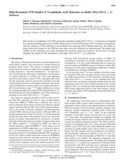
High-Resolution STM Studies of Terephthalic Acid Molecules on ...
High-Resolution STM Studies of Terephthalic Acid Molecules on ...
High-Resolution STM Studies of Terephthalic Acid Molecules on ...

You also want an ePaper? Increase the reach of your titles
YUMPU automatically turns print PDFs into web optimized ePapers that Google loves.
<str<strong>on</strong>g>High</str<strong>on</strong>g>-<str<strong>on</strong>g>Resoluti<strong>on</strong></str<strong>on</strong>g> <str<strong>on</strong>g>STM</str<strong>on</strong>g> <str<strong>on</strong>g>Studies</str<strong>on</strong>g> <str<strong>on</strong>g>of</str<strong>on</strong>g> <str<strong>on</strong>g>Terephthalic</str<strong>on</strong>g> <str<strong>on</strong>g>Acid</str<strong>on</strong>g> <str<strong>on</strong>g>Molecules</str<strong>on</strong>g> <strong>on</strong> Rutile TiO2(110)-(1 × 1)<br />
Surfaces<br />
I. Introducti<strong>on</strong><br />
Jakub S. Prauzner-Bechcicki,* Szym<strong>on</strong> Godlewski, Ant<strong>on</strong>i Tekiel, † Piotr Cyganik,<br />
Janusz Budzioch, and Marek Szym<strong>on</strong>ski<br />
Research Centre for Nanometer-scale Science and AdVanced Materials (NANOSAM), Faculty <str<strong>on</strong>g>of</str<strong>on</strong>g> Physics,<br />
Astr<strong>on</strong>omy and Applied Computer Science, Jagiell<strong>on</strong>ian UniVersity, Reym<strong>on</strong>ta 4, 30-059, Krakow, Poland<br />
ReceiVed: February 9, 2009; ReVised Manuscript ReceiVed: March 20, 2009<br />
The structure <str<strong>on</strong>g>of</str<strong>on</strong>g> terephthalic acid (TPA) molecules adsorbed <strong>on</strong> rutile TiO2(110)-(1 × 1) has been investigated<br />
by scanning tunneling microscopy (<str<strong>on</strong>g>STM</str<strong>on</strong>g>). Molecularly resolved <str<strong>on</strong>g>STM</str<strong>on</strong>g> images show formati<strong>on</strong> <str<strong>on</strong>g>of</str<strong>on</strong>g> m<strong>on</strong>olayer<br />
with the tendency <str<strong>on</strong>g>of</str<strong>on</strong>g> TPA molecules to form dimer rows al<strong>on</strong>g the [001] substrate directi<strong>on</strong>. The ability to<br />
image functi<strong>on</strong>al groups by the <str<strong>on</strong>g>STM</str<strong>on</strong>g> tip, and single molecule diffusi<strong>on</strong> are dem<strong>on</strong>strated. The quality and<br />
stability <str<strong>on</strong>g>of</str<strong>on</strong>g> the m<strong>on</strong>olayer are tested, including the resistance against air exposure. On the basis <str<strong>on</strong>g>of</str<strong>on</strong>g> results<br />
obtained, the model <str<strong>on</strong>g>of</str<strong>on</strong>g> TPA adsorpti<strong>on</strong> <strong>on</strong> the rutile TiO2(110)-(1 × 1) is proposed.<br />
The surface <str<strong>on</strong>g>of</str<strong>on</strong>g> titanium dioxide is the most <str<strong>on</strong>g>of</str<strong>on</strong>g>ten studied metal<br />
oxide surface, which is also c<strong>on</strong>cerned as a model system for<br />
transiti<strong>on</strong> metal oxides. 1 The interest in titanium dioxide is<br />
mainly driven by its diverse applicati<strong>on</strong>s including catalysis, 2-5<br />
coating materials, 6-8 and sensor producti<strong>on</strong> 9-12 to name just a<br />
few. C<strong>on</strong>sidering this wide range <str<strong>on</strong>g>of</str<strong>on</strong>g> applicati<strong>on</strong>s, the adsorpti<strong>on</strong><br />
<str<strong>on</strong>g>of</str<strong>on</strong>g> organic molecules is certainly <strong>on</strong>e <str<strong>on</strong>g>of</str<strong>on</strong>g> the most appealing<br />
directi<strong>on</strong>s in the surface science <str<strong>on</strong>g>of</str<strong>on</strong>g> TiO2. Although metal oxides<br />
provide anisotropic surfaces and specific adsorpti<strong>on</strong> sites that<br />
would facilitate growth <str<strong>on</strong>g>of</str<strong>on</strong>g> well-ordered m<strong>on</strong>olayers, the chemical<br />
functi<strong>on</strong>alizati<strong>on</strong> <str<strong>on</strong>g>of</str<strong>on</strong>g> these surfaces by organic molecules is<br />
still in its preliminary stage. 1,13-20 In this c<strong>on</strong>tributi<strong>on</strong>, we focus<br />
<strong>on</strong> scanning tunneling microscopy (<str<strong>on</strong>g>STM</str<strong>on</strong>g>) measurements <str<strong>on</strong>g>of</str<strong>on</strong>g> 1,4benzenedicarboxylic<br />
acid (terephthalic acid, TPA) m<strong>on</strong>olayers<br />
adsorbed <strong>on</strong> the rutile TiO2(110)-(1 × 1) surface.<br />
Our interest in studying adsorpti<strong>on</strong> <str<strong>on</strong>g>of</str<strong>on</strong>g> TPA is driven by the<br />
fact that TPA molecules are used in some <str<strong>on</strong>g>of</str<strong>on</strong>g> metal-organic<br />
frameworks (MOFs). 21 MOFs form a relatively new class <str<strong>on</strong>g>of</str<strong>on</strong>g><br />
porous materials which c<strong>on</strong>sist <str<strong>on</strong>g>of</str<strong>on</strong>g> metal i<strong>on</strong>s linked by specific<br />
organic molecules like the TPA. Due to their potential applicati<strong>on</strong><br />
in gas storage, molecular separati<strong>on</strong>, and heterogeneous<br />
catalysis MOFs attract growing attenti<strong>on</strong>. Although MOFs are<br />
usually produced in the form <str<strong>on</strong>g>of</str<strong>on</strong>g> a powder, it was dem<strong>on</strong>strated<br />
recently that thin films <str<strong>on</strong>g>of</str<strong>on</strong>g> this material can be very selectively<br />
grown <strong>on</strong> surfaces functi<strong>on</strong>alized by m<strong>on</strong>olayers <str<strong>on</strong>g>of</str<strong>on</strong>g> organic<br />
molecules which mimic the functi<strong>on</strong>ality <str<strong>on</strong>g>of</str<strong>on</strong>g> organic linkers used<br />
for MOFs synthesis. 22-24 Formati<strong>on</strong> <str<strong>on</strong>g>of</str<strong>on</strong>g> MOF thin films <strong>on</strong><br />
desired substrates, called SURMOFs, 25 clearly opens up new<br />
opportunities for MOFs applicati<strong>on</strong>, e.g., in design <str<strong>on</strong>g>of</str<strong>on</strong>g> sensors,<br />
as it was dem<strong>on</strong>strated very recently. 26 Al<strong>on</strong>g this line <str<strong>on</strong>g>of</str<strong>on</strong>g><br />
research, formati<strong>on</strong> <str<strong>on</strong>g>of</str<strong>on</strong>g> well-defined and stable TPA m<strong>on</strong>olayers<br />
<strong>on</strong> the TiO2 surface would enable later research <strong>on</strong> SURMOFs<br />
formati<strong>on</strong> <strong>on</strong> this substrate, and at the same time, possibility <str<strong>on</strong>g>of</str<strong>on</strong>g><br />
merging these two important classes <str<strong>on</strong>g>of</str<strong>on</strong>g> materials. For example,<br />
joining in <strong>on</strong>e composite device the catalytic properties <str<strong>on</strong>g>of</str<strong>on</strong>g> the<br />
* To whom corresp<strong>on</strong>dence should be addressed: E-mail: jakub.prauznerbechcicki@uj.edu.pl.<br />
† Present address: Department <str<strong>on</strong>g>of</str<strong>on</strong>g> Physics, McGill University, 3600<br />
University Street, M<strong>on</strong>treal QC, H3A 2T8, Canada.<br />
J. Phys. Chem. C 2009, 113, 9309–9315 9309<br />
substrate with extraordinary storage capacity <str<strong>on</strong>g>of</str<strong>on</strong>g> MOFs for<br />
accumulati<strong>on</strong> <str<strong>on</strong>g>of</str<strong>on</strong>g> products <str<strong>on</strong>g>of</str<strong>on</strong>g> catalytic reacti<strong>on</strong>s could be very<br />
advantageous. It is also worth menti<strong>on</strong>ing that in c<strong>on</strong>trast to<br />
previous studies <strong>on</strong> SURMOFs growth, formati<strong>on</strong> <str<strong>on</strong>g>of</str<strong>on</strong>g> stable TPA<br />
m<strong>on</strong>olayers <strong>on</strong> TiO2 allows studying direct applicati<strong>on</strong> <str<strong>on</strong>g>of</str<strong>on</strong>g> MOFs<br />
c<strong>on</strong>stituencies for initiating SURMOFs.<br />
In our previous work, 20 we have reported <strong>on</strong> the first results<br />
<str<strong>on</strong>g>of</str<strong>on</strong>g> combined <str<strong>on</strong>g>STM</str<strong>on</strong>g> and n<strong>on</strong>-c<strong>on</strong>tact atomic force microscopy<br />
(nc-AFM) measurements <str<strong>on</strong>g>of</str<strong>on</strong>g> TPA molecules adsorbed <strong>on</strong> the<br />
rutile TiO2(110)-(1 × 1) surface. It was c<strong>on</strong>cluded that TPA<br />
molecules form a m<strong>on</strong>olayer <str<strong>on</strong>g>of</str<strong>on</strong>g> upright standing molecules that<br />
supposedly expose <strong>on</strong>e <str<strong>on</strong>g>of</str<strong>on</strong>g> their carboxyl groups to the vacuum<br />
interface. Here, we extended previous measurements by further<br />
<str<strong>on</strong>g>STM</str<strong>on</strong>g> studies addressing details <str<strong>on</strong>g>of</str<strong>on</strong>g> the film structure, defects<br />
formati<strong>on</strong> and stability <str<strong>on</strong>g>of</str<strong>on</strong>g> TPA molecules <strong>on</strong> the rutile TiO2(110)-<br />
(1 × 1) surface.<br />
II. Experimental Secti<strong>on</strong><br />
The experiments were carried out in a multichamber ultrahigh<br />
vacuum (UHV) system, which c<strong>on</strong>sisted <str<strong>on</strong>g>of</str<strong>on</strong>g> a microscope,<br />
preparati<strong>on</strong>, surface analysis, and radial distributi<strong>on</strong> chambers.<br />
The base pressure in the microscope chamber, equipped with<br />
Omicr<strong>on</strong> variable temperature scanning probe microscope<br />
(VT <str<strong>on</strong>g>STM</str<strong>on</strong>g>/AFM), was 4 × 10-11 mbar. In the remaining<br />
chambers <str<strong>on</strong>g>of</str<strong>on</strong>g> the UHV system, base pressure was in the low<br />
10-10 mbar range. The preparati<strong>on</strong> chamber was supplied with<br />
a noble gas i<strong>on</strong> gun, effusi<strong>on</strong> cells, an infrared pyrometer, and<br />
a quartz microbalance thickness m<strong>on</strong>itor. The surface analysis<br />
chamber was equipped with a low energy electr<strong>on</strong> diffracti<strong>on</strong><br />
(LEED) system. The TiO2(110) wafers (MaTecK GmbH) were<br />
mounted <strong>on</strong> sample holders with a silic<strong>on</strong> wafer as a resistive<br />
heater. The TiO2 samples were first annealed for 6hat800K,<br />
and subsequently subjected to repetitive cycles <str<strong>on</strong>g>of</str<strong>on</strong>g> 1 keV Ar +<br />
sputtering for 15 min and annealing at 960 K for 15 min until<br />
clean surface was reached. The annealing temperature was<br />
c<strong>on</strong>trolled by the infrared pyrometer. The quality <str<strong>on</strong>g>of</str<strong>on</strong>g> the obtained<br />
TiO2(110)-(1 × 1) surface was m<strong>on</strong>itored by <str<strong>on</strong>g>STM</str<strong>on</strong>g> and LEED<br />
measurements. The TPA molecules were evaporated at about<br />
370 K from a Knudsen cell <strong>on</strong> the TiO2(110) surface kept at<br />
room temperature. Pressure in the preparati<strong>on</strong> chamber during<br />
TPA depositi<strong>on</strong> was 9 × 10⁻¹⁰ mbar. Prior to the evaporati<strong>on</strong><br />
10.1021/jp901184t CCC: $40.75 © 2009 American Chemical Society<br />
Published <strong>on</strong> Web 04/30/2009
9310 J. Phys. Chem. C, Vol. 113, No. 21, 2009 Prauzner-Bechcicki et al.<br />
Figure 1. Images (a), (b), and (c) show different <str<strong>on</strong>g>STM</str<strong>on</strong>g> c<strong>on</strong>trasts obtained at the same scanning parameters (I ) 2 pA, U ) 1.8-2.0 V) for the same<br />
sample <str<strong>on</strong>g>of</str<strong>on</strong>g> TPA/TiO2(110). The carto<strong>on</strong> located in the central part <str<strong>on</strong>g>of</str<strong>on</strong>g> the figure schematically shows the side view <str<strong>on</strong>g>of</str<strong>on</strong>g> the TPA m<strong>on</strong>olayer. Blue<br />
arrows pointing images (a), (b), and (c) in this carto<strong>on</strong> schematically show which part <str<strong>on</strong>g>of</str<strong>on</strong>g> the m<strong>on</strong>olayer is presumably visualized by the given <str<strong>on</strong>g>STM</str<strong>on</strong>g><br />
scan. The top view appearance <str<strong>on</strong>g>of</str<strong>on</strong>g> the m<strong>on</strong>olayer in <str<strong>on</strong>g>STM</str<strong>on</strong>g> scans shown in (a) and (b) is schematically presented in carto<strong>on</strong>s displayed below<br />
respective image. Arrows <strong>on</strong> sides <str<strong>on</strong>g>of</str<strong>on</strong>g> image (c) point to rows <str<strong>on</strong>g>of</str<strong>on</strong>g> bright spots (red arrow) and S-shaped structures (green arrow), respectively (see<br />
text for details).<br />
procedure, terephthalic acid powder (Fluka, g 99% purity) was<br />
outgassed in vacuum. The <str<strong>on</strong>g>STM</str<strong>on</strong>g> imaging was carried out in a<br />
c<strong>on</strong>stant current mode at positive bias voltages, i.e., emptystates<br />
imaging, with etched tungsten tips used as probes. The<br />
image processing and analysis was d<strong>on</strong>e using WSxM 4.0<br />
s<str<strong>on</strong>g>of</str<strong>on</strong>g>tware. 27<br />
III. Results and Discussi<strong>on</strong><br />
Structure <str<strong>on</strong>g>of</str<strong>on</strong>g> the M<strong>on</strong>olayer. Former studies <strong>on</strong> room<br />
temperature adsorpti<strong>on</strong> <str<strong>on</strong>g>of</str<strong>on</strong>g> m<strong>on</strong>ocarboxylic acids, such as formic,<br />
acetic, and trimethylacetic acids, 1,16 as well as, larger, aromatic<br />
m<strong>on</strong>ocarboxylic acids, such as benzoic and is<strong>on</strong>icotinic acids13-15 dem<strong>on</strong>strated that carboxylic acids follow a dissociative adsorpti<strong>on</strong><br />
path <strong>on</strong> the rutile TiO2(110)-(1 × 1) surface. The adsorpti<strong>on</strong><br />
process involves dissociati<strong>on</strong> <str<strong>on</strong>g>of</str<strong>on</strong>g> the carboxyl group that gives<br />
carboxylate and hydrogen. In the following step, the negatively<br />
charged carboxylate is bounded in a bridge form with the<br />
O-C-O plane aligned in the [001] surface directi<strong>on</strong> <strong>on</strong> a pair<br />
<str<strong>on</strong>g>of</str<strong>on</strong>g> 5-fold coordinated Ti atoms, and thus, occupies two surface<br />
lattice unit cells. The remaining hydrogen atom is presumably<br />
bounded to <strong>on</strong>e or two surface oxygen atoms, forming -OH<br />
group or bridging two oxygen atoms, respectively. Lyubinetsky<br />
et al. suggested that presence <str<strong>on</strong>g>of</str<strong>on</strong>g> such -OH residues or bridging<br />
-H atoms might play a role in stabilizing the overlayer <str<strong>on</strong>g>of</str<strong>on</strong>g><br />
trimethylacetic acid <strong>on</strong> the TiO2(110) surface. 28<br />
Since the primary motivati<strong>on</strong> <str<strong>on</strong>g>of</str<strong>on</strong>g> our research is well-defined,<br />
functi<strong>on</strong>alizati<strong>on</strong> <str<strong>on</strong>g>of</str<strong>on</strong>g> the TiO2(110) surface by TPA molecules<br />
to enable later growth <str<strong>on</strong>g>of</str<strong>on</strong>g> MOFs, we have focused our experiments<br />
<strong>on</strong> studying depositi<strong>on</strong> c<strong>on</strong>diti<strong>on</strong>s, which allow a full<br />
m<strong>on</strong>olayer coverage. The high-resoluti<strong>on</strong> <str<strong>on</strong>g>STM</str<strong>on</strong>g> data presented<br />
in Figure 1 show densely packed structure <str<strong>on</strong>g>of</str<strong>on</strong>g> TPA molecules<br />
obtained at the full m<strong>on</strong>olayer coverage (1 ML). Importantly,<br />
further increase <str<strong>on</strong>g>of</str<strong>on</strong>g> depositi<strong>on</strong> time does not result in observati<strong>on</strong><br />
<str<strong>on</strong>g>of</str<strong>on</strong>g> sec<strong>on</strong>d or higher order molecular layers, thus suggesting that<br />
adsorpti<strong>on</strong> <str<strong>on</strong>g>of</str<strong>on</strong>g> the TPA <strong>on</strong> the TiO2(110)-(1 × 1) surface at room<br />
temperature is a self-limiting process, up to 1 ML coverage.<br />
Four different c<strong>on</strong>trasts observed in our <str<strong>on</strong>g>STM</str<strong>on</strong>g> data are documented<br />
in Figure 1. Whereas the two first types <str<strong>on</strong>g>of</str<strong>on</strong>g> c<strong>on</strong>trast<br />
depicted in Figure 1, parts a and b, we have already reported, 20<br />
the other two c<strong>on</strong>trasts shown in Figure 1c, have not been<br />
identified in our previous experiments. We have observed<br />
random, reversible switching between these four <str<strong>on</strong>g>STM</str<strong>on</strong>g> c<strong>on</strong>trasts<br />
during data acquisiti<strong>on</strong>, which we attribute to a change <str<strong>on</strong>g>of</str<strong>on</strong>g> the<br />
<str<strong>on</strong>g>STM</str<strong>on</strong>g> tip terminati<strong>on</strong>. Several possibilities <str<strong>on</strong>g>of</str<strong>on</strong>g> a tip modificati<strong>on</strong><br />
leading to the observed changes <str<strong>on</strong>g>of</str<strong>on</strong>g> the <str<strong>on</strong>g>STM</str<strong>on</strong>g> c<strong>on</strong>trast can be<br />
c<strong>on</strong>sidered including: (1) adsorpti<strong>on</strong> <str<strong>on</strong>g>of</str<strong>on</strong>g> a TPA molecule, (2) its<br />
further reorientati<strong>on</strong>, or (3) transfer <str<strong>on</strong>g>of</str<strong>on</strong>g> the prot<strong>on</strong> that comes<br />
from dissociati<strong>on</strong> <str<strong>on</strong>g>of</str<strong>on</strong>g> TPA molecules and is supposedly adsorbed<br />
<strong>on</strong> the surface. Each <str<strong>on</strong>g>of</str<strong>on</strong>g> such changes <str<strong>on</strong>g>of</str<strong>on</strong>g> the <str<strong>on</strong>g>STM</str<strong>on</strong>g> tip apex may<br />
easily take place several times while scanning the m<strong>on</strong>olayer
<str<strong>on</strong>g>STM</str<strong>on</strong>g> <str<strong>on</strong>g>of</str<strong>on</strong>g> <str<strong>on</strong>g>Terephthalic</str<strong>on</strong>g> <str<strong>on</strong>g>Acid</str<strong>on</strong>g> <str<strong>on</strong>g>Molecules</str<strong>on</strong>g> J. Phys. Chem. C, Vol. 113, No. 21, 2009 9311<br />
<str<strong>on</strong>g>of</str<strong>on</strong>g> TPA molecules <strong>on</strong> TiO2(110) surface leading to reoccurrence<br />
<str<strong>on</strong>g>of</str<strong>on</strong>g> a given c<strong>on</strong>trast. The more strict identificati<strong>on</strong> <str<strong>on</strong>g>of</str<strong>on</strong>g> the tip<br />
modificati<strong>on</strong> resp<strong>on</strong>sible for a given <str<strong>on</strong>g>STM</str<strong>on</strong>g> c<strong>on</strong>trast requires a<br />
theoretical modeling which has not been d<strong>on</strong>e so far.<br />
In Figure 1a, a regular (2 × 1) pattern is clearly visible. This<br />
structure is l<strong>on</strong>g-ranged, as it is documented by the 50 × 50<br />
nm scans, which will be analyzed later <strong>on</strong>. Assuming that each<br />
bright spot in Figure 1a corresp<strong>on</strong>ds to <strong>on</strong>e molecule, we obtain<br />
a packing density corresp<strong>on</strong>ding to <strong>on</strong>e TPA molecule per two<br />
TiO2(110)-(1 × 1) substrate unit cells, i.e., 38 Å 2 per molecule<br />
(2.61 × 10 14 molecules/cm 2 ). These results indicate an upright<br />
orientati<strong>on</strong> <str<strong>on</strong>g>of</str<strong>on</strong>g> the TPA molecules c<strong>on</strong>sidering a previous study 15<br />
for shorter molecules <str<strong>on</strong>g>of</str<strong>on</strong>g> is<strong>on</strong>icotinic acid <strong>on</strong> the TiO2(110)-(1<br />
× 1) substrate, where combined <str<strong>on</strong>g>STM</str<strong>on</strong>g> and angle-dependent<br />
X-ray absorpti<strong>on</strong> spectroscopy data dem<strong>on</strong>strated upright orientati<strong>on</strong><br />
<str<strong>on</strong>g>of</str<strong>on</strong>g> these molecules adsorbed in the similar (2 × 1)<br />
pattern. We attribute the observed (2 × 1) pattern to the<br />
adsorpti<strong>on</strong> sites <str<strong>on</strong>g>of</str<strong>on</strong>g> the TPA molecules, i.e., deeper lying<br />
electr<strong>on</strong>ic structures since it was not observed in the corresp<strong>on</strong>ding<br />
nc-AFM measurements reported in our previous<br />
paper. 20 The other comm<strong>on</strong> <str<strong>on</strong>g>STM</str<strong>on</strong>g> c<strong>on</strong>trast, shown in Figure 1b,<br />
is the m<strong>on</strong>omer-dimer motif which has been also observed in<br />
our previous nc-AFM measurements, 20 and therefore, we<br />
attribute it to the higher lying electr<strong>on</strong>ic structures. In this<br />
pattern, molecular rows are aligned al<strong>on</strong>g the [001] substrate<br />
directi<strong>on</strong>. As documented by the data, m<strong>on</strong>omer rows frequently<br />
evolve into dimer rows and vice versa.<br />
The additi<strong>on</strong>al piece <str<strong>on</strong>g>of</str<strong>on</strong>g> informati<strong>on</strong>, regarding formati<strong>on</strong> <str<strong>on</strong>g>of</str<strong>on</strong>g><br />
this m<strong>on</strong>omer-dimer pattern, can be found in the two other<br />
much less frequently observed <str<strong>on</strong>g>STM</str<strong>on</strong>g> c<strong>on</strong>trasts visible in the<br />
upper and lower parts <str<strong>on</strong>g>of</str<strong>on</strong>g> the single <str<strong>on</strong>g>STM</str<strong>on</strong>g> image shown in Figure<br />
1c. Whereas the upper part <str<strong>on</strong>g>of</str<strong>on</strong>g> this image exhibits bright spots<br />
forming rows al<strong>on</strong>g the [001] substrate directi<strong>on</strong> (marked by<br />
the red arrow in Figure 1c), the lower part reveals dark lines<br />
separating rows <str<strong>on</strong>g>of</str<strong>on</strong>g> S-shaped structures (marked by the green<br />
arrow in Figure 1c). When comparing the positi<strong>on</strong> <str<strong>on</strong>g>of</str<strong>on</strong>g> the rows<br />
<str<strong>on</strong>g>of</str<strong>on</strong>g> bright spots in the upper part <str<strong>on</strong>g>of</str<strong>on</strong>g> Figure 1c with the rows <str<strong>on</strong>g>of</str<strong>on</strong>g><br />
S-shaped structures in the lower part <str<strong>on</strong>g>of</str<strong>on</strong>g> this image, it becomes<br />
clear that the rows <str<strong>on</strong>g>of</str<strong>on</strong>g> bright features corresp<strong>on</strong>d to the enhanced<br />
c<strong>on</strong>trast from the middle part <str<strong>on</strong>g>of</str<strong>on</strong>g> TPA dimer rows, i.e., S-shaped<br />
structures (compare dashed line in Figure 1c). Such a transiti<strong>on</strong><br />
<str<strong>on</strong>g>of</str<strong>on</strong>g> the row <str<strong>on</strong>g>of</str<strong>on</strong>g> bright spots <str<strong>on</strong>g>of</str<strong>on</strong>g> the enhanced c<strong>on</strong>trast into the<br />
middle part <str<strong>on</strong>g>of</str<strong>on</strong>g> the dimer rows was repeatedly observed.<br />
Therefore, we suppose that this enhanced c<strong>on</strong>trast originates<br />
from the sp<strong>on</strong>taneous tip apex modificati<strong>on</strong> by either an<br />
adsorbed TPA molecule or prot<strong>on</strong> transfer, which enables<br />
recogniti<strong>on</strong> <str<strong>on</strong>g>of</str<strong>on</strong>g> the -COOH groups in a m<strong>on</strong>olayer. The<br />
recogniti<strong>on</strong> <str<strong>on</strong>g>of</str<strong>on</strong>g> the functi<strong>on</strong>al -OH and -COOH groups via<br />
deliberate chemical modificati<strong>on</strong> <str<strong>on</strong>g>of</str<strong>on</strong>g> the <str<strong>on</strong>g>STM</str<strong>on</strong>g> tip with thiol selfassembled<br />
m<strong>on</strong>olayers (SAMs) has been dem<strong>on</strong>strated by Ito<br />
et al. for the flat-laying physisorbed m<strong>on</strong>olayers <str<strong>on</strong>g>of</str<strong>on</strong>g> 1-octadecanol<br />
and 1-octadecanoic acid <strong>on</strong> HOPG. 29 It has been suggested in<br />
this work that the c<strong>on</strong>trast enhancement <strong>on</strong> hydroxyl or carboxyl<br />
groups originates from the hydrogen b<strong>on</strong>d interacti<strong>on</strong> between<br />
the functi<strong>on</strong>al groups <str<strong>on</strong>g>of</str<strong>on</strong>g> the tip and the sample. Such a hydrogen<br />
b<strong>on</strong>d based c<strong>on</strong>trast enhancement effect was also observed for<br />
polymer-modified tips. 30 For the TPA m<strong>on</strong>olayer, observed<br />
c<strong>on</strong>trast enhancement probably comes from the adsorpti<strong>on</strong> <str<strong>on</strong>g>of</str<strong>on</strong>g> a<br />
TPA molecule (or molecules) <strong>on</strong> the tip and formati<strong>on</strong> <str<strong>on</strong>g>of</str<strong>on</strong>g> the<br />
hydrogen b<strong>on</strong>d between the decorated tip and the molecules in<br />
the m<strong>on</strong>olayer. Interestingly, the enhancement <str<strong>on</strong>g>of</str<strong>on</strong>g> the c<strong>on</strong>trast<br />
is observed <strong>on</strong>ly for TPA molecules in dimer rows. The origin<br />
<str<strong>on</strong>g>of</str<strong>on</strong>g> this drastic difference in the visualizati<strong>on</strong> <str<strong>on</strong>g>of</str<strong>on</strong>g> the upright<br />
standing molecules arranged in dimer and m<strong>on</strong>omer rows is<br />
Figure 2. In (a) <str<strong>on</strong>g>STM</str<strong>on</strong>g> image (2pA, 2V) exhibiting rows <str<strong>on</strong>g>of</str<strong>on</strong>g> dimers and<br />
m<strong>on</strong>omers <strong>on</strong> the TPA/TiO2(110) sample. The round shape and S-shape<br />
characteristic for m<strong>on</strong>omer and dimer rows, respectively, are marked<br />
in blue. The carto<strong>on</strong> in (b) schematically shows possible tilting and<br />
rotating <str<strong>on</strong>g>of</str<strong>on</strong>g> the TPA phenyl rings associated with the dimer formati<strong>on</strong>.<br />
For simplicity, <strong>on</strong>ly phenyl rings associated with the TPA molecules<br />
are shown in this carto<strong>on</strong>. The <str<strong>on</strong>g>STM</str<strong>on</strong>g> c<strong>on</strong>trast visible in (a) is<br />
schematically marked in (b) by yellow circles. The possible rotati<strong>on</strong><br />
by 25° <str<strong>on</strong>g>of</str<strong>on</strong>g> the TPA phenyl rings with respect to the O-C-O plane <str<strong>on</strong>g>of</str<strong>on</strong>g><br />
the carboxyl group through which TPA binds to the substrate al<strong>on</strong>g<br />
the [001] substrate directi<strong>on</strong> has been adopted from ref 32.<br />
not clear at the moment. However, it should be noted that in<br />
the previous experiments by Ito et al. 29 the drastic c<strong>on</strong>trast<br />
enhancement was observed in the m<strong>on</strong>olayers <str<strong>on</strong>g>of</str<strong>on</strong>g> physisorbed<br />
molecules where two neighboring -COOH or -OH groups<br />
were in c<strong>on</strong>tact (the molecules <str<strong>on</strong>g>of</str<strong>on</strong>g> 1-octadecanol or 1-octadecanoic<br />
acid <strong>on</strong> HOPG were physisorbed flat lying in the headto-head<br />
c<strong>on</strong>figurati<strong>on</strong>). Therefore, we suppose that higher<br />
c<strong>on</strong>trast <str<strong>on</strong>g>of</str<strong>on</strong>g> the TPA molecules in the dimer c<strong>on</strong>figurati<strong>on</strong> may<br />
result from their inclinati<strong>on</strong>, which enables c<strong>on</strong>tact between<br />
neighboring -COOH residues as it was observed in previous<br />
experiments by Ito et al. The other possibility for the <str<strong>on</strong>g>STM</str<strong>on</strong>g><br />
c<strong>on</strong>trast modificati<strong>on</strong>, which enables high chemical sensitivity,<br />
is by prot<strong>on</strong> transfer to the tip apex, as proposed in the previous<br />
high-resoluti<strong>on</strong> <str<strong>on</strong>g>STM</str<strong>on</strong>g> studies <str<strong>on</strong>g>of</str<strong>on</strong>g> the pristine TiO2 (110) surface. 31<br />
Another type <str<strong>on</strong>g>of</str<strong>on</strong>g> c<strong>on</strong>trast visible in the lower part <str<strong>on</strong>g>of</str<strong>on</strong>g> Figure<br />
1c is presented in more details in Figure 2a. In this <str<strong>on</strong>g>STM</str<strong>on</strong>g> image,<br />
TPA molecules in m<strong>on</strong>omer rows are round shaped, similar to<br />
that observed for the data presented in Figure 1, parts a and b.<br />
In c<strong>on</strong>trast, dimer rows are formed by the S-shaped structures<br />
with more el<strong>on</strong>gated shape <str<strong>on</strong>g>of</str<strong>on</strong>g> the TPA molecules (see blue<br />
markers in Figure 2a). We suppose that these differences reflect<br />
inclinati<strong>on</strong> <str<strong>on</strong>g>of</str<strong>on</strong>g> the TPA molecules in dimer rows and possible<br />
rotati<strong>on</strong> <str<strong>on</strong>g>of</str<strong>on</strong>g> the phenyl ring (see carto<strong>on</strong> in Figure 2b). The<br />
possible rotati<strong>on</strong> <str<strong>on</strong>g>of</str<strong>on</strong>g> the phenyl ring with respect to the O-C-O<br />
plane <str<strong>on</strong>g>of</str<strong>on</strong>g> the carboxyl group through which the molecule binds<br />
to the substrate is supported by the very recent theoretical<br />
calculati<strong>on</strong>s for the TPA molecules <strong>on</strong> Cu(110) substrate. 32<br />
Summarizing obtained results, we can propose a model <str<strong>on</strong>g>of</str<strong>on</strong>g><br />
the TPA adsorpti<strong>on</strong> <strong>on</strong> the TiO2(110)-(1 × 1) substrate, which<br />
is schematically depicted in Figure 1. The basic ingredients <str<strong>on</strong>g>of</str<strong>on</strong>g><br />
this model are as follows: (i) molecules bind to the surface in<br />
a bidentate fashi<strong>on</strong> <strong>on</strong> the two 5-fold coordinated Ti atoms, (ii)<br />
form rows al<strong>on</strong>g the [001] directi<strong>on</strong>, (iii) are oriented upward
9312 J. Phys. Chem. C, Vol. 113, No. 21, 2009 Prauzner-Bechcicki et al.<br />
Figure 3. In (a) high-resoluti<strong>on</strong> <str<strong>on</strong>g>STM</str<strong>on</strong>g> image obtained for TPA/<br />
TiO2(110) sample exhibiting 0.6 ML coverage (measured from <str<strong>on</strong>g>STM</str<strong>on</strong>g><br />
images with use <str<strong>on</strong>g>of</str<strong>on</strong>g> WSxM 4.0 program [ref 27]) dimer pattern clearly<br />
visible in selected areas <str<strong>on</strong>g>of</str<strong>on</strong>g> the image. In (b) and (c) two c<strong>on</strong>secutive<br />
<str<strong>on</strong>g>STM</str<strong>on</strong>g> images taken at the same locati<strong>on</strong> <str<strong>on</strong>g>of</str<strong>on</strong>g> the TPA/TiO2(110) sample<br />
show <str<strong>on</strong>g>STM</str<strong>on</strong>g> induced reducti<strong>on</strong> <str<strong>on</strong>g>of</str<strong>on</strong>g> the TPA coverage from 0.7 ML in<br />
(b) to 0.5 ML in (c). <str<strong>on</strong>g>STM</str<strong>on</strong>g> parameters: (a) I ) 2pAandU ) 2.0 V,<br />
(b) I ) 20 pA, U ) 2V, (c) I ) 20 pA, and U ) 2V.<br />
with -COOH group exposed to the vacuum interface, and (iv)<br />
incline and rotate toward each other to form dimers.<br />
Defects Formati<strong>on</strong>. Before discussing defects which are<br />
inherently present in the TPA m<strong>on</strong>olayer, it is crucial to c<strong>on</strong>sider<br />
the influence <str<strong>on</strong>g>of</str<strong>on</strong>g> the <str<strong>on</strong>g>STM</str<strong>on</strong>g> imaging <strong>on</strong> the film stability. The<br />
data presented in Figures 1 and 2 have been obtained at the<br />
extremely low current set point <str<strong>on</strong>g>of</str<strong>on</strong>g> 2 pA, and relatively high<br />
bias voltage <str<strong>on</strong>g>of</str<strong>on</strong>g> 1.8-2.0 V. Generally, our experiments show<br />
that even for the <str<strong>on</strong>g>STM</str<strong>on</strong>g> imaging with 2 orders <str<strong>on</strong>g>of</str<strong>on</strong>g> magnitude<br />
higher current set point (keeping the same bias voltage), no<br />
systematic modificati<strong>on</strong> <str<strong>on</strong>g>of</str<strong>on</strong>g> the TPA film structure could be<br />
observed. This statement is, however, true <strong>on</strong>ly for the TPA<br />
coverage close to a complete m<strong>on</strong>olayer. As it is exemplified<br />
by the data obtained for the TPA film corresp<strong>on</strong>ding to ∼0.6<br />
ML coverage (coverage measured from the respective <str<strong>on</strong>g>STM</str<strong>on</strong>g><br />
images with use <str<strong>on</strong>g>of</str<strong>on</strong>g> WSxM 4.0 program [ref 27]), the n<strong>on</strong>destructive<br />
high-resoluti<strong>on</strong> imaging <str<strong>on</strong>g>of</str<strong>on</strong>g> this sample could be<br />
performed at 2 pA current set point and 2 V bias voltage (Figure<br />
3a), i.e., at extremely high impedance c<strong>on</strong>diti<strong>on</strong>s. In c<strong>on</strong>trast,<br />
the <str<strong>on</strong>g>STM</str<strong>on</strong>g> imaging <str<strong>on</strong>g>of</str<strong>on</strong>g> such low-coverage TPA samples at the<br />
current set point increased up to <strong>on</strong>ly 20 pA (keeping the same<br />
bias voltage) results in well visible TPA film modificati<strong>on</strong>. This<br />
observati<strong>on</strong> is documented by the data shown in Figure 3, parts<br />
b and c, where two c<strong>on</strong>secutive scans reveal <str<strong>on</strong>g>STM</str<strong>on</strong>g>-tip induced<br />
increase <str<strong>on</strong>g>of</str<strong>on</strong>g> dark patches in Figure 3, parts b and c, which<br />
corresp<strong>on</strong>d to regi<strong>on</strong>s free from TPA molecules. The more<br />
detailed analysis <str<strong>on</strong>g>of</str<strong>on</strong>g> these data show that single <str<strong>on</strong>g>STM</str<strong>on</strong>g> scan, at<br />
this imaging c<strong>on</strong>diti<strong>on</strong>s, results in a decrease <str<strong>on</strong>g>of</str<strong>on</strong>g> the TPA<br />
coverage from the initial ∼0.7 down to ∼0.5 ML.<br />
Three important c<strong>on</strong>clusi<strong>on</strong>s can be drawn from the data<br />
shown in Figure 3. First, it is clear that n<strong>on</strong>destructive <str<strong>on</strong>g>STM</str<strong>on</strong>g><br />
imaging <str<strong>on</strong>g>of</str<strong>on</strong>g> TPA films at the subm<strong>on</strong>olayer coverage requires<br />
extremely high impedance c<strong>on</strong>diti<strong>on</strong>s; otherwise, the interpretati<strong>on</strong><br />
<str<strong>on</strong>g>of</str<strong>on</strong>g> the obtained results may lead to severe problems. Sec<strong>on</strong>d,<br />
the fact that the characteristic dimer packing motive <str<strong>on</strong>g>of</str<strong>on</strong>g> TPA<br />
molecules reported above for the full m<strong>on</strong>olayer coverage is<br />
also present even at a much earlier stage <str<strong>on</strong>g>of</str<strong>on</strong>g> the film formati<strong>on</strong>,<br />
i.e., at coverage <str<strong>on</strong>g>of</str<strong>on</strong>g> <strong>on</strong>ly 0.6 ML (see high-resoluti<strong>on</strong> data in<br />
Figure 3a), suggest rather important c<strong>on</strong>tributi<strong>on</strong> <str<strong>on</strong>g>of</str<strong>on</strong>g> dimerizati<strong>on</strong><br />
to the overall energetics <str<strong>on</strong>g>of</str<strong>on</strong>g> the TPA film formati<strong>on</strong> <strong>on</strong> the TiO2<br />
(110) substrate. Finally, the very high sensitivity <str<strong>on</strong>g>of</str<strong>on</strong>g> the TPA<br />
film to the <str<strong>on</strong>g>STM</str<strong>on</strong>g> imaging <strong>on</strong>ly at low coverage, with at the same<br />
time unchanged TPA packing structure between a m<strong>on</strong>olayer<br />
and subm<strong>on</strong>olayer coverage, and film destructi<strong>on</strong> starting from<br />
edges <str<strong>on</strong>g>of</str<strong>on</strong>g> TPA islands indicate that c<strong>on</strong>tributi<strong>on</strong> <str<strong>on</strong>g>of</str<strong>on</strong>g> the intermolecular<br />
interacti<strong>on</strong>s to the overall energetics <str<strong>on</strong>g>of</str<strong>on</strong>g> the film is<br />
significant. This c<strong>on</strong>clusi<strong>on</strong> is also supported by recent theoretical<br />
calculati<strong>on</strong>s 33 performed for an individual TPA molecule<br />
<strong>on</strong> the TiO2(110) surface which show TPA adsorpti<strong>on</strong> in flat<br />
laying geometry, and thus, indicate that formati<strong>on</strong> <str<strong>on</strong>g>of</str<strong>on</strong>g> standing<br />
up (2 × 1) structure observed in our studies is driven by<br />
intermolecular interacti<strong>on</strong>s. Importantly, both later c<strong>on</strong>clusi<strong>on</strong>s<br />
imply that dimerizati<strong>on</strong> process is related to the optimizati<strong>on</strong><br />
<str<strong>on</strong>g>of</str<strong>on</strong>g> the intermolecular interacti<strong>on</strong>, as it is assumed in the model<br />
proposed in Figure 1, rather than being an effect <str<strong>on</strong>g>of</str<strong>on</strong>g> the<br />
molecule-substrate binding geometry.<br />
The basic defects that are observed for the n<strong>on</strong>invasive<br />
imaging c<strong>on</strong>diti<strong>on</strong>s <str<strong>on</strong>g>of</str<strong>on</strong>g> the TPA m<strong>on</strong>olayer are missing molecules.<br />
The identificati<strong>on</strong> <str<strong>on</strong>g>of</str<strong>on</strong>g> these defects depends <strong>on</strong> the<br />
terminati<strong>on</strong> <str<strong>on</strong>g>of</str<strong>on</strong>g> the <str<strong>on</strong>g>STM</str<strong>on</strong>g> tip. In particularly, the two basic <str<strong>on</strong>g>STM</str<strong>on</strong>g><br />
c<strong>on</strong>trasts reported in Figure 1, parts a and b, exhibit a different<br />
appearance <str<strong>on</strong>g>of</str<strong>on</strong>g> these defects. Figure 4 shows the same area <str<strong>on</strong>g>of</str<strong>on</strong>g><br />
the TPA m<strong>on</strong>olayer, which was imaged several times and due<br />
to a sp<strong>on</strong>taneous tip change the two types <str<strong>on</strong>g>of</str<strong>on</strong>g> the <str<strong>on</strong>g>STM</str<strong>on</strong>g> c<strong>on</strong>trasts<br />
<str<strong>on</strong>g>of</str<strong>on</strong>g> the same surface area were recorded (at the same values <str<strong>on</strong>g>of</str<strong>on</strong>g><br />
the current set point and bias voltage). The c<strong>on</strong>trast type shown<br />
in Figure 4, parts a and c, which reveals the (2 × 1) structure,<br />
exhibits pr<strong>on</strong>ounced black patches in some sample areas. One<br />
might interpret these depressi<strong>on</strong>s as real changes in the sample<br />
topography and thus missing molecules. However, the c<strong>on</strong>trast<br />
type shown in Figure 4, parts b and d, which enables visualizati<strong>on</strong><br />
<str<strong>on</strong>g>of</str<strong>on</strong>g> dimer rows, reveals that these black patches can<br />
corresp<strong>on</strong>d to both molecular vacancies (see white dotted circles<br />
in Figure 4, parts c and d) and still adsorbed molecules (see<br />
yellow dashed ellipses in Figure 4, parts c and d). It should be<br />
noted at this point that the image shown in Figure 4, parts b<br />
and d, has been acquired after collecting the image shown in<br />
Figure 4, parts a and c, and therefore the misinterpretati<strong>on</strong><br />
originating from tip-induced damage <str<strong>on</strong>g>of</str<strong>on</strong>g> the sample can be<br />
excluded. Nevertheless, we suppose that in the case <str<strong>on</strong>g>of</str<strong>on</strong>g> a defected<br />
layer, the scanning tip can have some influence <strong>on</strong> its appearance,<br />
e.g., it can smear the molecules at the edge <str<strong>on</strong>g>of</str<strong>on</strong>g> the<br />
uncovered area and slope them into it without removing them<br />
from the surface. Thus, some regi<strong>on</strong>s without molecules appear<br />
disordered and blurred in Figure 4, parts b and d (compare<br />
defects marked by black ellipses in Figure 4, parts a and b).<br />
C<strong>on</strong>trary to the previous case, some <str<strong>on</strong>g>of</str<strong>on</strong>g> the black patches present<br />
in Figure 4, parts a and c do not corresp<strong>on</strong>d to a disordered<br />
c<strong>on</strong>trast in Figure 4, parts b and d (compare defects marked by<br />
yellow ellipses in Figure 4, parts c and d). Interestingly, in those<br />
regi<strong>on</strong>s, the molecules still build well-defined m<strong>on</strong>omers or
<str<strong>on</strong>g>STM</str<strong>on</strong>g> <str<strong>on</strong>g>of</str<strong>on</strong>g> <str<strong>on</strong>g>Terephthalic</str<strong>on</strong>g> <str<strong>on</strong>g>Acid</str<strong>on</strong>g> <str<strong>on</strong>g>Molecules</str<strong>on</strong>g> J. Phys. Chem. C, Vol. 113, No. 21, 2009 9313<br />
Figure 4. In (a) and (b); two <str<strong>on</strong>g>STM</str<strong>on</strong>g> images taken at the same locati<strong>on</strong> <str<strong>on</strong>g>of</str<strong>on</strong>g> the TPA/TiO2(110) sample. Black rectangles marked in (a) and (b)<br />
corresp<strong>on</strong>d to the magnified areas shown in (c) and (d), respectively. White dotted loops and dotted lines in (c) and (d) mark single molecule<br />
vacancy and translati<strong>on</strong>al domain boundary (see white lines, which mark molecular rows), respectively. Dashed ellipses (black and yellow) mark<br />
different appearance <str<strong>on</strong>g>of</str<strong>on</strong>g> defects (see text for details). All images where acquired with the same imaging parameters (I ) 2 pA, U ) 2.0 V). Image<br />
in (a) was taken before image in (b).<br />
dimers. We suppose that within these black patches, the TPA<br />
molecules are adsorbed in a different way in comparis<strong>on</strong> to the<br />
majority <str<strong>on</strong>g>of</str<strong>on</strong>g> TPA molecules bounded in a bridge form over two<br />
Ti surface atoms. Presumably, this might be caused by the<br />
presence <str<strong>on</strong>g>of</str<strong>on</strong>g> surface defects or complex intermolecular interacti<strong>on</strong>s.<br />
Although a molecule adsorbs differently, e.g., binding to<br />
a single Ti atom as m<strong>on</strong>odentate or bidentate, it still may adsorb<br />
in an upright orientati<strong>on</strong>, especially at a m<strong>on</strong>olayer coverage.<br />
This scenario allows for the creati<strong>on</strong> <str<strong>on</strong>g>of</str<strong>on</strong>g> well-defined m<strong>on</strong>omer<br />
and dimer rows but with locally perturbed adsorpti<strong>on</strong> geometry,<br />
which can lead to a different appearance <str<strong>on</strong>g>of</str<strong>on</strong>g> the imperfect regi<strong>on</strong><br />
<strong>on</strong>ly in <strong>on</strong>e <str<strong>on</strong>g>of</str<strong>on</strong>g> the discussed types <str<strong>on</strong>g>of</str<strong>on</strong>g> <str<strong>on</strong>g>STM</str<strong>on</strong>g> c<strong>on</strong>trasts.<br />
Another type <str<strong>on</strong>g>of</str<strong>on</strong>g> defects in the TPA film, marked by white<br />
dashed lines in Figure 4, parts c and d, is created by unidentified<br />
impurities, and translati<strong>on</strong> domain boundaries between (2 × 1)<br />
domains shifted by the single substrate lattice c<strong>on</strong>stant (2.95<br />
Å) al<strong>on</strong>g the [001] substrate directi<strong>on</strong>. In some cases, the domain<br />
boundary c<strong>on</strong>tains a kink (see Figure 5a), which can be
9314 J. Phys. Chem. C, Vol. 113, No. 21, 2009 Prauzner-Bechcicki et al.<br />
Figure 5. C<strong>on</strong>secutive <str<strong>on</strong>g>STM</str<strong>on</strong>g> images showing migrati<strong>on</strong> <str<strong>on</strong>g>of</str<strong>on</strong>g> a single molecule vacati<strong>on</strong> in TPA/TiO2(110) m<strong>on</strong>olayer. In all images, a crossed circle<br />
marks a reference point and the black arrow points to a defect that migrates al<strong>on</strong>g the translati<strong>on</strong>al domain boundary marked by the white line. The<br />
first (a) and last scan (e) present 10 × 10 nm area, whereas scans (b)-(d) show 5 × 5 nm. The area scanned in (b)-(d) scans is marked by black<br />
dashed rectangle in scans (a) and (e). All scans were acquired with I ) 2pAandU ) 2V.<br />
undoubtedly attributed to a molecular vacancy due to spatial<br />
limitati<strong>on</strong>. Our <str<strong>on</strong>g>STM</str<strong>on</strong>g> data show that this single molecular<br />
vacancy may move al<strong>on</strong>g the domain boundary, as is exemplified<br />
by the set <str<strong>on</strong>g>of</str<strong>on</strong>g> c<strong>on</strong>secutive scans shown in Figure 5. Since<br />
observed diffusi<strong>on</strong> was not correlated with the <str<strong>on</strong>g>STM</str<strong>on</strong>g> scanning<br />
directi<strong>on</strong>, we do not attribute it to the tip-induced modificati<strong>on</strong>.<br />
This observati<strong>on</strong> suggests a rather low barrier for surface<br />
diffusi<strong>on</strong> <str<strong>on</strong>g>of</str<strong>on</strong>g> TPA molecules in the m<strong>on</strong>olayer al<strong>on</strong>g the [001]<br />
substrate directi<strong>on</strong>.<br />
Exposure Experiment. C<strong>on</strong>sidering possible applicati<strong>on</strong> <str<strong>on</strong>g>of</str<strong>on</strong>g><br />
TPA m<strong>on</strong>olayers deposited <strong>on</strong> the TiO2(110)-(1 × 1) as<br />
substrates for MOF structures, the issue <str<strong>on</strong>g>of</str<strong>on</strong>g> the TPA film stability<br />
toward exposure to the air envir<strong>on</strong>ment becomes crucial. This<br />
problem arises because the TiO2 surface preparati<strong>on</strong> and TPA<br />
molecular depositi<strong>on</strong> is accomplished in the UHV system,<br />
whereas the MOFs growth process would be performed in the<br />
soluti<strong>on</strong> c<strong>on</strong>sisting <str<strong>on</strong>g>of</str<strong>on</strong>g> metal i<strong>on</strong>s and organic linkers. To verify<br />
the tolerance <str<strong>on</strong>g>of</str<strong>on</strong>g> the TPA m<strong>on</strong>olayer to the air envir<strong>on</strong>ment,<br />
corresp<strong>on</strong>ding sample was removed from the UHV system and<br />
kept in air for approximately <strong>on</strong>e hour, and then again<br />
reintroduced into the UHV system. Before measurements, the<br />
sample was kept in the introducti<strong>on</strong> chamber for several hours<br />
without heating to desorb water. The representative <str<strong>on</strong>g>STM</str<strong>on</strong>g> data<br />
recorded for such a sample after air exposure are presented in<br />
Figure 6. The results obtained at the molecular resoluti<strong>on</strong> level<br />
reveal that the molecular film has not suffered severe damage,<br />
and practically remained intact c<strong>on</strong>trary to the clean TiO2 (110)<br />
substrate, which is known to be unstable in ambient c<strong>on</strong>diti<strong>on</strong>s.<br />
Thus, our data justify the possibility <str<strong>on</strong>g>of</str<strong>on</strong>g> preparing high quality<br />
samples <str<strong>on</strong>g>of</str<strong>on</strong>g> TPA m<strong>on</strong>olayers <strong>on</strong> TiO2 substrate in the UHV<br />
system, and their later transport through the air envir<strong>on</strong>ment<br />
for further analysis or depositi<strong>on</strong> <str<strong>on</strong>g>of</str<strong>on</strong>g> other materials. Such a<br />
passivati<strong>on</strong> <str<strong>on</strong>g>of</str<strong>on</strong>g> the TiO2 surface through an adsorpti<strong>on</strong> <str<strong>on</strong>g>of</str<strong>on</strong>g> pivalate<br />
ani<strong>on</strong> was recently used as a method <str<strong>on</strong>g>of</str<strong>on</strong>g> protecti<strong>on</strong> against<br />
c<strong>on</strong>taminati<strong>on</strong> from the laboratory air in a study <str<strong>on</strong>g>of</str<strong>on</strong>g> black and<br />
ruthenium dyes adsorbed <strong>on</strong> a rutile TiO2(110). 18,34<br />
III. C<strong>on</strong>clusi<strong>on</strong>s<br />
<str<strong>on</strong>g>STM</str<strong>on</strong>g> measurements <str<strong>on</strong>g>of</str<strong>on</strong>g> TPA deposited <strong>on</strong> the rutile TiO2(110)-<br />
(1 × 1) revealed formati<strong>on</strong> <str<strong>on</strong>g>of</str<strong>on</strong>g> well ordered complete m<strong>on</strong>olayer<br />
Figure 6. <str<strong>on</strong>g>STM</str<strong>on</strong>g> image (I ) 2pA, U ) 2V) <str<strong>on</strong>g>of</str<strong>on</strong>g> TPA/TiO2(110) sample<br />
after <strong>on</strong>e hour exposure to the air envir<strong>on</strong>ment.<br />
at room temperature. The detailed analysis <str<strong>on</strong>g>of</str<strong>on</strong>g> different <str<strong>on</strong>g>STM</str<strong>on</strong>g><br />
c<strong>on</strong>trasts, which most probably corresp<strong>on</strong>d to imaging different<br />
parts <str<strong>on</strong>g>of</str<strong>on</strong>g> the film’s electr<strong>on</strong>ic structure, i.e., adsorpti<strong>on</strong> sites,<br />
molecular backb<strong>on</strong>e, and functi<strong>on</strong>al groups, allowed us to<br />
propose a model <str<strong>on</strong>g>of</str<strong>on</strong>g> the TPA adsorpti<strong>on</strong> <strong>on</strong> that surface. In this<br />
model, adsorpti<strong>on</strong> <str<strong>on</strong>g>of</str<strong>on</strong>g> the TPA molecules takes place in an<br />
upright orientati<strong>on</strong> in the (2 × 1) structure with a rather regular<br />
network <str<strong>on</strong>g>of</str<strong>on</strong>g> dimer rows al<strong>on</strong>g the [001] substrate directi<strong>on</strong>.<br />
Although the nature <str<strong>on</strong>g>of</str<strong>on</strong>g> the observed dimerizati<strong>on</strong> cannot be fully<br />
resolved <strong>on</strong> the basis <str<strong>on</strong>g>of</str<strong>on</strong>g> scanning probe techniques <strong>on</strong>ly, we<br />
propose that dimers are formed as a result <str<strong>on</strong>g>of</str<strong>on</strong>g> tilting and rotating<br />
<str<strong>on</strong>g>of</str<strong>on</strong>g> neighboring TPA molecules. Our results show that n<strong>on</strong>destructive<br />
<str<strong>on</strong>g>STM</str<strong>on</strong>g> imaging, enabling proper analysis <str<strong>on</strong>g>of</str<strong>on</strong>g> the film<br />
structure, requires rather high impedance, i.e., low current set<br />
point at relatively high bias voltage. This limitati<strong>on</strong> is particularly<br />
important for imaging TPA film at the subm<strong>on</strong>olayer<br />
coverage and correct identificati<strong>on</strong> <str<strong>on</strong>g>of</str<strong>on</strong>g> the inherent (i.e., not tip<br />
induced) defects within the film in the form <str<strong>on</strong>g>of</str<strong>on</strong>g> missing<br />
molecules, domain boundaries and impurities, documented in<br />
our studies. The n<strong>on</strong>invasive <str<strong>on</strong>g>STM</str<strong>on</strong>g> imaging also allows for direct<br />
observati<strong>on</strong> <str<strong>on</strong>g>of</str<strong>on</strong>g> defects diffusi<strong>on</strong> within the TPA m<strong>on</strong>olayer as<br />
exemplified by the detecti<strong>on</strong> <str<strong>on</strong>g>of</str<strong>on</strong>g> the single molecule vacancy<br />
migrati<strong>on</strong>. Nevertheless, our <str<strong>on</strong>g>STM</str<strong>on</strong>g> measurements show that these<br />
inherent defects do not significantly disturb the order and<br />
stability <str<strong>on</strong>g>of</str<strong>on</strong>g> the TPA m<strong>on</strong>olayer, as it is also dem<strong>on</strong>strated by
<str<strong>on</strong>g>STM</str<strong>on</strong>g> <str<strong>on</strong>g>of</str<strong>on</strong>g> <str<strong>on</strong>g>Terephthalic</str<strong>on</strong>g> <str<strong>on</strong>g>Acid</str<strong>on</strong>g> <str<strong>on</strong>g>Molecules</str<strong>on</strong>g> J. Phys. Chem. C, Vol. 113, No. 21, 2009 9315<br />
its resistance against exposure to the air envir<strong>on</strong>ment. The latter<br />
property is particularly important since it enables ex situ analysis<br />
<str<strong>on</strong>g>of</str<strong>on</strong>g> this system, and its further treatment in the n<strong>on</strong>-UHV<br />
envir<strong>on</strong>ment.<br />
Acknowledgment. This work was supported by the sixth<br />
Framework Program <str<strong>on</strong>g>of</str<strong>on</strong>g> the European Commissi<strong>on</strong> within the<br />
Specific Targeted Research Project “Anchoring <str<strong>on</strong>g>of</str<strong>on</strong>g> metal-organic<br />
frameworks, MOFs, to surfaces, SURMOF”, C<strong>on</strong>tract No.<br />
NMP4-CT-2006-032109. Three <str<strong>on</strong>g>of</str<strong>on</strong>g> us would like to acknowledge<br />
the support received from the Foundati<strong>on</strong> for Polish Science<br />
under: Subsidy No. 11/2007 (M.S. and S.G.) and Homing<br />
fellowship (P.C.).<br />
References and Notes<br />
(1) Diebold, U. Surf. Sci. Rep. 2003, 48, 53–229.<br />
(2) Satterfield, C. N. Heterogeneous Catalysis in Industrial Practice,<br />
2nd ed.; McGraw-Hill: New York, 1991.<br />
(3) Grirrane, A.; Corma, A.; Garcia, H. Science 2008, 322, 1661–1664.<br />
(4) Li, B.; Zhao, J.; Onda, K.; Jordan, O. K. D.; Yang, J.; Petek, H.<br />
Science 2006, 311, 1436–1440.<br />
(5) Valden, M.; Lai, X.; Goodman, D. W. Science 1998, 281, 1647–<br />
1650.<br />
(6) Lamaka, S. V.; Zheludkevich, M. L.; Yasakau, K. A.; Serra, R.;<br />
Poznyak, S. K.; Ferreira, M. G. S. Prog. Org. Coat. 2007, 58, 127–135.<br />
(7) Hernandez-Al<strong>on</strong>so, M. D.; Tejedor-Tejedor, I.; Cor<strong>on</strong>ado, J. M.;<br />
Soria, J.; Anders<strong>on</strong>, M. A. Thin Solid Films 2006, 502, 125–131.<br />
(8) Paulios, I.; Spathis, P.; Grigoriadou, A.; Delidou, K.; Tsoumparis,<br />
P. J. EnVir<strong>on</strong>. Sci. Health A 1999, 34, 1455–1471.<br />
(9) Sigmund, W.; Yuh, J.; Park, H.; Maneeratana, V.; Pyrgiotakis, G.;<br />
Daga, A.; Taylor, J.; Nino, J. C. J. Am. Ceram. Soc. 2006, 89, 395–407.<br />
(10) Ruiz, A. M.; Cornet, A.; Shimanoe, K.; Morante, J. R.; Yamazoe,<br />
N. Sens. Actuators, B 2005, 108, 34–40.<br />
(11) Wang, G.; Wang, Q.; Lu, W.; Li, J. H. J. Phys. Chem. B 2006,<br />
110, 22029–22034.<br />
(12) Mor, G. K.; Varghese, O. K.; Paulose, M.; Ong, K. G.; Grimes,<br />
C. A. Thin Solid Films 2006, 496, 42–48.<br />
(13) Guo, Q.; Williams, E. M. Surf. Sci. 1999, 433-436, 322–326.<br />
(14) Schnadt, J.; O’Shea, J. N.; Patthey, L.; Schliessling, J.; Krempasky,<br />
J.; Shi, M.; Martenss<strong>on</strong>, N.; Brühwiler, P. A. Surf. Sci. 2003, 544, 74–86.<br />
(15) Schnadt, J.; Schiessling, J.; O’Shea, J. N.; Gray, S. M.; Patthey,<br />
L.; Johanss<strong>on</strong>, M. K. J.; Shi, M.; Krempasky, J.; Ahlund, J.; Karlss<strong>on</strong>, P. G.;<br />
Perss<strong>on</strong>, P.; Martenss<strong>on</strong>, N.; Brühwiler, P. A. Surf. Sci. 2003, 540, 39–54.<br />
(16) Heanders<strong>on</strong>, M. A.; White, J. M.; Uetsuka, H.; Onishi, H. J. Am.<br />
Chem. Soc. 2003, 125, 14974–14975.<br />
(17) Qiu, T.; Barteau, M. A. J. Colloid Interface Sci. 2006, 303, 229–<br />
235.<br />
(18) Sasahara, A.; Pang, C. L.; Onishi, H. J. Phys. Chem. C 2006, 110,<br />
4751–4755.<br />
(19) Palmgren, P.; Yu, S.; Hennies, F.; Nils<strong>on</strong>, K.; Akermark, B.;<br />
Göthelied, M. J. J. Chem. Phys. 2008, 129, 074707.<br />
(20) Tekiel, A.; Prauzner-Bechcicki, J. S.; Godlewski, S.; Budzioch, J.;<br />
Szym<strong>on</strong>ski, M. J. Phys. Chem. C 2008, 112, 12606–12609.<br />
(21) Yaghi, O. M.; O’Keeffe, M.; Ockwig, N. W.; Chae, H. K.;<br />
Eddaoudi, M.; Kim, J. Nature (L<strong>on</strong>d<strong>on</strong>) 2003, 423, 705–714.<br />
(22) Shekhah, O.; Wang, H.; Kowarik, S.; Schreiber, F.; Pauluns, M.;<br />
Tolan, M.; Sternemann, C.; Evers, F.; Zacher, D.; Fischer, R. A.; Woll, C.<br />
J. Am. Chem. Sci. 2007, 129, 15118–15119.<br />
(23) Biemmi, E.; Scherb, C.; Bein, T. J. Am. Chem. Sci. 2007, 129,<br />
8054–8055.<br />
(24) Szelagowska-Kunstman, K.; Cyganik, P.; Goryl, M.; Zacher, D.;<br />
Puterova, Z.; Fischer, R. A.; Szym<strong>on</strong>ski, M. J. Am. Chem. Soc. 2008, 130,<br />
14446–14447.<br />
(25) SURMOF,-acr<strong>on</strong>ym for the EU-founded STREP type project<br />
NMP4-CT-2006-032109.<br />
(26) Allendorf, M. D.; Houk, R. J. T.; Andruszkiewicz, L.; Talin, A. A.;<br />
Pikarsky, J.; Choudhury, A.; Gall, K. A.; Hesketh, P. J. J. Am. Chem. Soc.<br />
2008, 130, 14404–14405.<br />
(27) Horcas, I.; Fernandez, R.; Gomez-Rodriguez, J.; Colchero, J.;<br />
Gomez-Herrero, J.; Baro, A. M. ReV. Sci. Instrum. 2007, 78, 013705.<br />
(28) Lyubinetsky, I.; Yu, Z. Q.; Henders<strong>on</strong>, M. A. J. Phys. Chem. C<br />
2007, 111, 4342–4346.<br />
(29) Ito, T.; Bühlmann, P.; Umazewa, Y. Anal. Chem. 1998, 70, 255–<br />
259.<br />
(30) Ito, T.; Bühlmann, P.; Umazewa, Y. Anal. Chem. 1999, 71, 1699–<br />
1705.<br />
(31) Wendt, S.; Schaub, R.; Matthiesen, J.; Vestergaard, E. K.; Wahlström,<br />
E.; Rasmussen, M. D.; Thostrup, P.; Molina, L. M.; Leagsgaard, E.;<br />
Stensgaard, I.; Hammer, B.; Besenbacher, F. Surf. Sci. 2005, 598, 226–<br />
245.<br />
(32) Atodiresei, N.; Caciuc, V.; Schroeder, K.; Blügel, S. Phys. ReV. B<br />
2007, 76, 115433.<br />
(33) Watkins, M.; Trevethan, T.; Sushko, M. L.; Shluger, A. J. Phys.<br />
Chem. C 2008, 112, 4226–4231.<br />
(34) Ikeda, M.; Koide, N.; Han, L.; Sasahara, A.; Onishi, H. Langmuir<br />
2008, 24, 8056–8060.<br />
JP901184T